Mahmoud Tafish Malik Sallam

Welcome message from author
This document is posted to help you gain knowledge. Please leave a comment to let me know what you think about it! Share it to your friends and learn new things together.
Transcript

Mahmoud Tafish
Malik Sallam

بسم الله الرحمن الرحيم
Firstly, we shall finish Cultivation and Assay of Viruses
from the last lecture:
In the course of viral multiplication within cells, virus-
specific structures called inclusion bodies may be
produced. They become far larger than the individual
virus particle and often have an affinity for acid dyes
(e.g., eosin). They may be situated in the nucleus
(herpesvirus), in the cytoplasm (poxvirus), or in both
(measles virus). In many viral infections, the inclusion
bodies are the site of development of the virions (the
viral factories).
Histopathologic changes can be used for the detection of viral infections, for
example: in Autopsy (تشريح الجثة) in rabies ( داء الكلب) , the presence of eosinophilic
cytoplasmic inclusions that are called nuclear bodies is very specific, and considered
pathognomonic, it is a characteristic of infection by rabies virus.
Variations in the appearance of inclusion material depend largely on the tissue
fixative used. The presence of inclusion bodies may be of considerable diagnostic
aid. The intracytoplasmic inclusion in nerve cells—the Negri body—is
pathognomonic for rabies.
Quantitation of Viruses: the number of viruses in a sample.
Extra: why should we know number of viruses in a sample?
This helps us in studying viruses and diagnosing the patient, more viruses means
more probability to be infected and more severity of the disease
The unit used for this operation will be the Titer, if I have a solution which contain
viruses and a plate which contain cells, the Titer would be the concentration of the
viruses in the solution which is able to infect 50% of the cells.
We can use physical of biological methods:
A. Physical methods:
• PCR, we can depend on quantitative real time PCR
• Enzyme Immunoassay EIA
• Radioimmunoassay RIA
• Agglutination ( تلصيق)/hemagglutination (التراص الدموي) {from the word glue},
to define the titer of the virus that present on the sample.
B. Biologic methods:
• End-point biologic assays depend on the measurement of animal death,
animal infection, or cytopathic effects in tissue culture at a series of
dilutions of the virus being tested.
The titer is expressed as the 50% infectious dose (ID50), which is the reciprocal of
the dilution of virus that produces the effect in 50% of the cells or animals
inoculated (Plaque assay, IFA). It is used in the plaque assay and
immunofluorescence assay.
Purification of Virus Particles
First, we do concertation be precipitation with ammonium sulfate, ethanol, or
polyethylene glycol or by ultrafiltration.
Then, viruses could be separated form host material by differential gradient
centrifugation, density gradient centrifugation, column chromatography, and
electrophoresis.
Viruses Reaction to Physical and Chemical Agents
It is very important topic specially in preparation for vaccines, and in the disinfection
and sterilization procedures.
Heat and Cold: There is great variability in the heat stability of different viruses, as a
general rule the envelope viruses are more sensitive to changes in temperatures.
Icosahedral viruses tend to be stable, losing little infectivity after several hours at
37°C. Enveloped viruses are much more heat labile, rapidly dropping in titer at 37°C.
Viral infectivity is generally destroyed by heating at 50–60°C for 30 minutes,
although there are some notable exceptions (e.g., hepatitis B virus, polyomaviruses:
polyoma is naked it is intrinsically more stable to changes in temperature).
Viruses can be preserved by storage at subfreezing temperatures, and some may
withstand lyophilization and can thus be preserved in the dry state at 4°C or even at
room temperature. Viruses are sensitive to repeated freezing and thawing.
For DNA viruses -20 is sufficient, for RNA viruses because of the nature of RNA
molecules which is more fragile compared to DNA molecules, the storage is done at
-70 to -80.
Important Note: multiple freeze-thaw cycles may affect the stability of RNA viruses,
so even if the virus is stored at -70 or -80, 2 to 3 cycles of freeze-thaw will make RNA
degradation take place.

Many viruses can be stabilized by salts in concentrations of 1 mol/L (i.e., the viruses
are not inactivated even by heating at 50°C for 1 hour) and the mechanism by which
the salts stabilize viral preparations is not known.
Viruses are preferentially stabilized by certain salts. MgCl2, 1 mol/L, stabilizes
picornaviruses and reoviruses; MgSO4, 1 mol/L, stabilizes orthomyxoviruses and
paramyxoviruses; and Na2SO4, 1 mol/L, stabilizes herpesviruses.
The stability of viruses is important in the preparation of vaccines. The ordinary non-
stabilized oral polio vaccine must be stored at freezing temperatures to preserve its
potency. However, with the addition of salts for stabilization of the virus, potency
can be maintained for weeks at ambient temperatures even in the high
temperatures of the tropics, and this is very helpful in low-income settings.
Viruses are usually stable between pH values of 5.0 and 9.0. Some viruses (e.g.,
enteroviruses) are resistant to acidic conditions. All viruses are destroyed by alkaline
conditions. In hemagglutination reactions, variations of less than 1 pH unit may
influence the result. As a general rule, naked viruses are more stable at lower PH.
Ultraviolet, x-ray, and high-energy particles inactivate viruses. The dose varies for
different viruses (we will talk about that when we talk about the specific virus
infections). Infectivity is the most radiosensitive property because replication
requires expression of the entire genetic contents. Irradiated particles that are
unable to replicate may still be able to express some specific functions in host cells.
Neutral red is commonly used to stain plaque assays so that plaques are more
readily seen. The assay plates must be protected from bright light after the neutral
red has been added; otherwise, there is the risk that progeny virus will be
inactivated and plaque development will cease. So, the bright light can make the
virus culture or the plaque assay as if there is no infection because of the altered
susceptibility to bright light if we add neutral red.
▪ Antibacterial antibiotics have no effect on viruses.
▪ Some antiviral drugs are available.
▪ Quaternary ammonium compounds are not effective against viruses.
▪ Organic iodine compounds are also ineffective.
▪ Larger concentrations of chlorine are required to destroy viruses than to kill
bacteria, especially in the presence of extraneous proteins. Usually if we want to get
rid of viruses from biological samples usually it is accompanied by blood containing a
lot or proteins so larger concentration of chlorine are needed.
▪ For example, the chlorine treatment of stools adequate to inactivate typhoid
bacilli is inadequate to destroy poliomyelitis virus present in feces.
▪ Alcohols, such as isopropanol and ethanol, are relatively ineffective against certain
viruses, especially picornaviruses(naked).
Common Methods of Inactivating Viruses for Various Purpose
▪ Viruses may be inactivated for various reasons, such as to sterilize laboratory
supplies and equipment, disinfect surfaces or skin, make drinking water safe, and
produce inactivated virus vaccines.
▪ Sterilization may be accomplished by steam under pressure, dry heat, ethylene
oxide, and γirradiation.
▪ Surface disinfectants include sodium hypochlorite, glutaraldehyde, formaldehyde,
and peracetic acid.
▪ Skin disinfectants include chlorhexidine, 70% ethanol (less effective), and
iodophores.
▪ Vaccine production may involve the use of formaldehyde, β-propiolactone,
psoralen + ultraviolet irradiation, or detergents (subunit vaccines) to inactivate the
vaccine virus.
Replication of Viruses
▪ The unique feature of viral multiplication is that soon after interaction with a host
cell (once the virion enter the target cell) the infecting virion is disrupted and its
measurable infectivity is lost (the total number of infectious virions decrease).
Usually, infection starts with a single of very few number of virions, after that there
will be disappearance of all infectious virions at this stage).
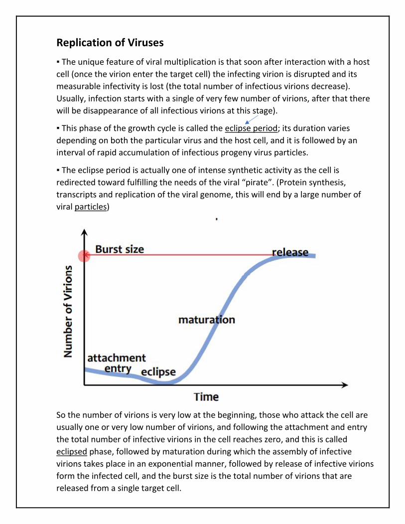
▪ This phase of the growth cycle is called the eclipse period; its duration varies
depending on both the particular virus and the host cell, and it is followed by an
interval of rapid accumulation of infectious progeny virus particles.
▪ The eclipse period is actually one of intense synthetic activity as the cell is
redirected toward fulfilling the needs of the viral “pirate”. (Protein synthesis,
transcripts and replication of the viral genome, this will end by a large number of
viral particles)
So the number of virions is very low at the beginning, those who attack the cell are
usually one or very low number of virions, and following the attachment and entry
the total number of infective virions in the cell reaches zero, and this is called
eclipsed phase, followed by maturation during which the assembly of infective
virions takes place in an exponential manner, followed by release of infective virions
form the infected cell, and the burst size is the total number of virions that are
released from a single target cell.
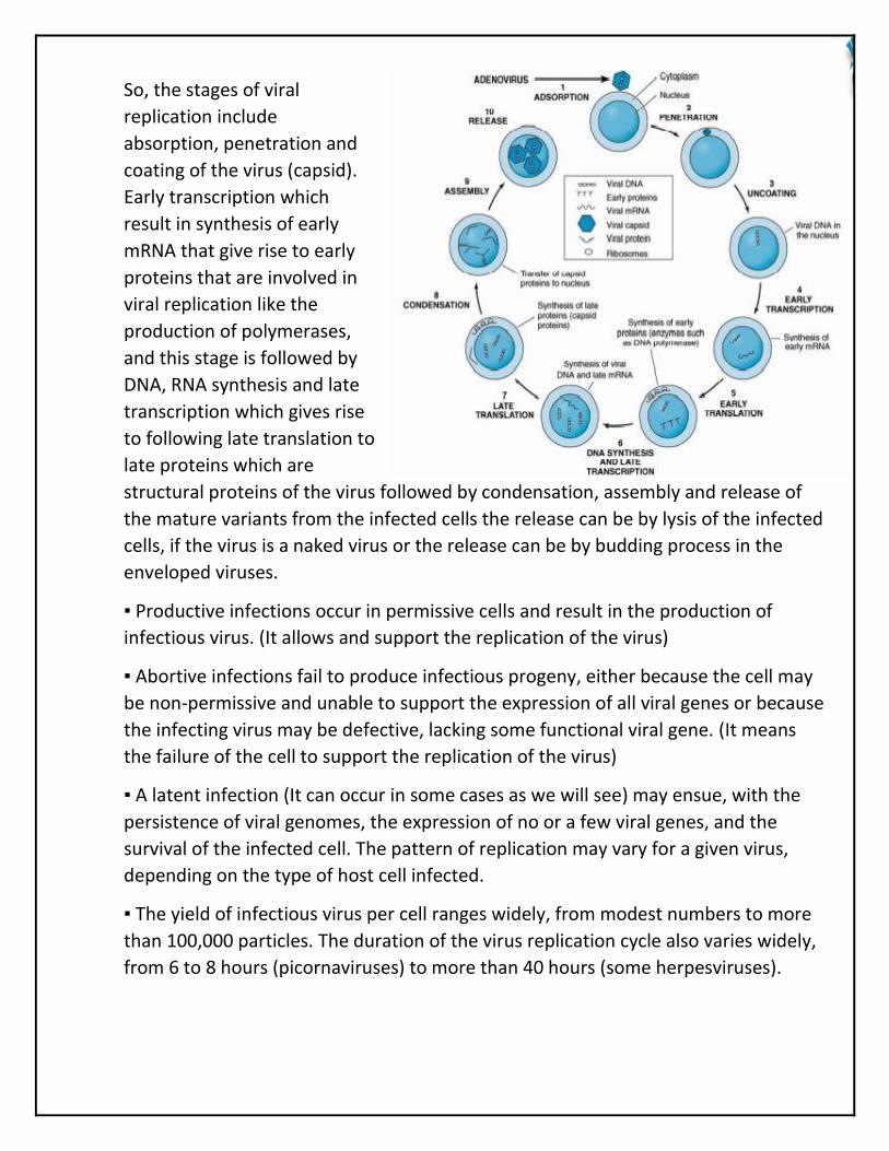
So, the stages of viral
replication include
absorption, penetration and
coating of the virus (capsid).
Early transcription which
result in synthesis of early
mRNA that give rise to early
proteins that are involved in
viral replication like the
production of polymerases,
and this stage is followed by
DNA, RNA synthesis and late
transcription which gives rise
to following late translation to
late proteins which are
structural proteins of the virus followed by condensation, assembly and release of
the mature variants from the infected cells the release can be by lysis of the infected
cells, if the virus is a naked virus or the release can be by budding process in the
enveloped viruses.
▪ Productive infections occur in permissive cells and result in the production of
infectious virus. (It allows and support the replication of the virus)
▪ Abortive infections fail to produce infectious progeny, either because the cell may
be non-permissive and unable to support the expression of all viral genes or because
the infecting virus may be defective, lacking some functional viral gene. (It means
the failure of the cell to support the replication of the virus)
▪ A latent infection (It can occur in some cases as we will see) may ensue, with the
persistence of viral genomes, the expression of no or a few viral genes, and the
survival of the infected cell. The pattern of replication may vary for a given virus,
depending on the type of host cell infected.
▪ The yield of infectious virus per cell ranges widely, from modest numbers to more
than 100,000 particles. The duration of the virus replication cycle also varies widely,
from 6 to 8 hours (picornaviruses) to more than 40 hours (some herpesviruses).
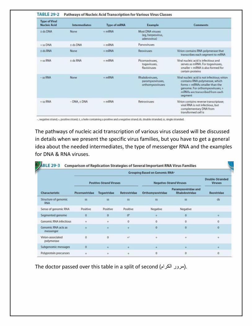
The pathways of nucleic acid transcription of various virus classed will be discussed
in details when we present the specific virus families, but you have to get a general
idea about the needed intermediates, the type of messenger RNA and the examples
for DNA & RNA viruses.
The doctor passed over this table in a split of second ( مرور الكرام(.
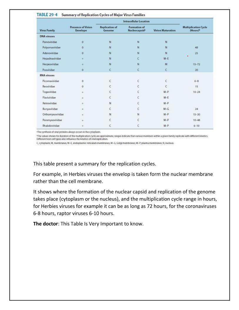
This table present a summary for the replication cycles.
For example, in Herbies viruses the envelop is taken form the nuclear membrane
rather than the cell membrane.
It shows where the formation of the nuclear capsid and replication of the genome
takes place (cytoplasm or the nucleus), and the multiplication cycle range in hours,
for Herbies viruses for example it can be as long as 72 hours, for the coronaviruses
6-8 hours, raptor viruses 6-10 hours.
The doctor: This Table Is Very Important to know.
Genetics of Animal Viruses
▪ Genetic analysis is a powerful approach toward understanding the structure and
function of the viral genome, its gene products, and their roles in infection and
disease, the epidemiology, evolution of viruses, pathogenesis of diseases, the
severity of certain virions, etc.
▪ Viruses that have stable antigens on their surfaces (poliovirus, measles virus) can
be controlled by vaccination. Other viruses that exist as many antigenic types
(rhinoviruses) or change frequently (influenza virus A) are difficult to control by
vaccination.
▪ Genetic analysis will help identify virus-specific processes that may be appropriate
targets for the development of antiviral therapy.
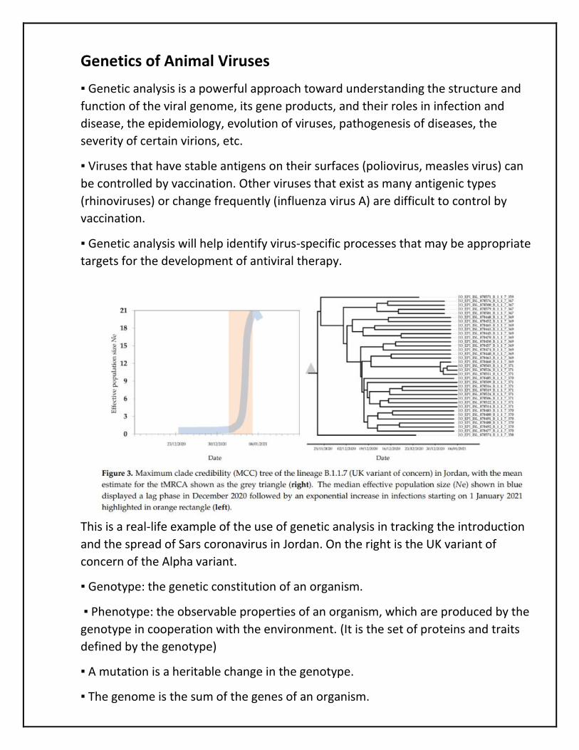
This is a real-life example of the use of genetic analysis in tracking the introduction
and the spread of Sars coronavirus in Jordan. On the right is the UK variant of
concern of the Alpha variant.
▪ Genotype: the genetic constitution of an organism.
▪ Phenotype: the observable properties of an organism, which are produced by the
genotype in cooperation with the environment. (It is the set of proteins and traits
defined by the genotype)
▪ A mutation is a heritable change in the genotype.
▪ The genome is the sum of the genes of an organism.
▪ Wildtype virus denotes the original virus from which mutants are derived and with
which the mutants are compared; the term may not accurately characterize the
virus as it is isolated in nature. Fresh virus isolates from the natural host are referred
to as field isolates or primary isolates.
Mapping of Viral Genomes
Biochemical and physical mapping can be done much more rapidly than genetic
mapping using classic genetic techniques. However, the accurate results are
obtained from genetic analysis by genotyping and genetic analysis of the viruses.
For isolates that can be cloned, sequence analysis and comparison with known
viruses is often used, and these comparisons are to draw entrances about the
evolutionary relatedness between different types of viruses or viruses strains of
variants or linages is done throw phylogenetic analysis.
Restriction endonucleases can be used for identification of specific strains of DNA
viruses.
Types of Virus Mutants
▪ Classic genetic studies with animal viruses require a sensitive and accurate
quantitative assay method, to compare the features (the virulence, the time) to
produce the effect for mutants compared to wild type) such as a plaque assay for
viral infectivity, and good mutants (resulting from single mutations) that are easily
scored and reasonably stable.
▪ Some markers commonly used include plaque morphology, antibody escape or
resistance to neutralizing antisera, loss of a virus protein, drug resistance, host
range, and inability to grow at low or high temperatures.
▪ Conditional-lethal mutants are mutants that are lethal (in that no infectious virus is
produced) (It is lethal for the virus itself under a set of conditions that is why it is
called conditional) under one set of conditions—termed nonpermissive conditions
(because no infectious variants are produced)—but that yield normal infectious
progeny under other conditions— termed permissive conditions

Defective Viruses
A defective virus is one that lacks one or more functional genes required for viral
replication (it might be completely defective if there is for example a certain
deletion mutant, or it could be able to replicate in the presence of a helper virus).
Defective viruses require helper activity from another virus for some step-in
replication or maturation.
One type of defective virus lacks a portion of its genome (i.e., deletion mutant).
Spontaneous deletion mutants may interfere with the replication of homologous
virus and are called defective interfering virus particles.
DIPs have lost essential segments of genome but contain normal capsid proteins;
they require infectious homologous virus as helper for replication, and they
interfere with the multiplication of that homologous virus.
Another category of defective virus requires an unrelated replication-competent
virus as helper.
Examples include the adeno-associated satellite viruses and hepatitis D virus (delta
agent), which replicate only in the presence of coinfecting human adenovirus or
hepatitis B virus, respectively.
The essential helper function supplied by the helper virus varies, depending on the
system.
Another type of defective viruses are Pseudovirions, they contain host cell DNA
rather than the viral genome, enclosed in within the viral capsid and its other
proteins.
During viral replication, the capsid sometimes encloses random pieces of host
nucleic acid rather than viral nucleic acid.
Such particles look like ordinary virus particles when observed by electron
microscopy, because they have the capsid and the other structural components of
the virus, but they are not able to replicate.

Interactions Among Viruses, which can result in production novel variants
Recombination results in the production of progeny virus (recombinant) that carries
traits not found together in either parent. The classic mechanism is that the nucleic
acid strands break, and part of the genome of one parent is joined to part of the
genome of the second parent, or there is jumping between different strands during
the replication of the viral genome which is seen in a verity of RNA viruses, there is
also the jumping between the two copies of RNA genome during the reverse
transcription process and DNA production and HIV replication cycle.
Complementation is the interaction of viral gene products in cells infected with two
viruses, one or both of which may be defective. It results in the replication of one or
both under conditions in which replication would not ordinarily occur. The basis for
complementation is that one virus provides a gene product in which the second is
defective, allowing the second virus to grow.
Infection of either cell cultures or whole animals with two viruses often leads to an
inhibition of multiplication of one of the viruses, an effect called Interference.
Natural History (Ecology) and Modes of Transmission of Viruses
Viruses may be transmitted in the following ways:
❖ Direct transmission.
❖ Indirect transmission.
❖ Transmission from animal to animal, with human as accidental host.
❖ Arthropod vector.

Transmission patters among arboviruses
It could be transmitted directly from the human to the arthropod and vice versa,
example include yellow fever dengue fever.
It could be from arthropod to lower vertebrate to arthropod, with human as an
accidental host, like Saint Louis encephalitis.
From arthropod to lower vertebrate or to human and from the lower vertebrate to
arthropod, so it is here involving the arthropod mainly, include lacrosse encephalitis
and Colorado tick fever. Here the viruses have their main host as arthropod, and
lower vertebrate and human as accidental.

Emerging Viral Diseases
An important concept to consider and we will touch upon that when we discuss
several virous infections like Ebola and Zika fever and of course discussing Sars
coronavirus ii
We have contributing factors that might help increase the number of emerging viral
infections in human, and these are:
Examples of Emerging Viral Infections
Ebola virus
Nipah virus
Hantavirus pulmonary disease
Hyman immunodeficiency virus
West Nile virus: its transmission from the middle-east into the US in late 1990s
Rift Valley fever
Emerging Coronaviruses

Bioterrorism Agents
▪ Microorganisms (or toxins) that could be used to produce death and disease in
humans, animals, or plants for terrorist purposes.
▪ Potential bioterrorism agents are classified into risk categories based on the ease
of dissemination or transmission from person to person, mortality rates, ability to
cause public panic, and requirement for public health preparedness.
▪ Viral agents in the highest risk category are smallpox and the viral haemorrhagic
fever.
This will be discussed in details when we touch upon the viral hemargic fevers and
when we discuss smallpox.
Related Documents