This article was downloaded by: [217.219.137.200] On: 26 September 2014, At: 22:57 Publisher: Taylor & Francis Informa Ltd Registered in England and Wales Registered Number: 1072954 Registered office: Mortimer House, 37-41 Mortimer Street, London W1T 3JH, UK Journal of Biomaterials Science, Polymer Edition Publication details, including instructions for authors and subscription information: http://www.tandfonline.com/loi/tbsp20 Magnetic/pH-sensitive κ-carrageenan/ sodium alginate hydrogel nanocomposite beads: preparation, swelling behavior, and drug delivery Gholam Reza Mahdavinia a , Zeinab Rahmani a , Shiva Karami a & Ali Pourjavadi b a Faculty of Science, Department of Chemistry, University of Maragheh, Maragheh 55181-83111, Iran b Polymer Research Laboratory, Department of Chemistry, Sharif University of Technology, Tehran 11365-9516, Iran Published online: 08 Sep 2014. To cite this article: Gholam Reza Mahdavinia, Zeinab Rahmani, Shiva Karami & Ali Pourjavadi (2014): Magnetic/pH-sensitive κ-carrageenan/sodium alginate hydrogel nanocomposite beads: preparation, swelling behavior, and drug delivery, Journal of Biomaterials Science, Polymer Edition, DOI: 10.1080/09205063.2014.956166 To link to this article: http://dx.doi.org/10.1080/09205063.2014.956166 PLEASE SCROLL DOWN FOR ARTICLE Taylor & Francis makes every effort to ensure the accuracy of all the information (the “Content”) contained in the publications on our platform. However, Taylor & Francis, our agents, and our licensors make no representations or warranties whatsoever as to the accuracy, completeness, or suitability for any purpose of the Content. Any opinions and views expressed in this publication are the opinions and views of the authors, and are not the views of or endorsed by Taylor & Francis. The accuracy of the Content should not be relied upon and should be independently verified with primary sources of information. Taylor and Francis shall not be liable for any losses, actions, claims, proceedings, demands, costs, expenses, damages, and other liabilities whatsoever or howsoever caused arising directly or indirectly in connection with, in relation to or arising out of the use of the Content. This article may be used for research, teaching, and private study purposes. Any substantial or systematic reproduction, redistribution, reselling, loan, sub-licensing,

Welcome message from author
This document is posted to help you gain knowledge. Please leave a comment to let me know what you think about it! Share it to your friends and learn new things together.
Transcript
This article was downloaded by: [217.219.137.200]On: 26 September 2014, At: 22:57Publisher: Taylor & FrancisInforma Ltd Registered in England and Wales Registered Number: 1072954 Registeredoffice: Mortimer House, 37-41 Mortimer Street, London W1T 3JH, UK
Journal of Biomaterials Science,Polymer EditionPublication details, including instructions for authors andsubscription information:http://www.tandfonline.com/loi/tbsp20
Magnetic/pH-sensitive κ-carrageenan/sodium alginate hydrogelnanocomposite beads: preparation,swelling behavior, and drug deliveryGholam Reza Mahdaviniaa, Zeinab Rahmania, Shiva Karamia & AliPourjavadiba Faculty of Science, Department of Chemistry, University ofMaragheh, Maragheh 55181-83111, Iranb Polymer Research Laboratory, Department of Chemistry, SharifUniversity of Technology, Tehran 11365-9516, IranPublished online: 08 Sep 2014.
To cite this article: Gholam Reza Mahdavinia, Zeinab Rahmani, Shiva Karami & Ali Pourjavadi(2014): Magnetic/pH-sensitive κ-carrageenan/sodium alginate hydrogel nanocomposite beads:preparation, swelling behavior, and drug delivery, Journal of Biomaterials Science, Polymer Edition,DOI: 10.1080/09205063.2014.956166
To link to this article: http://dx.doi.org/10.1080/09205063.2014.956166
PLEASE SCROLL DOWN FOR ARTICLE
Taylor & Francis makes every effort to ensure the accuracy of all the information (the“Content”) contained in the publications on our platform. However, Taylor & Francis,our agents, and our licensors make no representations or warranties whatsoever as tothe accuracy, completeness, or suitability for any purpose of the Content. Any opinionsand views expressed in this publication are the opinions and views of the authors,and are not the views of or endorsed by Taylor & Francis. The accuracy of the Contentshould not be relied upon and should be independently verified with primary sourcesof information. Taylor and Francis shall not be liable for any losses, actions, claims,proceedings, demands, costs, expenses, damages, and other liabilities whatsoever orhowsoever caused arising directly or indirectly in connection with, in relation to or arisingout of the use of the Content.
This article may be used for research, teaching, and private study purposes. Anysubstantial or systematic reproduction, redistribution, reselling, loan, sub-licensing,
systematic supply, or distribution in any form to anyone is expressly forbidden. Terms &Conditions of access and use can be found at http://www.tandfonline.com/page/terms-and-conditions
Dow
nloa
ded
by [
217.
219.
137.
200]
at 2
2:57
26
Sept
embe
r 20
14
Magnetic/pH-sensitive κ-carrageenan/sodium alginate hydrogelnanocomposite beads: preparation, swelling behavior, and
drug delivery
Gholam Reza Mahdaviniaa*, Zeinab Rahmania, Shiva Karamia and Ali Pourjavadib
aFaculty of Science, Department of Chemistry, University of Maragheh, Maragheh 55181-83111,Iran; bPolymer Research Laboratory, Department of Chemistry, Sharif University of Technology,
Tehran 11365-9516, Iran
(Received 15 April 2014; accepted 15 August 2014)
This work describes the preparation of magnetic and pH-sensitive beads based onκ-carrageenan and sodium alginate for use as drug-targeting carriers. Physical cross-linking using K+/Ca2+ ions was applied to obtain ionic cross-linked magnetic hydrogelbeads. The produced magnetite beads were thoroughly characterized by TEM, SEM/EDS, XRD, FTIR, and VSM techniques. While the water absorbency (WA) of mag-netic beads was enhanced by increasing the weight ratio of κ-carrageenan, introducingmagnetic nanoparticles caused a decrease in WA capacity from 15.4 to 6.3 g/g. Inves-tigation on the swelling of the hydrogel beads in NaCl, KCl, and CaCl2 solutionsrevealed the disintegration of beads depending on the composition of hydrogel beadsand the type of metal cations in swelling media. The swelling ratio of beads indicatedpH-dependent properties with maximum water absorbing at pH 7.4. Also, it was foundthat the strength of pH-sensitivity of magnetic beads was low for beads with the highcontent of carrageenan component. The in vitro drug release studies from hydrogelsexhibited significant behaviors on the subject of physiological-simulated pH valuesand external magnetic fields. The maximum cumulative releases obtained were 98 and43% at pH values 7.4 and 1.2, respectively. The Introducing magnetite nanoparticlesinfluenced the cumulative release of drug.
Keywords: sodium alginate; κ-carrageenan; magnetic beads; pH-sensitive; drugdelivery
1. Introduction
Hydrogels are hydrophilic and polymeric networks capable of absorbing large amountsof water or biological fluids without dissolving in media.[1] In recent years, due to bio-degradability and biocompatibility, polysaccharides have been emerged as candidatesfor the synthesis of hydrogels. Hydrogels composed of different polysaccharides havebeen widely used in many fields including drug delivery systems,[2] biomedical appli-cations,[3] enzyme immobilization,[4] and wastewater treatment.[5] Hydrogels whichswell and contract in response to external conditions have been explored.[6,7] Theseintelligent hydrogels have potential use in site-specific delivery of drugs to specificregions of the gastrointestinal tract and have been prepared for delivery of low molecu-lar weight protein drugs.[8] Many structural factors (e.g. charge, concentration, and pKa
of the ionizable group, degree of ionization, cross-link density, and hydrophilicity)
*Corresponding author. Email: [email protected]
© 2014 Taylor & Francis
Journal of Biomaterials Science, Polymer Edition, 2014http://dx.doi.org/10.1080/09205063.2014.956166
Dow
nloa
ded
by [
217.
219.
137.
200]
at 2
2:57
26
Sept
embe
r 20
14
influence the degree of swelling of ionic polymers.[9,10] In addition, properties of theswelling medium (e.g. pH, ionic strength, and the counter ion and its valency) affectthe swelling characteristics.[11,12] These responsive or smart hydrogels have becomean important area of research and development in the field of medicine, pharmacy, andbiotechnology. Recently, increasing interest has been dedicated to the investigation ofmagnetic hydrogels which show significant sensitivity to external magnetic field (EMF)as stimuli and can be used in drug delivery and biomedical fields.[13,14]
Sodium alginate contains pendant carboxylate and is known as polyanionic copoly-mer of 1,4-linked-α-L-guluronic acid and β-D-mannuronic acid residues found in brownseaweeds.[15] Physical cross-linking of sodium alginate could be done through electro-static interactions between polyvalent cations especially Ca2+ and aligned guluronicblocks of alginate chains.[15] Biocompatible polymers such as polyvinyl alcohol,[16]chitosan,[17] hdroxypropyl methylcellulose,[18] and starch [19] have been incorporatedin the synthesis of alginate beads with improved properties and evaluated in the drugdelivery and biomedical systems. In addition to biopolymers, inorganic and organicnanoparticles including silica,[20] clay,[21] carbon nanotubes,[22] magnetic Fe3O4,[23]and hydroxyapatite [24] have been used for the synthesis of alginate composite beadswith unique structure and properties. Among the nanoparticles, Fe3O4 with magneticproperties can be introduced into alginate beads. As a result of introducing magneticnanoparticles into alginate beads, a dual-sensitive (pH- and magnetic-sensitive) alginatehydrogels can be synthesized and applied in drug delivery.[23]
Similar to alginate, κ-carrageenan is a family naturally occurring of polysaccharidesextracted from red seaweed. They are used as cost-effective stabilizers, thermo-reversiblegelling agents, binders, thickeners, texture modifiers, and moisture retainers. There arenumerous reviews of their chemistry and applications in foods and drug deliverysystems.[25,26] The gelling of κ-carrageenan can be occurred physically and chemi-cally. κ-Carrageenan contains pendant anion sulfate on its backbones. Electrostaticinteractions between sulfate on carrageenan with metal cations (especially K+) [27] andcationic polyelectrolytes (especially chitosan [28]) are classes of physical cross-linkingthat have been used for synthesis of κ-carrageenan hydrogels. Chitosan/carrageenannanoparticles,[28] alginate/carrageenan beads,[29] and κ-carrageenan/barium ferrite [30]have been synthesized through physical cross-linking methods. Chemical cross-linkingis originated from the presence of reactive hydroxyl groups on κ-carrageenan. Chemicalcross-linking agents such as divinyl sulfone,[31] genipin,[32] and gamma-irradiation[33] have been used for preparation of carrageenan-based hydrogel.
Gelation mechanism of κ-carrageenan with K+ and sodium alginate with Ca2+ issimilar.[34] Hydrogel beads composed of mixture of κ-carrageenan and alginate havebeen developed in drug [29] and cell delivery [34] as well as wastewater treatment.[35]It has been reported that the hydrogels carrying sulfate pendants indicate high swellingcapacity in salt solution.[36] Also, while the pKa of carboxylate pendants on alginate isaround 3.5 (guluronic acid with pKa of 3.65 and mannuronic acid with pKa of 3.38),the pKa of the anionic sulfates on carrageenan is lower than 2.[37] These substantialbehaviors of κ-carrageenan were the main reason to select this polysaccharide for thepreparation of magnetic interpenetrating polymer network hydrogels by incorporationof alginate for drug delivery system. The main objective of the present study was toexamine the capability of drug release from hydrogels under variation of pH of mediaand applying EMF for oral administration of drug to colon site. Especially, this paperfocuses on the influence of different amounts of magnetic iron oxide nanoparticles andratio of the κ-carrageenan/alginate biopolymers on the swelling and drug release
2 G.R. Mahdavinia et al.
Dow
nloa
ded
by [
217.
219.
137.
200]
at 2
2:57
26
Sept
embe
r 20
14
behaviors of magnetic hydrogels. To the nutshell, we characterized the morphology andthe formation of magnetic iron oxide nanoparticles in structure of hydrogels by SEM,XRD, VSM, and FTIR spectroscopy. Riboflavin as a model drug was loaded intomagnetic beads and the release of drug from prepared magnetic beads was investigated.
2. Materials and methods
2.1. Materials
κ-Carrageenan was obtained from Condinson Co. (Denmark). Sodium alginate (man-nuronate/guluronate ratio of the alginate = 1.56, Mw= 270,000) was purchased fromFluka (Switzerland). KCl, CaCl2, FeCl2.4H2O, and FeCl3.6H2O were purchased fromMerck (Germany). Riboflavin was provided from Sigma (USA). All other chemicalswere of analytical grades and were used as received.
2.2. Synthesis of magnetic Fe3O4 nanoparticles
Distilled water of 200 mL was deoxygenated by bubbling with N2 gas for 30 min. Ironsalts (5.40 g of FeCl3·6H2O and 2.00 g of FeCl2·4H2O, Fe
+3/Fe+2 = 2) were poured in200 mL of deoxygenated distilled water an allowed to stir until complete dissolving ofsalts. The temperature of iron salts solution was adjusted at 50 °C. The magnetic nano-particles were obtained through the precipitation of iron salts by adding 2 M NH4OHsolution.[38] The dark solution confirmed the formation of magnetic particle. The pHof final solution was adjusted at 10 and allowed to stir at 50 °C for 1 h. Then, the tem-perature of the solution was decreased to ambient temperature, and magnetic nanoparti-cles were isolated by an EMF. The precipitate was washed with distilled water until thepH of solution reached 7. The magnetic nanoparticles were redispersed in 200 mL ofdistilled water containing 0.5 g of dissolved sodium citrate and allowed to stir at ambi-ent temperature for 2 h. Finally, the product was isolated from solution and washedthrice with distilled water. The purified magnetic nanoparticles were dried at ambienttemperature till constant weight.
2.3. Preparation of magnetic beads
The dispersion and dissolving of materials were carried out in a one-liter reactorequipped with a mechanical stirrer (RZR 2041 model, Heidolph, Germany). Differentmagnetic beads with required amounts of initial materials were tabulated in Table 1. Ingeneral, desired amount of magnetic Fe3O4 powder was dispersed in 50 mL of distilled
Table 1. Required amount of initial materials for the preparation of magnetic beads; EntrapmentEfficiency of drug in hydrogel beads (EE, %).
Na-Alginate (g) κ-Carrageenan (g) Fe3O4 (g) EE(%)
mCar0Alg100 2 0 0.125 62mCar25Alg75 1.5 0.5 0.125 –mCar50Alg50 1 1 0.125 68mCar75Alg25 0.5 1.5 0.125 –mCar100Alg0 0 2 0.125 63mCar50Alg50L 1 1 0.065 61mCar50Alg50H 1 1 0.25 65
Journal of Biomaterials Science, Polymer Edition 3
Dow
nloa
ded
by [
217.
219.
137.
200]
at 2
2:57
26
Sept
embe
r 20
14
water and stirred for 1 h (stirring speed 500 rpm). The reactor was placed in a waterbath adjusted at 70 °C. The dispersed magnetic solution was sonicated for 20 min. Theoperating frequency used in sonication was 70 kHz. Then, κ-carrageenan and sodiumalginate were poured in dispersed magnetic solution and allowed to stir until completedissolving of biopolymers (WNa–alginate + Wκ-carrageenan = 2 g). Then, the temperature ofsolution was decreased to 40 °C. The beads were obtained by dropwise addition of thesolution of magnetic biopolymers to the magnetically stirred cross-linking solution(comprising 3 wt% of KCl and 3 wt% of CaCl2; stirring speed 60 rpm) using a 5 mLglass syringe. It may be noted that the temperature of cross-linking solution wasadjusted at 40 °C. The beads were allowed to stir in salts solution for 1 h forhardening. Then, the beads were collected and immersed in excess water to removeun-participated ingredients. After purification, the beads were dried at 40 °C for con-stant weight. In Table 1, the suffixes y and z in mCaryAlgz show weight percentage ofcarrageenan and alginate in total mixture of biopolymers feed, respectively. The suf-fixes L and H in mCar50Alg50L and mCar50Alg50H indicate the low and high con-tents of magnetic nanoparticles, respectively. The prefix ‘m’ indicates magneticproperty of beads.
2.4. Swelling studies
The water absorbency (WA) of synthesized hydrogel beads was measured by immersingthe dried beads (~0.1 g) in distilled water or in 0.15 M of salt solutions (KCl, NaCl,and CaCl2). Then, the beads were removed from media at consecutive time intervalsand blotted with filter paper to remove surface water, weighed, and the WA wascalculated using Equation (1):
WAðg=gÞ ¼ Ws �Wd
Wd(1)
where Ws and Wd are the weights of the samples swollen in aqueous solutions and indry state, respectively. To investigate the effect of pH of solution on degree of swellingof hydrogel beads, the synthesized beads (~0.1 g) were immersed in phosphate-bufferedsolutions (pH 7.4) and hydrochloric acid buffered solution (HBS, pH 1.2), and WA wascalculated as mentioned above. All the swelling experiments were repeated thrice andthe mean of results are shown in the figures.
2.5. Loading/release studies
Loading of riboflavin was done before cross-linking of beads. Desired content ofriboflavin (0.050 g) was added into solution containing biopolymers and magnetic nano-particles and allowed to stir at 50 °C for 30 min. Cross-linking was carried out as above.The drug-loaded beads were dried at 40 °C for constant weight. To estimate theEntrapment Efficiency (EE%), the content of released riboflavin into cross-linking media(Ct, mg) was determined spectrophotometrically using UV–vis spectrophotometer. Thepercentage of Entrapment Efficiency (EE%) was obtained according to Equation 2:
Loading level ðEE%Þ ¼ C0 � Ct
C0� 100 (2)
where C0 (mg) is the initial amount of riboflavin added into the solution. The amountsof loading level for beads are given in Table 1. In vitro release of the riboflavin frommagnetic beads was evaluated by immersing certain amount of drug-loaded magnetic
4 G.R. Mahdavinia et al.
Dow
nloa
ded
by [
217.
219.
137.
200]
at 2
2:57
26
Sept
embe
r 20
14
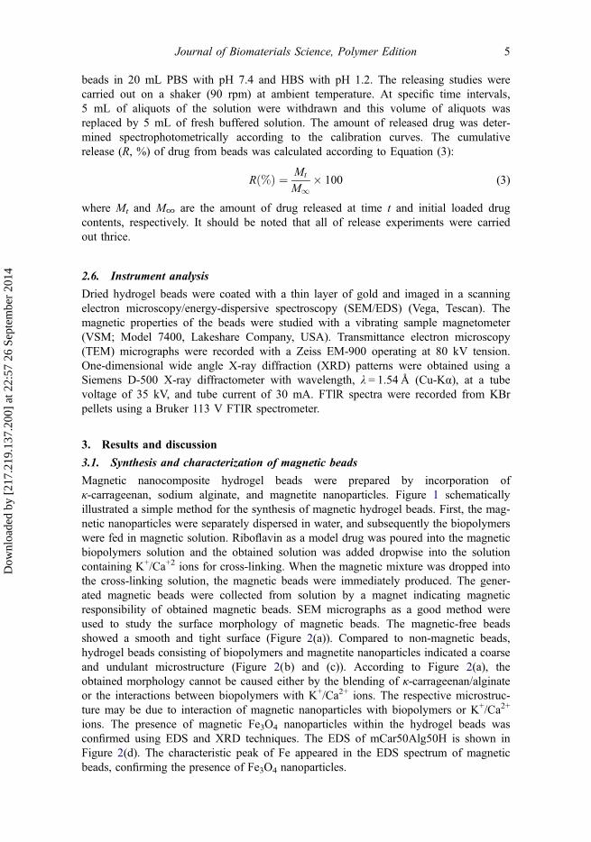
beads in 20 mL PBS with pH 7.4 and HBS with pH 1.2. The releasing studies werecarried out on a shaker (90 rpm) at ambient temperature. At specific time intervals,5 mL of aliquots of the solution were withdrawn and this volume of aliquots wasreplaced by 5 mL of fresh buffered solution. The amount of released drug was deter-mined spectrophotometrically according to the calibration curves. The cumulativerelease (R, %) of drug from beads was calculated according to Equation (3):
Rð%Þ ¼ Mt
M1� 100 (3)
where Mt and M∞ are the amount of drug released at time t and initial loaded drugcontents, respectively. It should be noted that all of release experiments were carriedout thrice.
2.6. Instrument analysis
Dried hydrogel beads were coated with a thin layer of gold and imaged in a scanningelectron microscopy/energy-dispersive spectroscopy (SEM/EDS) (Vega, Tescan). Themagnetic properties of the beads were studied with a vibrating sample magnetometer(VSM; Model 7400, Lakeshare Company, USA). Transmittance electron microscopy(TEM) micrographs were recorded with a Zeiss EM-900 operating at 80 kV tension.One-dimensional wide angle X-ray diffraction (XRD) patterns were obtained using aSiemens D-500 X-ray diffractometer with wavelength, λ = 1.54 Å (Cu-Kα), at a tubevoltage of 35 kV, and tube current of 30 mA. FTlR spectra were recorded from KBrpellets using a Bruker 113 V FTIR spectrometer.
3. Results and discussion
3.1. Synthesis and characterization of magnetic beads
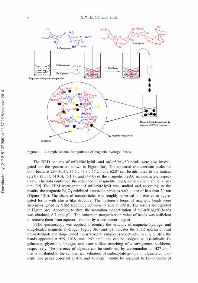
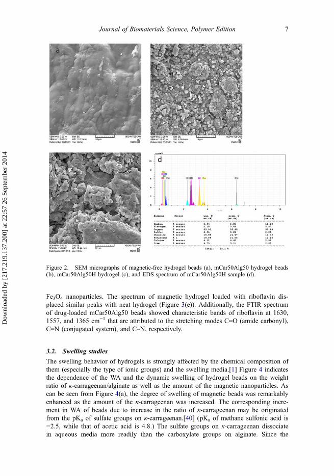
Magnetic nanocomposite hydrogel beads were prepared by incorporation ofκ-carrageenan, sodium alginate, and magnetite nanoparticles. Figure 1 schematicallyillustrated a simple method for the synthesis of magnetic hydrogel beads. First, the mag-netic nanoparticles were separately dispersed in water, and subsequently the biopolymerswere fed in magnetic solution. Riboflavin as a model drug was poured into the magneticbiopolymers solution and the obtained solution was added dropwise into the solutioncontaining K+/Ca+2 ions for cross-linking. When the magnetic mixture was dropped intothe cross-linking solution, the magnetic beads were immediately produced. The gener-ated magnetic beads were collected from solution by a magnet indicating magneticresponsibility of obtained magnetic beads. SEM micrographs as a good method wereused to study the surface morphology of magnetic beads. The magnetic-free beadsshowed a smooth and tight surface (Figure 2(a)). Compared to non-magnetic beads,hydrogel beads consisting of biopolymers and magnetite nanoparticles indicated a coarseand undulant microstructure (Figure 2(b) and (c)). According to Figure 2(a), theobtained morphology cannot be caused either by the blending of κ-carrageenan/alginateor the interactions between biopolymers with K+/Ca2+ ions. The respective microstruc-ture may be due to interaction of magnetic nanoparticles with biopolymers or K+/Ca2+
ions. The presence of magnetic Fe3O4 nanoparticles within the hydrogel beads wasconfirmed using EDS and XRD techniques. The EDS of mCar50Alg50H is shown inFigure 2(d). The characteristic peak of Fe appeared in the EDS spectrum of magneticbeads, confirming the presence of Fe3O4 nanoparticles.
Journal of Biomaterials Science, Polymer Edition 5
Dow
nloa
ded
by [
217.
219.
137.
200]
at 2
2:57
26
Sept
embe
r 20
14
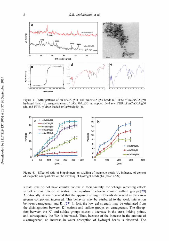
The XRD patterns of mCar50Alg50L and mCar50Alg50 beads were also investi-gated and the spectra are shown in Figure 3(a). The appeared characteristic peaks forboth beads at 2θ = 30.3°, 35.5°, 43.3°, 57.2°, and 62.8° can be attributed to the indices(2 2 0), (3 1 1), (4 0 0), (5 1 1), and (4 4 0) of the magnetic Fe3O4 nanoparticles, respec-tively. The data confirmed the existence of magnetite Fe3O4 particles with spinal struc-ture.[39] The TEM micrograph of mCar50Alg50 was studied and according to theresults, the magnetic Fe3O4 exhibited nanoscale particles with a size of less than 20 nm(Figure 3(b)). The shape of nanoparticles was roughly spherical and existed in aggre-gated forms with cluster-like structure. The hysteresis loops of magnetic beads werealso investigated by VSM technique between ±9 kOe at 298 K. The results are depictedin Figure 3(c). According to data, the saturation magnetization of mCar50Alg50 beadswas obtained, 6.7 emu g−1. The saturation magnetization value of beads was sufficientto remove them from aqueous solution by a permanent magnet.
FTIR spectroscopy was applied to identify the structure of magnetic hydrogel anddrug-loaded magnetic hydrogel. Figure 3(d) and (e) indicates the FTIR spectra of neatmCar50Alg50 and drug-loaded mCar50Alg50 samples, respectively. In Figure 3(d), thebands appeared at 925, 1038, and 1252 cm−1 and can be assigned to 3,6-anhydro-D-galactose, glycosidic linkage, and ester sulfate stretching of κ-carrageenan backbone,respectively. The presence of alginate can be confirmed by wavenumber at 1427 cm−1
that is attributed to the symmetrical vibration of carboxylate groups on alginate compo-nent. The peaks observed at 450 and 470 cm−1 could be assigned to Fe–O bonds of
Figure 1. A simple scheme for synthesis of magnetic hydrogel beads.
6 G.R. Mahdavinia et al.
Dow
nloa
ded
by [
217.
219.
137.
200]
at 2
2:57
26
Sept
embe
r 20
14
Fe3O4 nanoparticles. The spectrum of magnetic hydrogel loaded with riboflavin dis-placed similar peaks with neat hydrogel (Figure 3(e)). Additionally, the FTIR spectrumof drug-loaded mCar50Alg50 beads showed characteristic bands of riboflavin at 1630,1557, and 1365 cm−1 that are attributed to the stretching modes C=O (amide carbonyl),C=N (conjugated system), and C–N, respectively.
3.2. Swelling studies
The swelling behavior of hydrogels is strongly affected by the chemical composition ofthem (especially the type of ionic groups) and the swelling media.[1] Figure 4 indicatesthe dependence of the WA and the dynamic swelling of hydrogel beads on the weightratio of κ-carrageenan/alginate as well as the amount of the magnetic nanoparticles. Ascan be seen from Figure 4(a), the degree of swelling of magnetic beads was remarkablyenhanced as the amount of the κ-carrageenan was increased. The corresponding incre-ment in WA of beads due to increase in the ratio of κ-carrageenan may be originatedfrom the pKa of sulfate groups on κ-carrageenan.[40] (pKa of methane sulfonic acid is−2.5, while that of acetic acid is 4.8.) The sulfate groups on κ-carrageenan dissociatein aqueous media more readily than the carboxylate groups on alginate. Since the
Figure 2. SEM micrographs of magnetic-free hydrogel beads (a), mCar50Alg50 hydrogel beads(b), mCar50Alg50H hydrogel (c), and EDS spectrum of mCar50Alg50H sample (d).
Journal of Biomaterials Science, Polymer Edition 7
Dow
nloa
ded
by [
217.
219.
137.
200]
at 2
2:57
26
Sept
embe
r 20
14
sulfate ions do not have counter cations in their vicinity, the ‘charge screening effect’is not a main factor to restrict the repulsion between anionic sulfate groups.[29]Additionally, it was observed that the apparent strength of beads decreased as the carra-geenan component increased. This behavior may be attributed to the weak interactionbetween carrageenan and K+.[27] In fact, the low gel strength may be originated fromthe disintegration between K+ cations and sulfate groups on carrageenan. The disrup-tion between the K+ and sulfate groups causes a decrease in the cross-linking points,and subsequently the WA is increased. Thus, because of the increase in the amount ofκ-carrageenan, an increase in water absorption of hydrogel beads is observed. The
Figure 3. XRD patterns of mCar50Alg50L and mCar50Alg50 beads (a), TEM of mCar50Alg50hydrogel bead (b), magnetization of mCar50Alg50 vs. applied field (c), FTIR of mCar50Alg50(d), and FTIR of drug-loaded mCar50Alg50 (e).
Figure 4. Effect of ratio of biopolymers on swelling of magnetic beads (a), influence of contentof magnetic nanoparticles on the swelling of hydrogel beads (b) (mean ± 5%).
8 G.R. Mahdavinia et al.
Dow
nloa
ded
by [
217.
219.
137.
200]
at 2
2:57
26
Sept
embe
r 20
14
effect of the content of magnetic nanoparticles on the degree of swelling of hydrogelbeads was also studied while the other influential factors were kept constant. Increasedmagnetic Fe3O4 nanoparticles resulted in a considerable decrease in WA of hydrogelsbeads, as shown in Figure 4(b). This behavior can be attributed to: (a) decrease in theratio of hydrophilic and anionic functional groups on hydrogels and (b) the increase inthe cross-linking density of hydrogel beads through the interaction betweenmagnetic nanoparticles and polymeric chains, and thereby decrease in the swellingcapacity.[41,42]
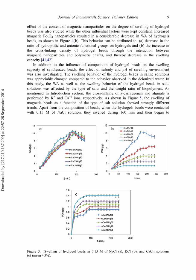
In addition to the influence of composition of hydrogel beads on the swellingcapacity of synthesized beads, the effect of salinity and pH of swelling environmentwas also investigated. The swelling behavior of the hydrogel beads in saline solutionswas appreciably changed compared to the behavior observed in the deionized water. Inthis study, the WA as well as the swelling behavior of the hydrogel beads in saltssolutions was affected by the type of salts and the weight ratio of biopolymers. Asmentioned in Introduction section, the cross-linking of κ-carrageenan and alginate isperformed by K+ and Ca+2 ions, respectively. As shown in Figure 5, the swelling ofmagnetic beads as a function of the type of salt solution showed strongly differenttrends. Apart from the composition of beads, when the hydrogels beads were contactedwith 0.15 M of NaCl solution, they swelled during 160 min and then began to
Figure 5. Swelling of hydrogel beads in 0.15 M of NaCl (a), KCl (b), and CaCl2 solutions(c) (mean ± 5%).
Journal of Biomaterials Science, Polymer Edition 9
Dow
nloa
ded
by [
217.
219.
137.
200]
at 2
2:57
26
Sept
embe
r 20
14
disintegrate (Figure 5(a)). After this period, the WA of hydrogel beads was notmeasurable. This observation indicated that the alginate/κ-carrageenan network cross-linked with Ca+2/K+ could be easily broken due to cation-exchange ability of thependant anionic on hydrogels.[16]
The effect of KCl and CaCl2 solutions on the swelling behavior of beads is shownin Figure 5(b) and (c), respectively. In both salt solutions, all of the hydrogel beadsbegan to swell without any disintegration. The swelling behavior of hydrogels beads asa function of the ratio of biopolymers in the CaCl2 solution was obtained different fromthat in the KCl solution. In CaCl2 solution, a decrement in swelling was observed byan increase in the weight ratio of alginate. This observation could be attributed to anincrease in cross-linking density due to interaction between carboxylate and Ca+2 cat-ions, and thereby a decrease in WA is observed (Figure 5(c)). In comparison to CaCl2solution, the swelling of hydrogel beads in KCl solution was reversed (Figure 5(b)). Asthe κ-carrageenan content increased, the WA of hydrogel beads decreased and could beexplained similar to CaCl2 solution. In addition, in hydrogel beads without κ-carra-geenan component (mCar0Alg100), the beads began to disintegrate similar to NaClsolution. This can be attributed to break of cross-linked -CO2−-Ca2+-−2OC-points dueto exchanging of Ca2+ with K+ cations.[43]
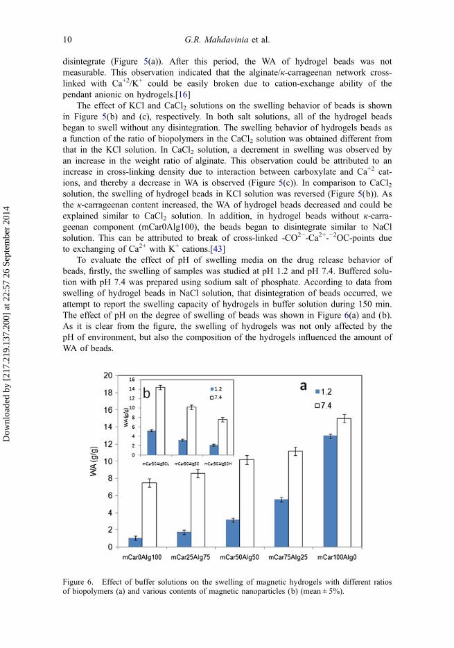
To evaluate the effect of pH of swelling media on the drug release behavior ofbeads, firstly, the swelling of samples was studied at pH 1.2 and pH 7.4. Buffered solu-tion with pH 7.4 was prepared using sodium salt of phosphate. According to data fromswelling of hydrogel beads in NaCl solution, that disintegration of beads occurred, weattempt to report the swelling capacity of hydrogels in buffer solution during 150 min.The effect of pH on the degree of swelling of beads was shown in Figure 6(a) and (b).As it is clear from the figure, the swelling of hydrogels was not only affected by thepH of environment, but also the composition of the hydrogels influenced the amount ofWA of beads.
Figure 6. Effect of buffer solutions on the swelling of magnetic hydrogels with different ratiosof biopolymers (a) and various contents of magnetic nanoparticles (b) (mean ± 5%).
10 G.R. Mahdavinia et al.
Dow
nloa
ded
by [
217.
219.
137.
200]
at 2
2:57
26
Sept
embe
r 20
14
The synthesized beads comprise κ-carrageenan, sodium alginate, and magneticnanoparticles. κ-Carrageenan is an ionic polysaccharide carrying sulfate groups(–OSO3
−). These pendants are completely dissociated in the overall pH range, and thehydrogels from this biopolymer show pH-independent swelling behavior.[44] (pKa ofmethane sulfonic acid is ~ −2.5.) While the κ-carrageenan component containspH-independent sulfate groups, the sodium alginate contains ionizable carboxylategroups (guluronic acid with pKa of 3.65 and mannuronic acid with pKa of 3.38). Whenthe pH of solution is 7.4, the sulfate groups on the κ-carrageenan and carboxylategroups on the alginate are completely dissociated and the repulsion between anionicgroups results in high swelling capacity. When the beads were contacted with acidicbuffered solution, while the sulfate groups on κ-carrageenan remain unchanged, the–COO− pendants on alginate are converted into –COOH. So, the decrease in the WAof beads can be attributed to absence of –COO− groups. Also, the swelling of beads inbuffer solutions revealed that the difference in WA of beads in acidic and basic buffersreduced as the κ-carrageenan component increased (Figure 6(a)). In fact, the strength ofpH-sensitivity of hydrogel beads was affected by the ratio of biopolymers and thisbehavior resulted from the low pKa of κ-Carrageenan biopolymer. The effect of pH onthe swelling of hydrogel beads containing different contents of magnetic nanoparticleswas also studied and shown in Figure 6(b). The content of two biopolymers in thesehydrogel beads was identical. The high swelling at pH 7.4 and low swelling at pH 1.2can be explained as mentioned above. A similar observation has been reported byKulkarni et al. that the swelling of κ-carrageenan-g-polyacrylamide at pH 1.2 has beenobtained more than the κ-carrageenan-g-polyacrylamide/sodium alginate hydrogels.[45]
3.3. Drug release studies
In general, the diffusion of drug from dried and glassy hydrogels is controlled by twomain factors including swelling behavior of hydrogels and chemical nature ofdrugs.[46,47] As shown above, the swelling of hydrogel beads was affected by thecomposition of beads and pH of swelling environment. So, we tried to investigate thereleasing of riboflavin from hydrogel beads as a function of composition of beads aswell as releasing media.
3.3.1. Effect of pH on drug release
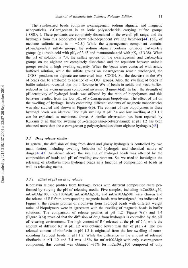
Riboflavin release profiles from hydrogel beads with different composition were per-formed by varying the pH of releasing media. Five samples, including mCar50Alg50,mCar0Alg100, mCar100Alg0, mCar50Alg50L, and mCar50Alg50H were chosen andthe release of RF from corresponding magnetic beads was investigated. As indicated inFigure 7, the release profiles of riboflavin from hydrogel beads with different weightratios of biopolymers were in agreement with the swelling of magnetic beads in buffersolutions. The comparison of release profiles at pH 1.2 (Figure 7(a)) and 7.4(Figure 7(b)) revealed that the diffusion of drug from hydrogels is controlled by the pHof releasing environment. The high content of RF released at the pH of 7.4, while theamount of diffused RF at pH 1.2 was obtained lower than that of pH 7.4. The lowreleased content of riboflavin in pH 1.2 is originated from the low swelling of corre-sponding hydrogel beads in pH 1.2. While the difference in the amount of releasedriboflavin in pH 1.2 and 7.4 was ~15% for mCar100Alg0 with only κ-carrageenancomponent, this content was obtained ~35% for mCar0Alg100 composed of only
Journal of Biomaterials Science, Polymer Edition 11
Dow
nloa
ded
by [
217.
219.
137.
200]
at 2
2:57
26
Sept
embe
r 20
14
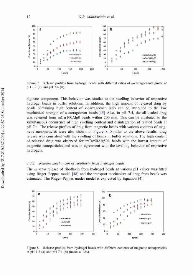
alginate component. This behavior was similar to the swelling behavior of respectivehydrogel beads in buffer solutions. In addition, the high amount of released drug bybeads containing high content of κ-carrageenan ratio can be attributed to the lowmechanical strength of κ-carrageenan beads.[45] Also, in pH 7.4, the all-loaded drugwas released from mCar100Alg0 beads within 200 min. This can be attributed to thesimultaneous occurrence of high swelling content and disintegration of related beads atpH 7.4. The release profiles of drug from magnetic beads with various contents of mag-netic nanoparticles were also shown in Figure 8. Similar to the above results, drugrelease was consistent with the swelling of beads in buffer solutions. The high contentof released drug was observed for mCar50Alg50L beads with the lowest amount ofmagnetic nanoparticles and was in agreement with the swelling behavior of respectivehydrogels.
3.3.2. Release mechanism of riboflavin from hydrogel beads
The in vitro release of riboflavin from hydrogel beads at various pH values was fittedusing Ritger–Peppas model [48] and the transport mechanism of drug from beads wasestimated. The Ritger–Peppas model model is expressed by Equation (4):
Figure 7. Release profiles from hydrogel beads with different ratios of κ-carrageenan/alginate atpH 1.2 (a) and pH 7.4 (b).
Figure 8. Release profiles from hydrogel beads with different contents of magnetic nanoparticlesat pH 1.2 (a) and pH 7.4 (b) (mean ± 3%).
12 G.R. Mahdavinia et al.
Dow
nloa
ded
by [
217.
219.
137.
200]
at 2
2:57
26
Sept
embe
r 20
14
Mt
M1¼ ktn (4)
where Mt is the amount of riboflavin released at time t, and M∞ is the total content ofriboflavin loaded into beads. k is kinetic constant; and n is the release exponent, whichis indicative of transport mechanism. The transport mechanism of release from matrixcan be Fickian-type diffusion (diffusion kinetic) and non-Fickian type diffusion (diffu-sion kinetic and polymer relaxation kinetic), when n be ≤0.45 and 0.45 < n < 0.89,respectively.[49] To investigate the transport mechanism of riboflavin from hydrogelbead into releasing media, the initial releasing data were fitted to the exponential heu-ristic equation for Mt/M∞≤ 0.6. The n and k values for samples can be achieved fromslope and intercept of plot of Ln Mt
M1against Lnt. The n values for samples at both pH
values are tabulated in Table 2. Apart from the pH of media, the results showed thatthe release of riboflavin from hydrogels followed both diffusion and polymer relaxationkinetics except mCar50Alg50L. In this case, the drug release was controlled by diffu-sion kinetic. This observation is due to high degree of swelling of respective beads and
Table 2. Parameters k and n according to Ritger–Peppas model for release of riboflavin frombeads in pH 7.4 and 1.2.
R2 n k Transport mechanism
pH 7.4 mCar0Alg100 0.99 0.53 0.04 Non-FickianmCar50Alg50 0.99 0.5 0.07 Non-FickianmCar100Alg0 0.99 0.46 0.1 Non-FickianmCar50Alg50L 0.97 0.36 0.16 FickianmCar50Alg50H 0.98 0.58 0.034 Non-Fickian
pH 1.2 mCar0Alg100 0.98 0.52 0.033 Non-FickianmCar50Alg50 0.97 0.53 0.051 Non-FickianmCar100Alg0 0.99 0.55 0.07 Non-FickianmCar50Alg50L 0.99 0.36 0.12 FickianmCar50Alg50H 0.98 0.77 0.014 Non-Fickian
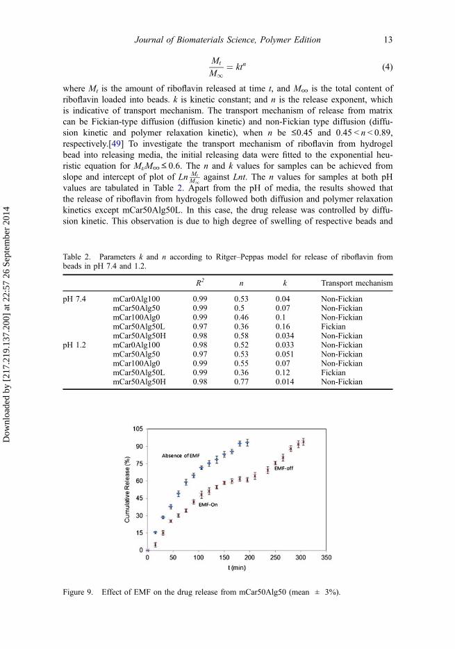
Figure 9. Effect of EMF on the drug release from mCar50Alg50 (mean ± 3%).
Journal of Biomaterials Science, Polymer Edition 13
Dow
nloa
ded
by [
217.
219.
137.
200]
at 2
2:57
26
Sept
embe
r 20
14
thereby high network expansion. When a polymer network has a high degree ofswelling, its pore size becomes larger and it can be assumed that the location of drug issimilar to a free solution.[50] So, the drug can be easily diffused into solution.
3.3.3. Effect of EMF on release
The effect of EMF on the release profile of drug from mCar50Alg50 was examinedand displayed in Figure 9. It is evident from figure that the amount of released drugwas decreased with applying EMF. In the presence of magnetic field, the magneticmoment of Fe3O4 nanoparticles can be aligned together and led to aggregate of Fe3O4
nanoparticles. This aggregation of magnetic nanoparticles could be led to decrease inpore size of magnetic hydrogel, and subsequently decrease in diffusion of drug intoreleasing media.[13] This behavior of ferrogel is known as ‘close’ configuration. Also,according to Hu et al., the formation of magnetic-sensitive walls restricts the diffusionof drug from hydrogels and results in a decrease in the amount of released drug.[51]As the magnetic force was disconnected, the release of riboflavin began to increase.
4. Conclusion
Magnetic/pH-sensitive nanocomposite hydrogel beads composed of κ-carrageenan,sodium alginate, and magnetic Fe3O4 nanoparticles were prepared through ionic-gela-tion technique by K+/Ca2+ ions. The swelling studies indicated that an increment inWA of beads is observed by increasing the κ-carrageenan component. In contrast, adecrement in swelling was seen by increasing the content of magnetic nanoparticles.Depending on the composition of hydrogels, the beads were disintegrated in salt solu-tion. In NaCl solution, regardless of the composition of hydrogels, the beads weredestroyed. When the beads were immersed in KCl solution, the hydrogel composed ofonly alginate component was disintegrated. Additionally, by immersing the hydrogel inthe CaCl2 solution, disintegration of beads did not occur. Swelling behavior of beads asa function of pH was investigated and it was found that the sensitivity of hydrogels tothe pH of media was reduced with the increase in κ-carrageenan ratio. Riboflavin, adrug model, was loaded in hydrogel beads and the release behavior of drug from beads.The drug release profiles indicated pH-dependent behavior with high releasing at pH7.4. The lowest content of releasing at acidic pH was observed for beads containingonly alginate component and this behavior is originated from pH sensitivity of alginatebiopolymer. By applying an EMF, a decrease in the amount of released drug wasobserved.
References[1] Mohamadnia Z, Zohuriaan-Mehr MJ, Kabiri K, Jamshidi A, Mobedi H. Ionically cross-
linked carrageenan-alginate hydrogel beads. J. Biomat. Sci.-Polym. E. 2007;22:342–356.[2] Matricardi P, Di-Meo C, Coviello T, Hennink WE, Alhaique F. Interpenetrating polymer net-
works polysaccharide hydrogels for drug delivery and tissue engineering. Adv. Drug Deliver.Rev. 2013;65:1172–1187.
[3] Leone G, Barbucci R. Polysaccharide Based Hydrogels for Biomedical Applications. In:Barbucci R, editor. Hydrogels. Milan: Springer; 2009. p. 25–41.
[4] Dumitriu S, Chornet E. Immobilization of xylanase in chitosan–xanthan hydrogels. Biotechnol.Progr. 1997;13:539–545.
14 G.R. Mahdavinia et al.
Dow
nloa
ded
by [
217.
219.
137.
200]
at 2
2:57
26
Sept
embe
r 20
14
[5] Crini G. Recent developments in polysaccharide-based materials used as adsorbents inwastewater treatment. Prog. Polym. Sci. 2005;30:38–70.
[6] Pourjavadi A, Barzegar Sh, Mahdavinia GR. MBA-crosslinked Na-Alg/CMC as a smartfull-polysaccharide superabsorbent hydrogels. Carbohydr. Polym. 2006;66:386–395.
[7] Xiao C, Xia C, Ma Y, He X. Preparation and characterization of dual sensitive carboxylatedmethyl cellulose/poly(vinyl alcohol) physical composite hydrogel. J. Appl. Polym. Sci.2013;127:4750–4755.
[8] Bajpai AK, Shukla SK, Bhanu S, Kankane S. Responsive polymers in controlled drugdelivery. Prog. Polym. Sci. 2008;33:1088–1118.
[9] Mahdavinia GR, Pourjavadi A, Hosseinzadeh H, Zohuriaan MJ. Modified chitosan 4. Super-absorbent hydrogels from poly(acrylic acid-co-acrylamide) grafted chitosan with salt- andpH-responsiveness properties. Eur. Polym. J. 2004;40:1399–1407.
[10] Shi X, Wang W, Wang A. pH-responsive sodium alginate-based superporous hydrogel gen-erated by an anionic surfactant micelle templating. Carbohydr. Polym. 2013;94:449–455.
[11] Gomez ML, Williams RJJ, Montejano HA, Previtali CM. Influence of the ionic character ofa drug on its release rate from hydrogels based on 2-hydroxyethylmethacrylate and acrylam-ide synthesized by photopolymerization. Express Polym. Lett. 2012;6:189–197.
[12] Islam A, Yasin T, Bano I, Riaz M. Controlled release of aspirin from pH-sensitive chitosan/poly(vinyl alcohol) hydrogel. J. Appl. Polym. Sci. 2012;124:4184–4192.
[13] Liu TY, Hu SH, Liu TY, Liu DM, Chen SY. Magnetic-sensitive behavior of intelligent ferro-gels for controlled release of drug. Langmuir. 2006;22:5974–5978.
[14] Meenach SA, Hilt JZ, Anderson KW. Poly(ethylene glycol)-based magnetic hydrogel nano-composites for hyperthermia cancer therapy. Acta Biomater. 2010;6:1039–1046.
[15] Liakos I, Rizzello L, Bayer IS, Pompa PP, Cingolani R, Athanassiou A. Controlled antisep-tic release by alginate polymer films and beads. Carbohydr. Polym. 2013;92:176–183.
[16] Hua S, Ma H, Li X, Yang H, Wang A. pH-sensitive sodium alginate/poly(vinyl alcohol)hydrogel beads prepared by combined Ca2+ crosslinking and freeze-thawing cycles for con-trolled release of diclofenac sodium. Int. J. Biol. Macromol. 2010;46:517–523.
[17] Sahasathian T, Praphairaksit N, Muangsin N. Mucoadhesive and floating chitosan-coated algi-nate beads for the controlled gastric release of amoxicillin. Arch. Pharm. Res. 2010;33:889–899.
[18] Fahmy RH. Statistical approach for assessing the influence of calcium silicate and HPMCon the formulation of novel alfuzosin hydrochloride mucoadhesive-floating beads as gastro-retentive drug delivery systems. AAPS PharmSciTech. 2012;13:990–1004.
[19] Kim YJ, Park HG, Yang YL, Yoon Y, Kim S, Oh E. Multifunctional drug delivery systemusing starch-alginate beads for controlled release. Biol. Pharm. Bull. 2005;28:394–397.
[20] Cao Z, He Y, Sun L, Cao X. Preparation of alginate/silica composite beads with in vitroapatite-forming ability. Adv. Mater. Res. 2011;236–238:1889–1892.
[21] Iliescu RI, Andronescu E, Ghitulica CD, Voicu G, Ficai A, Hoteteu M. Montmorillonite-alginate nanocomposite as a drug delivery system – incorporation and in vitro release of iri-notecan. Int. J. Pharm. 2014;463:184–192.
[22] Zhang X, Hui Z, Wan D, Huang H, Huang J, Yuan H, Yu J. Alginate microsphere filledwith carbon nanotube as drug carrier. Int. J. Biol. Macromol. 2010;47:389–395.
[23] Brulé S, Levy M, Wilhelm C, Letourneur D, Gazeau F, Ménager C, Le Visage CL. Doxoru-bicin release triggered by alginate embedded magnetic nanoheaters: a combined therapy.Adv. Mater. 2011;23:787–790.
[24] Zhang J, Wang Q, Wang A. In situ generation of sodium alginate/hydroxyapatite nanocom-posite beads as drug-controlled release matrices. Acta Biomater. 2010;6:445–454.
[25] Li L, Ni R, Shao Y, Mao S. Carrageenan and its applications in drug delivery. Carbohydr.Polym. 2014;103:1–11.
[26] Prajapati VD, Maheriya PM, Jani GK, Solanki HK. Carrageenan: a natural seaweed polysac-charide and its applications. Carbohydr. Polym. 2014;105:97–112.
[27] Morris ER, Rees DA, Robinson G. Cation-specific aggregation of carrageenan helices:Domain model of polymer gel structure. J. Mol. Biol. 1980;138:349–362.
[28] Rodrigues S, Costa AM, Grenha A. Chitosan/carrageenan nanoparticles: effect of cross-link-ing with tripolyphosphate and charge ratios. Carbohydr. Polym. 2012;89:282–289.
[29] Mohamadnia Z, Zohuriaan-Mehr MJ, Kabiri K, Jamshidi A, Mobedi H. pH-sensitive IPNhydrogel beads of carrageenan-alginate for controlled drug delivery. J. Bioact. Compat. Pol.2007;22:342–356.
Journal of Biomaterials Science, Polymer Edition 15
Dow
nloa
ded
by [
217.
219.
137.
200]
at 2
2:57
26
Sept
embe
r 20
14
[30] Mitsumata T, Sakai K, Takimoto J. Giant reduction in dynamic modulus of κ-carrageenanmagnetic gels. J. Phys. Chem. B. 2006;110:20217–20223.
[31] Sagbas S, Butun S, Sahiner N. Modifiable chemically crosslinked poli(κ-carrageenan) parti-cles. Carbohydr. Polym. 2012;87:2718–2724.
[32] Muhamad II, Fen LS, Hui NH, Mustapha NA. Genipin-cross-linked kappa-carrageenan/carboxymethyl cellulose beads and effects on beta-carotene release. Carbohydr. Polym.2011;83:1207–1212.
[33] Tranquilan-Aranilla C, Nagasawa N, Bayquen A, Dela Rosa AD. Synthesis and characteriza-tion of carboxymethyl derivatives of kappa-carrageenan. Carbohydr. Polym. 2012;87:1810–1816.
[34] Popa EG, Gomes ME, Reis RL. Cell delivery systems using alginate–carrageenan hydrogelbeads and fibers for regenerative medicine applications. Biomacromolecules. 2011;12:3952–3961.
[35] Mahdavinia GR, Aghaie H, Sheykhloie H, Vardini MT, Etemadi H. Synthesis of CarAlg/MMt nanocomposite hydrogels and adsorption of cationic crystal violet. Carbohydr. Polym.2013;98:358–365.
[36] Pourjavadi A, Harzandi AM, Hosseinzadeh H. Modified carrageenan 3. Synthesis of a novelpolysaccharide-based superabsorbent hydrogel via graft copolymerization of acrylic acidonto kappa-carrageenan in air. Eur. Polym. J. 2004;40:1363–1370.
[37] Mahdavinia GR, Massoudi A, Baghban A, Shokri E. Study of adsorption of cationic dye onmagnetic kappa-carrageenan/PVA nanocomposite hydrogels. J. Environ. Chem. Eng.2014;2:1578–1587.
[38] Zhang S, Zhou YF, Nie WY, Song LY. Preparation of Fe3O4/chitosan/poly(acrylic acid)composite particles and its application in adsorbing copper ion (II). Cellulose. 2012;19:2081–2091.
[39] Khan A. Preparation and characterization of magnetic nanoparticles embedded in microgels.Mater. Lett. 2008;62:898–902.
[40] Hosseinzadeh H, Pourjavadi A, Mahdavinia GR, Zohuriaan-Mehr MJ. Modified carra-geenan. 1 H-CARRAGPAM, a novel biopolymer-based superabsorbent hydrogel. J. Bioact.Compat. Pol. 2005;20:475–490.
[41] Philippova O, Barabanova A, Molchanov V, Khokhlov A. Magnetic polymer beads: Recenttrends and developments in synthetic design and applications. Eur. Polym. J. 2011;47:542–559.
[42] Bardajee GR, Hooshyar Z. A novel biocompatible magnetic iron oxide nanoparticles/hydro-gel based on poly (acrylic acid) grafted onto starch for controlled drug release. J. Polym.Res. 2013;20:298.
[43] Abd El-Ghaffar MA, Hashem MS, El-Awady MK, Rabie AM. pH-sensitive sodium alginatehydrogels for riboflavin controlled release. Carbohydr. Polym. 2012;89:667–675.
[44] Durmaz S, Okay O. Acrylamide/2-acrylamido-2-methylpropane sulfonic acid sodium salt-based hydrogels: synthesis and characterization. Polymer. 2000;41:3693–3704.
[45] Kulkarni RV, Boppana R, Krishna Mohan GK, Mutalik S, Kalyane NV. pH-responsive inter-penetrating network hydrogel beads of poly(acrylamide)-g-carrageenan and sodium alginatefor intestinal targeted drug delivery: Synthesis, in vitro and in vivo evaluation. J. ColloidInterf. Sci. 2012;367:509–517.
[46] Petrusic S, Jovancic P, Lewandowski M, Giraud S, Bugarski B, Djonlagic J, Koncar V. Syn-thesis, characterization and drug release properties of thermosensitive poly(N-isopropylacryl-amide) microgels. J. Polym. Res. 2013;19:9979.
[47] Gupta B, Kumari M, Ikram S. Drug release studies of N-isopropyl acrylamide/acrylic acidgrafted polypropylene nonwoven fabric. J. Polym. Res. 2013;20:95.
[48] Ritger PL, Peppas NA. A simple equation for description of solute release II. Fickian andanomalous release from swellable devices. J. Control. Release. 1987;5:37–42.
[49] Li L, Wang L, Shao Y, Ni R, Zhang T, Mao S. Drug release characteristics from chitosan–alginate matrix tablets based on the theory of self-assembled film. Int. J. Pharmaceut.2013;450:197–207.
[50] Soppirnath KS, Aminabhavi TM. Water transport and drug release study from cross-linkedpolyacrylamide grafted guar gum hydrogel microspheres for the controlled release applica-tion. Eur. J. Pharm. Biopharm. 2002;53:87–98.
[51] Hu SH, Liu TY, Liu DM, Chen SY. Nano-ferrosponges for controlled drug release. J.Control. Release. 2007;121:181–189.
16 G.R. Mahdavinia et al.
Dow
nloa
ded
by [
217.
219.
137.
200]
at 2
2:57
26
Sept
embe
r 20
14
Related Documents