Journal of Clinical Medicine Article Macular Perfusion Impairment in Von Hippel- Lindau Disease Suggests a Generalized Retinal Vessel Alteration Elisabetta Pilotto 1 , Elisabetta Beatrice Nacci 2, *, Alfonso Massimiliano Ferrara 3 , Gilda De Mojà 1 , Stefania Zovato 3 and Edoardo Midena 1,2 1 Department of Ophthalmology, University of Padova, 35122 Padova, Italy; [email protected] (E.P.); [email protected] (G.D.M.); [email protected] (E.M.) 2 IRCCS—Fondazione G. B. Bietti, 00198 Rome, Italy 3 Familial Tumor Unit, Veneto Institute of Oncology IOV-IRCCS, 35122 Padova, Italy; [email protected] (A.M.F.); [email protected] (S.Z.) * Correspondence: [email protected] Received: 10 July 2020; Accepted: 12 August 2020; Published: 18 August 2020 Abstract: Background: To evaluate macular perfusion in patients with Von Hippel–Lindau (VHL) disease. Methods: VHL patients with or without peripheral retinal hemangioblastomas (RHs) were consecutively enrolled. A group of healthy subjects served as controls. Macular perfusion was analyzed by means of OCT angiography (OCTA) in the superficial vascular plexus (SVP), and in the intermediate (ICP) and deep retinal capillary (DCP) plexuses. The following OCTA parameters were measured: Vessel Area Density (VAD), Vessel Length Fraction (VLF), Vessel Diameter Index (VDI) and Fractal Dimension (FD). Results: Sixty-three VHL patients (113 eyes) and 28 healthy controls (56 eyes) were enrolled. All OCTA quantitative parameters were reduced in VHL patients vs. controls, reaching statistical significance for VAD of the SVP (0.348 ± 0.07 vs. 0.369 ± 0.06, p = 0.0368) and VDI of all plexuses (p < 0.03 for all). No significant differences were detected between eyes without or with peripheral RHs. Conclusions: Macular perfusion is reduced in VHL patients demonstrating retinal vessel changes that are independent of the presence of peripheral RHs. VHL gene mutations disrupt the hypoxia-induced (HIF)/vascular endothelium growth factors (VEGF) pathway and the Notch signaling, both essential for the normal retinal vasculogenesis and angiogenesis. Therefore, an anomalous generalized retinal vascular development may be hypothesized in VHL disease. Keywords: Von Hippel Lindau disease; retinal hemangioblastoma; OCT; OCT angiography; retinal vascular plexuses; macular flow 1. Introduction Von Hippel–Lindau (VHL) disease (OMIM 193300) is an autosomal dominant heritable cancer syndrome with an incidence of 1 in 36,000 live births per year, caused by the oncosuppressor VHL gene mutation. In VHL disease, benign and malignant tumors and/or cysts develop throughout the central nervous system (CNS) and visceral organs (clear cell renal carcinoma, pheochromocytomas, neuroendocrine tumors and cysts of the pancreas, endolymphatic sac tumors, papillary cystadenomas of the epididymis and broad ligament) [1]. The mean age of onset is in the third decade of life and by the age of 65 years 90% of patients become symptomatic. Eugen von Hippel, a German ophthalmologist, first described retinal lesions (“angiomatosis retinae”) in 1904 while Arvid Lindau, a Swedish pathologist, recognized the association between retinal (RHs) and cerebellar hemangioblastomas (CHs) in 1927 [2,3]. RHs are well-circumscribed reddish-orange vascular lesions characterized by a tortuous feeding artery and draining vein that develop in the peripheral retina or in the iuxtapapillary J. Clin. Med. 2020, 9, 2677; doi:10.3390/jcm9082677 www.mdpi.com/journal/jcm

Welcome message from author
This document is posted to help you gain knowledge. Please leave a comment to let me know what you think about it! Share it to your friends and learn new things together.
Transcript
Journal of
Clinical Medicine
Article
Macular Perfusion Impairment in Von Hippel-Lindau Disease Suggests a Generalized RetinalVessel Alteration
Elisabetta Pilotto 1, Elisabetta Beatrice Nacci 2,*, Alfonso Massimiliano Ferrara 3, Gilda De Mojà 1,Stefania Zovato 3 and Edoardo Midena 1,2
1 Department of Ophthalmology, University of Padova, 35122 Padova, Italy; [email protected] (E.P.);[email protected] (G.D.M.); [email protected] (E.M.)
2 IRCCS—Fondazione G. B. Bietti, 00198 Rome, Italy3 Familial Tumor Unit, Veneto Institute of Oncology IOV-IRCCS, 35122 Padova, Italy;
[email protected] (A.M.F.); [email protected] (S.Z.)* Correspondence: [email protected]
Received: 10 July 2020; Accepted: 12 August 2020; Published: 18 August 2020�����������������
Abstract: Background: To evaluate macular perfusion in patients with Von Hippel–Lindau (VHL)disease. Methods: VHL patients with or without peripheral retinal hemangioblastomas (RHs) wereconsecutively enrolled. A group of healthy subjects served as controls. Macular perfusion wasanalyzed by means of OCT angiography (OCTA) in the superficial vascular plexus (SVP), and in theintermediate (ICP) and deep retinal capillary (DCP) plexuses. The following OCTA parameters weremeasured: Vessel Area Density (VAD), Vessel Length Fraction (VLF), Vessel Diameter Index (VDI)and Fractal Dimension (FD). Results: Sixty-three VHL patients (113 eyes) and 28 healthy controls(56 eyes) were enrolled. All OCTA quantitative parameters were reduced in VHL patients vs. controls,reaching statistical significance for VAD of the SVP (0.348 ± 0.07 vs. 0.369 ± 0.06, p = 0.0368) and VDIof all plexuses (p < 0.03 for all). No significant differences were detected between eyes without orwith peripheral RHs. Conclusions: Macular perfusion is reduced in VHL patients demonstratingretinal vessel changes that are independent of the presence of peripheral RHs. VHL gene mutationsdisrupt the hypoxia-induced (HIF)/vascular endothelium growth factors (VEGF) pathway and theNotch signaling, both essential for the normal retinal vasculogenesis and angiogenesis. Therefore, ananomalous generalized retinal vascular development may be hypothesized in VHL disease.
Keywords: Von Hippel Lindau disease; retinal hemangioblastoma; OCT; OCT angiography; retinalvascular plexuses; macular flow
1. Introduction
Von Hippel–Lindau (VHL) disease (OMIM 193300) is an autosomal dominant heritable cancersyndrome with an incidence of 1 in 36,000 live births per year, caused by the oncosuppressor VHLgene mutation. In VHL disease, benign and malignant tumors and/or cysts develop throughout thecentral nervous system (CNS) and visceral organs (clear cell renal carcinoma, pheochromocytomas,neuroendocrine tumors and cysts of the pancreas, endolymphatic sac tumors, papillary cystadenomasof the epididymis and broad ligament) [1]. The mean age of onset is in the third decade of life and by theage of 65 years 90% of patients become symptomatic. Eugen von Hippel, a German ophthalmologist,first described retinal lesions (“angiomatosis retinae”) in 1904 while Arvid Lindau, a Swedishpathologist, recognized the association between retinal (RHs) and cerebellar hemangioblastomas(CHs) in 1927 [2,3]. RHs are well-circumscribed reddish-orange vascular lesions characterized by atortuous feeding artery and draining vein that develop in the peripheral retina or in the iuxtapapillary
J. Clin. Med. 2020, 9, 2677; doi:10.3390/jcm9082677 www.mdpi.com/journal/jcm
J. Clin. Med. 2020, 9, 2677 2 of 9
area [1]. Small RHs are asymptomatic, while larger lesions can cause macular edema, exudative ortractional retinal detachment and vitreous bleeding [4]. By means of widefield fluorescein angiography,RH-associated peripheral retinal nonperfusion has been recently described in eyes with peripheral RH,suggesting a vascular steal phenomenon from the surrounding normal retina [5]. With the introductionin clinical practice of optical coherence tomography angiography (OCTA), it has now become possibleto explore in vivo, without any dye, the central and midperipheral retinal blood flow. Moreover,OCTA allows separate analysis of three single retinal vascular plexuses: the superficial vascular plexus(SVP), and two deeper capillary plexuses: the intermediate (ICP) and the deep capillary plexus (DCP),invisible to fluorescein angiography [6,7]. The aim of this study was to investigate macular perfusion,using OCTA, in VHL patients with or without peripheral RHs.
2. Experimental Section
2.1. Participants
In this cross-sectional study, all patients with VHL disease genetically confirmed of the FamilialTumor Unit, Veneto Institute of Oncology (IOV-IRCCS) undergoing scheduled eye examination,were consecutively recruited. All patients underwent complete ophthalmological evaluation including:best-corrected visual acuity (BCVA), using standard Early Treatment Diabetic Retinopathy Study(ETDRS) charts; slit-lamp examination of the anterior segment; intraocular pression measurement;indirect ophthalmoscopy; and 90-diopter-lens biomicroscopy. Exclusion criteria were: RHs or laserscars from previous treatments or retinal pigment epithelium changes involving the posterior pole;epiretinal macular membrane; history of inflammatory ocular diseases; concomitant presence of otherretinal vascular diseases; congenital or acquired anterior segment disorders; glaucoma; and refractiveerrors ≥ 6 diopters. The same examinations were performed on an age-matched healthy group forcomparison. The approval from the Ethics Committee for Clinical Practice of the Azienda Ospedalieradi Padova for the study was obtained (Prot. 34971/AOP/2018). Informed consent was obtained fromeach subject and data collection followed the tenets of the Declaration of Helsinki.
2.2. OCT and OCT Angiography
Optical coherence tomography (OCT) and OCTA were performed using Spectralis HRA + OCTA(Heidelberg Engineering, Heidelberg, Germany). All scans were acquired after pupil dilatation,obtained with 1% tropicamide solution, in a dark room, late in the morning, and the in-built eye-trackeralways activated. The scan protocol included a single horizontal scan (180◦ line scan, 9-mm length,automated real time (ART) set at 100 frames), a macular map of 20◦ × 20◦ (5.8 × 5.8 mm; 97 B-scansseparated by 60 micron) and an OCTA scan pattern of 10◦ × 10◦ (3.0 × 3.0 mm; 512 B-scans separated by6 micron) all centered onto the fovea. The inbuilt software automatically generated the enface OCTAimages of the full retina (extending from the inner limiting membrane to the Bruch membrane) andof the SVP, ICP and DCP, as automatically segmented by the device, to avoid segmentation manualerrors. For high quality OCTA images, a signal strength (SS) more than 30 in “Q score” (on a scale of 0to 40 for Spectralis, Heidelbeerg, Germany) was required [8,9]. An eye tracking system was used toguarantee correct foveal centration of the scans. A skilled technician performed all scans and checkedeach image after acquisition to detect any motion artifacts or segmentation errors and eventuallyrepeated examination.
2.3. OCTA en Face Image Analysis
Quantitative analysis of the OCTA en face images was performed using an open-source availableImageJ software (National Institutes of Health, Bethesda, MD, USA). The foveal avascular zone (FAZ)was measured in the en face OCTA image of the full retina as previously suggested [7]. The followingquantitative parameters were analyzed in each vascular plexus (SVP, ICP and DCP): Vessel AreaDensity (VAD), Vessel Length Fraction (VLF), and Vessel Diameter Index (VDI) [10]. Briefly, OCTA en
J. Clin. Med. 2020, 9, 2677 3 of 9
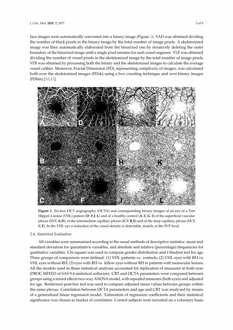
face images were automatically converted into a binary image (Figure 1). VAD was obtained dividingthe number of black pixels in the binary image by the total number of image pixels. A skeletonizedimage was then automatically elaborated from the binarized one by iteratively deleting the outerboundary of the binarized image until a single pixel remains for each vessel segment. VLF was obtaineddividing the number of vessel pixels in the skeletonized image by the total number of image pixels.VDI was obtained by processing both the binary and the skeletonized images to calculate the averagevessel caliber. Moreover, Fractal Dimension (FD), representing complexity of images, was calculatedboth over the skeletonized images (FDsk) using a box counting technique and over binary images(FDbin) [10,11].
J. Clin. Med. 2020, 9, 2677 3 of 9
The following quantitative parameters were analyzed in each vascular plexus (SVP, ICP and DCP): Vessel Area Density (VAD), Vessel Length Fraction (VLF), and Vessel Diameter Index (VDI) [10]. Briefly, OCTA en face images were automatically converted into a binary image. (Figure 1) VAD was obtained dividing the number of black pixels in the binary image by the total number of image pixels. A skeletonized image was then automatically elaborated from the binarized one by iteratively deleting the outer boundary of the binarized image until a single pixel remains for each vessel segment. VLF was obtained dividing the number of vessel pixels in the skeletonized image by the total number of image pixels. VDI was obtained by processing both the binary and the skeletonized images to calculate the average vessel caliber. Moreover, Fractal Dimension (FD), representing complexity of images, was calculated both over the skeletonized images (FDsk) using a box counting technique and over binary images (FDbin) [10,11].
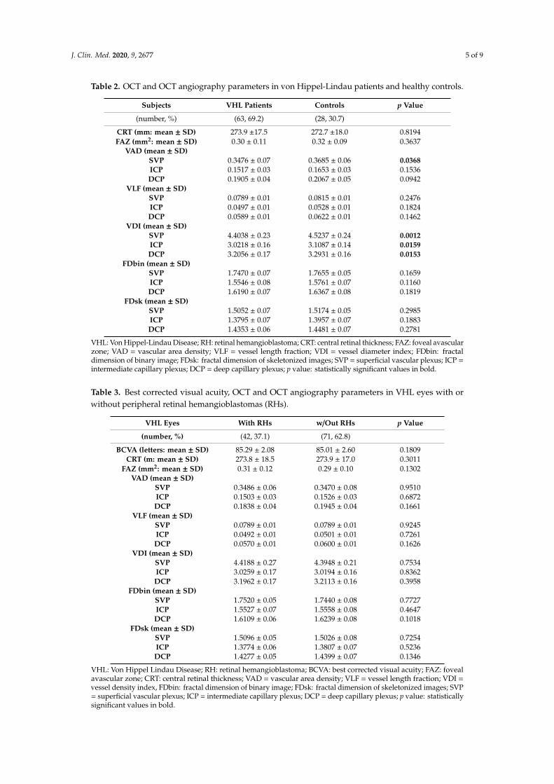
Figure 1. En-face OCT angiography (OCTA) and corresponding binary images of an eye of a Von Hippel–Lindau (VHL) patient (D–F,J–L) and of a healthy control (A–C,G–I) of the superficial vascular plexus (SVP, A,D), of the intermediate capillary plexus (ICP, B,E) and of the deep capillary plexus (DCP, C,F). In the VHL eye a reduction of the vessel density is detectable, mainly at the SVP level.
2.4. Statistical Evaluation
All variables were summarized according to the usual methods of descriptive statistics: mean and standard deviation for quantitative variables, and absolute and relative (percentage) frequencies for qualitative variables. Chi-square was used to compare gender distribution and t-Student test for age. Three groups of comparison were defined: (1) VHL patients vs. controls; (2) VHL eyes with RH vs. VHL eyes without RH; (3) eyes with RH vs. fellow eyes without RH in patients with monocular lesions. All the models used in these statistical analyses accounted for replication of measures in both eyes (PROC MIXED of SAS 9.4 statistical software). CRT and OCTA parameters were compared between groups using a mixed effects two-way ANOVA model, with repeated measures (both eyes) and adjusted for age. Bonferroni post-hoc test was used to compare adjusted mean values between groups within the same plexus. Correlation between OCTA parameters and age and CRT was analyzed by means of a generalized linear regression model. Estimation of regression coefficients and their statistical significance was chosen as marker of correlation. Control subjects were recruited on
Figure 1. En-face OCT angiography (OCTA) and corresponding binary images of an eye of a VonHippel–Lindau (VHL) patient (D–F,J–L) and of a healthy control (A–C,G–I) of the superficial vascularplexus (SVP, A,D), of the intermediate capillary plexus (ICP, B,E) and of the deep capillary plexus (DCP,C,F). In the VHL eye a reduction of the vessel density is detectable, mainly at the SVP level.
2.4. Statistical Evaluation
All variables were summarized according to the usual methods of descriptive statistics: mean andstandard deviation for quantitative variables, and absolute and relative (percentage) frequencies forqualitative variables. Chi-square was used to compare gender distribution and t-Student test for age.Three groups of comparison were defined: (1) VHL patients vs. controls; (2) VHL eyes with RH vs.VHL eyes without RH; (3) eyes with RH vs. fellow eyes without RH in patients with monocular lesions.All the models used in these statistical analyses accounted for replication of measures in both eyes(PROC MIXED of SAS 9.4 statistical software). CRT and OCTA parameters were compared betweengroups using a mixed effects two-way ANOVA model, with repeated measures (both eyes) and adjustedfor age. Bonferroni post-hoc test was used to compare adjusted mean values between groups withinthe same plexus. Correlation between OCTA parameters and age and CRT was analyzed by meansof a generalized linear regression model. Estimation of regression coefficients and their statisticalsignificance was chosen as marker of correlation. Control subjects were recruited on a voluntary basis.
J. Clin. Med. 2020, 9, 2677 4 of 9
The total sample size of enrolled subjects was calculated in order to recognize differences of at least 18%of the reference values as statistically significant, using a 2-sided t test with type I error alpha = 0.05,and a power of 0.80. Moreover, an enrollment ratio of 2.25 patients per 1 control was consideredadequate for comparison within subgroups. In addition, since both eyes of patients and controlscontributed to the study, the sample size was adjusted applying the factor 1/(1-rho), where rho = 0.60 isthe estimated correlation between eye measures.
Statistical significance was set at p < 0.05. All the analyses were performed by SAS® 9.4 statisticalsoftware (SAS Institute, Cary, NC, USA).
3. Results
Sixty-three VHL patients and 28 healthy controls were consecutively enrolled from December2018 to January 2020. All subjects were Caucasian. Patients and controls did not differ for mean ageand sex distribution. BCVA was significantly lower in VHL patients than in controls (84.4 ± 8.4 lettersvs. 87.8 letters ± 3.7 letters, p = 0.0003) (Table 1).
Table 1. Demographic characteristics of enrolled von Hippel-Lindau patients and healthy controls.
VHL Patients Controls p Value
Subjects, (number) 63 28Mean age (years: mean, SD) 40.5, 14.4 40.0, 11.8 0.8824
Female:Male (number, %) 36:27, 57.1:42.9 17:11, 60.7:39.3 0.7498BCVA (letters: mean, SD) 84.4, 8.4 87.8, 3.7 0.0003
Peripheral RH (number, %)Present 36, 57 - -Absent 27, 43 - -
VHL: Von Hippel–Lindau disease; BCVA: best corrected visual acuity; RH: retinal hemangioblastoma; p value:statistically significant values in bold.
In 13 patients only one eye was included; the fellow eye was excluded because of: RH at theposterior pole (5 cases), band keratopathy (2 cases), previous total retinal detachment related to RH(1 case), epiretinal macular membrane (3 cases), myelinated nerve fibers (1 case), and retinal pigmentepithelium changes involving the macula (1 case). Therefore, 113 eyes of 63 VHL patients (71 eyeswithout RHs (62.8%) and 42 (37.1%) with peripheral RHs) and 56 eyes of 28 healthy subjects wereincluded in the analysis.
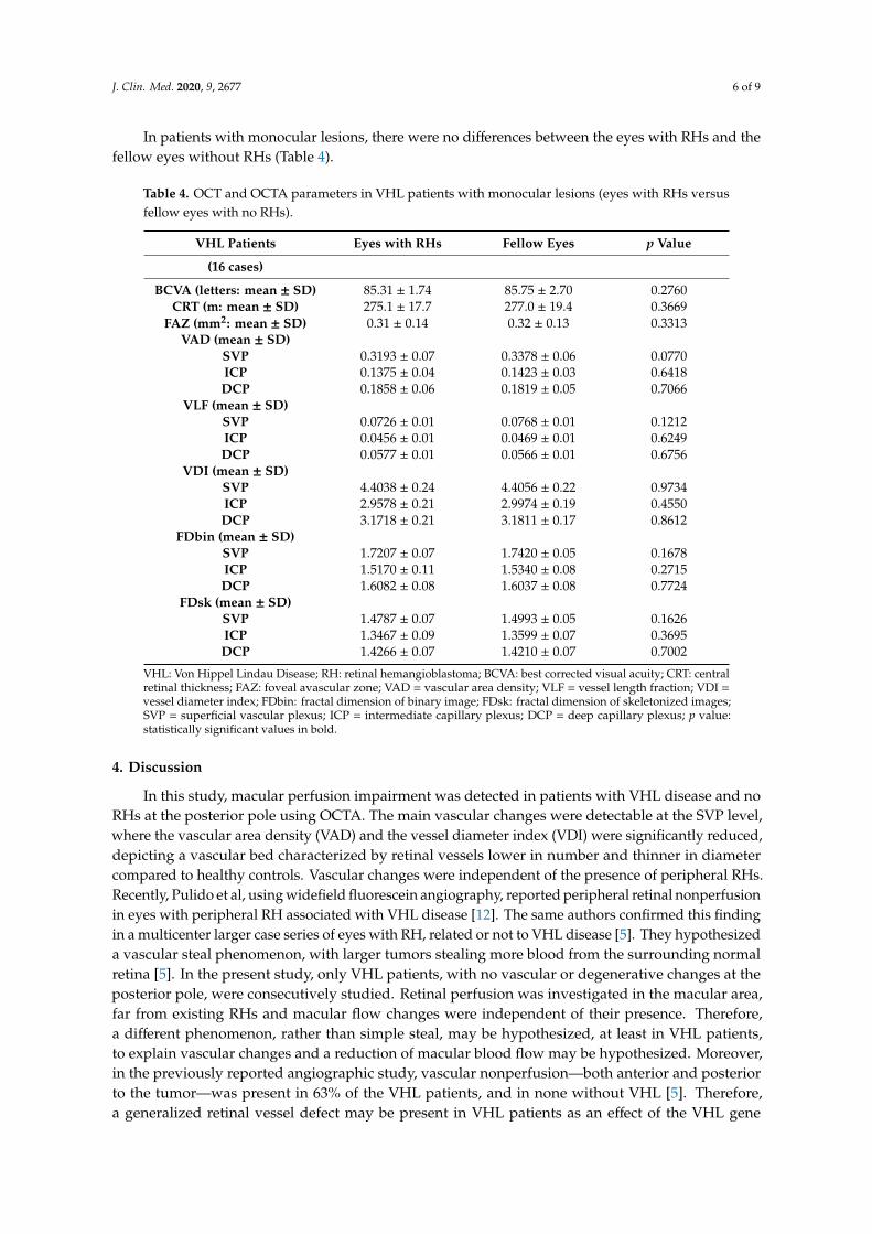
Sixteen patients (25.4%) had only one eye affected by peripheral RHs. Mean SS of the OCTAimages was 37.53 ± 2.76 “Q score” in VHL patients and 38.18 ± 3.34 “Q score” in controls. CRT and FAZwere not significantly different between VHL patients and controls (273.9 ±17.5 µm vs. 272.7 ± 18.0 µm,p = 0.8194 and 0.30 ± 0.11 mm2 vs. 0.32 ± 0.09 mm2, p = 0.3637; respectively). All the other OCTAparameters were reduced in VHL patients versus controls, reaching statistical significance for the VADof the SVP (0.348 ± 0.07 vs. 0.369 ± 0.06, p = 0.0368), and for the VDI of all remaining plexuses (p < 0.02for all) (Table 2 and Figure 1).
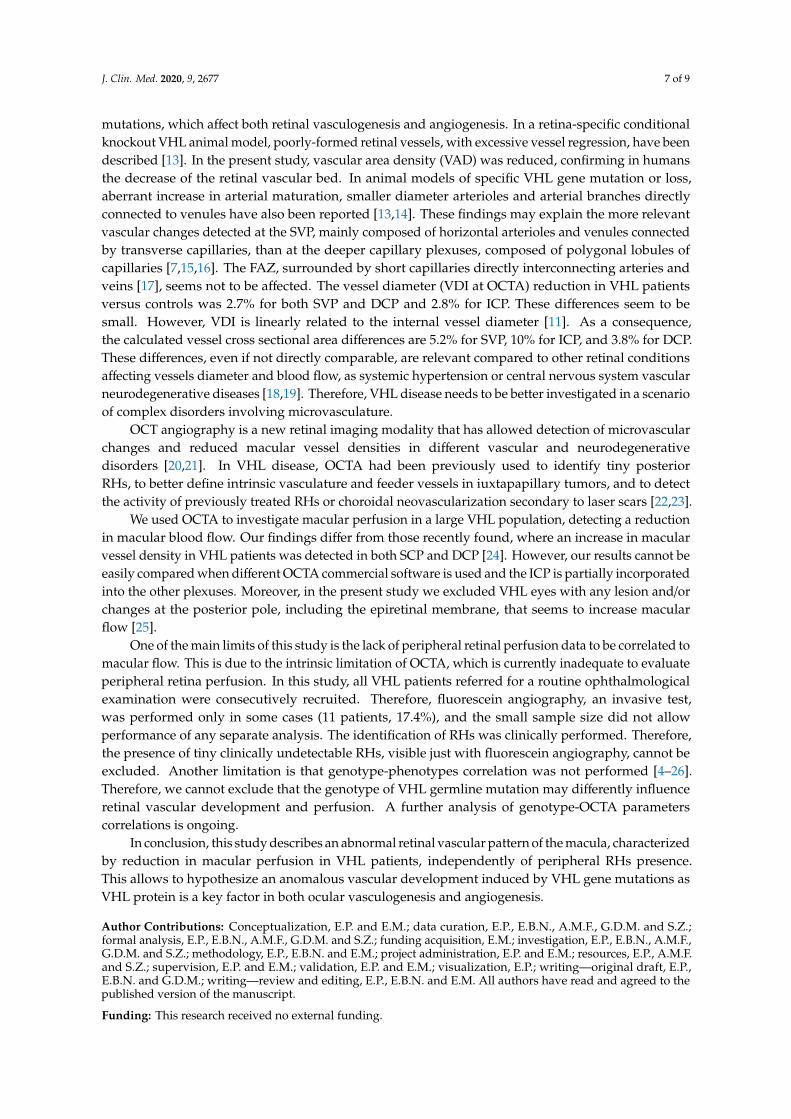
CRT and OCTA parameters were not significantly different between eyes without (71 eyes) orwith RHs (42 eyes) (Table 3).
J. Clin. Med. 2020, 9, 2677 5 of 9
Table 2. OCT and OCT angiography parameters in von Hippel-Lindau patients and healthy controls.
Subjects VHL Patients Controls p Value
(number, %) (63, 69.2) (28, 30.7)
CRT (mm: mean ± SD) 273.9 ±17.5 272.7 ±18.0 0.8194FAZ (mm2: mean ± SD) 0.30 ± 0.11 0.32 ± 0.09 0.3637
VAD (mean ± SD)SVP 0.3476 ± 0.07 0.3685 ± 0.06 0.0368ICP 0.1517 ± 0.03 0.1653 ± 0.03 0.1536DCP 0.1905 ± 0.04 0.2067 ± 0.05 0.0942
VLF (mean ± SD)SVP 0.0789 ± 0.01 0.0815 ± 0.01 0.2476ICP 0.0497 ± 0.01 0.0528 ± 0.01 0.1824DCP 0.0589 ± 0.01 0.0622 ± 0.01 0.1462
VDI (mean ± SD)SVP 4.4038 ± 0.23 4.5237 ± 0.24 0.0012ICP 3.0218 ± 0.16 3.1087 ± 0.14 0.0159DCP 3.2056 ± 0.17 3.2931 ± 0.16 0.0153
FDbin (mean ± SD)SVP 1.7470 ± 0.07 1.7655 ± 0.05 0.1659ICP 1.5546 ± 0.08 1.5761 ± 0.07 0.1160DCP 1.6190 ± 0.07 1.6367 ± 0.08 0.1819
FDsk (mean ± SD)SVP 1.5052 ± 0.07 1.5174 ± 0.05 0.2985ICP 1.3795 ± 0.07 1.3957 ± 0.07 0.1883DCP 1.4353 ± 0.06 1.4481 ± 0.07 0.2781
VHL: Von Hippel-Lindau Disease; RH: retinal hemangioblastoma; CRT: central retinal thickness; FAZ: foveal avascularzone; VAD = vascular area density; VLF = vessel length fraction; VDI = vessel diameter index; FDbin: fractaldimension of binary image; FDsk: fractal dimension of skeletonized images; SVP = superficial vascular plexus; ICP =intermediate capillary plexus; DCP = deep capillary plexus; p value: statistically significant values in bold.
Table 3. Best corrected visual acuity, OCT and OCT angiography parameters in VHL eyes with orwithout peripheral retinal hemangioblastomas (RHs).
VHL Eyes With RHs w/Out RHs p Value
(number, %) (42, 37.1) (71, 62.8)
BCVA (letters: mean ± SD) 85.29 ± 2.08 85.01 ± 2.60 0.1809CRT (m: mean ± SD) 273.8 ± 18.5 273.9 ± 17.0 0.3011
FAZ (mm2: mean ± SD) 0.31 ± 0.12 0.29 ± 0.10 0.1302VAD (mean ± SD)
SVP 0.3486 ± 0.06 0.3470 ± 0.08 0.9510ICP 0.1503 ± 0.03 0.1526 ± 0.03 0.6872DCP 0.1838 ± 0.04 0.1945 ± 0.04 0.1661
VLF (mean ± SD)SVP 0.0789 ± 0.01 0.0789 ± 0.01 0.9245ICP 0.0492 ± 0.01 0.0501 ± 0.01 0.7261DCP 0.0570 ± 0.01 0.0600 ± 0.01 0.1626
VDI (mean ± SD)SVP 4.4188 ± 0.27 4.3948 ± 0.21 0.7534ICP 3.0259 ± 0.17 3.0194 ± 0.16 0.8362DCP 3.1962 ± 0.17 3.2113 ± 0.16 0.3958
FDbin (mean ± SD)SVP 1.7520 ± 0.05 1.7440 ± 0.08 0.7727ICP 1.5527 ± 0.07 1.5558 ± 0.08 0.4647DCP 1.6109 ± 0.06 1.6239 ± 0.08 0.1018
FDsk (mean ± SD)SVP 1.5096 ± 0.05 1.5026 ± 0.08 0.7254ICP 1.3774 ± 0.06 1.3807 ± 0.07 0.5236DCP 1.4277 ± 0.05 1.4399 ± 0.07 0.1346
VHL: Von Hippel Lindau Disease; RH: retinal hemangioblastoma; BCVA: best corrected visual acuity; FAZ: fovealavascular zone; CRT: central retinal thickness; VAD = vascular area density; VLF = vessel length fraction; VDI =vessel density index, FDbin: fractal dimension of binary image; FDsk: fractal dimension of skeletonized images; SVP= superficial vascular plexus; ICP = intermediate capillary plexus; DCP = deep capillary plexus; p value: statisticallysignificant values in bold.
J. Clin. Med. 2020, 9, 2677 6 of 9
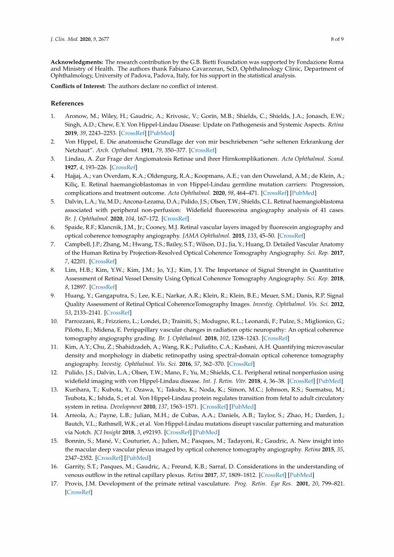
In patients with monocular lesions, there were no differences between the eyes with RHs and thefellow eyes without RHs (Table 4).
Table 4. OCT and OCTA parameters in VHL patients with monocular lesions (eyes with RHs versusfellow eyes with no RHs).
VHL Patients Eyes with RHs Fellow Eyes p Value
(16 cases)
BCVA (letters: mean ± SD) 85.31 ± 1.74 85.75 ± 2.70 0.2760CRT (m: mean ± SD) 275.1 ± 17.7 277.0 ± 19.4 0.3669
FAZ (mm2: mean ± SD) 0.31 ± 0.14 0.32 ± 0.13 0.3313VAD (mean ± SD)
SVP 0.3193 ± 0.07 0.3378 ± 0.06 0.0770ICP 0.1375 ± 0.04 0.1423 ± 0.03 0.6418DCP 0.1858 ± 0.06 0.1819 ± 0.05 0.7066
VLF (mean ± SD)SVP 0.0726 ± 0.01 0.0768 ± 0.01 0.1212ICP 0.0456 ± 0.01 0.0469 ± 0.01 0.6249DCP 0.0577 ± 0.01 0.0566 ± 0.01 0.6756
VDI (mean ± SD)SVP 4.4038 ± 0.24 4.4056 ± 0.22 0.9734ICP 2.9578 ± 0.21 2.9974 ± 0.19 0.4550DCP 3.1718 ± 0.21 3.1811 ± 0.17 0.8612
FDbin (mean ± SD)SVP 1.7207 ± 0.07 1.7420 ± 0.05 0.1678ICP 1.5170 ± 0.11 1.5340 ± 0.08 0.2715DCP 1.6082 ± 0.08 1.6037 ± 0.08 0.7724
FDsk (mean ± SD)SVP 1.4787 ± 0.07 1.4993 ± 0.05 0.1626ICP 1.3467 ± 0.09 1.3599 ± 0.07 0.3695DCP 1.4266 ± 0.07 1.4210 ± 0.07 0.7002
VHL: Von Hippel Lindau Disease; RH: retinal hemangioblastoma; BCVA: best corrected visual acuity; CRT: centralretinal thickness; FAZ: foveal avascular zone; VAD = vascular area density; VLF = vessel length fraction; VDI =vessel diameter index; FDbin: fractal dimension of binary image; FDsk: fractal dimension of skeletonized images;SVP = superficial vascular plexus; ICP = intermediate capillary plexus; DCP = deep capillary plexus; p value:statistically significant values in bold.
4. Discussion
In this study, macular perfusion impairment was detected in patients with VHL disease and noRHs at the posterior pole using OCTA. The main vascular changes were detectable at the SVP level,where the vascular area density (VAD) and the vessel diameter index (VDI) were significantly reduced,depicting a vascular bed characterized by retinal vessels lower in number and thinner in diametercompared to healthy controls. Vascular changes were independent of the presence of peripheral RHs.Recently, Pulido et al, using widefield fluorescein angiography, reported peripheral retinal nonperfusionin eyes with peripheral RH associated with VHL disease [12]. The same authors confirmed this findingin a multicenter larger case series of eyes with RH, related or not to VHL disease [5]. They hypothesizeda vascular steal phenomenon, with larger tumors stealing more blood from the surrounding normalretina [5]. In the present study, only VHL patients, with no vascular or degenerative changes at theposterior pole, were consecutively studied. Retinal perfusion was investigated in the macular area,far from existing RHs and macular flow changes were independent of their presence. Therefore,a different phenomenon, rather than simple steal, may be hypothesized, at least in VHL patients,to explain vascular changes and a reduction of macular blood flow may be hypothesized. Moreover,in the previously reported angiographic study, vascular nonperfusion—both anterior and posteriorto the tumor—was present in 63% of the VHL patients, and in none without VHL [5]. Therefore,a generalized retinal vessel defect may be present in VHL patients as an effect of the VHL gene
J. Clin. Med. 2020, 9, 2677 7 of 9
mutations, which affect both retinal vasculogenesis and angiogenesis. In a retina-specific conditionalknockout VHL animal model, poorly-formed retinal vessels, with excessive vessel regression, have beendescribed [13]. In the present study, vascular area density (VAD) was reduced, confirming in humansthe decrease of the retinal vascular bed. In animal models of specific VHL gene mutation or loss,aberrant increase in arterial maturation, smaller diameter arterioles and arterial branches directlyconnected to venules have also been reported [13,14]. These findings may explain the more relevantvascular changes detected at the SVP, mainly composed of horizontal arterioles and venules connectedby transverse capillaries, than at the deeper capillary plexuses, composed of polygonal lobules ofcapillaries [7,15,16]. The FAZ, surrounded by short capillaries directly interconnecting arteries andveins [17], seems not to be affected. The vessel diameter (VDI at OCTA) reduction in VHL patientsversus controls was 2.7% for both SVP and DCP and 2.8% for ICP. These differences seem to besmall. However, VDI is linearly related to the internal vessel diameter [11]. As a consequence,the calculated vessel cross sectional area differences are 5.2% for SVP, 10% for ICP, and 3.8% for DCP.These differences, even if not directly comparable, are relevant compared to other retinal conditionsaffecting vessels diameter and blood flow, as systemic hypertension or central nervous system vascularneurodegenerative diseases [18,19]. Therefore, VHL disease needs to be better investigated in a scenarioof complex disorders involving microvasculature.
OCT angiography is a new retinal imaging modality that has allowed detection of microvascularchanges and reduced macular vessel densities in different vascular and neurodegenerativedisorders [20,21]. In VHL disease, OCTA had been previously used to identify tiny posteriorRHs, to better define intrinsic vasculature and feeder vessels in iuxtapapillary tumors, and to detectthe activity of previously treated RHs or choroidal neovascularization secondary to laser scars [22,23].
We used OCTA to investigate macular perfusion in a large VHL population, detecting a reductionin macular blood flow. Our findings differ from those recently found, where an increase in macularvessel density in VHL patients was detected in both SCP and DCP [24]. However, our results cannot beeasily compared when different OCTA commercial software is used and the ICP is partially incorporatedinto the other plexuses. Moreover, in the present study we excluded VHL eyes with any lesion and/orchanges at the posterior pole, including the epiretinal membrane, that seems to increase macularflow [25].
One of the main limits of this study is the lack of peripheral retinal perfusion data to be correlated tomacular flow. This is due to the intrinsic limitation of OCTA, which is currently inadequate to evaluateperipheral retina perfusion. In this study, all VHL patients referred for a routine ophthalmologicalexamination were consecutively recruited. Therefore, fluorescein angiography, an invasive test,was performed only in some cases (11 patients, 17.4%), and the small sample size did not allowperformance of any separate analysis. The identification of RHs was clinically performed. Therefore,the presence of tiny clinically undetectable RHs, visible just with fluorescein angiography, cannot beexcluded. Another limitation is that genotype-phenotypes correlation was not performed [4–26].Therefore, we cannot exclude that the genotype of VHL germline mutation may differently influenceretinal vascular development and perfusion. A further analysis of genotype-OCTA parameterscorrelations is ongoing.
In conclusion, this study describes an abnormal retinal vascular pattern of the macula, characterizedby reduction in macular perfusion in VHL patients, independently of peripheral RHs presence.This allows to hypothesize an anomalous vascular development induced by VHL gene mutations asVHL protein is a key factor in both ocular vasculogenesis and angiogenesis.
Author Contributions: Conceptualization, E.P. and E.M.; data curation, E.P., E.B.N., A.M.F., G.D.M. and S.Z.;formal analysis, E.P., E.B.N., A.M.F., G.D.M. and S.Z.; funding acquisition, E.M.; investigation, E.P., E.B.N., A.M.F.,G.D.M. and S.Z.; methodology, E.P., E.B.N. and E.M.; project administration, E.P. and E.M.; resources, E.P., A.M.F.and S.Z.; supervision, E.P. and E.M.; validation, E.P. and E.M.; visualization, E.P.; writing—original draft, E.P.,E.B.N. and G.D.M.; writing—review and editing, E.P., E.B.N. and E.M. All authors have read and agreed to thepublished version of the manuscript.
Funding: This research received no external funding.
J. Clin. Med. 2020, 9, 2677 8 of 9
Acknowledgments: The research contribution by the G.B. Bietti Foundation was supported by Fondazione Romaand Ministry of Health. The authors thank Fabiano Cavarzeran, ScD, Ophthalmology Clinic, Department ofOphthalmology, University of Padova, Padova, Italy, for his support in the statistical analysis.
Conflicts of Interest: The authors declare no conflict of interest.
References
1. Aronow, M.; Wiley, H.; Gaudric, A.; Krivosic, V.; Gorin, M.B.; Shields, C.; Shields, J.A.; Jonasch, E.W.;Singh, A.D.; Chew, E.Y. Von Hippel-Lindau Disease: Update on Pathogenesis and Systemic Aspects. Retina2019, 39, 2243–2253. [CrossRef] [PubMed]
2. Von Hippel, E. Die anatomische Grundlage der von mir beschriebenen “sehr seltenen Erkrankung derNetzhaut”. Arch. Opthalmol. 1911, 79, 350–377. [CrossRef]
3. Lindau, A. Zur Frage der Angiomatosis Retinae und ihrer Hirnkomplikationen. Acta Ophthalmol. Scand.1927, 4, 193–226. [CrossRef]
4. Hajjaj, A.; van Overdam, K.A.; Oldengurg, R.A.; Koopmans, A.E.; van den Ouweland, A.M.; de Klein, A.;Kiliç, E. Retinal haemangioblastomas in von Hippel-Lindau germline mutation carriers: Progression,complications and treatment outcome. Acta Ophthalmol. 2020, 98, 464–471. [CrossRef] [PubMed]
5. Dalvin, L.A.; Yu, M.D.; Ancona-Lezama, D.A.; Pulido, J.S.; Olsen, T.W.; Shields, C.L. Retinal haemangioblastomaassociated with peripheral non-perfusion: Widefield fluoresceina angiography analysis of 41 cases.Br. J. Ophthalmol. 2020, 104, 167–172. [CrossRef]
6. Spaide, R.F.; Klancnik, J.M., Jr.; Cooney, M.J. Retinal vascular layers imaged by fluorescein angiography andoptical coherence tomography angiography. JAMA Ophthalmol. 2015, 133, 45–50. [CrossRef]
7. Campbell, J.P.; Zhang, M.; Hwang, T.S.; Bailey, S.T.; Wilson, D.J.; Jia, Y.; Huang, D. Detailed Vascular Anatomyof the Human Retina by Projection-Resolved Optical Coherence Tomography Angiography. Sci. Rep. 2017,7, 42201. [CrossRef]
8. Lim, H.B.; Kim, Y.W.; Kim, J.M.; Jo, Y.J.; Kim, J.Y. The Importance of Signal Strenght in QuantitativeAssessment of Retinal Vessel Density Using Optical Coherence Tomography Angiography. Sci. Rep. 2018,8, 12897. [CrossRef]
9. Huang, Y.; Gangaputra, S.; Lee, K.E.; Narkar, A.R.; Klein, R.; Klein, B.E.; Meuer, S.M.; Danis, R.P. SignalQuality Assessment of Retinal Optical CoherenceTomography Images. Investig. Ophthalmol. Vis. Sci. 2012,53, 2133–2141. [CrossRef]
10. Parrozzani, R.; Frizziero, L.; Londei, D.; Trainiti, S.; Modugno, R.L.; Leonardi, F.; Pulze, S.; Miglionico, G.;Pilotto, E.; Midena, E. Peripapillary vascular changes in radiation optic neuropathy: An optical coherencetomography angiography grading. Br. J. Ophthalmol. 2018, 102, 1238–1243. [CrossRef]
11. Kim, A.Y.; Chu, Z.; Shahidzadeh, A.; Wang, R.K.; Puliafito, C.A.; Kashani, A.H. Quantifying microvasculardensity and morphology in diabetic retinopathy using spectral-domain optical coherence tomographyangiography. Investig. Ophthalmol. Vis. Sci. 2016, 57, 362–370. [CrossRef]
12. Pulido, J.S.; Dalvin, L.A.; Olsen, T.W.; Mano, F.; Yu, M.; Shields, C.L. Peripheral retinal nonperfusion usingwidefield imaging with von Hippel-Lindau disease. Int. J. Retin. Vitr. 2018, 4, 36–38. [CrossRef] [PubMed]
13. Kurihara, T.; Kubota, Y.; Ozawa, Y.; Takubo, K.; Noda, K.; Simon, M.C.; Johnson, R.S.; Suematsu, M.;Tsubota, K.; Ishida, S.; et al. Von Hippel-Lindau protein regulates transition from fetal to adult circulatorysystem in retina. Development 2010, 137, 1563–1571. [CrossRef] [PubMed]
14. Arreola, A.; Payne, L.B.; Julian, M.H.; de Cubas, A.A.; Daniels, A.B.; Taylor, S.; Zhao, H.; Darden, J.;Bautch, V.L.; Rathmell, W.K.; et al. Von Hippel-Lindau mutations disrupt vascular patterning and maturationvia Notch. JCI Insight 2018, 3, e92193. [CrossRef] [PubMed]
15. Bonnin, S.; Mané, V.; Couturier, A.; Julien, M.; Pasques, M.; Tadayoni, R.; Gaudric, A. New insight intothe macular deep vascular plexus imaged by optical coherence tomography angiography. Retina 2015, 35,2347–2352. [CrossRef] [PubMed]
16. Garrity, S.T.; Pasques, M.; Gaudric, A.; Freund, K.B.; Sarraf, D. Considerations in the understanding ofvenous outflow in the retinal capillary plexus. Retina 2017, 37, 1809–1812. [CrossRef] [PubMed]
17. Provis, J.M. Development of the primate retinal vasculature. Prog. Retin. Eye Res. 2001, 20, 799–821.[CrossRef]
J. Clin. Med. 2020, 9, 2677 9 of 9
18. Pellegrini, M.; Vagge, A.; Ferro Desideri, L.; Bernabei, F.; Triolo, G.; Mastropasqua, R.; Del Noce, C.; Borrelli, E.;Sacconi, R.; Iovino, C.; et al. Optical Coherence Tomography Angiography in Neurodegenerative Disorders.J. Clin. Med. 2020, 9, 1706. [CrossRef]
19. Huaa, D.; Xua, Y.; Zengb, X.; Yanga, N.; Jianga, M.; Zhanga, X.; Yanga, J.; Hea, T.; Xinga, Y. Use of opticalcoherence tomography angiography for assessment of microvascular changes in the macula and opticnerve head in hypertensive patients without hypertensive retinopathy. Microvasc. Res. 2020, 129, 103969.[CrossRef]
20. Yarmohammadi, A.; Zangwill, L.M.; Diniz-Filiho, A.; Diniz-Filho, A.; Suh, M.H.; Yousefi, S.; Saunders, L.J.;Belghith, A.; Manalastas, P.I.C.; Medeiros, F.A.; et al. Relationship between optical coherence tomographyangiography vessel density and severity of visual field loss in glaucoma. Ophthalmology 2016, 123, 2498–2508.[CrossRef]
21. Lee, G.; Park, K.; Oh, S.Y.; Kong, D. Analysis of optic chiasmal compression caused by brain tumors usingoptical coherence tomography angiography. Sci. Rep. 2020, 10, 2088. [CrossRef] [PubMed]
22. Sagar, P.; Rajesh, R.; Shanmugam, M.; Konana, V.K.; Mishra, D. Comparison of optical coherencetomography angiography and fundus fluorescein angiography features of retinal capillary hemangioblastoma.Indian J. Ophthalmol. 2018, 66, 872–876. [PubMed]
23. Lang, S.J.; Cakir, B.; Evers, C.; Ludwig, F.; Lange, C.A.K.; Agostini, H.T. Value of Optical CoherenceTomography angiography imaging in diagnosis and treatment of hemangioblastomas in von Hippel-Lindaudisease. Ophthalmic Surg. Lasers Imaging 2016, 47, 935–946. [CrossRef] [PubMed]
24. Lu, Y.; Wang, J.C.; Zeng, R.; Nagata, T.; Katz, R.; Mukai, S.; Miller, J.B. Detection of retinal microvascularchanges in von Hippel-Lindau disease using optical coherence tomography angiography. PLoS ONE 2020,15, e0229213. [CrossRef] [PubMed]
25. Mao, J.; Lao, J.; Liu, C.; Zhang, C.; Chen, Y.; Tao, J.; Shen, L. A study analyzing macular microvasculaturefeatures after vitrectomy using OCT angiography in patients with idiopathic macular epiretinal membrane.BMC Ophthlamol. 2020, 20, 165. [CrossRef]
26. Dollfus, H.; Massin, P.; Taupin, P.; Nemeth, C.; Amara, S.; Giraud, S.; Béroud, C.; Dureau, P.; Gaudric, A.;Landais, P.; et al. Retinal hemangioblastomas in von Hippel-Lindau disease: A clinical and molecular study.Investig. Ophthalmol. Vis. Sci. 2002, 43, 3067–3074.
© 2020 by the authors. Licensee MDPI, Basel, Switzerland. This article is an open accessarticle distributed under the terms and conditions of the Creative Commons Attribution(CC BY) license (http://creativecommons.org/licenses/by/4.0/).
Related Documents


![Von Hippel Lindau Disease [VHL]: Magnetic Resonance ... · Kata kunci: penyakit Von Hippel Lindau, hemangioblastoma, karsinoma sel ginjal, kista ginjal. ABSTRACT In this case, we](https://static.cupdf.com/doc/110x72/5e56f6c31708e23e51691672/von-hippel-lindau-disease-vhl-magnetic-resonance-kata-kunci-penyakit-von.jpg)