RESEARCH ARTICLE Macrophages Modulate Migration and Invasion of Human Tongue Squamous Cell Carcinoma Emma Pirilä 1,2,3 *, Otto Väyrynen 1,2,3 , Elias Sundquist 1,2,3 , Kaisa Päkkilä 1,2,3 , Pia Nyberg 1,2,3 , Sini Nurmenniemi 1,2,3 , Virve Pääkkönen 10 , Paula Pesonen 4 , Dan Dayan 5 , Marilena Vered 5,6 , Lars Uhlin-Hansen 7,8 , Tuula Salo 1,2,3,9 1 Oulu Center for Cell-Matrix Research, University of Oulu, Oulu, Finland, 2 Department of Diagnostics and Oral Medicine, Institute of Dentistry, University of Oulu, Oulu, Finland, 3 Oulu Medical Research Center, University of Oulu and Oulu University Hospital, Oulu, Finland, 4 Department of Community Dentistry, Institute of Dentistry, University of Oulu, Oulu, Finland, 5 Department of Oral Pathology and Oral Medicine, School of Dental Medicine, Tel Aviv University, Tel Aviv, Israel, 6 Institute of Pathology, The Chaim Sheba Medical Center, Tel Hashomer, Tel Aviv, Israel, 7 Institute of Medical Biology, Faculty of Health Sciences, University of Tromsø, Tromsø, Norway, 8 Department of Pathology, University Hospital of Northern Norway, Tromsø, Tromsø, Norway, 9 Oulu University Hospital, Oulu, Finland, 10 Department of Pedodontics, Cariology and Endodontology, University of Oulu, Oulu, Finland * [email protected] Abstract Oral tongue squamous cell carcinoma (OTSCC) has a high mortality rate and the incidence is rising worldwide. Despite advances in treatment, the disease lacks specific prognostic markers and treatment modality. The spreading of OTSCC is dependent on the tumor mi- croenvironment and involves tumor-associated macrophages (TAMs). Although the pres- ence of TAMs is associated with poor prognosis in OTSCC, the specific mechanisms underlying this are still unknown. The aim here was to investigate the effect of macrophages (Mfs) on HSC-3 tongue carcinoma cells and NF-kappaB activity. We polarized THP-1 cells to M1 (inflammatory), M2 (TAM-like) and R848 (imidazoquinoline-treated) type Mfs. We then investigated the effect of Mfs on HSC-3 cell migration and NF-kappaB activity, cytokine production and invasion using several different in vitro migration models, a human 3D tissue invasion model, antibody arrays, confocal microscopy, immunohistochemistry and a mouse invasion model. We found that in co-culture studies all types of Mfs fused with HSC-3 cells, a process which was partially due to efferocytosis. HSC-3 cells induced expression of epi- dermal growth factor and transforming growth factor-beta in co-cultures with M2 Mfs. Direct cell-cell contact between M2 Mfs and HSC-3 cells induced migration and invasion of HSC-3 cells while M1 Mfs reduced HSC-3 cell invasion. M2 Mfs had an excess of NF-kappaB p50 subunit and a lack of p65 subunits both in the presence and absence of HSC-3 cells, indi- cating dysregulation and pro-tumorigenic NF-kappaB activation. TAM-like cells were abun- dantly present in close vicinity to carcinoma cells in OTSCC patient samples. We conclude that M2 Mfs/TAMs have an important role in OTSCC regulating adhesion, migration, inva- sion and cytokine production of carcinoma cells favouring tumor growth. These results PLOS ONE | DOI:10.1371/journal.pone.0120895 March 26, 2015 1 / 34 OPEN ACCESS Citation: Pirilä E, Väyrynen O, Sundquist E, Päkkilä K, Nyberg P, Nurmenniemi S, et al. (2015) Macrophages Modulate Migration and Invasion of Human Tongue Squamous Cell Carcinoma. PLoS ONE 10(3): e0120895. doi:10.1371/journal. pone.0120895 Academic Editor: Xin-Yuan Guan, The University of Hong Kong, CHINA Received: November 8, 2014 Accepted: January 27, 2015 Published: March 26, 2015 Copyright: © 2015 Pirilä et al. This is an open access article distributed under the terms of the Creative Commons Attribution License, which permits unrestricted use, distribution, and reproduction in any medium, provided the original author and source are credited. Data Availability Statement: All relevant data are within the paper and its Supporting Information files. Funding: This work was partially funded with grants from the Finnish Dental Society Apollonia (E.Pirilä, P. Nyberg), the Finnish Cultural Foundation (E.Pirilä), Northern Finland Cancer Foundation (E.Pirilä) and Academy of Finland grant number 130140 (T.Salo). The funders had no role in study design, data collection and analysis, decision to publish, or preparation of the manuscript.

Welcome message from author
This document is posted to help you gain knowledge. Please leave a comment to let me know what you think about it! Share it to your friends and learn new things together.
Transcript

RESEARCH ARTICLE
Macrophages Modulate Migration andInvasion of Human Tongue Squamous CellCarcinomaEmma Pirilä1,2,3*, Otto Väyrynen1,2,3, Elias Sundquist1,2,3, Kaisa Päkkilä1,2,3,Pia Nyberg1,2,3, Sini Nurmenniemi1,2,3, Virve Pääkkönen10, Paula Pesonen4, Dan Dayan5,Marilena Vered5,6, Lars Uhlin-Hansen7,8, Tuula Salo1,2,3,9
1 Oulu Center for Cell-Matrix Research, University of Oulu, Oulu, Finland, 2 Department of Diagnostics andOral Medicine, Institute of Dentistry, University of Oulu, Oulu, Finland, 3 Oulu Medical Research Center,University of Oulu and Oulu University Hospital, Oulu, Finland, 4 Department of Community Dentistry,Institute of Dentistry, University of Oulu, Oulu, Finland, 5 Department of Oral Pathology and Oral Medicine,School of Dental Medicine, Tel Aviv University, Tel Aviv, Israel, 6 Institute of Pathology, The Chaim ShebaMedical Center, Tel Hashomer, Tel Aviv, Israel, 7 Institute of Medical Biology, Faculty of Health Sciences,University of Tromsø, Tromsø, Norway, 8 Department of Pathology, University Hospital of Northern Norway,Tromsø, Tromsø, Norway, 9 Oulu University Hospital, Oulu, Finland, 10 Department of Pedodontics,Cariology and Endodontology, University of Oulu, Oulu, Finland
AbstractOral tongue squamous cell carcinoma (OTSCC) has a high mortality rate and the incidence
is rising worldwide. Despite advances in treatment, the disease lacks specific prognostic
markers and treatment modality. The spreading of OTSCC is dependent on the tumor mi-
croenvironment and involves tumor-associated macrophages (TAMs). Although the pres-
ence of TAMs is associated with poor prognosis in OTSCC, the specific mechanisms
underlying this are still unknown. The aim here was to investigate the effect of macrophages
(Mfs) on HSC-3 tongue carcinoma cells and NF-kappaB activity. We polarized THP-1 cells
to M1 (inflammatory), M2 (TAM-like) and R848 (imidazoquinoline-treated) type Mfs. We
then investigated the effect of Mfs on HSC-3 cell migration and NF-kappaB activity, cytokine
production and invasion using several different in vitro migration models, a human 3D tissue
invasion model, antibody arrays, confocal microscopy, immunohistochemistry and a mouse
invasion model. We found that in co-culture studies all types of Mfs fused with HSC-3 cells,
a process which was partially due to efferocytosis. HSC-3 cells induced expression of epi-
dermal growth factor and transforming growth factor-beta in co-cultures with M2 Mfs. Direct
cell-cell contact between M2 Mfs and HSC-3 cells induced migration and invasion of HSC-3
cells while M1 Mfs reduced HSC-3 cell invasion. M2 Mfs had an excess of NF-kappaB p50
subunit and a lack of p65 subunits both in the presence and absence of HSC-3 cells, indi-
cating dysregulation and pro-tumorigenic NF-kappaB activation. TAM-like cells were abun-
dantly present in close vicinity to carcinoma cells in OTSCC patient samples. We conclude
that M2 Mfs/TAMs have an important role in OTSCC regulating adhesion, migration, inva-
sion and cytokine production of carcinoma cells favouring tumor growth. These results
PLOS ONE | DOI:10.1371/journal.pone.0120895 March 26, 2015 1 / 34
OPEN ACCESS
Citation: Pirilä E, Väyrynen O, Sundquist E, PäkkiläK, Nyberg P, Nurmenniemi S, et al. (2015)Macrophages Modulate Migration and Invasion ofHuman Tongue Squamous Cell Carcinoma. PLoSONE 10(3): e0120895. doi:10.1371/journal.pone.0120895
Academic Editor: Xin-Yuan Guan, The University ofHong Kong, CHINA
Received: November 8, 2014
Accepted: January 27, 2015
Published: March 26, 2015
Copyright: © 2015 Pirilä et al. This is an openaccess article distributed under the terms of theCreative Commons Attribution License, which permitsunrestricted use, distribution, and reproduction in anymedium, provided the original author and source arecredited.
Data Availability Statement: All relevant data arewithin the paper and its Supporting Information files.
Funding: This work was partially funded with grantsfrom the Finnish Dental Society Apollonia (E.Pirilä, P.Nyberg), the Finnish Cultural Foundation (E.Pirilä),Northern Finland Cancer Foundation (E.Pirilä) andAcademy of Finland grant number 130140 (T.Salo).The funders had no role in study design, datacollection and analysis, decision to publish, orpreparation of the manuscript.

demonstrate that OTSCC patients could benefit from therapies targeting TAMs, polarizing
TAM-like M2 Mfs to inflammatory macrophages and modulating NF-kappaB activity.
IntroductionOral squamous cell carcinoma (OSCC) is the eight most common cancer in the world. Annual-ly, 640 000 new oral cancers are reported worldwide [1]. The World Health Organizationexpects a worldwide increase in incidence in the next few decades. The most common siteaffected by OSCC is the tongue and at this site OSCC is particularly aggressive. The 5-year sur-vival rate for tongue cancer has remained at approximately 50% without significant improve-ments [2]. An alarming report showed that tongue cancer is increasing especially in youngadults and none of the typical etiological factors such as tobacco, alcohol or human papillomavirus can be accounted for [3].
Currently, there is no specific marker in clinicopathological use to identify aggressive earlystage OSCC tumors [4]. In recent years, it has been accepted that to effectively treat cancer, thetumor must be considered as an entity containing both the cancer cells and the surroundingtissue which together forms the tumor microenvironment (TME). In certain types of cancer,including squamous cell carcinomas, the TME may even be dominant over cancer cell malig-nancy [5]. TME contains a mixture of heterotypic cells such as cancer-associated fibroblasts(CAFs), smooth muscle cells, endothelial cells, neutrophils, lymphocytes and macrophages [6].Of these, especially CAFs and macrophages (Mfs) are known to promote tumor progression intongue cancer [7–9].
Links between cancer and inflammation were made already in the 19th century by RudolfVirchow, demonstrating that tumor tissues includes chronic inflammation and contain leuko-cytes [6]. The inflammatory infiltrate of a TME is an essential source of cytokines, chemokines,growth factors, angiogenetic factors and enzymes produced by inflammatory cells associatedwith tumor growth and progression. This type of inflammatory infiltrate compromise a “badinflammation” in contrast to components of the inflammatory infiltrate that represent an anti-tumorigenic force and a “good inflammation” [8]. This opens up new prognostic and therapeu-tic possibilities because the tumor-associated stromal cells are genetically more stable than can-cer cells and should therefore also be less prone to develop chemoresistance. However, in someinstances the tumor stromal cells can also contribute to tumor chemoresistance, so targetingstromal cells or their products may be a viable strategy [10,11].
Macrophages are a heterogenous population of innate myeloid cells derived from monocyticprecursors in the blood and undergo specific differentiation depending on the signaling in thetissue. Mfs are highly plastic in regard to phenotypes but can roughly be divided into two sub-types based on surface receptors, cytokine production and reactivity: the classically activatedinflammatory M1 Mfs and the alternatively activated M2 Mfs. Cancer-associated inflammationincludes the so called tumor-associated macrophages (TAMs) which are generally thought toresemble M2-type Mfs. TAMs suppress the Th1 immune response, possess anti-cytotoxic ef-fects, promote angiogenesis and thus benefit survival and spreading of tumor cells [12,13].TAMs release reactive oxygen species, tumor necrosis factor (TNF)-α, Interleukin [IL]-6 andIL-1β, promoting DNA damage, transformation and cancer cell survival. The abundance ofTAMs in TME is associated with poor prognosis in ovarian, breast, bladder, prostate and renalcell cancer but not in colon and gastric cancer [14]. The presence of TAMs are associated witha poor prognosis in oropharyngeal and oral cavity cancers [8,15–18]. A higher percentage of
MacrophageModulation of Tongue Carcinoma
PLOSONE | DOI:10.1371/journal.pone.0120895 March 26, 2015 2 / 34
Competing Interests: The authors have declaredthat no competing interests exist.

TAMs in metastatic OSCC in contrast to non-metastatic OSCC has been found and also con-tributed to decreased patient survival [18]. The TME and inflammation in tongue carcinoma isnot very well characterized or studied but is of critical importance for metastasis [19].
The transcription factor nuclear factor kappa B (NF-κB) is an important and critical regula-tor of inflammation. NF-κB is a dimer formed from multiple subunits consisting of p65 (RelA), Rel B, c-Rel, p105/p50 (NF-κB1), and p100/p52 (NF-κB2). In the classical NF-κB signalingpathway, the p50/p65 subunits are stored in the cytosol bound to their inhibitor IκBα. Uponactivation, IκB kinase 2 phosphorylates IκBα which leads to ubiquitination and degradation ofthe inhibitor complex. p50/p65 then translocates into the nucleus, binds to consensus DNA se-quences and activates transcription of target genes [20]. Increased oral carcinoma cell migra-tion and invasion was associated with enhanced NF-κB activity and inhibition suppressed bothinvasion and proliferation of OSCC cells [21]. Etiological factors activating oral carcinogenesissuch as tobacco and alcohol are known to activate NF-κB signaling. However, recent researchdemonstrate that while inflammation and subsequent NF-κB activation may contribute to car-cinogenesis, the TME becomes tolerated which favors tumor progression [22]. In particular,the tolerization of recruited TAMs induces the formation of NF-κB p50 homodimers in the nu-cleus which represses NF-κB signaling and leads to down-regulation of NF-κB induced genesand upregulation of so called non-tolerable genes which are thought to contribute to tumorprogression [23].
Imiquimod is an imidazoquinoline which induces immune responses and is used for super-ficial treatment of basal and squamous cell carcinomas of the skin. Imiquimod is a toll-like-re-ceptor (TLR)-7/8 agonist activating the myeloid differentiation primary response protein 88(MyD88) and subsequently leading to NF-κB activation [24]. Imiquimod is known to act onskin carcinomas through the modulation of the immune cells and polarization of Mfs in theTME [24,25]. Recently, imiquimod-containing mucoadhesive films have been developed forthe treatment of OSCC [26].
Here we studied the interaction of aggressive HSC-3 tongue carcinoma cells and THP-1 Mfspolarized to type M1 and M2 Mfs. In addition, we wanted to study the direct effect of imiqui-mod on macrophage polarization in vitro. We found that in monolayer co-cultures, Mfschanged the migratory properties of HSC-3 cells and in addition, both cell types exhibited al-tered cell surface markers, cytokine content and NF-κB activity. By using the human hypoxic3D invasion model developed by our group [27,28] we also found that M2 Mfs, but not M1Mfs, induced invasion of HSC-3 cells. The imiquimod-polarized Mfs did not affect HSC-3 cellinvasion, however, they reduced proliferation of HSC-3 cells.
Materials and Methods
Cell cultureHSC-3 tongue carcinoma cells (Japanese Collection of Research Bioresources Cell Bank. Cat.no: JCRB0623) were cultured in DMEM/F12 supplied with 10% fetal bovine serum (both fromLife Technologies, CA, USA), 50 μg/ml ascorbic acid, 0.4 μg/ml hydrocortisone, 100 U/ml pen-icillin, 100 μg/ml streptomycin and 250 ng/ml Fungizone (all from Sigma-Aldrich Co.LLC,MO, USA).
THP-1 leukemia cells (ATCC, VA, USA. Cat.no. TIB-202) were cultured in advanced RPMIsupplied with 10% fetal bovine serum (both from Life Technologies, CA, USA) supplementedwith 2 mM L-Glutamine (Life Technologies, CA, USA), 100 U/ml penicillin, 100 μg/ml strepto-mycin and 250 ng/ml fungizone (all from Sigma-Aldrich Co. LLC, MO, USA). For both celllines, passages only up to 30 were used. Both cell lines have been characterized by the
MacrophageModulation of Tongue Carcinoma
PLOSONE | DOI:10.1371/journal.pone.0120895 March 26, 2015 3 / 34

manufacturers. In addition, they were subjected to short tandem repeat-profiling by Identicell(Århus, Denmark) and found to match HSC-3 and THP-1 cell lines (S1 and S2 Tablesrespectively).
THP-1 cells were polarized into Mfs as previously described [54] by priming for 6 h with200 ng/ml phorbol 12-myristate 13-acetate (Sigma-Aldrich Co.LLC, MO, USA). Cells werethen polarized toward M1-type by incubating with 20 ng/ml interferon-γ (ProSpec, Ness-Ziona, Israel) and 100 ng/ml lipopolysaccharide (Sigma-Aldrich Co.LLC, MO, USA) for anoth-er 18 hours or toward M2-type by incubating with 20 ng/ml Il-4 and 20 ng/ml IL-13 (bothfrom ProSpec, Ness-Ziona, Israel) for 18 hours. In addition, we tested the effect of an imidazo-quinoline-compound R848, which act as a TLR7/TLR8-agonist for the polarization of THP-1cells to Mfs. THP-1 cells were first primed with PMA as described above and then incubatedwith 5 ng/ml R848 (InvivoGen, CA USA) for another 18 hours.
For co-culture experiments, HSC-3 cells were labeled with Vybrant CM-Dil (red) and Mfswere labeled green with Vybrant DiO (both stains from Life Technologies, CA, USA). HSC-3cells and polarized Mfs were seeded to TC Lab-Tek Chamber slides (Thermo Fisher Scientific,MA, USA) at approximately a 1:2 ratio of HSC-3 cells and Mfs. Incubation was done in 50%HSC-3 medium/50% THP-1 medium w/wo 10% FBS. For some experiments, conditionedmedia (CM) from HSC-3 cells, polarized Mfs or co-cultures were collected. Cells were washedwith phosphate-buffered saline (PBS) and then serum free (SF)-medium supplemented with0.1% bovine serum albumin (from Sigma Aldrich Co.LLC, MO, USA) was added to the cells.Cells were incubated o/n where after media was collected. In some experiments, cells were sup-plied with normal growth medium and medium collected at additional days, accordingly.
Efferocytosis2500 Vybrant CM-Dil-labeled HSC-3 cells and 5000 Vybrant DiO-labeled Mfs were co-cul-tured in TC Lab-Tek Chamber slides (Thermo Fisher Scientific, MA, USA). The cells were al-lowed to attach o/n in normal growth medium. Thereafter cells were washed with PBS, andmedium changed to SF Optimem (Life Technologies, CA, USA). The following experimentalgroups were created (n = 4): co-cultures of HSC-3 cells and Mfs (M1 Mf, M2 Mf, R848 Mf)were incubated with either DMSO (1:20 000), 0.3 mM Amiloride (Sigma-Aldrich Co.LLC,MO, USA), 3 mM Amiloride, 5 μMNF-κB inhibitor BAY 11-7082 (MerckMillipore, MA,USA) or 3 mM Amiloride plus 5 μM BAY 11-7082. Also monocultures of HSC-3 cells and Mfswere incubated with the same experimental molecules. Incubation was continued for up to 7days and monitored once every day with the Evos FL Cell Imaging System (Life Technologies,CA, USA).
Antibody arraySerum-free media from the mono and co-culture experiments (day 2) were collected as de-scribed above. The media were analyzed with RayBio Human Cytokine Antibody Array G5(RayBiotech, GA, USA) for the detection of 80 cytokines. The values were normalized andbackground was reduced. Thereafter the values were normalized against sample proteinamount.
Horizontal migration analysesHorizontal cell migration was first tested with the Oris Pro cell migration assay (Platypus tech-nologies, WI, USA). Vybrant CM-Dil (red) HSC-3 and Vybrant DiO (green) THP-1 cells(5000 each) were seeded onto Oris Pro cell migration 96-well plates and allowed to attach withinserts overnight. Thereafter inserts were removed, the cells washed with PBS and medium
MacrophageModulation of Tongue Carcinoma
PLOSONE | DOI:10.1371/journal.pone.0120895 March 26, 2015 4 / 34
with 1% serum was added. Migration was monitored for 48 hours by photographing wells withan Evos FL Cell Imaging System (Life Technologies, CA, USA) and the area of migrated, la-beled cells was analyzed with QWin3 Software (Leica Microsystems, Wetzlar, Germany). Eachexperimental group was done in quadruplicate.
In another experiment, horizontal cell migration was tested with Ibidi inserts (Ibidi, Pla-negg/Martinsried, Germany). 20 000 cells were applied and allowed to grown to confluencewith the inserts forming an empty area in the middle of the plate (similar setting to the OrisPro cell migration 96-well plates). Inserts were removed and cell migration into the empty areawas monitored using an Evos FL Cell Imaging System (Life Technologies, CA, USA). Theempty area without cells was measured with the ImageJ software (Research Services Branch,National Institute of Mental Health, Maryland, USA). Each experimental group was done intriplicate. The effect of M1 and M2 conditioned medium was tested, in addition to the effect ofNF-κB inhibitor BAY 11-7082 at 10 μM.
Adhesion assay6000 HSC-3 cells were seeded to a 96-well plate coated with fibronectin (10 μg/ml in PBS,Sigma Aldrich Co.LLC, MO, USA). Cells were seeded in SF medium supplemented with either10% FBS, 0.1% BSA, DMSO (1:20 000), 5 μM BAY 11-7082, M1 Mf- CM, M2 Mf-CM or R848Mf CM (n = 6 per group). Cells were allowed to attach for 2 hours where after the wells werewashed with PBS and the cells were fixed with 10% trichloracid (TCA, from Sigma Aldrich Co.LLC, MO, USA), washed with water and air dried. The wells were then stained with 0.1% crys-tal violet (Sigma Aldrich Co.LLC, MO, USA). The cell density was determined by measuringthe absorbance at 540 nm with a Victor X3 plate reader (Perkin Elmer, CA, USA).
Chemotactic migration assaysFor chemotactic migration assays, Transwell membrane inserts (Corning Inc., MA, USA) werecoated with 10 μg/ml fibronectin (in PBS and from Sigma Aldrich Co.LLC, MO, USA) for 2hours at 37°C, blocked for 2 hours at room temperature with BSA/PBS and thereafter washedwith PBS. 25 000 HSC-3 cells were seeded to the upper chambers and 250 000 HSC-3-cells,M1- or M2- or PMA-Mfs were seeded to the bottom of the lower chamber. In addition somelower chambers were left without cells.
In a second Transwell experiment 50 000 M1-, M2- or PMAMfs or HSC-3 cells were seededto the upper chambers and 240 000 HSC-3 cells or THP-1 cells were seeded to the lower cham-ber. Some lower chambers were left without cells. All incubations were done using mediumwith 50% HSC-3 medium/50% THP-1 medium with 0.1% BSA instead of serum. Incubationswere done for 17 hours and after the incubation the membrane inserts were fixed with 10%TCA, washed with water and air dried. The membranes were then stained with 0.1% crystal vi-olet. Cells attached to the upper side of the membranes were removed by gentle scrubbing. Thecells that had migrated to the lower site of the membrane insert were photographed with aLeica DFC 480-camera attached to a Leica DMRB microscope (Leica microsystems, Wetzlar,Germany). The area of the stained cells that had migrated to the lower side of the membranewas further analysed with a Leica Qwin V3 software. Each experimental group was donein triplicate.
Chemotactic cell migration was also tested with the xCELLigence system (Roche Diagnos-tics AS, Oslo, Norway). 50 000 HSC-3 cells in SF medium with 0.1% BSA were seeded to theupper inserts containing electrodes in migration plates. Mfs-CMs were added to the lowerchambers and the cells were allowed to migrate for 48 hours. Cell migration was monitored
MacrophageModulation of Tongue Carcinoma
PLOSONE | DOI:10.1371/journal.pone.0120895 March 26, 2015 5 / 34

with the xCelligence system detecting changes in impedance over time as cells migrate tothe electrodes.
Myoma organotypic cultureTo investigate the effect of Mfs and Mf CM on HSC-3 cell invasion we utilized the previouslycharacterized myoma invasion model which has been found to mimic the human tumor mi-croenvironment well [27]. Briefly, human uterine leiomyoma tissue was obtained during rou-tine surgeries after the informed consent of the donors (see Ethics statement). The myomadiscs were cut with an 8 mm biopsy punch and placed into Transwell inserts (diameter 6.5mm; Corning). HSC-3 cells (400 000 cells) alone or in co-culture with M1, M2 or R848 Mfs(400 000 cells) in 50 μl of normal growth medium were seeded on top of the myoma discs. Inanother experiment, myomas were pre-incubated with either M1, M2 or R848 Mf CM. Thesame CM was also used throughout the experiment as incubation medium. To these myomas,HSC-3 cells (400 000 cells) were put on top of the myomas. One set of myomas were also pre-incubated with either 10 μM BAY 11-7082, 50 ng/ml R848 solution or a similar amount ofDMSO in SF-medium. These compounds were also added to the incubation medium duringthe experiment. The next day myomas were transferred onto nylon membranes placed on steelgrids and 1 ml of THP-1/HSC-3 mixed medium was added to the wells with or without experi-mental compounds. In the second experiment, Mf CMs was used. Medium was collected andchanged on days 4 and 7 of the culture period which was 10 days long. Each experimentalgroup was done in triplicate. After 10 days of incubation, myomas were cut in half vertically inthe middle of the myoma. One half was fixed in formalin for 24 hours and the other half wassnap-frozen in liquid nitrogen to be used for biochemical analysis. After formalin fixationmyoma halves were processed into paraffin blocks and 5 μm tissue sections were cut from thesectional area. The sections were deparaffinized and stained with Mayer’s hematoxylin&eosin(H&E). Thereafter myoma sections were immunostained for pancytokeratin with AE1/AE3antibody (Dako AS, Glostrup, Denmark) as previously described [27]. The immunostained sec-tions were photographed with a Leica DFC 480 camera attached to a Leica DMRB microscopeusing the LAS v3.8 software. Invasion depth and invasion area was analysed using the QWinV3 software as previously described [27]. The areas of immunostained noninvading and invad-ing cells were calculated and the invasion index (1 − [noninvading area/total area]) was calcu-lated as previously described [27], and the maximal invasion depth per microscopic field (thedistance of the invaded cell clusters from the lower surface of the non-invasive cell layer)was measured.
ZymographyThe Mf-CM which was used in the myoma organotypic experiment and collected at days 4 and8 was analysed by gelatin zymography as previously described [27].
Matrigel invasion assayThe BD Biosciences (CA, USA) BioCoat Tumor Invasion system was utilized for the testing ofcell invasion on Matrigel according to the manufacturer’s instructions. In one set of experi-ments, 10 000 unlabeled Mfs or HSC-3 cells were cultured at the bottom of the lower chamberswhile Vybrant DiO-labeled HSC-3 cells were cultured in the upper insert in serum-free medi-um with 0.1% BSA. Cells in the lower chambers were cultured with or without serum. Cellswere cultured for up to 96 hours and readings were measured with a Victor X3 plate reader(Perkin Elmer, CA, USA) at 490/535nm Ex/Em at 24, 48 and 96 hours. An identical set of
MacrophageModulation of Tongue Carcinoma
PLOSONE | DOI:10.1371/journal.pone.0120895 March 26, 2015 6 / 34

experiments were done with an uncoated otherwise identical reference plate to measure the mi-gration of cells.
Mouse xenograft modelMyoma or rat tail collagen gels with embedded gingival fibroblasts [63] were transplanted sub-cutaneously to 11 weeks old Balb-c nude (nu/nu) mice (n = 5 per group, totally 20 mice, seeEthics statement). Mice were fed and given water ad libitum. The myomas and collagen gelswere pre-cultured either with or without 400 000 HSC-3 cells for 10 days prior to transplanta-tion as described [27]. A PET plastic dome (1 cm in diameter) was carefully placed on top ofthe myomas and gels prior to transplantation to prevent smearing of the top HSC-3 cell layerduring the transplantation procedure. Two groups of nude mice with myomas/collagen gelswere left without HSC-3 cells. Mice were sacrificed after 6 weeks. Implanted xenografts werecollected in 4% formalin and embedded in paraffin. Xenografts were sectioned and stainedwith Mayer’s H&E or immunostained for mouse macrophage marker F4/80 (clone A3–1,Abcam, Cambridge, UK). The sections were photographed using a Leica DFC480 camera at-tached to a Leica DMRB microscope with the LAS v. 3.8 software. The percentage area of Mfswere measured from the macrophage dense interface between gel/myoma and mouse host tis-sue and macrophage cell counts were calculated from the gel/myoma tissue itself.
Immunohistochemistry of myomas and myoma/gel transplantsSix μmmyoma tissue sections were deparaffinized and subjected to immunohistochemistry aspreviously described [27]. Briefly, endogenous peroxidase activity was blocked with 0.3% H2O2
in methanol, followed by microwaving in citrate buffer (Real Target Retrieval Solution, Dako,Glostrup, Denmark). Non-specific antibody binding was inhibited by normal goat/horseserum (Vector Laboratories, Burlingame, CA, USA) in 2% BSA and then incubated with eitherrat monoclonal F4/80 (clone A3–1, Abcam, Cambridge, UK) or mouse monoclonal AE1/AE3pancytokeratin (Dako, Glostrup, Denmark) at 37°C for 30 min, followed by incubation at 4°Co/n. The samples were stained using the Vectastain Elite ABC kits with the respective biotiny-lated secondary antibodies (Vector Laboratories, CA, USA). Visualization was done with3-amino-9-ethylcarbazole (Vector Laboratories, CA, USA) as chromogen, counterstained withMayer`s hematoxylin (Sigma Aldrich Co.LLC, MO, USA), and mounted in Glycergel (Dako,Glostrup, Denmark) or Aquamount (BDH Laboratory Supplies, Poole, UK).
For double staining, 5 μm thick paraffin-fixed tissue sections from human tongue carcino-mas (see Ethics statement) were deparaffinized, peroxidase blocked with Peroxidase block(Dako, Glostrup, Denmark) and antigen retrieval was done by microwaving in citrate buffer.Non-specific antibody binding was blocked as described above. First, sections were incubatedwith either polyclonal rabbit NF-κB p50 (1:200, NLS, Santa Cruz Biotechnology, TX, USA) orpolyclonal rabbit NF-κB p65 (1:200, Enzo Life Sciences, NY, USA). The antibodies were ap-plied overnight where after horseradish peroxidase-conjugated secondary antibody (1:200 in0.1%BSA/PBS) was applied to the tissues for 1 h. SG colour (grey/black colour; Vector Labora-tories, CA, USA) was used to visualize the antibodies. Thereafter sections were incubated witha second set of antibodies: polyclonal rabbit NF-κB p65 (1:200, Enzo Life Sciences, NY, USA)or monoclonal mouse CD163 (1:200, EDHu-1, AbD Serotec, NC, USA). The second set of anti-bodies was visualized with AEC (red colour; Vector Laboratories, CA, USA). Finally, the sam-ples were mounted with Aquatex (MerckMillipore, MA, USA. To validate the staining, sometissue sections were treated with non-immune serum (mouse/rabbit, Dako, Glostrup, Den-mark). All immunostained tissue sections were photographed using a Leica DFC480 camera at-tached to a Leica DMRB microscope with the LAS v.3.8 software.
MacrophageModulation of Tongue Carcinoma
PLOSONE | DOI:10.1371/journal.pone.0120895 March 26, 2015 7 / 34

ImmunofluorescenceVybrantCM-Dil labeled HSC-3 cells (3000 cells) with our without Vybrant DiO-labeled M1-,M2- or R848 Mfs (6000 cells) were grown in 8-well TC Lab-Tek Chamber Slides (Thermo Fi-scher Scientific, MA, USA). Cells were then washed with PBS, fixed with 4% paraformaldehydefor 10 min at +4°C and permeabilized with methanol at -20°C. Non-specific binding wasblocked with normal goat/horse serum diluted 1:50 in 2% BSA/PBS for 30 min at RT. Polyclon-al rabbit NF-κB p50 (1:400, NLS, Santa Cruz Biotechnology, TX, USA), polyclonal rabbit NF-κB p65 (1:200, Enzo Life Sciences, NY, USA), monoclonal mouse CD163 (1:100, EDHu-1,AbD Serotec, NC, USA), monoclonal mouse CD68/SR-D1 (1:100, R&D Systems, MN, USA) ormonoclonal mouse AE1/AE3 pancytokeratin (1:400, Dako, Glostrup, Denmark) in 1% BSA/PBS were applied and kept for 30 min at 37°C followed by o/n at +4°C. Secondary antibodieswere diluted 1:200 in 0.1% BSA/PBS and kept for 1 h at RT in dark. After washing sampleswere mounted with Shandon Immu-Mount with DAPI (Thermo Fisher Scientific, MA, USA).Images were recorded using a Leica TSC SPS confocal laser microscope and the Leica Applica-tion Suite Advanced Fluorescence software.
Ethical statementThe animals study was approved by the Animal Care and Use Committee at the University ofOulu and the National Animal Care and Use Committee of Finland (OLH-2006-02521/Ym-23, OLH-2006–01987/Ym-22, ESLH-2008-09631-Ym-23, ESLH-2008-03956-Ym-23). Patients(oral cancer tissue samples), had signed an informed consent form and data inquiry has beenapproved by the National Supervisory Authority for Welfare and Health (VALVIRA), #6865/05.01.00.06/2010 (5.10.2010), and the Ethics Committee of the Northern Ostrobothnia Hospi-tal District, statement 49/2010 (16.8.2010). Use of patient material (myomas) for this study wasapproved by the Northern Ostrobothnia Hospital District Ethics Committee (statement #8/2006, amendment 19/10/2006 and statement #35/2014, 28.4.2014).
Statistical analysisThe results from the Oris Pro, Transwell, xCelligence migration assays and from the Matrigelinvasion assay were analysed statistically by using One-way Anova. The results from themyoma experiment were analyzed statistically by using Mann-Whitney U-Test except invasionindex was calculated using One-Way Anova. For multiple comparisons test in the xCelligencemigration assay analysis, Tamhane T2-method was used because the variance of the comparedgroups differed. P-values of less than 0.05 were considered significant. All statistical analyseswere performed using SPSS 20-software (SPSS Inc., Chicago, IL, USA).
Results and Discussion
HSC-3 carcinoma cells merge with Mfs in co-culturesCostea et al. [18] showed that carcinoma cells and TAMs appear to be in close vicinity in theoral TME. It is not yet fully clear to which extent tumor cells and TAMs interact through cell-cell contact and paracrine signaling in vivo. Keller et al. [29] showed that in cell culture condi-tions, pro-inflammatory M1 type Mfs can kill and phagocytose tumor cells. However, studiesaddressing the direct interaction of M2/TAM type Mfs and tumor cells are lacking. We there-fore wanted to study in more detail Mfs of different subtypes in co-culture with tumor cells.THP-1 cells were polarized to M1, M2 and R848 Mf subtypes. When cell density of Mfs grownwithout HSC-3 cells was optically measured, M1 Mfs slightly increased in number up today 7 while M2 and R848 Mfs showed survival up to day 4 after which cell density decreased
MacrophageModulation of Tongue Carcinoma
PLOSONE | DOI:10.1371/journal.pone.0120895 March 26, 2015 8 / 34

(S1 Fig.). This indicates that in long-term cultures, M1 Mfs are viable for a longer period oftime compared to M2 and R848 Mfs.
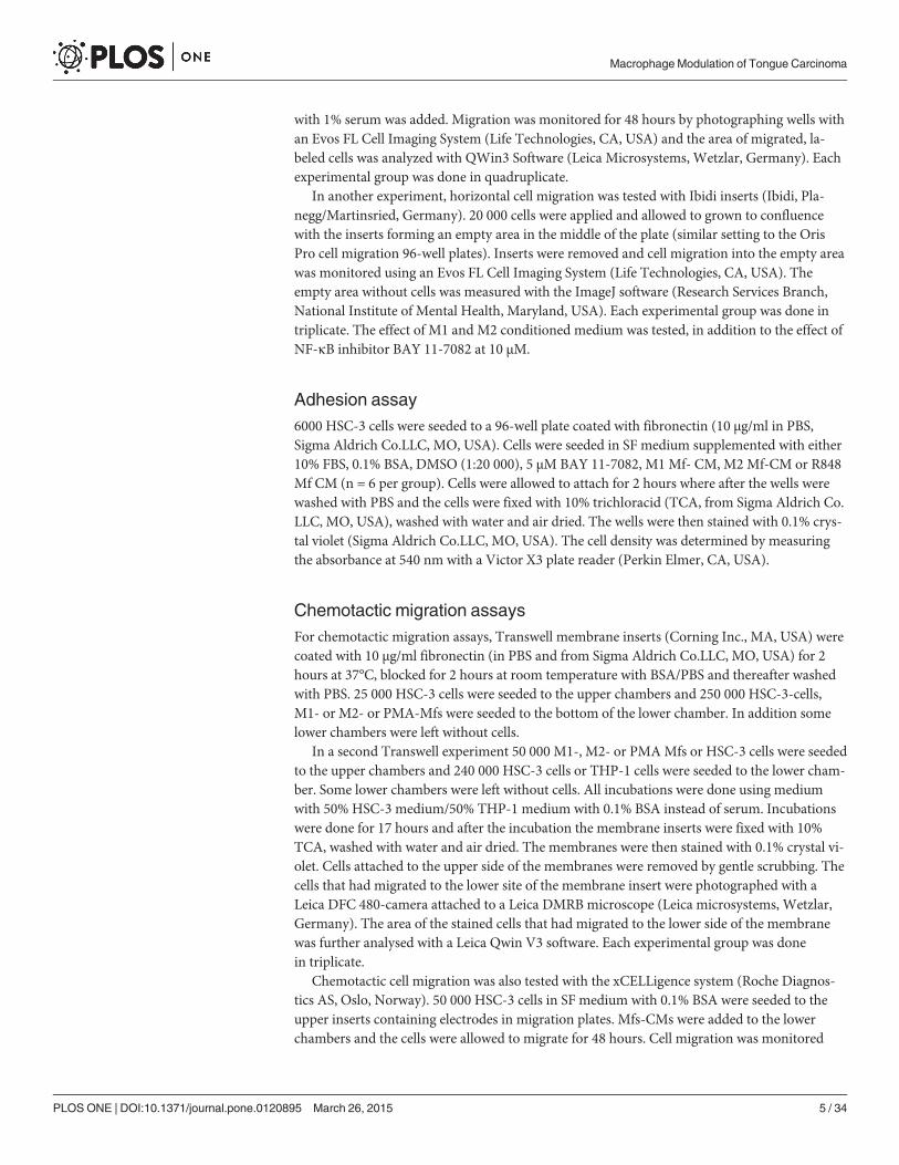
Thereafter, we co-cultured labeled HSC-3 tongue carcinoma cells (red) with labeled THP-1Mfs (green). We first established that HSC-3 cell proliferation was not affected by any secretedfactor from any of the used Mf subtypes and that HSC-3 cells proliferated normally (notshown). At day 2, the Mfs formed islands within the more uniform HSC-3 layer (Fig. 1A,D,G).At day 5, co-localization of HSC-3 and Mfs could be seen (Fig. 1B,E,H white arrows) as yellowmerging. Whether this was fusion of HSC-3 and Mfs or phagocytosis by either cell line couldnot be established in this experiment. The cells were labeled with Vybrant DilO and CM-Dil(from Life Technologies) respectively which are incorporated into the lipid membranes. Thus,the merging would indicate fusion of membranes.
Interestingly, at days 7–10 of co-cultures, the amount of M1 (Fig. 1C) and M2Mfs (Fig. 1F)had diminished in the co-cultures while the amount of R848 Mfs was still abundant (Fig. 1I).Also, while only few dispersed fused cells could be seen in the HSC-3/M1 (Fig. 1C) and HSC-3/M2 Mfs co-cultures at days 7–10 (Fig. 1F), clusters of fused cells were abundant in R848 Mfsand HSC-3 co-cultures (Fig. 1I). When co-cultures were optically analyzed, we found that overtime, the amount of M1 and M2Mfs in co-cultures was similar to that found in cultures with-out HSC-3 cells (S1 Fig.). In contrast, the amount of R848 Mfs in co-cultures increased up today 7 after which the amount dropped (S1 Fig.). When the amount of HSC-3 cells in co-cul-tures was analyzed, the amount was growing up to day 7 after which it diminished in contrastto the HSC-3 cultures without Mfs where HSC-3 cells continuously proliferated even after 14days (Fig. 1J). A reduction in the amount of HSC-3 cells appeared at day 7 simultaneously withthe drop in macrophage amount. Interestingly, while R848 Mfs appeared to benefit from theco-culture with HSC-3 in comparison to being cultured alone, HSC-3 cell proliferation on theother hand was readily reduced by co-culture with R848 Mfs. This indicates that cell-cell con-tact between R848 Mfs and HSC-3 cells can reduce the amount of tumor cells without affectingthe Mfs themselves. The polarization of monocytes to tumor-reducing-macrophages may beone way in which Imiquimod reduces tumor growth.
Co-cultures were further analyzed by confocal microscopy by which HSC-3 cells and Mfswere found in close vicinity prior to merging. Fusion of plasma membranes could clearly be ob-served (Fig. 1K-M). In a further closer view, macrophage cell content appeared to be incorpo-rated into HSC-3 cells that changed color from red to yellow (Fig. 1K-M).
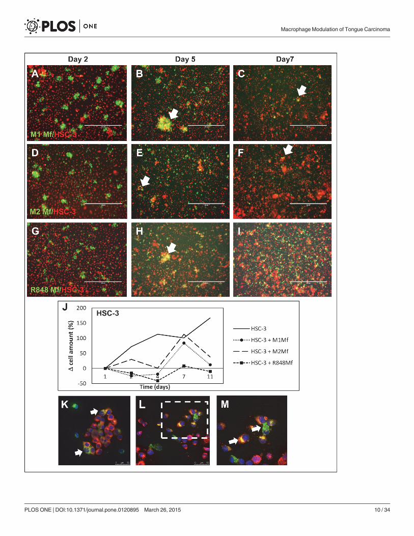
Merging of HSC-3 cells and Mfs is partially due to efferocytosisTo evaluate if the observed fusion of HSC-3 cells and Mfs was due to efferocytosis, a processwhere dying cells are removed by phagocytic cells (usually Mfs), we treated the cells with an in-hibitor for efferocytosis/macropinocytosis, amiloride (AMI) [30]. Based on previous publica-tions, the effect of AMI is concentration dependent and we therefore tested 0.3 and 3 mMconcentrations [30]. In addition, NF-κB is known to regulate TAMs and cancer-related pro-cesses and thus, we also tested the effect of NF-κB inhibitor BAY 11–7082 on co-cultures. Wefirst tested the compounds on the cell lines separately. Dimethylsulfoxide (DMSO; used as ve-hicle) had no effect on the cells cultured alone. At day 2 both HSC-3 and Mfs were slightlyrounded by BAY 11-7082 treatment while AMI at 3 mM induced HSC-3 cells to form elongat-ed connections between neighboring cells forming a network-like organization of cells (notshown).
In DMSO-treated co-cultures at day 2, merging of Mfs and HSC-3 cells was visible as ex-pected (Fig. 2, only HSC-3/M2 Mf co-cultures are shown). Merging was also observed in BAY11–708 (Fig. 2) and 0.3 mM AMI (not shown) treated co-cultures although to a lesser extent.
MacrophageModulation of Tongue Carcinoma
PLOSONE | DOI:10.1371/journal.pone.0120895 March 26, 2015 9 / 34
MacrophageModulation of Tongue Carcinoma
PLOSONE | DOI:10.1371/journal.pone.0120895 March 26, 2015 10 / 34
Fusion cells were almost absent in cultures treated with 3 mM AMI (Fig. 2). Thus, we conclud-ed that the observed merging of Mfs and HSC-3 cells was at least partially due to efferocytosis.There is strong evidence that Mfs can fuse with tumor cells to form heterotypic giant cells andthese fusion cells have been indicated in metastatic processes [31]. The heterotypic fusion cellshave historically been difficult to identify in individuals but there are a few cases that have beenverified [32]. Experiments with fusion cells has shown that typical characteristics are increasedmetastatic potential, increased sensitivity to pro-migratory signals and increased resistance tochemo- and radiotherapy [33]. Fusion might arise through aberrant phagocytosis of apoptotictumor cells by Mfs [34]. This could very well be the case in our study, since at least some of the
Fig 1. Co-culture of HSC-3 cells and Mfs. Vybrant CM-Dil labeled HSC-3 (red) and Vybrant DiO labeled Mfs (green) were co-cultured for up to 11 days innormal growth medium and photographed with an Evos FL Cell Imaging Systemmicroscope. Cell density was analyzed optically using Leica QWin3Software. Co-culture of labeled HSC-3 and M1 Mfs (A-C), HSC-3 and M2 Mfs (D-F) and HSC-3 and R848 Mfs (G-I) at days 2, 5 and 7. White arrows showsmerged/fused cells. (J) Relative HSC-3 cell density in co-cultures. Scale bars 400 μm and n = 2. Representative confocal microscope pictures of co-culturedHSC-3 and R848 Mfs are shown in K-M. Fusion of membranes was observed between the two cell types (white arrows). An R848 macrophage appears todeliver parts to surrounding HSC-3 cells (L), larger picture in M. Scale bars 50 μm in K,L and 25 μm in M.
doi:10.1371/journal.pone.0120895.g001
Fig 2. Efferocytosis in HSC-3 andMf co-cultures. Vybrant CM-Dil labeled HSC-3 (red) and Vybrant DiO-labeled M2Mfs (green) were co-cultured, treatedwith 3 mM efferocytosis-inhibitor amiloride (AMI) or 10 μMNF-κB inhibitor BAY 11-7082. Co-cultures of HSC-3 cells and M2Mfs were photographed with anEVOS FL Cell Imaging Systemmicroscope. Pictures presented are from day 2 (n = 4). Fusion of membranes was observed between HSC-3 cells and M2 Mfsin DMSO (vehicle)- and BAY-treated cultures (white arrows in Overlay-section). Scale bars 200 μm.
doi:10.1371/journal.pone.0120895.g002
MacrophageModulation of Tongue Carcinoma
PLOSONE | DOI:10.1371/journal.pone.0120895 March 26, 2015 11 / 34

fusion of HSC-3 cells and Mfs was inhibited by AMI. It has also been suggested that the fusionis a way of tumor cells to transfer pro-tumorigenic material to surrounding stromal cells andthis has been partially shown in vitro [34].
Co-culture of HSC-3 cells and Mfs alter the cytokine content in vitroDifferent types of Mfs are known to produce specific cytokines while there is no comprehensivecytokine profile of HSC-3 cells. We hypothesized that co-culturing HSC-3 cells and Mfs wouldalter the cytokine profiles. Therefore, SF-media from HSC-3 cells, the three Mf subtypes andthe three co-cultures (HSC-3 with M1-, M2 or R848 Mfs) at day 2 were collected and analyzedfor 80 cytokines with the RayBio Human Cytokine Antibody Array. The array values were nor-malized against positive control, background and sample protein content. The analysis showedthat HSC-3 cells contained low levels of all the analyzed cytokines as compared to the Mfs witha few exceptions. Therefore, the antibody array values for HSC-3 were chosen for calculation offold change in comparison to the other groups. Cytokines with inter-group fold changes of atleast 2x were considered of interest. Of all groups, the M1 Mfs cultured alone showed the high-est levels of all cytokines. For representative presentation, cytokines with similar fold changes(e.g. max fold change 25x in figure A etc.) were grouped into the same graphs. The full anti-body array data is provided in the supporting information (S1 Dataset).
M1 Mfs medium contained higher amounts of TNF-α, Granulocyte—Macrophage Colony-Stimulating Factor (GM-CSF), Monokine Induced by Gamma Interferon (MIG), Tissue Inhibi-tor of Metalloproteinases (TIMP)-1 (Fig. 3B) and Monocyte Chemoattractant Protein-3(MCP-3) (Fig. 3A) than the other groups. Co-culture of M1 Mfs and HSC-3 cells reduced thesecretion of these cytokines several-fold down to levels seen in the other groups (Fig. 3A,B).TNF-α has been shown to be the major inflammatory signal for macrophage lytic activityagainst tumor cells [29]. However, TNF-α is also associated with sustained chronic inflamma-tion of the TME through NF-κB regulation [35]. High levels of TNF-α have been found inhead and neck (HNSCC) patient serum and in malignant oral carcinoma cell lines [36]. Thus,while TNF-α, MCP-3 and GM-CSF are pro-tumorigenic, we found that co-culture with anti-inflammatory M1 Mfs and HSC-3 cells actually down-regulated these cytokines.
MIG on the other hand is involved in T cell trafficking and has been found to be anti-cancerous and also previously found in high amounts in M1 Mfs [37]. Interestingly, a previousstudy found that NF-κB mediated polarization of M2 Mfs to M1Mfs was associated with anincrease in MIG secretion [38]. In addition, TIMP-1 is a potent inhibitor of several matrixmetalloproteinases (MMPs), including MMP-1, -8 and -9. Thus, down-regulation of MIG andTIMP-1 in HSC-3 cells and M1 Mfs is in favour of pro-tumoral processes.
Transforming Growth Factor (TGF)-β, Epidermal Growth Factor (EGF) and MacrophageColony-Stimulating Factor (M-CSF) (Fig. 3B) were all found in higher amounts in M1Mfswhile the levels were slightly lower in M2 and R848 Mfs. However, co-culturing M1 Mfs andHSC-3 resulted in several-fold lower levels of all these cytokines while M2 Mfs and HSC-3 co-cultures resulted in higher levels as compared to the M2Mfs alone (Fig. 3B). TGF-β is associat-ed with M2 type Mfs and tumor cell proliferation, tissue fibrosis, immunosuppressive activityand epithelial-to-mesenchymal-transformation [23]. Specifically, TGF-β promotes fibroblast-to-CAF trans-differentiation and accumulation of CAFs is significantly correlated to poorprognosis in tongue cancer [8]. In this regard, high levels of TGF-β have been found in OSCCTAMs and has been associated with metastasis [18]. EGF is a growth factor highly associatedwith progression of OSCC [36] and is intricately associated with M-CSF, a cytokine involved inmacrophage recruitment and differentiation [39]. Similar to our results, Yang et al. [40] foundthat M-CSF production was increased in Mfs of mouse breast carcinoma cells and Mfs co-
MacrophageModulation of Tongue Carcinoma
PLOSONE | DOI:10.1371/journal.pone.0120895 March 26, 2015 12 / 34
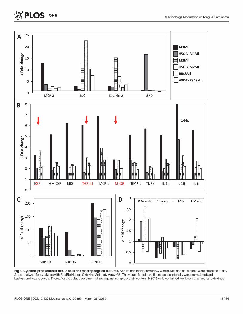
Fig 3. Cytokine production in HSC-3 cells andmacrophage co-cultures. Serum-free media from HSC-3 cells, Mfs and co-cultures were collected at day2 and analyzed for cytokines with RayBio Human Cytokine Antibody Array G5. The values for relative fluorescence intensity were normalized andbackground was reduced. Thereafter the values were normalized against sample protein content. HSC-3 cells contained low levels of almost all cytokines
MacrophageModulation of Tongue Carcinoma
PLOSONE | DOI:10.1371/journal.pone.0120895 March 26, 2015 13 / 34

cultures inducing polarization fromM1 to M2 type Mfs through NF-κB p50 induced c-Jun ex-pression. In vivo, M-CSF produced by carcinoma cells attracts TAMs to nearby blood vesselswhich in turn promote the expression of EGF by Mfs [33]. In our study, M-CSF was producedby HSC-3 cells although to a lower extent than in the Mfs. The EGF in turn attracts tumor cellsto the blood vessels and a positive feedback loop is created, feeding the growth and local inva-sion of tumor cells [33,36]. Clearly, co-culture of HSC-3 and M2 Mfs in this study induced ex-pression of both EGF and M-CSF as well as TGF-β further underlining the important role forthese three cytokines in oral cancer progression (Fig. 3B, underlined and indicated with redcolor).
All groups showed a similar fold change of all the studied interleukins with little inter-groupvariations, except for the M1Mfs cultured alone which showed a higher fold change for all in-terleukins and a 144x fold change for IL-1β (Fig. 3B). Interestingly, co-culturing M2 or R848Mfs with HSC-3 did not particularly affect interleukin levels as compared to Mfs culturedalone. However, incubating M1 Mfs with HSC-3 cells consequently reduced interleukin secre-tion several-fold as compared to the M1Mfs alone (Fig. 3B). A study by Engström et al. [37]showed very similar results in terms of interleukins secreted by M1Mfs, further re-enforcingour results. IL-1β, secreted to a high amount by the M1 Mfs is pro-inflammatory, however ithas also been found to participate in trans-differentation of fibroblasts-to-CAFs [41].
The highest fold-changes (up to 200x) were found for Macrophage inflammatory protein(MIP)-1β, MIP-3α and RANTES (Fig. 3C). When M1 Mfs were co-cultured with HSC-3 cells,the levels were reduced and conversely, when M2 Mfs were incubated with HSC-3 cells the lev-els increased (Fig. 3C). We found low amounts of MIP-3α and RANTES in HSC-3 cells in con-cordance with previous publications [42,43] and also MIP-1β (not shown) which has notpreviously been described in OSCC cell lines. Previous work has confirmed that RANTES,MIP-1α and MIP-1β and their receptor C-C Chemokine Receptor type 5 (CCR5) are involvedin the recruitment of M1 Mfs to tumor sites [39,44]. Dysregulated CCR5 expression on Mfshave been found in OTSCC patients which may contribute to reduced anti-tumoricidal activity[44].
The only cytokines that were expressed to a higher level in HSC-3 than in some Mfs werePlateled-derived growth factor (PDGF)-BB, angiogenin, Macrophage migration Inhibitory Fac-tor (MIF) and TIMP-2 (Fig. 3D). In addition, the fold change of all four cytokines increased inco-cultures of HSC-3 /Mfs as compared to Mfs alone (Fig. 3D). Circulating tumor/macrophagehybrid cells have been found to release MIF through exosomes which then creates metastaticniches [32]. MIF has previously been found to be secreted by M2 Mfs [33] but we found thatHSC-3 cells secreted more MIF than M2 Mfs. Yaddanapudi et al. [45] showed that MIF con-trols activation of M2 Mfs in melanoma tumor-carrying mice. The role of MIF in OSCC pro-gression is controversial [46,47] but inhibiting MIF activity in OSCC cells has been found toinhibit invasion and proliferation [46,48]. TIMP-2 expression has been found in tumor cells atthe invasive front as well as in the stromal cells [49]. TIMPs expression in OSCC is mostly asso-ciated with poor outcome [50] and our results corroborate well with this. Interestingly, whileHSC-3 cells secreted more PDGF-BB than the Mfs, co-culture increased secretion regardless ofmacrophage subtype. PDGF is involved in the modulation of the TME [51] and a recent studyshowed that TAMs expressed PDGF-BB only when they adhered to cancer stem cell-like breastcancer cells and this was modulated through hyaluronan synthase [52].
(with a few exceptions) as compared to the Mfs and the antibody array values for HSC-3 were chosen for calculation of fold change in comparison to the othergroups. Cytokine data in the figure are grouped and presented based on mean fold change levels: up to 25x (A), up to 8x (B) and up to 200x (C). Fourcytokines had fold changes that were higher in HSC-3 cells than in Mfs (D). EGF, TGF-β1 and M-CSF are marked in red with red arrows.
doi:10.1371/journal.pone.0120895.g003
MacrophageModulation of Tongue Carcinoma
PLOSONE | DOI:10.1371/journal.pone.0120895 March 26, 2015 14 / 34

Taken together, we found that cytokines shown to be associated with OSCC and increasedinvasion, such as EGF, M-CSF, TGF-β, TIMPs, PDGF-BB and MIF were increased upon co-culture with HSC-3 and M2 Mfs, and on the other hand down-regulated in HSC-3/M1 Mfs co-cultures. Thus, these cytokines are well suited for further investigation as therapeutic targets inOSCC.
An M2Mf-secreted soluble factor induces migration of HSC-3 tonguecarcinoma cellsWe continued with investigating the effect of Mfs on HSC-3 cell migration, something whichhas not previously been characterized. We used different migration systems to investigate theeffect of Mfs on HSC-3 cell migration.
First, we tested the effect of macrophage-HSC-3 cell-cell contact on HSC-3 cell migration ina horizontal plane using the Oris Pro cell migration system. In this system, cells are grown in a96-well plate around an insert which is then removed, allowing cells to migrate to the emptyspace.
In homogenic cell cultures Mfs migrated to the empty space and filled it to approximately40% at 48 hours while HSC-3 cells had filled the space almost completely at 24 hours withminor changes at 48 hours (not shown). In co-cultures, HSC-3 cells (red) were found to mi-grate more efficiently in co-culture with M2Mfs (Fig. 4B,C; Mfs in green), although not statisti-cally significant (Fig. 4A). HSC-3 cells migrated slightly slower in co-culture with M1 Mfs (S1Fig. and Fig. 4A) and similarly with and without R848 Mfs (S1 Fig. and Fig. 4A). When the mi-gratory front was observed more closely, co-migration together with merging of Mfs and HSC-3 cells could be seen with all Mfs subtypes (Fig. 4B,C and S1 Fig.), white arrows show mergedcells). The Mfs appeared to migrate slightly in front of the HSC-3 cells. In co-cultures with M1Mfs and HSC-3 cells, Mfs were more abundant at 48 hours (S1 Fig.) which could partially ex-plain the reduced migration of HSC-3 cells. These results indicate that when cell-cell contact ispropagated in a horizontal plane, M2 Mfs induce migration of HSC-3 cells while M1 Mfsslightly inhibit migration.
We then tested the effect of Mf-conditioned medium (CM) on horizontal HSC-3 migrationusing Ibidi cell culture inserts. Cells were allowed to attach overnight on both sides of the in-serts and thereafter inserts were removed, medium changed to Mf-CM with or without BAY11-7082 and cells allowed to migrate into the empty space. In this experimental setting, HSC-3cells treated with Mf-CMmigrated slower than DMSO-treated cells (Fig. 4D,E, only M2 Mf-CM is shown). While no statistical significance could be reached, it appeared that M2 Mf-CMhad the least effect and M1 Mf-CM slowed HSC-3 cells down more effectively. Interestingly,while BAY 11-7082 did not affect HSC-3 cell migration, addition of BAY 11-7082 slightly re-versed the Mf-CM inhibition of HSC-3 cell migration (Fig. 4E). This indicates that the migra-tion inhibitory factor(s) present in Mf-CM is/are partially NF-κB-dependent.
Next, chemotactic migration using the Transwell culture inserts was assessed. HSC-3 cellswere cultured in the upper inserts and Mfs in the lower chambers. All Mfs induced migrationof HSC-3 cells to a similar efficacy although only M1 Mfs showed statistical significance(Fig. 4F). For comparison, HSC-3 cells were also cultured to the lower chamber of some wellsand did not induce migration of the HSC-3 cells in the upper chamber (Fig. 4F). This indicatesthat the migration inducing factor is specifically produced by Mfs. We also tested if HSC-3cells were able to attract Mfs, an event that would be critical in vivo. Here, HSC-3 cells wereseeded to the lower chamber in Transwell-plates while Mfs were seeded to the upper chamberin SF-medium. We found that HSC-3 cells induced migration of Mfs, although statistical sig-nificance was not reached (S2 Fig.).
MacrophageModulation of Tongue Carcinoma
PLOSONE | DOI:10.1371/journal.pone.0120895 March 26, 2015 15 / 34
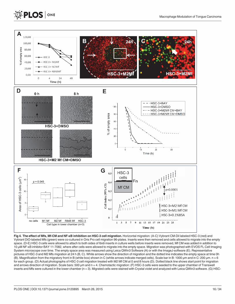
Fig 4. The effect of Mfs, Mf-CM and NF-κB inhibition on HSC-3 cell migration.Horizontal migration: (A-C) Vybrant CM-Dil labeled HSC-3 (red) andVybrant DiO-labeled Mfs (green) were co-cultured in Oris Pro cell migration 96-plates. Inserts were then removed and cells allowed to migrate into the emptyspace. (D-E) HSC-3 cells were allowed to attach to both sides of Ibidi inserts in culture wells before inserts were removed, Mf CM was added in addition to10 μMNF-κB inhibitor BAY 11-7082, where after cells were allowed to migrate into the empty space. Migration was photographed with EVOS FL Cell ImagingSystemmicroscope over time. The empty space area was measured using Leica QWin3 Software (A) or with the ImageJ-software (E). Representativepictures of HSC-3 and M2Mfs migration at 24 h (B, C). White arrows show the direction of migration and the dotted line indicates the empty space at time 0h(B). Magnification from the migratory front in B (white box) shown in C (white arrows indicate merged cells). Scale bar in B: 1000 μm and in C: 200 μm. n = 6for each group. (D) Actual photographs of HSC-3 cell migration treated with M2 Mf CM at 0 and 8 hours (D). Dotted black line shows start point for migrationand arrows direction of migration. Scale bars: 500 μm and n = 4. Chemotactic migration: (F) HSC-3 cells were seeded to the upper chamber of Transwellinserts and Mfs were cultured in the lower chamber (n = 3). Migrated cells were stained with Crystal violet and analyzed with Leica QWin3 software. (G) HSC-
MacrophageModulation of Tongue Carcinoma
PLOSONE | DOI:10.1371/journal.pone.0120895 March 26, 2015 16 / 34

The xCelligence migration assay was further used to analyze the effect of Mf-secreted che-motactic factors on HSC-3 migration. HSC-3 cells were seeded to fibronectin-coated upperchambers and Mf-CM was added to the lower chambers. Significantly more HSC-3 cells mi-grated towards M2 Mf-CM than towards non-CM (Fig. 4G, p<0.0001). Also M1Mf-CM in-duced HSC-3 cell migration significantly (Fig. 4G, p<0.0001), however, not as efficiently asM2 Mf-CM. The experiment was repeated on gelatin-coated chambers and by using uncoatedchambers with a highly similar result (not shown). BAY 11-7082 was tested in this experimen-tal setting but was not found to affect chemotactic HSC-3 cell migration. Mf-CMs were alsonot found to induce proliferation of HSC-3 cells as analysed by xCelligence (not shown).Thus, soluble factors secreted by TAM-like Mfs act as chemotactic agents on highly aggressiveHSC-3 cells but this mechanism is not NF-κB dependent. As found in the cytokine antibodyarray, Mf-CMs contain many chemotactic cytokines such as EGF, M-CSF and MIF. This isthe first time that the direct effect of Mfs and Mf-CM on tongue SCC cell migration has beendemonstrated.
Interestingly, while the Mf-CM clearly contains chemotactic signals for HSC-3 cells, the Mf-CM also contains inhibitory factor(s) acting directly on HSC-3 cells if applied on horizontalmigration settings (haptotactic migration). In chemotactic migration, the CM acts from belowcreating a gradient of migratory factors for HSC-3 cells while in the horizontal migration set-tings, the medium acts on the top of cells. The mode of action is clearly dependent on tumorcell orientation.
Mfs induce HSC-3 cell invasion by cell-cell contactMigration studies only give information about the mobility of cells in a given 2D environment.To further investigate the effect of Mfs on HSC-3 cell mobility, we also studied invasion whichrequires cells to get through a 3D-barrier. We first tested if Mf-CM had any effect on HSC-3cell adhesion since this would affect invasion. We found that significantly less HSC-3 cells ad-hered on fibronectin when incubated in serum free (SF)-medium (supplemented with bovineserum albumin) as compared to HSC-3 cells in medium with fetal bovine serum (Fig. 5A,p<0.0001). DMSO at the same concentration used as vehicle did not affect adhesion as com-pared to cells in SF-medium with BSA. Cells were then treated with Mf-CM to which BAY 11-7082 or DMSO was added. BAY 11-7082 or M1Mf-CM alone did not affect adhesion whileM2 and R848 Mf-CMs slightly reduced HSC-3 cell adhesion (Fig. 5A, p = 0.07). Only the com-bination of BAY 11-7082 and Mf-CM significantly reduced HSC-3 cell adhesion as comparedto DMSO alone (Fig. 5A, p<0.0001 for M1 Mf-CM+BAY; p = 0.002 for M2 Mf-CM+BAY;p = 0.02 for R848 Mf-CM+BAY). This indicates that while HSC-3 cell adhesion in the absenceof Mfs is not NF-κB dependent, Mf-CM (regardless of subtype) contains an NF-κB regulatedmolecule which promotes HSC-3 cell adhesion.
The strongest effect was seen with M1 Mf-CM, indicating that M1 Mfs activity could bedownregulated and NF-κB dysregulated in favour of oral cancer. In a study by Chengye et al.[53] peroxisome proliferator-activated receptor gamma-1-related co-activator was found to in-hibit expression of adhesion-mediating molecules through inhibition of NF-κB activation.
We then tested HSC-3 invasion using the Transwell-based invasion assay in which HSC-3cells were cultured on top of Matrigel and the Mfs were seeded to the lower chamber. For com-parison, we used also phorbol myristate acetate (PMA) Mfs which are THP-1 cells that havebeen polarized to Mfs with PMA alone and resemble TAM-like Mfs [54]. As controls we seeded
3 cells were seeded to fibronectin (Fn)-coated xCelligence inserts and Mf-CM was added to the lower chambers (n = 4). Migration was monitored for up to 24hours. The xCelligence systemmeasures changes in cell impedance as cells migrate through the insert membrane and data is presented as cell index.
doi:10.1371/journal.pone.0120895.g004
MacrophageModulation of Tongue Carcinoma
PLOSONE | DOI:10.1371/journal.pone.0120895 March 26, 2015 17 / 34

Fig 5. Co-culture of HSC-3 cells and Mfs induce invasion in an organotypic 3D tissuemodel. (A) HSC-3 cells were treated with Mf-CM and/or 10 μMBAY 11-7082 (n = 6) and adhesion to fibronectin-coated wells was analysed after 2 hours with Crystal violet-staining. Stained wells were analysed bymeasuring absorbance at 540 nm. (B) Labeled HSC-3 cells were cultured on top of Matrigel and Mfs, HSC-3 or THP-1 cells were cultured in the lowerchambers (n = 6). Invaded cells were analyzed from the membrane by measuring fluorescence. HSC-3 cells and Mfs were co-cultured on top of humanmyoma tissue (C,D, n = 3) for 10 days where after tissues were fixed and processed for immunohistochemistry. Pan-cytokeratin stained sections (in C, scalebars: 500 μm) were photographed and invasion areas and invasion depths were analysed with the Leica Qwin3 software. Conditioned medium was collectedat day 4 and 8 frommyomas and medium containing 0.5 (Mf CMs) or 15 μg (HSC-3) protein were subjected to gelatin zymography (E, day 4). Samples weredone in triplicate, representative picture of samples is shown in E. Recombinant MMP-2 and -9 were used as positive controls. Relative intensity of MMP-9bands were analyzed with the ImageJ-software (F).
doi:10.1371/journal.pone.0120895.g005
MacrophageModulation of Tongue Carcinoma
PLOSONE | DOI:10.1371/journal.pone.0120895 March 26, 2015 18 / 34

HSC-3 or THP-1 cells in the lower chambers to exclude that any cell type could secrete invasivesignals for HSC-3 cells. M2- and PMA-type Mfs significantly induced HSC-3 cell migrationcompared with controls (Fig. 5B, p<0.05). In this setting, also M1-type Mfs induced invasionof HSC-3 cells although not statistically significant (Fig. 5B).
We then used the fully human 3D hypoxic myoma-based model [27,28] where HSC-3 cellsand Mfs were first co-cultured on top of the myoma. After 10 days, the myomas were fixed andfurther analysed by histology and semi-quantitative computer analysis. We found that bothM2- and PMAMfs significantly induced invasion of HSC-3 cells in myoma tissue (Fig. 5C,D,p = 0.02 and 0.002 respectively) compared to HSC-3 cells alone (Fig. 5C,D). The M1-type Mfson the other hand significantly reduced HSC-3 cell migration (Fig. 5C,D, p = 0.034). The inva-sion index was calculated and results were comparable for the invasion depth (not shown).Thus, M2 Mfs act pro-invasive on HSC-3 cells while M1 Mfs inhibits invasion. In another ex-periment, we also tested the ability of R848 Mfs to induce invasion of HSC-3 in myomas in ad-dition to NF-κB inhibitor BAY 11-7082 and Imiquimod-solution (R848). There were nosignificant differences in HSC-3 invasion depth between the groups (S2 Fig.). When the inva-sion index was calculated, R848 Mfs were found to decrease invasion of HSC-3 cells, as didalso BAY 11-7082, but the effect was not statistically significant (S2 Fig.). In a second myomaexperiment, HSC-3 cells were cultured on top of the myoma and myomas were incubated withMf-CM. As opposed to the results from migration assays, Mf-CM did not induce invasion ofHSC-3 cells (S2 Fig.). This is an important finding showing that although Mf-CM containspro-migratory signals for HSC-3 cells, it does not necessary induce invasion. Taken together,for the first time we show that the close vicinity of Mfs to oral cancer cells is crucial for boththe induction and inhibition of tumor cell invasion through tissues. In addition, R848 Mfs donot appear to affect invasion or migration substantially. However, as observed in the initial co-culture experiments, cell-cell contact with R848 Mfs appears to reduce HSC-3 cell proliferation(Fig. 1J) which might be one of the mechanisms by which these artificial Mfs work.
Medium from the myoma experiment performed with the Mf-CM was collected on days 4and 8 and analysed by gelatin zymography. We have shown in previous publications that medi-um collected from cell-free myomas contain only minor amounts MMP-9 but culturing HSC-3cells on top of myomas results in secretion of MMP-9 into the incubation medium [27]. Alsohere, SF-medium from the myoma with HSC-3 cells on top contained both MMP-2 and-9(Fig. 5E, day 4). The uncropped zymograms can be viewed in the supporting information (S3Fig.).
Mf-CM from the myomas incubated with HSC-3 cells was found to contain high levels ofactive MMP-9 (Fig. 5E,F). Interestingly, M2 Mf-CM contained the least MMP-9 and theMMP-9 level also decreased between day 4 and 8 (Fig. 5F). In contrast, M1 Mf-CM (p = 0.055at day 4 and p = 0.020 at day 8) and R848 Mf-CM (p = 0.023 at day 4 and p = 0.004 at day 8)contained significantly more MMP-9 than M2 Mf-CM (Fig. 5F). In addition, in myomas incu-bated with HSC-3 cells and M2 Mf-CM, MMP-9 was present mostly in the pro-form while inmyomas incubated with HSC-3 cells and M1 Mf-CM or R848 Mf-CM, MMP-9 was presentboth in the active and pro-form. MMP-2 was present in all myoma incubation media and therewere no major differences between the groups (not shown). We have recently shown that inHSC-3 cells, MMP-9 actually might have a protective role (Väyrynen et al. manuscript in prep-aration). In this regard, it is interesting to note that when HSC-3 cells are invading tissue in thepresence of M1 Mf-CM and R848 Mf-CM, the MMP-9 level remains high, while in the pres-ence of M2 Mf-CM, the MMP-9 level is actually decreasing over time. Zajac et al. [55] foundthat M2Mfs induced angiogenesis which was mediated through production of high MMP-9 se-cretion. Similar to our findings, they also found that M1 Mfs produced high levels of MMP-9.However, M1 Mfs also produced high levels of TIMP-1 while M2 Mfs did not and thus, only
MacrophageModulation of Tongue Carcinoma
PLOSONE | DOI:10.1371/journal.pone.0120895 March 26, 2015 19 / 34

M2Mf-produced MMP-9 activated angiogenesis. In parallel with our results, M1 Mfs pro-duced significantly more TIMP-1 as compared to the M2Mfs. Interestingly, co-culture withHSC-3 cells reduced TIMP-1 production also in M1 Mfs. In this regard, Koontongkaew et al.[56] found that direct contact between HNSCC cells and fibroblasts activated MMP-9.
Modulation of NF-κB in HSC-3 and macrophage co-culturesThe activity of the nuclear transcription factor complex NF-κB is associated with oral carcino-mas and is known to be dysregulated in TAMs. The findings in the migration experiments thatMf-CM contains NF-κB regulated molecules acting inhibitory on HSC-3 cell migration lead usto investigate the previously uncharacterized expression and activity of NF-κB subunits p50and p65 in HSC-3 and macrophage co-cultures.For this purpose, we performed immunofluo-rescence on cultured HSC-3 and Mfs analyzed by confocal microscopy. All samples were sub-ject to immunostaining with polyclonal and monoclonal non-specific immunoglobulins andshowed no unspecific staining (not shown).
M1 Mfs were CD68 positive (S4 Fig.) and CD163 negative (marker for TAM/M2, notshown) while M2 Mfs were positive for both CD68 and CD163 (S4 Fig.). R848 Mfs were CD68positive (S4 Fig.) but some cells were also CD163 positive (S4 Fig.). HSC-3 cells cultured alonestained positive for pan-cytokeratin (S4 Fig.) and were negative for macrophage markersCD68 or CD163. Both the NF-κB p50 (Fig. 6A) and NF-κB 65 (Fig. 6B) subunits were foundin the cytoplasm of HSC-3 cells and TNF-α treatment induced translocation to the nucleus(Fig. 6A,B). BAY 11-7082 mostly but not entirely inhibited nuclear translocation of both sub-units. This indicates that the NF-κB activation mechanism itself would be normal in HSC-3cells. In M2 Mfs, the p50 subunit appeared in the nucleus of resting cells while in the TNF-αtreated cells there was less visible p50 in the nuclei. BAY 11–782 reduced the overall expressionof p50 (Fig. 6A). Interestingly, the p65 subunit was mostly detected in the cytoplasm of bothresting, activated and inhibited M2 Mfs (Fig. 6B). This indicates that there could be an exces-sive amount of p50 subunits in the M2 type Mfs, perhaps causing a dysregulation of NF-κB ac-tivation. In the TME, TAMs are thought to be tolerized by inflammatory stimuli through theformation of repressive p50 homodimers in the nucleus which in turn activate non-tolerable,tumor-progressive favouring genes [23,57]. The formation of p50 homodimers also inhibitsthe polarization of M1 Mfs [23]. IL-10, prostaglandin E2 and TGF-β, all produced by TAMsand many tumors, induce the formation of p50 homodimers [23]. In this regard, we found thatco-culturing HSC-3 cells and M2 Mfs increased TGF-β secretion (Fig. 3B). Connelly et al. [38]showed that re-activation of NF-κB signaling reduced tumor burden in mice. While p50 homo-dimers have been found in complex with bcl-3, acting on gene transcription [58], the p50/p50complex is mostly thought to repress gene activation. Also, the p50/p50/bcl-3 complex hasbeen found in nasopharyngeal carcinomas but is rare in other HNSCCs [59].
M1 and R848 Mfs were similar in that p50 was present in the nuclei of resting Mfs andTNF-α treatment did not change this appearance as expected (S5 Fig.). The NF-κB p65 subuniton the other hand was similarly present in the cytoplasm of both resting M1 and R848 Mfs,however in the TNF-α activated cells, a nuclear translocation of p65 could be observed in theM1 but curiously not in the R848 Mfs (S5 Fig.). BAY 11–782 treatment decreased the overallappearance of p50 and p65 and also the nuclear translocation in both cell types (S5 Fig.). Thus,the NF-κB activation mechanism present in R848 Mfs is different from both the M1 and M2Mfs. R848 is a TLR7/8 agonist and is thus expected to induce NF-κB through the MyD88- NF-κB pathway [24]. Imiquimod has so far been used for the treatment of skin carcinomas throughthe modulation of immune cells and polarization of Mfs in the TME [24,25]. Recently,mucoadhesive films with imiquimods for the treatment of OSCC have been developed [26].
MacrophageModulation of Tongue Carcinoma
PLOSONE | DOI:10.1371/journal.pone.0120895 March 26, 2015 20 / 34

MacrophageModulation of Tongue Carcinoma
PLOSONE | DOI:10.1371/journal.pone.0120895 March 26, 2015 21 / 34

Our results indicate that R848 Mfs are not directly polarized to a type clearly identified as M1or M2. Hadler-Olsen et al. [19] showed that in terms of skin and tongue carcinoma cells, theTME directs tumor progression. We thus suggest that the R848 Mfs deserve further experi-ments to evaluate if imiquimod modulation of TME inflammatory cells will be as beneficial intongue cancer as it is in skin cancer.
In M2Mfs and HSC-3 cell co-cultures, CD68 positivity was seen also in HSC-3 cells andthis expression was abolished by TNF-α treatment (Fig. 7, red arrow). CD163 positivity wasseen only in M2 Mfs (not shown). NF-κB p50 subunit was observed in both the cytoplasm andnuclei of M2 Mfs (Fig. 7, green arrows) but in HSC-3 cells it was mostly confined to the nucleiin co-cultures (Fig. 7, red arrows). TNF-α treatment induced translocation of p50 to the nucleiof M2 Mfs (Fig. 7 green arrow) while the nuclear p50 found in HSC-3 cells was unchanged inappearance (Fig. 7, red arrow). Nuclear translocation of the NF-κB p65 subunit was observedafter TNF-α treatment in HSC-3 cells (Fig. 7, red arrow) while in the M2 Mfs this translocationwas almost absent (Fig. 7, green arrow). It was also lacking in HSC-3 cells in a close proximityto M2 Mfs (Fig. 7, red arrow). BAY 11-7082 abolished p50 expression in almost all cells and in-hibited p65 translocation to most but not all cells (Fig. 7). It thus appears that similarly to thecultures with M2 Mfs alone, also in co-cultures the M2Mfs are lacking in p65 translocation in-dicating the presence of p50 homodimers and dysregulated NF-κB activation. However, theM2 Mfs also appear to induce dysregulation of NF-κB signaling (no translocation of p65) inHSC-3 cells at least in a close proximity to the M2 Mfs. This might be one way in which TAMsadd to tumor progression and also explaining why cell-cell contact appears to be importantfor interaction.
In M1Mfs and HSC-3 co-cultures, M1 Mfs continued to be CD68 positive (not shown),however some M1 Mfs also showed clear CD163 positivity (Fig. 8A, green arrow). Also HSC-3cells were CD163 positive after TNF-α treatment but this was abolished after BAY treatment(Fig. 8A, red arrows). This indicates that the proximity of carcinoma cells can induce CD163expression and polarization in M1 Mfs to TAMs. In a study by Maniecki et al. [60] both Mfsand cancer cells were CD163 positive in bladder cancer biopsies. In co-cultures with Mfs, thebladder cancer cells started to express CD163 indicative of either fusion or exosome RNA/pro-tein transfer. In any case, cytokines alone were not found to induce endogenous expression ofCD163 in bladder cancer cells [60]. Also breast and colorectal cancer cells are known to expressCD163 which is associated with reduced patient survival compared to CD163 negative cancers[33]. Thus, CD163 expression in cancer cells is a measurement of aggressiveness. HSC-3 cellsstarted to express CD163 after TNF-α stimulation and this may be partially NF-κB dependentsince BAY 11–782 abolished the expression pattern. Therefore, CD163 could be a potentiallyimportant diagnostic tool for tongue cancer. NF-κB deposition in HSC-3 cells co-cultured withM1 Mfs appeared similar to that observed in culture without Mfs (Fig. 8A). However, M1 Mfsshowed p50 deposition as seen in cultures without HSC-3 cells (S6 Fig.) while translocation ofp65 was weak (Fig. 8A). This indicates that in co-cultures with M1 Mfs, HSC-3 cells retaintheir NF-κB activity mechanism while changing the NF-κB activity in M1 Mfs (lack of p65translocation). Again, this indicates that HSC-3 cells in a close vicinity to M1 Mfs act to changethe polarization towards a more tumor-progressive type. Co-cultures of R848 Mfs and HSC-3cells were CD163 negative (not shown) but CD68 positive so that also HSC-3 cells were
Fig 6. Expression of NF-κB p50 and p65 subunit in HSC-3 and M2Mfs. Vybrant CM-Dil labeled HSC-3 cells (red) and unlabeled M2 Mfs were incubatedwith DMSO or 10 ng/ml TNF-α for 30 min where after cells were fixed for immunofluorescence with polyclonal NF-κB p50 (A) or NF-κB p65 (B) antibodies.Some samples were pre-incubated with 10 μMBAY 11-7082 prior to TNF-α activation. AlexaFluor488-conjugated secondary antibody was used forvisualization. Samples were mounted with DAPI- mountain medium to visualize nuclei (blue). Samples were photographed with a Leica Confocal microscopewith 63x oil immersion objective. Scale bars 50 μm.
doi:10.1371/journal.pone.0120895.g006
MacrophageModulation of Tongue Carcinoma
PLOSONE | DOI:10.1371/journal.pone.0120895 March 26, 2015 22 / 34

positive after TNF-α treatment (Fig. 8 B red arrows). The expression was abolished by BAYtreatment. Co-culturing HSC-3 and R848 Mfs resulted in reduced p50 appearance in both celltypes (Fig. 8B) while TNF-α treatment increased expression and BAY 11–782 abolished it. NF-κB p65 was readily detected in the cytoplasm of HSC-3 and R848 Mfs in co-culture (S6 Fig.).Nuclear translocation was observed in HSC-3 cells after TNF-α treatment but with less nucleardeposition in R848 Mfs. BAY 11-7082 almost completely blocked translocation (S6 Fig.).
Fig 7. Expression of Mf-markers and NF-κB subunits in HSC-3 and M2Mf co-culture. Vybrant CM-Dil labeled HSC-3 cells (red) and unlabeled M2 Mfswere incubated with DMSO or 10 ng/ml TNF-α for 30 min where after cells were fixed for immunofluorescence with antibodies for CD68, NF-κB p50 and p65subunits. Some samples were pre-incubated with 10 μMBAY 11-7082 prior to TNF-α activation. AlexaFluor488-conjugated secondary antibody was used forvisualization. Samples were mounted with DAPI- mountain medium to visualize nuclei (blue). Samples were photographed with a Leica Confocal microscopewith 63x oil immersion objective. Red arrows indicate HSC-3 cells and green arrows Mfs. Scale bars 50 μm.
doi:10.1371/journal.pone.0120895.g007
MacrophageModulation of Tongue Carcinoma
PLOSONE | DOI:10.1371/journal.pone.0120895 March 26, 2015 23 / 34

Fig 8. Expression of Mf-markers and NF-κB subunits in co-cultures of HSC-3 cells and M1 or R848 Mfs. Vybrant CM-Dil labeled HSC-3 cells (red) andunlabeled M1- or R848 Mfs were incubated with DMSO or 10 ng/ml TNF-α for 30 min where after cells were fixed for immunofluorescence with antibodies for
MacrophageModulation of Tongue Carcinoma
PLOSONE | DOI:10.1371/journal.pone.0120895 March 26, 2015 24 / 34

These findings indicate that co-culturing R848 Mfs with HSC-3 cells changes the expressionand deposition of p50 subunit while p65 deposition and nuclear translocation appearsrather normal.
The results from the NF-κB activation experiment are summarized in Fig. 9.
The role of TME for macrophage migration in vivoTo study the role of TME in vivo, myomas and collagen gels containing gingival fibroblastswith or without HSC-3 cells were transplanted into the back of nude mice (depicted in the in-sert in Fig. 10B). HSC-3 cells on myoma tissue generated a stromal reaction in the mice and in-vasion could be observed (Fig. 10A, magnification in a). HSC-3 cells transplanted on collagengels were also found to invade to some extent; however the stromal reaction was lacking(Fig. 10B, magnification in b). Transplanted myoma tissue reactions with HSC-3 cells (Fig. 10,insert in A) were more necrotic and cancer-like in appearance than benign myoma tissue im-plants without HSC-3 cells (Fig. 10, insert in A).
Mouse F4/80 positive-Mfs had migrated to the transplanted human myoma-HSC-3 tumortissue (Fig. 10C,E). Mfs within the tumor tissue were polyglonal in shape and some were granu-lar (Fig. 10C,E). Mfs at the tumor-mouse tissue interface were abundant and more elongated inshape (Fig. 10D, interface marked with red line). Mouse Mfs were found to migrate also to thetransplanted collagen gel with or without HSC-3 cells (Fig. 10 F,H). In addition, Mfs also mi-grated to the cell-free transplanted myoma tissue in a similar fashion as to the myomas withHSC-3 cells (Fig. 10 G,I). The Mfs were studied in more detail by counting cells both in thetumor and at the tumor-mouse tissue interface. No differences in macrophage number was de-tected in the dense macrophage area at the gel/myoma—mouse tissue interface (not shown).However, the dense area contained significantly more Mfs when the gel/myoma containedHSC-3 cells compared to gel/myoma tissue alone (not shown). When the gel/myoma tissuewas analysed for Mfs (by counting the F4/80-positive cells), it was again observed that the gel/myoma containing HSC-3 cells also contained significantly more Mfs than cell-free gel/myoma(Fig. 10 J, p<0.001 for both). Interestingly, while the collagen gel containing HSC-3 cells alsocontained more Mfs than the myoma tissue (Fig. 10 J, p<0.001), the cell-free myoma tissuecontained significantly more Mfs than the cell-free collagen gel (Fig. 10 J, p<0.001). These re-sults show that in an in vivo setting, the TME itself act as a chemoattractant for Mfs but withtumor cells present, the effect is even more profound. In addition, while the Mfs more easilypenetrate the collagen gel than myoma tissue when HSC-3 cells serve as chemoattractants, thecell-free myoma tissue itself contains pro-migratory signals for Mfs that are lacking in the arti-ficial collagen gel. These observations reinforce our previous observations that myoma tissuemimics the TME well.
TAMs are localized to evading carcinoma cells in tongue carcinomatissueTo define the role of TAMs more precisely in vivo, we stained human tongue carcinoma tissuesections obtained from cancer patients at the Oulu University Hospital, Finland. Tissue sec-tions were double-stained with antibodies for TAMs (CD163) and NF-κB subunits p50 and
CD68, CD163, NF-κB p50 and p65 subunits. Some samples were pre-incubated with 10 μMBAY 11-7082 prior to TNF-α activation. AlexaFluor488-conjugated secondary antibody was used for visualization. Samples were mounted with DAPI- mountain medium to visualize nuclei (blue). Samples werephotographed with a Leica Confocal microscope with 63x oil immersion objective. CD163 and NF-κB p65 immunofluorescence from HSC-3 cell and M1Mfco-cultures (A). CD68 and NF-κB p50 immunofluorescence from HSC-3 cell and R848 Mf co-cultures (B). Red arrows indicate HSC-3 cells and green arrowsMfs. Scale bars 50 μm.
doi:10.1371/journal.pone.0120895.g008
MacrophageModulation of Tongue Carcinoma
PLOSONE | DOI:10.1371/journal.pone.0120895 March 26, 2015 25 / 34

p65. CD163-positive cells were found in a close vicinity to invasive carcinoma cell islands(Fig. 11, depicted with a blue line), leading the way into the muscular layer (Fig. 11 A-D).CD163-positive cells were also abundant around evading carcinoma cells (Fig. 11 C,D,G,H). Insome areas, CD163-positive cells appeared to be in direct contact with evading carcinoma cells(Fig. 11 G,H, blue arrows indicating carcinoma cells). Interestingly, while NF-κB p65 was ex-pressed in carcinoma tissue (Fig. 11 C-F), NF-κB p50 was lacking or much less abundant(Fig. 11 A,B,E,F). However, not all carcinoma tissue showed strong staining for NF-κB p65(Fig. 11 G,H). On the other hand, while most CD163-positive cells were negative for NF-κBp65 staining (Fig. 11 D,H), expression for NF-κB p50 was detected (Fig. 11 B,black arrows).Double-staining for both p50 and p65 showed that while the carcinoma tissue was mostly ex-pressing p65, the stromal cells were expressing either subunit or both subunits (Fig. 11 E,F).Some areas of the carcinoma lesions did not show any CD163 positive cells or NF-κB expres-sion (not shown). Tissue sections treated with non-immune serum showed no staining (S7Fig.). These results indicate that while the carcinoma tissue itself expresses NF-κB p65, there isless expression of NF-κB p50 while the situation is reversed in TAMs, further evidencing theexistence of p50 homodimer formation in TAMs. A high number of TAMs are associated withmetastasis and correlate with low survival in OSCC [18,61]. In addition, several studies showthat primary HNSCC tumors with high number of TAMs are associated with lymph node me-tastasis, extracellular capsular spread and advanced stage [36,62]. Our findings corroborate
Fig 9. Summary of NF-κB translocation/activation in HSC-3 cell and Mfs co-cultures. In monocultures, a typical activation of the NF-κB dimer (p65/p50translocation to nucleus) is seen in HSC-3 cells and M1 Mfs while M2 Mfs present with a p50/p50 homodimer pattern also present in R848 Mfs. In co-cultures,HSC-3 cells induced a shift in M1 Mfs NF-κB activation towards a p50/p50 profile. In HSC-3 and M2Mfs co-cultures on the other hand, NF-κB activation in theHSC-3 cells was shifted if the HSC-3 cells were in a close proximity to the M2Mfs. The NF-κB activation profile of R848 Mfs in co-cultures was more crypticbut translocation of p50/p65 could be observed. The p65/p50 heterodimer activates NF-κB inducible genes while the p50/p50 homodimer is associated withrepressed gene transcription or induction of tumor-progressive genes.
doi:10.1371/journal.pone.0120895.g009
MacrophageModulation of Tongue Carcinoma
PLOSONE | DOI:10.1371/journal.pone.0120895 March 26, 2015 26 / 34

Fig 10. Mfs migrate to transplanted myoma tissue in nudemice.Myoma tissue or collagen gels with fibroblasts and with or without HSC-3 cells weretransplanted into nude mice (small insert figure in B, n = 5). Appearance of myoma after transplantation with and without HSC-3 cells as small inserts in A.After removal, tissues were processed for routine H&E staining (A,B, a,b) and immunohistochemistry with mouse macrophage marker F4/80 (C-E). Overviewof myoma-HSC-3 cells tissue (A) and invasive area in (a). Overview of collagen gel-HSC-3 cells (B) and invasive area in (b). Scale bars 100 μm. Mfs migratedinto the myoma-HSC-3 tumors (C,E) as well as to the myoma-mouse tissue interface (D, interface marked with red dotted line). Analysis of Mfs in tissuesections from the gel or myoma—mouse host tissue interface (F-J). All images were photographed using a Leica microscope-camera system. Scale bars50 μm. The Mfs counted from the myoma/gel stroma using ImageJ software (J).
doi:10.1371/journal.pone.0120895.g010
MacrophageModulation of Tongue Carcinoma
PLOSONE | DOI:10.1371/journal.pone.0120895 March 26, 2015 27 / 34
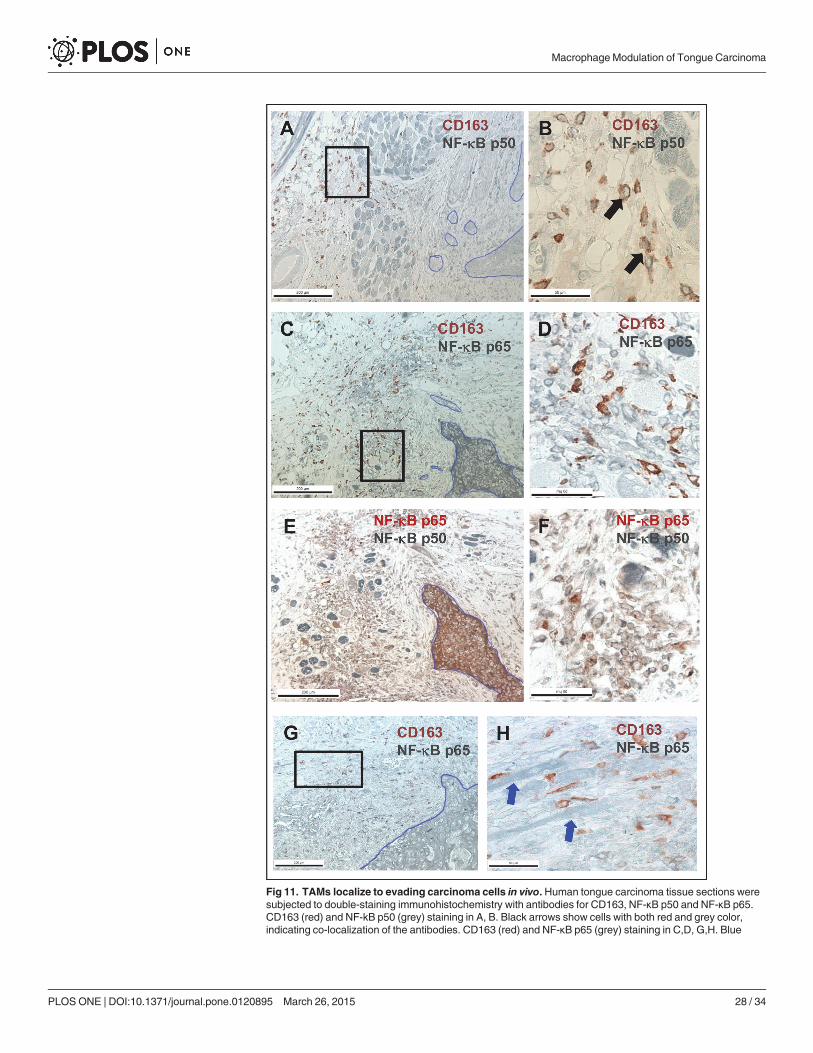
Fig 11. TAMs localize to evading carcinoma cells in vivo. Human tongue carcinoma tissue sections weresubjected to double-staining immunohistochemistry with antibodies for CD163, NF-κB p50 and NF-κB p65.CD163 (red) and NF-kB p50 (grey) staining in A, B. Black arrows show cells with both red and grey color,indicating co-localization of the antibodies. CD163 (red) and NF-κB p65 (grey) staining in C,D, G,H. Blue
MacrophageModulation of Tongue Carcinoma
PLOSONE | DOI:10.1371/journal.pone.0120895 March 26, 2015 28 / 34

previous studies and further strengthens the paradigm of using CD163 as a diagnostic tool forOSCC.
ConclusionsWe here show for the first time that HSC-3 cells fuse with Mfs and especially the interactionbetween HSC-3 cells and M2 Mfs induced migration and also invasion in the 3D myomamodel. Co-culturing altered the phenotype of both HSC-3 cells and Mfs as well as completelyaltered the cytokine secretion profile. Especially the expression of EGF, M-CSF and TGF-,known to act pro-tumorigenic in OSCC, were induced in co-cultures with HSC-3 cells and M2Mfs. In addition, M2 Mfs were found to contain an excess of NF-κB p50 homodimers whichcan be critical for oral tumor progression. In vivo, HSC-3 cells and to a lesser extent also theTME in myoma tissue, attracted Mfs and in addition, tongue cancer patient tissue sections areredundant in M2Mfs. These results show that TAMs are actively involved in tongue cancerprogression and should be considered as therapeutic targets in tongue cancer treatment.
Supporting InformationS1 Table. Short Tandem Repeat (STR) profiling of commercial HSC-3 cells purchased fromthe Japanese Collection of Research Bioresources Cell Bank, Japan.HSC-3 catalogue num-ber: JCRB0623. The STR-profiling was done by Identicell, Aarhus, Denmark.(PDF)
S2 Table. Short Tandem Repeat (STR) profiling of commercial THP-1 cells purchasedfrom ATCC (cat.no. ATCC TIB-202). The STR-profiling was done by Identicell, Aarhus,Denmark.(PDF)
S1 Dataset. Serum-free media from HSC-3 cells, Mfs and co-cultures were collected at day 2and analyzed for cytokines with RayBio Human Cytokine Antibody Array G5. The tableshows the raw data of signal intensity from the antibody array.(XLS)
S1 Fig. (A) Relative Mf cell density in co-cultures. Vybrant CM-Dil-labeled HSC-3 cells (red)and Vybrant DiO-labeled Mfs (green) were co-cultured for up to 11 days in normal growthmedium and photographed with an Evos FL Cell Imaging System microscope. Cell density wasanalyzed optically using Leica QWin3 Software. (B-E) Vybrant CM-Dil labeled HSC-3 (red)and Vybrant DiO-labeled macrophages (green) were co-cultured in Oris Pro cell migration96-plates. Inserts were then removed and cells allowed to migrate into the empty space. Cellswere photographed with an EVOS FL Cell Imaging System microscope. HSC-3 and R848 Mfsmigration at 24 hours (B) and magnification from the migratory front in B (white box) shownin C. HSC-3 and M1 Mfs migration at 48 hours in (D) and magnification from the migratoryfront in D (white box) shown in E. White arrows indicate merged cells. Scale bars in B,D:1000 μm and in C,E: 200 μm. n = 6.(TIF)
S2 Fig. Mfs were seeded to the upper chamber of Transwell-inserts and HSC-3 cells wereseeded to the lower chambers in SF-medium (A). Cells were allowed to migrate for 24 hours
arrows in H show tongue carcinoma cells. NF-kB p50 (grey) and NF-kB p65 (red) staining in E,F. Black boxesin A, C and G indicates site of magnification in B, D and H respectively. Scale bars in A,C,E and G 200 μmand in B, D, F, and H 50 μm.
doi:10.1371/journal.pone.0120895.g011
MacrophageModulation of Tongue Carcinoma
PLOSONE | DOI:10.1371/journal.pone.0120895 March 26, 2015 29 / 34

and then cells were stained with Crystal violet, photographed and analysed with Leica Qwin3software. Results are presented as mean area of cells in inserts (n = 4). HSC-3 cells and R848Mfs were co-cultured on top of human myoma tissue (B,C) or HSC-3 cells were cultured ontop of myoma tissue treated with NF-κB inhibitor BAY 11-7082 (10 μM) or R848 solution(50 nM) which was also added to the incubation medium (B,C). HSC-3 cells were cultured ontop of myoma tissues using Mf-CMs as incubation media (D). Incubation was continued for10 days where after tissues were fixed and processed for immunohistochemistry. Pan-cytokera-tin stained sections were photographed and invasion depths (B,D) and invasion indexes (C)were analysed with the Leica Qwin3 software. All myoma experiments were done in triplicate.(TIF)
S3 Fig. Conditioned medium was collected at day 4 and 8 from myomas and medium con-taining 0.5 (left) or 15 μg (right) protein were subjected to gelatin zymography. The figureto the left shows the uncropped zymogram from day 4 myoma medium with 0.5 μg loaded pro-tein which is presented slightly cropped in Fig. 5E to be more representative and more easily in-terpreted. The figure to the right shows the uncropped zymogram from day 4 myoma mediumwith 14 μg loaded protein to show the gelatinases in the HSC-3 sample which were not visiblewhen loading 0.5 μg protein.(TIF)
S4 Fig. Vybrant CM-Dil labeled HSC-3 cells (red) and unlabeled M1-, M2 and R848 Mfswere incubated with DMSO where after cells were fixed for immunofluorescence with anti-bodies for CD68, CD163 and pancytokeratin. AlexaFluor488-conjugated secondary antibodywas used for visualization. Samples were mounted with DAPI- mounting medium to visualizenuclei (blue). Samples were photographed with a Leica Confocal microscope with 63x oil im-mersion objective. CD68 marker staining in DMSO-treated M1Mfs (A), M2 Mfs (B) and R848Mfs (C). CD163 marker staining in DMSO-treated M2 Mfs treated (D) and R848 Mfs (E).DMSO-treated HSC-3 cells (red) stained with pancytokeratin (green) in F. Scale bars 50 μm.(TIF)
S5 Fig. Unlabeled M1 and R848 Mfs were incubated with DMSO or 10 ng/ml TNF-α for 30min where after cells were fixed for immunofluorescence with polyclonal NF-κB p50 (A) orp65 antibody (B). Some samples were pre-incubated with 10 μM BAY 11-7082 prior to TNF-α activation. AlexaFluor488-conjugated secondary antibody was used for visualization. Sam-ples were mounted with DAPI- mountain medium to visualize nuclei (blue). Samples werephotographed with a Leica Confocal microscope with 63x oil immersion objective. Scale bars50 μm.(TIF)
S6 Fig. Vybrant CM-Dil labeled HSC-3 cells (red) and unlabeled M1-and R848 Mfs wereincubated with DMSO or 10 ng/ml TNF-α for 30 min where after cells were fixed forimmunofluorescence with antibodies for NF-κB p50 and p65 subunits. Some samples werepre-incubated with 10 μM BAY 11-7082 prior to TNF-α activation. AlexaFluor488-conjugatedsecondary antibody was used for visualization. Samples were mounted with DAPI- mountainmedium to visualize nuclei (blue). Samples were photographed with a Leica Confocal micro-scope with 63x oil immersion objective. Red arrows indicate HSC-3 cells (red) and green ar-rows macrophages (unlabeled). Scale bars 50 μm.(TIF)
S7 Fig. Control immunohistochemical staining of human tongue carcinoma tissue sec-tions. Non-specific mouse immunoglobulins (A), rabbit immunoglobulins (B) and both
MacrophageModulation of Tongue Carcinoma
PLOSONE | DOI:10.1371/journal.pone.0120895 March 26, 2015 30 / 34

mouse and rabbit immunoglobulins (C). Scale bars 200 μm.(TIF)
AcknowledgmentsWe wish to thank laboratory technicians Ms. Maija-Leena Lehtonen and Tanja Kuusisto forvaluable assistance with laboratory experiments.
Author ContributionsConceived and designed the experiments: EP PNMV TS. Performed the experiments: EP OVKP PN SN. Analyzed the data: EP ES KP VP PP DD. Contributed reagents/materials/analysistools: EP LUH TS. Wrote the paper: EP PN DDMV LUH TS.
References1. Scully C, Bagan J. Oral squamous cell carcinoma overview. Oral Oncol. 2009; 45: 301–308. doi: 10.
1016/j.oraloncology.2009.01.004 PMID: 19249237
2. Sano D, Myers JN. Metastasis of squamous cell carcinoma of the oral tongue. Cancer Metastasis Rev.2007; 26: 645–62. PMID: 17768600
3. Annertz K, Anderson H, Palmér K, Wennerberg J. The increase in incidence of cancer of the tongue inthe Nordic countries continues into the twenty-first century. Acta Otolaryngol. 2012; 132: 552–557. doi:10.3109/00016489.2011.649146 PMID: 22339663
4. Marsh D, Suchak K, Moutasim KA, Vallath S, Hopper C, Jerjes W et al. Stromal features are predictiveof disease mortality in oral cancer patients. J Pathol. 2011; 223: 470–481. doi: 10.1002/path.2830PMID: 21294121
5. Hendrix MJ, Seftor EA, Seftor RE, Kasemeier-Kulesa J, Kulesa PM, Postovit LM. Reprogramming met-astatic tumour cells with embryonic microenvironments. Nat Rev Cancer 2007; 7: 246–255. PMID:17384580
6. Balkwill F, Mantovani A. Inflammation and cancer: back to Virchow? Lancet 2001; 357: 539–545.PMID: 11229684
7. Vered M, Dayan D, Salo T. The role of the tumour microenvironment in the biology of head and neckcancer: lessons frommobile tongue cancer. Nat Rev Cancer 2011; 11: 382. doi: 10.1038/nrc2982-c1PMID: 21455256
8. Dayan D, Salo T, Salo S, Nyberg P, Nurmenniemi S, Costea DE et al. Molecular crosstalk between can-cer cells and tumor microenvironment components suggests potential targets for new therapeutic ap-proaches in mobile tongue cancer. Cancer Med. 2012; 1: 128–140. doi: 10.1002/cam4.24 PMID:23342263
9. Salo T, VeredM, Bello IO, Nyberg P, Bitu CC, Zlotogorski Hurvitz A et al. Insights into the role of compo-nents of the tumor microenvironment in oral carcinoma call for new therapeutic approaches. Exp CellRes. 2014; 325: 58–64. doi: 10.1016/j.yexcr.2013.12.029 PMID: 24462456
10. De Palma M,Lewis CE. Cancer: Macrophages limit chemotherapy. Nature 2011; 472: 303–304. doi: 10.1038/472303a PMID: 21512566
11. Udagawa T, Wood M. Tumor-stromal cell interactions and opportunities for therapeutic intervention.Curr Opin Pharmacol. 2010; 10: 369–374. doi: 10.1016/j.coph.2010.06.010 PMID: 20638903
12. Sica A, Mantovani A. Macrophage plasticity and polarization: in vivo veritas. J Clin Invest. 2012; 122:787–795. doi: 10.1172/JCI59643 PMID: 22378047
13. Wyckoff JB, Wang Y, Lin EY, Li JF, Goswami S, Stanley ER et al. Direct visualization of macrophage-assisted tumor cell intravasation in mammary tumors. Cancer Res. 2007; 67: 2649–2656. PMID:17363585
14. Biswas SK, Allavena P, Mantovani A. Tumor-associated macrophages: functional diversity, clinical sig-nificance, and open questions. Semin Immunopathol. 2013; 35: 585–600. doi: 10.1007/s00281-013-0367-7 PMID: 23657835
15. Marcus B, Arenberg D, Lee J, Kleer C, Chepeha DB, Schmalbach CE et al. Prognostic factors in oralcavity and oropharyngeal squamous cell carcinoma. Cancer 2004; 101: 2779–2787. PMID: 15546137
16. Li C, Shintani S, Terakado N, Nakashiro K, Hamakawa H. Infiltration of tumor-associated macrophagesin human oral squamous cell carcinoma. Oncol Rep. 2002; 9: 1219–1223. PMID: 12375023
MacrophageModulation of Tongue Carcinoma
PLOSONE | DOI:10.1371/journal.pone.0120895 March 26, 2015 31 / 34

17. Liu SY, Chang LC, Pan LF, Hung YJ, Lee CH, Shieh YS. Clinicopathologic significance of tumor cell-lined vessel and microenvironment in oral squamous cell carcinoma. Oral Oncol. 2008; 44: 277–285.PMID: 17475541
18. Costea DE, Hills A, Osman AH, Thurlow J, Kalna G, Huang X et al. Identification of two distinct carcino-ma-associated fibroblast subtypes with differential tumor-promoting abilities in oral squamous cell carci-noma. Cancer Res. 2013; 73: 3888–3901. doi: 10.1158/0008-5472.CAN-12-4150 PMID: 23598279
19. Hadler-Olsen E, Wetting HL, Rikardsen O, Steigen SE, Kanapathippillai P, Grénman R et al. impact ontumor growth and lymphangiogenesis in human carcinoma xenografts. Virchows Arch. 2010; 457:677–692. doi: 10.1007/s00428-010-0980-y PMID: 20890764
20. Oeckinghaus A, Hayden MS, Ghosh S. Crosstalk in NF-κB signaling pathways. Nat Immunol. 2011; 12:695–708. doi: 10.1038/ni.2065 PMID: 21772278
21. Tanaka T, Nakayama H, Yoshitake Y, Irie A, Nagata M, Kawahara K et al. Selective inhibition of nuclearfactor-κB by nuclear factor-κB essential modulator-binding domain peptide suppresses the metastasisof highly metastatic oral squamous cell carcinoma. Cancer Sci. 2012; 103: 455–463. doi: 10.1111/j.1349-7006.2011.02174.x PMID: 22136381
22. Hagemann T, Biswas SK, Lawrence T, Sica A, Lewis CE. Regulation of macrophage function in tumors:the multifaceted role of NF-kappaB. Blood 2009; 113: 3139–3146. doi: 10.1182/blood-2008-12-172825PMID: 19171876
23. Saccani A, Schioppa T, Porta C, Biswas SK, Nebuloni M, Vago L et al. p50 nuclear factor-kappaB over-expression in tumor-associated macrophages inhibits M1 inflammatory responses and antitumor resis-tance. Cancer Res. 2006; 66: 11432–40. PMID: 17145890
24. Drobits B, Holcmann M, Amberg N, Swiecki M, Grundtner R, Hammer M et al. Imiquimod clears tumorsin mice independent of adaptive immunity by converting pDCs into tumor-killing effector cells. J Clin In-vest. 2012; 122: 575–585. doi: 10.1172/JCI61034 PMID: 22251703
25. Smith KJ, Hamza S, Skelton H. Topical imidazoquinoline therapy of cutaneous squamous cell carcino-ma polarizes lymphoid and monocyte/macrophage populations to a Th1 and M1 cytokine pattern. ClinExp Dermatol. 2004; 29: 505–512. PMID: 15347337
26. Ramineni SK, Cunningham LL Jr, Dziubla TD, Puleo DA. Development of imiquimod-loaded mucoad-hesive films for oral dysplasia. J Pharm Sci. 2013; 102: 593–603. doi: 10.1002/jps.23386 PMID:23192692
27. Nurmenniemi S, Sinikumpu T, Alahuhta I, Salo S, Sutinen M, Santala M et al. A novel organotypicmodel mimics the tumor microenvironment. Am J Pathol 2009; 175: 1281–1291. doi: 10.2353/ajpath.2009.081110 PMID: 19679876
28. Teppo S, Sundquist E, Vered M, Holappa H, Parkkisenniemi J, Rinaldi T et al. The hypoxic tumor micro-environment regulates invasion of aggressive oral carcinoma cells. Exp Cell Res. 2013; 319: 376–389.doi: 10.1016/j.yexcr.2012.12.010 PMID: 23262025
29. Keller R, Keist R, Wechsler A, Leist TP, van der Meide PH. Mechanisms of macrophage-mediatedtumor cell killing: a comparative analysis of the roles of reactive nitrogen intermediates and tumor ne-crosis factor. Int J Cancer 1990; 46: 682–686. PMID: 2120138
30. Vandivier RW, Richens TR, Horstmann SA, deCathelineau AM, Ghosh M, Reynolds SD et al. Dysfunc-tional cystic fibrosis transmembrane conductance regulator inhibits phagocytosis of apoptotic cells withproinflammatory consequences. Am J Physiol Lung Cell Mol Physiol. 2009; 297: L677–L686. doi: 10.1152/ajplung.00030.2009 PMID: 19633071
31. Adams DL, Martin SS, Alpaugh RK, Charpentier M, Tsai S, Bergan RC et al. Circulating giant macro-phages as a potential biomarker of solid tumors. Proc Natl Acad Sci U S A 2014; 111: 3514–3529. doi:10.1073/pnas.1320198111 PMID: 24550495
32. Clawson GA. Cancer. Fusion for moving. Science 2013; 342: 699–700. doi: 10.1126/science.1244270PMID: 24202164
33. Dittmar T, Nagler C, Niggemann B, Zänker KS. The dark side of stem cells: triggering cancer progres-sion by cell fusion. Curr Mol Med. 2013; 13: 735–750. PMID: 23642055
34. Pawelek JM. Tumour-cell fusion as a source of myeloid traits in cancer. Lancet Oncol. 2005; 6: 988–993. PMID: 16321767
35. Baay M, Brouwer A, Pauwels P, Peeters M, Lardon F. Tumor cells and tumor-associated macrophages:secreted proteins as potential targets for therapy. Clin Dev Immunol. 2011;565187.
36. Koontongkaew S. The tumor microenvironment contribution to development, growth, invasion and me-tastasis of head and neck squamous cell carcinomas. J Cancer 2013; 4: 66–83. doi: 10.7150/jca.5112PMID: 23386906
MacrophageModulation of Tongue Carcinoma
PLOSONE | DOI:10.1371/journal.pone.0120895 March 26, 2015 32 / 34

37. Engström A, Erlandsson A, Delbro D, Wijkander J. Conditioned media frommacrophages of M1, butnot M2 phenotype, inhibit the proliferation of the colon cancer cell lines HT-29 and CACO-2. Int J Oncol.2014; 44: 385–392. doi: 10.3892/ijo.2013.2203 PMID: 24296981
38. Connelly L, BarhamW, Onishko HM, Chen L, Sherrill TP, Zabuawala T et al. NF-kappaB activationwithin macrophages leads to an anti-tumor phenotype in a mammary tumor lung metastasis model.Breast Cancer Res. 2011; 13: R83. doi: 10.1186/bcr2935 PMID: 21884585
39. Cook J, Hagemann T. Tumour-associated macrophages and cancer. Curr Opin Pharmacol. 2013; 13:595–601. doi: 10.1016/j.coph.2013.05.017 PMID: 23773801
40. Yang Y, Qin J, Lan L, Li N, Wang C, He P et al. M-CSF cooperating with NFκB induces macrophagetransformation fromM1 to M2 by upregulating c-Jun: Cancer Biol Ther. 2014; 15: 99–107. doi: 10.4161/cbt.26718 PMID: 24100343
41. Erez N, Truitt M, Olson P, Arron ST, Hanahan D. Cancer-Associated Fibroblasts Are Activated in Incipi-ent Neoplasia to Orchestrate Tumor-Promoting Inflammation in an NF-kappaB-Dependent Manner.Cancer Cell 2010; 17: 135–47. doi: 10.1016/j.ccr.2009.12.041 PMID: 20138012
42. Wolff HA, Rolke D, Rave-Fränk M, Schirmer M, Eicheler W, Doerfler A et al. Analysis of chemokine andchemokine receptor expression in squamous cell carcinoma of the head and neck (SCCHN) cell lines.Radiat Environ Biophy. 2011; 50: 145–154. doi: 10.1007/s00411-010-0341-x PMID: 21085979
43. Abiko Y, Nishimura M, Kusano K, Nakashima K, Okumura K, Arakawa T et al. Expression of MIP-3alpha/CCL20, a macrophage inflammatory protein in oral squamous cell carcinoma. Arch Oral Biol.2003; 48: 171–175. PMID: 12642237
44. Chakraborty K, Bose A, Goswami KK, Mukherjee KK, Goswami S, Ghosh D et al. Dysregulated CC re-ceptor/ligand in monocytes/macrophages from tongue squamous cell carcinoma patients is partiallyrectified by interferon α-2b. Hum Immunol. 2012; 73: 38–47. doi: 10.1016/j.humimm.2011.10.015PMID: 22085486
45. Yaddanapudi K, Putty K, Rendon BE, Lamont GJ, Faughn JD, Satoskar A et al. Control of tumor-asso-ciated macrophage alternative activation by macrophage migration inhibitory factor. J Immunol. 2013;190: 2984–2993. doi: 10.4049/jimmunol.1201650 PMID: 23390297
46. Kindt N, Laurent G, Nonclercq D, Journé F, GhanemG, Duvillier H et al. Pharmacological inhibition ofmacrophagemigration inhibitory factor interferes with the proliferation and invasiveness of squamouscarcinoma cells. Int J Oncol. 2013; 43: 185–193. doi: 10.3892/ijo.2013.1944 PMID: 23677331
47. França CM, Batista AC, Borra RC, Ventiades-Flores JA, Mendonça EF, Deana AM et al. Macrophagemigration inhibitory factor and oral cancer. J Oral Pathol Med. 2013; 42: 368–73. doi: 10.1111/jop.12011 PMID: 23067233
48. Dumitru CA, Gholaman H, Trellakis S, Bruderek K, Dominas N, Gu X et al. Tumor-derived macrophagemigration inhibitory factor modulates the biology of head and neck cancer cells via neutrophil activation.Int J Cancer 2011; 129: 859–869. doi: 10.1002/ijc.25991 PMID: 21328346
49. Mashhadiabbas F, Mahjour F, Mahjour SB, Fereidooni F, Hosseini FS. The immunohistochemical char-acterization of MMP-2, MMP-10, TIMP-1, TIMP-2, and podoplanin in oral squamous cell carcinoma.Oral Surg Oral Med Oral Pathol Oral Radiol. 2012; 114: 240–250. doi: 10.1016/j.oooo.2012.04.009PMID: 22769410
50. Pérez-Sayáns García M, Suárez-Peñaranda JM, Gayoso-Diz P, Barros-Angueira F, Gándara-Rey JM,García-García A. Tissue inhibitor of metalloproteinases in oral squamous cell carcinomas—a therapeu-tic target? Cancer Lett. 2012; 323: 11–19. doi: 10.1016/j.canlet.2012.03.040 PMID: 22484495
51. Abbas O, MahalingamM. Desmoplasia: not always a bad thing. Histopathology 2011; 58: 643–659.doi: 10.1111/j.1365-2559.2010.03617.x PMID: 20718872
52. Okuda H, Kobayashi A, Xia B, Watabe M, Pai SK, Hirota S et al. Hyaluronan synthase HAS2 promotestumor progression in bone by stimulating the interaction of breast cancer stem-like cells with macro-phages and stromal cells. Cancer Res. 2012; 72: 537–547. doi: 10.1158/0008-5472.CAN-11-1678PMID: 22113945
53. Chengye Z, Daixing Z, Qiang Z, Shusheng L. PGC-1-related coactivator (PRC) negatively regulates en-dothelial adhesion of monocytes via inhibition of NF κB activity. Biochem Biophys Res Commun. 2013;439: 121–125. doi: 10.1016/j.bbrc.2013.08.015 PMID: 23954632
54. Tjiu JW, Chen JS, Shun CT, Lin SJ, Liao YH, Chu CY et al. Tumor-associated macrophage-induced in-vasion and angiogenesis of human basal cell carcinoma cells by cyclooxygenase-2 induction. J InvestDermatol. 2009; 129: 1016–1025. doi: 10.1038/jid.2008.310 PMID: 18843292
55. Zajac E, Schweighofer B, Kupriyanova TA, Juncker-Jensen A, Minder P, Quigley JP et al. Angiogeniccapacity of M1- and M2-polarized macrophages is determined by the levels of TIMP-1 complexed withtheir secreted proMMP-9. Blood 2013; 122: 4054–4067. doi: 10.1182/blood-2013-05-501494 PMID:24174628
MacrophageModulation of Tongue Carcinoma
PLOSONE | DOI:10.1371/journal.pone.0120895 March 26, 2015 33 / 34

56. Koontongkaew S, Amornphimoltham P, Monthanpisut P, Saensuk T, Leelakriangsak M. Fibroblastsand extracellular matrix differently modulate MMP activation by primary and metastatic head and neckcancer cells. Med Oncol. 2012; 29: 690–703. doi: 10.1007/s12032-011-9871-6 PMID: 21380786
57. Ivashkiv LB. Inflammatory signaling in macrophages: transitions from acute to tolerant and alternativeactivation states. Eur J Immunol. 2011; 41: 2477–2481. doi: 10.1002/eji.201141783 PMID: 21952800
58. Kurland JF, Kodym R, Story MD, Spurgers KB, McDonnell TJ, Meyn RE. NF-kappaB1 (p50) homodi-mers contribute to transcription of the bcl-2 oncogene. J Biol Chem. 2001; 276: 45380–45386. PMID:11567031
59. Chung GT, LouWP, Chow C, To KF, Choy KW, Leung AW et al. Constitutive activation of distinct NF-κB signals in EBV-associated nasopharyngeal carcinoma. J Pathol. 2013; 231: 311–322 doi: 10.1002/path.4239 PMID: 23868181
60. Maniecki MB, Etzerodt A, Ulhøi BP, Steiniche T, Borre M, Dyrskjøt L et al. Tumor-promoting macro-phages induce the expression of the macrophage-specific receptor CD163 in malignant cells. Int J Can-cer 2012; 131: 2320–2331. doi: 10.1002/ijc.27506 PMID: 22362417
61. Fujii N, Shomori K, Shiomi T, Nakabayashi M, Takeda C, Ryoke K et al. Cancer-associated fibroblastsand CD163-positive macrophages in oral squamous cell carcinoma: their clinicopathological and prog-nostic significance. J Oral Pathol Med. 2012; 41: 444–451. doi: 10.1111/j.1600-0714.2012.01127.xPMID: 22296275
62. Weber M, Büttner-Herold M, Hyckel P, Moebius P, Distel L, Ries J et al. Small oral squamous cell carci-nomas with nodal lymphogenic metastasis show increased infiltration of M2 polarized macrophages—An immunohistochemical analysis. J Craniomaxillofac Surg. 2014;pii:S1010–5182.
63. Kylmäniemi M, Oikarinen A, Oikarinen K, Salo T. Effects of dexamethasone and cell proliferation on theexpression of matrix metalloproteinases in human mucosal normal and malignant cells. J Dent Res.1996; 75: 919–26. PMID: 8675803
MacrophageModulation of Tongue Carcinoma
PLOSONE | DOI:10.1371/journal.pone.0120895 March 26, 2015 34 / 34
Related Documents