Hindawi Publishing Corporation Journal of Parasitology Research Volume 2012, Article ID 413052, 12 pages doi:10.1155/2012/413052 Review Article Macrophage Migration Inhibitory Factor in Protozoan Infections Marcelo T. Bozza, Yuri C. Martins, Let´ ıcia A. M. Carneiro, and Claudia N. Paiva Laborat´ orio de Inflamac ¸˜ ao e Imunidade, Departamento de Imunologia, Instituto de Microbiologia, Universidade Federal do Rio de Janeiro, 21941-902, Rio de Janeiro, RJ, Brazil Correspondence should be addressed to Marcelo T. Bozza, [email protected] Received 2 September 2011; Revised 1 November 2011; Accepted 7 November 2011 Academic Editor: Marcela F. Lopes Copyright © 2012 Marcelo T. Bozza et al. This is an open access article distributed under the Creative Commons Attribution License, which permits unrestricted use, distribution, and reproduction in any medium, provided the original work is properly cited. Macrophage migration inhibitory factor (MIF) is a cytokine that plays a central role in immune and inflammatory responses. In the present paper, we discussed the participation of MIF in the immune response to protozoan parasite infections. As a general trend, MIF participates in the control of parasite burden at the expense of promoting tissue damage due to increased inflammation. 1. Introduction The immune/inflammatory response triggered during infec- tion has an essential role in eliminating the infectious agent and in promoting tissue repair [1]. The very existence of multicellular organisms in an environment replete of infectious agents is made possible by an effective immune system, indicating that the ability to control infections has been throughout evolution an important selective pressure to mold the immune system. However, it is not unusual that the tissue damage observed during infectious processes is caused by the immune/inflammatory response itself. Innate immune receptors recognize conserved microbial molecules from all classes of microorganisms [1, 2]. The activation of these receptors elicits selective intracellular signaling cas- cades that result in the production of cytokines, chemokines, lipid mediators, and reactive oxygen/nitrogen species. Both the intensity and the quality of the inflammatory responses are determined by the detection of combinations of micro- bial molecules and molecules from host origin such as cytokines, ATP, and ROS [3, 4]. This activation of the immune system is considered essential for pathogen killing but, on the other hand, is also critically involved in tissue damage and sepsis [1–4]. Thus, the pathology of infectious diseases can result either from a direct effect of the infectious agents or from the immune/inflammatory response, both of which can cause metabolic changes, cellular malfunctioning, and cell death. In fact, the pathology of most infectious diseases is the intricate result of these two forces. Macrophage migration inhibitory factor (MIF) activity was described in the sixties and it is considered one of the first cytokines to be described [5, 6]. The MIF gene was cloned in 1989 using a functional assay based on its ability to inhibit the random migration of macrophages [7]. A major breakthrough in the characterization of MIF was achieved by a remarkable study that identified proteins secreted by the pituitary gland upon stimulation by LPS [8]. Among these proteins was MIF, and the authors went on to show that blockade of MIF protected mice from LPS-induced lethality, indicating its prominent proinflammatory role in endotoxemia. These studies led to renewed scientific interest on the biology of MIF and opened research avenues in several fields. In the 20 years of research following the cloning of MIF a complex scenario of its biology has emerged and it is now clear that MIF is an important inflammatory mediator that participates in both innate and adaptive immune responses [9]. Preformed MIF protein is found in many cell types and is released in response to different stimuli, such as infections and cytokine stimulation [9]. Physiological increases in glucocorticoid concentrations induce immune cells to secrete MIF, and, once released, MIF can counterregulate the anti- inflammatory effects of steroids on cytokine production [10, 11]. The pro-inflammatory activities of MIF include the induction/production of inflammatory mediators such as tumor necrosis factor (TNF), interleukin-1 (IL-1), and nitric oxide (NO) by macrophages, the production of arachidonic acid and eicosanoids through the induction of phospholipase

Welcome message from author
This document is posted to help you gain knowledge. Please leave a comment to let me know what you think about it! Share it to your friends and learn new things together.
Transcript

Hindawi Publishing CorporationJournal of Parasitology ResearchVolume 2012, Article ID 413052, 12 pagesdoi:10.1155/2012/413052
Review Article
Macrophage Migration Inhibitory Factor in Protozoan Infections
Marcelo T. Bozza, Yuri C. Martins, Letıcia A. M. Carneiro, and Claudia N. Paiva
Laboratorio de Inflamacao e Imunidade, Departamento de Imunologia, Instituto de Microbiologia,Universidade Federal do Rio de Janeiro, 21941-902, Rio de Janeiro, RJ, Brazil
Correspondence should be addressed to Marcelo T. Bozza, [email protected]
Received 2 September 2011; Revised 1 November 2011; Accepted 7 November 2011
Academic Editor: Marcela F. Lopes
Copyright © 2012 Marcelo T. Bozza et al. This is an open access article distributed under the Creative Commons AttributionLicense, which permits unrestricted use, distribution, and reproduction in any medium, provided the original work is properlycited.
Macrophage migration inhibitory factor (MIF) is a cytokine that plays a central role in immune and inflammatory responses. Inthe present paper, we discussed the participation of MIF in the immune response to protozoan parasite infections. As a generaltrend, MIF participates in the control of parasite burden at the expense of promoting tissue damage due to increased inflammation.
1. Introduction
The immune/inflammatory response triggered during infec-tion has an essential role in eliminating the infectious agentand in promoting tissue repair [1]. The very existenceof multicellular organisms in an environment replete ofinfectious agents is made possible by an effective immunesystem, indicating that the ability to control infections hasbeen throughout evolution an important selective pressureto mold the immune system. However, it is not unusual thatthe tissue damage observed during infectious processes iscaused by the immune/inflammatory response itself. Innateimmune receptors recognize conserved microbial moleculesfrom all classes of microorganisms [1, 2]. The activationof these receptors elicits selective intracellular signaling cas-cades that result in the production of cytokines, chemokines,lipid mediators, and reactive oxygen/nitrogen species. Boththe intensity and the quality of the inflammatory responsesare determined by the detection of combinations of micro-bial molecules and molecules from host origin such ascytokines, ATP, and ROS [3, 4]. This activation of theimmune system is considered essential for pathogen killingbut, on the other hand, is also critically involved in tissuedamage and sepsis [1–4]. Thus, the pathology of infectiousdiseases can result either from a direct effect of the infectiousagents or from the immune/inflammatory response, both ofwhich can cause metabolic changes, cellular malfunctioning,and cell death. In fact, the pathology of most infectiousdiseases is the intricate result of these two forces.
Macrophage migration inhibitory factor (MIF) activitywas described in the sixties and it is considered one of thefirst cytokines to be described [5, 6]. The MIF gene wascloned in 1989 using a functional assay based on its ability toinhibit the random migration of macrophages [7]. A majorbreakthrough in the characterization of MIF was achievedby a remarkable study that identified proteins secreted bythe pituitary gland upon stimulation by LPS [8]. Amongthese proteins was MIF, and the authors went on to showthat blockade of MIF protected mice from LPS-inducedlethality, indicating its prominent proinflammatory role inendotoxemia. These studies led to renewed scientific intereston the biology of MIF and opened research avenues in severalfields. In the 20 years of research following the cloning of MIFa complex scenario of its biology has emerged and it is nowclear that MIF is an important inflammatory mediator thatparticipates in both innate and adaptive immune responses[9].
Preformed MIF protein is found in many cell types andis released in response to different stimuli, such as infectionsand cytokine stimulation [9]. Physiological increases inglucocorticoid concentrations induce immune cells to secreteMIF, and, once released, MIF can counterregulate the anti-inflammatory effects of steroids on cytokine production[10, 11]. The pro-inflammatory activities of MIF includethe induction/production of inflammatory mediators such astumor necrosis factor (TNF), interleukin-1 (IL-1), and nitricoxide (NO) by macrophages, the production of arachidonicacid and eicosanoids through the induction of phospholipase
2 Journal of Parasitology Research
MIFPreformed
GlucocorticoidsInfectionsCytokines
Anti-inflammatoryeffects of steroids
CXCR2
CXCR4CD44/CD74
IL-1β
NO
PLA2
Arachidonicacid
Adhesionmolecules
Chemotaxis andsurvival ofleukocytes
ERK-1/2
COX
PGE2
p53
Growth arrest
TLRs
↑
↑
↑↑
↓
Apoptosis
TNF-α
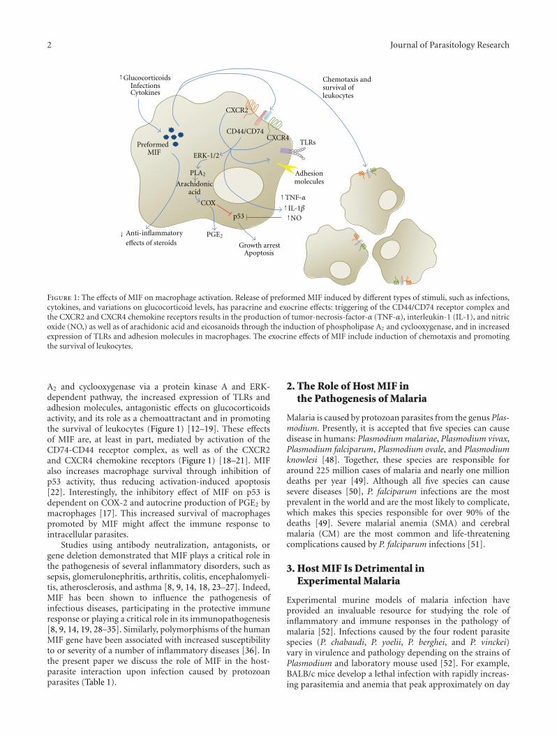
Figure 1: The effects of MIF on macrophage activation. Release of preformed MIF induced by different types of stimuli, such as infections,cytokines, and variations on glucocorticoid levels, has paracrine and exocrine effects: triggering of the CD44/CD74 receptor complex andthe CXCR2 and CXCR4 chemokine receptors results in the production of tumor-necrosis-factor-α (TNF-α), interleukin-1 (IL-1), and nitricoxide (NO,) as well as of arachidonic acid and eicosanoids through the induction of phospholipase A2 and cyclooxygenase, and in increasedexpression of TLRs and adhesion molecules in macrophages. The exocrine effects of MIF include induction of chemotaxis and promotingthe survival of leukocytes.
A2 and cyclooxygenase via a protein kinase A and ERK-dependent pathway, the increased expression of TLRs andadhesion molecules, antagonistic effects on glucocorticoidsactivity, and its role as a chemoattractant and in promotingthe survival of leukocytes (Figure 1) [12–19]. These effectsof MIF are, at least in part, mediated by activation of theCD74-CD44 receptor complex, as well as of the CXCR2and CXCR4 chemokine receptors (Figure 1) [18–21]. MIFalso increases macrophage survival through inhibition ofp53 activity, thus reducing activation-induced apoptosis[22]. Interestingly, the inhibitory effect of MIF on p53 isdependent on COX-2 and autocrine production of PGE2 bymacrophages [17]. This increased survival of macrophagespromoted by MIF might affect the immune response tointracellular parasites.
Studies using antibody neutralization, antagonists, orgene deletion demonstrated that MIF plays a critical role inthe pathogenesis of several inflammatory disorders, such assepsis, glomerulonephritis, arthritis, colitis, encephalomyeli-tis, atherosclerosis, and asthma [8, 9, 14, 18, 23–27]. Indeed,MIF has been shown to influence the pathogenesis ofinfectious diseases, participating in the protective immuneresponse or playing a critical role in its immunopathogenesis[8, 9, 14, 19, 28–35]. Similarly, polymorphisms of the humanMIF gene have been associated with increased susceptibilityto or severity of a number of inflammatory diseases [36]. Inthe present paper we discuss the role of MIF in the host-parasite interaction upon infection caused by protozoanparasites (Table 1).
2. The Role of Host MIF inthe Pathogenesis of Malaria
Malaria is caused by protozoan parasites from the genus Plas-modium. Presently, it is accepted that five species can causedisease in humans: Plasmodium malariae, Plasmodium vivax,Plasmodium falciparum, Plasmodium ovale, and Plasmodiumknowlesi [48]. Together, these species are responsible foraround 225 million cases of malaria and nearly one milliondeaths per year [49]. Although all five species can causesevere diseases [50], P. falciparum infections are the mostprevalent in the world and are the most likely to complicate,which makes this species responsible for over 90% of thedeaths [49]. Severe malarial anemia (SMA) and cerebralmalaria (CM) are the most common and life-threateningcomplications caused by P. falciparum infections [51].
3. Host MIF Is Detrimental inExperimental Malaria
Experimental murine models of malaria infection haveprovided an invaluable resource for studying the role ofinflammatory and immune responses in the pathology ofmalaria [52]. Infections caused by the four rodent parasitespecies (P. chabaudi, P. yoelii, P. berghei, and P. vinckei)vary in virulence and pathology depending on the strains ofPlasmodium and laboratory mouse used [52]. For example,BALB/c mice develop a lethal infection with rapidly increas-ing parasitemia and anemia that peak approximately on day
Journal of Parasitology Research 3
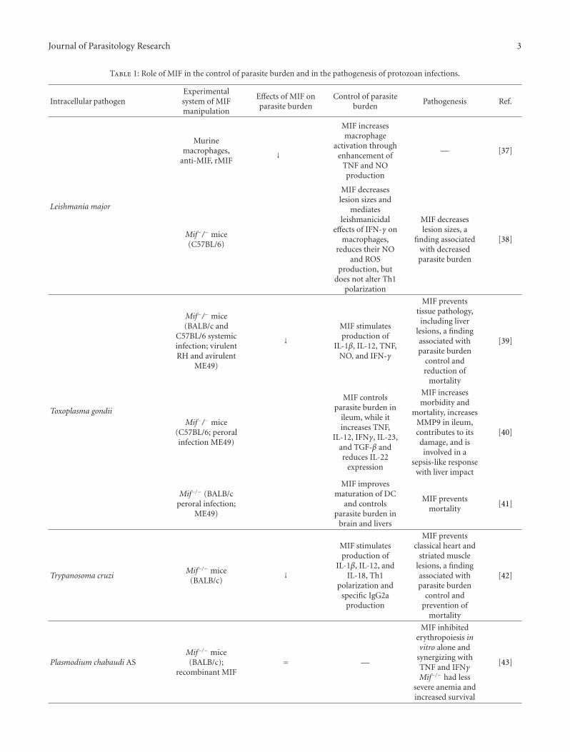
Table 1: Role of MIF in the control of parasite burden and in the pathogenesis of protozoan infections.
Intracellular pathogenExperimentalsystem of MIFmanipulation
Effects of MIF onparasite burden
Control of parasiteburden
Pathogenesis Ref.
Leishmania major
Murinemacrophages,
anti-MIF, rMIF↓
MIF increasesmacrophage
activation throughenhancement of
TNF and NOproduction
— [37]
Mif−/− mice(C57BL/6)
MIF decreaseslesion sizes and
mediatesleishmanicidal
effects of IFN-γ onmacrophages,
reduces their NOand ROS
production, butdoes not alter Th1
polarization
MIF decreaseslesion sizes, a
finding associatedwith decreasedparasite burden
[38]
Toxoplasma gondii
Mif−/− mice(BALB/c and
C57BL/6 systemicinfection; virulentRH and avirulent
ME49)
↓MIF stimulatesproduction of
IL-1β, IL-12, TNF,NO, and IFN-γ
MIF preventstissue pathology,including liver
lesions, a findingassociated withparasite burden
control andreduction of
mortality
[39]
Mif−/− mice(C57BL/6; peroralinfection ME49)
MIF controlsparasite burden in
ileum, while itincreases TNF,
IL-12, IFNγ, IL-23,and TGF-β andreduces IL-22
expression
MIF increasesmorbidity and
mortality, increasesMMP9 in ileum,contributes to itsdamage, and isinvolved in a
sepsis-like responsewith liver impact
[40]
Mif−/− (BALB/cperoral infection;
ME49)
MIF improvesmaturation of DC
and controlsparasite burden in
brain and livers
MIF preventsmortality
[41]
Trypanosoma cruziMif−/− mice
(BALB/c)↓
MIF stimulatesproduction of
IL-1β, IL-12, andIL-18, Th1
polarization andspecific IgG2a
production
MIF preventsclassical heart and
striated musclelesions, a findingassociated withparasite burden
control andprevention of
mortality
[42]
Plasmodium chabaudi ASMif−/− mice(BALB/c);
recombinant MIF= —
MIF inhibitederythropoiesis invitro alone and
synergizing withTNF and IFNγMif−/− had less
severe anemia andincreased survival
[43]

4 Journal of Parasitology Research
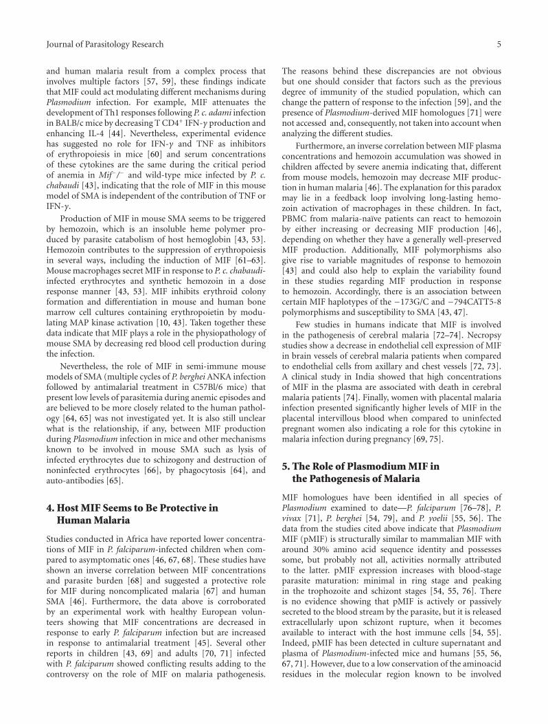
Table 1: Continued.
Intracellular pathogenExperimentalsystem of MIFmanipulation
Effects of MIF onparasite burden
Control of parasiteburden
Pathogenesis Ref.
Plasmodium chabaudi adami
Mif−/− mice(BALB/c);
Ab-neutralizedMIF
↑
MIF promotes Th2polarization (in itsabsence, cells react
better toIL-12/anti-IL-4
with Th1polarization)
— [44]
Plasmodium falciparum
Human volunteerssubmitted to
infection;correlation
= —
MIF is associatedwith a number of
circulatinglymphocytes
[45]
Infected childrenin endemic zone;
correlation↓ —
MIFconcentrations inplasma and MIF
produced byleukocytes in vitro
inversely correlatedwith severity ofmalarial anemia
[46]
Infected childrenin endemic zone;
associationbetween
polymorphism ofMIF promoter and
pathology
= —
MIF peripherallevels are
associated withpromoter
polymorphismsand with
susceptibility tosevere malarial
anemia
[47]
8 of infection when inoculated with P. chabaudi chabaudiAS [53]. For this reason, this parasite-mouse combinationis considered an experimental model of SMA. On the otherhand, the same strain of mouse develops a nonlethal self-resolving infection with peak parasitemia also on day 8of infection followed by cell-mediated parasite killing andtotal parasite clearance on day 15 when inoculated with P.chabaudi adami DK [43]. This second model is consideredsuitable to study the interactions between macrophagesand T cells involved in parasite elimination. Alternatively,C57Bl/6 mice develop a neurologic syndrome similar tohuman CM and characterized by ataxia, convulsions, andcoma upon infection with P. berghei ANKA or P. yoelii17XL [54–56]. Interestingly, P. yoelii 17XL, but not P. bergheiANKA, also induces CM when inoculated in BALB/c mice[52].
However, none of the rodent Plasmodium strains arenatural pathogens of laboratory mouse strains and the courseof infection and complications observed in some mouse-parasite combinations including SMA and CM differ fromthe human spectrum of disease [52]. For instance, peakanemia in the P. c. chabaudi AS-BALB/c model correlateswith a peak parasitemia of around 20%, which makes thedestruction of infected erythrocytes a major contributorto the physiopathogenesis of anemia in this model [52,57]. Although it also occurs in acute hyperparasitemic
infections, the development of SMA in humans occursmainly in chronic infections with low parasitemias (<5%)and appears to be more related to other mechanismssuch as the destruction of uninfected erythrocytes andthe suppression of the erythropoietic response [57]. Themajority of mouse models of CM are characterized by theadhesion of leukocytes, instead of infected erythrocytes in thebrain microvasculature as occurs in human CM [58].
Studies using mouse models of malaria indicate that MIFplays a detrimental role during the infection [43, 44, 53].Mif−/− mice in the BALB/c background or animals treatedwith anti-MIF neutralizing monoclonal antibodies are moreresistant to Plasmodium chabaudi adami infection than wild-type controls presenting a significant reduction in peakand cumulative parasitemia [44]. Accordingly, the infectionof BALB/c mice with P. chabaudi chabaudi AS, a mousemodel of SMA, revealed that elevated concentrations of MIFin the plasma are associated with severity of anemia andsuppression of erythropoiesis [43, 53]. In addition, Mif−/−
mice infected with P. c. chabaudi develop a parasitemia curvesimilar to that of wild-type controls but present less severeanemia, less inhibition of erythroid colony formation, and ahigher survival [43].
It is not clear why blockade of MIF reduces parasitemiaduring P. c. adami but not P. c. chabaudi infection. Asthe development of immunity and/or anemia in mouse
Journal of Parasitology Research 5
and human malaria result from a complex process thatinvolves multiple factors [57, 59], these findings indicatethat MIF could act modulating different mechanisms duringPlasmodium infection. For example, MIF attenuates thedevelopment of Th1 responses following P. c. adami infectionin BALB/c mice by decreasing T CD4+ IFN-γ production andenhancing IL-4 [44]. Nevertheless, experimental evidencehas suggested no role for IFN-γ and TNF as inhibitorsof erythropoiesis in mice [60] and serum concentrationsof these cytokines are the same during the critical periodof anemia in Mif−/− and wild-type mice infected by P. c.chabaudi [43], indicating that the role of MIF in this mousemodel of SMA is independent of the contribution of TNF orIFN-γ.
Production of MIF in mouse SMA seems to be triggeredby hemozoin, which is an insoluble heme polymer pro-duced by parasite catabolism of host hemoglobin [43, 53].Hemozoin contributes to the suppression of erythropoiesisin several ways, including the induction of MIF [61–63].Mouse macrophages secret MIF in response to P. c. chabaudi-infected erythrocytes and synthetic hemozoin in a doseresponse manner [43, 53]. MIF inhibits erythroid colonyformation and differentiation in mouse and human bonemarrow cell cultures containing erythropoietin by modu-lating MAP kinase activation [10, 43]. Taken together thesedata indicate that MIF plays a role in the physiopathology ofmouse SMA by decreasing red blood cell production duringthe infection.
Nevertheless, the role of MIF in semi-immune mousemodels of SMA (multiple cycles of P. berghei ANKA infectionfollowed by antimalarial treatment in C57Bl/6 mice) thatpresent low levels of parasitemia during anemic episodes andare believed to be more closely related to the human pathol-ogy [64, 65] was not investigated yet. It is also still unclearwhat is the relationship, if any, between MIF productionduring Plasmodium infection in mice and other mechanismsknown to be involved in mouse SMA such as lysis ofinfected erythrocytes due to schizogony and destruction ofnoninfected erythrocytes [66], by phagocytosis [64], andauto-antibodies [65].
4. Host MIF Seems to Be Protective inHuman Malaria
Studies conducted in Africa have reported lower concentra-tions of MIF in P. falciparum-infected children when com-pared to asymptomatic ones [46, 67, 68]. These studies haveshown an inverse correlation between MIF concentrationsand parasite burden [68] and suggested a protective rolefor MIF during noncomplicated malaria [67] and humanSMA [46]. Furthermore, the data above is corroboratedby an experimental work with healthy European volun-teers showing that MIF concentrations are decreased inresponse to early P. falciparum infection but are increasedin response to antimalarial treatment [45]. Several otherreports in children [43, 69] and adults [70, 71] infectedwith P. falciparum showed conflicting results adding to thecontroversy on the role of MIF on malaria pathogenesis.
The reasons behind these discrepancies are not obviousbut one should consider that factors such as the previousdegree of immunity of the studied population, which canchange the pattern of response to the infection [59], and thepresence of Plasmodium-derived MIF homologues [71] werenot accessed and, consequently, not taken into account whenanalyzing the different studies.
Furthermore, an inverse correlation between MIF plasmaconcentrations and hemozoin accumulation was showed inchildren affected by severe anemia indicating that, differentfrom mouse models, hemozoin may decrease MIF produc-tion in human malaria [46]. The explanation for this paradoxmay lie in a feedback loop involving long-lasting hemo-zoin activation of macrophages in these children. In fact,PBMC from malaria-naıve patients can react to hemozoinby either increasing or decreasing MIF production [46],depending on whether they have a generally well-preservedMIF production. Additionally, MIF polymorphisms alsogive rise to variable magnitudes of response to hemozoin[43] and could also help to explain the variability foundin these studies regarding MIF production in responseto hemozoin. Accordingly, there is an association betweencertain MIF haplotypes of the −173G/C and −794CATT5-8polymorphisms and susceptibility to SMA [43, 47].
Few studies in humans indicate that MIF is involvedin the pathogenesis of cerebral malaria [72–74]. Necropsystudies show a decrease in endothelial cell expression of MIFin brain vessels of cerebral malaria patients when comparedto endothelial cells from axillary and chest vessels [72, 73].A clinical study in India showed that high concentrationsof MIF in the plasma are associated with death in cerebralmalaria patients [74]. Finally, women with placental malariainfection presented significantly higher levels of MIF in theplacental intervillous blood when compared to uninfectedpregnant women also indicating a role for this cytokine inmalaria infection during pregnancy [69, 75].
5. The Role of Plasmodium MIF inthe Pathogenesis of Malaria
MIF homologues have been identified in all species ofPlasmodium examined to date—P. falciparum [76–78], P.vivax [71], P. berghei [54, 79], and P. yoelii [55, 56]. Thedata from the studies cited above indicate that PlasmodiumMIF (pMIF) is structurally similar to mammalian MIF witharound 30% amino acid sequence identity and possessessome, but probably not all, activities normally attributedto the latter. pMIF expression increases with blood-stageparasite maturation: minimal in ring stage and peakingin the trophozoite and schizont stages [54, 55, 76]. Thereis no evidence showing that pMIF is actively or passivelysecreted to the blood stream by the parasite, but it is releasedextracellularly upon schizont rupture, when it becomesavailable to interact with the host immune cells [54, 55].Indeed, pMIF has been detected in culture supernatant andplasma of Plasmodium-infected mice and humans [55, 56,67, 71]. However, due to a low conservation of the aminoacidresidues in the molecular region known to be involved
6 Journal of Parasitology Research
in the catalytic sites, it seems that the tautomerase andoxidoreductase activities are highly depressed in pMIF whencompared to mammalian MIF. Alternatively, pMIF mighthave a different substrate specificity and the physiologicalsubstrate has yet to be identified [54–56].
In terms of functional studies, in vitro assays and animalmodels have shown that pMIF shares some biologicalproperties with mammalian MIF. Indeed, both pMIF andmammalian MIF reduce AP-1 expression, interact withhuman CD74, induce macrophage chemotaxis, and inhibiterythropoiesis and macrophage apoptosis [54–56, 79, 80].On the other hand, pMIF does not stimulate the releaseof IL-8, TNF, or IL-12 from mice and human monocytesor enhance the response of these cells to LPS [55, 78], akey function of mammalian MIF. Finally, a study showedthat pMIF and mouse MIF act synergistically to activate theMAPK-ERK1/2 signaling pathway at very low concentrationsbut act antagonistically at higher concentrations [56], indi-cating that pMIF and mammalian MIF can interact in acomplex way.
The role of pMIF during malaria infection is also notcompletely understood. Although counterintuitive, studiesin mouse models indicate that pMIF attenuates Plasmodiumvirulence by modulating host immune responses [54–56].C57Bl/6 and BALB/c mice showed a reduction in diseaseseverity when infected with transgenic strains of P. yoelii17X and P yoelii 17XL that constitutively overexpress P. yoeliiMIF (PyMIF) [55] or when treated with recombinant PyMIF[56]. This was phenotypically manifested by a decreasein peak and cumulative parasitemia in mice infected withthe nonlethal strain P. yoelii 17X and prolonged course ofinfection with a reduction in overall mortality rate in animalsinfected with the lethal strain P yoelii 17XL [55, 56]. On theother hand, the development of cerebral complications inC57BL/6 mice and hyperparasitemia and severe anemia inBALB/c mice did not differ upon infection with P. bergheiwild-type or P. berghei MIF knockout parasites [54]. Onceagain, studies in humans failed to recapitulate observationsfrom mouse models as pMIF amounts in uncomplicatedmalaria patients are positively correlated with parasitemia,disease severity, and plasma concentrations of TNF, IL-10,and MCP-1 [71]. Thus, future studies are required to definethe role of host and Plasmodium MIF in the pathogenesis ofmalaria.
6. Critical Role of MIF inToxoplasma gondii Infection
Toxoplasma gondii is an intracellular parasite of the phylumApicomplexa that is highly adapted to infect differentcell types and tissues. T. gondii enters its host via thegastrointestinal tract and the innate immune response in theintestine is triggered by the recognition of parasite moleculesby enterocytes, macrophages, and dendritic cells (DCs)[81]. The establishment of an antigen-specific Th1 responseis essential to protective immunity but also potentiallydetrimental as excessive intestinal inflammation and tissuenecrosis can lead to bacterial translocation and death [81].
The proinflammatory cytokines IL-12, TNF, IFN-γ, and IL-1β promote resistance against T. gondii in part due to thegeneration of NO by macrophages, an important mechanismresponsible for parasite elimination.
A model of systemic infection with T. gondii through theintraperitoneal route demonstrated an increased susceptibil-ity of Mif−/− mice when compared to wild-type mice [39].Mif−/− mice presented higher parasite burden in brains andperitoneal macrophages and reduced plasma concentrationsof IL-12, TNF, IFN-γ, IL-1β, and nitrite during infection[39]. These findings were expected considering that MIF isan enhancer of IL-12 and TNF production by macrophages.A recent study using a model of oral T. gondii infectionin the BALB/c background also demonstrated an increasedlethality and tissue parasitism with reduced IL-12 productionand DC activation on Mif−/− mice compared to wild-typemice [41]. DCs obtained from spleens and mesenteric lymphnodes from Mif−/− mice orally infected with T. gondii hadimpaired maturation, with decreased expression of CD80,CD86, CD40, and MHC class II [41]. Thus, the protectiverole of MIF in T. gondii infection is apparently related to theproduction of proinflammatory cytokines, the activation ofDC, and the better control of parasite burden.
BALB/c mice are naturally resistant to oral infection withT. gondii, while those of C57BL/6 are highly susceptibledisplaying intestinal inflammation especially in the ileum[82–84]. This increased lethality of C57BL/6 is related tothe extensive intestinal inflammation, tissue necrosis, and asepsis-like syndrome. Using the peroral route of infection inC57BL/6 mice, it was shown that Mif−/− mice have reducedintestinal and systemic inflammation and survive longercompared to wild-type mice, despite an increase in intestinalparasite burden [40]. Lack of MIF caused a reduction of TNF,IL-12, IFN-γ, and IL-23 and an increased expression of IL-22 in ileal mucosa. Signs of systemic inflammation includingthe increased concentrations of inflammatory cytokines inthe plasma and liver damage were less pronounced in Mif−/−
mice compared to wild-type mice [40]. Although MIF hasbeen regarded as essential in host protection during T. gondiiinfection, these findings demonstrated a pathogenic role ofMIF in natural T. gondii infection in susceptible hosts. Thisdichotomy seems to depend on the route of infection andthe genetic background of the host. Thus, MIF is necessaryto control parasite burden in resistant and susceptible hosts,but it increases intestinal tissue damage causing death insusceptible hosts while it is essential for survival in resistanthosts.
A major consequence of human T. gondii-infection is thesevere congenital malformations when the primary infectionoccurs in the first trimester of pregnancy. A series of studiesdemonstrated a putative role of MIF on placental biologyupon infection with T. gondii. Infection or stimulation ofchorionic explants with molecules of T. gondii, IFN-γ, andIL-12 evoked the secretion of MIF [85, 86]. MIF and itsreceptor, CD74, are present in the syncytiotrophoblast layerand mesenchyme [86]. MIF induces ICAM-1 expressionincreasing the interaction of villous explants with monocytes[85]. These results suggest that MIF, by influencing therecruitment of T. gondii infected monocytes, could facilitate

Journal of Parasitology Research 7
the dissemination of the infection into the deep placentaltissues or increase the tissue damage due to inflammation.The same group recently demonstrated, however, that MIF isimportant for control of placental T. gondii infection in firsttrimester of pregnancy [86].
7. MIF Is Protective in Leishmania Infection
Leishmaniasis, caused by the protozoan parasites from thegenus Leishmania, comprises a large spectrum of clinicalmanifestations including benign ulcer, destructive muco-cutaneous lesions, disseminated cutaneous lesions, andsystemic visceral forms [87]. In the mammalian host,Leishmania sp. is an obligatory intracellular parasite infectingmainly macrophages. Parasite killing requires macrophageactivation with ensuing NO and ROS production [87].Infection with Leishmania major causes skin lesions, whichin general parallel the parasite load. A highly polarized Th1response is effective against L. major, activating macrophagesto produce NO and resulting in resolving skin lesions. Addi-tion of MIF to macrophage cell cultures results in increasedL. major elimination [37]. Though the MIF concentrationrequired to reduce L. major burden in macrophages is high(1 μg/mL, 100 times that of other cytokines with leishmani-cidal effects, such as IFN-γ), this concentration is withinthe range reached in inflammatory conditions. The MIF-induced leishmanicidal effect requires the production of TNFand NO by infected macrophages, and can be reversed by theaddition of IL-10, TGF-β or IL-13, indicating that it dependson an M1 activation status [37]. The expression of MIFincreases during L. major footpad inoculation in popliteallymph node, but the kinetics of its expression compared tothat of MIF secretion by T cells upon antigenpresentationsuggests that lymph node MIF comes from another cellularsource [37]. Consistent with the observed role of MIF asan enhancer of macrophage leishmanicidal function, oraladministration of Salmonella typhimurium transfected withMIF reduces the size of skin lesions [88], while Mif−/− miceare highly susceptible to L. major, developing severe skinlesions late after infection [38]. MIF does not affect Thpolarization in L. major infection, as indicated by the similarIFN-γ and IL-4 production among T cells from Mif−/−
and wild-type mice. However, IFN-γ-activated macrophagesfrom Mif−/− mice infected in vitro with L. major haveslightly decreased parasite clearance [38], indicating thateither they are somewhat insensitive to IFN-γ or MIFproduction is partially required as an intermediary step toIFN-γ-induced leishmanicidal activity. The contribution ofMIF produced by CD4+ lymphocytes to protective immunityagainst cutaneous leishmaniasis was demonstrated using amodel of vaccination with the L. pifanoi antigen P-4 [89].BALB-c mice immunized with P-4 expressed around 10-fold higher amounts of MIF, TNF, and IFN-γ mRNAs thanthe adjuvant controls. Moreover, blockage of MIF with anti-MIF antibody significantly reduced the leishmanicidal abilityof macrophages cultured with CD4+ lymphocytes obtainedfrom P-4-immunized mice.
Patients with visceral leishmaniasis due to infection withL. donovani presented CD4+ lymphocytes expressing low
amounts of CD2, IFN-γ, and MIF [90]. Antileishmanialtreatment caused immunological recovery with increasedexpression of CD2 and production of MIF. On the otherhand, a recent study demonstrated that patients with visceralleishmaniasis caused by L. chagasi have increased plasmaconcentrations of MIF [91]. The MIF concentrations werehigher in patients with the active form compared to patientsin remission. Interestingly, the authors identify an increase ofLPS in the plasma of patients with active disease and the LPSconcentrations positively correlated with MIF.
8. Identification of Leishmanial MIF andIts Role in Infection
The complete genome sequencing of L. major revealed twogenes with significant sequence similarities to human MIF(22% identity) [92]. Cloning and expression of one of theseleishmanial orthologues of MIF allowed detailed functionaland structural characterizations [93, 94]. The X-ray crystalstructure of Lm1740MIF/LmjMIF1 demonstrated an overallglobal topology similar to that of human MIF, but thecatalytic site has substantial differences that correlate withthe low tautomerase activity of Lm1740MIF/LmjMIF1 andthe lack of inhibitory effect of ISO-1, a MIF antagonist thatbinds to the catalytic site [93, 94]. Similar to the other MIFstructures, the L. major orthologue proteins adopt trimericring architecture. Lm1740MIF binds to CD74, the MIFreceptor, indicating a putative role of L. major MIF affectinghost immunity [93]. In fact, LM1740 induces a signalingcascade on monocytes dependent on CD74 and similarto the one triggered by mammalian MIF. This includesthe ability of L. major MIF orthologues to induce ERK1/2phosphorylation, to cause the reduction of Ser15-p53 in thecytoplasm, and to protect macrophages from NO-inducedapoptosis [93]. Since the macrophage is the main cell typehosting Leishmania, the ability of L. major MIF to increasethe survival of macrophages might represent an importantselective advantage that guarantees more efficient amastigotereplication.
9. MIF Is Protective inTrypanosoma cruzi Infection
Trypanosoma cruzi is an intracellular protozoan that caninfect many cell types, including macrophages. The effec-tive response to T. cruzi comprises innate activation ofmacrophages to induce NO production and, ultimately, theestablishment of antigen-specific Th1 CD4 and CTL CD8responses [95]. Mice genetically deficient in Mif also aremore susceptible to Trypanosoma cruzi infection [42]. Thisincrease in susceptibility is accompanied by decreased plasmaconcentrations of IL-12 and IFN-γ along with acute infectionand also decreased IL-12 and IFN-γ production by spleno-cytes stimulated with T. cruzi antigens early in the acutephase, indicating that in contrast to the trypanosomatid,L. major, MIF participates in Th1 polarization in T. cruziinfection. This deficient Th1 polarization is reflected bydecreased titers of anti-T. cruzi IgG2a (but not IgG1). Also,

8 Journal of Parasitology Research
Mif−/− mice have decreased plasma concentrations of TNF,IL-1β, and IL-18, suggesting that decreased production ofproinflammatory cytokines underlies their susceptibility toT. cruzi infection. The deficient Th1 polarization, specificIgG and pro-inflammatory cytokine secretion are all highlycompatible with susceptibility to T. cruzi infection, but thereis currently no functional data to support this hypothesis. Infact, IFN-γ-activated macrophages have a prominent role inT. cruzi clearance through NO production, a function thatcan be enhanced by TNF production. As MIF controls TNFproduction by macrophages in a number of cases and, alongwith TNF, enhances production of NO by macrophages andthe elimination of trypanosomatid L. major [37], it seemslikely that MIF enhances macrophage trypanocidal activity.Interestingly, increased expression of MIF was observedin myocardium and skeletal muscles from acutely T. cruziinfected BALB/c mice and positively correlated with parasiteburden and myopathic alterations [96].
A prior intracellular infection can sensitize the organismto septic shock by priming monocytes to overreact in thepresence of very low amounts of TLR ligands, as happensin influenza [97], VSV [98], LCMV infection [99], amongothers. T. cruzi-infected mice are highly susceptible to sys-temic inflammation, which can be caused by infection itselfin mice lineages that develop severe inflammatory responseor by administration of TNF, anti-CD3 [100], SEB [98], orLPS [101]. The lethal synergism between T. cruzi infectionand LPS inoculation likely results from redundant lethalpathways induced by TNF and MIF: although both Mif−/−
and Tnfr1 −/− infected mice succumb to LPS administration,treatment with anti-MIF rescues Tnfr1-deficient mice fromlethal shock [102]. However, at present there are no studiesdemonstrating a contribution of MIF to human mortality inChagas disease.
Almost no information is available on MIF biologyin Chagasic patients. The only study that addressed thisissue demonstrated that the MIF-173G/C polymorphismconfers susceptibility to Chagas disease in two cohorts fromColombia and Peru [103]. Future studies are essential tocharacterize the participation of MIF in the physiopathologyand immunity to T. cruzi infection.
10. Future Directions
In this paper we described the involvement of MIF in sev-eral models of protozoan infections, considering commonthemes and certain peculiarities specific to each parasite. Ingeneral, MIF seems to participate in the control of parasiteburden but, in many cases, with the cost of promotingtissue damage due to increased inflammation. The essentialrole of MIF in the pathogenesis of infectious diseases and,consequently, the concept that it might be used as therapeutictarget still require extensive clinical studies. Thus, for theyears to come, several aspects of the biology of MIF andits participation in the response to infectious diseases,including parasitic diseases, need to be addressed openingup new highways of research and, possibly, novel therapeuticstrategies.
References
[1] R. Medzhitov, “Recognition of microorganisms and activa-tion of the immune response,” Nature, vol. 449, no. 7164, pp.819–826, 2007.
[2] S. Akira, S. Uematsu, and O. Takeuchi, “Pathogen recogni-tion and innate immunity,” Cell, vol. 124, no. 4, pp. 783–801,2006.
[3] C. Nathan, “Points of control in inflammation,” Nature, vol.420, no. 6917, pp. 846–852, 2002.
[4] K. A. Powers, K. Szaszi, R. G. Khadaroo et al., “Oxidativestress generated by hemorrhagic shock recruits Toll-likereceptor 4 to the plasma membrane in macrophages,” Journalof Experimental Medicine, vol. 203, no. 8, pp. 1951–1961,2006.
[5] J. R. David, “Delayed hypersensitivity in vitro: its mediationby cell-free substances formed by lymphoid cell-antigeninteraction,” Proceedings of the National Academy of Sciencesof the United States of America, vol. 56, no. 1, pp. 72–77, 1966.
[6] B. R. Bloom and B. Bennett, “Mechanism of a reaction invitro associated with delayed-type hypersensitivity,” Science,vol. 153, no. 3731, pp. 80–82, 1966.
[7] W. Y. Weiser, P. A. Temple, J. S. Witek-Giannotti, H. G.Remold, S. C. Clark, and J. R. David, “Molecular cloning of acDNA encoding a human macrophage migration inhibitoryfactor,” Proceedings of the National Academy of Sciences of theUnited States of America, vol. 86, no. 19, pp. 7522–7526, 1989.
[8] J. Bernhagen, T. Calandra, R. A. Mitchell et al., “MIF is apituitary-derived cytokine that potentiates lethal endotox-aemia,” Nature, vol. 365, no. 6448, pp. 756–759, 1993.
[9] T. Calandra and T. Roger, “Macrophage migration inhibitoryfactor: a regulator of innate immunity,” Nature ReviewsImmunology, vol. 3, no. 10, pp. 791–800, 2003.
[10] T. Nishino, J. Bernhagen, H. Shiiki, T. Calandra, K. Dohi, andR. Bucala, “Localization of macrophage migration inhibitoryfactor (MIF) to secretory granules within the corticotrophicand thyrotrophic cells of the pituitary gland,” MolecularMedicine, vol. 1, no. 7, pp. 781–788, 1995.
[11] T. Calandra, J. Bernhagen, C. N. Metz et al., “MIF as aglucocorticoid-induced modulator of cytokine production,”Nature, vol. 377, no. 6544, pp. 68–71, 1995.
[12] T. Calandra, J. Bernhagen, R. A. Mitchell, and R. Bucala, “Themacrophage is an important and previously unrecognizedsource of macrophage migration inhibitory factor,” Journal ofExperimental Medicine, vol. 179, no. 6, pp. 1895–1902, 1994.
[13] J. Bernhagen, R. A. Mitchell, T. Calandra, W. Voelter, A.Cerami, and R. Bucala, “Purification, bioactivity, and sec-ondary structure analysis of mouse and human macrophageMigration Inhibitory Factor (MIF),” Biochemistry, vol. 33,no. 47, pp. 14144–14155, 1994.
[14] M. Bozza, A. R. Satoskar, G. Lin et al., “Targeted disruptionof migration inhibitory factor gene reveals its critical role insepsis,” Journal of Experimental Medicine, vol. 189, no. 2, pp.341–346, 1999.
[15] R. A. Mitchell, C. N. Metz, T. Peng, and R. Bucala,“Sustained mitogen-activated protein kinase (MAPK) andcytoplasmic phospholipase A2 activation by macrophagemigration inhibitory factor (MIF): regulatory role in cellproliferation and glucocorticoid action,” Journal of BiologicalChemistry, vol. 274, no. 25, pp. 18100–18106, 1999.
[16] T. Roger, J. David, M. P. Glauser, and T. Calandra, “MIFregulates innate immune responses through modulation ofToll-like receptor 4,” Nature, vol. 414, no. 6866, pp. 920–924,2001.
Journal of Parasitology Research 9
[17] R. A. Mitchell, H. Liao, J. Chesney et al., “Macrophagemigration inhibitory factor (MIF) sustains macrophageproinflammatory function by inhibiting p53: regulatory rolein the innate immune response,” Proceedings of the NationalAcademy of Sciences of the United States of America, vol. 99,no. 1, pp. 345–350, 2002.
[18] J. Bernhagen, R. Krohn, H. Lue et al., “MIF is a noncognateligand of CXC chemokine receptors in inflammatory andatherogenic cell recruitment,” Nature Medicine, vol. 13, no.5, pp. 587–596, 2007.
[19] E. S. Magalhaes, C. N. Paiva, H. S. P. Souza et al.,“Macrophage migration inhibitory factor is critical to inter-leukin-5-driven eosinophilopoiesis and tissue eosinophiliatriggered by Schistosoma mansoni infection,” FASEB Journal,vol. 23, no. 4, pp. 1262–1271, 2009.
[20] L. Leng, C. N. Metz, Y. Fang et al., “MIF signal transductioninitiated by binding to CD74,” Journal of ExperimentalMedicine, vol. 197, no. 11, pp. 1467–1476, 2003.
[21] X. Shi, L. Leng, T. Wang et al., “CD44 Is the signalingcomponent of the macrophage migration inhibitory factor-CD74 receptor complex,” Immunity, vol. 25, no. 4, pp. 595–606, 2006.
[22] J. D. Hudson, M. A. Shoaibi, R. Maestro, A. Carnero, G.J. Hannon, and D. H. Beach, “A proinflammatory cytokineinhibits p53 tumor suppressor activity,” Journal of Experi-mental Medicine, vol. 190, no. 10, pp. 1375–1382, 1999.
[23] H. Y. Lan, M. Bacher, N. Yang et al., “The pathogenic role ofmacrophage migration inhibitory factor in immunologicallyinduced kidney disease in the rat,” Journal of ExperimentalMedicine, vol. 185, no. 8, pp. 1455–1465, 1997.
[24] Y. P. de Jong, A. C. Abadia-Molina, A. R. Satoskar et al.,“Development of chronic colitis is dependent on the cytokineMIF,” Nature Immunology, vol. 2, no. 11, pp. 1061–1066,2001.
[25] C. M. Denkinger, M. Denkinger, J. J. Kort, C. Metz,and T. G. Forsthuber, “In vivo blockade of macrophagemigration inhibitory factor ameliorates acute experimentalautoimmune encephalomyelitis by impairing the homingof encephalitogenic T cells to the central nervous system,”Journal of Immunology, vol. 170, no. 3, pp. 1274–1282, 2003.
[26] Y. Mizue, S. Ghani, L. Leng et al., “Role for macrophagemigration inhibitory factor in asthma,” Proceedings of theNational Academy of Sciences of the United States of America,vol. 102, no. 40, pp. 14410–14415, 2005.
[27] E. S. Magalhaes, D. S. Mourao-Sa, A. Vieira-de-Abreu etal., “Macrophage migration inhibitory factor is essential forallergic asthma but not for Th2 differentiation,” EuropeanJournal of Immunology, vol. 37, no. 4, pp. 1097–1106, 2007.
[28] T. Calandra, B. Echtenacher, D. Le Roy et al., “Protectionfrom septic shock by neutralization of macrophage migrationinhibitory factor,” Nature Medicine, vol. 6, no. 2, pp. 164–170,2000.
[29] Y. Al-Abed, D. Dabideen, B. Aljabari et al., “ISO-1 bindingto the tautomerase active site of MIF inhibits its pro-inflammatory activity and increases survival in severe sepsis,”Journal of Biological Chemistry, vol. 280, no. 44, pp. 36541–36544, 2005.
[30] M. Oddo, T. Calandra, R. Bucala, and P. R. A. Meylan,“Macrophage migration inhibitory factor reduces the growthof virulent Mycobacterium tuberculosis in human macro-phages,” Infection and Immunity, vol. 73, no. 6, pp. 3783–3786, 2005.
[31] H. Koebernick, L. Grode, J. R. David et al., “Macrophagemigration inhibitory factor (MIF) plays a pivotal role in
immunity against Salmonella typhimurium,” Proceedings ofthe National Academy of Sciences of the United States ofAmerica, vol. 99, no. 21, pp. 13681–13686, 2002.
[32] H. Sashinami, H. Sakuraba, Y. Ishiguro, A. Munakata, J.Nishihira, and A. Nakane, “The role of macrophage migra-tion inhibitory factor in lethal Listeria monocytogenes infec-tion in mice,” Microbial Pathogenesis, vol. 41, no. 2-3, pp.111–118, 2006.
[33] A. Arjona, H. G. Foellmer, T. Town et al., “Abrogation ofmacrophage migration inhibitory factor decreases West Nilevirus lethality by limiting viral neuroinvasion,” Journal ofClinical Investigation, vol. 117, no. 10, pp. 3059–3066, 2007.
[34] I. Assuncao-Miranda, F. A. Amaral, F. A. Bozza et al.,“Contribution of macrophage migration inhibitory factor tothe pathogenesis of dengue virus infection,” FASEB Journal,vol. 24, no. 1, pp. 218–228, 2010.
[35] L. J. Herrero, M. Nelson, A. Srikiatkhachorn et al., “Criticalrole for macrophage migration inhibitory factor (MIF) inRoss River virus-induced arthritis and myositis,” Proceedingsof the National Academy of Sciences of the United States ofAmerica, vol. 108, no. 29, pp. 12048–12053, 2011.
[36] P. Renner, T. Roger, and T. Calandra, “Gene polymorphismsand susceptibility to inflammatory diseases,” Clinical Infec-tious Diseases, vol. 41, no. 7, pp. S513–S519, 2005.
[37] S. Juttner, J. Bernhagen, C. N. Metz, M. Rollinghoff, R.Bucala, and A. Gessner, “Migration inhibitory factor induceskilling of Leishmania major by macrophages: dependenceon reactive nitrogen intermediates and endogenous TNF-α,”Journal of Immunology, vol. 161, no. 5, pp. 2383–2390, 1998.
[38] A. R. Satoskar, M. Bozza, M. R. Sosa, G. Lin, and J.R. David, “Migration-inhibitory factor gene-deficient miceare susceptible to cutaneous Leishmania major infection,”Infection and Immunity, vol. 69, no. 2, pp. 906–911, 2001.
[39] M. Flores, R. Saavedra, R. Bautista et al., “Macrophagemigration inhibitory factor (MIF) is critical for the hostresistance against Toxoplasma gondii,” FASEB Journal, vol. 22,no. 10, pp. 3661–3671, 2008.
[40] M. G. Cavalcanti, J. S. Mesquita, K. Madi et al., “Mif partici-pates in Toxoplasma gondii-induced pathology following oralinfection,” PLoS ONE, vol. 6, no. 9, Article ID e25259, 2011.
[41] C. A. Terrazas, I. Juarez, L. I. Terrazas, R. Saavedra, E.A. Calleja, and M. Rodriguez-Sosa, “Toxoplasma gondii:impaired maturation and pro-inflammatory response ofdendritic cells in MIF-deficient mice favors susceptibility toinfection,” Experimental Parasitology, vol. 126, no. 3, pp. 348–358, 2010.
[42] J. L. Reyes, L. I. Terrazas, B. Espinoza et al., “Macrophagemigration inhibitory factor contributes to host defenseagainst acute Trypanosoma cruzi Infection,” Infection andImmunity, vol. 74, no. 6, pp. 3170–3179, 2006.
[43] M. A. McDevitt, J. Xie, G. Shanmugasundaram et al., “Acritical role for the host mediator macrophage migrationinhibitory factor in the pathogenesis of malarial anemia,”Journal of Experimental Medicine, vol. 203, no. 5, pp. 1185–1196, 2006.
[44] D. T. Malu, B. Belanger, Desautels F et al., “Macrophagemigration inhibitory factor: a downregulator of early T cell-dependent IFN-gamma responses in Plasmodium chabaudiadami (556 KA)-infected mice,” The Journal of Immunology,vol. 186, pp. 6271–6279, 2011.
[45] Q. De Mast, F. C. G. J. Sweep, M. McCall et al., “Adecrease of plasma macrophage migration inhibitory factorconcentration is associated with lower numbers of circu-lating lymphocytes in experimental Plasmodium falciparum

10 Journal of Parasitology Research
malaria,” Parasite Immunology, vol. 30, no. 3, pp. 133–138,2008.
[46] G. A. Awandare, Y. Ouma, C. Ouma et al., “Role ofmonocyte-acquired hemozoin in suppression of macrophagemigration inhibitory factor in children with severe malarialanemia,” Infection and Immunity, vol. 75, no. 1, pp. 201–210,2007.
[47] G. A. Awandare, J. J. Martinson, T. Were et al., “MIF(Macrophage Migration Inhibitory Factor) promoter poly-morphisms and susceptibility to severe malarial anemia,”Journal of Infectious Diseases, vol. 200, no. 4, pp. 629–637,2009.
[48] B. Singh, L. K. Sung, A. Matusop et al., “A large focusof naturally acquired Plasmodium knowlesi infections inhuman beings,” Lancet, vol. 363, no. 9414, pp. 1017–1024,2004.
[49] WHO, “World malaria report,” Geneva, Switzerland, WHOPress, 2010.
[50] P. K. Sarkar, G. Ahluwalia, V. K. Vijayan, and A. Talwar,“Critical care aspects of malaria,” Journal of Intensive CareMedicine, vol. 25, no. 2, pp. 93–103, 2010.
[51] C. C. John, E. Kutamba, K. Mugarura, and R. O. Opoka,“Adjunctive therapy for cerebral malaria and other severeforms of Plasmodium falciparum malaria,” Expert Review ofAnti-Infective Therapy, vol. 8, no. 9, pp. 997–1008, 2010.
[52] T. J. Lamb, D. E. Brown, A. J. Potocnik, and J. Langhorne,“Insights into the immunopathogenesis of malaria usingmouse models,” Expert Reviews in Molecular Medicine, vol.8, no. 6, pp. 1–22, 2006.
[53] J. A. Martiney, B. Sherry, C. N. Metz et al., “Macrophagemigration inhibitory factor release by macrophages afteringestion of Plasmodium chabaudi-infected erythrocytes:possible role in the pathogenesis of malarial anemia,” Infec-tion and Immunity, vol. 68, no. 4, pp. 2259–2267, 2000.
[54] K. D. Augustijn, R. Kleemann, J. Thompson et al., “Func-tional characterization of the Plasmodium falciparum andP. berghei homologues of macrophage migration inhibitoryfactor,” Infection and Immunity, vol. 75, no. 3, pp. 1116–1128,2007.
[55] S. Thorat, T. M. Daly, L. W. Bergman, and J. M. Burns,“Elevated levels of the Plasmodium yoelii homologue ofmacrophage migration inhibitory factor attenuate blood-stage malaria,” Infection and Immunity, vol. 78, no. 12, pp.5151–5162, 2010.
[56] D. Shao, X. Zhong, Y. F. Zhou et al., “Structural andfunctional comparison of MIF ortholog from Plasmodiumyoelii with MIF from its rodent host,” Molecular Immunology,vol. 47, no. 4, pp. 726–737, 2010.
[57] K. Haldar and N. Mohandas, “Malaria, erythrocytic infec-tion, and anemia,” Hematology / the Education Program ofthe American Society of Hematology. American Society ofHematology. Education Program, pp. 87–93, 2009.
[58] J. B. de Souza, J. C. R. Hafalla, E. M. Riley, and K. N.Couper, “Cerebral malaria: why experimental murine modelsare required to understand the pathogenesis of disease,”Parasitology, vol. 137, no. 5, pp. 755–772, 2010.
[59] J. C. Hafalla, O. Silvie, and K. Matuschewski, “Cell biologyand immunology of malaria,” Immunological Reviews, vol.240, no. 1, pp. 297–316, 2011.
[60] G. S. Yap and M. M. Stevenson, “Inhibition of in vitroerythropoiesis by soluble mediators in Plasmodium chabaudiAS malaria: lack of a major role for interleukin 1, tumornecrosis factor alpha, and gamma interferon,” Infection andImmunity, vol. 62, no. 2, pp. 357–362, 1994.
[61] C. Casals-Pascual, O. Kai, J. O. P. Cheung et al., “Suppressionof erythropoiesis in malarial anemia is associated withhemozoin in vitro and in vivo,” Blood, vol. 108, no. 8, pp.2569–2577, 2006.
[62] A. A. Lamikanra, M. Theron, T. W. A. Kooij, and D. J.Roberts, “Hemozoin (Malarial pigment) directly promotesapoptosis of erythroid precursors,” PLoS ONE, vol. 4, no. 12,Article ID e8446, 2009.
[63] O. A. Skorokhod, L. Caione, T. Marrocco et al., “Inhibitionof erythropoiesis in malaria anemia: role of hemozoin andhemozoin-generated 4-hydroxynonenal,” Blood, vol. 116, no.20, pp. 4328–4337, 2010.
[64] K. J. Evans, D. S. Hansen, N. Van Rooijen, L. A. Buckingham,and L. Schofield, “Severe malarial anemia of low parasiteburden in rodent models results from accelerated clearanceof uninfected erythrocytes,” Blood, vol. 107, no. 3, pp. 1192–1199, 2006.
[65] G. K. Helegbe, N. T. Huy, T. Yanagi et al., “Rate of red bloodcell destruction varies in different strains of mice infectedwith Plasmodium berghei-ANKA after chronic exposure,”Malaria Journal, vol. 8, no. 1, article 91, 2009.
[66] P. R. R. Totino, A. D. Magalhaes, L. A. Silva, D. M. Banic, C.T. Daniel-Ribeiro, and M. D. Ferreira-da-Cruz, “Apoptosis ofnon-parasitized red blood cells in malaria: a putative mech-anism involved in the pathogenesis of anaemia,” MalariaJournal, p. 350, 2010.
[67] G. A. Awandare, J. B. Hittner, P. G. Kremsner et al.,“Decreased circulating macrophage migration inhibitoryfactor (MIF) protein and blood mononuclear cell MIFtranscripts in children with Plasmodium falciparum malaria,”Clinical Immunology, vol. 119, no. 2, pp. 219–225, 2006.
[68] G. A. Awandare, P. G. Kremsner, J. B. Hittner et al., “Shortreport: higher production of peripheral blood macrophagemigration inhibitory factor in healthy children with a historyof mild malaria relative to children with a history of severemalaria,” American Journal of Tropical Medicine and Hygiene,vol. 76, no. 6, pp. 1033–1036, 2007.
[69] S. C. Chaiyaroj, A. S. M. Rutta, K. Muenthaisong, P.Watkins, M. Na Ubol, and S. Looareesuwan, “Reducedlevels of transforming growth factor-β1, interleukin-12 andincreased migration inhibitory factor are associated withsevere malaria,” Acta Tropica, vol. 89, no. 3, pp. 319–327,2004.
[70] A. A. M. Fernandes, L. J. D. M. Carvalho, G. M. Zaniniet al., “Similar cytokine responses and degrees of anemiain patients with Plasmodium falciparum and Plasmodiumvivax infections in the Brazilian Amazon region,” Clinical andVaccine Immunology, vol. 15, no. 4, pp. 650–658, 2008.
[71] C. Han, Y. Lin, G. Shan et al., “Plasma concentration ofmalaria parasite-derived macrophage migration inhibitoryfactor in uncomplicated malaria patients correlates withparasitemia and disease severity,” Clinical and VaccineImmunology, vol. 17, no. 10, pp. 1524–1532, 2010.
[72] I. Clark and M. Awburn, “Migration inhibitory factor in thecerebral and systemic endothelium in sepsis and malaria,”Critical Care Medicine, vol. 30, supplement, no. 5, pp. S263–S267, 2002.
[73] I. A. Clark, M. M. Awburn, R. O. Whitten et al., “Tissuedistribution of migration inhibitory factor and induciblenitric oxide synthase in falciparum malaria and sepsis inAfrican children,” Malaria Journal, vol. 2, no. 1, article 6, pp.2–6, 2003.
[74] V. Jain, S. McClintock, A. C. Nagpal et al., “Macrophagemigration inhibitory factor is associated with mortality in
Journal of Parasitology Research 11
cerebral malaria patients in India,” BMC Research Notes, vol.2, article 36, 2009.
[75] S. Chaisavaneeyakorn, J. M. Moore, C. Othoro et al.,“Immunity to placental malaria. IV. Placental malaria isassociated with up-regulation of macrophage migrationinhibitory factor in intervillous blood,” Journal of InfectiousDiseases, vol. 186, no. 9, pp. 1371–1375, 2002.
[76] M. J. Gardner, N. Hall, E. Fung et al., “Genome sequence ofthe human malaria parasitePlasmodium falciparum,” Nature,vol. 419, no. 6906, pp. 498–511, 2002.
[77] Z. F. Han, D. D. Shao, and H. Wang, “Cloning and expressionof a homologue of human macrophage migration inhibitoryfactor from P. falciparum 3D7,” Acta Academiae MedicinaeSinicae, vol. 26, no. 5, pp. 515–518, 2004.
[78] D. V. Cordery, U. Kishore, S. Kyes et al., “Characterization ofa Plasmodium falciparum macrophage-migration inhibitoryfactor homologue,” Journal of Infectious Diseases, vol. 195, no.6, pp. 905–912, 2007.
[79] S. E. Dobson, K. D. Augustijn, J. A. Brannigan et al., “Thecrystal structures of macrophage migration inhibitory factorfrom Plasmodium falciparum and Plasmodium berghei,”Protein Science, vol. 18, no. 12, pp. 2578–2591, 2009.
[80] D. Shao, Z. Han, Y. Lin et al., “Detection of Plasmodiumfalciparum derived macrophage migration inhibitory factorhomologue in the sera of malaria patients,” Acta Tropica, vol.106, no. 1, pp. 9–15, 2008.
[81] D. Buzoni-Gatel and C. Werts, “Toxoplasma gondii andsubversion of the immune system,” Trends in Parasitology,vol. 22, no. 10, pp. 448–452, 2006.
[82] O. Liesenfeld, J. Kosek, J. S. Remington, and Y. Suzuki, “Asso-ciation of CD4+ T cell-dependent, interferon-γ-mediatednecrosis of the small intestine with genetic susceptibility ofmice to peroral infection with Toxoplasma gondii,” Journal ofExperimental Medicine, vol. 184, no. 2, pp. 597–607, 1996.
[83] O. Liesenfeld, H. Kang, D. Park et al., “TNF-α, nitric oxideand IFN-γ are all critical for development of necrosis in thesmall intestine and early mortality in genetically susceptiblemice infected perorally with Toxoplasma gondii,” ParasiteImmunology, vol. 21, no. 7, pp. 365–376, 1999.
[84] Y. Suzuki, A. Sher, G. Yap et al., “IL-10 is required forprevention of necrosis in the small intestine and mortalityin both genetically resistant BALB/c and susceptible C57BL/6mice following peroral infection with Toxoplasma gondii,”Journal of Immunology, vol. 164, no. 10, pp. 5375–5382, 2000.
[85] E. A. V. Ferro, J. R. Mineo, F. Ietta et al., “Macrophagemigration inhibitory factor is up-regulated in human first-trimester placenta stimulated by soluble antigen of Toxo-plasma gondii, resulting in increased monocyte adhesion onvillous explants,” American Journal of Pathology, vol. 172, no.1, pp. 50–58, 2008.
[86] A. De Oliveira Gomes, D. A. De Oliveira Silva, N. M. Silva etal., “Effect of macrophage migration inhibitory factor (MIF)in human placental explants infected with Toxoplasma gondiidepends on gestational age,” American Journal of Pathology,vol. 178, no. 6, pp. 2792–2801, 2011.
[87] P. Kaye and P. Scott, “Leishmaniasis: complexity at the host-pathogen interface,” Nature Reviews Microbiology, vol. 9, no.8, pp. 604–615, 2011.
[88] D. Xu, S. J. McSorley, L. Tetley et al., “Protective effecton Leishmania major infection of migration inhibitoryfactor, TNF-α, and IFN-γ, administered orally via attenuated
Salmonella typhimurium,” Journal of Immunology, vol. 160,no. 3, pp. 1285–1289, 1998.
[89] S. Kar, C. Metz, and D. McMahon-Pratt, “CD4+ T cells play adominant role in protection against new world leishmaniasisinduced by vaccination with the P-4 amastigote antigen,”Infection and Immunity, vol. 73, no. 6, pp. 3823–3827, 2005.
[90] S. Bimal, S. K. Singh, V. N. R. Das et al., “Leishmaniadonovani: effect of therapy on expression of CD2 antigenand secretion of macrophage migration inhibition factor byT-cells in patients with visceral leishmaniasis,” ExperimentalParasitology, vol. 111, no. 2, pp. 130–132, 2005.
[91] J. R. Santos-Oliveira, E. G. Regis, C. R.B. LealCa, R. V.Cunha, P. T. BozzaPatrı, and A. M. Da-Cruz, “Evidence thatlipopolisaccharide may contribute to the cytokine storm andcellular activation in patients with visceral leishmaniasis,”PLoS Neglected Tropical Diseases, vol. 5, no. 7, Article IDe1198, 2011.
[92] A. C. Ivens, C. S. Peacock, E. A. Worthey et al., “The genomeof the kinetoplastid parasite, Leishmania major,” Science, vol.309, no. 5733, pp. 436–442, 2005.
[93] D. Kamir, S. Zierow, L. Leng et al., “A Leishmania orthologof macrophage migration inhibitory factor modulates hostmacrophage responses,” Journal of Immunology, vol. 180, no.12, pp. 8250–8261, 2008.
[94] J. M. Richardson, L. S. Morrison, N. D. Bland et al.,“Structures of Leishmania major orthologues of macrophagemigration inhibitory factor,” Biochemical and BiophysicalResearch Communications, vol. 380, no. 3, pp. 442–448, 2009.
[95] C. Junqueira, B. Caetano, D. C. Bartholomeu et al., “Theendless race between Trypanosoma cruzi and host immunity:lessons for and beyond Chagas disease,” Expert reviews inmolecular medicine, vol. 12, p. e29, 2010.
[96] R. A. Cutrullis, M. Postan, P. B. Petray, and R. S. Corral,“Timing of expression of inflammatory mediators in skeletalmuscles from mice acutely infected with the RA strain ofTrypanosoma cruzi,” Pathobiology, vol. 76, no. 4, pp. 170–180,2009.
[97] W. J. Zhang, S. Sarawar, P. Nguyen et al., “Lethal synergismbetween influenza infection and staphylococcal enterotoxin Bin mice,” Journal of Immunology, vol. 157, no. 11, pp. 5049–5060, 1996.
[98] A. Nansen and A. R. Thomsen, “Viral infection causes rapidsensitization to lipopolysaccharide: central role of IFN-αβ,”Journal of Immunology, vol. 166, no. 2, pp. 982–988, 2001.
[99] A. Nansen, J. P. Christensen, O. Marker, and A. R. Thomsen,“Sensitization to lipopolysaccharide in mice with asymp-tomatic viral infection: role of T cell-dependent productionof interferon-γ,” Journal of Infectious Diseases, vol. 176, no. 1,pp. 151–157, 1997.
[100] F. Jacobs, C. Dubois, Y. Carlier, and M. Goldman, “Admin-istration of anti-CD3 monoclonal antibody during experi-mental Chagas’ disease induces CD8+ cell-dependent lethalshock,” Clinical and Experimental Immunology, vol. 103, no.2, pp. 233–238, 1996.
[101] C. N. Paiva, A. S. Pyrrho, J. Lannes-Vieira, M. Vacchio, M. B.Soares, and C. R. Gattass, “Trypanosoma cruzi sensitizes miceto fulminant SEB-induced shock: overrelease of inflamma-tory cytokines and independence of Chagas’ disease or TCRVbeta-usage,” Shock, vol. 19, no. 2, pp. 163–168, 2003.
[102] C. N. Paiva, R. H. Arras, L. P. Lessa et al., “Unraveling thelethal synergism between Trypanosoma cruzi infection and

12 Journal of Parasitology Research
LPS: a role for increased macrophage reactivity,” EuropeanJournal of Immunology, vol. 37, no. 5, pp. 1355–1364, 2007.
[103] O. A. Torres, J. E. Calzada, Y. Beraun et al., “Association of themacrophage migration inhibitory factor -173G/C polymor-phism with Chagas disease,” Human Immunology, vol. 70, no.7, pp. 543–546, 2009.
Related Documents