Immunopathology and Infectious Diseases Macrophage-Mediated Dorsal Root Ganglion Damage Precedes Altered Nerve Conduction in SIV-Infected Macaques Victoria A. Laast,* † Beom Shim, ‡ Lisa M. Johanek, ‡ Jamie L. Dorsey,* Peter E. Hauer, § Patrick M. Tarwater, ¶ Robert J. Adams,* Carlos A. Pardo, †§ Justin C. McArthur, § Matthias Ringkamp, ‡ and Joseph L. Mankowski* †§ From the Departments of Molecular and Comparative Pathobiology,* Pathology, † Neurosurgery, ‡ and Neurology, § Johns Hopkins University School of Medicine, Baltimore, Maryland; and the Division of Biostatistics and Epidemiology, ¶ Paul L. Foster School of Medicine, El Paso, Texas Peripheral neuropathy is the most common neurologi- cal complication of HIV-1 infection, affecting over one- third of infected individuals, including those treated with antiretroviral therapy. To study the pathogenesis of HIV-induced peripheral nervous system disease, we established a model in which SIV-infected macaques de- veloped changes closely resembling alterations re- ported in components of the sensory pathway in HIV- infected individuals. Significant declines in epidermal nerve fiber density developed in SIV-infected macaques, similar to that of HIV-infected individuals with neurop- athy. Changes in dorsal root ganglia (DRG) included macrophage infiltration, SIV replication in macro- phages, immune activation of satellite cells, and neuro- nal loss. To determine whether dorsal root ganglion damage was associated with altered nerve function, we measured unmyelinated C-fiber conduction velocities (CV) in nerves of SIV-infected macaques and compared CV changes with DRG alterations. Twelve weeks postinocula- tion, SIV-infected macaques had significantly lower C-fiber conduction velocity in sural nerves than uninfected ani- mals and the magnitude of conduction velocity decline correlated strongly with extent of DRG macrophage infil- tration. Thus, injury to neurons in the DRG—mediated by activated macrophages—preceded altered conduction of unmyelinated nerve fibers in SIV-infected macaques, suggesting that macrophage-mediated DRG damage may be the initiating event in HIV-induced sensory neuropathy. (Am J Pathol 2011, 179:2337–2345; DOI: 10.1016/j.ajpath.2011.07.047) Peripheral neuropathy is currently the most common neu- rological complication associated with HIV-1 infection with over one-third of infected individuals exhibiting signs of peripheral neuropathy. 1,2 The most typical clinical manifes- tation of HIV-induced peripheral nervous system disease is a debilitating syndrome called distal sensory polyneurop- athy (DSP) with gradual onset of bilateral pain described as aching, painful numbness or burning that is most severe in the soles of the feet. 3,4 Although not life threatening, HIV- induced peripheral neuropathy markedly affects the quality of life of many HIV-infected patients. 1,5 The effects of antiretroviral therapy on HIV-induced peripheral nervous system (PNS) disease remain to be de- termined. Although the incidence of HIV neuropathy may decrease with antiretroviral treatment, many treated HIV- infected individuals still suffer from HIV-induced PNS dis- ease. It is well known that treatment of HIV infection with nucleoside analogues as well as protease inhibitors to con- trol systemic viral replication appears to induce unintended toxic neuropathy as a side effect, thereby further complicat- ing clinically based studies of the pathogenesis of HIV- induced PNS disease. 1,6 For these reasons, estimates of the incidence of HIV neuropathy in highly active antiretrovi- ral therapy–treated individuals vary widely (from 13% to 52%) depending on the cohort evaluated. 3,7–9 Although HIV-induced PNS disease exhibits clinical features consistent with length-dependent nerve dam- age, the pathogenesis of HIV-induced damage to the sensory nociceptive pathway is poorly understood, and Supported by NIH grants NS055651 (J.L.M.), RR07002 (V.A.L.), NS44807, and U54NS043011 (J.C.M.); animal studies were also sup- ported by NIH MN61189. Accepted for publication July 11, 2011. Current address of V.A.L., Covance Laboratories, Chantilly, VA. Address reprint requests to Joseph L. Mankowski, D.V.M., Ph.D., Johns Hopkins University School of Medicine, Department of Molecular and Comparative Pathobiology, 733 N. Broadway, 835 Broadway Research Bldg., Baltimore, MD 21205-2196. E-mail: [email protected]. The American Journal of Pathology, Vol. 179, No. 5, November 2011 Copyright © 2011 American Society for Investigative Pathology. Published by Elsevier Inc. All rights reserved. DOI: 10.1016/j.ajpath.2011.07.047 2337

Welcome message from author
This document is posted to help you gain knowledge. Please leave a comment to let me know what you think about it! Share it to your friends and learn new things together.
Transcript
The American Journal of Pathology, Vol. 179, No. 5, November 2011
Copyright © 2011 American Society for Investigative Pathology.
Published by Elsevier Inc. All rights reserved.
DOI: 10.1016/j.ajpath.2011.07.047
Immunopathology and Infectious Diseases
Macrophage-Mediated Dorsal Root GanglionDamage Precedes Altered Nerve Conduction
in SIV-Infected MacaquesVictoria A. Laast,*† Beom Shim,‡
Lisa M. Johanek,‡ Jamie L. Dorsey,*Peter E. Hauer,§ Patrick M. Tarwater,¶
Robert J. Adams,* Carlos A. Pardo,†§
Justin C. McArthur,§ Matthias Ringkamp,‡ andJoseph L. Mankowski*†§
From the Departments of Molecular and Comparative
Pathobiology,* Pathology,† Neurosurgery,‡ and Neurology,§ Johns
Hopkins University School of Medicine, Baltimore, Maryland;
and the Division of Biostatistics and Epidemiology,¶ Paul L.
Foster School of Medicine, El Paso, Texas
Peripheral neuropathy is the most common neurologi-cal complication of HIV-1 infection, affecting over one-third of infected individuals, including those treatedwith antiretroviral therapy. To study the pathogenesisof HIV-induced peripheral nervous system disease, weestablished a model in which SIV-infected macaques de-veloped changes closely resembling alterations re-ported in components of the sensory pathway in HIV-infected individuals. Significant declines in epidermalnerve fiber density developed in SIV-infected macaques,similar to that of HIV-infected individuals with neurop-athy. Changes in dorsal root ganglia (DRG) includedmacrophage infiltration, SIV replication in macro-phages, immune activation of satellite cells, and neuro-nal loss. To determine whether dorsal root gangliondamage was associated with altered nerve function, wemeasured unmyelinated C-fiber conduction velocities(CV) in nerves of SIV-infected macaques and compared CVchanges with DRG alterations. Twelve weeks postinocula-tion, SIV-infected macaques had significantly lower C-fiberconduction velocity in sural nerves than uninfected ani-mals and the magnitude of conduction velocity declinecorrelated strongly with extent of DRG macrophage infil-tration. Thus, injury to neurons in the DRG—mediated byactivated macrophages—preceded altered conduction ofunmyelinated nerve fibers in SIV-infected macaques,suggesting that macrophage-mediated DRG damage may
be the initiating event in HIV-induced sensoryneuropathy. (Am J Pathol 2011, 179:2337–2345; DOI:
10.1016/j.ajpath.2011.07.047)
Peripheral neuropathy is currently the most common neu-rological complication associated with HIV-1 infection withover one-third of infected individuals exhibiting signs ofperipheral neuropathy.1,2 The most typical clinical manifes-tation of HIV-induced peripheral nervous system disease isa debilitating syndrome called distal sensory polyneurop-athy (DSP) with gradual onset of bilateral pain described asaching, painful numbness or burning that is most severe inthe soles of the feet.3,4 Although not life threatening, HIV-induced peripheral neuropathy markedly affects the qualityof life of many HIV-infected patients.1,5
The effects of antiretroviral therapy on HIV-inducedperipheral nervous system (PNS) disease remain to be de-termined. Although the incidence of HIV neuropathy maydecrease with antiretroviral treatment, many treated HIV-infected individuals still suffer from HIV-induced PNS dis-ease. It is well known that treatment of HIV infection withnucleoside analogues as well as protease inhibitors to con-trol systemic viral replication appears to induce unintendedtoxic neuropathy as a side effect, thereby further complicat-ing clinically based studies of the pathogenesis of HIV-induced PNS disease.1,6 For these reasons, estimates ofthe incidence of HIV neuropathy in highly active antiretrovi-ral therapy–treated individuals vary widely (from 13% to52%) depending on the cohort evaluated.3,7–9
Although HIV-induced PNS disease exhibits clinicalfeatures consistent with length-dependent nerve dam-age, the pathogenesis of HIV-induced damage to thesensory nociceptive pathway is poorly understood, and
Supported by NIH grants NS055651 (J.L.M.), RR07002 (V.A.L.),NS44807, and U54NS043011 (J.C.M.); animal studies were also sup-ported by NIH MN61189.
Accepted for publication July 11, 2011.
Current address of V.A.L., Covance Laboratories, Chantilly, VA.
Address reprint requests to Joseph L. Mankowski, D.V.M., Ph.D., JohnsHopkins University School of Medicine, Department of Molecular andComparative Pathobiology, 733 N. Broadway, 835 Broadway Research
Bldg., Baltimore, MD 21205-2196. E-mail: [email protected].2337
2338 Laast et alAJP November 2011, Vol. 179, No. 5
the site of primary damage in HIV-induced PNS diseaseremains to be established. Damage to the cell bodies ofsensory neurons located in the dorsal root ganglia (DRG)may be the initiating event leading to HIV neuropathy,with neuronal damage resulting in a secondary dying-back process including axonal degeneration.10 Anotherpossibility is that the primary pathological change is adistal axonopathy resulting from primary nerve fiber dam-age.3,11 Distinguishing between damage to these tworegions is difficult to study in HIV-infected individualsbecause of our inability to repeatedly sample differentcomponents of the PNS throughout infection.
It is not clear whether HIV-induced peripheral neurop-athy is a consequence of persistent release of neurotoxicproducts by activated macrophages (including the cyto-kines tumor necrosis factor-� or IL-1�), of neurotoxic viralproteins such as HIV glycoprotein (gp)41, gp120, or Tat,or of the combined effects of macrophage activation andviral proteins.12 Pathogenesis studies are complicated bythe treatment of the majority of HIV-infected individualswith antiretroviral therapy. Animal models are thus essen-tial to determine where and when initial neuronal injuryoccurs and to dissect out the effects of HIV infectionseparate from the effects of potentially neurotoxic antiret-roviral drugs.13,14 It is crucial to define the mechanismsunderlying HIV PNS disease because this knowledgewould direct appropriate therapeutic strategies.
To study the pathogenesis of HIV-induced PNS disease,we established an animal model in which SIV-infected ma-caques developed PNS changes closely resembling alter-ations seen in HIV-infected individuals, including macro-phage infiltration, SIV replication in macrophages, andneuronal loss in sensory ganglia. In our previous studies, wedocumented alterations in the trigeminal ganglia of SIV-infected macaques, demonstrating that replication of SIV inmacrophages of sensory ganglia closely resembles the al-terations reported in the DRG from HIV-infected individu-als.15 In this report, we have extended our studies of SIV-infected macaques to define the relationship betweendamage to critical components of the sensory pathway andthe corresponding alterations in nerve function. This studyspecifically examined morphological alterations in two keycomponents of the peripheral sensory pathway, the lumbardorsal root ganglia and the epidermal nerve fibers that areconnected by the small unmyelinated C-fibers that coursein peripheral nerves. To determine whether altered periph-eral nerve function closely corresponded with either sen-sory ganglia alterations or changes in epidermal nerve fiberdensity, we measured unmyelinated C-fiber conduction ve-locities (CV) in sural nerves of SIV-infected macaques andthen compared changes in sural nerve conduction proper-ties with corresponding lumbar DRG and epidermal nervefiber alterations.
Materials and Methods
Animal Studies
Twenty-eight pigtailed macaques were inoculated intra-
venously simultaneously with the neurovirulent clone SIV/17E-Fr and the immunosuppressive swarm SIV/DeltaB670 and then euthanized at either 6 weeks (n �11), 8 weeks (n � 10), or 12 weeks postinoculation (p.i.)(n � 7).16 This particular SIV/macaque model has beenpreviously characterized in detail in studies of SIV centralnervous system disease pathogenesis; SIV-infected ani-mals uniformly progress to AIDS with comparable highplasma viral loads within 12 weeks p.i.17,18 Six additionalage-matched uninfected pigtailed macaques served asuninoculated, virus-negative controls. Lumbar (L3 to L6)dorsal root ganglia that contain the cell bodies of sensoryneurons innervating the skin of distal hind limbs and suraland sciatic nerves were harvested from all pigtailed ma-caques. The animal procedures in this study were per-formed according to the principles set forth by the Insti-tutional Animal Care and Use Committee at JohnsHopkins University and the National Research Council’sGuide for the care and use of laboratory animals.
Measurements of Epidermal Nerve Fiber Density
To measure epidermal nerve fiber (ENF) density in theskin of the plantar footpad surface of the hind limb, 3-mm-diameter punch footpad samples were obtained at nec-ropsy from the identical locale. Footpad skin sectionswere fixed for 12 to 24 hours in 2% paraformaldehyde/lysine/periodate fixative at 4°C, rinsed with 0.08 mol/LSorensen’s phosphate buffer, and then transferred tocryoprotective buffer (20% glycerol in 0.08 mol/L Sorens-en’s phosphate buffer) until processed as previously de-scribed.19 Cryoprotected samples were sectioned at athickness of 50 �m on a sliding microtome and thenimmunostained for PGP9.5, a panaxonal marker (1:2000;Chemicon, Temecula, CA), as previously described.20,21
ENF density was measured using a modification of themethod used by Kennedy et al13 and McCarthy et al.20
Briefly, 15 adjacent, nonoverlapping collapsed Z-stackimages were obtained for each PGP9.5 immunostainedskin section. Serial Z-stack images for each microscopicfield were collected at 0.5-�m intervals using �400 mag-nification on a Zeiss microscope equipped with a z-motor(Carl Zeiss, Oberkochen, Germany). PGP9.5 immunore-activity in the collapsed Z-stack images was then measuredby digital image analysis using iVision software (BioVisionTechnologies, Exton, PA).17 Results were normalized to thethickness of each skin sample (the z distance of the stack)for each image to control for any variations in the thicknessof immunostained skin sections.
Immunohistochemistry
Immunohistochemistry was performed on Streck-fixed(Streck tissue fixative; Streck Laboratories, Omaha, NE),paraffin-embedded sections of dorsal root ganglia fromSIV-infected and control animals. Tissue sections weredeparaffinized in changes of Histo-Clear (National Diag-nostics, Atlanta, GA) and then rehydrated in a gradientseries of alcohol. After antigen retrieval in sodium citratebuffer for 8 minutes, sections were washed and thenblocked against endogenous peroxidase followed by in-
cubation in the appropriate antibody dilution (KK41
SIV/Macaque Model of HIV PNS Disease 2339AJP November 2011, Vol. 179, No. 5
1:4000; NIH AIDS Research and Reference Reagent Pro-gram; CD68, 1:2000; clone KP1 and GFAP, 1:4,00,Z0334; DAKO, Carpinteria, CA) for an hour at room tem-perature. Sections were then incubated sequentially inbiotinylated secondary multilink antibody and horserad-ish peroxidase–labeled streptavidin (Biogenex, San Ra-mon, CA). The chromogen reaction was subsequentlydetected by incubating the sections in substrate 3,3=-diaminobenzidine. The washed and cleared tissue sec-tions were then coverslipped with Permount mountingmedium (Fisher Scientific, Pittsburgh, PA). The amount ofimmunostaining for the macrophage marker CD68 and thesatellite cell activation marker GFAP was measured by dig-ital image analysis as described previously.17 Twenty non-overlapping adjacent fields at �200 magnification werecaptured with a Retiga 2000R digital camera (QImaging,Surrey, BC, Canada) mounted on a Nikon E600 microscope(Nikon, Tokyo, Japan). Binarized images were then ana-lyzed using iVision imaging software (BioVision Technolo-gies, Exton, PA). The mean total area occupied by immu-nopositive pixels in the DRG was then calculated for eachanimal.
Viral Load in DRG
To measure SIV RNA levels, real-time RT-PCR was per-formed on RNA isolated from DRG. At postmortem, sam-ples were snap frozen in liquid nitrogen for RNA isolation aspreviously described.20 RNA was isolated from 25 mg oftissue using the RNA STAT-60 Kit (Tel-Test, Inc., Friend-swood, TX) and an RNeasy Mini Kit (Qiagen, Valencia, CA).Quantitation of SIV RNA in extracted brain tissue was donewith SIV gag region primers and probe. Real-time RT-PCRdetected both cell-associated full-length viral transcriptsand genomic RNA present in tissue-associated virions.
Confocal Microscopy for Colocalization of SIVand Macrophages
To perform colocalization studies using confocal micros-copy, 7-�m-thick sections of paraffin-embedded DRG ongelatin-subbed glass slides were deparaffinized and re-hydrated. Sections were microwaved in sodium citrate buf-fer for 8 minutes to expose antigen epitopes, followingwhich, sections were washed and then blocked in 3% nor-mal goat serum containing 1% triton for an hour at roomtemperature. Incubation of sections in appropriate antibodydilution (KK41 1:4000; NIH AIDS Research and ReferenceReagent Program; Iba-1 1:250; WAKO Lab Chemicals,Richmond, VA) was performed for 48 hours at 4°C. Sectionswere then incubated in fluorophore-tagged secondary an-tibodies (Cy3-conjugated goat anti-rabbit IgG, 1:2000;Jackson ImmunoResearch Lab Inc, West Grove, PA), Alex-aFlour-488–conjugated goat anti-mouse IgG (1:1000; Mo-lecular Probes, Eugene, OR) for 2 hours at room tempera-ture in the dark. Sections were subsequently washed,coverslipped with Mowiol mounting medium, and visualizedin a single X:Y plane using a Nikon C1 confocal laser mi-croscopy system mounted on a Nikon Eclipse TE2000-E
microscope.Quantitation of Neuronal Density in DRG
Neuronal density in L5 DRG was measured in H&E-stainedsections using the unbiased stereological method of areafractionation via the Stereo Investigator fractionator program(MicroBrightField Inc, Colchester, VT).22–24 Between 35 and40 counting frames were systematically and randomlyplaced within the delineated area of each DRG section.Each counting grid contained a group of 20 crosses, andfor each sample, the sum of the crosses covering neuronswas divided by the total number of grid crosses sampled toestimate the fraction of the area (fractional area) occupiedby neurons.22
Electrophysiology Studies to Measure C-FiberConduction Velocity
To examine whether SIV infection induced functional al-terations in peripheral nerves, nerve conduction studieswere performed on isolated sural nerves obtained fromSIV-infected pigtailed macaques and from uninfectedcontrol macaques (n � 6) for comparison. To measurethe conduction velocity of small sensory C-fibers, a 6-cm-long segment of mid-sural nerve was harvested from thesame location (lower limb, calf region) from either SIV-infected or control animals at time of sacrifice. The har-vested nerve segment was transferred to a two-chamberin vitro recording system that has been described in detailpreviously.25 Inside the bathing chamber of this system,the nerve was superfused with oxygenated synthetic in-terstitial fluid at physiological pH and temperature (pH7.4 and 32°C). The proximal segment of the nerve wasthreaded through an opening into a recording chamberfilled with paraffin oil. On a dissecting platform inside therecording chamber, the sural nerve was carefullydesheathed, and small bundles of the nerve were teasedapart using watchmaker forceps. Small filaments werethen placed on a silver wire electrode to record extracel-lularly the neuronal activity from single nerve fibers. Theneurophysiological methods used for signal filtering anddata recording were described in detail previously.25,26
Conduction latencies of nerve fibers were measured in re-sponse to a single electrical stimulus (up to 30 mA, 0.1 ms)applied through a suction electrode, which was placed atthe distal end of the nerve inside the bathing chamber. Forthe calculation of the conduction velocity, the distance be-tween the stimulation and the recording electrode was di-vided by the measured conduction latency. Nerve fiberswith a conduction velocity below 2 m/second were classi-fied as unmyelinated C-fibers.26 From SIV-infected and un-infected control animals, recordings from 71 and 138 C-fibers, respectively, were obtained and analyzed. Theexperimenters performing the electrophysiological record-ings were blind to the SIV infection status of the animal.
Statistical Analysis
All statistical inferences (P values) were calculated usingparametric methods. Groups of SIV-infected animals anduninfected control animals were compared using one-
way analysis of variance (analysis of variance). Subse-
2340 Laast et alAJP November 2011, Vol. 179, No. 5
quent comparisons between any two of the groups usedthe Bonferroni method to adjust for multiple comparisons.The Pearson correlation coefficient was used to analyze therelationships between continuous variables. To compareC-fiber conduction velocity data between SIV-infected ma-caques and uninfected control animals, the two-tailed Stu-dent’s t-test was used. In all analyses, statistical signifi-cance was defined as a P value of less than 0.05.
Results
Epidermal Nerve Fiber Density Declines with SIVInfection
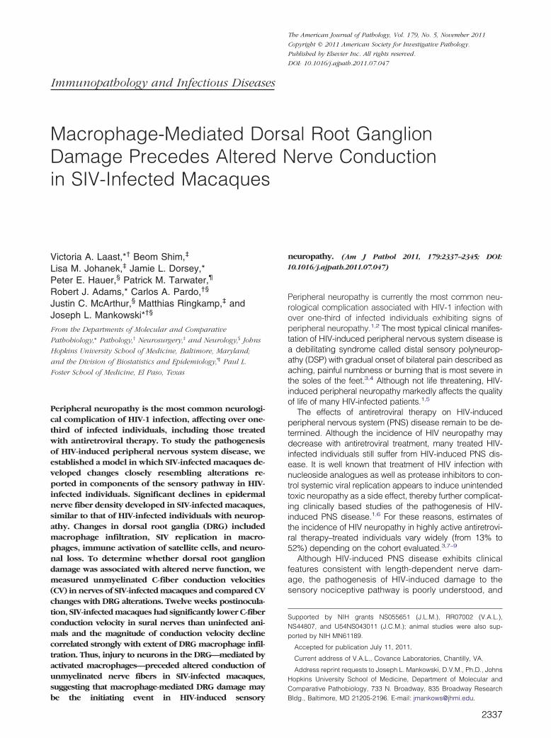
Skin biopsies have proven to be an invaluable tool todiagnose small sensory fiber neuropathies, largely re-placing sural nerve biopsies for clinical assessments ofsmall fiber sensory neuropathies.8,19,20,27–29 Previousstudies of skin biopsies taken from HIV-infected patientshave shown a close correlation between epidermal nervefiber (ENF) densities measured at the distal leg regionand the presence of neuropathic pain. In those studies,reduced ENF density at the distal leg was associated withincreased neuropathic pain, lower CD4 counts, andhigher plasma viral loads.29 To determine whether epi-dermal nerve fiber density loss occurred with SIV infec-tion, epidermal nerve fiber densities in footpad samplesfrom SIV-infected macaques were immunostained forPGP9.5, and then were compared with ENF density inuninfected control animals (Figure 1). Immunostaining todetect PGP9.5, a pan-neuronal marker present as a cy-toplasmic protein in peripheral nerves, is currently themost consistent method to visualize ENF.30,31 A progres-sive decrease in epidermal nerve fiber density over timedeveloped in SIV-infected macaques resembling ENFdecreases reported in HIV-infected individuals. A signif-icant decline in ENF density developed 8 weeks postin-oculation and was reduced further at the 12-week post-inoculation time point (P � 0.001; Figure 1).
SIV-Induced Dorsal Root Ganglia Pathology
The neuropathological changes in the DRG associatedwith HIV-induced distal sensory polyneuropathy includeinflammatory infiltrates of activated macrophages, fewerlymphocytes, decreased numbers of dorsal root ganglionneurons, and the presence of nodules of Nageotte.4,32,33
To investigate the histopathological lesions in the DRG ofSIV-infected macaques, hematoxylin and eosin sectionsof the fifth lumbar DRG from SIV-inoculated macaquesand uninfected controls were examined. DRG from unin-fected control macaques did not contain lesions. In con-trast, DRG examined from SIV-infected macaques con-tained multifocal aggregates of infiltrating mononuclearcells including macrophages as well as scattered lym-phocytes. Nageotte nodules, with neuronal cell bodiesreplaced by a mixture of infiltrating mononuclear cellsand Schwann cells, and evidence of neuronophagia, withinfiltrating macrophages abutting degenerate neurons
within the neuronal compartment, were also observed inDRG of animals with moderate to severe ganglionitis.These lesions in DRG of SIV-infected macaques demon-strated that SIV infection induces DRG pathology closelyresembling the lesions reported in dorsal root ganglia ofHIV-infected individuals with distal sensory polyneurop-athy.1,4 In contrast with DRG lesions, microscopic exami-nation of plastic-embedded, toluidine blue–stained sectionsof sural nerve and peroneal nerves did not reveal evidenceof neuritis or damage to myelinated fibers in SIV-infectedmacaques despite the presence of inflammatory changesin the lumbar DRG. Inflammatory lesions may still be pres-ent multifocally in peripheral nerve but may be difficult todetect given the extensive length of peripheral nerves. Asimilar paucity of lesions in sural nerve has been reported inHIV neuropathy.34,35
Dorsal Root Ganglia Macrophage and SatelliteCell Activation in SIV-Infected Macaques
Macrophage infiltration has been reported in previousimmunopathological studies performed on DRG fromHIV-infected patients with distal sensory polyneuropathyas well as in trigeminal ganglia in SIV-infected ma-
Figure 1. Epidermal nerve fiber density is reduced with SIV infection.PGP9.5 immunostained sections of footpad from control (A) and SIV-infectedmacaques (B) showed a decline in epidermal nerve fiber density. Nervefibers (red) in skin are present in both epidermis (E), and in dermis (D), withdermal-epidermal junction traced in green. C: Measurement of ENF density incontrol and SIV-infected macaques demonstrated reduced ENF density inSIV-infected macaques (triangles) compared to uninfected controls (cir-cles), with significant ENF decline beginning 8 weeks p.i., then decliningfurther at the 12-week time point. P � 0.001, analysis of variance). PI,postinoculation; ROI, region of interest.
caques.14,32 To investigate whether a similar process
SIV/Macaque Model of HIV PNS Disease 2341AJP November 2011, Vol. 179, No. 5
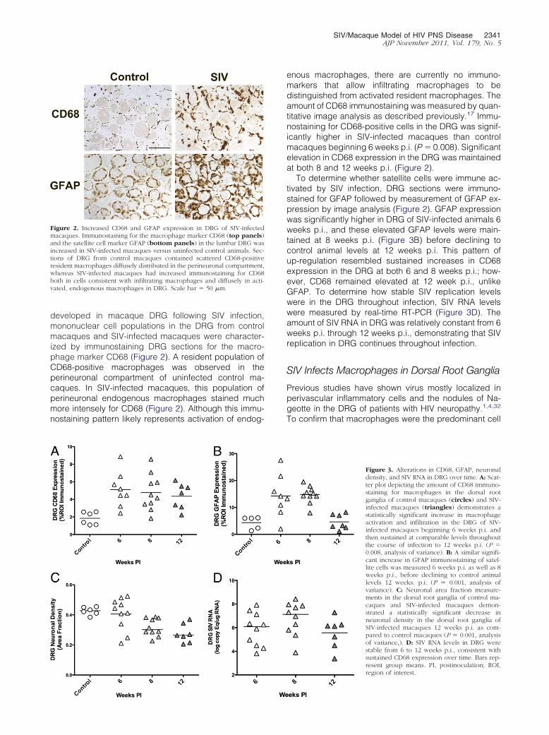
developed in macaque DRG following SIV infection,mononuclear cell populations in the DRG from controlmacaques and SIV-infected macaques were character-ized by immunostaining DRG sections for the macro-phage marker CD68 (Figure 2). A resident population ofCD68-positive macrophages was observed in theperineuronal compartment of uninfected control ma-caques. In SIV-infected macaques, this population ofperineuronal endogenous macrophages stained muchmore intensely for CD68 (Figure 2). Although this immu-nostaining pattern likely represents activation of endog-
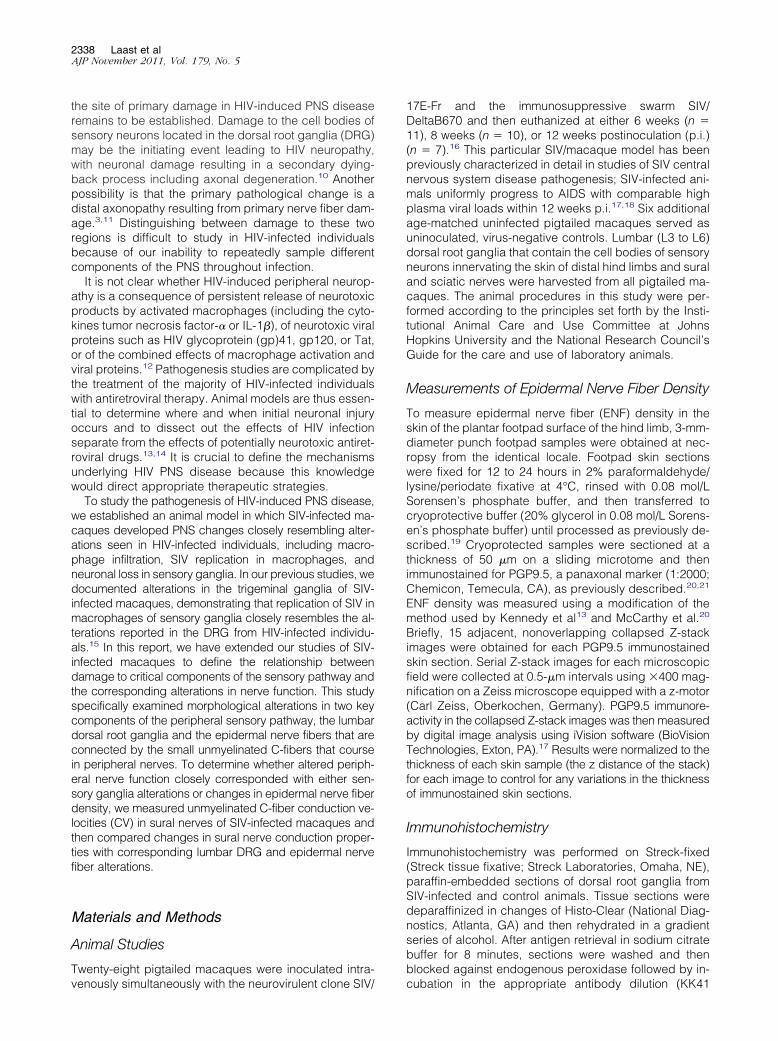
Figure 2. Increased CD68 and GFAP expression in DRG of SIV-infectedmacaques. Immunostaining for the macrophage marker CD68 (top panels)and the satellite cell marker GFAP (bottom panels) in the lumbar DRG wasincreased in SIV-infected macaques versus uninfected control animals. Sec-tions of DRG from control macaques contained scattered CD68-positiveresident macrophages diffusely distributed in the perineuronal compartment,whereas SIV-infected macaques had increased immunostaining for CD68both in cells consistent with infiltrating macrophages and diffusely in acti-vated, endogenous macrophages in DRG. Scale bar � 50 �m.
enous macrophages, there are currently no immuno-markers that allow infiltrating macrophages to bedistinguished from activated resident macrophages. Theamount of CD68 immunostaining was measured by quan-titative image analysis as described previously.17 Immu-nostaining for CD68-positive cells in the DRG was signif-icantly higher in SIV-infected macaques than controlmacaques beginning 6 weeks p.i. (P � 0.008). Significantelevation in CD68 expression in the DRG was maintainedat both 8 and 12 weeks p.i. (Figure 2).
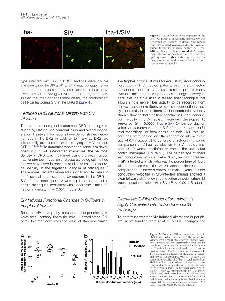
To determine whether satellite cells were immune ac-tivated by SIV infection, DRG sections were immuno-stained for GFAP followed by measurement of GFAP ex-pression by image analysis (Figure 2). GFAP expressionwas significantly higher in DRG of SIV-infected animals 6weeks p.i., and these elevated GFAP levels were main-tained at 8 weeks p.i. (Figure 3B) before declining tocontrol animal levels at 12 weeks p.i. This pattern ofup-regulation resembled sustained increases in CD68expression in the DRG at both 6 and 8 weeks p.i.; how-ever, CD68 remained elevated at 12 week p.i., unlikeGFAP. To determine how stable SIV replication levelswere in the DRG throughout infection, SIV RNA levelswere measured by real-time RT-PCR (Figure 3D). Theamount of SIV RNA in DRG was relatively constant from 6weeks p.i. through 12 weeks p.i., demonstrating that SIVreplication in DRG continues throughout infection.
SIV Infects Macrophages in Dorsal Root Ganglia
Previous studies have shown virus mostly localized inperivascular inflammatory cells and the nodules of Na-geotte in the DRG of patients with HIV neuropathy.1,4,32
To confirm that macrophages were the predominant cell
Figure 3. Alterations in CD68, GFAP, neuronaldensity, and SIV RNA in DRG over time. A: Scat-ter plot depicting the amount of CD68 immuno-staining for macrophages in the dorsal rootganglia of control macaques (circles) and SIV-infected macaques (triangles) demonstrates astatistically significant increase in macrophageactivation and infiltration in the DRG of SIV-infected macaques beginning 6 weeks p.i. andthen sustained at comparable levels throughoutthe course of infection to 12 weeks p.i. (P �0.008, analysis of variance). B: A similar signifi-cant increase in GFAP immunostaining of satel-lite cells was measured 6 weeks p.i. as well as 8weeks p.i., before declining to control animallevels 12 weeks. p.i. (P � 0.001, analysis ofvariance). C: Neuronal area fraction measure-ments in the dorsal root ganglia of control ma-caques and SIV-infected macaques demon-strated a statistically significant decrease inneuronal density in the dorsal root ganglia ofSIV-infected macaques 12 weeks p.i. as com-pared to control macaques (P � 0.001, analysisof variance,). D: SIV RNA levels in DRG werestable from 6 to 12 weeks p.i., consistent withsustained CD68 expression over time. Bars rep-resent group means. PI, postinoculation; ROI,region of interest.
2342 Laast et alAJP November 2011, Vol. 179, No. 5
type infected with SIV in DRG, sections were doubleimmunostained for SIV gp41 and the macrophage markerIba-1, and then examined by laser confocal microscopy.Colocalization of SIV gp41 within macrophages demon-strated that macrophages were clearly the predominantcell type harboring SIV in the DRG (Figure 4).
Reduced DRG Neuronal Density with SIVInfection
The main morphological features of DRG pathology in-duced by HIV include neuronal injury and axonal degen-eration. Relatively few reports have demonstrated neuro-nal loss in the DRG in addition to injury as DRG areinfrequently examined in patients dying of HIV-inducedDSP.13,14,33,36 To determine whether neuronal loss devel-oped in DRG of SIV-infected macaques, the neuronaldensity in DRG was measured using the area fractionfractionator technique, an unbiased stereological methodthat we have used in previous studies to estimate neuro-nal density in the trigeminal ganglia of macaques.15
These measurements revealed a significant decrease inthe fractional area occupied by neurons in the DRG ofSIV-infected macaques 12 weeks p.i. as compared tocontrol macaques, consistent with a decrease in the DRGneuronal density (P � 0.001, Figure 3C).
SIV Induces Functional Changes in C-Fibers inPeripheral Nerves
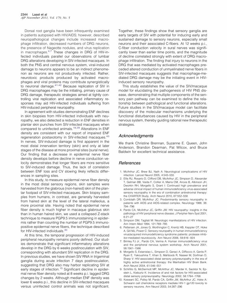
Because HIV neuropathy is suspected to principally in-volve small sensory fibers (ie, small, unmyelinated C-fi-bers), this markedly limits the value of standard clinical
electrophysiological studies for evaluating nerve conduc-tion, both in HIV-infected patients and in SIV-infectedmacaques, because such assessments predominantlyevaluate the conductive properties of large sensory fi-bers. We therefore used a teased fiber technique thatallows single nerve fiber activity to be recorded fromunmyelinated nerve fibers to measure conduction veloc-ity specifically in these fibers. C-fiber conduction velocitystudies showed that significant decline in C-fiber conduc-tion velocity in SIV-infected macaques developed 12weeks p.i. (P � 0.0003, Figure 5A). C-fiber conductionvelocity measurements from SIV-infected macaques (71total recordings) or from control animals (138 total re-cordings) were pooled, and then separated into bins (binsize of 0.1 m/second) to generate a histogram allowingcomparison of C-fiber conduction in SIV-infected ma-caques 12 weeks postinfection versus the uninfectedcontrol macaques (Figure 5B). The percentage of fiberswith conduction velocities below 0.5 m/second increasedin SIV-infected animals, whereas the percentage of fiberswith conduction velocities �0.5 m/second decreased ascompared to uninfected control animals. Overall, C-fiberconduction velocities in SIV-infected animals showed aclear leftward shift to lower conduction velocity values 12weeks postinoculation with SIV (P � 0.001; Student’st-test).
Decreased C-Fiber Conduction Velocity IsHighly Correlated with SIV-Induced DRGPathology
To determine whether SIV-induced alterations in periph-eral nerve function were related to DRG changes, the
Figure 4. SIV infection of macrophages in theDRG. Confocal laser scanning microscopy wasperformed on sections of dorsal root gangliafrom SIV-infected macaques double immuno-stained for the macrophage marker Iba-1 (red,left) and SIV gp41 (green, middle). A mergedimage showed colocalization of Iba-1 and SIVgp41 (yellow, right), indicating that macro-phages were the predominant SIV-infected celltype in sensory ganglia.
Figure 5. Decreased C-fiber conduction velocity inSIV-infection. A: Mean sural nerve C-fiber conductionvelocity for the group of SIV-infected animals evalu-ated 12 weeks p.i. was significantly slower than foruninfected control animals as well as for the groupsof SIV-infected animals evaluated 6 and 8 weekspostinoculation (P � 0.003, analysis of variance). B:To characterize the functional alterations in periph-eral nerves that developed with SIV infection, theconduction velocities of C-fibers in sural nerves fromSIV-infected monkeys collected 12 weeks p.i. werecompared with the conduction velocities in unin-fected control animals. The histogram containing thepooled C-fiber CV measurements for SIV-infected(black bars) and control macaques (white bars)showed an increase in the percentage of nerve fiberswith slower conduction velocities in SIV-infected ma-
caques 12 weeks p.i. as compared to controls (P �0.001; Student’s t-test). PI, postinoculation.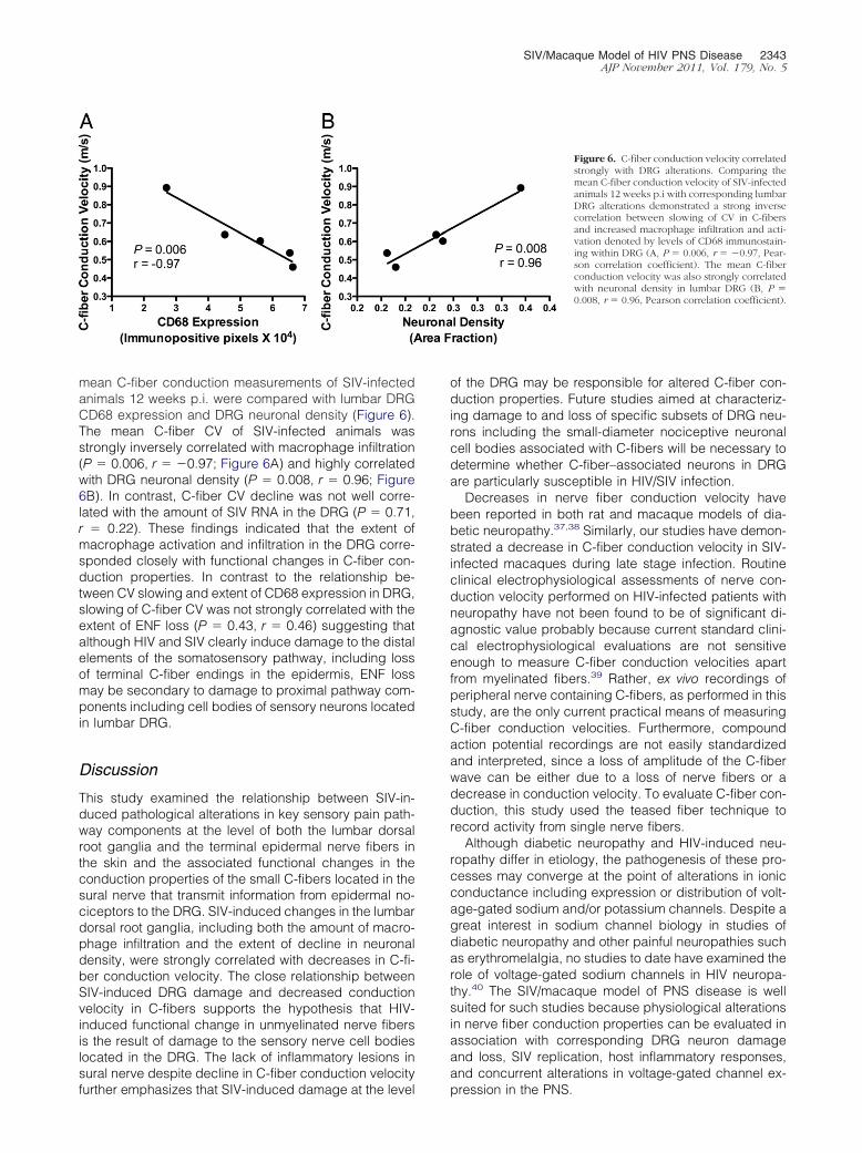
SIV/Macaque Model of HIV PNS Disease 2343AJP November 2011, Vol. 179, No. 5
mean C-fiber conduction measurements of SIV-infectedanimals 12 weeks p.i. were compared with lumbar DRGCD68 expression and DRG neuronal density (Figure 6).The mean C-fiber CV of SIV-infected animals wasstrongly inversely correlated with macrophage infiltration(P � 0.006, r � �0.97; Figure 6A) and highly correlatedwith DRG neuronal density (P � 0.008, r � 0.96; Figure6B). In contrast, C-fiber CV decline was not well corre-lated with the amount of SIV RNA in the DRG (P � 0.71,r � 0.22). These findings indicated that the extent ofmacrophage activation and infiltration in the DRG corre-sponded closely with functional changes in C-fiber con-duction properties. In contrast to the relationship be-tween CV slowing and extent of CD68 expression in DRG,slowing of C-fiber CV was not strongly correlated with theextent of ENF loss (P � 0.43, r � 0.46) suggesting thatalthough HIV and SIV clearly induce damage to the distalelements of the somatosensory pathway, including lossof terminal C-fiber endings in the epidermis, ENF lossmay be secondary to damage to proximal pathway com-ponents including cell bodies of sensory neurons locatedin lumbar DRG.
Discussion
This study examined the relationship between SIV-in-duced pathological alterations in key sensory pain path-way components at the level of both the lumbar dorsalroot ganglia and the terminal epidermal nerve fibers inthe skin and the associated functional changes in theconduction properties of the small C-fibers located in thesural nerve that transmit information from epidermal no-ciceptors to the DRG. SIV-induced changes in the lumbardorsal root ganglia, including both the amount of macro-phage infiltration and the extent of decline in neuronaldensity, were strongly correlated with decreases in C-fi-ber conduction velocity. The close relationship betweenSIV-induced DRG damage and decreased conductionvelocity in C-fibers supports the hypothesis that HIV-induced functional change in unmyelinated nerve fibersis the result of damage to the sensory nerve cell bodieslocated in the DRG. The lack of inflammatory lesions insural nerve despite decline in C-fiber conduction velocity
further emphasizes that SIV-induced damage at the levelof the DRG may be responsible for altered C-fiber con-duction properties. Future studies aimed at characteriz-ing damage to and loss of specific subsets of DRG neu-rons including the small-diameter nociceptive neuronalcell bodies associated with C-fibers will be necessary todetermine whether C-fiber–associated neurons in DRGare particularly susceptible in HIV/SIV infection.
Decreases in nerve fiber conduction velocity havebeen reported in both rat and macaque models of dia-betic neuropathy.37,38 Similarly, our studies have demon-strated a decrease in C-fiber conduction velocity in SIV-infected macaques during late stage infection. Routineclinical electrophysiological assessments of nerve con-duction velocity performed on HIV-infected patients withneuropathy have not been found to be of significant di-agnostic value probably because current standard clini-cal electrophysiological evaluations are not sensitiveenough to measure C-fiber conduction velocities apartfrom myelinated fibers.39 Rather, ex vivo recordings ofperipheral nerve containing C-fibers, as performed in thisstudy, are the only current practical means of measuringC-fiber conduction velocities. Furthermore, compoundaction potential recordings are not easily standardizedand interpreted, since a loss of amplitude of the C-fiberwave can be either due to a loss of nerve fibers or adecrease in conduction velocity. To evaluate C-fiber con-duction, this study used the teased fiber technique torecord activity from single nerve fibers.
Although diabetic neuropathy and HIV-induced neu-ropathy differ in etiology, the pathogenesis of these pro-cesses may converge at the point of alterations in ionicconductance including expression or distribution of volt-age-gated sodium and/or potassium channels. Despite agreat interest in sodium channel biology in studies ofdiabetic neuropathy and other painful neuropathies suchas erythromelalgia, no studies to date have examined therole of voltage-gated sodium channels in HIV neuropa-thy.40 The SIV/macaque model of PNS disease is wellsuited for such studies because physiological alterationsin nerve fiber conduction properties can be evaluated inassociation with corresponding DRG neuron damageand loss, SIV replication, host inflammatory responses,and concurrent alterations in voltage-gated channel ex-
Figure 6. C-fiber conduction velocity correlatedstrongly with DRG alterations. Comparing themean C-fiber conduction velocity of SIV-infectedanimals 12 weeks p.i with corresponding lumbarDRG alterations demonstrated a strong inversecorrelation between slowing of CV in C-fibersand increased macrophage infiltration and acti-vation denoted by levels of CD68 immunostain-ing within DRG (A, P � 0.006, r � �0.97, Pear-son correlation coefficient). The mean C-fiberconduction velocity was also strongly correlatedwith neuronal density in lumbar DRG (B, P �0.008, r � 0.96, Pearson correlation coefficient).
pression in the PNS.
2344 Laast et alAJP November 2011, Vol. 179, No. 5
Dorsal root ganglia have been infrequently examinedin patients autopsied with HIV/AIDS; however, describedneuropathological changes include increased macro-phage infiltration, decreased numbers of DRG neurons,the presence of Nageotte nodules, and virus replicationin macrophages.4,7 These changes in DRG of HIV-in-fected individuals parallel our observations of lumbarDRG alterations developing in SIV-infected macaques. Inboth the PNS and central nervous system, viral-induceddamage to neurons appears to be an indirect phenome-non as neurons are not productively infected. Rather,neurotoxic products produced by activated macro-phages and viral proteins may contribute synergisticallyto neuronal damage.41,42 Because replication of SIV inDRG macrophages may be the initiating, primary cause ofDRG damage, therapeutic strategies aimed at tightly con-trolling viral replication and associated inflammatory re-sponses may aid HIV-infected individuals suffering fromHIV-induced peripheral neuropathy.
In agreement with studies demonstrating ENF declinesin skin biopsies from HIV-infected individuals with neu-ropathy, we also detected a reduction in ENF densities inplantar skin punches from SIV-infected macaques whencompared to uninfected animals.19,29 Alterations in ENFdensity are consistent with our report of impaired ENFregeneration postaxotomy in SIV-infected macaques.43
In nerves, SIV-induced damage is first apparent in themost distal innervation territory (skin) and only at laterstages of the disease at more proximal sites (sural nerve).Our finding that a decrease in epidermal nerve fiberdensity develops before decline in nerve conduction ve-locity demonstrates that longer fibers are more sensitiveto SIV-induced damage. Thus, the lack of correlationbetween ENF loss and CV slowing likely reflects differ-ences in sampling sites.
In this study, to measure epidermal nerve fiber densityin the most distal sensory regions, skin samples wereharvested from the glabrous (non-haired) skin of the plan-tar footpad of SIV-infected macaques. Skin biopsy sam-ples from humans, in contrast, are generally harvestedfrom haired skin at the level of the lateral malleolus, amore proximal site. Having noted that epidermal nervefiber density is much higher in macaque glabrous skinthan in human haired skin, we used a collapsed Z-stacktechnique to measure PGP9.5 immunostaining in epider-mis rather than counting the number of PGP9.5 immuno-positive epidermal nerve fibers, the technique describedfor HIV-infected individuals.30
At this time, the temporal progression of HIV-inducedperipheral neuropathy remains poorly defined. Our stud-ies demonstrate that significant inflammatory alterationsdevelop in the DRG by 6 weeks postinoculation with SIV,corresponding with abundant SIV replication in the DRG.In previous studies, we have shown SIV RNA in trigeminalganglia during acute infection 7 days postinoculation,suggesting that DRG also may harbor replicating SIV atearly stages of infection.15 Significant decline in epider-mal nerve fiber density noted at 8 weeks p.i. lagged DRGchanges by 2 weeks. Although median ENF density waslower 6 weeks p.i., this decline in SIV-infected macaques
versus uninfected control animals was not significant.Together, these findings show that sensory ganglia areearly targets of SIV with potential for inducing early andsustained damage to sensory neurons, especially smallneurons and their associated C-fibers. At 12 weeks p.i.,C-fiber conduction velocity in sural nerves was signifi-cantly lower than earlier time points, and the magnitudeof decline correlated strongly with extent of DRG macro-phage infiltration. The finding that injury to neurons in theDRG that was mediated by activated macrophages pre-ceded altered conduction of unmyelinated nerve fibers inSIV-infected macaques suggests that macrophage-me-diated DRG damage may be the initiating event in HIV-induced sensory neuropathy.
This study establishes the value of the SIV/macaquemodel for elucidating the pathogenesis of HIV PNS dis-ease, demonstrating that multiple components of the sen-sory pain pathway can be examined to define the rela-tionship between pathological and functional alterations.Future studies in the SIV/macaque model can facilitatediscovery of the molecular mechanisms underlying thefunctional disturbances caused by HIV in the peripheralnervous system, thereby guiding rational new therapeuticstrategies.
Acknowledgments
We thank Christine Brennan, Suzanne E. Queen, JohnAnderson, Brandon Dearman, Pat Wilcox, and BruceBaldwin for excellent technical assistance.
References
1. McArthur JC, Brew BJ, Nath A: Neurological complications of HIVinfection. Lancet Neurol 2005, 4:543–555
2. Ellis RJ, Rosario D, Clifford DB, McArthur JC, Simpson D, AlexanderT, Gelman BB, Vaida F, Collier A, Marra CM, Ances B, Atkinson JH,Dworkin RH, Morgello S, Grant I: Continued high prevalence andadverse clinical impact of human immunodeficiency virus-associatedsensory neuropathy in the era of combination antiretroviral therapy:the CHARTER Study. Arch Neurol 2010, 67:552–558
3. Cornblath DR, McArthur JC: Predominantly sensory neuropathy inpatients with AIDS and AIDS-related complex. Neurology 1988, 38:794–796
4. Pardo CA, McArthur JC, Griffin JW: HIV neuropathy: insights in thepathology of HIV peripheral nerve disease. J Peripher Nerv Syst 2001,6:21–27
5. Simpson DM, Tagliati M: Neurologic manifestations of HIV infection.Ann Intern Med 1994, 121:769–785
6. Pettersen JA, Jones G, Worthington C, Krentz HB, Keppler OT, HokeA, Gill MJ, Power C: Sensory neuropathy in human immunodeficiencyvirus/acquired immunodeficiency syndrome patients: protease inhib-itor-mediated neurotoxicity. Ann Neurol 2006, 59:816–824
7. Brinley FJ Jr., Pardo CA, Verma A: Human immunodeficiency virusand the peripheral nervous system workshop. Arch Neurol 2001,58:1561–1566
8. Morgello S, Estanislao L, Simpson D, Geraci A, DiRocco A, Gerits P,Ryan E, Yakoushina T, Khan S, Mahboob R, Naseer M, Dorfman D,Sharp V: HIV-associated distal sensory polyneuropathy in the era ofhighly active antiretroviral therapy: the Manhattan HIV Brain Bank.Arch Neurol 2004, 61:546–551
9. Schifitto G, McDermott MP, McArthur JC, Marder K, Sacktor N, Ep-stein L, Kieburtz K: Incidence of and risk factors for HIV-associateddistal sensory polyneuropathy. Neurology 2002, 58:1764–1768
10. Keswani SC, Polley M, Pardo CA, Griffin JW, McArthur JC, Hoke A:
Schwann cell chemokine receptors mediate HIV-1 gp120 toxicity tosensory neurons. Ann Neurol 2003, 54:287–296
SIV/Macaque Model of HIV PNS Disease 2345AJP November 2011, Vol. 179, No. 5
11. Bradley WG, Shapshak P, Delgado S, Nagano I, Stewart R, Rocha B:Morphometric analysis of the peripheral neuropathy of AIDS. MuscleNerve 1998, 21:1188–1195
12. Watkins LR, Goehler LE, Relton J, Brewer MT, Maier SF: Mechanismsof tumor necrosis factor-alpha (TNF-alpha) hyperalgesia. Brain Res1995, 692:244–250
13. Kennedy JM, Hoke A, Zhu Y, Johnston JB, van Marle G, Silva C,Zochodne DW, Power C: Peripheral neuropathy in lentivirus infection:evidence of inflammation and axonal injury. AIDS 2004, 18:1241–1250
14. Zhu Y, Jones G, Tsutsui S, Opii W, Liu S, Silva C, Butterfield DA,Power C: Lentivirus infection causes neuroinflammation and neuronalinjury in dorsal root ganglia: pathogenic effects of STAT-1 and induc-ible nitric oxide synthase. J Immunol 2005, 175:1118–1126
15. Laast VA, Pardo CA, Tarwater PM, Queen SE, Reinhart TA, Ghosh M,Adams RJ, Zink MC, Mankowski JL: Pathogenesis of simian immu-nodeficiency virus-induced alterations in macaque trigeminal gan-glia. J Neuropathol Exp Neurol 2007, 66:26–34
16. Zink MC, Suryanarayana K, Mankowski JL, Shen A, Piatak M Jr,Spelman JP, Carter DL, Adams RJ, Lifson JD, Clements JE: High viralload in the cerebrospinal fluid and brain correlates with severity ofsimian immunodeficiency virus encephalitis. J Virol 1999, 73:10480–10488
17. Mankowski JL, Clements JE, Zink MC: Searching for clues: trackingthe pathogenesis of human immunodeficiency virus central nervoussystem disease by use of an accelerated, consistent simian immu-nodeficiency virus macaque model. J Infect Dis 2002, 186 (Suppl2):S199–208
18. Zink MC, Laast VA, Helke KL, Brice AK, Barber SA, Clements JE,Mankowski JL: From mice to macaques–animal models of HIV ner-vous system disease. Curr HIV Res 2006, 4:293–305
19. Holland NR, Stocks A, Hauer P, Cornblath DR, Griffin JW, McArthurJC: Intraepidermal nerve fiber density in patients with painful sensoryneuropathy. Neurology 1997, 48:708–711
20. McCarthy BG, Hsieh ST, Stocks A, Hauer P, Macko C, Cornblath DR,Griffin JW, McArthur JC: Cutaneous innervation in sensoryneuropathies: evaluation by skin biopsy. Neurology 1995, 45:1848–1855
21. Nolano M, Provitera V, Crisci C, Stancanelli A, Wendelschafer-CrabbG, Kennedy WR, Santoro L: Quantification of myelinated endings andmechanoreceptors in human digital skin. Ann Neurol 2003, 54:197–205
22. Vargas DL, Nascimbene C, Krishnan C, Zimmerman AW, Pardo CA:Neuroglial activation and neuroinflammation in the brain of patientswith autism. Ann Neurol 2005, 57:67–81
23. West MJ, Coleman PD, Flood DG, Troncoso JC: Differences in thepattern of hippocampal neuronal loss in normal ageing and Alzhei-mer’s disease. Lancet 1994, 344:769–772
24. Kril JJ, Patel S, Harding AJ, Halliday GM: Neuron loss from thehippocampus of Alzheimer’s disease exceeds extracellular neurofi-brillary tangle formation. Acta Neuropathol (Berl) 2002, 103:370–376
25. Ali Z, Ringkamp M, Hartke TV, Chien HF, Flavahan NA, Campbell JN,Meyer RA: Uninjured C-fiber nociceptors develop spontaneous ac-tivity and alpha-adrenergic sensitivity following L6 spinal nerve liga-tion in monkey. J Neurophysiol 1999, 81:455–466
26. Wu G, Ringkamp M, Hartke TV, Murinson BB, Campbell JN, GriffinJW, Meyer RA: Early onset of spontaneous activity in uninjured C-fiber
nociceptors after injury to neighboring nerve fibers. J Neurosci 2001,21:RC14027. Herrmann DN, McDermott MP, Henderson D, Chen L, Akowuah K,Schifitto G: Epidermal nerve fiber density, axonal swellings and QSTas predictors of HIV distal sensory neuropathy. Muscle Nerve 2004,29:420–427
28. Herrmann DN, Griffin JW, Hauer P, Cornblath DR, McArthur JC:Epidermal nerve fiber density and sural nerve morphometry in pe-ripheral neuropathies. Neurology 1999, 53:1634–1640
29. Polydefkis M, Yiannoutsos CT, Cohen BA, Hollander H, Schifitto G,Clifford DB, Simpson DM, Katzenstein D, Shriver S, Hauer P, Brown A,Haidich AB, Moo L, McArthur JC: Reduced intraepidermal nerve fiberdensity in HIV-associated sensory neuropathy. Neurology 2002, 58:115–119
30. Kennedy WR, Wendelschafer-Crabb G, Johnson T: Quantitation ofepidermal nerves in diabetic neuropathy. Neurology 1996, 47:1042–1048
31. Horiuchi Y, Bae S, Katayama I: Nerve growth factor (NGF) andepidermal nerve fibers in atopic dermatitis model NC/Nga mice. JDermatol Sci 2005, 39:56–58
32. Keswani SC, Pardo CA, Cherry CL, Hoke A, McArthur JC: HIV-associated sensory neuropathies. AIDS 2002, 16:2105–2117
33. Esiri MM, Morris CS, Millard PR: Sensory and sympathetic ganglia inHIV-1 infection: immunocytochemical demonstration of HIV-1 viralantigens, increased MHC class II antigen expression and mild reac-tive inflammation. J Neurol Sci 1993, 114:178–187
34. Gonzalez-Duarte A, Cikurel K, Simpson DM: Managing HIV periph-eral neuropathy. Curr HIV/AIDS Rep 2007, 4:114–118
35. Gonzalez-Duarte A, Robinson-Papp J, Simpson DM: Diagnosis andmanagement of HIV-associated neuropathy. Neurol Clin 2008, 26:821–832
36. Jones G, Zhu Y, Silva C, Tsutsui S, Pardo CA, Keppler OT, McArthurJC, Power C: Peripheral nerve-derived HIV-1 is predominantly CCR5-dependent and causes neuronal degeneration and neuroinflamma-tion. Virology 2005, 334:178–193
37. Price SA, Agthong S, Middlemas AB, Tomlinson DR: Mitogen-acti-vated protein kinase p38 mediates reduced nerve conduction veloc-ity in experimental diabetic neuropathy: interactions with aldose re-ductase. Diabetes 2004, 53:1851–1856
38. Cornblath DR, Hillman MA, Striffler JS, Herman CN, Hansen BC:Peripheral neuropathy in diabetic monkeys. Diabetes 1989, 38:1365–1370
39. Hahn K, Arendt G, Braun JS, von Giesen HJ, Husstedt IW, MaschkeM, Straube ME, Schielke E: A placebo-controlled trial of gabapentinfor painful HIV-associated sensory neuropathies. J Neurol 2004, 251:1260–1266
40. Keswani SC, Hoke A: Incidence of and risk factors for HIV-associateddistal sensory polyneuropathy, Neurology 2003, 61:279; author reply279–280
41. Babas T, Munoz D, Mankowski JL, Tarwater PM, Clements JE, ZinkMC: Role of microglial cells in selective replication of simian immu-nodeficiency virus genotypes in the brain. J Virol 2003, 77:208–216
42. Mankowski JL, Queen SE, Tarwater PM, Fox KJ, Perry VH: Accumu-lation of beta-amyloid precursor protein in axons correlates with CNSexpression of SIV gp41. J Neuropathol Exp Neurol 2002, 61:85–90
43. Ebenezer GJ, Laast VA, Dearman B, Hauer P, Tarwater PM, AdamsRJ, Zink MC, McArthur JC, Mankowski JL: Altered cutaneous nerveregeneration in a simian immunodeficiency virus/macaque intracuta-
neous axotomy model. J Comp Neurol 2009, 514:272–283
Related Documents


