Macro-to-micro porous special bioactive glass and ceftriaxone–sulbactam composite drug delivery system for treatment of chronic osteomyelitis: an investigation through in vitro and in vivo animal trial Biswanath Kundu • Samit Kumar Nandi • Sudip Dasgupta • Someswar Datta • Prasenjit Mukherjee • Subhasis Roy • Aruna Kumari Singh • Tapan Kumar Mandal • Partha Das • Rupnarayan Bhattacharya • Debabrata Basu Received: 1 September 2010 / Accepted: 19 December 2010 / Published online: 8 January 2011 Ó Springer Science+Business Media, LLC 2010 Abstract A systematic and extensive approach incorpo- rating in vitro and in vivo experimentation to treat chronic osteomyelitis in animal model were made using antibiotic loaded special bioactive glass porous scaffolds. After thorough characterization for porosity, distribution, surface charge, a novel drug composite were infiltrated by using vacuum infiltration and freeze-drying method which was subsequently analyzed by SEM–EDAX and studied for in vitro drug elution in PBS and SBF. Osteomyelitis in rab bit was induced by inoculation of Staphylococcus aureus and optimum drug-scaffold were checked for its efficacy over control and parenteral treated animals in terms of histopathology, radiology, in vivo drug concentration in bone and serum and implant-bone interface by SEM. It was optimized that 60P samples with 60–65% porosity (bimo- dal distribution of macro- to micropore) with average pore size *60 lm and higher interconnectivity, moderately high antibiotic adsorption efficiency (*49%) was ideal. Results after 42 days showed antibiotic released higher than MIC against S. aureus compared to parenteral treat- ment (2 injections a day for 6 weeks). In vivo drug phar- macokinetics and SEM on bone-defect interface proved superiority of CFS loaded porous bioactive glass implants over parenteral group based on infection eradication and new bone formation. Abbreviations HAp Hydroxyapatite b-TCP Beta-tri calcium phosphate SEM–EDAX Scanning electron microscopy–energy dispersive analysis of X-ray HPLC High performance liquid chromatography PBS Phosphate buffered saline SBF Simulated body fluid PMMA Poly-methylmethacrylate MIC Minimum inhibitory concentration CFT Ceftriaxone sodium SUL Sulbactam sodium CFS Combination of CFT and SUL drug XRD X-ray diffraction FTIR Fourier-transformed infrared spectroscopy FESEM Field emission scanning electron microscopy ASTM American Society for Testing and Materials CFA Colony forming unit RBC Red blood cell B. Kundu Á S. Dasgupta Á S. Datta Á D. Basu (&) Bioceramics and Coating Division, Central Glass and Ceramic Research Institute, 196, Raja S. C. Mullick Road, Kolkata 700028, India e-mail: [email protected] S. K. Nandi (&) Á P. Mukherjee Á S. Roy Department of Veterinary Surgery and Radiology, West Bengal University of Animal and Fishery Sciences, 37 and 68, Kshudiram Bose Sarani, Kolkata 700037, India e-mail: [email protected] A. K. Singh Á T. K. Mandal Department of Veterinary Pharmacology and Toxicology, West Bengal University of Animal and Fishery Sciences, Kolkata, India P. Das Department of Anatomy and Histology, West Bengal University of Animal and Fishery Sciences, Kolkata, India R. Bhattacharya Department of Plastic Surgery, R. G. Kar Medical College and Hospital, Kolkata, India 123 J Mater Sci: Mater Med (2011) 22:705–720 DOI 10.1007/s10856-010-4221-3

Welcome message from author
This document is posted to help you gain knowledge. Please leave a comment to let me know what you think about it! Share it to your friends and learn new things together.
Transcript

Macro-to-micro porous special bioactive glassand ceftriaxone–sulbactam composite drug delivery systemfor treatment of chronic osteomyelitis: an investigation throughin vitro and in vivo animal trial
Biswanath Kundu • Samit Kumar Nandi • Sudip Dasgupta • Someswar Datta •
Prasenjit Mukherjee • Subhasis Roy • Aruna Kumari Singh • Tapan Kumar Mandal •
Partha Das • Rupnarayan Bhattacharya • Debabrata Basu
Received: 1 September 2010 / Accepted: 19 December 2010 / Published online: 8 January 2011
� Springer Science+Business Media, LLC 2010
Abstract A systematic and extensive approach incorpo-
rating in vitro and in vivo experimentation to treat chronic
osteomyelitis in animal model were made using antibiotic
loaded special bioactive glass porous scaffolds. After
thorough characterization for porosity, distribution, surface
charge, a novel drug composite were infiltrated by using
vacuum infiltration and freeze-drying method which was
subsequently analyzed by SEM–EDAX and studied for in
vitro drug elution in PBS and SBF. Osteomyelitis in rab
bit was induced by inoculation of Staphylococcus aureus
and optimum drug-scaffold were checked for its efficacy
over control and parenteral treated animals in terms of
histopathology, radiology, in vivo drug concentration in
bone and serum and implant-bone interface by SEM. It was
optimized that 60P samples with 60–65% porosity (bimo-
dal distribution of macro- to micropore) with average pore
size *60 lm and higher interconnectivity, moderately
high antibiotic adsorption efficiency (*49%) was ideal.
Results after 42 days showed antibiotic released higher
than MIC against S. aureus compared to parenteral treat-
ment (2 injections a day for 6 weeks). In vivo drug phar-
macokinetics and SEM on bone-defect interface proved
superiority of CFS loaded porous bioactive glass implants
over parenteral group based on infection eradication and
new bone formation.
Abbreviations
HAp Hydroxyapatite
b-TCP Beta-tri calcium phosphate
SEM–EDAX Scanning electron microscopy–energy
dispersive analysis of X-ray
HPLC High performance liquid chromatography
PBS Phosphate buffered saline
SBF Simulated body fluid
PMMA Poly-methylmethacrylate
MIC Minimum inhibitory concentration
CFT Ceftriaxone sodium
SUL Sulbactam sodium
CFS Combination of CFT and SUL drug
XRD X-ray diffraction
FTIR Fourier-transformed infrared spectroscopy
FESEM Field emission scanning electron
microscopy
ASTM American Society for Testing and
Materials
CFA Colony forming unit
RBC Red blood cell
B. Kundu � S. Dasgupta � S. Datta � D. Basu (&)
Bioceramics and Coating Division, Central Glass and Ceramic
Research Institute, 196, Raja S. C. Mullick Road, Kolkata
700028, India
e-mail: [email protected]
S. K. Nandi (&) � P. Mukherjee � S. Roy
Department of Veterinary Surgery and Radiology,
West Bengal University of Animal and Fishery Sciences,
37 and 68, Kshudiram Bose Sarani, Kolkata 700037, India
e-mail: [email protected]
A. K. Singh � T. K. Mandal
Department of Veterinary Pharmacology and Toxicology,
West Bengal University of Animal and Fishery Sciences,
Kolkata, India
P. Das
Department of Anatomy and Histology, West Bengal University
of Animal and Fishery Sciences, Kolkata, India
R. Bhattacharya
Department of Plastic Surgery, R. G. Kar Medical College
and Hospital, Kolkata, India
123
J Mater Sci: Mater Med (2011) 22:705–720
DOI 10.1007/s10856-010-4221-3

1 Introduction
Treatment of orthopaedic infections with antibacterial
agents by oral or intravenous route often leads the clini-
cians to be pessimistic about patient outcome [1]; as the
condition is frequently associated with poor vascular
perfusion accompanied by infection of the surrounding
tissue [2]. Following surgical debridement, it is necessary
to maintain a highly effective concentration of the anti-
biotic in the infected area for a sufficient period of time
(usually 4–6 weeks) to allow the healing process to
complete [3].
Different antibiotic impregnated implants based on
various kinds of carrier materials have been tried [4].
With the growing interest for combination devices that
could release drug and as well enhance or support tissue
regeneration, approaches with bioactive ceramics have
proved to improve the prognosis of orthopaedic infections
than polymers used conventionally for novel drug delivery
systems [5]. However, recently a new gentamicin-
vancomycin-impregnated (2:1) poly-methyl methacrylate
(PMMA) coating nail has been introduced as a drug
delivery device which could treat bone and intramedullary
infections [caused by methicillin-resistant Staphylococcus
aureus (MRSA)] effectively after surgical debridement and
immediate implantation [6]. Porous bioceramic scaffolds
made with hydroxyapatite, b-TCP have also proved their
suitability in releasing drug and accepting bone ingrowth
[7, 8]. Macroporosity with pore diameter above 100 lm is
desired to permit bone infiltration, on the other hand, big-
ger the pore size, faster would be the drug elution from the
scaffolds [4]. Pore interconnection is another key factor
that dominates bone ingrowth; and high pore interconnec-
tivity is generally obtained by high pore volume [9].
Now, CFT is a broad-spectrum semi-synthetic third-
generation cephalosporin with a potent bactericidal activity
against a wide range of gram-positive and gram-negative
bacteria and SUL is b-lactamase inhibitor. CFS have long
been considered the drugs of choice for the treatment of
chronic osteomyelitis because of their favorable penetra-
tion into poorly vascularized sites of infection, their
advantageous bactericidal effects against all probable
pathogens of chronic osteomyelitis, and the lack of serious
adverse reactions [10].
Studies on effect of macro- and microporosity alongwith
pore interconnection on the performance of porous bioac-
tive glass scaffolds are yet to be reported to the best of our
knowledge which was used as local drug delivery system.
Our previous study on goat indicated bioactive glass
scaffolds are at least marginally better osteoinductive
compared to HAp and b-TCP scaffolds [11]. Hence, in the
present study we reported a porous bioactive glass based
drug delivery system and extensively studied its suitability
in releasing the drug both in vitro and in vivo for a pro-
longed time period and supporting new bone formation and
hence dead space management. The bioactive glass com-
position used had been developed earlier in our lab and
proved to be bioactive and noncytotoxic in vitro [12].
Ceftriaxone–sulbactam combination was selected as model
drugs as the combination of beta-lactam antibiotic with an
irreversible b-lactamase inhibitor decrease the MICs of
hydrolyzed b-lactams to normal [13] and expands the
antimicrobial spectrum to include previously b-lactam
resistant microorganisms [14].
2 Materials and methods
2.1 Preparation and characterization of bioactive glass
powder
A composition of bioactive glass powders was prepared
using different raw materials as source of network former
and modifiers. The raw materials include silica (SiO2),
calcium carbonate (CaCO3), dry soda ash (Na2CO3),
decahydrated borax (Na2B4O7�10H2O), titania (TiO2),
di-ammonium hydrogen ortho-phosphate (all chemicals
were analytical grade from M/s SD Fine-Chem Limited,
India). The powder was prepared by following usual glass
melting (1400�C) procedures, details of which could be
found elsewhere [11, 15]. Final chemical composition of
this glass, which was used subsequently for fabrication of
porous scaffolds, is given in Table 1.
Surface charge in terms of zeta potential of the
as-prepared powder (1400�C) as well as powders fired at
725�C/5 min (optimized sintering temperature of porous
scaffolds) was also estimated for indirect estimation of
interaction between these particles and CFS. Zeta poten-
tial of these particles was determined using suspensions
containing 0.01% (w/v) of such particles in a 10-3 M KCl
solution. The measurements were taken as a function of
pH at 20�C. The pH was adjusted with 0.1 M KOH and
1 N HCl solutions for basic and acidic conditions,
respectively.
Table 1 Final composition of
the bioactive glassComposition wt%
SiO2 43.70
CaO 19.20
P2O5 5.46
B2O3 9.40
Na2O 22.24
706 J Mater Sci: Mater Med (2011) 22:705–720
123
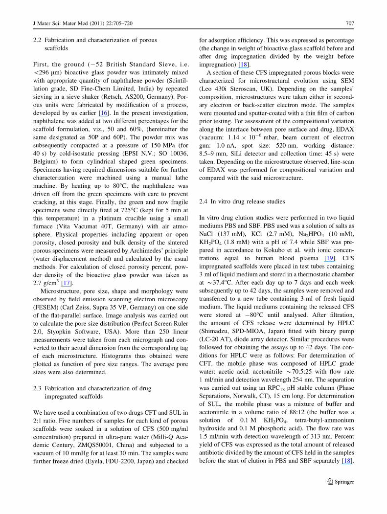
2.2 Fabrication and characterization of porous
scaffolds
First, the ground (-52 British Standard Sieve, i.e.
\296 lm) bioactive glass powder was intimately mixed
with appropriate quantity of naphthalene powder (Scintil-
lation grade, SD Fine-Chem Limited, India) by repeated
sieving in a sieve shaker (Retsch, AS200, Germany). Por-
ous units were fabricated by modification of a process,
developed by us earlier [16]. In the present investigation,
naphthalene was added at two different percentages for the
scaffold formulation, viz., 50 and 60%, (hereinafter the
same designated as 50P and 60P). The powder mix was
subsequently compacted at a pressure of 150 MPa (for
40 s) by cold-isostatic pressing (EPSI N.V.; SO 10036,
Belgium) to form cylindrical shaped green specimens.
Specimens having required dimensions suitable for further
characterization were machined using a manual lathe
machine. By heating up to 80�C, the naphthalene was
driven off from the green specimens with care to prevent
cracking, at this stage. Finally, the green and now fragile
specimens were directly fired at 725�C (kept for 5 min at
this temperature) in a platinum crucible using a small
furnace (Vita Vacumat 40T, Germany) with air atmo-
sphere. Physical properties including apparent or open
porosity, closed porosity and bulk density of the sintered
porous specimens were measured by Archimedes’ principle
(water displacement method) and calculated by the usual
methods. For calculation of closed porosity percent, pow-
der density of the bioactive glass powder was taken as
2.7 g/cm3 [17].
Microstructure, pore size, shape and morphology were
observed by field emission scanning electron microscopy
(FESEM) (Carl Zeiss, Supra 35 VP, Germany) on one side
of the flat-parallel surface. Image analysis was carried out
to calculate the pore size distribution (Perfect Screen Ruler
2.0, Styopkin Software, USA). More than 250 linear
measurements were taken from each micrograph and con-
verted to their actual dimension from the corresponding tag
of each microstructure. Histograms thus obtained were
plotted as function of pore size ranges. The average pore
sizes were also determined.
2.3 Fabrication and characterization of drug
impregnated scaffolds
We have used a combination of two drugs CFT and SUL in
2:1 ratio. Five numbers of samples for each kind of porous
scaffolds were soaked in a solution of CFS (500 mg/ml
concentration) prepared in ultra-pure water (Milli-Q Aca-
demic Century, ZMQS50001, China) and subjected to a
vacuum of 10 mmHg for at least 30 min. The samples were
further freeze dried (Eyela, FDU-2200, Japan) and checked
for adsorption efficiency. This was expressed as percentage
(the change in weight of bioactive glass scaffold before and
after drug impregnation divided by the weight before
impregnation) [18].
A section of these CFS impregnated porous blocks were
characterized for microstructural evolution using SEM
(Leo 430i Steroscan, UK). Depending on the samples’
composition, microstructures were taken either in second-
ary electron or back-scatter electron mode. The samples
were mounted and sputter-coated with a thin film of carbon
prior testing. For assessment of the compositional variation
along the interface between pore surface and drug, EDAX
(vacuum: 1.14 9 10-6 mbar, beam current of electron
gun: 1.0 nA, spot size: 520 nm, working distance:
8.5–9 mm, SiLi detector and collection time: 45 s) were
taken. Depending on the microstructure observed, line-scan
of EDAX was performed for compositional variation and
compared with the said microstructure.
2.4 In vitro drug release studies
In vitro drug elution studies were performed in two liquid
mediums PBS and SBF. PBS used was a solution of salts as
NaCl (137 mM), KCl (2.7 mM), Na2HPO4 (10 mM),
KH2PO4 (1.8 mM) with a pH of 7.4 while SBF was pre-
pared in accordance to Kokubo et al. with ionic concen-
trations equal to human blood plasma [19]. CFS
impregnated scaffolds were placed in test tubes containing
3 ml of liquid medium and stored in a thermostatic chamber
at *37.4�C. After each day up to 7 days and each week
subsequently up to 42 days, the samples were removed and
transferred to a new tube containing 3 ml of fresh liquid
medium. The liquid mediums containing the released CFS
were stored at -80�C until analysed. After filtration,
the amount of CFS release were determined by HPLC
(Shimadzu, SPD-MIOA, Japan) fitted with binary pump
(LC-20 AT), diode array detector. Similar procedures were
followed for obtaining the assays up to 42 days. The con-
ditions for HPLC were as follows: For determination of
CFT, the mobile phase was composed of HPLC grade
water: acetic acid: acetonitrile *70:5:25 with flow rate
1 ml/min and detection wavelength 254 nm. The separation
was carried out using an RPC18 pH stable column (Phase
Separations, Norwalk, CT), 15 cm long. For determination
of SUL, the mobile phase was a mixture of buffer and
acetonitrile in a volume ratio of 88:12 (the buffer was a
solution of 0.1 M KH2PO4, tetra-butyl-ammonium
hydroxide and 0.1 M phosphoric acid). The flow rate was
1.5 ml/min with detection wavelength of 313 nm. Percent
yield of CFS was expressed as the total amount of released
antibiotic divided by the amount of CFS held in the samples
before the start of elution in PBS and SBF separately [18].
J Mater Sci: Mater Med (2011) 22:705–720 707
123

2.5 Bacterial isolate
Staphylococcus aureus (coagulase positive) from an animal
with chronic osteomyelitis was used for development of
experimental model in rabbit. Pure cultures of the bacteria
were obtained on blood agar at 37�C and standardized
suspensions (3 9 106 CFU/ml) were prepared in saline.
This sample (1 ml) was introduced into the medullary
cavity of rabbit tibiae and confirmed successful induction
of osteomyelitis by S. aureus based on mannitol salt agar
test. Twenty-one (21) days of post inoculation, the swab
specimen was collected from the infected site from animals
of all groups and was streaked on mannitol 10% salt agar
slant and incubated at 37�C for overnight. From single
colony, bacterial growth was collected and stained by
Gram’s staining method. At the time that the animals were
sacrificed (12, 21 and 42 days for group II and III animals),
swab specimen was collected from the implanted site of
bone and similarly, inoculated to mannitol 10% salt agar
and incubated at 37�C for overnight.
2.6 In vivo studies
According to the model of Norden [20], osteomyelitis was
induced in the right tibia of 24 (twenty-four) nos. of adult
New Zealand white rabbits (2.5–3 kg body weight). The
proximal part of the tibia was exposed anteriorly after
anaesthesia with Nembutal 0.5 mg/kg IV (Thiopentone
sodium, Thiosol�, Neonlab, Mumbai, India), and a hole
was drilled through the cortex into the medullary cavity
using a 1.2 mm diameter dental burr. 1 ml of S. aureus
suspension containing approximately 3 9 106 CFU/ml was
injected into the drilled medullary cavity and the hole was
sealed with bone wax to prevent bacterial leakage into the
surrounding soft tissues. The animals were monitored after
surgery. All the animals received standard postoperative
pain medication (Carprofen; 4 mg/kg of body weight) for
3 days. The animals which developed osteomyelitis after 3
weeks of inoculation were only considered for present
study. By using the previous surgical approach, the prox-
imal tibia was exposed and bone defects were created by
micromotor dental drill. CFS impregnated bioactive glass
blocks were implanted in the defect area of infected bone
and same postoperative management was followed. All the
animal experimentations were carried out following the
procedures conforming to the standards of the Institutions
Animal Ethical Committee of the West Bengal University
of Animal and Fishery Sciences, India. The 24 animals
were divided into 3 Groups hereinafter would be desig-
nated as group I, II and III. The details of the experimen-
tation with these animals are given in Table 2. All the
samples and study parameters were obtained on 12, 21 and
42 days post osteomyelitis. Animals were pharmacologi-
cally euthanised under general anaesthesia after 42 days.
The implanted bone/antibiotic impregnated bioactive glass
implants were collected and then thoroughly washed
implants were fixed in 10% formalin for 7 days and sub-
sequently decalcified in Goodling and Stewart’s fluid
containing formic acid 15 ml, formalin 5 ml and distilled
water 80 ml solution. Decalcification was confirmed by
flexible and transparent section easily penetrable with pin.
Decalcified bone specimens were first embedded with
paraffin and sections were cut (3–4 mm thick) with rotary
microtome (HM 360, Microm, Germany). Hematoxyline
and eosin stained decalcified cross sections were consid-
ered for histological examinations. Histological images
were analyzed digitally as par different cellular events
occurred with time and following features were noted and
Table 2 Design of experiment for in vivo animal experimentation
Groups No. of
animals
Implant Days of
experiment
Experiment
Group I 6 Not given After 21 days Six animals were sacrificed for histological, radiographic
and microbiological examination to confirm development
of osteomyelitis
Group II 9 CFS injection parenterally
(15 mg/kg, bid) twice daily
for 6 weeks
12 days Three animals were sacrificed for histological and estimation
of drug concentration in bone and serum
After 21 days Three animals were sacrificed for histological and estimation
of drug concentration in bone and serum
After 42 days Three animals were sacrificed for histological and estimation
of drug concentration in bone and serum
Group III 9 Ceftriaxone–sulbactam
impregnated bioactive
glass blocks
12 days Three animals were sacrificed for histological and estimation
of drug concentration in bone and serum
After 21 days Three animals were sacrificed for histological and estimation
of drug concentration in bone and serum
After 42 days Three animals were sacrificed for histological, radiographic,
and estimation of drug concentration in bone and serum
708 J Mater Sci: Mater Med (2011) 22:705–720
123

rated (from 4 to 1) for absent (4), scanty/mild (3), moderate
(2) and abundant (1): (i) degenerative changes, (ii) fibro-
vascular proliferation, (iii) infiltration with mononuclear
cells, (iv) osteoclastic activity, (v) fibropurullent reaction,
(vi) mucin deposit, (vii) vascularity and (viii) presence of
giant cells.
Radiographic images (300 mA medical diagnostic
X-ray machine, M.E. X-Ray, India) of the subjected bones
were taken under direct radiographic magnification. Simi-
larly, radiographic images were semi-quantitatively digi-
tized for different events of bone formation/destruction
including (i) periosteal reaction, (ii) visible bone defect,
(iii) endosteal reaction, (iv) radiodensity, (v) resorptive
changes and (vi) cortical continuity and rated as par a
scoring from 1 to 4 [marked (1), moderate (2), mild (3) and
absent (4)] for (i)–(iii) and from 4 to 1 [marked (4),
moderate (3), mild (2) and absent (1)] for (iv)–(vi). All
parameters for both histopathology and radiology were
analyzed by SPSS (v. 14) software with one-way ANOVA
(analysis of variance) analysis. Blood samples from the ear
vein and pulverized, homogenized, centrifuged supernatant
fluid from cortico-cancellous portion of tibia (after
removing bone marrow) were collected for estimation of
antibiotic (separately for CFT and SUL) by HPLC tech-
niques by the methods described earlier. The results were
expressed as means ± standard deviations.
Specimens were also collected for SEM analysis from
the cortical part of the bone of animals from all the three
groups after 42 days; while from group III, samples were
also collected after 21 days, post-operatively. For SEM
specimens, 5% glutaraldehyde phosphate solution was used
for fixing the samples, washed twice for 30 min with PBS
(pH 7.4) and distilled water, dehydrated in a series of
graded ethanol followed by final drying with hexame-
thyldisilizane (HMDS). A gold conductive coating was
given by ion sputtering (JEOL ion sputter, Model JFC
1100, Japan) at 7–10 mA and 1–2 kV for 5 min. The resin
mounted sample surfaces were then examined under SEM
(JEOL JSM 5200 model, Japan) after proper alignment.
3 Results
3.1 Characterization of the powders
Details of the characterization of both powders and scaf-
folds could be found elsewhere [11, 15]. In summary, both
X-ray diffraction (XRD) and Fourier transformed infra-red
spectroscopy (FTIR) confirmed the amorphous nature and
prevalence of Si–O functional groups. It was also found
that the relative positions of the hump as seen from the
XRD pattern were unchanged with temperature. There was
no incipient formation of crystals, which was undesirable
for actual in vivo applications. FTIR spectra showed well-
defined transmission bands characteristic of the samples
prepared at 1400�C and fabricated at 725�C/5 min with
sharp split bands. All the transmission spectra showed a
broad band at around 3455 cm-1 which was assigned to
OH- group or silanol (Si–OH) group. There were bands at
around 1094, 776 and 416 cm-1 which were due to Si–O–Si
asymmetric stretching of bridging oxygen atoms within the
tetrahedra, Si–O–Si symmetric stretching of bridging
oxygen atoms within the tetrahedral and Si–O–Si bending,
respectively. This observation was also correlated the
observation of Hench.
3.2 Characterization of porous scaffolds
Physical parameters such as percent open and closed
porosity, bulk density of the porous bioactive glass scaf-
folds measured by Archimedes’ water displacement
method are given in Table 3. It was found that the increase
in naphthalene content in the formulation resulted higher
porosity and hence lower bulk density in 60P samples.
Increase of naphthalene by 10% resulted in increment of
open porosity by *6%. SEM microstructures of the porous
body fabricated using different percentage of naphthalene
(50P and 60P) are presented in Fig. 1a and b, respectively.
Histograms based on image analysis were plotted as
function of pore size ranges and are presented in Fig. 2a
and b for 50P and 60P samples, respectively. The micro-
structures were more non-uniform in 50P than 60P samples
with high interconnectivity of pores for the later. 50P
samples had granular microstructures with large amount of
micropores. For the both the cases monomodal distribution
of pores was noticed. Both micro-pores (\50 lm) and
macro-pores ([50 lm) were evidenced from micrograph
with higher amount of macro-pores in 60P samples than the
50P one. The average pore size for both these kinds of
samples were calculated and found to be *18.1 and
60.2 lm for 50P and 60P samples, respectively. Sub-sur-
face interconnections were found in the range from 25 to
95 lm throughout the microstructures of 60P samples and
about 15–50 lm for 50P samples. Both the microstructures
were mainly amorphous character with absence of grain
boundary between particles. Micropores were restricted on
the surface with pore closures could be seen on the sub-
surface of 50P samples. In this case, the pores were
Table 3 Physical parameters of the porous scaffolds before drug
impregnation
Sample Bulk density
(g/cm3)
Open porosity
(%)
Closed porosity
(%)
50P 1.25 ± 0.07 50.89 ± 2.53 9.58 ± 0.05
60P 1.14 ± 0.05 56.67 ± 2.44 7.08 ± 0.05
J Mater Sci: Mater Med (2011) 22:705–720 709
123

moderately interconnected and there was no as such
geometry of the pore morphology resembling the escape of
naphthalene while dried. On the other hand, better inter-
connections could be observed for 60P samples with
macro-to-micro sized pores for better biological fluid
exposure in vivo.
3.3 Characterization of drug loaded porous scaffolds
Adsorption efficiency of the drug CFS for 50P and 60P
samples were on an average found to be *32 and 49%,
respectively. Adsorption efficiency actually increased with
increase of pore percentage and distribution of pores. Due
to increase in the surface area of the pores, the drug
adsorption efficiency was also increased for 60P samples.
Surface charge in terms of zeta potential for bioactive glass
powders fired at 1400�C (powders obtained after glass
melting at this temperature) and 725�C (sintering temper-
ature) with varying pH is given in Fig. 3. Bioactive glass
had moderately high negative potential throughout the
observed pH spectrum and hence could safely be said that
in the physiologic pH spectrum it will behave as anionic.
At physiological pH, it was found to be *-21.7 mV for
725�C sintered samples where glass particles at green state
started to coalesce. Moreover, it is the surface charge,
which also had a definite role for adsorption to the surface
of porous bioactive glass as well as the cohesion between
drug CFS and glass.
We have also studied the surface morphology of bio-
active glass scaffolds loaded with drug (Fig. 4). The SEM
photomicrographs, e.g., in case of 50P, showed micro-
structure of non-crystalline bioactive glass and its closest
approximated drug at the interface infused into the sub-
surface pores. Cracks were visible at the drug surface only.
CFS was intruded through the granular structure of porous
bioactive glass. Corresponding EDAX taken at regions
marked as A, B and C are given in LHS of Fig. 4 and
further crosschecked for compositional variation (if any).
Fig. 1 Microstructure of a 50P and b 60P sample
Fig. 2 Histogram to show the pore size distribution of (a) 50P and
(b) 60P sample
Fig. 3 Variation of zeta potential with pH for as-prepared bioactive
glass powder and scaffolds prepared at 725�C
710 J Mater Sci: Mater Med (2011) 22:705–720
123

At point C, there were elements like S (sulphur), Na
(sodium), nitrogen (N) and carbon (C) with complete
absence of peak corresponding to Si (silicon), Ti (tita-
nium), Ca (calcium), P (phosphorous) and O (oxygen).
This indicated the prevalence of drug molecules (CFS) in
those areas. White at point A, abundance of later elements
could be seen with absence of elements corresponding the
drug, which indicated the bioactive glass composition.
Point B showed all the elements corresponding the drug
and bioactive glass molecule, could infer the interfacial
region between theses two. Wherever available, drug
molecules were attached on the surface of bioactive glass,
might be due to the electrostatic interaction between them.
There were no interfacial cracks/gap between glass and
infused drug.
3.4 In vitro drug elution study
Release profiles of the drugs CFT and SUL in PBS and
SBF were plotted separately in Figs. 5 and 6, respectively.
On an average, it was found that the drug yield after
42 days of elution in contact with PBS were *46.3% and
30.2% for 50P and 60P samples respectively. In general,
there was a high release of drugs observed initially from all
the samples followed by a much restricted release profile.
There was *0.88 and 0.57 mg/ml of CFT and 0.16 and
0.11 mg/ml of SUL release in the very first day for two
kinds of samples, the rate was subsequently dropped down
up to 4th day and subsequently the release become very
very slow which continued up to 42 days. Faster drug
release was observed for 50P samples owing to its lower
pore surface area and its granular microstructure distribu-
tion of pores. Similar trend was observed for 60P samples
however it had slower rate of drug release. On the other
Fig. 4 SEM of the section of CFS impregnated bioactive glass (50P). EDAX were taken at the points A, B and C (LHS)
Fig. 5 Elution of the drugs CFT and SUL up to 42 days in contact
with PBS at 37�C for 50P and 60P samples
J Mater Sci: Mater Med (2011) 22:705–720 711
123

hand, in contact with SBF, higher releases of both the drugs
were observed in the total study period (Fig. 6) of 42 days.
Percent drug yield on an average was found to be *51 and
84.6% for 50P and 60P samples respectively. There was
higher burst release of the individual drugs in the first day
(13.35 and 9.78 mg/ml CFT and 6.66 and 7.2 mg/ml SUL
from 50P and 60P samples respectively) in contact with
SBF, but the rate of release was very uniform until 42 days.
After 42 days, higher CFT release was observed for 60P
samples than 50P samples while SUL showed the release
rate close to each other. PBS had less effect on the drug
elution rate for all the samples than SBF. In general, the
elution rate of the drug CFS was much higher throughout
the study period for SBF than PBS.
3.5 Bacterial colony counts at various sampling points
For group I samples, the organisms were gram-positive
coccid arranged in single or diploid similar to the organism
inoculated. No bacterial growth of S. aureus was found for
group II; group III after 21 and 42 days post-implantation
of CFS impregnated bioactive glass.
3.6 Histopathological examination
Histological section on 12 days in group I revealed osteo-
myelitic changes characterized by degenerative changes of
haemopoigenesis centre, degeneration of osteophytes, fat
cells along with mild fibrovascular proliferation of connec-
tive tissue. Bone marrow in the peripheral region showed
infiltration with mononuclear cells and osteoclast (Fig. 7a).
The histological features on 21 days (Fig. 7b) and 42 days
Fig. 6 Elution of the drugs CFT and SUL up to 42 days in contact
with SBF at 37�C for 50P and 60P samples
Fig. 7 Histopathology of group I taken after a 12 days, b 21 days
and c 42 days post-operatively. a Degeneration of osteoblast indicat-
ing osteomyelitis (HE 9220); (1) Bony matrix, (2) osteocyte, (3)
osteoclast and (4) immature bony osteoid. b Proliferating osteoblastic
activity in Haversian canal (1); osteoclasts (2); supervening edema
and cellular infiltration (3) (HE 920). c Vascular proliferation in and
around Haversian canal and canalicular spaces (1); scanty RBCs (2);
cells (3) and some vacuolation in the osteoblastic stroma (4) indicates
acute phase of osteomyelitis (HE 9220)
712 J Mater Sci: Mater Med (2011) 22:705–720
123

(Fig. 7c) were more intensified and aggressive in terms of
osteomyelitic changes. Sections taken on 12 days from
group II showed fibropurullent reaction with mucin deposit
and presence of polymorphonuclear cells. Some portion of
medulla was replaced by cellular clumps of mononuclear
cells with eosinophilic exudation. This section was featured
with chronic infection characterized by scanty vascularity of
the cortico-medullary junction and presence of giant cells
(Fig. 8a). The section of 21 days showed a degenerative
stage of bony lamina with scanty cellular reaction and evi-
dence of exudation with edematous fluid. The other struc-
tures were indistinct (Fig. 8b). Retained Haversian system
with moderate parenchymal vascularity and formation of
partial callus around the osteoid was evidenced on 42nd day
section (Fig. 8c). On the other hand, histological section in
group III on day 12 (Fig. 9a), illustrated the architectural
details of bony laminae showing regenerative reaction,
including exudation and mild cellular infiltration. Deposition
of osteoids around the corner of Haversian canals was
marked. The section on 21 days (Fig. 9b) showed a marked
angiogenesis, characterized by haemorrhagic exudation and
few osteoclasts. Proliferation of fibrocartilaginous structures
in different directions was well marked. The section on
42 days (Fig. 9c) showed moderately repaired bony archi-
tectures characterized by proliferating osteoblasts and
osteoclasts clustering all through the bony parenchyma. Few
laminar portions showed mild degenerative changes with
osteoclastic activity. The same observations analyzed
quantitatively for different cellular events mentioned earlier,
a score sheet were prepared and given in Table 4 alongwith
the statistical analyses in Table 5.
3.7 Radiological examination
Induction of osteomyelitis was successful in animals of all
groups following inoculation of S. aureus as evidenced by
periosteal reaction and radiodense lamellated new bone
formation. Lytic changes and thinning of bony cortex were
evident. Prominent endosteal reaction with more radio-
dense bone marrow was visible. Both the osteophytic and
lytic changes were suggestive of osteomyelitis (Fig. 10).
In group II animals, the radiograph on 12 days showed
increased radio-opacity along with loss of characteristics of
cancellous bone in proximal metaphysis of tibia (Fig. 11a).
Both the phytic and lytic changes were prominent in
proximal metaphysis. Formation of new bone was of
amorphous type. Moderate endosteal reaction was clearly
visible. Anterior cortical border of proximal metaphysis of
tibia showed discontinuation in some places. Epiphyseal
cartilage showed secondary osteophytic changes. The
radiograph on 21 days showed variable radiodensity in
Fig. 8 Histopathology of group II taken after a 12 days, b 21 days
and c 42 days post-operatively. a Fibropurullent reaction with mucin
deposit (1) and presence of polymorphonuclear cells (2); eosinophilic
exudation (3); presence of giant cells (4) indicate the chronicity of the
disease (5) (HE 9220). b Degenerative stage of bony lamina (1) with
scanty cellular reaction; Evidence of exudation with edematous fluid
(2) (HE 9240). c Formation of partial callus around the osteoid (1);
Haversian system (2) retains their structures. Vascularity of the bony
parenchyma is moderate (3) (HE 9210)
J Mater Sci: Mater Med (2011) 22:705–720 713
123

anterior border of proximal metaphysis along with dis-
continuation of cortex in few places (Fig. 11b). Endosteal
reaction was mild. Reestablishment of the medullary cavity
and remodelling of cortex were noticeable. On day 42,
radiograph showed few radiolucent zones characteristic of
osteoclastic changes. Absence of periosteal and endosteal
reaction, reestablishment of medullary cavity along with
cortical continuation demonstrated the radiograph as under
process of healing (Fig. 11c).
In group III on day 12 (Fig. 12a) radiograph showed
unabsorbed antibiotic impregnated bioactive glass in prox-
imal metaphysis of tibia. Continuation of periosteum and
characteristic appearance of spongy bone with establish-
ment of medullary cavity is restored suggesting remodeling.
The focus of inoculation is evident as radiolucent small
circular area on the proximal metaphysis. Lateral radiograph
on 21 days (Fig. 12b) showed unaltered radiodense antibi-
otic impregnated bioactive glass block in proximal metaph-
ysis of tibia. Continuation of periosteum with appearance of
characteristic metaphyseal bone is clearly established.
Endosteal reaction is not evident. Periosteal reaction is
Table 4 Score sheet prepared for different cellular events after
observing the histological images for all groups of animals
Category (i) (ii) (iii) (iv) (v) (vi) (vii) (viii) Total
score
Gr. I—12 days 1 1 2 1 3 4 3 4 19
Gr. I—21 days 1 2 1 1 3 3 2 4 17
Gr. I—42 days 1 2 1 1 2 3 2 3 15
Gr. II—12 days 3 3 2 4 2 1 3 2 21
Gr. II—21 days 3 2 2 4 4 4 2 4 25
Gr. II—42 days 4 4 3 4 4 4 2 4 29
Gr. III—12 days 3 3 3 3 4 4 3 4 27
Gr. III—21 days 3 4 3 3 4 4 3 4 28
Gr. III—42 days 3 4 4 3 4 4 3 4 29
(i) Degenerative changes; (ii) fibro-vascular proliferation; (iii) infil-
tration with mononuclear cells; (iv) osteoclastic activity; (v) fibrop-
urullent reaction; (vi) mucin deposit; (vii) vascularity; (viii) presence
of giant cells
Table 5 Statistical analyses based on the results of Table 4
12 days 21 days 42 days
Gr. I 19a ± 0.58 17a ± 0.58 15a ± 0.58
Gr. II 21b ± 0.58 25b ± 0.58 29b ± 0.58
Gr. III 27c ± 0.58 28c ± 0.58 29b ± 0.58
Means having different superscripts differ significantly at 5% level
Fig. 9 Histopathology of group III taken after a 12 days, b 21 days
and c 42 days post-operatively. a Architectural details of bony
laminae showing regenerative reaction including exudation (1) and
mild cellular infiltration (2); deposition of osteoids (3) around the
corner of Haversian canals are marked (HE 9240). b Marked
angiogenesis (1) characterized by haemorrhagic exudation (2) of
RBCs with fibrin and few scavenger cells; osteoclasts (3). Prolifer-
ation of fibrocartilaginous structures (4) in different directions are
well marked (HE 9210). c Moderately repaired bony architectures
characterized by proliferating osteoblasts (1) and osteoclasts (2)
clustering all through the bony parenchyma. Few laminar portion
shows mild degenerative changes (3) with osteoclastic activity
(HE 9240)
b
714 J Mater Sci: Mater Med (2011) 22:705–720
123

radiographically absent. Radiodensity in and around the
antibiotic impregnated block is comparatively more than
other area proximal metaphysis. Lytic changes are not
viewed. Remodeling of bone is appeared to be nearly to
complete. Radiograph on 42 days (Fig. 12c) of tibia–fibula
showed rectangular shaped intact radiodense antibiotic
impregnated block at proximal metaphysis of tibia. Meta-
physeal region showed complete disappearance of radio-
dense hard tissue aggregation as observed in the early days
of osteomyelitis along with complete establishment of
continuation of periosteum. Radiodensity of medullary
cavity as well as cortical bone was homogenous to that of
unaffected diaphyseal bony tissue. As before, score sheet
prepared observing different bony events are presented in
Table 6 with statistical analyses in Table 7.
3.8 In vivo drug concentration
In vivo, initial high release of both CFT and SUL were
observed in bioactive glass implanted group (group III) as
compared to group II in bone and serum on day 12 fol-
lowed by decrease in concentration (in bone) on days 21
and 42 for group III animals (Fig. 13). Parenteral therapy
(group II) maintained the drug concentration almost con-
stant throughout the study period although very close to the
MIC values (in bone). SUL concentration in group II ani-
mals showed almost similar and constant drug release in
the observed days (12, 21 and 42), again very close to MIC.
On the contrary, both drug concentrations were found to be
almost constant in serum for group III animals through the
study period and the values were almost 15 times higher
than the MIC of S. aureus (Fig. 13a, b).
3.9 SEM of the extracted cortical bone
Microstructures of bone defect sites for all the group of
animals are given in Fig. 14a–d. Figure 14a and b display
the status of bone after 42 days when no treatment was
provided (group I) after development of osteomyelitis (i.e.
control) and the site when parenterally treated (group II) by
CFS, respectively. Figure 14c and d portray the status of
bone after 21 and 42 days when implanted with drug loa-
ded porous bioactive glass implants (group III). It wasFig. 10 Radiograph of tibia–fibula of group I animals after 42 days
Fig. 11 Radiographs of tibia–
fibula of group II animals after
a 12 days, b 21 days and
c 42 days
J Mater Sci: Mater Med (2011) 22:705–720 715
123

observed that after 42 days in group I, there was abundance
of RBC cells together with decalcification of the bony
matrix, indicative of osteolytic activity of the S. aureus at
the control site. There was recognizable presence of
bridging callus and fibrocartilaginous tissues but absence
of mature bone cells formation after parenteral treatment of
42 days. On the other hand, drug incorporated porous
bioactive glass scaffolds showed reticulo formation of
collagenous structure and penetration of bony trabeculae
into the porous structure by 21 days and complete absence
of any RBC cells indicating faster bone mineralization with
no reincarnation of the bacteria at the defect site. However,
by 42 days, matured bone could not cover the defect site.
Inside of such implant, there were still some abstract col-
lagenous tissues, indicating bone mineralization process
was continuing at the inside.
4 Discussion
Jones et al. varied sintering temperature to fabricate porous
bioactive glass and found a very simple and effective way
of controlling the texture of the gel-glass foams to control
the ion release for gene stimulation as long as the scaffold
is a glass [21]. They also found that changing the textural
porosity of the foams had an effect on the dissolution of the
scaffolds and accordingly suggested for sintering the bio-
active glass composition below 800�C. The same justifi-
cation is also applied in the present investigation.
Due to lower quantity of naphthalene in the 50P green
matrix and closer particle compaction, after drying,
micropores were predominant in the sample after sintering
owing to its further shrinkage. This was not the case for
Fig. 12 Radiographs of tibia–
fibula of group III animals after
a 12 days, b 21 days and
c 42 days
Table 6 Score sheet prepared
observing different bony events
of radiology images in all
groups of animals
Category Periosteal
reaction
Radiodensity Visible
bone defect
Resorptive
changes
Cortical
continuity
Endosteal
reaction
Total
score
Gr. I—12 days 1 3 1 1 1 1 8
Gr. I—21 days 1 3 1 1 1 1 8
Gr. I—42 days 1 3 1 1 1 1 8
Gr. II—12 days 2 3 2 1 1 2 11
Gr. II—21 days 3 3 3 2 2 3 16
Gr. II—42 days 4 3 4 3 4 4 22
Gr. III—12 days 3 3 3 4 4 3 20
Gr. III—21 days 4 4 4 4 4 4 24
Gr. III—42 days 4 4 4 4 4 4 24
Table 7 Statistical analyses based on the results of Table 6
12 days 21 days 42 days
Gr. I 8a ± 0.58 8a ± 0.58 8a ± 0.58
Gr. II 11b ± 0.58 16b ± 0.58 22b ± 0.58
Gr. III 20c ± 0.58 24c ± 0.58 24c ± 0.58
Means having different superscripts differ significantly at 5% level
716 J Mater Sci: Mater Med (2011) 22:705–720
123

60P; it had more quantity of naphthalene, less close particle
contact in the green state after drying and a good distri-
bution of macro- and micropore in the matrix after sinter-
ing. 50P sample also showed some disruption of the
microstructure which may be due to the uneven shrinkage
of both pore and glass particles during sintering. 50P
samples consisted of micropores, which also influenced
poorer drug loading. Macropores ([50 lm) are of an
appropriate scale to influence tissue function, for example,
pores greater than 300 lm in size are typically recom-
mended as optimal for bone in-growth in relation to vas-
cularization of the construct. Micropores (\50 lm) are of a
scale to influence cell function (e.g., cell attachment) given
that mammalian cells typically are 10–20 lm in size.
Nanoporosity refers to pore architectures or surface tex-
tures on a nano scale (1–1000 nm) [22, 23]. There are
reports on efficacy of mesoporous special bioactive glass
with good bone bonding ability which, when used for a
local drug delivery system could not elute the drug for a
prolonged period of time, most probably due to this mes-
oporosity and poor interconnectivity. Only mesopore or
micropore cannot elute the drug in a systematic and pro-
longed manner. The reason of course is obvious. Drug
adsorption efficiency is poor for nanopore or mesopore
while macropore cannot hold the drug in the physiological
fluid. Depending on the composition, some of the drug
molecules attached through secondary bonding mostly on
the surface and to a lesser extent inside the mesopore
channel and hence faster drug elution in PBS/SBF [24].
There are some specific distributions of pore, which can
serve the purpose. Apart from the chemical interaction
between the drug moiety and bioactive glass composition,
surface charge in aqueous medium also had a role to play
for CFT and SUL adsorption inside bioactive glass pore
channels. To the best of our knowledge, there is no report
on this parameter w.r.t. drug adhesion on this glass surface.
Surface charge of this particular material decreases from
the as-prepared condition in different pH condition. Fur-
ther, lower surface charge cannot adsorb a significant load-
ing of drug at the physiological pH condition (-21.7 mV
in this case). It is being negative zeta potential, all the
cations would be exposed on the surface surrounded by a
negatively charged Stern layer. As the zeta potential
becomes gradually more and more negative, more numbers
of cations are exposed on the surface, which can interact
with the anionic part of the drug molecule with a conse-
quent increase in drug loading. An ideal local drug delivery
system which can elute the drug for a very prolonged time
should have either higher surface charge and/or scaffold
should be designed in a manner so that it contains both
micro- and macropore (i.e. bimodal distribution of pore)
[25]. Due to these reasons, mesoporous scaffolds had poor
drug loading efficiency [24, 26]. Due to the same reason, in
the present study, distribution of CFS in 50P porous scaf-
fold as evidenced from Fig. 4, was not homogenous. From
the chemical structure of CFS molecule, it was understood
that -COOH group interacted with glass network former.
However, the binding capacity of the drug to the bioma-
terial is an import factor to retard the drug release [27].
From the surface charge point of view, both 50P and 60P
samples should adsorb with lower efficiency but due to
bimodal distribution of both micro- and macropore, 60P
samples had a higher drug loading efficiency.
In contact with PBS, 50P samples eluted both CFT and
SUL much faster than the 60P sample. Since PBS was
unreactive with the bioactive glass matrix, drug elution was
fastest in contact with it. Again, due to bimodal distribution
of micro- and macropore, 60P samples eluted slower in
contact with PBS. On the other hand, SBF was reactive to
the bioactive glass matrix. Hench was first who proposed
Fig. 13 In vivo concentration of a ceftriaxone sodium and b sulbac-
tam sodium in different groups over different days interval found in
bone and serum (group II: parenteral injection of antibiotic and group
III: CFS impregnated bioactive glass implanted group)
J Mater Sci: Mater Med (2011) 22:705–720 717
123

that bioactive glass undergoes the steps including rapid
exchange of Na? or K? with H? or H3O? from SBF, loss
of soluble silica in the form of Si(OH)4 at the glass-solution
interface, condensation and repolymerization of a silica-
rich layer, ultimately forming a carbonated calcium phos-
phate layer on the bioactive glass surface [28]. This layer
becomes matured with due course of time. In the initial
days, dissolution of both Ca2? and PO43- were predomi-
nant and after that, deposition of calcium phosphate starts.
Due to this, elution rates of CFT and SUL were faster in
contact with SBF like the PBS in the initial stage. How-
ever, the rate decreased after 6 days due to initiation of
deposition of calcium phosphate phase. Zhao et al. [26]
pointed out that CaO content in bioactive glass composi-
tion influence the release kinetics because of the chelation
between the drug and the calcium species. Our observation
also corroborate the same finding, where we have seen that
the rate of drug release was very uniform with time. The
more pronounced burst release of drugs in PBS than SBF
may be attributed to the above stated reasons. Similar
observation was also noticed by Rai et al., who demon-
strated that SBF-soaked composites of polycaprolactone-
tricalcium phosphate had delayed release of biomolecules
and absence of total release in the total study period [29].
Finally, from the above observations we had selected the
60P samples for further animal trial owing to its 60–65%
porosity (bimodal) with an average pore size *60 lm,
having higher interconnectivity, moderately high CFS
adsorption efficiency (*49%) and finally with much
slower, prolonged and homogeneous elution profile up to
42 days (higher than the MIC values) in contact with SBF.
Now, as far as the animal trial is concerned, previous
histological findings on biodegradable implants demon-
strated that infection was subsided by 3 weeks and 6 weeks
and inflammatory cells were replaced with bone forming
cells upon treatment of osteomyelitis [8, 30–32]. Histopa-
thology and microbiology are the best evidence of treat-
ment efficiency in osteomyelitis [33]. Besides, histological
observations provide more detailed knowledge about the
cellular events during incorporation of different types of
ceramic implants. In this study, from acceptability of the
implant and effective delivery of the antibiotic to osteo-
myelitic bone confirmed the minimal reaction towards
biomaterial and gradual new bone formation in the area.
From radiology, it was shown that initial bacterial coloni-
zation progressing with inflammation, hyperaemia, forma-
tion of abscess [34], production of purulent exudates
entering the cortical bone via Haversian and Volkmann’s
canals ultimately leads to necrosis of bone fragments and
development of osteomyelitis. The treatment of osteomy-
elitis in orthopaedic surgery poses a great challenge. Due to
inherent characteristic of bony tissue, the success of
treatment of osteomyelitis, even with different highly
sensitive antibiotics, are very limited that may be mastered
with local effective antibiotic delivery with desired con-
centration. In group III animals, newly grown periosteal
bone was predominant suggesting bone healing and
remodelling that might be due to the desired level of
antibiotic concentration at the site controlling the infection.
Further, the results of statistical analyses of both histopa-
thology and radiology with time showed that there was
significant difference exist between the different groups of
Fig. 14 SEM photomicrograph
of the extracted bone for the
samples of a group I animal
after 42 days, b group II animal
after 42 days and c, d group III
animal after 21 and 42 days,
respectively
718 J Mater Sci: Mater Med (2011) 22:705–720
123

animals at a given time but not within the groups at dif-
ferent time interval. Different implantable delivery system
having similar radiographic features of implant with well
toleration and gradual new bone formation have been
investigated [5, 30, 32]. The present study design had
certain limitations too. An additional experimental group
of animals that received combined local and short-term
systemic antimicrobial treatment would have served as a
group in which the comparison of efficacy of present
research methodologies of treating osteomyelitis can be
best judged. Yet, for comparison, a group of animals that
received systemic antimicrobial treatment alone for the
whole 6-week treatment period has been made in the
present article, which would also have given a reference for
efficacy in comparison with that of traditional systemic
treatment.
The efficacy of systemically applied antibiotic for pre-
cluding osteomyelitis seems to be very poor duo to
impermeability of this antibiotic in attaining desirable
concentration at the target site due to blood-bone barrier
[35]. The administration of antibiotics for 4–6 weeks is
usually recommended for the treatment of chronic osteo-
myelitis [36, 37]. The local antibiotic treatment may use
the blood-bone barrier effectively as a protection of the
body against a very high local antibiotic concentration
without systemic side effects [38]. In the present study, the
reason for the efficacy of this bioactive glass composition
in the treatment of osteomyelitis is probably the advanta-
geous pharmacokinetics at the site of infection. The phar-
macokinetics of the composites in vivo showed that
therapeutic concentration of antibiotic was maintained at
the site of implantation, which was adequate to provide
antimicrobial activity. The MIC of CFS against S. aureus is
1 mg/l [39]. The results clearly indicate the superiority of
these bioactive glass based implants in maintaining higher
concentrations of antibiotics at the site than the multi-dose
parenteral (2 injections a day for 6 weeks including cost
and chances of systemic toxicity). Compared with similar
biodegradable antibiotic releasing bone grafts, potentiality
of this biomaterial is paramount considering maintenance
of implant integrity and release of antibiotic at an adequate
concentration. Normally, first order release is not desired
and generally a constant release rate is expected by the
researchers. However, an early burst could actually be
quite useful in the initial treatment of the microorganisms
at the infection site, and the later, lower rate of release,
could maintain the medium sterile.
From the SEM study of the bone-biomaterial interface
of all groups also, it was found that group III animals had
better bone ingrowth into the deep pores through the inter-
pore connections after 21 days, which became matured at
day 42. These findings also corroborated our previous
finding about porous bioactive glass which had bone
in-growth into the deep pores [11]. Intra-implant vascu-
larization for group III animals’ cortical site after 21 days
had a favourable gradient for draining out of the drug CFS
through capillary of the bone tissue, which was occurring
even after 42 days, and meanwhile the new bone on the
surface became matured and there was sign of further bone
mineralization process subsurface. This finding also cor-
roborated the findings of Castro et al. [40].
5 Conclusion
In this study, a rational approach was made to combine
drugs irreversible b-lactamase inhibitor and b-lactam
antibiotic with a special bioactive glass matrix, which can
deliver the drug locally and sustainably. Two kinds of pore
percentage with differing distribution of pores sizes were
used. It was recognized that higher pore percentage with a
distribution of macro- to micropores were found to be more
efficient for prolonged time drug elution both in vitro and
in vivo compared to the data available for mesoporous
scaffolds. The criteria was matched with 60P samples
which had 60–65% porosity (bimodal) with an average
pore size *60 lm, having higher interconnectivity, mod-
erately high CFS adsorption efficiency (*49%). CFS
release from the implants was much faster in PBS com-
pared to in contact with SBF. Both the results of in vitro
and in vivo drug elution after 42 days showed drug release
at least 10–15 times higher than minimum inhibitory con-
centration of CFS against S. aureus compared to parenteral
treatment (2 injections a day for 6 weeks). In vivo drug
pharmacology and SEM on bone-implant and/or defect
interface also proved the superiority of CFS loaded porous
special bioactive glass implants than parenteral group
based on eradication of infection and new bone formation,
which could be safely applied for treatment of chronic
osteomyelitis patient of animal and human subjects.
Acknowledgments The authors wish to express their sincere thanks
for the financial support by Department of Science and Technology,
India [T.1 (7)/TIFA/2006-CGCRI] and the Director, CGCRI, India
and Vice Chancellor, West Bengal University of Animal and Fishery
Sciences, Kolkata, India for their generous and kind support to this
work. All the personnel related to the characterization of the materials
are sincerely acknowledged.
References
1. Walenkamp GHIM. Chronic osteomyelitis. Acta Orthop Scand.
1997;68(5):497–506.
2. Mader JT, Landon GC, Calhoun J. Antimicrobial treatment of
osteomyelitis. Clin Orthop Relat Res. 1993;295:87–95.
3. Kanellakopoulou K, Giamarellos-Bourboulis EJ. Carrier systems
for the local delivery of antibiotics in bone infections. Drugs.
2000;59(6):1223–32.
J Mater Sci: Mater Med (2011) 22:705–720 719
123

4. Soundrapandian C, Datta S, Sa B. Drug-eluting implants for
osteomyelitis. Crit Rev Ther Drug Carrier Syst. 2007;24(6):
493–545.
5. Nandi SK, Mukherjee P, Roy S, Kundu B, De DK, Basu D. Local
antibiotic delivery systems for the treatment of osteomyelitis—a
review. Mater Sci Eng C. 2009;29(8):2478–85.
6. Giavaresi G, Borsari V, Fini M, Giardino R, Sambri V, Gaibani P,
et al. Preliminary investigations on a new gentamicin and van-
comycin-coated PMMA nail for the treatment of bone and
intramedullary infections: an experimental study in the rabbit.
J Orthop Res. 2008;26(6):785–92.
7. Kundu B, Soundrapandian C, Nandi SK, Mukherjee P, Dandapat
N, Roy S, et al. development of new localized drug delivery
system based on ceftriaxone–sulbactam composite drug impreg-
nated porous hydroxyapatite: a systematic approach for in vitro
and in vivo animal trial. Pharm Res. 2010;27(8):1659–76.
8. Nandi SK, Kundu B, Ghosh SK, Mandal TK, Datta S, De DK,
et al. Cefuroxime-impregnated calcium phosphates as an
implantable delivery system in experimental osteomyelitis.
Ceram Int. 2009;35(4):1367–76.
9. Hing KA. Bioceramic bone graft substitutes: influence of porosity
and chemistry. Int J Appl Ceram Technol. 2005;2(3):184–99.
10. Mader JT, Shirtliff ME, Bergquist SC, Calhoun J. Antimicrobial
treatment of chronic osteomyelitis. Clin Orthop Relat Res.
1999;360:47–65.
11. Nandi SK, Kundu B, Datta S, De DK, Basu D. The repair of
segmental bone defects with porous bioglass: an experimental
study in goat. Res Vet Sci. 2009;86(1):162–73.
12. Bharati S, Soundrapandian C, Basu D, Datta S. Studies on a novel
bioactive glass and composite coating with hydroxyapatite on
titanium based alloys: effect of c-sterilization on coating. J Eur
Ceram Soc. 2009;29(12):2527–35.
13. Caron F, Gutmann L, Bure A, Pangon B, Vallois JM, Pechinot A,
et al. Ceftriaxone–sulbactam combination in rabbit endocarditis
caused by a strain of Klebsiella pneumoniae producing extended-
broad-spectrum TEM-3 beta-lactamase. Antimicrob Agents
Chemother. 1990;34(11):2070–4.
14. Foulds G, Stankewich JP, Marshall DC, O’Brien MM, Hayes SL,
Weidler DJ, et al. Pharmacokinetics of sulbactam in humans.
Antimicrob Agents Chemother. 1983;23(5):692–9.
15. Ghosh SK, Nandi SK, Kundu B, Datta S, De DK, Roy SK, et al.
In vivo response of porous hydroxyapatite and beta-tricalcium
phosphate prepared by aqueous solution combustion method and
comparison with bioglass scaffolds. J Biomed Mater Res.
2008;86(1):217–27.
16. Kundu B, Sinha MK, Mitra MK, Basu D. Fabrication and char-
acterization of porous hydroxyapatite ocular implant followed by
an in vivo study in dogs. Bull Mater Sci. 2004;27(2):133–40.
17. Hench LL. The story of Bioglass�. J Mater Sci: Mater Med. 2006;
17(11):967–78.
18. Hasegawa M, Sudo A, Komlev VS, Barinov SM, Uchida A. High
release of antibiotic from a novel hydroxyapatite with bimodal
pore size distribution. J Biomed Mater Res. 2004;70(2):332–9.
19. Kokubo T, Kushitani H, Sakka S, Kitsugi T, Yamamuro T.
Solutions able to reproduce in vivo surface-structure changes in
bioactive glass–ceramic A–W. J Biomed Mater Res. 1990;24(6):
721–34.
20. Norden CW. Experimental osteomyelitis. I. A description of the
model. J Infect Dis. 1970;122(5):410–8.
21. Jones JR, Ehrenfried LM, Hench LL. Optimising bioactive glass
scaffolds for bone tissue engineering. Biomaterials. 2006;27(7):
964–73.
22. Gibson LJ, Ashby MF. Cellular solids: structure and properties.
2nd ed. Cambridge: Cambridge University Press; 1997.
23. D883-99. Standard terminology relating to plastics. American
Standard for Testing and Methods. ASTM; 1999.
24. Zhu Y, Kaskel S. Comparison of the in vitro bioactivity and drug
release property of mesoporous bioactive glasses (MBGs) and
bioactive glasses (BGs) scaffolds. Microporous Mesoporous
Mater. 2009;118(1–3):176–82.
25. Komlev VS, Barinov SM, Koplik EV. A method to fabricate
porous spherical hydroxyapatite granules intended for time-con-
trolled drug release. Biomaterials. 2002;23(16):3449–54.
26. Zhao YF, Loo SC, Chen YZ, Boey FY, Ma J. In situ SAXRD
study of sol–gel induced well-ordered mesoporous bioglasses for
drug delivery. J Biomed Mater Res A. 2008;85(4):1032–42.
27. Burgos AE, Belchior JC, Sinisterra RD. Controlled release of
rhodium (II) carboxylates and their association complexes with
cyclodextrins from hydroxyapatite matrix. Biomaterials.
2002;23(12):2519–26.
28. Hench LL. Bioceramics: from concept to clinic. J Am Ceram Soc.
1991;74(7):1487–510.
29. Rai B, Teoh SH, Ho KH. An in vitro evaluation of PCL–TCP
composites as delivery systems for platelet-rich plasma. J Control
Release. 2005;107(2):330–42.
30. Itokazu M, Ohno T, Tanemori T, Wada E, Kato N, Watanabe K.
Antibiotic-loaded hydroxyapatite blocks in the treatment of
experimental osteomyelitis in rats. J Med Microbiol. 1997;46(9):
779–83.
31. Korkusuz F, Korkusuz P, Eksioglu F, Gursel I, Hasirci V. In vivo
response to biodegradable controlled antibiotic release systems.
J Biomed Mater Res. 2001;55(2):217–28.
32. Sanchez E, Baro M, Soriano I, Perera A, Evora C. In vivo–in
vitro study of biodegradable and osteointegrable gentamicin bone
implants. Eur J Pharm Biopharm. 2001;52(2):151–8.
33. Joosten U, Joist A, Frebel T, Brandt B, Diederichs S, von Eiff C.
Evaluation of an in situ setting injectable calcium phosphate as a
new carrier material for gentamicin in the treatment of chronic
osteomyelitis: studies in vitro and in vivo. Biomaterials. 2004;
25(18):4287–95.
34. Woodward JC, Riser WH. Morphology of fracture non union and
osteomyelitis. Vet Clin North Am Small Anim Pract. 1991;21:
313–44.
35. Barza M. Anatomical barriers for antimicrobial agents. Eur J Clin
Microbiol Infect Dis. 1993;12(Suppl 1):31–5.
36. Gordon L, Chiu EJ. Treatment of infected non-unions and seg-
mental defects of the tibia with staged microvascular muscle
transplantation and bone-grafting. J Bone Joint Surg. 1988;70(3):
377–86.
37. Calhoun J, Mader JT. Antibiotic beads in the management of
surgical infections. Am J Surg. 1989;157(4):443–9.
38. Walenkamp GHIM, Vree TB, Van Rens TJG. Gentamicin-
PMMA beads: pharmacokinetic and nephrotoxicological study.
Clin Orthop Relat Res. 1986;205:171–83.
39. Shrivastava SM, Saurabh S, Rai D, Dwivedi VK, Chaudhary M.
In vitro microbial efficacy of sulbactomax: a novel fixed dose
combination of ceftriaxone sulbactam and ceftriaxone alone. Curr
Drug Ther. 2009;4(1):73–7.
40. Castro C, Sanchez E, Delgado A, Soriano I, Nunez P, Baro M,
et al. Ciprofloxacin implants for bone infection. In vitro-in vivo
characterization. J Control Release. 2003;93(3):341–54.
720 J Mater Sci: Mater Med (2011) 22:705–720
123
Related Documents