Bull. Pharm. Sci., Assiut University, Vol. 31, Part 1, 2008, pp. 1-28. ــــــــــــــــــــــــــــــــــــــــــــــــــــــــــــــــــــــــــReceived in 18/11/2007, Received in revised form in 27/3/2008 & Accepted in 29/3/2008 MACRO- AND MICROMORPHOLOGY STUDIES OF THE LEAF, STEM AND STEM BARK OF FICUS PANDURATA HANCE. CULTIVATED IN EGYPT M. A. Ramadan 1 , A. S. Ahmad 1 , A. M. Nafady 2 and A. I. Mansour 2 1 Department of Pharmacognosy, Faculty of Pharmacy, Assiut University, Assiut, Egypt 2 Department of Pharmacognosy, Faculty of Pharmacy, Al-Azhar University, Assiut, Egypt " " . . Ficus pandurata (Hance) Fiddle leaf fig (Family, Moraceae) is a tree indigenous to South Africa and cultivated in Egypt for its shade in public and private gardens. Previous investigations of Ficus species showed many medicinal uses; externally they have been used for treatment of leprosy, ulcers, itching, leucoderma and warts. Internally used as anti-inflammatory, to reduce fever, cure tuberculosis and against intestinal parasites. In the present work, the detailed macro-and micromorphological characters of the leaf, stem and stem bark of Ficus pandurata Hance were studied with the aim to find out the diagnostic elements of these organs, which facilitate their identification in both entire and powdered forms. INTRODUCTION The family Moraceae known as mulberry family comprises over 73 genera and 1000 species 1-8 . The plants belonging to this family (trees, shrubs, herbs, or vines, mainly woody) are widely spread in

Welcome message from author
This document is posted to help you gain knowledge. Please leave a comment to let me know what you think about it! Share it to your friends and learn new things together.
Transcript
_³ªªK=m¦~°«K=p¡§KI=^±±§³²=r¬§´£°±§²·IsªKPNIm~°²NIOMMUI®®KNJOUK
ṓṓṓṓṓṓṓṓṓṓṓṓṓṓṓṓṓṓṓṓṓṓṓṓṓṓṓṓṓṓṓṓṓṓṓṓṓṓṓṓṓṓṓṓṓṓṓṓṓṓṓṓṓṓṓṓṓṓṓṓṓṓṓṓṓṓṓṓṓṓṓṓṓṓo£¡£§´£¢=§¬NULNNLOMMTIo£¡£§´£¢=§¬=°£´§±£¢=¤°«=§¬OTLPLOMMU=C ¡¡£®²£¢=§¬OVLPLOMMU
MACRO- AND MICROMORPHOLOGY STUDIES OF THELEAF, STEM AND STEM BARK OF FICUS PANDURATAHANCE. CULTIVATED IN EGYPT
M. A. Ramadan1, A. S. Ahmad1, A. M. Nafady2 and A. I. Mansour2
1Department of Pharmacognosy, Faculty of Pharmacy, AssiutUniversity, Assiut, Egypt
2Department of Pharmacognosy, Faculty of Pharmacy, Al-AzharUniversity, Assiut, Egypt
" "
.
.
Ficus pandurata (Hance) Fiddle leaf fig (Family, Moraceae) isa tree indigenous to South Africa and cultivated in Egypt for itsshade in public and private gardens. Previous investigations ofFicus species showed many medicinal uses; externally they havebeen used for treatment of leprosy, ulcers, itching, leucoderma andwarts. Internally used as anti-inflammatory, to reduce fever, curetuberculosis and against intestinal parasites. In the present work,the detailed macro-and micromorphological characters of the leaf,stem and stem bark of Ficus pandurata Hance were studied withthe aim to find out the diagnostic elements of these organs, whichfacilitate their identification in both entire and powdered forms.
INTRODUCTION
The family Moraceae known asmulberry family comprises over 73
genera and 1000 species1-8. Theplants belonging to this family(trees, shrubs, herbs, or vines,mainly woody) are widely spread in
jK=^K=o~«~¢~¬I£²~ªK
2
tropical and subtropical regions,less common in temperate areas1-8.
The genus Ficus is a very largegenus of at least 800 species andwidely spread in tropical andsubtropical countries1-8.
The statement mentioned in holybook, Koran, about some materialsof plant origin have drown, thealmighty god, swore with Ficus andOlives in surah of EL-Tin, this maypoint to the importance of Ficusamong the plant kingdom.
Some of Ficus species are grownfor their edible fruits, (Ficussycomorus Linn.), while others forproviding shade and as ornamentalplants.
In folk medicine, Ficus plantsare reported to have hypotensiveand antidiabetic activities, also theyare used to treat cough, scrofula,chest condition, and also they areused as mild laxative, andantirheumatic, digestive and asanthelmintic against intestinalparasites. It has been used as anti-inflammatory in urinary tract, insore throat, ulcerated nose, toreduce fever, to cure tuberculosisand piles9-11.
Externally, they have been usedto treat postulous, eczema, to curetinea, leprosy, to treat cracks in thesoles of the feet and dressing toboils. Several Ficus species areindigenous to Egypt, such as Ficussyocomorus Linn., Ficuspseudosycomorus Decene., Ficus
salicifolia Vahl., other species arerecently introduced, such as Ficusasprima Linn., Ficus benjaminaLinn., Ficus elastica Roxb., Ficusglomerata Roxb., Ficuseriobatriods Linn., Ficus hisptidalinn., Ficus infectoria Roxb., Ficusmacrophylla Linn., Ficus platibodaLinn., Ficus pareelli Veitch., Ficusreligiosa Linn., and Ficuspandurata Hance.
Ficus pandurata Hance (Ficuslyrata), fiddle leaf is indigenous totropical central and West Africa. Itis 5-11 meters tall, evergreen tree ofupright-spreading, irregular growth,produces 20 to 38 centimeter longand 25 centimeter wide, dull green,thick, fiddle shaped leaves whichare quite attractive, the trunk cangrow to several feet thick. Ficuspandurata used as container orabove-ground planter, shade tree,suitable for growing indoors12&13.
HabitatFicus pandurata (Hance) is a
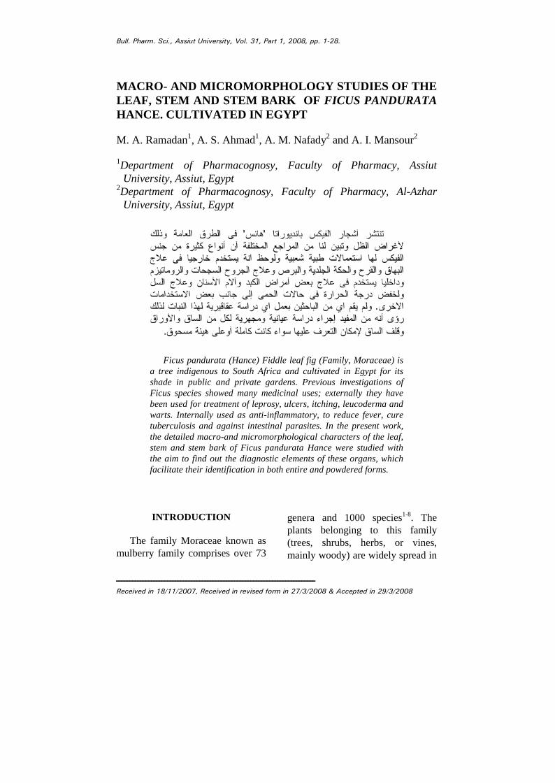
large ever-green tree attaining up to11 meters in height (Fig. 1). Thetree bears simple coriaceous,obovate leaves with cordate orsquared basal ends and slightlywavy and scalloped margins. Theflowers are inconspicuous and notshowy. Fruits are succulent,enlarged hollow, cup-shaped closedr e c e pt a c l e s , e nc l os i ng a c he ne � s -likebodies (syconus); they areyellowish-brown when ripe12&13.
3
Fig. 1: Photo of the plant.
MaterialsThe plant materials used in this
work consisted of the separatedfresh samples of the separate organs(stems, leaves and stem bark) ofFicus pandurata Hance as well assamples preserved in mixture ofalcohol - glycerin - water (1:1:1)and stored in well tightly closedcontainers. The plant materials werecollected in the period from Marchto April 2004 from the front ofFaculty of Pharmacy, AssiutUniversity. The plant was kindlyidentified and authenticated byProf. Dr. Salah EL-Nagar(Professor of botany, Faculty ofScience, Assiut University). Thesamples were gathered duringflowering and fruiting stages.
Macromorphological study ofFicus pandurata (Hance)
Macromorphology1- The stem: (Figs. 1 & 3C)
The main trunk of the plant(Figs. 1 & 3C) is erect, cylindrical,
woody mono-podially branched,reaching about 5-11 meters inheight and 35-75 cm in diameternear the base.
The outer surface is dark-brown,rough, wrinkled, showing lenticels,the terminal and lateral branches arethinner, green in color, faintlylongitudinally striated and haveshort internodes (about 2-5 cm inlength), the older lower branchesare brown with rough surface andcarry scars of fallen leaves. Thestems are odorless with a bitteracrid taste.
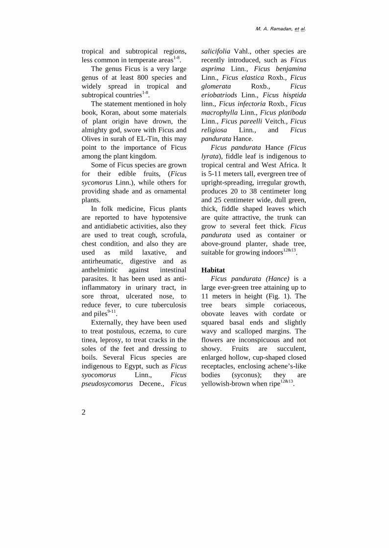
The bark (Figs. 2B, C & 3D) ishardly separated from the wood andusually broken into vertical fissures.The outer surface of the bark is darkgray to almost black withlongitudinal wrinkles, transversefissures and lenticels.
2- Leaves: (Figs. 2A & 3B)The leaves are simple, obovate
with cordate or squared symmetricbases and slightly wavy andscalloped margins, acuminateapices with very tough and leatherytexture. The leaves measure about18 to 30 cm in length and 9 to 20cm in width. Both surfaces aresmooth, shining and glabrous, theupper surface is dark deep glossygreen, while the lower one is lighterwith prominent midrib and lateralveins. The venation is pinnate. Thecommon name fiddle-leaf figperfectly describes their shape.
The petiole is nearly cylindricalin shape green, smooth, glabrous
jK=^K=o~«~¢~¬I£²~ªK
4
(A)
(C) (B)
Fig. 2: A. Photo of the leaves and fruits.B. Photo of the outer surface of the bark.C. Photo of the inner surface of the bark.
5
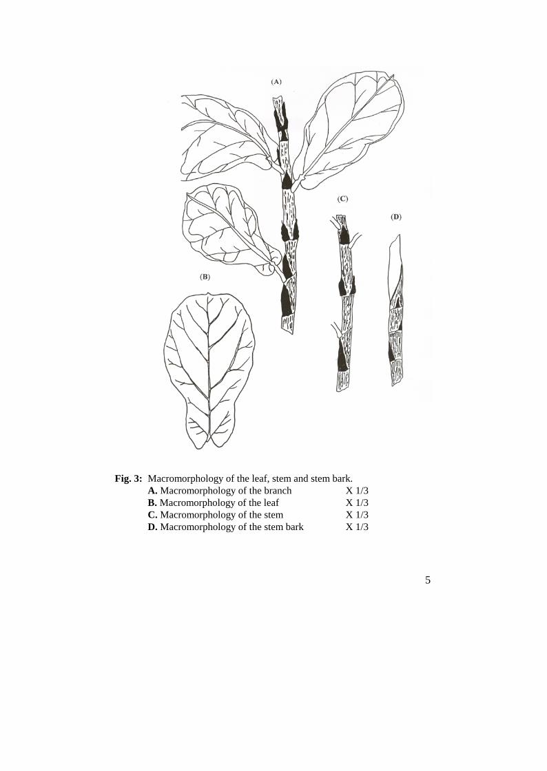
Fig. 3: Macromorphology of the leaf, stem and stem bark.A. Macromorphology of the branch X 1/3B. Macromorphology of the leaf X 1/3C. Macromorphology of the stem X 1/3D. Macromorphology of the stem bark X 1/3
jK=^K=o~«~¢~¬I£²~ªK
6
and measures 1.5-2.5 cm in lengthand about 0.5-0.7 cm in diameter.The leaf has faint odour and bitterastringent taste.
Micromorphological study ofFicus pandurata (Hance)
A- The stem
1- The young stemA transverse section through the
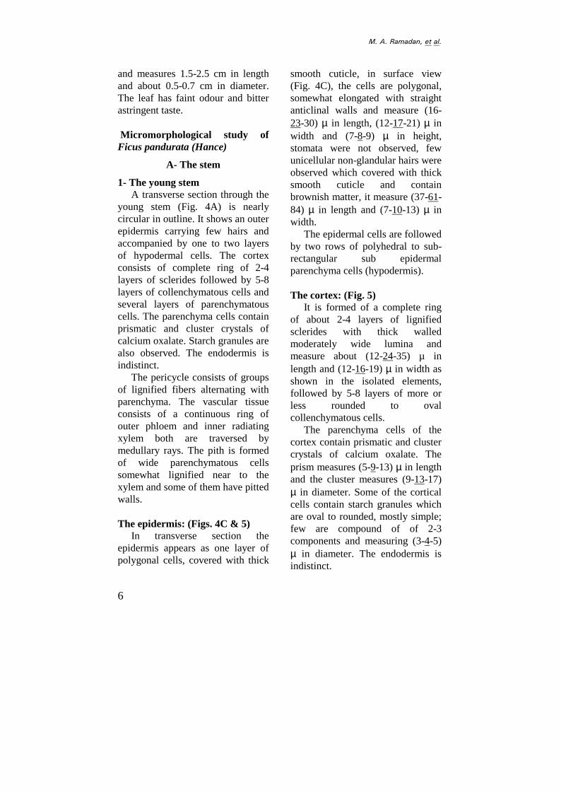
young stem (Fig. 4A) is nearlycircular in outline. It shows an outerepidermis carrying few hairs andaccompanied by one to two layersof hypodermal cells. The cortexconsists of complete ring of 2-4layers of sclerides followed by 5-8layers of collenchymatous cells andseveral layers of parenchymatouscells. The parenchyma cells containprismatic and cluster crystals ofcalcium oxalate. Starch granules arealso observed. The endodermis isindistinct.
The pericycle consists of groupsof lignified fibers alternating withparenchyma. The vascular tissueconsists of a continuous ring ofouter phloem and inner radiatingxylem both are traversed bymedullary rays. The pith is formedof wide parenchymatous cellssomewhat lignified near to thexylem and some of them have pittedwalls.
The epidermis: (Figs. 4C & 5)In transverse section the
epidermis appears as one layer ofpolygonal cells, covered with thick
smooth cuticle, in surface view(Fig. 4C), the cells are polygonal,somewhat elongated with straightanticlinal walls and measure (16-23-30) µ in length, (12-17-21) µ inwidth and (7-8-9) µ in height,stomata were not observed, fewunicellular non-glandular hairs wereobserved which covered with thicksmooth cuticle and containbrownish matter, it measure (37-61-84) µ in length and (7-10-13) µ inwidth.
The epidermal cells are followedby two rows of polyhedral to sub-rectangular sub epidermalparenchyma cells (hypodermis).
The cortex: (Fig. 5)It is formed of a complete ring
of about 2-4 layers of lignifiedsclerides with thick walledmoderately wide lumina andmeasure about (12-24-35) µ inlength and (12-16-19) µ in width asshown in the isolated elements,followed by 5-8 layers of more orless rounded to ovalcollenchymatous cells.
The parenchyma cells of thecortex contain prismatic and clustercrystals of calcium oxalate. Theprism measures (5-9-13) µ in lengthand the cluster measures (9-13-17)µ in diameter. Some of the corticalcells contain starch granules whichare oval to rounded, mostly simple;few are compound of of 2-3components and measuring (3-4-5)µ in diameter. The endodermis isindistinct.
7
Fig. 4: A. Diagrammatic T. S of young stem X 62.5B. Diagrammatic T. S of old stem X 62.5C. Surface preparation of young stem X 500D. Cork cells X 500
ca.ox., calcium oxalate; ck., cork; coll., collenchyma; cor., cortex; epi.,epidermis; int.ph., intraxylary phloem; hypo., hypodermis; n.gl.h., non-glandular hair; pe., pericycle; ph., phloem; pi., pith; st.gr., starch granules;scl., scleride; xyl., xylem.
st.gr.
ca.oxint.ph.
jK=^K=o~«~¢~¬I£²~ªK
8
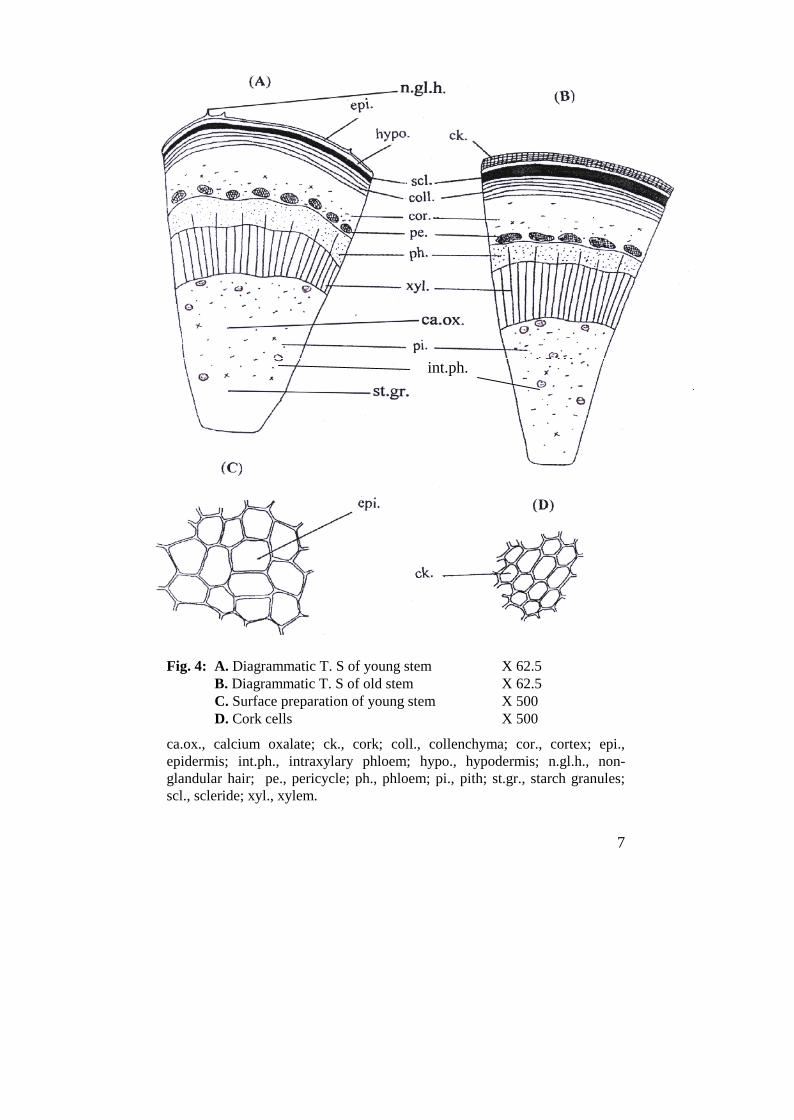
Fig. 5: Detailed T.S. of the young stem X 400
ca.ox., calcium oxalate; cam., cambium; coll., collenchyma; hypo.,hypodermis; epi.,epidermis; int.ph., intraxylary phloem; l.v., laticiferousvessel; m.r., medullary ray; n.gl.h., non glandular hair; par., parenchyma;ph., phloem; p.f., pericyclic fiber; pi., pith; st.gr., starch granules; xyl.v.,xylem vessel.
int.ph
int.ph.
xyl.v.
9
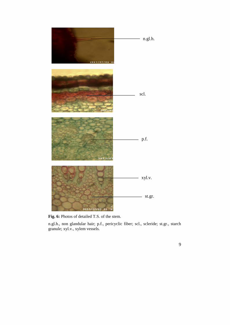
Fig. 6: Photos of detailed T.S. of the stem.
n.gl.h., non glandular hair; p.f., pericyclic fiber; scl., scleride; st.gr., starchgranule; xyl.v., xylem vessels.
scl.
n.gl.h.
p.f.
xyl.v.
st.gr.
jK=^K=o~«~¢~¬I£²~ªK
10
The pericycle: (Figs. 5 & 7)The pericycle is formed of
parenchyma cells alternating withgroups of lignified fibers. Thefibers are elongated, straight withthick lignified walls, moderatelywide lumina and blunt to roundedapices, they measure (212-247-281)µ in length and (12-14.5-17) µ indiameter.
The vascular system: (Fig. 5)The vascular system consists of
continuous ring of phloem,cambium and xylem.
The phloem: (Figs. 5 & 7)The phloem consists of shining
thin-walled cellulosic soft elementsof sieve tubes and phloemparenchyma. Some laticiferoustubes, which are non-branched withcontents that stained yellowish-brown with iodine (T.S), wereobserved in the phloem. Thephloem is separated from xylem bycambial zone.
The cambium: (Fig. 5)The cambium is formed of few
layers of thin walled sub-rectangular, tangentially elongatedand radialy arranged cellulosiccells.
The xylem: (Figs. 5 & 7)The xylem consists of a
comparatively wide zone oflignified thick-walled, radialyarranged elements traversed by unito biseriate medullary rays. The
xylem elements include xylemvessels, wood fibers, woodparenchyma and tracheids. Thevessels are arranged in radial rowsand show sclariform, spiral andpitted thickening and measure (22-39-56) µ in diameter. The tracheidsare elongated with lignified pittedwalls, rounded to blunt ends andmeasure (54-71-88) µ in length and(9-11-13) µ in width. The woodfibers have lignified walls,moderately narrow to wide luminaand acute apices some times it maybe septated. They measure (160-203-245) µ in length and (8-11-14)µ in diameter.
Wood parenchyma consists ofrectangular to sub rectangular cellswith lignified pitted walls. Theymeasure (23-29-35) µ in length and(21-23-25) µ in width. Themedullary rays are uni to biseriateof sub rectangular cells with thicklignified pitted walls in xylemregion, while in the phloem regionthey are thin-walled cellulosic cells.
The pith: (Fig. 5)The pith is formed of a wide
central zone of rounded to ovalparenchymatous cells. The outerlayers of the pith are formed ofparenchymatous cells with thicklignified walls which contain starchgranules. Starch granules aresimple; few are compound of 2-3components. The inner part of thepith is formed of thin-walledparenchyma cells with intercellularspaces. The cells contain prismatic
11
and cluster crystals of calciumoxalate. The prisms measure (4-8.5-13 µ in length and the cluster (8-12.5-17) µ in diameter. Severalgroups of intraxyllary phloem withsoft cellulosic elements scatteredare at the periphery of the pith.
2- The old stem: (Fig. 4B)A transverse section through the
old stem is nearly similar to that ofyoung stem (Fig. 4B). However itshows an outer brownish, narrow tomoderately wide cork formed ofthin suberised and tangentiallyelongated cells. The stone cells andthe vascular system are similar tothose of the young stem but moredeveloped.
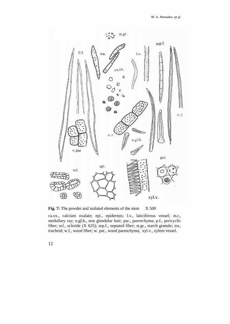
The powder and isolated elementsof stem: (Fig. 7)
The powdered stem of Ficuspandurata (Hance) has dark greencolor with a faint odour and bitteracrid taste. The powder and isolatedelements of stem are characterizedmicroscopically by the following:1- Fragments of polygonal, mainly
axially elongated epidermalcells with straight anticlinalwalls and covered with thickcuticle, stomata are notobserved.
2- Unicellular, non-glandularhairs covered with thick cuticlehaving acute apices, containbrownish matter.
3- Fragments of thin-walledparenchyma cells either from
the cortex or the pith,containing prismatic, clustercrystals of calcium oxalate andstarch granules.
4- Fragments of pericyclic fiberswith straight thick lignifiedwalls moderately wide luminaand blunt to rounded apices.
5- Fragments of lignified xylemvessels with spiral, sclariformand pitted thickenings.
6- Fragments of wood fibers withstraight walls, moderately wideto narrow lumina, acute apicesand sometime septated lignifiedwalls.
7- Fragments showing lignifiedand pitted parenchyma cells ofthe xylem in addition to pittedlignified cells of medullary ray.
8- Fragments of tracheids, withrounded apices and lignifiedpitted walls
9- Fragments of laticiferousvessels which are simple, non-branched and containinggranular contents, stainingyellowish-brown with iodine(T.S).
10- Fragments of lignified sclerideswith thick walls and mostlybranched narrow lumina.
11- Numerous prisms and clustersof calcium oxalate arescattered.
12- Numerous starch granules arescattered, mainly simple; feware compound of 2-3components.
jK=^K=o~«~¢~¬I£²~ªK
12
Fig. 7: The powder and isolated elements of the stem X 500
ca.ox., calcium oxalate; epi., epidermis; l.v., laticiferous vessel; m.r.,medullary ray; n.gl.h., non glandular hair; par., parenchyma; p.f., pericyclicfiber; scl., scleride (X 625); sep.f., septated fiber; st.gr., starch granule; tra.,tracheid; w.f., wood fiber; w. par., wood parenchyma; xyl.v., xylem vessel.
xyl.v.
13
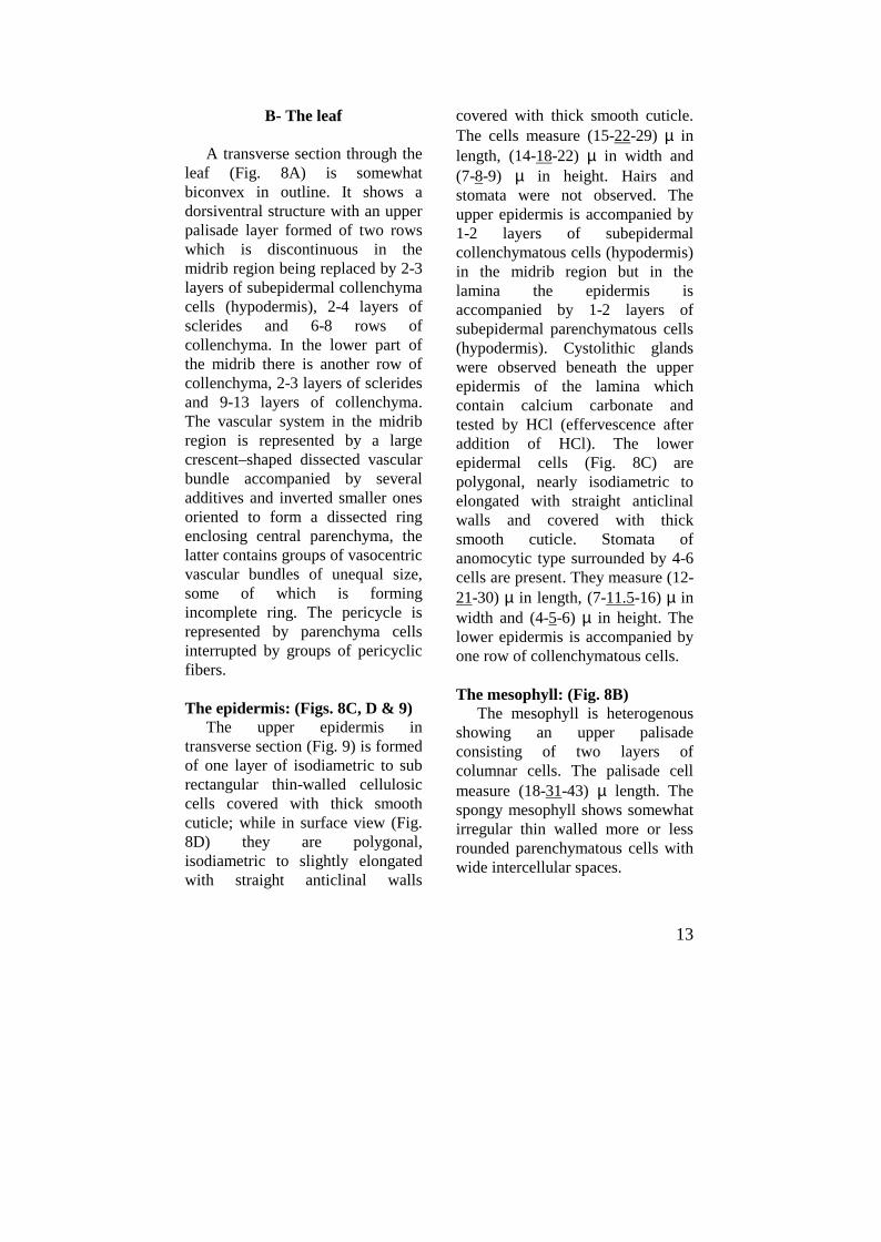
B- The leaf
A transverse section through theleaf (Fig. 8A) is somewhatbiconvex in outline. It shows adorsiventral structure with an upperpalisade layer formed of two rowswhich is discontinuous in themidrib region being replaced by 2-3layers of subepidermal collenchymacells (hypodermis), 2-4 layers ofsclerides and 6-8 rows ofcollenchyma. In the lower part ofthe midrib there is another row ofcollenchyma, 2-3 layers of scleridesand 9-13 layers of collenchyma.The vascular system in the midribregion is represented by a largecrescent � shaped dissected vascularbundle accompanied by severaladditives and inverted smaller onesoriented to form a dissected ringenclosing central parenchyma, thelatter contains groups of vasocentricvascular bundles of unequal size,some of which is formingincomplete ring. The pericycle isrepresented by parenchyma cellsinterrupted by groups of pericyclicfibers.
The epidermis: (Figs. 8C, D & 9)The upper epidermis in
transverse section (Fig. 9) is formedof one layer of isodiametric to subrectangular thin-walled cellulosiccells covered with thick smoothcuticle; while in surface view (Fig.8D) they are polygonal,isodiametric to slightly elongatedwith straight anticlinal walls
covered with thick smooth cuticle.The cells measure (15-22-29) µ inlength, (14-18-22) µ in width and(7-8-9) µ in height. Hairs andstomata were not observed. Theupper epidermis is accompanied by1-2 layers of subepidermalcollenchymatous cells (hypodermis)in the midrib region but in thelamina the epidermis isaccompanied by 1-2 layers ofsubepidermal parenchymatous cells(hypodermis). Cystolithic glandswere observed beneath the upperepidermis of the lamina whichcontain calcium carbonate andtested by HCl (effervescence afteraddition of HCl). The lowerepidermal cells (Fig. 8C) arepolygonal, nearly isodiametric toelongated with straight anticlinalwalls and covered with thicksmooth cuticle. Stomata ofanomocytic type surrounded by 4-6cells are present. They measure (12-21-30) µ in length, (7-11.5-16) µ inwidth and (4-5-6) µ in height. Thelower epidermis is accompanied byone row of collenchymatous cells.
The mesophyll: (Fig. 8B)The mesophyll is heterogenous
showing an upper palisadeconsisting of two layers ofcolumnar cells. The palisade cellmeasure (18-31-43) µ length. Thespongy mesophyll shows somewhatirregular thin walled more or lessrounded parenchymatous cells withwide intercellular spaces.
jK=^K=o~«~¢~¬I£²~ªK
14
Fig. 8: A. Diagrammatic T.S. of the leaf X 62.5B. Detailed T.S. of the lamina X 312.5C. Surface preparation of the leaf (lower epidermis) X 500D. Surface preparation of the leaf (upper epidermis) X 500
ca.ox., calcium oxalate; coll., collenchyma; cor., cortex; g., cystolithic gland;hypo., hypodermis; int.ph., intraxylary phloem; l.epi., lower epidermis; l.v.,laticiferous vessel; mes., mesophyll; pal., palisade; pe., pericycle; ph.,phloem; scl, sclerides; st., stomata; u.epi., upper epidermis; vas. c. v. b.,vasocentric vascular bundles; xyl, xylem.
int.ph
15
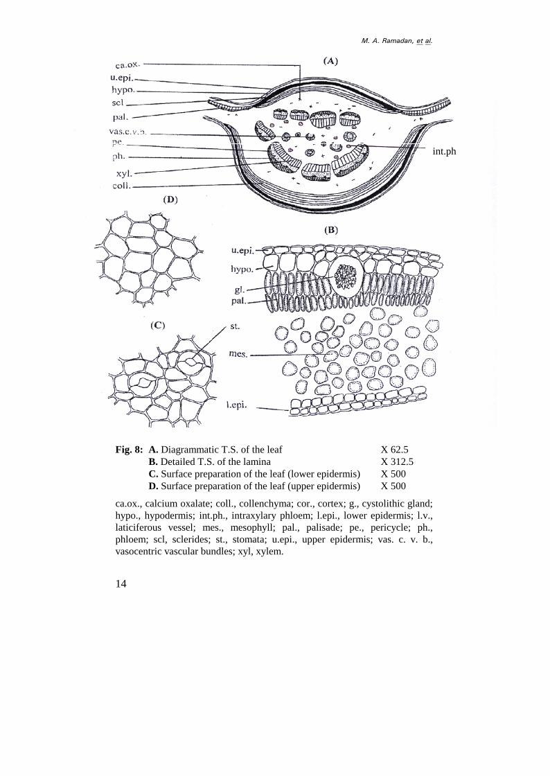
Fig. 9: Detailed T.S. of the leaf X 400
ca.ox., calcium oxalate; coll., collenchyma; hypo., hypodermis; l.epi., lowerepidermis; l.v., laticiferous vessel; m.r., medullary ray; par., parenchyma;ph., phloem; p.f. , pericyclic fiber; scl., scleride; u.epi., upper epidemis; v.,vessel; vas.c.v.b., vasocentric vascular bundle.
coll.
hypo.
scl.
xyl.v.
ca.ox
jK=^K=o~«~¢~¬I£²~ªK
16
(A) (B)
(B)
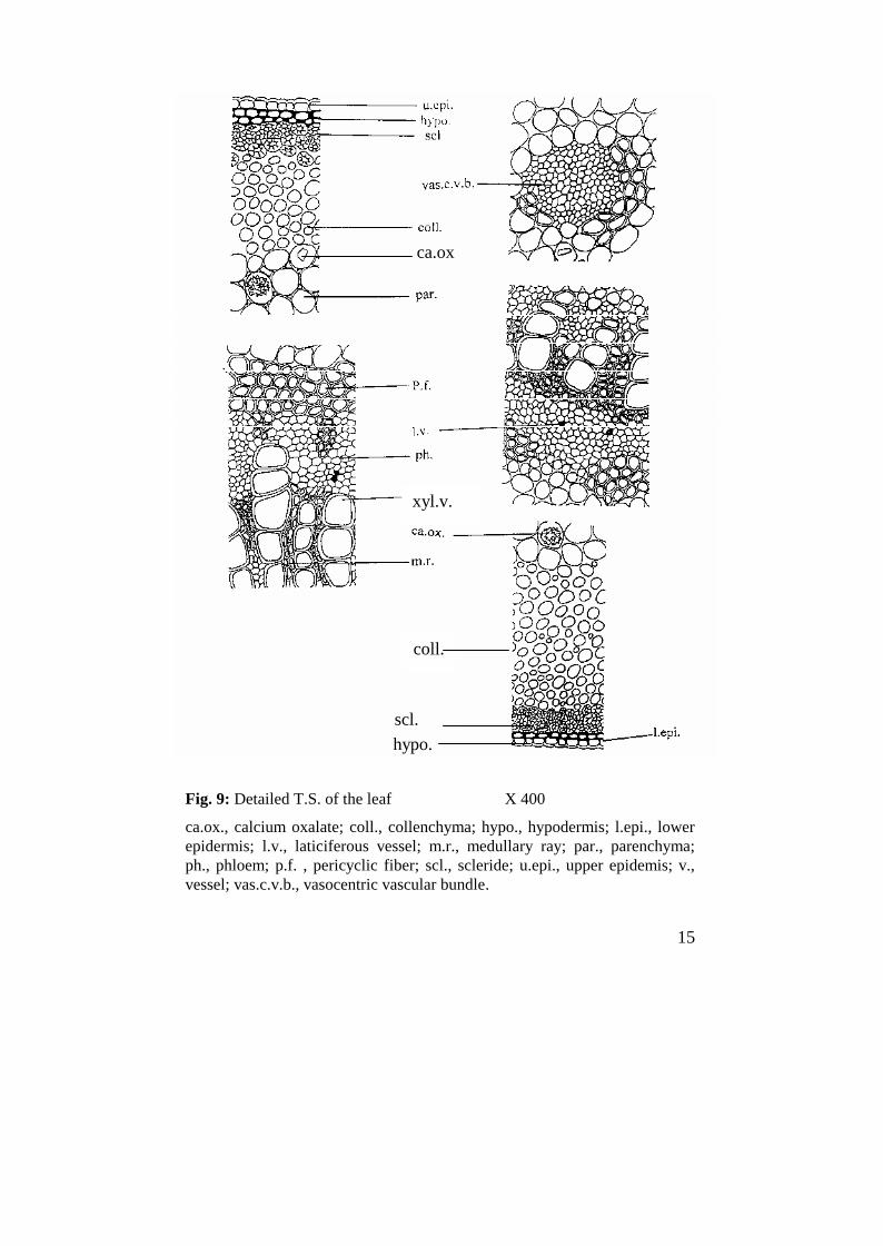
Fig.10: A. Photo of diagrammatic T.S. of the leaf.B. Photos of detailed T.S. of the leaf.
coll., collenchyma; p.f., pericyclic fiber; ph., phloem; scl., sclerides; v.,vessel; vas.c.v.b., vasocentric vascular bundle.
coll.
scl.
p.f.
ph.
v.
vas.c.v.b.
p.f.
17
The cortical tissue: (Fig. 9)The cortical tissue is formed of
about 2-4 layers of sclerides withthick lignified walls and moderatelywide lumina and measure about (9-14-18) µ in length and (9-11-12) µin width, followed by 6-8 rows ofrounded to oval collenchyma cells,in the lower part of the midrib thecollenchyma is formed of 9-13 rowsof rounded to oval collenchymacells. The remaining cortical tissueis represented by several layers ofwide parenchymatous cellssurrounding the vascular bundle. Itcontains cluster and prismaticcrystals of calcium oxalate, theprisms measure (4-9-14) µ in lengthand the clusters measure (7-11-15)µ in diameter. Several groups ofintraxyllary phloem with softcellulosic elements were observed.
The vascular system: (Fig. 9)It is represented by a large
crescent-shaped main dissectedvascular bundle enclosing centralparenchyma. Each systemsurrounded by a pericycle.
The pericycle: (Figs. 9 & 13)The pericycle is represented by
an arc of pericyclic fibers above thevascular system. Each arc is formedof 3-6 rows of pericyclic fibers. Thepericyclic fibers are straight, withlignified moderately thick walls,narrow to moderately wide lumina,acute to rounded apices andmeasure (114-187-260) µ in lengthand (11-14-17) µ in diameter. The
pericyclic fibers penetrate into thephloem region to form an arm intothe phloem.
The xylem: (Figs. 9 & 13)The xylem region consists of
xylem vessels, wood fibers, woodparenchyma, and trachieds andtraversed by 1-2 layers ofrectangular pitted lignifiedmedullary ray cells. The vesselshave spiral, scalariform and pittedthickening measuring (14-18-22) µin diameter, the wood fibers (Fig.13) are few with lignified walls,narrow lumina, rounded to acuteapices, and tracheids (Fig. 13)having blunt or pointed end withlignified pitted thickening. Thewood fibers measure (32-111-190)µ in length and (8-11-14) µ indiameter. The wood parenchymaconsists of rectangular to subrectangular cells with lignifiedpitted walls, wide lumina andmeasure (20-29-38) µ in length and(13-14-15) µ in diameter.
The phloem: (Fig. 9)The phloem is formed of soft
thin walled cellulosic elements.Laticiferous tubes were observedwhich have granular contents,staining yellowish � brown withiodine (T.S). The cambium is notclear.
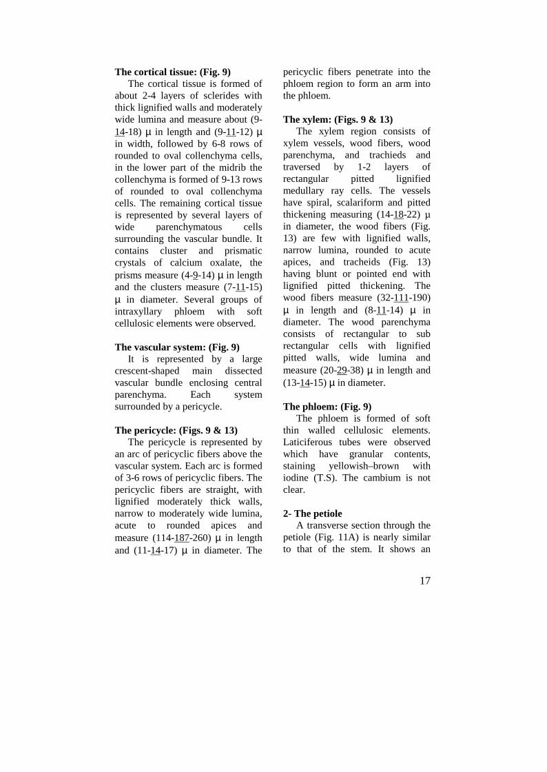
2- The petioleA transverse section through the
petiole (Fig. 11A) is nearly similarto that of the stem. It shows an
jK=^K=o~«~¢~¬I£²~ªK
18
outer epidermis accompanied by 1-2layers of subepidermal parenchymahypodermal cells. The cortexcontains a ring of 2-4 layers oflignified stone cells, 4-6 layers ofcollenchymatous cells and widezone of parenchymatous cells.
The pericycle consists ofparenchyma cells alternating withgroups of pericyclic fibers. Thevascular system is formed of adissected bundles, each consists ofradiating xylem and outer phloemenclosing comparatively wide pith.Groups of intra xylary phloem arescattered at periphery of the pith.
The epidermis: (Fig. 11C)The epidermis in transverse
section (Fig. 11B) consists of onelayer of isodiametric to subrectangular cells. In surface view(Fig. 11C) they are polygonalmostly sub rectangular cells withstraight anticlinal walls and coveredwith smooth cuticle. The cellsmeasure (13-24-35) µ in length,(13-19-25) µ in width and (7-8.5-10) µ in height. Few non-glandularunicellular hairs were observed,stomata are not observed. Theepidermal cells are followed by 1-2layers of hypodermal parenchymacells similar to those of the stem.
The cortex: (Fig. 11B)It consists of a ring of an outer
zone of 2-4 layers of lignified thickwalled and moderately wide luminasclerides followed by 4-6 rows ofrounded collenchymatous cells. The
remaining of the cortex consists ofwide zone of thin walledparenchymatous cells containingprismatic and cluster crystals ofcalcium oxalate.
The pericycle: (Figs. 11B & 13)The pericycle is formed of thin
walled parenchyma cells interruptedby groups of pericyclic fibers. Thepericyclic fibers are similar to thoseof the stem and measure (113-186.5-260) µ in length and (10-13-16) µ in diameter; the fibers havemoderately thick lignified walls andnarrow to moderately wide luminawith blunt to rounded apices.
The vascular system (Figs. 11B &13)
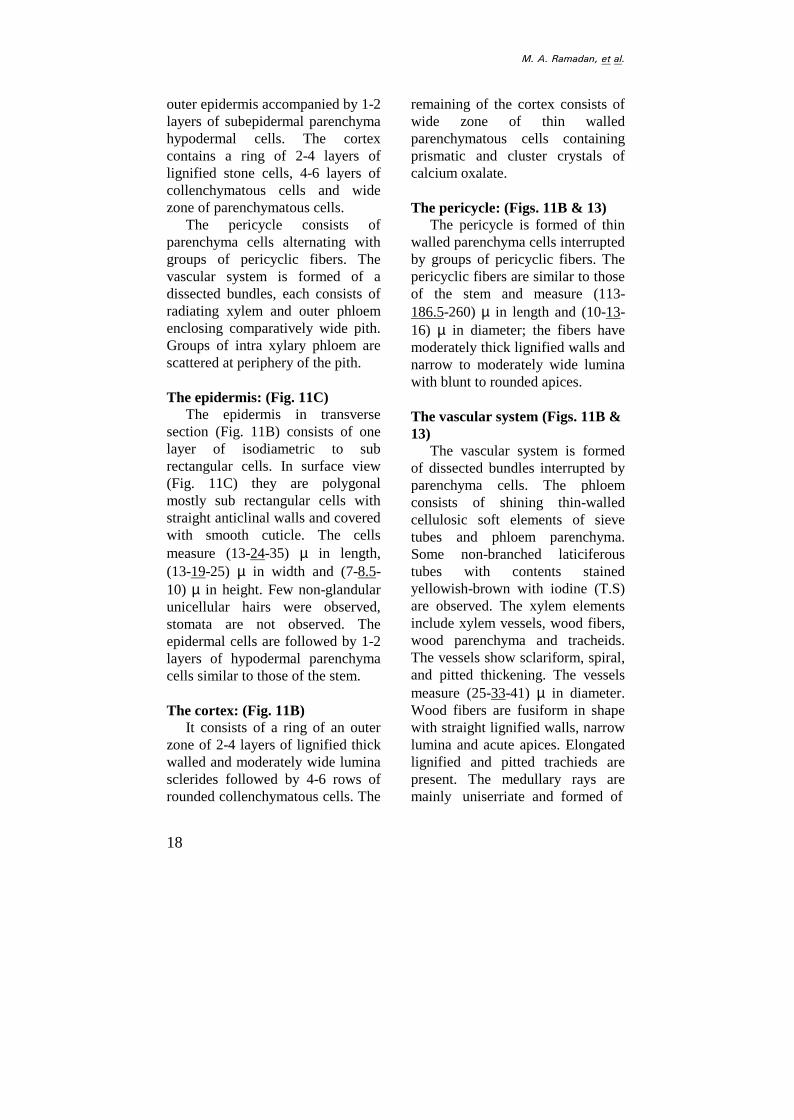
The vascular system is formedof dissected bundles interrupted byparenchyma cells. The phloemconsists of shining thin-walledcellulosic soft elements of sievetubes and phloem parenchyma.Some non-branched laticiferoustubes with contents stainedyellowish-brown with iodine (T.S)are observed. The xylem elementsinclude xylem vessels, wood fibers,wood parenchyma and tracheids.The vessels show sclariform, spiral,and pitted thickening. The vesselsmeasure (25-33-41) µ in diameter.Wood fibers are fusiform in shapewith straight lignified walls, narrowlumina and acute apices. Elongatedlignified and pitted trachieds arepresent. The medullary rays aremainly uniserriate and formed of
19
Fig. 11: A. Diagrammatic T.S. of the petiole X 62.5B. Detailed T.S. of the petiole X 400C. Surface preparation of the petiole X 500
ca.ox., calcium oxalate; coll., collenchyma; cor., cortex; hypo., hypodermis;epi., epidermis; int.ph., intraxylary phloem; l.v., laticiferous vessel; n.gl.h.,nonglandular hair; par., parenchyma; pe., pericycle; p.f., pericyclic fiber; ph.,phloem; pi., pith; scl., scleride; xyl., xylem; xyl.v., xylem vessel.
n.gl.h.
xyl.v.
jK=^K=o~«~¢~¬I£²~ªK
20
Fig. 12: Photos of detailed T. S. of the petiole.
coll., collenchyma; cor., cortex; int. ph., intraxylary phloem; xyl.v., xylemvessels.
xyl. v.
21
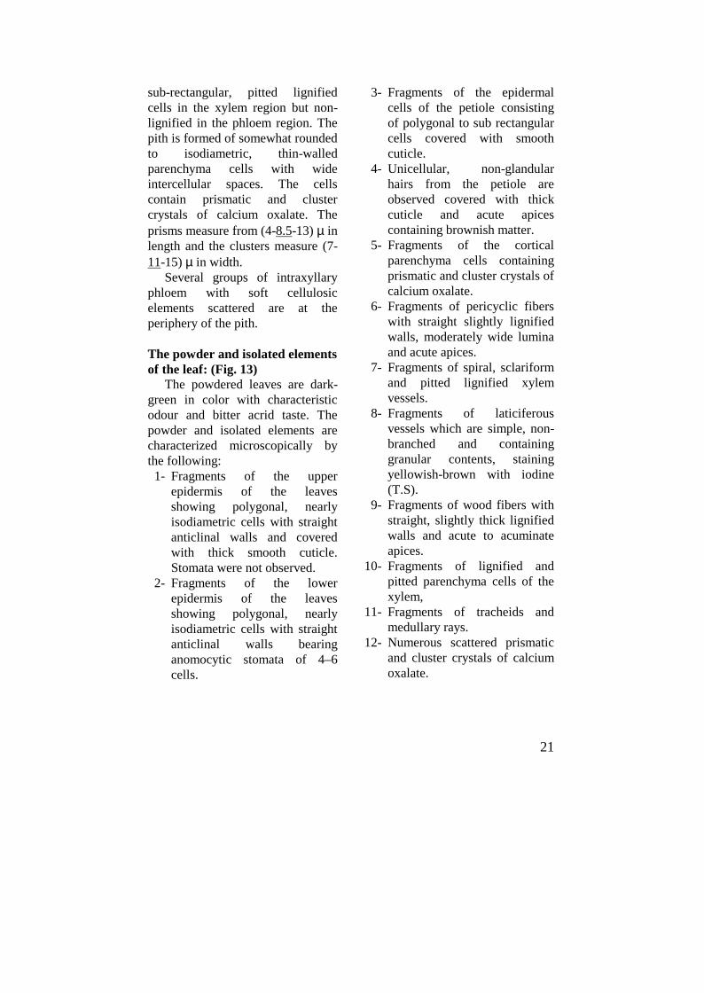
sub-rectangular, pitted lignifiedcells in the xylem region but non-lignified in the phloem region. Thepith is formed of somewhat roundedto isodiametric, thin-walledparenchyma cells with wideintercellular spaces. The cellscontain prismatic and clustercrystals of calcium oxalate. Theprisms measure from (4-8.5-13) µ inlength and the clusters measure (7-11-15) µ in width.
Several groups of intraxyllaryphloem with soft cellulosicelements scattered are at theperiphery of the pith.
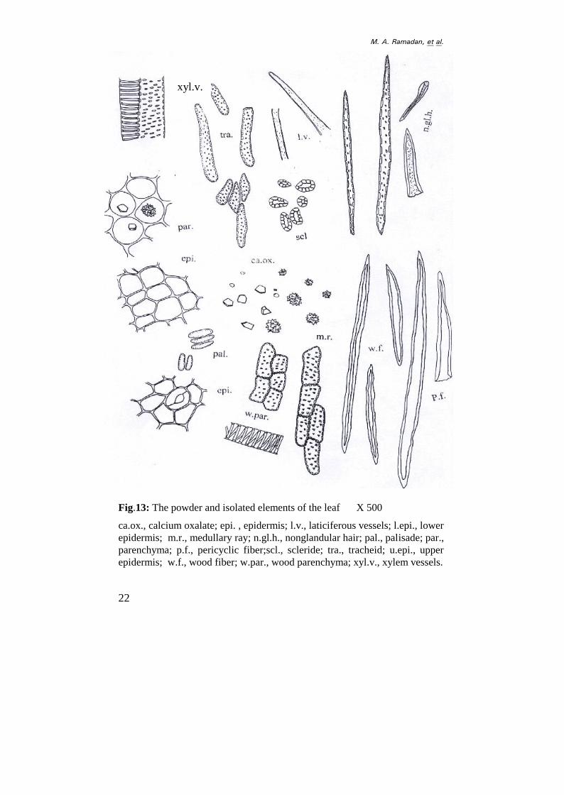
The powder and isolated elementsof the leaf: (Fig. 13)
The powdered leaves are dark-green in color with characteristicodour and bitter acrid taste. Thepowder and isolated elements arecharacterized microscopically bythe following:1- Fragments of the upper
epidermis of the leavesshowing polygonal, nearlyisodiametric cells with straightanticlinal walls and coveredwith thick smooth cuticle.Stomata were not observed.
2- Fragments of the lowerepidermis of the leavesshowing polygonal, nearlyisodiametric cells with straightanticlinal walls bearinganomocytic stomata of 4� 6cells.
3- Fragments of the epidermalcells of the petiole consistingof polygonal to sub rectangularcells covered with smoothcuticle.
4- Unicellular, non-glandularhairs from the petiole areobserved covered with thickcuticle and acute apicescontaining brownish matter.
5- Fragments of the corticalparenchyma cells containingprismatic and cluster crystals ofcalcium oxalate.
6- Fragments of pericyclic fiberswith straight slightly lignifiedwalls, moderately wide luminaand acute apices.
7- Fragments of spiral, sclariformand pitted lignified xylemvessels.
8- Fragments of laticiferousvessels which are simple, non-branched and containinggranular contents, stainingyellowish-brown with iodine(T.S).
9- Fragments of wood fibers withstraight, slightly thick lignifiedwalls and acute to acuminateapices.
10- Fragments of lignified andpitted parenchyma cells of thexylem,
11- Fragments of tracheids andmedullary rays.
12- Numerous scattered prismaticand cluster crystals of calciumoxalate.
jK=^K=o~«~¢~¬I£²~ªK
22
Fig.13: The powder and isolated elements of the leaf X 500
ca.ox., calcium oxalate; epi. , epidermis; l.v., laticiferous vessels; l.epi., lowerepidermis; m.r., medullary ray; n.gl.h., nonglandular hair; pal., palisade; par.,parenchyma; p.f., pericyclic fiber;scl., scleride; tra., tracheid; u.epi., upperepidermis; w.f., wood fiber; w.par., wood parenchyma; xyl.v., xylem vessels.
xyl.v.
23
C- The Stem Bark
Micromorphology of the stembark
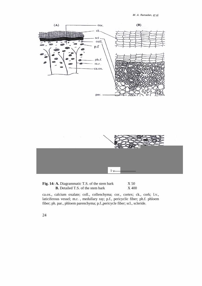
A transverse section through thestem bark (Fig. 14A) shows threedistinct regions, the cork, cortexand phloem regions.
The cork consists of severalrows of radially arrangedtangentially elongated thin-walledcells. The cortex is comparativelywide regions formed of outer 4-7rows of sclerides followed by thin-walled irregular shaped parenchymacells containing prisms of calciumoxalate. The cortical tissueinterrupted by groups ofcollenchyma cells. The inner mostlayer of cortex shows groups oflignified pericyclic fibers. Thephloem appears as wide zoneconsists of phloem parenchyma andphloem fibers, they are traversed byfunnel shaped medullary rays.
The cork: (Figs. 14B & 15)The cork layer is formed of
several rows (8-13 layers) ofradially arranged tangentiallyelongated, tabular to subrectangularcells with thin walls. The outerrows of the cork contain yellowishbrown pigments, while the restbeing free of the of the pigments.The cork cells in surface view (Fig.15), appear polygonal toisodiametric, slightly elongatedwith straight anticlinal walls,measure (12-18-24) µ in length, (9-
12-15) µ in width and (6-10-14) µin height.
The cortex: (Figs. 14B & 16)The cortex is formed of outer 4-
7 rows of thick-walled lignifiedstone cells, with narrow tomoderately wide lumen, the stonecells measuring (24-34-44) µ inlength and (20-25-30) µ in width.,followed by thin walled, rounded tooval parenchyma cells. Few stonecells are scattered in the cortexwhich characterized by thick,lignified walls. The parenchymacells contain prisms of calciumoxalate measure (4-8-12) µ inlength and (4-7-10) µ in width.Groups of rounded to ovalcollenchyma cells are present inouter cortex.
The innermost layer of thecortex contain few groups ofpericyclic fibers(Figs. 14B & 15) which have thicklignified walls and they measure upto (235-262.5-290) µ in length and(11-14-17) µ in width.
The phloem: (Figs. 14B & 16)The phloem zone is
comparatively wide formed ofphloem parenchyma and phloemfibers, traversed by funnel shapedmedullary rays. The phloemparenchyma consists of rectangularto sub rectangular, somewhat ovalthin-walled, non-lignified cellscontaining prisms of calciumoxalate.
jK=^K=o~«~¢~¬I£²~ªK
24
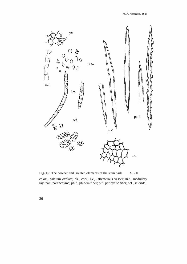
Fig. 14: A. Diagrammatic T.S. of the stem bark X 50B. Detailed T.S. of the stem bark X 400
ca.ox., calcium oxalate; coll., collenchyma; cor., cortex; ck., cork; l.v.,laticiferous vessel; m.r. , medullary ray; p.f., pericyclic fiber; ph.f. phloemfiber; ph. par., phloem parenchyma; p.f.,pericycle fiber; scl., scleride.
l.v
25
(B) (A)
(C) (D)
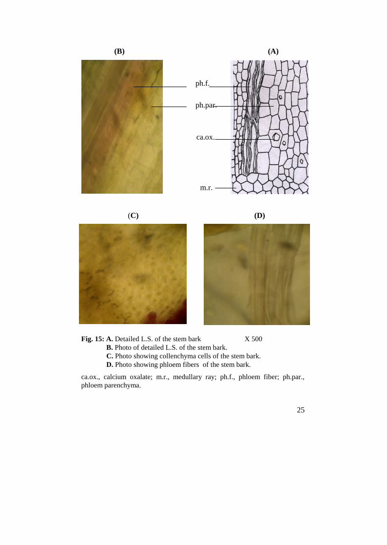
Fig. 15: A. Detailed L.S. of the stem bark X 500B. Photo of detailed L.S. of the stem bark.C. Photo showing collenchyma cells of the stem bark.D. Photo showing phloem fibers of the stem bark.
ca.ox., calcium oxalate; m.r., medullary ray; ph.f., phloem fiber; ph.par.,phloem parenchyma.
ph.f.
ph.par.
ca.ox.
m.r.
Related Documents


![1.0 Introduction - UMstudentsrepo.um.edu.my/4770/5/4Thesis_3.pdfB. rotunda [formerly known as Kaempferia pandurata Roxb. or Boesenbergia pandurata (Roxb. Schltr)] belongs to the Zingiberaceae](https://static.cupdf.com/doc/110x72/60d3fd4ed783b061f106c107/10-introduction-b-rotunda-formerly-known-as-kaempferia-pandurata-roxb-or-boesenbergia.jpg)

