Module 2: Chest Pain Page 1 of 25 MACEP Risk Management Course Module 2: Chest Pain Alison Sullivan, M.D. Thomas G. Horejsi, M.D. Sutin Chen, M.D. James A. Feldman, M.D., M.P.H, FACEP Course Objectives State the major life threatening diagnoses that are the most common triggers for liability risk in non-traumatic chest pain as chief complaint. Indicate the key components of documentation in the evaluation, treatment and disposition of a patient with chest pain as chief complaint. Identify specific risk factors that are associated with acute coronary syndrome (ACS), thoracic aortic dissection (TAD) and pulmonary embolism (PE). Describe specific systems of care that can potentially minimize chest pain liability risk. State other diagnoses that are associated with chest pain liability risk Introduction Evaluation of non-traumatic chest pain remains one of the highest-risk chief complaints in emergency medicine. Based upon most recent data from a review of malpractice claims from 2005-2009, cardiovascular diagnoses accounted for 26% (58/224) of closed claims and 29% (72/247) of open claims as of 10/2010. Prior reviews 1,2 of malpractice claims and prospective research studies have consistently indentified specific chest pain diagnoses that have been the most problematic. The most prevalent diagnoses that are associated with adverse outcomes are 1) acute coronary syndrome (ACS, missed myocardial infarction (MI)) 2) thoracic aortic dissection (TAD); and 3) pulmonary embolism (PE). It is also important to note that myocarditis, bacterial endocarditis and other cardiovascular diagnoses are a source of risk management concerns. These are briefly covered in a category for “other” liability concerns. The average payout for chest pain cases closed with payment is approximately $600,000 (average for missed ACS: $560,000; missed dissection: $700,000). This module presents a summary of the most prevalent triggers of liability and the most important aspects of the clinical evaluation, diagnostic testing, treatment and disposition that are associated with adverse outcomes. A consistent theme remains the failure of clinicians to

Welcome message from author
This document is posted to help you gain knowledge. Please leave a comment to let me know what you think about it! Share it to your friends and learn new things together.
Transcript
Module 2: Chest Pain Page 1 of 25
MACEP Risk Management Course
Module 2: Chest Pain
Alison Sullivan, M.D.
Thomas G. Horejsi, M.D.
Sutin Chen, M.D.
James A. Feldman, M.D., M.P.H, FACEP
Course Objectives
State the major life threatening diagnoses that are the most common triggers for liability risk
in non-traumatic chest pain as chief complaint.
Indicate the key components of documentation in the evaluation, treatment and disposition
of a patient with chest pain as chief complaint.
Identify specific risk factors that are associated with acute coronary syndrome (ACS),
thoracic aortic dissection (TAD) and pulmonary embolism (PE).
Describe specific systems of care that can potentially minimize chest pain liability risk.
State other diagnoses that are associated with chest pain liability risk
Introduction
Evaluation of non-traumatic chest pain remains one of the highest-risk chief complaints in
emergency medicine. Based upon most recent data from a review of malpractice claims from
2005-2009, cardiovascular diagnoses accounted for 26% (58/224) of closed claims and 29%
(72/247) of open claims as of 10/2010. Prior reviews1,2
of malpractice claims and prospective
research studies have consistently indentified specific chest pain diagnoses that have been the
most problematic. The most prevalent diagnoses that are associated with adverse outcomes are 1)
acute coronary syndrome (ACS, missed myocardial infarction (MI)) 2) thoracic aortic
dissection (TAD); and 3) pulmonary embolism (PE). It is also important to note that
myocarditis, bacterial endocarditis and other cardiovascular diagnoses are a source of risk
management concerns. These are briefly covered in a category for “other” liability concerns. The
average payout for chest pain cases closed with payment is approximately $600,000 (average for
missed ACS: $560,000; missed dissection: $700,000).
This module presents a summary of the most prevalent triggers of liability and the most
important aspects of the clinical evaluation, diagnostic testing, treatment and disposition that are
associated with adverse outcomes. A consistent theme remains the failure of clinicians to

Module 2: Chest Pain Page 2 of 25
diagnose a high risk condition, obtain or interpret routine diagnostic tests or to appreciate the
limitations of diagnostic tests such as cardiac biomarkers or plain chest x-ray as screening tests.
Each section will briefly review the target condition, key elements of the history and
examination that are associated with making or failing to make the diagnosis, the role of
diagnostic testing, and specific aspects of disposition and system design from a risk management
perspective.
An extensive review of the various diagnostic modalities potentially available or in depth review
of treatment considerations is beyond the scope of the module. The availability of diagnostic
testing and treatment options is highly dependent on the practice setting. Each section will
provide references for further reading about technologies to aide in the diagnosis of ACS or
recommendation for the treatment of ACS, PE or TAD based upon consensus guidelines or
evidence-based reviews.
Acute Coronary Syndrome
Coronary heart disease remains the most common cause of mortality in the United States.
According to the American Heart Association there are an estimated 935,000 cases of
myocardial infarction per year in the US and an additional 86,000 cases of unstable angina.
About 20% of those are fatal.3 Our goal in the emergency department (ED) is to identify and
appropriately treat patients presenting with acute coronary syndrome (ACS). ACS encompasses
both myocardial infarction (including ST-segment elevation MI and non ST-segment elevation
MI) and unstable angina. While patients with unstable angina have better outcomes than patients
with non-ST segment MI, these syndromes are clinically indistinguishable at presentation.4
Determining which patients have ACS can be difficult, resulting in a miss rate of 2-4 %.5,6
Chest
pain is the second most common chief complaint in the emergency department,7 responsible for
an estimated 5.5 million visits to emergency departments in 2008.8 A minority of patients
presenting with chest pain are diagnosed with ACS.8 A significant percentage of patients with
ACS do not have chest pain as their chief complaint.9,10
Patients with atypical symptoms (i.e.
those without chest pain) are at higher risk of misdiagnosis, are less likely to receive standard of
care medical treatment, and have a higher mortality.9,10
Regardless of the presenting complaint,
missed ACS results in a much higher mortality and morbidity rate,1,2,6,11-13
and is a common
source of liability for the emergency physician.1,2,6
Claims relating to myocardial infarction (MI) accounted for 7.6% of the emergency medicine
closed claims in 2005-2009, and resulted in 23% of the indemnity paid. Failure to diagnose is
particularly high risk, with 61.5% of claims for failure to diagnose MI being closed with
indemnity paid, compared to 32% of overall claims against emergency physicians closed with
indemnity paid. This is consistent with prior malpractice claims studies showing that failure to
diagnose makes up the majority of claims for all diagnoses.1,13,14
Errors in diagnosis most
commonly result from: 1) failure to order a diagnostic test; 2) inadequate history and
physical; 3) incorrect interpretation of test results; and 4) failure to order an appropriate
consult.14
Some of the lawsuits resulting in liability included discharging patients with positive
troponin and misinterpretation of ECGs.
Patient characteristics which put them at higher risk of being misdiagnosed include female sex,
age < 55, nonwhite race, a normal ECG and shortness of breath as the chief complaint.5,6
As
providers, it is imperative that we consider ACS in patients with atypical symptoms, interpret

Module 2: Chest Pain Page 3 of 25
ECGs carefully, take time to look at laboratory and diagnostic testing, and are not falsely
reassured by a negative stress test or even a negative cardiac catheterization in a high-risk
patient.
History
Patients with ACS classically present with chest discomfort, but may also present with anginal
equivalents such as shortness of breath, nausea, arm/neck pain, unexplained fatigue, syncope, or
abdominal pain.4,6,9,10
History-taking in patients who are being evaluated for possible ACS
should focus on the characterization of the pain or discomfort, timing of symptoms, relationship
to exertion, and associated symptoms. Failure to appropriately characterize the patients’
pain is common in missed diagnoses.13
The classic chest pain of ACS is usually described as a
pressure or burning type pain located in the left chest or substernal area which radiates to one or
both arms and is worsened with exertion or worse than the patient’s typical angina.15
Associated
symptoms include nausea and/or vomiting, diaphoresis, and shortness of breath.15
While the
presence of each of these symptoms contributes to a higher relative risk of ACS, the absence of
these symptoms does not rule the patient out for having ACS.4,6,15
Additionally, symptoms which
are not characteristic of ACS, such as pleuritic or reproducible chest pain, do not rule out the
possibility of ACS.4,15
The patient’s history, along with the physician’s opinion of the patient’s
level of likelihood of ACS (high, intermediate or low), should be carefully documented in the
chart.
ACS is commonly misdiagnosed as gastro-esophageal reflux disease.1 Remember that patients
with ACS may present with epigastric pain rather than chest pain. Caution must be taken in these
patients, especially in those whose symptoms otherwise seem characteristic for angina (i.e.
relationship to exertion, relief with rest, associated shortness of breath or diaphoresis).
Improvement with GI cocktail should not be used as a diagnostic maneuver.15
In conjunction with characterizing the nature of the patient’s symptoms, the patient should be
asked about risk factors for coronary artery disease (CAD).4 Important risk factors for CAD
include personal history of CAD, family history of CAD, age > 70, male sex, diabetes mellitus,
smoking history, hypertension, and hyperlipidemia.4 Although determining classic Framingham
risk factors has not been demonstrated to improve triage decisions because the absence of these
risk factors does not reduce the risk of ACS, documentation can support a reasonable medical
evaluation. Patients should also be asked about less traditional risk factors such as cocaine or
stimulant use, or a history of hypercoagulabilty.4,16
A recent negative workup for ACS is important to note in the history, but care must be taken
when interpreting these results. Note the type of workup done, whether a stress test was
performed and whether the level of stress reached made the test diagnostic or not. Most
importantly, consider the patient’s pre-test probability and consider their presenting symptoms. If
the patient is at high risk by clinical history and therefore has a high pre-test probability, a
negative stress test (or other diagnostic test for ACS) is more likely a false negative test result
than truly negative, and thus does not rule out ACS.17,18
Module 2: Chest Pain Page 4 of 25
Physical Examination
The primary goals of the physical exam in the patient with suspected ACS is to assess for causes
of ACS and to document comorbid conditions which may complicate their course. The physical
exam should include assessment of vital signs, a careful cardiac examination including
identification of evidence of CHF such as S3 gallop, elevated JVP, pulmonary edema. While a
careful physical exam should be performed for all patients with possible ACS to assess for the
above findings, it should be recognized that the majority of patients with ACS will have a normal
physical exam. In addition, pain which is reproducible on palpation or with movement does not
rule out ACS, as approximately 7% of patients with ACS have reproducible chest pain.4
ECG
ECGs are a low cost, rapid screening test. As such, they should be performed early, with a low
threshold, and should be carefully interpreted and documented. High risk complaints, such as
chest pain and unexplained dyspnea, should prompt an ECG within 10 minutes of presentation.4
While the ECG is at the center of the evaluation of the patient with suspected ACS, it should be
recognized that a single ECG has a low sensitivity, and cannot be used to rule out ACS.4 Rather,
the ECG is used to decide the next step in management. Patients with STEMI should be
considered candidates for PCI, transfer to a center capable of PCI, or thrombolysis, while those
with new ST depressions or T wave inversions should be considered at high risk for unstable
angina or non ST-elevation MI.
Serial ECGs should be performed in order to increase sensitivity. Suggestions range from 15-60
minute intervals or at recurrence of symptoms.4,19
Dynamic ST depressions of 0.5 mm or more
which occur with symptoms, are particularly concerning for active ischemia.4
ECGs must be interpreted carefully, with specific attention to features which are high risk for
ischemia including ST segment elevations, ST segment depressions, and T wave inversions. Best
practice includes documenting absence of these findings as pertinent negatives. Misinterpretation
of ischemic ECGs in the ED is fairly common, estimated to be approximately 12%,20
and results
in substandard treatment and higher inpatient mortality.20
While ST elevations were less
frequently missed than T wave inversions and ST depressions, they were missed in 8% of ST
elevation MIs.20
Patients with atypical symptoms (i.e. those presenting without chest pain) were
at a higher risk of ECG misinterpretation,20
suggesting that emergency physicians less carefully
interpret ECGs when they have a lower suspicion for ACS.
Laboratory Testing
Laboratory testing in patients with ACS is used to identify patients who have had myocardial
necrosis. Recent studies have shown increased sensitivity of sensitive troponin assays
(“ultrasensitive troponin”) within 3 hours of symptom onset. Not all hospitals have access to the
more sensitive assays, and each still misses a significant percentage of patients within that time
frame. The troponins that most hospitals have access to are only 41-73% sensitive at 4-6 hours
after symptom onset.19
It is important to recognize that negative cardiac biomarkers cannot
be used to exclude unstable angina. This is a clinical diagnosis that cannot be excluded based
on ECG or laboratory evidence.
Module 2: Chest Pain Page 5 of 25
ACEP recommends using one of the following three approaches to exclude non-ST-elevation MI:19
1. A single negative CK-MB, Troponin I or Troponin T measured 8-12 hours after symptom
onset.
2. A negative myoglobin in conjunction with a negative CK-MB or negative troponin when
measured at baseline and 90 minutes in patients presenting less than 8 hours after
symptom onset.
3. A negative 2 hour delta CK-MB in conjunction with a negative 2 hour delta troponin in
patients presenting less than 8 hours after symptom onset.
Risk Stratification
There are several risk stratification tools that integrate the above information in order to
determine patients’ mortality risk. The ACC/AHA recommends their use in the evaluation of
patients with suspected ACS.4 However, their use is controversial,
21,22 is not considered standard
of care, and does not allow for safe discharge from the emergency department. Several studies
have shown that those with a TIMI risk score of 0 still have a significant risk of ACS.4,21-24
Clinicians can consider using risk score stratification as part of the clinical evaluation and to help
select level of care and therapy for a patient.
Consultation
Interventional cardiology consultation should be undertaken immediately in patients with ECG
findings concerning for ST segment elevation MI. This should be part of the standard protocol
for STEMI. A formal protocol for reperfusion or transfer protocol for patients eligible for
reperfusion is an important component of medical care and risk management.
Consider cardiology consultation for patients with other high risk features on ECG, especially
those with deep T wave inversions or biphasic T waves in the anterior leads concerning for
Wellen’s syndrome. These patients are at risk for high grade LAD stenosis, and should be
considered candidates for inpatient cardiac catheterization. Other high risk patients include those
with a history of CABG or coronary stenting, prior myocardial infarction or known CAD. One
should consider a cardiology consultation if unsure about the ECG interpretation and ensure that
there is a system for prompt ECG consultation.
Disposition
Patients with high risk of ACS based on history, ECG and biomarkers should be admitted to the
hospital for further management. Intermediate and low risk patients with suspected ACS can be
admitted or observed in an ED chest pain unit with serial measurements of cardiac biomarkers
and serial ECGs performed.
Further Testing
The ACC recommends stress testing for patients with low or intermediate risk of ACS after two
sets of negative cardiac biomarkers, with coronary CT angiography (CCTA) as a reasonable
alternative in those who cannot undergo provocative testing.4 As with any diagnostic test, it is
important to consider the pre-test probability of a patient prior to further testing. Given their low
Module 2: Chest Pain Page 6 of 25
pre-test probability for CAD, patients who are at very low risk of ACS are likely to have a high
false positive rate of stress test and coronary CTA.25,26
This may result in unnecessary
downstream testing, perhaps exposing the patient and the health care system to unnecessary costs
and risk. Part of the clinical evaluation should include discussing the clinical decision making
and risks and benefits of diagnostic testing with the patient and family.
Management
The management of ACS is well known to most emergency physicians. As the choice of
pharmacologic agents and the type of reperfusion therapy available vary by hospital, each
hospital should have standardized treatment protocols for patients with UA/NSTEMI and those
with STEMI. Each practicing emergency physician should be aware of the recommendations of
the ACC/AHA and their institutional treatment algorithm.4,27
Discharge Instructions
Patients who are thought not to have ACS or another serious cause of chest pain based upon the
clinical evaluation can be discharged from the ED. Because the evaluation of chest pain remains
imperfect, indicating the limitations of the evaluation and need for follow up is important for
patient care and risk management. Several standardized chest pain instructions are available.
Most importantly, patients should be advised to follow up within a short time frame with their
physician or cardiologist and to return to the nearest emergency department should their
symptoms recur.
Thoracic Aortic Dissection
Background
Claims relating to thoracic aortic dissection represented only 2/72 closed claims for
cardiovascular conditions in Massachusetts from 2005-2009; both resulted in death. Claims
included not obtaining a family history of dissection and not ordering a diagnostic study (chest x-
ray). Cost per claim was $750,000 and $450,000. Prior ProMutual reviews indicated 4/41 (10%)
of the chest pain cases closed with payment were from TAD. Present open claims indicate an
increasing liability risk related to TAD. 11/72 (15%) of open cardiac claims are for TAD. A 2008
study by Elefteriades analyzed patterns in 33 nontraumatic thoracic aorta related legal cases in
which 23 patients (69.7%) had aortic dissection. All but one of the dissection cases resulted in
death with the lone survivor suffering a severe stroke with neurologic deficits. The most
common malpractice claim was failure to diagnose.28
Because missed TAD can lead to
unexpected and preventable death and can affect young patients and pregnant women, TAD must
be considered as a possible diagnosis in all patients who present with chest pain as a chief
complaint.
According to the CDC the broad category “disease of the Aorta” accounts for 43,000- 47,000
deaths annually in the United States.29
Whereas the incidence of myocardial infarction in the
United States has been estimated at 4,400 per 1 million people per year, the incidence of aortic

Module 2: Chest Pain Page 7 of 25
dissection has been estimated from 5-30 per 1 million people per year.30
(These data represent
aortic dissection ranging from 100 to 1,000 times less prevalent than myocardial infarction.)
Autopsy studies conducted prior to the modern era estimated 48 hour mortality from a proximal
dissection to be 40-50%. Modern estimates of over-all in hospital 30 day mortality were reported
as 27.4%. Highest mortality occurred in patients with type A dissection not receiving surgery
(58.0%), while patients receiving surgery had an improved mortality (26%). Patients with type B
dissection treated medically have a lower mortality (10.7%).30
It has been estimated that in the setting of acute chest pain, back pain, or both, acute coronary
syndromes outweigh the frequency of acute aortic dissection by 80:1, with diagnostic separation
failing in 0.01%.31
Additional studies have shown that the diagnosis of aortic dissection is
frequently missed on initial evaluation and not made until postmortem examination in 27-
55% of patients.28
It has been reported that 38% of dissections are missed on initial
evaluation. There are no validated decision rules for the clinical diagnosis of aortic dissection.32
Modern aggressive treatment of ACS presents another challenge to the clinician. The
consequence of rapid ACS treatment with antiplatelet, antithrombin, and fibrinolytic agents
showed a higher rate of the primary end point of mortality or major bleeding (composite
frequency of 54% vs. 23%).33
Due to the severity of outcome, its time dependent nature, and
similarity of presentation to acute coronary syndrome, aortic dissection continues to be a
quagmire of medical malpractice litigation.
History
The classic presentation of aortic dissection is often described as chest pain with “a ripping or
tearing sensation” radiating to the back. It is most often considered in middle aged men with a
history of hypertension. In fact, emergency physicians suspected aortic dissection most
commonly when both chest and back pain were the presenting complaints, considering the
diagnosis in 86% of cases.34
Unfortunately there can be significant variation in presentation. The
same study by Sullivan et al. found that emergency physicians suspected the diagnosis only 45%
of the time when chest pain was the sole presenting complaint and only 8% of the time when
patients presented with epigastric, abdominal, or flank pain.34
It has been estimated that up to
20% of affected patients present without typical signs and symptoms of chest pain or
neurological dysfunction.35
The data surrounding presentation of aortic dissection are quite variable. A meta-analysis of 21
studies (1848 patients) by Klompas et al. found that most patients with thoracic aortic dissection
present with a history of severe pain (pooled sensitivity 90%) with sudden onset (sensitivity
84%). They also found that the absence of sudden pain onset lowers the likelihood of dissection
by a negative likelihood ratio of 0.3.36
Interestingly, a history of hypertension only showed
sensitivity of 64%, “ripping or tearing” quality showed only 39%, migrating pain only 31%,
syncope only 9%, and Marfan’s a mere 5% sensitivity. The specific location of pain (anterior,
posterior, back, or abdomen) showed limited sensitivity with ranges from 57% to 23%
respectively.36
Additionally, neurological symptoms can be part of the presentation. They have
been reported as dramatic, varied, and may dominate the clinical picture. In a large case series
Module 2: Chest Pain Page 8 of 25
and review article, Gaul et al. report that the frequency of neurological symptoms varied from
17- 40%.37
The most common risk factor has traditionally been reported as hypertension with sensitivity as
high as 72%.38
A large meta-analysis found that history of hypertension had a pooled sensitivity
of 64% with an increased likelihood ratio of 1.6 (95% CI, 1.2-2.0).36
Traditionally aortic
dissection has been described as a disease affecting men in the later decades of life. It has been
reported that 95% of patients present at age >40 with a mean age of 65 years.39
A recent IRAD
study of 464 aortic dissection patients noted that two thirds were male and that the mean age for
all patients was 63 years.30
Clinicians are, however, frequently presented with patients who are younger than 40 complaining
of chest pain. A 2004 IRAD study of 1078 AD patients found that 6.4% were younger than age
40. They also found that traditional risk factors such as hypertension were less common in this
group while Marfan’s syndrome, bicuspid aortic valve, and prior aortic valve surgery were
significantly higher.40
The International Registry of Aortic Dissection (IRAD) lists the following
risk factors: Long standing arterial hypertension: smoking, dyslipidemia, cocaine/crack;
Connective tissue disorders: Hereditary fibrillinopathies (Marfan’s and Ehlers Danlos);
Hereditary vascular disease (Bicuspid aortic valve, Coarctation); Vascular inflammation (Giant
cell arteritis, Takayasu arteritis, Behcet’s disease, Syphillis, Ormond’s disease).41
The 2010
ACCS/AHA Guidelines for the Diagnosis and Management of Patients With Thoracic Aortic
Disease: Executive Summary lists similar categories and adds pheochromocytoma, weight
lifting/valsalva maneuver, Turner’s syndrome, Loeys-Deitz syndrome, pregnancy, polycystic
kidney disease, chronic corticosteroid use or immunosuppresion agent administration, and
infections involving the aortic wall.
Despite the varied presentation and risk factors, it is important to take a history that minimally
inquires about quality, radiation, and intensity at onset. One investigation of 83 patients with
subsequent confirmed aortic dissection noted that only 42% of conscious patients were asked all
three questions. The study went on to show that if all 3 questions were asked diagnostic accuracy
improved to 91%. If one or more of the three questions were omitted, suspicion fell to 49%.42
(Of course the retrospective nature of this study is limiting. It is possible that physicians were
more likely to ask questions if they had an initially high degree of clinical suspicion from other
data: chest xray, physical exam, etc. Nonetheless, history matters.)
Physical Exam
Although a history of hypertension is considered a significant risk factor for the development of
aortic dissection it is less commonly seen on initial presentation. An IRAD review noted that
elevated blood pressure was present in 36% of type A dissection and 70% of type B 56.30
Pooled
sensitivity from a large meta-analysis shows acute hypertension to be present in 64% of cases.36
Impaired blood flow can present as a pulse deficit in any of the major associated vessels and is
often a much more subtle finding. It was found to be present in the carotid, brachial, or femoral
arteries infrequently (31%). However, when present in the setting of chest pain or back pain the
positive likelihood ratio increased to 5.7 (95% CI 1.4-23).36
Module 2: Chest Pain Page 9 of 25
A diastolic heart murmur has often been discussed as one possible manifestation of a type A
dissection however it has little value in predicting dissection. One third of patients with aortic
dissection will have a diastolic murmur (pooled sensitivity 28%). However the positive
likelihood ratio for this finding was found to be only 1.4 and the negative likelihood ratio was
found to be 0.9. Whereas a new murmur might be helpful in making the diagnosis, it is unlikely
that an emergency physician would be able to make such an observation given limited familiarity
with most patients.36
Focal neurological deficits, although rare (17% of cases), can be very helpful in diagnosing
dissection. The high specificity of focal neurological deficits yielded a positive likelihood ratio
ranging from (6.6-33).31,43
Unfortunately the absence of focal neurological deficits does not rule
out dissection.36
Neurological deficits can be isolated. In one study, up to 10% of dissection
patients presented with neurological symptoms, and no chest pain.37
Evaluation
ECG on its own is of limited value in diagnosing aortic dissection. One study of 464 patients
found the ECG to be normal 31% of the time. Nonspecific ST and T wave changes were
identified in 42% of patients. Ischemic changes were seen in 15% of patients and those with type
A (ascending) dissection showed evidence of infarction 5% of the time.30
Meta-analysis showed
new Q waves or ST segment elevation in 7% of admission ECGs. Yet, in the same analysis
normal ECGs were documented an average of 22% of the time.36
Chest x-ray also has limited utility as the sole diagnostic study, however, when used in
combination with key history and physical exam findings it can be quite helpful.31
In a study of
216 patients chest radiography had a sensitivity of 64% and a specificity of 86%. Sensitivity was
lower for pathology involving the proximal aorta (47%) and better for disease involving distal
aortic segments (77%).31
A review of 464 patients found mediastinal widening present in 63
percent with type A dissections and 56 percent of type B dissections. The same study
unfortunately reported no abnormality whatsoever in 11% of type A and 16% of type B
patients.30
Because chest radiography can yield variable or indeterminate results, sensitivity and
specificity are somewhat limited. Almost all patients with suspected aortic disease receive more
definitive testing.
CT is considered the most frequently used definitive diagnostic test. Its sensitivity exceeds 95%
with specificities ranging from 87-100%.38
Ultrasound: Transthoracic echocardiography (TTE) has limited value in imaging the entire aorta
but can help with identifying involvement of the proximal aorta and with assessing some of the
complications of dissection (aortic insufficiency, pericardial tamponade, and regional LV
dysfunction). An appropriately trained EP can identify aortic widening or an intimal flap using
TTE-findings with high specificity for TAD in a patient with chest pain. Transesophageal
echocardiography (TEE) requires experienced staff and is often not obtainable on an emergent
basis. However, it can be performed at the bedside in an unstable patient and its availability is
institution dependent. It has been reported to reach a sensitivity of 99% with a specificity of
89%. MRI is less commonly used in the acute evaluation, but may be used for preoperative
staging.38
Module 2: Chest Pain Page 10 of 25
D-dimer has been discussed as a potential biomarker to screen for TAD and much hope has been
placed in finding a future biomarker that will help “rule out” this disease. Initial investigations
were promising; however, a recent meta-analysis of studies using D-dimer as the “sole screening
tool” for acute dissection found that there is a subset of patients who develop a thrombosed false
lumen, which may be less likely to stimulate the clotting cascade than those with luminal
extension, resulting in negative D-dimer results. Additionally none of the studies defined a
specific patient population eligible for D-dimer screening and many studies had wide confidence
intervals because of low patient numbers. Despite high sensitivity, D-dimer could not be
recommended as the sole screening tool for acute aortic dissection.44
In the future, biomarkers
along with other key clinical variables and ancillary studies may play a role in the formation of
algorithms and decision rules to better identify aortic dissection.
Summary of History, Physical Exam, and Evaluation: An analysis of 250 patients with acute
chest and/or back pain (128 with a dissection) found that 96 percent of acute aortic dissections
could be identified based upon some combination of the following three clinical features.31
Abrupt onset of thoracic or abdominal pain with a sharp, tearing and/or ripping
quality Variation in pulse (absence of a proximal extremity or carotid pulse) and/or blood
pressure (>20 mmHg difference between the right and left arm) Mediastinal and/or aortic widening on chest radiograph
(Probability of dissection was low with absence of all 3 variables: 7%.)
“Aortic pain” alone (sudden, tearing, ripping) has a positive likelihood ratio of 2.6. The presence
of both aortic pain and pulse or blood pressure differentials increased the likelihood ratio to
10.5%. The addition of abnormal widening of the mediastinum on chest radiograph essentially
seals the diagnosis with a likelihood ratio of 66.0. Unfortunately only 27% of all dissection
patients presented with this triad.36
Although many patients suspected to have dissection turn out
not to have acute aortic disease, anywhere 50-75% are diagnosed with alternative serious
disease. This should encourage a thorough work-up in any patient suspected of having aortic
pathology.31,43
Treatment and Management
Treatment and management of aortic dissection is somewhat controversial and somewhat
dependent upon the resources available to the clinician. The 2010
ACCF/AHA/AATS/ACR/ASA/SCA/SCAI/SIR/STS/SVM Guidelines for Diagnosis and
Management of Patients With Thoracic Aortic Disease: Executive Summary provides detailed
recommendations on how to manage aortic disease.45
All patients with suspected dissection
should undergo further diagnostic evaluation immediately under close monitoring. Patients
should be admitted to an intensive care unit or transferred (with appropriate staff and monitoring)
to a facility with cardiothoracic surgery capabilities. Pain can be controlled with opiates, and a
systolic blood pressure of 110 mm Hg should be the goal of therapy. Beta-blockers are generally
the antihypertensive of choice and usually sufficient, however, additional vasodilating agents
may be required. If beta blockers are contraindicated, intravenous verapamil or diltiazem may
also be used. Surgical services must be consulted early in all cases of suspected aortic
Module 2: Chest Pain Page 11 of 25
dissection and transfer to an appropriate facility with appropriate interfacility care at sites
without thoracic surgery.
Documentation
From a risk management perspective, documentation of the history of pain, clinical evaluation
and clinical reasoning are important. Some risk managers have suggested that symmetrical blood
pressures and symmetrical pulses should be documented in all chest pain patients because of
the liability risk with missed dissection. Consider documenting these findings in patients with
presentation suggestive of TAD.
Pulmonary Embolism
Background
Over 500,000 cases of pulmonary embolism (PE) events occur each year in the United States. Of
these cases, over 50% are fatal.46
Failure to diagnose pulmonary embolism remains a liability
concern for emergency physicians (8% of open cardiovascular claims as of 2010). The
diagnosis of pulmonary embolism must be considered in all patients with acute cardiovascular
complaints including chest pain, dyspnea, palpitations and syncope. Infectious diagnoses
(pneumonia, pleurisy) or respiratory diagnoses (COPD or asthma) are often sources of diagnostic
error. The clinical presentation of PE can vary from mild dyspnea on exertion or unilateral leg
swelling to severe respiratory distress and cardiac arrest.47
Although historically mortality rates
for untreated or missed PE were 26-30%, a recent study shows that with new technologies to
diagnose PE, mortality and recurrence rates are now less than 5%.48
However, because
anticoagulation therapy has been shown to decrease mortality rates,49
failure to diagnose PE can
be associated with what is considered a preventable adverse outcome.48
The availability of
validated risk stratification tools and rapid access to diagnostic imaging has advanced the ability
of emergency physicians to rapidly diagnose PE. However, failure to consider the diagnosis
and errors in diagnosis remain liability concerns.
Risk Factors
In patients involved in the PIOPED II study, fewer than 20% of patients with PE present with
the classic triad of hemoptysis, dyspnea, and chest pain. However, 94% had at least 1 identified
risk factor.47
Reviewing a patient’s risk factors is a key component of assessing the likelihood of
PE. Risk factors can be either hereditary or acquired.
Acquired risk factors for PE are more common than inherited thrombophilias. Acute medical
illness or reduced mobility lead to the majority of episodes of PE: examples include hospital or
nursing home confinement, recent surgery, trauma, malignancy, paresis (stroke), etc.47,50,51
Extended travel times have also been associated with PE.52
For female patients, oral
contraceptive use or hormone replacement therapy are additional risk factors for PE.53
Module 2: Chest Pain Page 12 of 25
Hypercoagulable states, whether inherited or acquired, are associated with increased PE events.
Inherited thrombophilias may be suspected in patients who present with PE at a young age (see
Table 1). Acquired hypercoagulable states that elevate risk of PE include polycythemia vera and
antiphospholipid syndrome (e.g. lupus) and active inflammatory bowel disease.54
In addition, a
personal or family history of prior venous thromboembolism elevates risk for PE.50
Patients with
a prior history of VTE are 8 times more likely to have recurrent VTE compared to patients
without a history of DVT or PE.55
Therefore noting patient level factors or a family history of
DVT/PE is an important part of risk stratification.
Clinical Evaluation
A minority of patients present with the classic symptoms associated with PE: dyspnea, pleuritic
chest pain and hemoptysis. Clinical findings associated with PE can include the following:
history-dyspnea, chest pain, unilateral calf pain or swelling, physical exam-tachypnea,
hypoxemia, tachycardia, and exam findings suggestive of deep venous thrombosis (DVT).50
Of
note orthopnea, which is not historically associated with PE, was a moderately frequent
complaint (38%) in patients with PE in the PIOPED II study.47
Hemoptysis does not have any
predictive value for diagnosing PE.47,50
No single physical exam finding is sensitive (rules out) or specific (rules in) for PE. Half of the
patients in the PIOPED II study with PE did not have tachypnea; three quarters of PE patients
were not tachycardiac. The presence of fever does not exclude PE nor make an infectious process
such as pneumonia more likely in a patient with respiratory symptoms. Only 30% of PE patients
have abnormal lung exam findings.47
Lower leg swelling or tenderness may suggest DVT.
However these findings were neither sensitive nor specific for PE.47,54
From a risk management
perspective, one can not use a normal heart rate, respiratory rate or oxygen saturation to
exclude the diagnosis of PE.
Atypical Presentations
“Atypical presentations” of PE include syncope or cardiac arrest.56
Syncope is present in 8-13%
of all patients with PE and is more frequent in patients with massive PEs (20%) compared to
those with smaller PEs (4%). Pulseless electrical activity is the primary presenting rhythm in
patients presenting with cardiac arrest secondary to PE.
PE can be challenging to diagnose in certain populations. The frequency of PE events increases
with age; thus the likelihood that a patient with PE will have multiple comorbidities is raised as
well. Patients with cardiopulmonary diseases such as congestive heart failure (CHF), coronary
artery disease (CAD), or chronic obstructive pulmonary disease (COPD) may have similar
complaints of chest pain or shortness of breath that obscure the process of diagnosing PE. In one
study, 19.9% of patients hospitalized for acute COPD exacerbations were later found to have
objective evidence for PE requiring therapeutic anticoagulation.57
Clinicians should have a low
threshold to evaluate patients for COPD if they are being admitted for acute COPD
exacerbations, especially if there is a history of prior VTE or active malignancy. The risk of
missed PE is higher in these populations as overall mortality for PE is higher in COPD (12%)
and CHF (17%) patients compared to those without either diagnosis (10%).58
Module 2: Chest Pain Page 13 of 25
In addition, clinicians may overlook PE in atypical population groups, such as children or
pregnant women, leading to cases of missed PEs. Pulmonary embolism is rare in children56
and
is due to an inherited or acquired hypercoagulable state. The most common acquired risk factor
in children is central venous access devices. Other factors include infection, renal disease,
autoimmune diseases, vasculitis, severe inflammatory bowel disease, malignancy, surgery and
trauma.
The incidence of PE is 5 times higher in pregnant women compared to non-pregnant women.
The postpartum period is associated with an even higher risk of PE. However, PE is especially
difficult to diagnose as dyspnea is a common complaint during pregnancy. Physiologic dyspnea
of pregnancy is usually mild, with no symptoms present at rest. Symptoms tend to remain stable
as the fetus matures. Syncope, hemoptysis, chest pain, shortness of breath at rest, or rapid onset
of symptoms should raise red flags for further workup and not be attributed to physiologic
dyspnea.
Pretest Probability
If PE is on the differential diagnosis, further evaluation is based on risk factors, history and
physical exam. An objective clinical assessment tool should be used to determine the pretest
probability prior to instigating further workup54,59
(See Appendix, Tables 2-4 for prediction
tools). Comparison between the Canadian (Wells) scoring system with either the revised or the
original Geneva criteria has shown similar accuracy in predicting the likelihood of PE in
emergency department patients.60,61
However, the Geneva score has not been as well tested as the
revised Wells criteria and revisions of the original Geneva criteria have not been tested for
clinical usefulness in outcome studies.
Preliminary Workup
Based on clinical assessment (formed from history, physical exam, risk factor profile, etc.),
standard workup for chest pain or shortness of breath should be considered. ECG findings are
nonspecific, but may be consistent with acute right heart strain such as right axis deviation, right
bundle branch block or the “classic” PE ECG finding of the S1Q3T3 pattern. Nonspecific t-wave
changes often trigger an admission for possible ACS in a patient with “atypical chest” pain
leading to a failure to consider and diagnose PE. The CXR is often non-diagnostic for PE, and
findings (Hampton’s hump, Westermark’s sign) are rare but could identify another cause of chest
pain and dyspnea (e.g. pneumothorax etc). ABG results have not been shown to be of diagnostic
value in clinical studies.62
In conjunction with pre-test probability assessment, an enzyme-linked
immunosorbent assay (ELISA)-based D-dimer tests (sensitivity 97-100% and negative-predictive
value 99.6%) enables use of a negative result to rule out PE in patients with low pretest
probability.63
The primary drawback to D-dimer assays is that they can be positive in any
patients with inflammatory states, such as pregnancy, infection, cancer, trauma, etc., and
therefore cannot be used uniformly for all patients.64
Kline et al. developed the pulmonary
embolism rule-out criteria (PERC), an eight variable decision rule to validate not ordering further
diagnostic testing in patients determined to be of low clinical suspicion for PE.65
A prospective
multicenter study later showed that a combination of low clinical suspicion for PE with a
negative PERC result reduced the probability of VTE to less than 2%. There is controversy about
whether the PERC rule can be used in clinical practice or requires further testing.
Module 2: Chest Pain Page 14 of 25
Definitive Diagnosis and Management
Computed thoracic (CT) pulmonary angiography has become the test of choice for definitive
diagnosis of PE.66
Treatment of segmental or subsegmental PEs seen on CT, is controversial and
the investigators of the PIOPED II study recommend reassessing the certainty of the CT
diagnosis as the cause of the patient’s symptoms.59,66
Patients who have contraindications to CT
pulmonary angiography should undergo other diagnostic imaging studies (usually a V/Q scan).
In patients with a high pretest probability for PE but for whom the initial imaging scan is
negative, consideration for further diagnostic testing is warranted. Alternatively, imaging studies
may be ordered to detect DVT. In unstable patients, bedside transthoracic echocardiography
(TTE) may be suggestive of either PE or DVT and can be performed by an appropriately trained
EP. In one study assessing massive PE in hemodynamically unstable patients, if any two of three
variables were positive (high clinical probability of PE, bedside TTE, or lower extremity
ultrasonography), the sensitivity was 97% and the negative predictive value was 98%.67
In patients who have moderate-to-high pretest probability for PE, consider initiating treatment
with antithrombin therapy prior to obtaining diagnostic testing results.59
Patients with
contraindications to anticoagulation or reoccurrence of VTEs despite therapeutic anticoagulation
may require a venous filter.59
Thrombolytics should be used in patients with shock secondary to
a massive PE; however, in patients who are hemodynamically stable, thrombolytics have not
been shown to reduce mortality or recurrent PE.68
In patients with massive PE, surgical
thrombectomy and percutaneous catheter fragmentation can also be considered.54,69
An
institutional pathway for DVT/PE (including those with massive PEs) can encourage a consistent
management strategy based upon an institution’s resources and a patient’s clinical condition.
Other Chest Pain Diagnoses Although the diagnoses responsible for medical legal risk for emergency physicians are ACS,
TAD and PE, it is important to note that there are other diagnoses that are associated with
liability exposure. Among these are myocarditis (4 of the open claims pending as of 2010),
bacterial endocarditis and heart failure.
Myocarditis can be a difficult diagnosis, present with nonspecific symptoms especially
during a time when viral illnesses associated with myocarditis, such as influenza, may be
prevalent. Exercise caution when a patient, especially child or young adult with either a
suspected viral syndrome or recent viral syndrome, reports cardiovascular complaints such as
dyspnea, has low oxygen saturation or either unexplained or persistent tachycardia.
Cardiomegaly on CxR or depressed ejection function on transthoracic ECHO (can be done
by an appropriately trained emergency physician) can support the diagnosis of myocarditis.
Because of the risk of death from ventricular dysrhythmia or heart failure and the young age
of patients whose adverse outcomes have often triggered malpractice suites, emergency
physicians should be aware of myocarditis as a liability risk.
Bacterial endocarditis (BE): Exercise caution when a patient with chest pain has fever,
murmur or is at increased risk for BE (injection drug use, abnormal or prosthetic valve). If
Module 2: Chest Pain Page 15 of 25
blood cultures are sent in a patient at low risk for BE, ensure a follow up system to contact
the patient if positive.
Heart failure (CHF): Heart failure or cardiogenic pulmonary edema is identified in 7 of the
open malpractice claims.
SUMMARY POINTS
History: Pain: timing, location, quality, radiation, mitigating and exacerbating factors, severity
Associated symptoms
Previous similar symptoms
Previous cardiac history
Previous cardiac studies: ECG, stress test, catheterization
Risk factors
Family history of ACS, TAD, PE
Physical examination:
Look for complications of ischemia (rales, edema, S3 gallop, rales, or a new systolic
murmur).
May identify an alternate diagnosis.
Beware of chest wall tenderness.
Consider documenting symmetric BP, presence of pulses.
Evaluation: ECG: have a low threshold for ordering it.
A normal electrocardiogram does not exclude cardiac ischemia.
Compare with previous ECGs.
Use as a supplement to your clinical judgment.
Cardiac biomarkers: most useful when performed serially.
A single enzyme determination cannot rule out cardiac ischemia. Interpret in clinical
context.
If a cardiac troponin is obtained, discharge with an elevated level is a high risk decision.
Obtain and interpret other tests (chest x-ray, d-dimer) in clinical context.
Treatment: Have a protocol in place for reperfusion therapy.
Clinical pathways (low risk chest pain, other chest pain diagnoses) can standardize care
and reduce liability exposure
Risk Management: Documentation is crucial
Document history, risk factors, past cardiac history, physical examination.
Module 2: Chest Pain Page 16 of 25
Write your ECG findings on the chart and document comparison with previous ECGs
if available.
Document discussions with consultants and the patient's physician.
Discharge Instructions:
Ensure the instructions include specific symptoms for which they should return for
recheck.
ACS Summary: 1. Carefully document a complete history and physical exam as well as the clinical
reasoning behind testing and management decisions
2. Consider the diagnosis of ACS in patients presenting with atypical complaints such as
shortness of breath, nausea or weakness
3. A “GI” presentation for ACS is high risk. Consider diagnoses such as “GERD” or
“esophageal spasm” to be high risk diagnoses in patients with chest pain or epigastric
pain and possible ACS.
4. Have a low threshold to obtain an ECG, and spend time carefully interpreting and
comparing the ECG to an old ECG
5. Do not rely on a single set of cardiac enzymes in patients presenting within 12 hours
of symptom onset or if the onset is unable to be reliably determined
6. Consider unstable angina and further testing in patients who have ruled out for MI.
7. Give clear discharge instructions to patients regarding follow up and when to return
to the emergency department
TAD Summary:
1. TAD has a low incidence but high rate of malpractice claims 2. TAD is easily mistaken for ACS and both diagnoses can occur simultaneously. 3. Document that these essential chest pain history questions were asked:
- quality - radiation - intensity at onset
4. Pursue the diagnosis of TAD in a patient with these essential findings: - severe pain with sudden onset - pulse deficit in any of the major associated vessels - chest x-ray showing mediastinal widening
5. Do not let a negative chest x-ray rule out dissection is a patient with high suspicion. 6. Manage pain and blood pressure appropriately 7. Consult surgery early or stabilize and transfer to an appropriate facility. 8. Be aware of common TAD presentations
i. -Chest pain with neurologic symptoms b. Chest pain with limb ischemia c. Chest pain with syncope
9. Consider TAD in young patients, pregnant women and patients with risk factors for TAD
PE Summary:

Module 2: Chest Pain Page 17 of 25
1. Consider PE in differential of acute cardiovascular complaints especially chest pain,
dyspnea and syncope, any patient with tachypnea.
2. PE should be considered in patients with chest pain and t-wave abnormalities.
Diagnosing ACS and missing PE is a noted error.
3. Consider important aspects of the history (risk factors, family history) and exam to
risk stratify. Documentation of this evaluation can indicate that medical decision-
making was reasonable.
4. Consider the use of a validated decision rule to risk stratify for diagnostic testing
5. Admission and disposition may be dependent on patient stability and availability of
testing.
6. In patients who are discharged home, provide appropriate instructions to return for
worsening of symptoms
Module 2: Chest Pain Page 18 of 25
Appendix
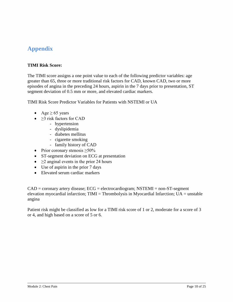
TIMI Risk Score:
The TIMI score assigns a one point value to each of the following predictor variables: age
greater than 65, three or more traditional risk factors for CAD, known CAD, two or more
episodes of angina in the preceding 24 hours, aspirin in the 7 days prior to presentation, ST
segment deviation of 0.5 mm or more, and elevated cardiac markers.
TIMI Risk Score Predictor Variables for Patients with NSTEMI or UA
Age ≥ 65 years ≥3 risk factors for CAD
- hypertension - dyslipidemia - diabetes mellitus - cigarette smoking - family history of CAD
Prior coronary stenosis ≥50% ST-segment deviation on ECG at presentation ≥2 anginal events in the prior 24 hours Use of aspirin in the prior 7 days Elevated serum cardiac markers
CAD = coronary artery disease; ECG = electrocardiogram; NSTEMI = non-ST-segment
elevation myocardial infarction; TIMI = Thrombolysis in Myocardial Infarction; UA = unstable
angina
Patient risk might be classified as low for a TIMI risk score of 1 or 2, moderate for a score of 3
or 4, and high based on a score of 5 or 6.

Module 2: Chest Pain Page 19 of 25
Table 1. Risk Factors for Pulmonary Embolism51,54
Inherited Acquired
Antithrombin III deficiency
Protein C deficiency
Protein S deficiency
Factor V Leiden
Activated protein C resistance (most common)
without factor V Leiden
Prothrombin gene mutation
Dysfibrinogenemia
Plaminogen deficiency
*Prior patient or family history of venous
thromboembolism
Immobilization
- Travel
- Paralysis/Spinal Cord Injury
- Bedridden state
- Immobilizer or cast
Surgery
Trauma
Acute medical illness
Malignancy (active)
Hypercoagulability state
- Polycythemia vera
- Antiphospholipid antibody syndrome
Central venous access devices
Pregnancy and the puerperium
Oral contraceptives/hormone replacement therapy
Advanced age
Obesity
Inflammatory bowel disease
Table 2. Revised Canadian (Wells) Prediction Score70
Variable Score
DVT symptoms and signs 3.0
PE as likely as or more likely than alternative
diagnosis
3.0
Heart rate >100 beats/min 1.5
Immobilization or surgery in previous 4 weeks 1.5
Previous DVT or PE 1.5
Hemoptysis 1.0
Cancer 1.0
Total Score
Score Pretest Probability
<2.0 Low
2.0 – 6.0 Moderate
>6.0 High
Module 2: Chest Pain Page 20 of 25
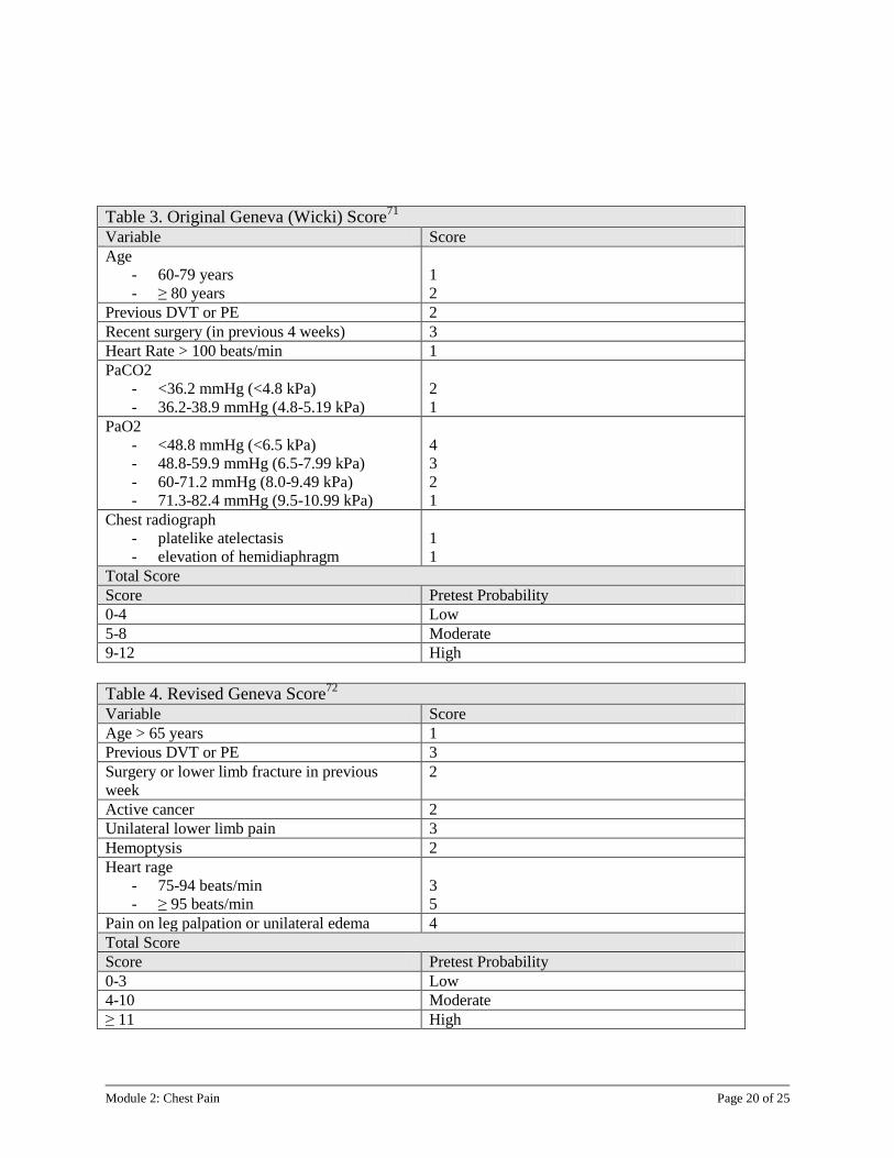
Table 3. Original Geneva (Wicki) Score71
Variable Score
Age
- 60-79 years
- ≥ 80 years
1
2
Previous DVT or PE 2
Recent surgery (in previous 4 weeks) 3
Heart Rate > 100 beats/min 1
PaCO2
- <36.2 mmHg (<4.8 kPa)
- 36.2-38.9 mmHg (4.8-5.19 kPa)
2
1
PaO2
- <48.8 mmHg (<6.5 kPa)
- 48.8-59.9 mmHg (6.5-7.99 kPa)
- 60-71.2 mmHg (8.0-9.49 kPa)
- 71.3-82.4 mmHg (9.5-10.99 kPa)
4
3
2
1
Chest radiograph
- platelike atelectasis
- elevation of hemidiaphragm
1
1
Total Score
Score Pretest Probability
0-4 Low
5-8 Moderate
9-12 High
Table 4. Revised Geneva Score72
Variable Score
Age > 65 years 1
Previous DVT or PE 3
Surgery or lower limb fracture in previous
week
2
Active cancer 2
Unilateral lower limb pain 3
Hemoptysis 2
Heart rage
- 75-94 beats/min
- ≥ 95 beats/min
3
5
Pain on leg palpation or unilateral edema 4
Total Score
Score Pretest Probability
0-3 Low
4-10 Moderate
≥ 11 High
Module 2: Chest Pain Page 21 of 25
Module 2: Chest Pain References
1. Karcz A, Holbrook J, Bruce SA, et al. Preventability of malpractice claims in emergency medicine: A closed claims study. Ann Emerg Med 1990;19:865-873.
2. Karcz A, Korn R, Burke MC, et al. Malpractice claims against emergency physicians in Massachusetts: 1975–1993. Am J Emerg Med 1996;14:341-5.
3. Medical Professional Mutual Insurance Company (“ProMutual”); Closed Case Analysis, Emergency Medicine, 1995-2003 & 2005-2009
4. American Heart Association. Heart Disease & Stroke Statistics: Our guide to current statistics and the supplement to our Heart and Stroke Facts. 2010 Update At-A-Glance. , 2010.
5. Anderson JL, Adams CD, Antman EM, et al. ACC/AHA 2007 Guidelines for the Management of Patients With Unstable Angina/Non-ST-Elevation Myocardial Infarction: A Report of the American College of Cardiology/American Heart Association Task Force on Practice Guidelines (Writing Committee to Revise the 2002 Guidelines for the Management of Patients With Unstable Angina/Non-ST-Elevation Myocardial Infarction) Developed in Collaboration with the American College of Emergency Physicians, the Society for Cardiovascular Angiography and Interventions, and the Society of Thoracic Surgeons Endorsed by the American Association of Cardiovascular and Pulmonary Rehabilitation and the Society for Academic Emergency Medicine. J Am Coll Cardiol 2007;50:e1-157.
6. McCarthy BD, Beshansky JR, D'Agostino RB, Selker HP. Missed diagnoses of acute myocardial infarction in the emergency department: Results from a multicenter study. Ann Emerg Med 1993;22:579-82.
7. Pope JH, Aufderheide TP, Ruthazer R, et al. Missed Diagnoses of Acute Cardiac Ischemia in the Emergency Department. N Engl J Med 2000;342:1163-70.
8. Pitts SR, Niska RW, Xu J, Burt CW. National Hospital Ambulatory Medical Care Survey: 2006 emergency department summary. Natl Health Stat Report 2008;(7):1-38.
9. Bhuiya FA, Pitts SR, McCaig LF. Emergency department visits for chest pain and abdominal pain: United States, 1999-2008. NCHS Data Brief 2010;(43):1-8.
10. Brieger D, Eagle KA, Goodman SG, et al. Acute Coronary Syndromes Without Chest Pain, An Underdiagnosed and Undertreated High-Risk Group*. Chest 2004;126:461-9.
11. Canto JG, Shlipak MG, Rogers WJ, et al. Prevalence, Clinical Characteristics, and Mortality Among Patients With Myocardial Infarction Presenting Without Chest Pain. JAMA 2000;283:3223-9.
12. Lee TH, Rouan GW, Weisberg MC, et al. Clinical characteristics and natural history of patients with acute myocardial infarction sent home from the emergency room. Am J Cardiol 1987;60:219-24.
13. Lee TH, Goldman L. Evaluation of the Patient with Acute Chest Pain. N Engl J Med 2000;342:1187-95.
14. Rusnak RA, Stair TO, Hansen K, Fastow JS. Litigation against the emergency physician: Common features in cases of missed myocardial infarction. Ann Emerg Med 1989;18:1029-34.

Module 2: Chest Pain Page 22 of 25
15. Kachalia A, Gandhi TK, Puopolo AL, et al. Missed and Delayed Diagnoses in the Emergency Department: A Study of Closed Malpractice Claims From 4 Liability Insurers. Ann Emerg Med 2007;49:196-205.
16. Swap CJ, Nagurney JT. Value and limitations of chest pain history in the evaluation of patients with suspected acute coronary syndromes. JAMA 2005;294:2623-9.
17. McCord J, Jneid H, Hollander JE, et al. Management of cocaine-associated chest pain and myocardial infarction: a scientific statement from the American Heart Association Acute Cardiac Care Committee of the Council on Clinical Cardiology. Circulation 2008;117:1897-907.
18. Duseja R, Feldman JA. Missed acute cardiac ischemia in the ED: limitations of diagnostic testing. Am J Emerg Med 2004;22:219-25.
19. Osborne DW, Brown DFM, Nadel ES. Chest pain. J Emerg Med 2004;26:447-50.
20. Fesmire FM, Decker WW, Diercks DB, et al. Clinical policy: critical issues in the evaluation and management of adult patients with non-ST-segment elevation acute coronary syndromes. Ann Emerg Med 2006;48:270-301.
21. Masoudi FA, Magid DJ, Vinson DR, et al. Implications of the Failure to Identify High-Risk Electrocardiogram Findings for the Quality of Care of Patients With Acute Myocardial Infarction: Results of the Emergency Department Quality in Myocardial Infarction (EDQMI) Study. Circulation 2006;114:1565-71.
22. Hess EP, Agarwal D, Chandra S, et al. Diagnostic accuracy of the TIMI risk score in patients with chest pain in the emergency department: a meta-analysis. CMAJ 2010;182:1039-44.
23. Manini AF, Dannemann N, Brown DF, et al. Limitations of risk score models in patients with acute chest pain. Am J Emerg Med 2009;27:43-8.
24. Campbell CF, Chang AM, Sease KL, et al. Combining Thrombolysis in Myocardial Infarction risk score and clear-cut alternative diagnosis for chest pain risk stratification. Am J Emerg Med 2009;27:37-42.
25. Farkouh ME, Aneja A, Reeder GS, et al. Clinical risk stratification in the emergency department predicts long-term cardiovascular outcomes in a population-based cohort presenting with acute chest pain: primary results of the Olmsted county chest pain study. Medicine (Baltimore) 2009;88:307-13.
26. Hermann LK, Weingart SD, Duvall WL, Henzlova MJ. The Limited Utility of Routine Cardiac Stress Testing in Emergency Department Chest Pain Patients Younger Than 40 Years. Ann Emerg Med 2009;54:12-6.
27. Hendel RC. Is computed tomography coronary angiography the most accurate and effective noninvasive imaging tool to evaluate patients with acute chest pain in the emergency department? CT coronary angiography is the most accurate and effective noninvasive imaging tool for evaluating patients presenting with chest pain to the emergency department: antagonist viewpoint. Circ Cardiovasc Imaging 2009;2:264,75; discussion 275.
28. Kushner FG, Hand M, Smith SC,Jr, et al. 2009 Focused Updates: ACC/AHA Guidelines for the Management of Patients With ST-Elevation Myocardial Infarction (Updating the 2004 Guideline and 2007 Focused Update) and ACC/AHA/SCAI Guidelines on Percutaneous Coronary Intervention (Updating the 2005 Guideline and 2007 Focused Update): A Report of the American College of Cardiology Foundation/American Heart Association Task Force on Practice Guidelines. J Am Coll Cardiol 2009;54:2205-41.

Module 2: Chest Pain Page 23 of 25
29. Elefteriades JA, Barrett PW, Kopf GS. Litigation in nontraumatic aortic diseases--a tempest in the malpractice maelstrom. Cardiology 2008;109:263-72.
30. Svensson LG, Rodriguez ER. Aortic Organ Disease Epidemic, and Why Do Balloons Pop? Circulation 2005;112:1082-4.
31. Hagan PG, Nienaber CA, Isselbacher EM, et al. The International Registry of Acute Aortic Dissection (IRAD): new insights into an old disease. JAMA 2000;283:897-903.
32. von Kodolitsch Y, Schwartz AG, Nienaber CA. Clinical Prediction of Acute Aortic Dissection. Arch Intern Med 2000;160:2977-82.
33. Bushnell J, Brown J. Evidence-based emergency medicine/rational clinical examination abstract. Clinical assessment for acute thoracic aortic dissection. Ann Emerg Med 2005;46:90-2.
34. Hansen MS, Nogareda GJ, Hutchison SJ. Frequency of and inappropriate treatment of misdiagnosis of acute aortic dissection. Am J Cardiol 2007;99:852-6.
35. Sullivan PR, Wolfson AB, Leckey RD, Burke JL. Diagnosis of acute thoracic aortic dissection in the emergency department. Am J Emerg Med 2000;18:46-50.
36. Erbel R, Alfonso F, Boileau C, et al. Diagnosis and management of aortic dissection. European Heart Journal 2001;22:1642-81.
37. Klompas M. Does This Patient Have an Acute Thoracic Aortic Dissection? JAMA 2002;287:2262-72.
38. Gaul C, Dietrich W, Friedrich I, Sirch J, Erbguth FJ. Neurological Symptoms in Type A Aortic Dissections. Stroke 2007;38:292-7.
39. Tsai TT, Nienaber CA, Eagle KA. Acute Aortic Syndromes. Circulation 2005;112:3802-13.
40. Haro LH, Krajicek M, Lobl JK. Challenges, controversies, and advances in aortic catastrophes. Emerg Med Clin North Am 2005;23:1159-77.
41. Januzzi JL, Isselbacher EM, Fattori R, et al. Characterizing the young patient with aortic dissection: results from the International Registry of Aortic Dissection (IRAD). J Am Coll Cardiol 2004;43:665-9.
42. Nienaber CA, Eagle KA. Aortic Dissection: New Frontiers in Diagnosis and Management: Part I: From Etiology to Diagnostic Strategies. Circulation 2003;108:628-35.
43. Rosman HS, Patel S, Borzak S, Paone G, Retter K. Quality of history taking in patients with aortic dissection. Chest 1998;114:793-5.
44. Armstrong WF, Bach DS, Carey LM, Froehlich J, Lowell M, Kazerooni EA. Clinical and echocardiographic findings in patients with suspected acute aortic dissection. Am Heart J 1998;136:1051-60.
45. Sutherland A, Escano J, Coon TP. D-dimer as the sole screening test for acute aortic dissection: a review of the literature. Ann Emerg Med 2008;52:339-43.
46. American College of Cardiology Foundation Task Force on Expert Consensus Documents, Mark DB, Berman DS, et al. ACCF/ACR/AHA/NASCI/SAIP/SCAI/SCCT 2010 Expert Consensus Document on Coronary Computed Tomographic Angiography. J Am Coll Cardiol 2010;.
47. Heit JA, Cohen AT, Anderson FA, Jr., on Behalf of the VTE Impact Assessment Group,. Estimated Annual Number of Incident and Recurrent, Non-Fatal and Fatal Venous Thromboembolism (VTE) Events in the US. ASH Annual Meeting Abstracts 2005;:910.
Module 2: Chest Pain Page 24 of 25
48. Stein PD, Beemath A, Matta F, et al. Clinical characteristics of patients with acute pulmonary embolism: data from PIOPED II. Am J Med 2007;120:871-9.
49. Calder KK, Herbert M, Henderson SO. The mortality of untreated pulmonary embolism in emergency department patients. Ann Emerg Med 2005;45:302-10.
50. Horlander KT, Mannino DM, Leeper KV. Pulmonary Embolism Mortality in the United States, 1979-1998: An Analysis Using Multiple-Cause Mortality Data. Arch Intern Med 2003;163:1711-7.
51. Courtney DM, Kline JA, Kabrhel C, et al. Clinical features from the history and physical examination that predict the presence or absence of pulmonary embolism in symptomatic emergency department patients: results of a prospective, multicenter study. Ann Emerg Med 2010;55:307,315.e1.
52. Heit JA, Silverstein MD, Mohr DN, Petterson TM, O'Fallon WM, Melton LJ,3rd. Risk factors for deep vein thrombosis and pulmonary embolism: a population-based case-control study. Arch Intern Med 2000;160:809-15.
53. Dalen JE. Economy Class Syndrome: Too Much Flying or Too Much Sitting? Arch Intern Med 2003;163:2674-6.
54. Gomes MPV, Deitcher SR. Risk of Venous Thromboembolic Disease Associated With Hormonal Contraceptives and Hormone Replacement Therapy: A Clinical Review. Arch Intern Med 2004;164:1965-76.
55. Tapson VF. Acute pulmonary embolism. N Engl J Med 2008;358:1037-52.
56. Samama M, for the Sirius Study Group,. An Epidemiologic Study of Risk Factors for Deep Vein Thrombosis in Medical Outpatients: The Sirius Study. Arch Intern Med 2000;160:3415-20.
57. Laack TA, Goyal DG. Pulmonary embolism: an unsuspected killer. Emerg Med Clin North Am 2004;22:961-83.
58. Rizkallah J, Man SFP, Sin DD. Prevalence of Pulmonary Embolism in Acute Exacerbations of COPD. Chest 2009;135:786-93.
59. Monreal M, Muñoz-Torrero JFS, Naraine VS, et al. Pulmonary Embolism in Patients with Chronic Obstructive Pulmonary Disease or Congestive Heart Failure. Am J Med 2006;119:851-8.
60. Stein PD, Woodard PK, Weg JG, et al. Diagnostic pathways in acute pulmonary embolism: recommendations of the PIOPED II investigators. Am J Med 2006;119:1048-55.
61. Chagnon I, Bounameaux H, Aujesky D, et al. Comparison of two clinical prediction rules and implicit assessment among patients with suspected pulmonary embolism. Am J Med 2002;113:269-75.
62. Klok FA, Mos ICM, Nijkeuter M, et al. Simplification of the Revised Geneva Score for Assessing Clinical Probability of Pulmonary Embolism. Arch Intern Med 2008;168:2131-6.
63. Rodger MA, Carrier M, Jones GN, et al. Diagnostic value of arterial blood gas measurement in suspected pulmonary embolism. Am J Respir Crit Care Med 2000;162:2105-8.
64. Dunn KL, Wolf JP, Dorfman DM, Fitzpatrick P, Baker JL, Goldhaber SZ. Normal D-dimer levels in emergency department patients suspected of acute pulmonary embolism. J Am Coll Cardiol 2002;40:1475-8.
65. Kelly J, Rudd A, Lewis RR, Hunt BJ. Plasma D-Dimers in the Diagnosis of Venous Thromboembolism. Arch Intern Med 2002;162:747-56.

Module 2: Chest Pain Page 25 of 25
66. Kline JA, Mitchell AM, Kabrhel C, Richman PB, Courtney DM. Clinical criteria to prevent unnecessary diagnostic testing in emergency department patients with suspected pulmonary embolism. J Thromb Haemost 2004;2:1247-55.
67. Schoepf UJ, Costello P. CT angiography for diagnosis of pulmonary embolism: state of the art. Radiology 2004;230:329-37.
68. Grifoni S, Olivotto I, Cecchini P, et al. Utility of an integrated clinical, echocardiographic, and venous ultrasonographic approach for triage of patients with suspected pulmonary embolism. Am J Cardiol 1998;82:1230-5.
69. Arcasoy SM, Kreit JW. Thrombolytic therapy of pulmonary embolism: a comprehensive review of current evidence. Chest 1999;115:1695-707.
70. Kucher N. Catheter embolectomy for acute pulmonary embolism. Chest 2007;132:657-63.
71. Wells PS, Anderson DR, Rodger M, et al. Derivation of a simple clinical model to categorize patients probability of pulmonary embolism: increasing the models utility with the SimpliRED D-dimer. Thromb Haemost 2000;83:416-20.
72. Wicki J, Perneger TV, Junod AF, Bounameaux H, Perrier A. Assessing Clinical Probability of Pulmonary Embolism in the Emergency Ward: A Simple Score. Arch Intern Med 2001;161:92-7.
73. Le Gal G, Righini M, Roy PM, et al. Prediction of pulmonary embolism in the emergency department: the revised Geneva score. Ann Intern Med 2006;144:165-71.
Related Documents