Cushing's Syndrome : hormonal secretion patterns, treatment and outcome. Aken, M.O. van Citation Aken, M. O. van. (2005, March 17). Cushing's Syndrome : hormonal secretion patterns, treatment and outcome. Retrieved from https://hdl.handle.net/1887/3748 Version: Corrected Publisher’s Version License: Licence agreement concerning inclusion of doctoral thesis in the Institutional Repository of the University of Leiden Downloaded from: https://hdl.handle.net/1887/3748 Note: To cite this publication please use the final published version (if applicable).

Welcome message from author
This document is posted to help you gain knowledge. Please leave a comment to let me know what you think about it! Share it to your friends and learn new things together.
Transcript

Cushing's Syndrome : hormonal secretion patterns, treatment andoutcome.Aken, M.O. van
CitationAken, M. O. van. (2005, March 17). Cushing's Syndrome : hormonal secretionpatterns, treatment and outcome. Retrieved from https://hdl.handle.net/1887/3748 Version: Corrected Publisher’s Version
License: Licence agreement concerning inclusion of doctoral thesis inthe Institutional Repository of the University of Leiden
Downloaded from: https://hdl.handle.net/1887/3748 Note: To cite this publication please use the final published version (if applicable).

Cushing’s Syndrome
Hormonal secretion patterns, treatment and outcome

Omslag: Harvey Cushing. Portret afkomstig van 1e dag envelop, bij uitgifte van postzegel met afbeelding van H. Cushing, in de serie “Great Americans” (1988).
Cover, lay-out and printed by Optima Grafi sche Communicatie, Rotterdam

Cushing’s Syndrome
Hormonal secretion patterns, treatment and outcome
Proefschrift
ter verkrijging van de graad van Doctor aan de
Universiteit Leiden,
op gezag van de
Rector Magnifi cus Dr. D.D. Breimer,
hoogleraar in de faculteit der Wiskunde en
Natuurwetenschappen en die der Geneeskunde,
volgens besluit van het College voor Promoties
te verdedigen op donderdag
17 maart 2005 klokke 14.15 uur
door
Maarten Otto van Aken
Geboren te Delft in 1968

PROMOTIECOMMISSIE
Promotores: Prof. dr. J.A. Romijn Prof. dr. S.W.J. Lamberts, Erasmus Universiteit Rotterdam
Copromotor: dr. W.W. de Herder, Erasmus Universiteit Rotterdam
Referent: Prof. dr. A.R.M.M. Hermus, Universitair Medisch Centrum St. Radboud
Overige leden: Prof. dr. E.R. de Kloet Prof. dr. S.E. Papapoulos Prof. dr. H.A.P. Pols, Erasmus Universiteit Rotterdam dr. F. Roelfsema Prof. dr. J.M. Wit

Aan ManonAan Pepijn, Jasmine en Friso


CONTENTS
Chapter 1Introduction
Chapter 2Irregular and frequent cortisol secretory episodes with preserved diurnal rhythmicity in primary adrenal Cushing’s syndrome.Journal of Clinical Endocrinology and Metabolism, online publication dec. 2004
Chapter 3Growth hormone secretion in primary adrenal Cushing’s syndrome is disorderly and inversely correlated with body mass index.American Journal of Physiology: Endocrinology and Metabolism 2005: 288(1); E63-70
Chapter 4Profound amplifi cation of secretory-burst mass and anomalous regularity of ACTH secretory process in patients with Nelson’s syndrome compared with Cushing’s disease.Clinical Endocrinology 2004:60;765-772
Chapter 5Salivary cortisol in the diagnosis of Cushing’s syndrome.Published in part in Clinical Chemistry 2003:49(8);1408-1409
Chapter 6Risk factors for meningitis after transsphenoidal surgery.Clinical Infectious Diseases 1997;25:852-6
Chapter 7Cerebrospinal fl uid leakage during transsphenoidal surgery: postoperative external lumbar drainage reduces the risk for meningitis.Pituitary 2004;7: 93-97
Chapter 8Postoperative metyrapone test in the early assessment of outcome of pituitary surgery for Cushing’s disease.Clinical Endocrinology 1997:47;145-149
9
31
49
67
83
101
111
121

Chapter 9Long-term predictive value of postsurgical cortisol concentrations for cure and risk of recurrence in Cushing’s disease.Journal of Clinical Endocrinology and Metabolism 2003: 88;5858-5864
Chapter 10Decreased quality of life in patients despite long-term biochemical cure of Cushing’s disease Submitted
Chapter 11Summary and conclusions.
Samenvatting
Nawoord
Curriculum Vitae
Publicaties
131
147
163
175
183
185
186

Chapter 1
Introduction


11
Introduction
CUSHING’S SYNDROME
Endogenous Cushing’s syndrome is a clinical state resulting from prolonged, inappropriate exposure to excessive secretion of cortisol (1). The syndrome has been named after Harvey Cushing, a neurosurgeon, born in 1869, trained at Johns’s Hopkins Medical School and professor at the Peter Brigham Hospital in Harvard from 1912 to 1932 (2). From 1932 until his death in 1939, he worked as professor of neurology at Yale. In 1912, Harvey Cushing described a woman with a syndrome of painfull obesity, hypertrichiosis and amenorrhoea, but the cause of this syndrome was not recognized. It was only twenty years later, in 1932, that he described a series of patients with the same symptoms, who, at postmortem examination were found to have a tumor of the pituitary gland. In retrospect, the fi rst case of a patient with Cushing’s syndrome has probably been described by William Osler, professor of surgery at John’s Hopkins University, in 1899 (3,4). The title of the paper was “An acute myxoedematous condition, with tachycardia, glycosuria, melaena, mania and death”, describing a case of a 37-year old male patient. A striking clinical feature was a gain in weight of 17 kg in 3 months, which apparently led Osler to consider the diagnosis of acute myxedema, and treat the patient with thyroid-grains. However, in the same report Osler points out a group of clinical symptoms not been reported before, including abdominal fat accumulation, reddish-purple striae and a bloated face. The patient died shortly after presentation. Unfortunately, post-mortem examination was not performed. Interestingly, in 1899, at the time of this case, Harvey Cushing worked as a resident at John’s Hopkins under mentorship of William Osler. There is, however, no evidence that the two ever discussed the case (4).
Several other classic papers have documented patients suffering from symptoms of hypercortisolism, including a paper by Achard and Thiers, who under the title “diabete des femmes a barbe”, describe a series of patients with hirsutism, glysosuria and adrenal lesions at autopsy (5)
In classical cases, the clinical features of Cushing’s syndrome are the triad of obesity, hypertension and diabetes mellitus(6). In general, the clinical picture may vary considerably, with a variety of symptoms associated with hypercortisolemia including weight gain, lethargy, weakness, menstrual irregularities, loss of libido, depression, hirsutism, acne, purplish skin striae, thinned skin and hyperpigmentation (7).
Biochemically, Cushing’s syndrome is characterized by loss of the normal feedback mechanism of the hypothalamo-pituitary-adrenal (HPA)-axis and the normal circadian rhythm of cortisol secretion. The etiology of Cushing’s syndrome is classically divided in ACTH-dependent and ACTH-independent causes (table 1) (1). ACTH-dependent Cushing’s syndrome is usually caused be excessive ACTH production from an adenoma in the pituitary gland, or, more rarely, by ectopic ACTH secretion from a non-pituitary tumor. ACTH-independent Cushing’s syndrome is caused by excessive secretion of cortisol by an adrenocortical adenoma or carcinoma, or, rarely, micro-or macronodular bilateral adrenal hyperplasia.

12
Chapter 1
Table 1: Causes of Cushing’s syndrome
Diagnosis Percent of patients
ACTH-dependent Cushing’s syndrome
Pituitary adenoma (Cushing’s disease) 68
Ectopic ACTH production 12
Ectopic CRH production < 1
ACTH-independent Cushing’s syndrome
Adrenal adenoma 10
adrenal carcinoma 8
Micronodular hyperplasia 1
Macronodular hyperplasia 1
(Adapted from: Orth DN. Cushing’s syndrome. N Engl J Med 1995; 332(12):791-803).
UNRESOLVED ISSUES IN CUSHING’S SYNDROME
Since 1932, Harvey Cushing’s description of the syndrome that results from long-term glucocorticoid-excess has not improved upon, but our understanding of its pathophysiologic features and our ability to diagnose and treat the disorder have increased dramatically (7). However, more than in any other area of clinical endocrinology, diagnosis, differential diagnosis and management continue to challenge the physician and occasionally cause considerable controversy. This is refl ected by our limited understanding of the biology of the different causes of Cushing’s syndrome and the intrinsic limitations of practically every diagnostic tests used in the diagnosis of Cushing’s syndrome in clinical practice (8-13). Finally, once a defi nitive diagnosis of the cause of Cushing’s syndrome has been made, there is frequently not a simple, straightforward surgical and/or medical treatment available, that can cure the disease without inducing (permanent) side-effects or necessitating additional treatment (14-22). Moreover, even after initial succesfull treatment, recurrence of the disease can occur in a signifi cant number of patients (23-30).
In the following paragraphs, several unresolved issues in Cushing’s syndrome will be discussed, especially concerning the characterization of temporal changes in hormonal secretion, diagnostic problems involved in Cushing’s syndrome, treatment of pituitary-dependent Cushing’s disease and its complications, assessment of cure and risk of relapse after treatment of Cushing’s disease and fi nally quality of life after succesfull treatment of Cushing’s disease.

13
Introduction
CHARACTERIZATION OF TEMPORAL CHANGES IN HORMONAL SECRETION
Cushing’s syndrome is the result of chronic exposure to excess of cortisol, released from the zona reticularis of the adrenal cortex (31). In healthy subjects, cortisol is released episodically. The main rhythm is circadian, in which cortisol level is at its peak around the time of awakening (around 07:00 am) and at is nadir around midnight (32-34). The adrenals are under feed-back control of the hypothalamo-pituitary adrenal (HPA) axis. Figure 1 shows the normal regulation of the HPA axis. In the hypothalamus, the suprachiasmatic nucleus harbours the regulation of the circadian rhythm. From this nucleus, neuronal input activates the paraventricular nucleus, where CRH is released into capillaries in the median eminence, draining into the anterior pituitary through the portal veins. In the anterior pituitary, CRH stimulates ACTH release from the corticotroph cells. Subsequently, ACTH binds to its receptor on the adrenals, resulting in cortisol-release. Finally, ACTH release from the pituitary corticotrophs is inhibited through glucocorticoid feedback, which causes cortisol released from the adrenals to ultimately restrain its own release.
In addition to the circadian changes in cortisol secretion, cortisol is secreted in a episodic, pulsatile fashion (35). This temporal architecture of plasma hormone secretion constitutes an additional mechanism, which may modulate signal transduction and which may also prevent down-regulation of the response of the target organ. In general, the sensitivity for pulsatile regulation within the hypothalamo-pituitary-adrenal axis may not be similar for each level of this axis. For instance, ACTH secretion is more sensitive for pulsatile than for continuous administration of ovine CRH (36). Cortisol secretion is hardly dependent on the pulsatile characteristics of ACTH release, because cortisol levels are similar after prolonged continuous and intermittend ACTH administration. This is not true for another, indirect biological effect of ACTH, like aldosterone secretion. Pulsatile ACTH administration results in higher aldosterone levels than continuous ACTH secretion (37). Finally, at present it is unclear to which extent the biological effects of cortisol are dependent on pulsatile characteristics of cortisol secretion.
For the corticotropic axis, the pulsatile release of ACTH and cortisol is altered in several conditions. In major depression, ACTH pulse frequency was increased, and the nadir of cortisol secretion occurred 3 hours earlier compared to healthy subjects (38,39). In women with the polycystic ovary syndrome, the amplitude of ACTH-pulses at night appeared to be increased. In addition, daytime cortisol secretion was more disorderly compared to controls (40).
The physiological relevance of the pulsatile release of pituitary hormones in general is also clearly demonstrated for the gonadotropic axis in women, where changes in the release pattern of the gonadotropins and gonadal steroids are mandatory for maintaining a normal menstrual cycle (41,42). In men, puberty can only be induced by pulsatile GnRH therapy, thereby underlining the importance of a properly functioning GnRH oscillator (43). Finally, for the somatotropic axis, pulsatile release is also of physiological importance, as shown by studies in

14
Chapter 1
growth-hormone defi cient children, where a greater response in growth rate was demonstrated with a more frequent regimen of GH injections than with the same total but weekly dose (44,45).
Figure 1. The hypothalamic–pituitary–adrenal control system.
Neural pathways into the paraventricular nucleus of the hypothalamus can be classifi ed as “stress” inputs (for example, hypoglycemia) directly and through the hippocampus and daily rhythm input (circadian rhythm). These inputs result in an activation of parvocellular neurons that release corticotrophin-releasing factor (CRH) and arginine vasopressin (AVP) into the capillary plexus of the median eminence, which forms long portal veins. These drain into the anterior pituitary, where CRH and AVP infl uence the corticotrophs to increase release of adrenocorticotrophic hormone (ACTH). The hormone enters the systemic circulation and stimulates the adrenal cortex to increase cortisol production. Cortisol exerts its biological effects through the glucocorticoid receptor. Plasma cortisol also drains to saliva and the urine. The hypothalamic–pituitary–adrenal axis loop is completed through glucocorticoid-negative feedback exerted at the anterior pituitary, hypothalamus, and hippocampus. Diagnostic tests associated with each physiologic process are shown in pink. CBG = corticosteroid-binding globulin; CS = Cushing syndrome.
Reprinted with permission from H. Raff and JW Findling in: A physiologic approach to diagnosis of the Cushing syndrome. Ann Intern Med. 2003 Jun 17;138(12):980-91.

15
Introduction
Changes in the pulsatile release of hormones are also observed in patients with various endocrine diseases, including pituitary diseases and non-pituitary-diseases (46-50). In general, in these disease-states, the augmented hormonal release is due to an increase in the ammount secreted per burst and/or an increase in pulse frequency. In addition, the orderliness of hormonal secretion is frequently disrupted (48,51,52).
As for Cushing’s syndome, studying the pathophysiology of episodic hormonal secretion can help understanding the biology of possible causes of hypercortisolism, and may be even exploited as a diagnostic tool (33). From studies in patients with pituitary-dependent Cushing’s disease it appeared that hypercortisolism is characterized by increased basal and increased pulsatile cortisol secretion due to an increased number of pulses and an increased mass secreted per pulse (53). Furthermore, the cortisol-secretion pattern showed more disorderliness compared to healthy controls (54,55,55). At present, it is unclear to which extent the cortisol secretory patterns are different among the different causes of Cushing’s syndrome, especially in primary adrenal Cushing’s syndrome.
In young patients, who still have open epiphysial growth plates, it is well known that the occurrence of Cushing’s syndrome precipitates an inhibition in growth and a decreased fi nal height. Pulsatile growth hormone secretion was shown to be preserved in Cushing’s disease, except for severe hypercortisolism. In addition, secretion of GH was remarkably disorderly in patients with Cushing’s disease (56). However, it is unclear to which extent these changes are related to excess cortisol concentrations per se, rather than to changes within the pituitary associated with the consequences of an ACTH producing pituitary adenoma. At present, however, there is no detailed information available on the secretory profi le GH in ACTH-independent Cushing’s syndrome, which could contribute to the understanding of the regulation of growth hormone secretion in conditions of cortisol excess.
Nelson’s syndrome was fi rst described in 1958 as the constellation of a pituitary macroadenoma, markedly elevated ACTH concentrations, and hyperpigmentation of the skin in a patient after bilateral adrenalectomy for pituitary-dependent hypercortisolism (Cushing’s disease) (122). The syndrome develops in 8 - 38% of adults requiring bilateral adrenalectomy for Cushing’s disease (123,124) and occurs infrequently in patients aged 40 yr or more at the time of bilateral adrenalectomy, in contrast to patients treated at an early age (125).
Cushing’s disease and Nelson’s syndrome are considered to be distinct pathological presentations of the same primary biological entity. For example, impaired responsiveness to glucocorticoid enforced negative feedback on ACTH is common to both (126). In addition, under in vitro conditions the secretion of POMC-derived peptides was similar in tumoral tissue derived from patients with Cushing’s disease and Nelson’s syndrome (127). However, CRH infusion stimulates greater and prolonged ACTH secretion in patients with Nelson’s syndrome than Cushing’s disease (128). The pathogenetic mechanisms underlying tumorigenesis and unrestrained ACTH secretion in Nelson’s syndrome are not well understood.

16
Chapter 1
Quantitative comparisons of neurosecretory control of tumoral ACTH secretion in Nelson’s syndrome and Cushing’s disease have not been reported, but could shed light on the pathophysiology of these two clinical entities.
The pulsatile and diurnal changes in release of ACTH- and cortisol (as well as other pituitary hormones) can be studied by frequent sampling of plasma in combination with mathematical tools to analyze the respective secretion patterns. Multiparameter deconvolution analysis is a technique which resolves the serum hormone concentration profi le into its constituent secretory contributions and simultaneously estimates the hormone half-life. This analysis can be used to quantify underlying basal and pulsatile hormone secretion and to estimate the corresponding (endogenous) half-life (57). The orderliness of hormone release patterns over 24 h can be quantitated by approximate entropy (ApEn) analysis. ApEn provides a scale-invariant and model-independent quantitation of relative disorderliness, in which higher ApEn values denote greater relative disorderliness or reduced regularity of the release process(58,59). Finally, cosinor analysis can be used to study twenty-four-hour variations in hormonal concentrations, with quantitation of the 24-h cosine amplitude (50% of the nadir-zenith difference), mesor (rhythmic mean) and acrophase (clock-time of maximal value) (60).
DIAGNOSIS OF CUSHING’S SYNDROME
In patients with severe clinical symptoms of hypercortisolism, the pretest likelihood of the presence of Cushing’s syndrome is high and the positive predictive value of the biochemical tests is high. Therefore, in these severe cases, the biochemical confi rmation usually offers no specifi c problems. However, it can be diffi cult to distinguish between mild forms of Cushing’s syndrome and situations referred to as pseudo-Cushing states, such as the metabolic syndrome, depression and alcoholism. In these conditions the pretest likelihood of the presence of disease is low and the positive predictive value of the biochemical tests is negatively affected. This may often lead to confusing results of biochemical tests. Since Cushing’s syndrome is associated with considerable morbidity and mortality and correction of hypercortisolism may substantially improve the metabolic consequences of excess cortisol, even a low index of suspicion should mandate at least a screening evaluation for Cushing’s syndrome. Consequently, with increasing awareness among doctors and the widespread availability of biochemical testing, patients are screened in an earlier phase of their disease, in which only minor abnormalities of cortisol secretion are present, making biochemical conformation even more diffi cult.
There are several diagnostic tools available for the biochemical screening for hypercortisolism. However, as will be discussed below, no test can be used as a single screening test for the detection of CS, since every test has its specifi c limitations in sensitivity and specifi city.

17
Introduction
- Twenty-four hour urine collection for the measurement of urinary free cortisol (UFC) has been considered a gold standard for the diagnosis of CS (1). However, this method has several limitations, including the infl uence of fl uid intake and impaired renal function, false elevation by specifi c medications (e.g. carbamazepin and digoxin), mild elevations in pseudo-Cushing’s states and pregnant women, and normal values in mild, subclinical Cushing’s syndrome (61). For these reasons, UFC cannot be considered as a universal single screening test for the detection of Cushing’s syndrome.
- The low-dose dexamethasone suppression test (DST) is the second screening test and is based on the assumption that in patients with CS, the negative feedback by glucocorticoids on the hypothalamic-pituitary-adrenal (HPA) axis is diminished. In the classical two-days DST, the suppression of urinary 17-hydroxycorticosteroids was used as an indicator of cortisol suppression (62). Measurement of serum cortisol at 09:00 after the administration of 0.5 mg dexamethasone every 6 h for 48 h and a cut-off value of 50 nmol/l has been reported to have a sensitivity of 98% in the diagnosis of Cushing’s syndrome (63). The overnight low-dose DST consists of the oral intake of 1 mg dexamethasone at 2300 h, and the measurement of fasting plasma cortisol concentration between 0800 and 0900 h the following morning. The international consensus on the criterion for normal level of suppresion, has recently been lowered to 50 nmol/l (31), increasing the sensitivity of this test, obviously at the cost of lower specifi city. The specifi city is further reduced by other interfering factors, such as increased concentrations of cortisol binding globulin, acute and chronic illness, pseudo-Cushing states, decreased dexamethasone absorption and drugs enhancing hepatic dexamethasone metabolism (phenytoin, carbamazepin, rifampicine). In addition, feedback-sensitivity to glucocorticoids varies between subjects, explained by polymorphisms in the glucocorticoid receptor gene (64,65). For these reasons, DST cannot adequately distinguish all patients with Cushing’s syndrome from other subjects.
- The third screeeningtest for hypercortisolism is measurement of midnight serum cortisol concentration. In healthy subjects, serum cortisol concentrations follow a circadian rhythm, with a peak at 0700 – 0900 h and falling levels thereafter until a subsequent rise at 0300 – 0400 h (66). In Cushing’s syndrome, several studies have suggested a loss of this circadian rhythm (67-71). However, other and more recent studies have shown that the diurnal pattern of cortisol secretion is preserved in certain patients but with levels that are set abnormally high (46,72-75). At midnight, the overlap of serum cortisol levels between patients with Cushing’s syndrome and the normal range was shown to be minimal (76). Therefore, measurement of midnight serum cortisol levels could be a useful tool in identifying patients with Cushing’s syndrome.
In two studies, the measurement of midnight serum cortisol concentration in the documentation of hypercortisolism was studied (63,77). In both studies, a single midnight serum cortisol value correctly identifi ed almost all subjects with Cushing’s syndrome. The discrepancy in cut-off levels between these studies (> 50 nmol/l and

18
Chapter 1
> 207 nmol/l respectively) is explained by the fact that in the fi rst study, patients had to be asleep before taking a midnight blood sample (63), while in the second study patients were awake. However, the need for hospitalization to obtain an unstressed midnight blood sample for the measurement of serum cortisol makes this a very impracticle test for the primary assessment of hypercortisolism, unsuitable for daily clinical practice.
- The fourth screening test is the measurement of late-night cortisol-concentrations in saliva. Measurement of salivary cortisol concentration has been described since the early 70’s (78-80). Salivary cortisol is a valid indicator of the plasma free cortisol concentration and is not affected by the rate of saliva production (81,82). An increase in plasma cortisol is refl ected by a change in salivary cortisol concentration within a few minutes. The circadian rhythm of plasma cortisol is similarly refl ected in the diurnal variation of salivary cortisol concentration, with a peak in the early morning and nadir around midnight (82,83). The concentration of cortisol in saliva is about 5% of the total plasma cortisol concentration, making the sensitivity of a salivary cortisol assay a very important issue.
The study of Laudat et al. was among the fi rst to document the effectiviness of diurnal salivary cortisol sampling to diagnose CS, with an elevated salivary cortisol level in all patients with CS (83). Since that study, new assay technologies in measuring cortisol concentration in saliva have emerged and several clinical studies have been performed using salivary cortisol as a fi rst line test in screening for CS (84-90). In all studies, late-night salivary cortisol-concentration performed well in establishing hypercortisolism, with sensitivity and specifi city ranging from 92 – 100%. However, these studies have mostly been performed in a research setting, with in-patients, under ideal conditions to accomplish the desired outcome. Moreover, patient series in all studies were referral-based samples, introducing possible selection bias. Finally, different cut-off levels of salivary cortisol for the diagnosis of CS were reported, ranging from > 3.6 nmol/l to > 15.2 nmol/l, at least partly explained by the use of different assays.
In patients with equivocal results, a combined dexamethasone suppression-test and CRH-test can be performed. This test was shown by one center to be highly accurate in distinguishing patients with CS from subjects with pseudo-CS (91,92).
If CS is confi rmed by one or more of the screening tests, determination of plasma ACTH values is the next step to establish the cause of hypercortisolism (66). ACTH concentrations below the level of detection or below 2 pmol/l suggest an ACTH-independent cause of CS (93). Plasma ACTH concentrations greater than 4 pmol/l are compatible with an ACTH-dependent cause. However, ACTH-levels may not be fully suppressed in some patients with CS and intermittent or moderate hypercortisolism. In these cases, a CRH stimulation test is indicated, with measurement of plasma ACTH (31).

19
Introduction
If ACTH is suppressed, the next step is computed tomography (CT) and /or magnetic resonance imaging (MRI) to identify the type of adrenal lesion(s) responsible for CS.
In patients with ACTH-dependent CS, a pituitary MRI with gadolineum enhancement should be performed, which has a sensitivity ranging from 50-60% in identifying a pituitary adenoma (94,95). In patients without a clear pituitary adenoma on MRI, bilateral inferior petrosal sinus sampling (BIPSS) should be performed to establish the source of ACTH secretion. An inferior petrosal sinus (IPS) to peripheral ACTH ratio greater than 2.0 in the basal state, and/or a ratio greater than 3.0 after CRH stimulation is consistent with pituitary-dependent Cushing’s disease (96). However, false-negative results may occur, due to technical factors as well as anomalous venous drainage(31). Also, false-positive results can be obtained in rare cases of ectopic CRH secreting tumors (97).In conclusion, the biochemical confi rmation of CS remains a challenge to the clinician, with limitations of every test employed. Measurement of late-night salivary cortisol concentration appears to be a promising, patient-friendly additional test in this challenging fi eld.
TREATMENT: TRANSSPHENOIDAL SURGERY AND COMPLICATIONS
Transsphenoidal surgery (TSS) is the treatment of choice for most lesions in the sellar region. Disadvantages of the transsphenoidal approach are a restricted fi eld of surgery and generally absent visualization of the optic nerves. In experienced hands, it is a safe procedure with low morbidity and mortality rates. In a large national survey in the USA among 958 neurosurgeons performing transsphenoidal operations, the mean operative mortality was 0.9% (98). Postoperative anterior pituitary insuffi ciency (19.4%) and diabetes insipidus (17.8%) were complications with the highest incidence of occurrence.
Pituitary adenomas originate below the diaphragma sellae, and, thus, outside the arachnoid membrane and the subarachnoid space. Therefore, the transsphenoidal removal of a pituitary tumor usually can be performed entirely outside the arachnoid membrane. However, the arachnoid membrane can be injured and penetrated in the process of opening the dura (in the case of a low-situated anterior arachnoid recess), maneuvers in the anterior-superior aspect of the sella-exposure or removal of a macroadenoma, when the distended alevated arachnoid membrane begins to invert into the sella. A subsequent CSF fi stula exposes the patient to the risk of developing postoperative meningitis. The incidence of CSF fi stula in the earlier mentioned national survey was 3.9% and of meningitis 1.5% (98). In the literature, the incidence of postoperative meningitis ranges from 0.4% to 9% (21).
The management of intraoperative CSF leakage consists primarily of meticulous, watertight reconstruction of the sellar fl oor. Several techniques of sella closure have been described in the literature. In addition, an external lumbar drain (ELD) can be

20
Chapter 1
inserted to prevent postoperative rhinorrhea and fi stula formation, a method which is employed by some, but rejected by others (99-102). However, the effect of ELD insertion on the risk of postoperative meningitis, has not been described yet.
POSTOPERATIVE EVALUATION AND FOLLOW-UP
Although TSS allows cure of Cushing’s disease, the reported success rates vary from 50 to almost 90% (103-107). The skill and experience of the neurosurgeon is a very important factor determining this outcome of TS (18). Additional factors determining the high variability in success rate are differences in criteria used to defi ne remission and differences in duration of follow up, which may result in a low rate of late relapses during short-term follow up.
Immediate postoperative assessment of outcome of pituitary surgery is important in order to plan further treatment in patients with persistent hypercortisolism.
For the assessment of cure after pituitary surgery for Cushing’s disease, several methods have been used. First, unmeasurable postoperative fasting serum cortisol levels appears to be a valuable indicator for remission. This is explained by suppression of non-tumorous corticotrophs by longstanding exposure to elevated levels of glucocorticoids. Histologically, this is characterized by so called Crooke’s hyaline atrophy (108). However, with measurable serum cortisol levels, long-term remission is also possible (109). Similarly, determination of the 24-hour urinary cortisol excretion has not much practical value in the assessment of cure after pituitary surgery for Cushing’s disease. Finally, failure to suppress with 1 mg dexamethasone in the early postoperative period, is not necessarily associated with surgical failure (110).
Early postoperative assessment is also valuable as an early marker of the risk of relapse of Cushing’s syndrome. In this respect, an undetectable postoperative fasting serum cortisol is not always associated with long-term remission (23,111). Similarly, 24-hour urinary cortisol excretion cannot identify patients at risk for relapse. In several studies, the role of CRH testing in the early postoperative period for the assessment of risk for relapse in Cushing’s disease has been evaluated. Subjects with a subnormal cortisol and/of ACTH response to CRH generally remain in remission, however again with exceptions.
The metyrapone test was introduced 35 years ago to assess the functional capacity of the hypothalamo-pituitary-adrenocortical axis. Since then, it has been used widely for this purpose. It has also been used for the differential diagnosis of Cushing’s syndrome. Until recently, the metyrapone test has not been used in the early postoperative assessment after TSS for Cushing’s disease.
The rate of recurrence of Cushing’s disease depends on the criteria for initial cure, and varies from 5 to 24% in the literature (23,24,111-113). When pituitary surgery has failed to cure Cushing’s disease or in case of recurrence of Cushing’s disease, there are several options for further treatment. Pituitary irradiation is nowadays

21
Introduction
considered as the most appropriate treatment in these cases (24). Newer techniques of stereotactic radiotherapy, such as gammaknife radiosurgery, might even improve the outcome, but experience and follow-up time are still limited (114-118). Medical treatment has the major major disadvantage of the need for lifelong therapy, but can be used to overcome the waiting-time for the effect of radiotherapy. Steroidogenesis inhibitors, including ketoconazole, metyrapone and mitotane are effective in a majority of patients (119). Compounds modulating ACTH release from a pituitary tumor, such as dopamine-agonists, PPAR-gamma agonists, and somatostatin analogs are interesting agents that need further investigation (120,121).
Bilateral adrenalectomy is another option in patients with persistent or recurrent Cushing’s disease, especially in patients with severe hypercortisolism that requires prompt reversal or after failure of radiotherapy. Disadvantages are the need for life-long replacement-therapy with gluco- and mineralocorticoids, the risk of acute adrenal insuffi ciency and the risk of development of Nelson’s syndrome.
QUALITY OF LIFE
TSS allows cure of Cushing’s disease in a large proportion of patients, whereas pituitary irradiation and/or bilateral adrenalectomy can correct hypercortisolism in the remaining patients. Despite this excellent prognosis (from a hormonal perspective), physical recovery is remarkably slow and often incomplete, with residual impairments such as osteoporosis, hypertension and pituitary defi ciencies. Similarly, disappearance of psychological distress does no always occur upon proper endocrine treatment (129,130). These persisting physical and psychological impairments may affect quality of life in patients with Cushing’s disease despite long-term biochemical cure. However, the long-term impact of Cushing’s disease on subjective well-being after successful treatment of cortisol excess is unclear.
SCOPE OF THIS THESIS
In this thesis, severel aspects of Cushing’s syndrome will be adressed, including characterization of temporal changes in hormonal secretion, diagnosis of Cushing’s syndrome, transsphenoidal surgery and complications, postoperative evaluation and follow-up.
Characterization of temporal changes in hormonal secretion
ACTH-independent Cushing’s syndrome is caused by excessive secretion of cortisol by an adrenocortical adenoma or carcinoma, or, rarely, micro-or macronodular bilateral adrenal hyperplasia. We investigated whether the differences between these adrenal causes of Cushing’s syndrome might also be refl ected in the temporal architecture of cortisol concentrations. Therefore, in chapter 2, the secretory profi les

22
Chapter 1
of cortisol in primary adrenal Cushing’s syndrome are described. Quantitative data were analyzed of basal and pulsatile secretion, diurnal rhythmicity and secretory process regularity, comparing unilateral adenoma and bilateral macronodular hyperplasia. Moreover, neurosecretory control of tumoral cortisol secretion in primary adrenal hypercortisolism was compared with that in pituitary dependent Cushing’s disease.
In pituitary-dependent hypercortisolism, the diminished growth hormone (GH) response to various stimuli, including GHRH, insulin-induced hypoglycaemia and ghrelin, is well-known. In addition, GH secretion was negatively correlated to 24 hr urinary cortisol excretion and the GH secretorion regularity was signifi cantly decreased. These GH secretory abnormalities could be the result of the presence of the pituitary adenoma itself, a tumoral product acting as a paracrine signal on the somatotrope or the result of cortisol excess per se. In order to further explore these observations, in chapter 3, the dynamics of spontaneous GH secretion in patients with primary adrenal Cushing’s syndrome are described, since these patients lack a pituitary adenoma, but otherwise suffer from chronic endogenous cortisol excess. The prime question was whether such patients diplay low-amplitude and/or disorderly GH secretion compared with BMI-matched controls, as we previously found in pituitary-dependent hypercortisolism.
When transsphenoidal surgery fails to induce remission of Cushing’s disease, bilateral adrenalectomy can be performed. However, this harbours the risk of developing Nelson’s syndrome, characterized by grossly elevated ACTH concentrations, a sellar mass and skin hyperpigmentation. In chapter 4, the issue is addressed whether the mechanisms directing ACTH secretion differ in Nelson’s syndrome and untreated Cushing’s disease, by analyzing 24 h ACTH profi les in these distinct conditions.
Diagnosis of Cushing’s syndrome
In the case of clinical suspicion of Cushing’s syndrome, biochemical screening for hypercortisolism is performed. Twenty-four hour urine collection for the measurement of urinary free cortisol excretion and the low-dose dexamethasone suppression test (1 mg) have been used extensively as fi rst line screenings test for CS, but neither test has proven fully capable of distinguishing all cases of CS from other individuals. Since several years, measuring late night salivary cortisol concentration has emerged as a screening test for the presence of hypercortisolism. Despite its simplicity, the use of salivary cortisol measurement to screen for Cushing’s syndrome has been slow to catch on. In chapter 5, several aspects of salivary cortisol measurement in the diagnosis of Cushing’s syndrome are discussed. Physiology of cortisol in saliva, collection methods, technical aspects of measuring salivary cortisol concentration, especially the validation of an automated assay on the Roche immunoanalyzer, confounding factors and establishment of reference ranges are described. Furthermore, published data on the clinical use of late-night salivary cortisol measurement in the diagnosis of Cushing’s syndrome are discussed.

23
Introduction
Treatment of Cushing’s disease: transsphenoidal surgery and complications
Transsphenoidal surgery (TSS) is the treatment of choice for most lesions in the sellar region, including pituitary-dependent Cushing’s disease. Meningitis still occurs as a complication of TSS, with its incidence ranging from 0.4% to 9%. In chapter 6, possible risk factors for meningitis after transsphenoidal surgery are adressed. We also studied the value of preoperative nasal cultures in relation to the pathogens isolated from the cerebrospinal fl uid (CSF).
Meningitis after TSS is considered to occur by infection via a CSF fi stula in the postoperative period. Therefore, when intraoperative CSF leakage is observed, meticilous, watertight reconstruction of the sellar fl oor should be performed, in order to prevent the formation of a CSF fi stula. In addition, an external lumbar drain (ELD) can be inserted to prevent postoperative rhinorrhea and fi stula formation. However, the effect of ELD insertion on the risk of postoperative meningitis, has not been described yet. In chapter 7, the question is addressed whether routine postoperative external cerobro-spinal fl uid (CSF) drainage in case of intraoperative CSF-leakage, can reduce the risk of postoperative meningitis.
Postoperative evaluation and long term follow-up after transsphenoidal surgery for Cushing’s disease
Following transspenoidal surgery for pituitary-dependent Cushing’s disease, usually an extensive biochemical evaluation is employed in order to establish remission of disease and future prognosis. Chapter 8 provides data on the use of a postoperative metyrapone test in the early assessment of outcome of pituitary surgery for Cushing’s disease.
In chapter 9, the outcome of transsphenoidal surgery and the long-term predictive value of postsurgical cortisol concentrations is described in establishing cure and risk of recurrence in Cushing’s disease in patients treated by transsphenoidal surgery.
With a few exceptions, most studies on the success of treatment of Cushing’s disease have focussed on hard biochemical outcome rather than functional recovery. Therefore, in chapter 10, we evaluated various physical and psychological aspects of quality of life in cured patients with Cushing’s disease, using four validated health-related quality of life questionnaires and comparing the results with a control group with equal age and sex distribution and with literature reference ranges.
Finally, chapter 11 provides the summary and conclusions from this thesis.

24
Chapter 1
REFERENCES
1. Orth DN. Cushing’s syndrome. N Engl J Med 1995; 332(12):791-803.2. Laws ER, Jr. A neurosurgical way of life. The 1998 presidential address. J Neurosurg 1998;
89(6):901-910.3. Osler W. An acute myxoedematous condition, with tachycardia, glycosuria, melaena, mania and
death. J Nerv Ment Dis 26, 65-71. 1899. Ref Type: Generic4. Altschule MD. Occasional notes. A near miss--Osler’s early description of Cushing’s syndrome
with, regrettably, no post-mortem examination. N Engl J Med 1980; 302(20):1153-1155.5. Jeffcoate W, Kong MF. Diabete des femmes a barbe: a classic paper reread. Lancet 2000;
356(9236):1183-1185.6. Cushing H. The basophil adenomas of the pituitary body and their clinical manifestations
(pituitary basophilism). Bulletin of Johns Hopkins Hospital 50, 137-195. 1932. Ref Type: Generic
7. Newell-Price J, Grossman AB. The differential diagnosis of Cushing’s syndrome. Ann Endocrinol (Paris) 2001; 62(2):173-179.
8. Dahia PL, Grossman AB. The molecular pathogenesis of corticotroph tumors. Endocr Rev 1999; 20(2):136-155.
9. Morris DG, Grossman AB. Dynamic tests in the diagnosis and differential diagnosis of Cushing’s syndrome. J Endocrinol Invest 2003; 26(7 Suppl):64-73.
10. Nieman LK. Diagnostic tests for Cushing’s syndrome. Ann N Y Acad Sci 2002; 970:112-8.:112-118.
11. Newell-Price J, Grossman AB. The differential diagnosis of Cushing’s syndrome. Ann Endocrinol (Paris) 2001; 62(2):173-179.
12. Boscaro M, Barzon L, Sonino N. The diagnosis of Cushing’s syndrome: atypical presentations and laboratory shortcomings. Arch Intern Med 2000; 160(20):3045-3053.
13. Ross RJ, Trainer PJ. Endocrine investigation: Cushing’s syndrome. Clin Endocrinol (Oxf) 1998; 49(2):153-155.
14. Reitmeyer M, Vance ML, Laws ER, Jr. The neurosurgical management of Cushing’s disease. Mol Cell Endocrinol 2002; 197(1-2):73-79.
15. Morris D, Grossman A. The medical management of Cushing’s syndrome. Ann N Y Acad Sci 2002; 970:119-33.:119-133.
16. Lanzi R, Montorsi F, Losa M et al. Laparoscopic bilateral adrenalectomy for persistent Cushing’s disease after transsphenoidal surgery. Surgery 1998; 123(2):144-150.
17. Miller JW, Crapo L. The medical treatment of Cushing’s syndrome. Endocr Rev 1993; 14(4):443-458.
18. Ludecke DK. Transnasal microsurgery of Cushing’s disease 1990. Overview including personal experiences with 256 patients. Pathol Res Pract 1991; 187(5):608-612.
19. Estrada J, Boronat M, Mielgo M et al. The long-term outcome of pituitary irradiation after unsuccessful transsphenoidal surgery in Cushing’s disease. N Engl J Med 1997; 336(3):172-177.
20. Reitmeyer M, Vance ML, Laws ER, Jr. The neurosurgical management of Cushing’s disease. Mol Cell Endocrinol 2002; 197(1-2):73-79.
21. Laws ER, Jr., Thapar K. Surgical management of pituitary adenomas. Baillieres Clin Endocrinol Metab 1995; 9(2):391-405.
22. Black PM, Zervas NT, Candia GL. Incidence and management of complications of transsphenoidal operation for pituitary adenomas. Neurosurgery 1987; 20(6):920-924.
23. Yap LB, Turner HE, Adams CB, Wass JA. Undetectable postoperative cortisol does not always predict long-term remission in Cushing’s disease: a single centre audit. Clin Endocrinol (Oxf) 2002; 56(1):25-31.
24. Sonino N, Zielezny M, Fava GA, Fallo F, Boscaro M. Risk factors and long-term outcome in pituitary-dependent Cushing’s disease. J Clin Endocrinol Metab 1996; 81(7):2647-2652.
25. Leinung MC, Kane LA, Scheithauer BW, Carpenter PC, Laws ER, Jr., Zimmerman D. Long term follow-up of transsphenoidal surgery for the treatment of Cushing’s disease in childhood. J Clin Endocrinol Metab 1995; 80(8):2475-2479.

25
Introduction
26. Vignati F, Berselli ME, Loi P. Early postoperative evaluation in patients with Cushing’s disease: usefulness of ovine corticotropin-releasing hormone test in the prediction of recurrence of disease. Eur J Endocrinol 1994; 130(3):235-241.
27. Toms GC, McCarthy MI, Niven MJ, Orteu CH, King TT, Monson JP. Predicting relapse after transsphenoidal surgery for Cushing’s disease. J Clin Endocrinol Metab 1993; 76(2):291-294.
28. Tahir AH, Sheeler LR. Recurrent Cushing’s disease after transsphenoidal surgery. Arch Intern Med 1992; 152(5):977-981.
29. Pieters GF, Hermus AR, Meijer E, Smals AG, Kloppenborg PW. Predictive factors for initial cure and relapse rate after pituitary surgery for Cushing’s disease. J Clin Endocrinol Metab 1989; 69(6):1122-1126.
30. Avgerinos PC, Chrousos GP, Nieman LK, Oldfi eld EH, Loriaux DL, Cutler GB, Jr. The corticotropin-releasing hormone test in the postoperative evaluation of patients with cushing’s syndrome. J Clin Endocrinol Metab 1987; 65(5):906-913.
31. Arnaldi G, Angeli A, Atkinson AB et al. Diagnosis and complications of Cushing’s syndrome: a consensus statement. J Clin Endocrinol Metab 2003; 88(12):5593-5602.
32. Liu JH, Kazer RR, Rasmussen DD. Characterization of the twenty-four hour secretion patterns of adrenocorticotropin and cortisol in normal women and patients with Cushing’s disease. J Clin Endocrinol Metab 1987; 64(5):1027-1035.
33. Raff H, Findling JW. A physiologic approach to diagnosis of the Cushing syndrome. Ann Intern Med 2003; 138(12):980-991.
34. Veldhuis JD, Iranmanesh A, Johnson ML, Lizarralde G. Amplitude, but not frequency, modulation of adrenocorticotropin secretory bursts gives rise to the nyctohemeral rhythm of the corticotropic axis in man. J Clin Endocrinol Metab 1990; 71(2):452-463.
35. Roelfsema F, van den BG, Frolich M et al. Sex-dependent alteration in cortisol response to endogenous adrenocorticotropin. J Clin Endocrinol Metab 1993; 77(1):234-240.
36. Vierhapper H, Waldhausl W. Superior effi cacy of pulsatile versus continuous administration of ovine corticotropin releasing hormone in healthy man. Exp Clin Endocrinol 1986; 88(3):355-359.
37. Seely EW, Conlin PR, Brent GA, Dluhy RG. Adrenocorticotropin stimulation of aldosterone: prolonged continuous versus pulsatile infusion. J Clin Endocrinol Metab 1989; 69(5):1028-1032.
38. Mortola JF, Liu JH, Gillin JC, Rasmussen DD, Yen SS. Pulsatile rhythms of adrenocorticotropin (ACTH) and cortisol in women with endogenous depression: evidence for increased ACTH pulse frequency. J Clin Endocrinol Metab 1987; 65(5):962-968.
39. Linkowski P, Mendlewicz J, Leclercq R et al. The 24-hour profi le of adrenocorticotropin and cortisol in major depressive illness. J Clin Endocrinol Metab 1985; 61(3):429-438.
40. Invitti C, De Martin M, Delitala G, Veldhuis JD, Cavagnini F. Altered morning and nighttime pulsatile corticotropin and cortisol release in polycystic ovary syndrome. Metabolism 1998; 47(2):143-148.
41. Marshall JC, Kelch RP. Gonadotropin-releasing hormone: role of pulsatile secretion in the regulation of reproduction. N Engl J Med 1986; 315(23):1459-1468.
42. Reame NE, Sauder SE, Case GD, Kelch RP, Marshall JC. Pulsatile gonadotropin secretion in women with hypothalamic amenorrhea: evidence that reduced frequency of gonadotropin-releasing hormone secretion is the mechanism of persistent anovulation. J Clin Endocrinol Metab 1985; 61(5):851-858.
43. Hoffman AR, Crowley WF, Jr. Induction of puberty in men by long-term pulsatile administration of low-dose gonadotropin-releasing hormone. N Engl J Med 1982; 307(20):1237-1241.
44. Ranke MB, Guilbaud O. Growth response in prepubertal children with idiopathic growth hormone defi ciency during the fi rst year of treatment with human growth hormone. Analysis of the Kabi International Growth Study. Acta Paediatr Scand Suppl 1990; 370:122-30.:122-130.
45. Albertsson WK, Alm F, Aronsson S et al. Effect of growth hormone (GH) during puberty in GH-defi cient children: preliminary results from an ongoing randomized trial with different dose regimens. Acta Paediatr Suppl 1999; 88(428):80-84.
46. van den BG, Frolich M, Veldhuis JD, Roelfsema F. Combined amplifi cation of the pulsatile and basal modes of adrenocorticotropin and cortisol secretion in patients with Cushing’s disease:

26
Chapter 1
evidence for decreased responsiveness of the adrenal glands. J Clin Endocrinol Metab 1995; 80(12):3750-3757.
47. van den BG, Pincus SM, Veldhuis JD, Frolich M, Roelfsema F. Greater disorderliness of ACTH and cortisol release accompanies pituitary-dependent Cushing’s disease. Eur J Endocrinol 1997; 136(4):394-400.
48. Siragy HM, Vieweg WV, Pincus S, Veldhuis JD. Increased disorderliness and amplifi ed basal and pulsatile aldosterone secretion in patients with primary aldosteronism. J Clin Endocrinol Metab 1995; 80(1):28-33.
49. Harms HM, Schlinke E, Neubauer O et al. Pulse amplitude and frequency modulation of parathyroid hormone in primary hyperparathyroidism. J Clin Endocrinol Metab 1994; 78(1):53-57.
50. Hartman ML, Pincus SM, Johnson ML et al. Enhanced basal and disorderly growth hormone secretion distinguish acromegalic from normal pulsatile growth hormone release. J Clin Invest 1994; 94(3):1277-1288.
51. Groote VR, van den BG, Pincus SM, Frolich M, Veldhuis JD, Roelfsema F. Increased episodic release and disorderliness of prolactin secretion in both micro- and macroprolactinomas. Eur J Endocrinol 1999; 140(3):192-200.
52. Hartman ML, Pincus SM, Johnson ML et al. Enhanced basal and disorderly growth hormone secretion distinguish acromegalic from normal pulsatile growth hormone release. J Clin Invest 1994; 94(3):1277-1288.
53. van den BG, Frolich M, Veldhuis JD, Roelfsema F. Combined amplifi cation of the pulsatile and basal modes of adrenocorticotropin and cortisol secretion in patients with Cushing’s disease: evidence for decreased responsiveness of the adrenal glands. J Clin Endocrinol Metab 1995; 80(12):3750-3757.
54. van den BG, Pincus SM, Veldhuis JD, Frolich M, Roelfsema F. Greater disorderliness of ACTH and cortisol release accompanies pituitary-dependent Cushing’s disease. Eur J Endocrinol 1997; 136(4):394-400.
55. Roelfsema F, Pincus SM, Veldhuis JD. Patients with Cushing’s disease secrete adrenocorticotropin and cortisol jointly more asynchronously than healthy subjects. J Clin Endocrinol Metab 1998; 83(2):688-692.
56. Veldman RG, Frolich M, Pincus SM, Veldhuis JD, Roelfsema F. Growth hormone and prolactin are secreted more irregularly in patients with Cushing’s disease. Clin Endocrinol (Oxf) 2000; 52(5):625-632.
57. Veldhuis JD, Carlson ML, Johnson ML. The pituitary gland secretes in bursts: appraising the nature of glandular secretory impulses by simultaneous multiple-parameter deconvolution of plasma hormone concentrations. Proc Natl Acad Sci U S A 1987; 84(21):7686-7690.
58. Pincus SM. Approximate entropy as a measure of system complexity. Proc Natl Acad Sci U S A 1991; 88(6):2297-2301.
59. Veldhuis JD, Pincus SM. Orderliness of hormone release patterns: a complementary measure to conventional pulsatile and circadian analyses. Eur J Endocrinol 1998; 138(4):358-362.
60. Veldhuis JD, Iranmanesh A, Johnson ML, Lizarralde G. Twenty-four-hour rhythms in plasma concentrations of adenohypophyseal hormones are generated by distinct amplitude and/or frequency modulation of underlying pituitary secretory bursts. J Clin Endocrinol Metab 1990; 71(6):1616-1623.
61. Samuels MH, Brandon DD, Isabelle LM et al. Cortisol production rates in subjects with suspected Cushing’s syndrome: assessment by stable isotope dilution methodology and comparison to other diagnostic methods. J Clin Endocrinol Metab 2000; 85(1):22-28.
62. LIDDLE GW. Tests of pituitary-adrenal suppressibility in the diagnosis of Cushing’s syndrome. J Clin Endocrinol Metab 1960; 20:1539-60.:1539-1560.
63. Newell-Price J, Trainer P, Perry L, Wass J, Grossman A, Besser M. A single sleeping midnight cortisol has 100% sensitivity for the diagnosis of Cushing’s syndrome. Clin Endocrinol (Oxf) 1995; 43(5):545-550.
64. Huizenga NA, Koper JW, De Lange P et al. A polymorphism in the glucocorticoid receptor gene may be associated with and increased sensitivity to glucocorticoids in vivo. J Clin Endocrinol Metab 1998; 83(1):144-151.

27
Introduction
65. Huizenga NA, Koper JW, De Lange P et al. Interperson variability but intraperson stability of baseline plasma cortisol concentrations, and its relation to feedback sensitivity of the hypothalamo-pituitary-adrenal axis to a low dose of dexamethasone in elderly individuals. J Clin Endocrinol Metab 1998; 83(1):47-54.
66. Newell-Price J, Trainer P, Besser M, Grossman A. The diagnosis and differential diagnosis of Cushing’s syndrome and pseudo-Cushing’s states. Endocr Rev 1998; 19(5):647-672.
67. DOE RP, VENNES JA, FLINK EB. Diurnal variation of 17-hydroxycorticosteroids, sodium, potassium, magnesium and creatinine in normal subjects and in cases of treated adrenal insuffi ciency and Cushing’s syndrome. J Clin Endocrinol Metab 1960; 20:253-65.:253-265.
68. Krieger DT, Allen W, Rizzo F, Krieger HP. Characterization of the normal temporal pattern of plasma corticosteroid levels. J Clin Endocrinol Metab 1971; 32(2):266-284.
69. Sederberg-Olsen P, Binder C, Kehlet H, Neville AM, Nielsen LM. Episodic variation in plasma corticosteroids in subjects with Cushing’s syndrome of differing etiology. J Clin Endocrinol Metab 1973; 36(5):906-910.
70. Hagen C, Kehlet H, Binder C. Diurnal variation in plasma cortisol and prolactin in patients with Cushing’s syndrome. Acta Endocrinol (Copenh) 1978; 88(4):737-743.
71. Boyar RM, Witkin M, Carruth A, Ramsey J. Circadian cortisol secretory rhythms in Cushing’s disease. J Clin Endocrinol Metab 1979; 48(5):760-765.
72. Hellman L, Weitzman ED, Roffwarg H, Fukushima DK, Yoshida K. Cortisol is secreted episodically in Cushing’s syndrome. J Clin Endocrinol Metab 1970; 30(5):686-689.
73. Glass AR, Zavadil AP, III, Halberg F, Cornelissen G, Schaaf M. Circadian rhythm of serum cortisol in Cushing’s disease. J Clin Endocrinol Metab 1984; 59(1):161-165.
74. Van Cauter E, Refetoff S. Evidence for two subtypes of Cushing’s disease based on the analysis of episodic cortisol secretion. N Engl J Med 1985; 312(21):1343-1349.
75. Refetoff S, Van Cauter E, Fang VS, Laderman C, Graybeal ML, Landau RL. The effect of dexamethasone on the 24-hour profi les of adrenocorticotropin and cortisol in Cushing’s syndrome. J Clin Endocrinol Metab 1985; 60(3):527-535.
76. Crapo L. Cushing’s syndrome: a review of diagnostic tests. Metabolism 1979; 28(9):955-977. 77. Papanicolaou DA, Yanovski JA, Cutler GB, Jr., Chrousos GP, Nieman LK. A single midnight
serum cortisol measurement distinguishes Cushing’s syndrome from pseudo-Cushing states. J Clin Endocrinol Metab 1998; 83(4):1163-1167.
78. Walker RF, Riad-Fahmy D, Read GF. Adrenal status assessed by direct radioimmunoassay of cortisol in whole saliva or parotid saliva. Clin Chem 1978; 24(9):1460-1463.
79. Riad-Fahmy D, Read GF, Walker RF. Salivary steroid assays for screening endocrine function. Postgrad Med J 1980; 56 Suppl 1:75-8.:75-78.
80. Hiramatsu R. Direct assay of cortisol in human saliva by solid phase radioimmunoassay and its clinical applications. Clin Chim Acta 1981; 117(2):239-249.
81. Vining RF, McGinley RA. The measurement of hormones in saliva: possibilities and pitfalls. J Steroid Biochem 1987; 27(1-3):81-94.
82. Vining RF, McGinley RA, Maksvytis JJ, Ho KY. Salivary cortisol: a better measure of adrenal cortical function than serum cortisol. Ann Clin Biochem 1983; 20(Pt 6):329-335.
83. Laudat MH, Cerdas S, Fournier C, Guiban D, Guilhaume B, Luton JP. Salivary cortisol measurement: a practical approach to assess pituitary-adrenal function. J Clin Endocrinol Metab 1988; 66(2):343-348.
84. Raff H, Raff JL, Findling JW. Late-night salivary cortisol as a screening test for Cushing’s syndrome. J Clin Endocrinol Metab 1998; 83(8):2681-2686.
85. Castro M, Elias PC, Quidute AR, Halah FP, Moreira AC. Out-patient screening for Cushing’s syndrome: the sensitivity of the combination of circadian rhythm and overnight dexamethasone suppression salivary cortisol tests. J Clin Endocrinol Metab 1999; 84(3):878-882.
86. Gafni RI, Papanicolaou DA, Nieman LK. Nighttime salivary cortisol measurement as a simple, noninvasive, outpatient screening test for Cushing’s syndrome in children and adolescents. J Pediatr 2000; 137(1):30-35.
87. Papanicolaou DA, Mullen N, Kyrou I, Nieman LK. Nighttime salivary cortisol: a useful test for the diagnosis of Cushing’s syndrome. J Clin Endocrinol Metab 2002; 87(10):4515-4521.

28
Chapter 1
88. Putignano P, Toja P, Dubini A, Giraldi FP, Corsello SM, Cavagnini F. Midnight salivary cortisol versus urinary free and midnight serum cortisol as screening tests for Cushing’s syndrome. J Clin Endocrinol Metab 2003; 88(9):4153-4157.
89. Martinelli CE, Jr., Sader SL, Oliveira EB, Daneluzzi JC, Moreira AC. Salivary cortisol for screening of Cushing’s syndrome in children. Clin Endocrinol (Oxf) 1999; 51(1):67-71.
90. Yaneva M, Mosnier-Pudar H, Dugue MA, Grabar S, Fulla Y, Bertagna X. Midnight salivary cortisol for the initial diagnosis of Cushing’s syndrome of various causes. J Clin Endocrinol Metab 2004; 89(7):3345-3351.
91. Yanovski JA, Cutler GB, Jr., Chrousos GP, Nieman LK. Corticotropin-releasing hormone stimulation following low-dose dexamethasone administration. A new test to distinguish Cushing’s syndrome from pseudo-Cushing’s states. JAMA 1993; 269(17):2232-2238.
92. Yanovski JA, Cutler GB, Jr., Chrousos GP, Nieman LK. The dexamethasone-suppressed corticotropin-releasing hormone stimulation test differentiates mild Cushing’s disease from normal physiology. J Clin Endocrinol Metab 1998; 83(2):348-352.
93. Lytras N, Grossman A, Perry L et al. Corticotrophin releasing factor: responses in normal subjects and patients with disorders of the hypothalamus and pituitary. Clin Endocrinol (Oxf) 1984; 20(1):71-84.
94. Buchfelder M, Nistor R, Fahlbusch R, Huk WJ. The accuracy of CT and MR evaluation of the sella turcica for detection of adrenocorticotropic hormone-secreting adenomas in Cushing disease. AJNR Am J Neuroradiol 1993; 14(5):1183-1190.
95. Tabarin A, Laurent F, Catargi B et al. Comparative evaluation of conventional and dynamic magnetic resonance imaging of the pituitary gland for the diagnosis of Cushing’s disease. Clin Endocrinol (Oxf) 1998; 49(3):293-300.
96. Oldfi eld EH, Doppman JL, Nieman LK et al. Petrosal sinus sampling with and without corticotropin-releasing hormone for the differential diagnosis of Cushing’s syndrome. N Engl J Med 1991; 325(13):897-905.
97. Swearingen B, Katznelson L, Miller K et al. Diagnostic errors after inferior petrosal sinus sampling. J Clin Endocrinol Metab 2004; 89(8):3752-3763.
98. Ciric I, Ragin A, Baumgartner C, Pierce D. Complications of transsphenoidal surgery: results of a national survey, review of the literature, and personal experience. Neurosurgery 1997; 40(2):225-236.
99. Kumar A, Maartens NF, Kaye AH. Reconstruction of the sellar fl oor using Bioglue following transsphenoidal procedures. J Clin Neurosci 2003; 10(1):92-95.
100. Cappabianca P, Cavallo LM, Esposito F, Valente V, de Divitiis E. Sellar repair in endoscopic endonasal transsphenoidal surgery: results of 170 cases. Neurosurgery 2002; 51(6):1365-1371.
101. Grady RE, Horlocker TT, Brown RD, Maxson PM, Schroeder DR. Neurologic complications after placement of cerebrospinal fl uid drainage catheters and needles in anesthetized patients: implications for regional anesthesia. Mayo Perioperative Outcomes Group. Anesth Analg 1999; 88(2):388-392.
102. Cappabianca P, Cavallo LM, Mariniello G, de Divitiis O, Romero AD, de Divitiis E. Easy sellar reconstruction in endoscopic endonasal transsphenoidal surgery with polyester-silicone dural substitute and fi brin glue: technical note. Neurosurgery 2001; 49(2):473-475.
103. Mampalam TJ, Tyrrell JB, Wilson CB. Transsphenoidal microsurgery for Cushing disease. A report of 216 cases. Ann Intern Med 1988; 109(6):487-493.
104. Swearingen B, Biller BM, Barker FG et al. Long-term mortality after transsphenoidal surgery for Cushing disease. Ann Intern Med 1999; 130(10):821-824.
105. Trainer PJ, Lawrie HS, Verhelst J et al. Transsphenoidal resection in Cushing’s disease: undetectable serum cortisol as the defi nition of successful treatment. Clin Endocrinol (Oxf) 1993; 38(1):73-78.
106. Knappe UJ, Ludecke DK. Persistent and recurrent hypercortisolism after transsphenoidal surgery for Cushing’s disease. Acta Neurochir Suppl (Wien ) 1996; 65:31-4.:31-34.
107. Boggan JE, Tyrrell JB, Wilson CB. Transsphenoidal microsurgical management of Cushing’s disease. Report of 100 cases. J Neurosurg 1983; 59(2):195-200.
108. Kovacs K. The pathology of Cushing’s disease. J Steroid Biochem Mol Biol 1993; 45(1-3):179-182.

29
Introduction
109. McCance DR, Besser M, Atkinson AB. Assessment of cure after transsphenoidal surgery for Cushing’s disease. Clin Endocrinol (Oxf) 1996; 44(1):1-6.
110. McDonald SD, Von Hofe SE, Dorfman SG, Jordan RM, LaMorgese JR, Young RL. Delayed cure of Cushing’s disease after transsphenoidal surgery of pituitary microadenomas. Report of two cases. J Neurosurg 1978; 49(4):593-596.
111. Estrada J, Garcia-Uria J, Lamas C et al. The complete normalization of the adrenocortical function as the criterion of cure after transsphenoidal surgery for Cushing’s disease. J Clin Endocrinol Metab 2001; 86(12):5695-5699.
112. Chee GH, Mathias DB, James RA, Kendall-Taylor P. Transsphenoidal pituitary surgery in Cushing’s disease: can we predict outcome? Clin Endocrinol (Oxf) 2001; 54(5):617-626.
113. Rees DA, Hanna FW, Davies JS, Mills RG, Vafi dis J, Scanlon MF. Long-term follow-up results of transsphenoidal surgery for Cushing’s disease in a single centre using strict criteria for remission. Clin Endocrinol (Oxf) 2002; 56(4):541-551.
114. Kobayashi T, Kida Y, Mori Y. Gamma knife radiosurgery in the treatment of Cushing disease: long-term results. J Neurosurg 2002; 97(5 Suppl):422-428.
115. Laws ER, Reitmeyer M, Thapar K, Vance ML. Cushing’s disease resulting from pituitary corticotrophic microadenoma. Treatment results from transsphenoidal microsurgery and gamma knife radiosurgery. Neurochirurgie 2002; 48(2-3 Pt 2):294-299.
116. Hayashi M, Izawa M, Hiyama H et al. Gamma Knife radiosurgery for pituitary adenomas. Stereotact Funct Neurosurg 1999; 72 Suppl 1:111-8.:111-118.
117. Kim SH, Huh R, Chang JW, Park YG, Chung SS. Gamma Knife radiosurgery for functioning pituitary adenomas. Stereotact Funct Neurosurg 1999; 72 Suppl 1:101-10.:101-110.
118. Seo Y, Fukuoka S, Takanashi M, Sasaki T, Suematsu K, Nakamura J. Gamma knife surgery for Cushing’s disease. Surg Neurol 1995; 43(2):170-175.
119. Nieman LK. Medical therapy of Cushing’s disease. Pituitary 2002; 5(2):77-82. 120. Ambrosi B, Dall’Asta C, Cannavo S et al. Effects of chronic administration of PPAR-gamma
ligand rosiglitazone in Cushing’s disease. Eur J Endocrinol 2004; 151(2):173-178. 121. Pivonello R, Ferone D, de Herder WW et al. Dopamine receptor expression and function in
corticotroph pituitary tumors. J Clin Endocrinol Metab 2004; 89(5):2452-2462. 122. Nelson D, Meakin J, Dealy J, Matson D, Emerson K, Thorn G. ACTH-producing
tumor of the pituitary gland. New England Journal of Medicine 259, 161-164. 1958. Ref Type: Generic
123. Nagesser SK, van Seters AP, Kievit J, Hermans J, Krans HM, van de Velde CJ. Long-term results of total adrenalectomy for Cushing’s disease. World J Surg 2000; 24(1):108-113.
124. Kemink SA, Grotenhuis JA, De Vries J, Pieters GF, Hermus AR, Smals AG. Management of Nelson’s syndrome: observations in fi fteen patients. Clin Endocrinol (Oxf) 2001; 54(1):45-52.
125. Kemink SA, Wesseling P, Pieters GF, Verhofstad AA, Hermus AR, Smals AG. Progression of a Nelson’s adenoma to pituitary carcinoma; a case report and review of the literature. J Endocrinol Invest 1999; 22(1):70-75.
126. Cook DM, Kendall JW, Allen JP, Lagerquist LG. Nyctohemeral variation and suppressibility of plasma ACTH in various stages of Cushing’s disease. Clin Endocrinol (Oxf) 1976; 5(4):303-312.
127. Westphal M, Ludecke DK. In vitro secretion of pro-opiomelanocortin (POMC) derived peptides in human ACTH-producing pituitary adenomas: evaluation of hormone ratios in different functional states. Mol Cell Endocrinol 1984; 37(2):145-151.
128. Oldfi eld EH, Schulte HM, Chrousos GP et al. Corticotropin-releasing hormone (CRH) stimulation in Nelson’s syndrome: response of adrenocorticotropin secretion to pulse injection and continuous infusion of CRH. J Clin Endocrinol Metab 1986; 62(5):1020-1026.
129. Dorn LD, Burgess ES, Friedman TC, Dubbert B, Gold PW, Chrousos GP. The longitudinal course of psychopathology in Cushing’s syndrome after correction of hypercortisolism. J Clin Endocrinol Metab 1997; 82(3):912-919.
130. Sonino N, Fava GA. Psychosomatic aspects of Cushing’s disease. Psychother Psychosom 1998; 67(3):140-146.


Chapter 2Irregular and Frequent Cortisol Secretory Episodes with Preserved
Diurnal Rhythmicity in Primary Adrenal Cushing’s syndrome
M.O. van Aken 1
, A.M Pereira 1
, S.W. van Thiel 1
, G. van den Berg 1
, M. Frölich 1
, J.D.Veldhuis
2
, J. A. Romijn1
, F.Roelfsema1
1
Department of Endocrinology and Metabolic Diseases, Leiden University Medical Center, Leiden, The Netherlands and
2
Department of Endocrinology/Metabolism and Internal Medicine, Mayo Clinic, Rochester, MN 55905
Journal of Clinical Endocrinology and Metabolism,online publication dec. 2004

32
Chapter 2
ABSTRACT
To evaluate the pathophysiology of altered cortisol secretion in patients with primary adrenal hypercortisolism cortisol secretion was investigated in 12 patients, seven with an unilateral adenoma and 5 with ACTH-independent macronodular adrenal hyperplasia (AIMAH) compared with age- and gender-matched controls and with patients with pituitary-dependent hypercortisolism. Pulsatile secretion was increased two-fold (P=0.04), attributable to increased event frequency (P=0.002). All patients showed a signifi cant diurnal rhythm with a delay phase shift of 3 h (P=0.01). Approximate entropy ratio, a feedback-sensitive measure, was increased compared with controls (P=0.00003), but similar to that of pituitary-dependent hypercortisolism (P=0.77), denoting loss of autoregulation. Cortisol burst-mass tended to be smaller in patients with AIMAH than in unilateral adenoma (P=0.06). In conclusion, increased cortisol secretion in patients with primary adrenal Cushing’s syndrome is caused by amplifi ed pulsatile secretion via event frequency modulation. We speculate that partial preservation of secretory regularity and diurnal rhythmicity point to incomplete autonomy of these tumors.

33
Cortisol secretion in adrenal hypercorticism
INTRODUCTION
Primary adrenal Cushing’s syndrome is characterized biochemically by increased 24-h cortisol synthesis, low or undetectable plasma ACTH concentrations and a diminished diurnal rhythm. The primary adrenal form of Cushing’s syndrome is caused by either unilateral adrenal adenoma, exceptionally a cortisol-producing adrenal carcinoma and, rarely, bilateral pigmented micronodular hyperplasia or ACTH-independent bilateral macronodular adrenal hyperplasia (AIMAH). The hallmarks of the latter syndrome are bilateral nodular enlargement of the adrenal glands and clinical and biochemical signs of cortisol excess, associated with low or undetectable serum ACTH (1).
In patients with pituitary-dependent Cushing’s disease, ACTH and cortisol secretory activity has been studied in detail, by sampling blood at 10-min interval for 24 h. Hypercortisolism in this disease is characterized by increased basal and pulsatile secretion, due to increased secretory burst frequency and mean burst mass, and marked deterioration of secretory regularity (2). ACTH secretion displayed similar disruption, but to a more marked extent (3, 4). Clinically, cortisol excess from primary adrenal causes or from pituitary-(ACTH)-dependent disease leads to the same detrimental catabolic state; however, there is no detailed knowledge of cortisol secretory abnormalities in the primary adrenal form. The pathogenetic mechanisms underlying the various clinical forms of hypercortisolism are different, but since the same end-organ is involved, i.c. the adrenal gland, we postulated some comparability of the secretory process. In particular, we tested the hypotheses that, fi rst patients with adrenal Cushing’s syndrome display increased basal and pulsatile cortisol secretion, via increased burst frequency and burst mass, and more disorderly cortisol secretion patterns, compared with age- and gender-matched controls. Secondly, we speculated that fundamental secretory differences between unilateral and bilateral adrenal pathology provide insights into distinct secretory pathophysiologies (5, 6).
SUBJECTS AND METHODS
Twelve consecutive patients with primary adrenal Cushing’s syndrome, 12 patients with pituitary-dependent Cushing’s disease and 12 healthy controls matched for gender and age were studied. The diagnosis of primary adrenal Cushing’s syndrome was established by elevated 24-h urinary excretion of free cortisol, subnormal or absent suppression of plasma cortisol concentrations after administration of 1 mg dexamethasone overnight, absent or subnormal suppression of urinary cortisol excretion during a low-dose dexamethasone test and low or undetectable plasma ACTH concentrations. After establishing the biochemical diagnosis of ACTH-dependent Cushing’s syndrome, a CT-scan and/or MRI-scan of the adrenal glands was performed, to identify the adrenal source of cortisol-overproduction. All

34
Chapter 2
patients were operated, with resection of the abnormal adrenal gland(s), resulting in complete resolution of Cushing’s syndrome. Histological diagnosis confi rmed an adrenocortical adenoma in 7 patients and bilateral macronodular hyperplasia in the remaining 5 patients (table 1).
Patients with pituitary-dependent Cushing’s disease were diagnosed by elevated 24-h urinary excretion of free cortisol, subnormal or absent suppression of plasma cortisol after administration of 1 mg dexamethasone overnight, absent or subnormal suppression of urinary cortisol excretion during a low-dose dexamethasone test, suppression of plasma cortisol by 190 nmol/L or more during a 7 h iv infusion of dexamethasone 1 mg/h (7), positive pituitary adenoma immunostaining for ACTH and clinical cortisol dependency for several months after selective removal of the adenoma. Data on cortisol and ACTH secretory characteristics have been published before (2). Here the cortisol data are used for comparison with those of patients with primary adrenal cortisol excess.
Table 1 Clinical characteristics of twelve patients with primary adrenal Cushing’s syndrome.
patient sex age (yr) diagnosis urinary cortisol excretion (nmol/24 hr)
size of adrenal gland(s) (CT/MRI)
1 f 59 UAA 617 5 cm
2 f 48 UAA 1017 2.8 cm
3 f 43 UAA 300 3.5 cm
4 f 21 UAA 2414 2.5 cm
5 f 40 UAA 1677 2.0 cm
6 m 58 UAA 490 4.8 cm
7 f 25 UAA 1359 5.2 cm
8 m 78 AIMAH 399 right 3 cm, left 2 cm
9 f 41 AIMAH 1031 right 2.5 cm, left 3.4 cm
10 f 48 AIMAH 641 right 2.5 cm, Left 5 cm
11 f 50 AIMAH 407 right 2.8 cm, left 2 cm
12 f 45 AIMAH 429 right 4.8 cm, left 4.1 cm
UAA: unilateral adrenal adenoma; AIMAH: ACTH-independent macronodular adrenal hyperplasia. Normal upper limit for urinary free cortisol excretion is 220 nmol/24 h.
Methods
Patients and control subjects were admitted to the hospital on the day of the study. An indwelling iv cannula was inserted in a forearm vein at least 60 min prior to the start of blood sampling. Blood samples were withdrawn at 10-min intervals for 24 h, starting at 0900 h. A slow infusion of 0.9% NaCl and heparin (1 U/mL) was used to keep the line open. The subjects were confi ned to their room, and instructed not to sleep during the daytime. Meals were served at 0800, 1230 and 1730 h. Lights were turned off between 2200-2400 h. Plasma for cortisol

35
Cortisol secretion in adrenal hypercorticism
measurement was collected, centrifuged at 4o C for 10 min, and stored at –20o C until later analysis. The study was approved by the ethical board of the Leiden University Medical Center and informed written consent was obtained from all the patients and control subjects.
Assays
Plasma cortisol concentrations were measured by RIA (Sorin Biomedica, Milan, Italy). The detection limit of the assay was 25 nmol/L. The interassay variation varied from 2 – 4 % at the concentrations obtained in this study.
Deconvolution analysis
A multiparameter deconvolution technique was used to estimate relevant measures of cortisol secretion from the 24-h serum cortisol concentration profi les, as described previously (8). Initial estimates of basal cortisol secretion rate were calculated with two component half-lives, to approximate the lowest 5% of all plasma cortisol concentrations in the time series. Biexponential cortisol decay was defi ned by a rapid-phase half-life of 3.8 min; a slow-phase half-life determined analytically in each subject, and fractional (slow/total) decay amplitude of 0.67. The following four secretory and clearance measures of interest were estimated: 1) the number and locations of secretory events; 2) the amplitudes of secretory bursts; 3) the durations of randomly dispersed cortisol secretory bursts; and 4) the endogenous slow component subject-specifi c plasma half-life of cortisol. It was assumed the cortisol distribution volume and half-lives were time and concentration invariant. The following parameters were calculated: secretory burst frequency, mean inter-burst interval, slow component of half-life, burst mass, basal secretion rate (time-invariant), pulsatile secretion rate and their sum viz. total secretion rate (9). Secretory pulse identifi cation for cortisol required that the estimated secretory-burst amplitude exceeded zero by 95% joint statistical confi dence intervals. Based upon cortisol model simulations this statistical requirement affords 95% sensitivity and 93% specifi city of cortisol pulse detection for 10-min data (10).
Cluster analysis
Cluster, a largely model-free computerized peak-detection algorithm, was used to identify statistically signifi cant pulses in relation to dose-dependent measurement error in the cortisol concentration vs. time series (11). The 10-min samples were used to calculate cortisol burst frequency (number of signifi cant burst/24 h), interpulse interval (time separating consecutive peak maxima), burst duration in min, height ( maximal hormone concentration in a burst), area ( burst mass), and increment (increase above nadir), along with interpulse valley mean and nadir concentrations. The variance model used in Cluster analysis was the between-replicate SD expressed as a power function of dose. Test cluster sizes were 2x1 in the moving nadir and peak with t = 2.0 as the signifi cance level for both upstrokes and down-strokes in the data.

36
Chapter 2
Approximate Entropy
The univariate approximate entropy (ApEn) statistic was developed to quantify the degree of irregularity, or disorderliness, of a time series (12). High values of ApEn signify disruption of coordinate (interlinked) control of the secretory process, and thus refl ect degree of autonomy. Technically, ApEn quantifi es the summed logarithmic likelihood that templates (of length m) of patterns in the data that are similar (within r) remain similar (within the same tolerance r) on next incremental comparison and has been formally defi ned elsewhere (13). The ApEn calculation provides a single non-negative number, which is an ensemble estimate of relative process randomness, wherein larger ApEn values denote greater irregularity, as observed for ACTH in Cushing’s disease, GH in acromegaly, and PRL in prolactinomas (3, 14, 15). ApEn results are reported as the ratio of the absolute value to that of the mean of 1000 randomly shuffl ed data series. Ratio values that approach 1.0 thus denote mean empirical randomness. In addition, we applied ApEn to the serial interburst interval and burst-mass values from the deconvolution analysis. Thereby, we quantitate relative randomness of serial interburst interval and burst mass values. For these measures m = 1 and r = 85% are appropriate (16).
Nyctohemeral (24-h) rhythmicity
Diurnal variations in plasma cortisol concentrations were appraised by Cosinor analysis, as reported earlier (17). Ninety-fi ve percent statistical confi dence intervals were determined for the 24 h cosine amplitude (50% of the nadir-zenith difference), mesor (rhythmic mean) and acrophase (clock-time of maximal value).
Statistical analysis
Results are expressed as the mean ± SEM. Comparison between groups was done with one-way ANOVA, followed post hoc by Tukey’s honestly signifi cantly different (HSD) test to contrast means. Derived measures (deconvolution and ApEn) were transformed logarithmically before analysis to limit dispersion of variance. In addition, linear regression was applied to evaluate the relation between relevant variables. The two forms of primary adrenal disease (unilateral versus bilateral) were compared with the Kolmogorov-Smirnov test. Calculations were carried out with Systat (release 10, SPSS, Inc., Chicago, IL). Differences were considered signifi cant for P < 0.05.
RESULTS
The clinical characteristics of the twelve patients with primary adrenal Cushing’s syndrome are shown in Table 1. All patients met the biochemical criteria for primary adrenal Cushing’s syndrome. Radiological studies showed unilateral adrenal adenoma in 7 patients and bilateral macronodular hyperplasia in 5 patients.

37
Cortisol secretion in adrenal hypercorticism
Fig 1. Cortisol concentration profi les, obtained by 10-min blood sampling for 24 h. Data are from patients with unilateral adenoma (left), AIMAH. (middle) and controls (right).
Cortisol secretion
Fig 1 illustrates the plasma cortisol concentration profi les in 5 patients with unilateral disease and in 5 patients with bilateral pathology. Pulsatile and total secretion was increased 2-fold compared with healthy controls and attributable to increased pulse frequency (28.8±1.9 vs. 17.5±0.9 bursts/24 h, P =0.002, see Fig 2). Burst mass and half-life did not differ between the adrenal patient group and controls. In addition, no signifi cant differences in cortisol secretion were present between primary adrenal hypercortisolism and pituitary-dependent hypercortisolism (table 2). The fractional contribution of pulsatile secretion to total secretion was decreased in pituitary-dependent hypercortisolism, but comparable in adrenal disease and healthy controls (table 2).
Complementary to the deconvolution analysis the plasma cortisol profi les were analyzed by Cluster as a model-independent approach (fi g 3). In patients with the primary adrenal form of Cushing’s syndrome, the increased cortisol secretion, refl ected by the integrated area, was caused by increased burst frequency and increased valley (nadir) concentration. Secretory burst duration in patients was shorter and the pulse height increased compared with healthy controls resulting in no net increase of mean burst area. No signifi cant differences in any of these parameters were present between patients with pituitary-dependent and primary-adrenal hypercortisolism.

38
Chapter 2
10
15
20
25
30
35
40
45
Burst frequency
control adrenal pituitary
P=0.002
ANOVA P=0.002
Fig 2. Burst frequency (number of signifi cant
pulses /24 h) estimated by multiparameter
deconvolution analysis [Methods]. In primary
adrenal hypercortisolism, mean frequency
(events /24 h) was 29, in pituitary-dependent
hypercortisolism 25 and in controls 18.
nm
ol/L
/24
h
0
200
400
600
800
1000
P=0.002
AUC
µm
ol/L
0
100
200
300
400
500
600
700
P=0.00012
M ean concentration
P=0.002
AUC
µm
ol/L
0
200
400
600
800
P=0.002
Valley concentration
num
ber/2
4 h
0
10
20
30
40
P=0.003
Pulse frequency
min
0
10
20
30
40
50
60
P=0.003
Burst width
Burst height
P=0.0005
µm
ol/L
0
100
200
300
400
500
600
P=0.005
Fig 3. Derived parameters of cortisol output based on Cluster analysis. Solid bars represent control subjects, slashed bars patients with primary-adrenal hypercortisolism and crossed bars patients with pituitary-dependent hypercortisolism. P values indicate signifi cance of contrast between patients with primary-adrenal hypercortisolism and healthy subjects.

39
Cortisol secretion in adrenal hypercorticism
Table 2. Deconvolution of the plasma cortisol concentration profi les.
primary adrenal Cushing’s syndrome
pituitary Cushing’s disease controls ANOVA
Half-life (min) 65.4 ± 2.8 60.9 ± 2.9 62.0± 1.5 0.73
Secretory-burst half duration (min)
10.6 ± 2.3¶ 7.3 ± 2.0 12.5 ± 1.2 0.05
Mean inter-burst interval (min) 53 ± 4¶¶ 63 ± 7 81 ± 4 0.001
Burst mass (nmol/L) 260 ± 38 260 ± 39 214 ± 18 0.89
Basal secretion (nmol/L/24h) 1610 ± 620 710 ± 270 360 ± 60 0.43
Pulsatile secretion (nmol/L/24h) 7550 ± 1270§ 6390 ± 1240 3720 ± 240 0.01
Total secretion (nmol/L/24h) 9160 ± 1615§§ 7780 ± 1160 4125 ± 240 0.004
Percentage pulsatile secretion 85 ± 4 77 ± 5.0† 91 ± 1.0 0.048
Results are expressed as the mean ± SEM. Comparison between groups was done with one-way ANOVA, followed post hoc by Tuckey’s honestly signifi cantly different (HSD) test to contrast means. Derived measures were transformed logarithmically before analysis to limit dispersion of variance. P-values of primary adrenal Cushing’s syndrome vs. controls: ¶ : 0.002; ¶¶ :0.001; §: 0.04; §§ :0.005.†: P=0.038 vs controls. No signifi cant differences were found between pituitary-dependent and primary-adrenal hypercortisolism.
Nyctohemeral variation
Cosinor analysis showed a signifi cant diurnal rhythm in all patients with primary adrenal Cushing’s syndrome and in pituitary-dependent hypercortisolism. The mesor (mean) was increased in primary adrenal Cushing’s syndrome compared with controls, but similar in the two form of hypercortisolism. The amplitudes in the 3 groups were similar (table 3). Of note was that the acrophase in primary adrenal Cushing’s syndrome was about 3 hours delayed compared with controls and pituitary-dependent hypercortisolism (table 3).
Table 3. Cosinor analysis of the 24 h serum cortisol concentration series.
primary adrenal
pituitary disease
control P-value (primary adrenal vs pituitary Cushing’s disease)
P-value (primary adrenal Cushing’s syndrome vs controls)
Mesor1 (nmol/L) 390 ± 67 406 ± 63 136 ± 9 0.57 0.0009
Amplitude 2 (nmol/L) 83 ± 10.4 81 ± 18.5 79 ± 10.8 0.93 0.64
Ratio amplitude/mean 0.28 ± 0.06 0.21 ± 0.04 0.57 ± 0.04 0.65 0.0006
Acrophase 3
(clock hours ± min) 1346 ± 72 1020 ± 91 1025 ± 34 0.01 0.01
Data are shown as mean ± SEM. Comparison between groups was done with one-way ANOVA, followed post hoc by Tukey’s honestly signifi cantly different (HSD) test to contrast means. Derived measures were transformed logarithmically before analysis to limit dispersion of variance 1: mean value about which the 24-h rhythm varies. 2 : 50% of the nadir-to-zenith difference in cortisol concentration. 3: time of maximum value.

40
Chapter 2
Approximate Entropy
The secretory process regularity of cortisol was disrupted in primary adrenal Cushing’s syndrome compared with healthy controls, with an increased ApEn ratio (0.793±0.047 vs. 0.553±0.025, P = 0.00003), but similar to that in pituitary-dependent hypercortisolism (0.826 ±0.029, P= 0.77), see Fig 4. Both ratios are less than unity by more than 10 SD’s, thus denoting measurable orderliness and regulated feedback. A unit ApEn defi nes empirically mean random, or apparent complete loss of integrative control. We further quantitated the regularity of the burst mass and interval, estimated by deconvolution of the concentration-time series. Neither burst mass nor burst interval regularity differed signifi cantly between the 3 investigated groups (ANOVA P=0.38 and P=0.40, respectively). Thus, subordinate secretion rather than the pulse-renewal process is strongly disrupted in cortisol excess of adrenal and pituitary origin.
Comparisons between unilateral vs. bilateral nodular disease.
The mean cortisol mass released per burst tended to be decreased in patients with AIMAH (179±35 nmol/L vs. 317 ± 51 nmol/L, P = 0.06). However, basal, pulsatile and total cortisol secretions were similar in both groups (table 4).
Cosinor analysis of plasma cortisol concentration time series in unilateral adenoma’s disclosed a 2-fold increase in amplitude over values in AIMAH (table 5).
The regularity of the cortisol secretion process was equally disrupted for unilateral adenoma compared to hyperplasia (ApEn ratio 0.74 ±0.07 vs. 0.86±0.05, P = 0.19), see Fig 2. Mean urinary 24 h cortisol secretion was slightly but not signifi cantly higher in patients with a unilateral adenoma compared with bilateral adrenal enlargement (mean 1056 ± 311 vs. 581 ± 121 nmol/24 h, P = 0.25).
Approximate Entropy Ratio
0.4
0.5
0.6
0.7
0.8
0.9
1.0
controls adrenal
P <0.001
pituitary
Fig 4. Approximate entropy (ApEn, feedback-sensitive regularity), expressed as ratio of observed to random series, in patients with pituitary-dependent hypercortisolism, patients with primary-adrenal hypercortisolism and healthy subjects. Patients with unilateral adenoma are represented by triangles and patients with bilateral nodular hyperplasia by diamonds. The P value refl ects the difference in mean ApEn ratio between primary-adrenal hypercortisolism and controls. ApEn ratios were similar in the different forms of hypercortisolism.

41
Cortisol secretion in adrenal hypercorticism
Table 4. Deconvolution of plasma cortisol profi les of patients with unilateral and bilateral adrenal adenomas.
Unilateral (7) Bilateral (5) P-value
Basal secretory rate (nmol/L/min) 1.004 ± 0.502 1.278 ± 0.829 0.78
Half-life (min) 65.6 ± 1.4 65.2 ± 6.9 0.95
Secretory-burst half duration (min) 14.0 ± 3.5 8.8 ± 0.4 0.06
No. of secretory bursts/24h 27.1 ± 2.4 31.2 ± 3.3 0.35
Mean burst interval (min) 55 ± 6 48 ± 5 0.38
Burst mass (nmol/L) 317 ± 51 179 ± 35 0.06
Secretory burst amplitude (nmol/L/min) 32.5 ± 7.6 34.1 ± 8.1 0.88
Basal secretion (nmol/L/24h) 1445 ± 720 1840± 1190 0.78
Pulsatile secretion 8980 ± 1860 5555 ± 1280 0.16
Total secretion 10425 ± 2380 7400 ± 1990 0.35
Data shown as mean ± SEM. Comparison between groups was done with the two-tailed Student’s t-test.
Table 5. Cosinor analysis of plasma cortisol profi les in patients in patients with primary unilateral and bilateral adrenal hypercortisolism.
Unilateral (7) Bilateral (5) P-value
Mean 1 (nmol/L) 454 ± 106 302 ± 52 0.23
Amplitude 2 (nmol/L) 106 ± 10 51 ± 7.7 0.004
Ratio amplitude/mean 0.33 ± 0.08 0.20 ± 0.07 0.29
Acrophase 3 (clock hours ± min) 1450 ± 59 1218 ± 154 0.68
Data shown as mean ± SEM. Differences between the groups were calculated with Kruskal-Wallis test. 1: mean value about which the 24 h rhythm varies. 2: 50% of the nadir-to-zenith difference in cortisol concentration. 3: time of maximum value.
DISCUSSION
The present comprehensive analysis of 24-h cortisol secretory activity in consecutive patients with primary adrenal Cushing’s disease shows that hypercortisolism in this setting is caused by 2-fold increased pulsatile cortisol secretion. Augmented pulsatile secretion was primarily due to increased burst frequency. In addition, the regularity of the cortisol secretory process was decreased in patients. All patients had a signifi cant diurnal rhythm, but showed a 3-h delay in time of maximal serum concentrations. Unilateral adenoma and bilateral macronodular hyperplasia behaved similarly.
In contrast to pituitary-dependent hypercortisolism where adrenal secretion is driven by tumoral ACTH output, the basic abnormality in primary hypercortisolism is defi nitionally located in the adrenal gland(s). Nevertheless, cortisol secretory patterns were very similar. Pulsatile cortisol secretion in the primary adrenal

42
Chapter 2
form was enhanced predominantly via increased burst frequency, and not, in contradistinction with the pituitary-dependent form (and other pituitary adenomas, including prolactinoma and somatotropinomas), via amplitude and frequency modulation (2, 14, 15).
From a clinical perspective, the underlying cause of primary adrenal Cushing’s syndrome, e.g. unilateral adenoma versus AIMAH, usually cannot be established from the presence of specifi c signs or symptoms, and the present results demonstrate that the serum cortisol profi le also does not add to the differential diagnosis. In both circumstances, signs of cortisol-excess dominate the clinical picture. There is increasing evidence that pathologically excessive adrenocortical steroidogenesis may be mediated, at least in some cases, by non-ACTH circulating hormones for which their respective (functional) receptors are expressed in the adrenal tumors. Thus, several studies observed aberrant stimulation of cortisol secretion in response to gastric inhibitory peptide (GIP), exogenous arginine-vasopressin, catecholamines, LH/ HCG, serotonin receptor agonists, angiotensin II and leptin in AIMAH and, rarely in unilateral adrenal adenomas (1). For instance, in a recent study, aberrant receptors for GIP were found in 4 of eight AIMAH, but only one of 16 unilateral adenoma patients (6). In addition, the pathogenesis of primary adrenal Cushing’s syndrome may include persistent expression of the ACTH receptor (ACTH-R) on adrenocortical adenoma cells, with suppression of ACTH-R on neighbouring non-neoplastic cells (18). Indeed, a close linear correlation between P450scc mRNA, the rate limiting step in adrenal steroidogenesis and ACTH-R mRNA has been found in (benign) adrenal adenoma, and may explain the rise in serum cortisol after ACTH administration (18-20). However, it does not explain the pulsatile cortisol secretion as we observed here, since till now no activating mutation of the receptor was described in adenomas (21).
Because of the fundamentally different pathogeneses of the two forms of hypercortisolism (monoclonal vs. polyclonal) we did not expect that the secretion characteristics, estimated by two independent techniques in a limited number of patients to be comparable. In fact, differences were minor and limited to the magnitude of cortisol secretory-burst mass (22). A recent prospective study in 21 patients with unilateral adrenal incidentaloma with subclinical autonomous cortisol hypersecretion demonstrated aberrant adrenal sensitivity to multiple ligands in vivo( 23) and supports the emerging notion that functional differences between uninodular and bilateral adrenal adenoma might be less pronounced than has been assumed in the past.
The adrenal gland is a complex organ, richly innervated (both cortex and medulla) by splanchnic nerves and by an intrinsic peptidergic system. Interactions occur between chromaffi n cells and cortical cells, especially in the many dispersed islets of cortical cells in the medulla and islets of chromaffi n cells in the cortex. In addition, the peptidergic system in conjunction with sympathetic neuronal input supervises steroid (cortisol) output in (patho) physiology (24-26). Neuropeptides apparently involved in paracrine actions include vasoactive intestinal peptide, galanin,

43
Cortisol secretion in adrenal hypercorticism
vasopressin, neuropeptide Y, pituitary adenylate cyclase-activating polypeptide, atrial natriuretic peptide, enkephalin, orexin, corticotrophin releasing hormone, ghrelin and agouti-related protein (10, 27-32). Loss or partial loss of steroidogenic control by paracrine mechanisms may be relevant to the increased cortisol pulse frequency in adrenal adenoma and hyperplasia.
Decreased secretory regularity is observed in somatotropinomas and prolactinomas and also in parathyroid hyperplasia of renal failure. Thus, inferred erosion of negative feedback control may be a hallmark of endocrine tumors. In our patients cortisol secretion regularity was clearly decreased, but nevertheless highly signifi cantly (> 10 SD’s) different from purely random. These observations and others in tumoral states indicate that benign glandular tumors are still under measurable infl uence of controlling hormonal signals. Indeed, treatment of acromegalic patients with octreotide partially restores GH secretion regularity, similar to the effect of somatostatin in healthy individuals (33, 34). If aberrant receptors in bilateral nodular hyperplasia maintain responsiveness to the corresponding agonists, this pathway would impose partial (albeit abnormal) regularity of timing and mass of cortisol secretory events. In cortical adenomas, regularity might be enforced by paracrine effects of (peptidergic) neurons, which are found in these tumors (35). A potential negative feedback signal to steroid secretion contributing to regularity might be increased concentrations of leptin associated with hypercortisolism, which appears to suppress corticosteroid secretion by normal and adenomatous adrenal tissue (36-39).
A signifi cant diurnal rhythm persisted in all the patients studied here, albeit with a phase delay of about 3 hours. These observations indicate that ACTH, which is markedly suppressed, is not an absolute prerequisite for cortisol rhythmicity. For the maintenance of biological rhythms the suprachiasmatic nucleus (SCN) is essential. The axis between the SCN and the paraventricular nucleus of the hypothalamus (PVN) is crucial for the organization and synchronization of the neuroendocrine and autonomic nervous system with the time of day (40). The SCN-neuroendocrine PVN axis governs timely hormonal secretion, while at the same time the SCN-autonomic PVN axis fi nely tunes receptor-mediated actions of the corresponding hormones. Essential for the latter concept in case of the adrenal gland was the demonstration of an anatomical and functional multisynaptic pathway between the adrenal gland and the SCN (41). Depending on circadian timing a light signal decreased corticosterone in rats or increased cortisol in the human (41, 42). Other reports also pointed to the functional signifi cance of the autonomic system for glucocorticoid secretion. Adrenal innervation modulates sensitivity to ACTH stimulation in several species, including dog, calf and sheep, and sectioning of the splanchnic nerves decreases while stimulation enhances steroidogenic responsiveness (43, 44). In other experiments, nerve stimulation increased steroid release independently of ACTH, probably via local release of neurotransmitters (45). Moreover, men with spinal cord injuries manifest impaired adrenal stimulation by ACTH (46). Other basic studies relate adrenal innervation to the normal diurnal variation in cortisol
44
Chapter 2
secretion (47). Collectively, these experimental fi ndings in animals and clinical data in the human suggest that autonomic neuronal input via the SCN may contribute to the (modifi ed) diurnal cortisol rhythm observed in human adrenal tumors in the absence of the ACTH oscillatory signal.
The only other type of adrenal adenomas studied in a comparable way is the aldosteronoma. In 10 patients with proven primary aldosteronism, basal and pulsatile secretion was greatly amplifi ed, but in contrast to cortisol-producing adenomas pulsatile steroid secretion was enhanced by increased pulse mass rather than increased pulse frequency (48). Interestingly, all tumors had a signifi cant diurnal secretory rhythm, but without phase shifting of the acrophase, observed here in cortisol-producing adenomas. Similar to the present fi ndings, aldosterone-secreting adenomas had decreased secretory regularity. The contrasts in secretion characteristics suggest that different control mechanisms operate in the adrenal tumors originating from different steroidogenic cell types.
In conclusion, increased cortisol secretion in patients with primary adrenal Cushing’s syndrome is caused by an amplifi ed frequency of discrete secretory events with signifi cant but not complete loss of secretory regularity and preservation of a (modifi ed) diurnal rhythm. These collective features suggest that that intra- and/or extra-adrenal regulatory signals and attendant communication are key features allowing persistence and preservation of cortisol secretion characteristics and the diurnal rhythm, albeit clearly modifi ed, in primary adrenal hypercorticism.

45
Cortisol secretion in adrenal hypercorticism
REFERENCES
1. Lacroix A, Ndiaye N, Tremblay J, Hamet P 2001 Ectopic and abnormal hormone receptors in adrenal Cushing’s syndrome. Endocrine Rev 22: 75-110
2. Van den Berg G, Frölich M, Veldhuis JD, Roelfsema F 1995 Combined amplifi cation of the pulsatile and basal modes of adrenocorticotropin and cortisol secretion in patients with Cushing’s disease: evidence for decreased responsiveness of the adrenal glands. J Clin Endocrinol Metab 80: 3750-3757
3. Van den Berg G, Pincus SM, Veldhuis JD, Frölich M, Roelfsema F 1997 Greater disorderliness of ACTH and cortisol release accompanies pituitary-dependent Cushing’s disease. Eur J Endocrinol 136: 394-400
4. Roelfsema F, Pincus SM, Veldhuis JD 1998 Patients with Cushing’s disease secrete adrenocorticotropin and cortisol jointly more asynchronously than healthy subjects. J Clin Endocrinol Metab 83: 688-692
5. Beuschlein F, Reincke M, Karl M, Travis WD, Jaursch-Hancke C, Abdelhamid S, Chrousos GP, Allolio B 1994 Clonal composition of human adrenocortical neoplasms. Cancer Res 54: 4927-4932
6. Groussin L, Perlemoine K, Contesse V, Lefebvre H, Tabarin A, Thieblot P, Schlienger JL, Luton JP, Bertagna X, Bertherat J 2002 The ectopic expression of the gastric inhibitory polypeptide receptor is frequent in adrenocorticotropin-independent bilateral macronodular adrenal hyperplasia, but rare in unilateral tumors. J Clin Endocrinol Metab 87: 1980-1985
7. Biemond P, de Jong FH, Lamberts SW 1990 Continuous dexamethasone infusion for seven hours in patients with the Cushing syndrome. A superior differential diagnostic test. Ann Intern Med 112: 738-742
8. Johnson ML, Veldhuis JD 1995 Evolution of deconvolution analysis as a hormone pulse detection method. Methods in Neurosci 28: 1-24
9. Veldhuis JD, Carlson ML, Johnson ML 1987 The pituitary gland secretes in bursts: appraising the nature of glandular secretory impulses by simultaneous multiple-parameter deconvolution of plasma hormone concentrations. Proc Natl Acad Sci U SA 84: 7686-7890
10. Keenan DM, Licinio J, Veldhuis JD 2001 A feedback-controlled ensemble model of the stress-responsive hypothalamo-pituitary-adrenal axis. Proc Natl Acad Sci USA 98: 4028-4033
11. Veldhuis JD, Johnson ML 1986 Cluster analysis: a simple, versatile, and robust algorithm for endocrine pulse detection. Am J Physiol 250: E486-E493
12. Veldhuis JD, Pincus SM 1998 Orderliness of hormone release patterns: a complementary measure to conventional pulsatile and circadian analyses. Eur J Endocrinol 138: 358-362
13. Pincus SM 1991 Approximate entropy as a measure of system complexity. Proc Natl Acad Sci USA 88: 2297-2301
14. Hartman ML, Pincus SM, Johnson ML, Matthews DH, Faunt LM, Vance ML, Thorner MO, Veldhuis JD 1994 Enhanced basal and disorderly growth hormone secretion distinguish acromegalic from normal pulsatile growth hormone release. J Clin Invest 94: 1277-1288
15. Groote Veldman R, van den Berg G, Pincus SM, Frölich M, Veldhuis JD, Roelfsema F 1999 Increased episodic release and disorderliness of prolactin secretion in both micro- and macroprolactinomas. Eur J Endocrinol 140: 192-200
16. Pincus SM, Hartman ML, Roelfsema F, Thorner MO, Veldhuis JD 1999 Hormone pulsatility discrimination via coarse and short time sampling. Am J Physiol Endocrinol Metab 277: E948-E957
17. Veldhuis JD, Iranmanesh A, Johnson ML, Lizarralde G 1990 Twenty-four-hour rhythms in plasma concentrations of adenohypophyseal hormones are generated by distinct amplitude and/or frequency modulation of underlying pituitary secretory bursts. J Clin Endocrinol Metab 71: 1616-23
18. Imai T, Sarkar D, Shibata A, Funahashi H, Morita-Matsuyama T, Kikumori T, Ohmori S, Seo H 2001 Expression of adrenocorticotropin receptor gene in adrenocortical adenomas from patients with Cushing syndrome: possible contribution for the autonomous production of cortisol. Ann Surg 234: 85-91

46
Chapter 2
19. Reincke M, Beuschlein F, Latronico AC, Arlt W, Chrousos GP, Allolio B 1997 Expression of adrenocorticotrophic hormone receptor mRNA in human adrenocortical neoplasms: correlation with P450scc expression. Clin Endocrinol 46:619-626
20. Bertagna C, Orth DN 1981 Clinical and laboratory fi ndings and results of therapy in 58 patients with adrenocortical tumors admitted to a single medical center 91951-19780. Am J Med 71; 855-875
21. Latronico AC, Reincke M, Mendonca BB,Arai K, Mora P, Allolio B, Wajchenberg BL, Chrousos GP, Tsigos C 1995 No evidence for oncogenic mutations in the adrenocorticotropin receptor gene in human adrenocortical neoplasms. J Clin Endocrinol Metab 80:875-877.
22. Diaz-Cano SJ, de Miguel M, Blanes A, Tashjian R, Galera H, Wolfe HJ 2000 Clonality as expression of distinctive cell kinetics patterns in nodular hyperplasias and adenomas of the adrenal cortex. Am J Pathol 156: 311-319
23. Reznik Y, Lefebvre H, Rohmer V, Charbonel B, Tabarin A, Rodien P, Lecomte P, Bardet S, Coffi n C, Mahoudeau J for the REHOS Study Group 2004 Aberrant adrenal sensitivity to multiple ligands in unilateral incidentaloma with subclinical autonomous cortisol hypersecretion: a prospective clinical study. Clin Endocrinol 61: 311-319
24. Ehrhart-Bornstein M, Hinson JP, Bornstein SR, Scherbaum WA, Vinson GP 1998 Intraadrenal interactions in the regulation of adrenocortical steroidogenesis. Endocr Rev 19: 101-143.
25. Toth IE, Hinson JP 1995 Neuropeptides in the adrenal gland: distribution, localization of receptors, and effects on steroid hormone synthesis. Endocr Res 21: 39-51
26. Li Q, Johansson H, Kjellman M, Grimelius L 1998 Neuroendocrine differentiation and nerves in human adrenal cortex and cortical lesions. APMIS 106: 807-817
27. Dhillo WS, Small CJ, Gardiner JV, Bewick GA, Whitworth EJ, Jethwa PH, Seal LJ, Ghatei MA, Hinson JP, Bloom SR 2003 Agouti-related protein has an inhibitory paracrine role in the rat adrenal gland. Biochem Biophys Res Commun 301: 102-107
28. Andreis PG, Malendowicz LK, Frejter M, Neri G, Spinazzi R, Rossi GP, Nussdorfer GG 2003 Ghrelin and growth hormone secretagogue receptor are expressed in the rat adrenal cortex: evidence that ghrelin stimulates the growth but not the secretory activity of adrenal cells. FEBS Letters 536: 173-179
29. Mazzocchi G, Malendowicz LK, Gottardo L, Aragona F, Nussdorfer GG 2001 Orexin A stimulates cortisol secretion from human adrenal cortical cells through activation of the adenylate –dependent signalling cascade. J Clin Endocrinol Metab 86: 778-782
30. Randeva HS, Karteris E, Grammatopoulos D, Hillhouse EW 2001 Expression of orexin –A and functional orexin type 2 receptors in the human adult adrenals: implications for adrenal function and energy homeostasis. J Clin Endocrinol Metab 86: 4808-4813
31. Rossi GP, Andreis PG, Colonna S, Albertin G, Aragona F, Belloni AS, Nussdorfer GG 2002 Endothelin-1 [1-31]: a novel autocrine-paracrine regulator of human adrenal cortex secretion and growth. J Clin Endocrinol Metab 87: 322-328
32. Willenberg HS, Bornstein SR, Hiroi N, Path G, Goretzki PE, Scherbaum WA, Chrousos GP 2000 Effects of a novel corticotrophin-releasing-hormone receptor type I antagonist on human adrenal function. Mol Psychiatry 5: 137-141
33. Biermasz NR, Pereira AM, Frölich M, Romijn JA, Veldhuis JD, Roelfsema F 2004 Octreotide represses secretory-burst mass and nonpulsatile secretion but does not restore event frequency or orderly secretion in acromegaly. Am J Physiol Endocrinol Metab 286: E25-E30
34. Fahry LS, Veldhuis JD 2003 Joint pituitary-hypothalamic and intrahypothalamic autofeedback construct of pulsatile growth hormone secretion. Am J Physiol Integr Comp Physiol 285: R1240-R1249
35. Li Q, Johansson H, Grimelius L 1999 Innervation of human adrenal gland and adrenal cortical lesions. Virchows Arch 435: 580-589
36. Bornstein SR, Uhlmann K, Haidan A, Ehrhart-Bornstein M, Scherbaum WA. 1997 Evidence for a novel peropheral action of leptin as a metabolic signal to the adrenal gland: leptin inhibits cortisol release directly. Diabetes 46: 1235-1238.
37. Pralong FP, Roduit R, Waeber G, Castillo E, Mosimann F, Thoreus B, Gaillard RC. 1998 Leptin inhibits directly glucocorticoid secretion by normal human and rat adrenal gland. Endocrinology 139: 4264-4268.

47
Cortisol secretion in adrenal hypercorticism
38 Groote Veldman R, Frölich M, Pincus SM, Veldhuis JD, Roelfsema F 2001 Hyperleptinemia in women with Cushing’s disease is driven by high-amplitude pulsatile, but orderly and eurhythmic, leptin secretion. Eur J Endocrinol 144: 21-27
39. Szucs N, Varga I, Jakab C, Patocs A, Glaz E, Toth M, Kiss R, Racz K 2001 Leptin inhibits cortisol and corticosterone secretion in pathologic human adrenal cortical cells. Pituitary 4: 71-77
40. Buijs RM, van Eden CG, Goncharuk VD, Kalsbeek A 2003 The biological clock tunes the organs of the body: timing by hormones and the autonomic nervous system. J Endocrinology 177: 17-26
41 Buijs RM, Wortel J, van Heerikhuize JJ, Feenstra MGP, Ter Horst GJ, Herms J, Romijn J, Kalsbeek A 1999 Anatomical and functional demonstration of a multisynaptic suprachiasmatic nucleus adrenal (cortex) pathway. Eur J Neuroscience 11: 1535-1544
42. Scheer FAJL, Buijs RM 1999 Light affects morning salivary cortisol in humans. J Clin Endocrinol Metab 84 : 3395-3398
43. Edwards AV, Jones CT, Bloom SR 1986 Reduced adrenal cortical sensitivity to ACTH in lambs with cut splanchnic nerves. J Endocrinol 110: 81-85
44. Edwards AV, Jones CT 1987 The effect of splanchnic nerve stimulation on adrenal cortical activity in conscious calves. J Physiol 382: 385-396
45. Bornstein SR, Ehrhart-Bornstein M, Scherbaum WA, Pfeiffer EF, Holst JJ 1990 Effects of splanchnic nerve stimulation on the adrenal cortex may be mediated by chromaffi n in a paracrine manner. Endocrinology 127: 900-906
46. Wang YH, Huang TS 1999 Impaired adrenal reserve in men with spinal cord injury : results of low- and high-dose adrenocorticotropin stimulation tests. Arch Phys Med Rehabil 80: 863-866.
47. Dijkstra I, Binnekade R, Tilders FJH 1996 Diurnal variation in resting levels of corticosterone is not mediated by variation in adrenal responsiveness to adrenocorticotropin but involves splanchnic nerve integrity. Endocrinology 137: 540-547
48. Siragy HM, Vieweg WVR, Pincus S, Veldhuis JD 1995 Increased disorderliness and amplifi ed basal and pulsatile aldosterone secretion in patients with primary aldosteronism. J Clin Endocrinol Metab 80:28-33


Chapter 3
Growth Hormone Secretion in Primary Adrenal Cushing’s Syndrome is Disorderly and Inversely Correlated with
Body Mass Index
Maarten O. van Aken 1, Alberto M. Pereira, 1, Marijke Frölich 1, Johannes A. Romijn 1, Hanno Pijl 1, Johannes D Veldhuis2 and Ferdinand Roelfsema 1
1 Department of Endocrinology and Metabolic Diseases, Leiden University Medical Center, Leiden, the Netherlands and 2 Division of Endocrinology and Metabolism, Mayo Medical
and Graduate Schools of Medicine, Mayo Clinic, Rochester,MN 55905.
American Journal of Physiology: Endocrinology and Metabolism 2005:288(1);E63-70

50
Chapter 3
ABSTRACT
To evaluate the impact on the somatotropic axis of endogenous cortisol excess in the absence of primary pituitary disease, we investigated spontaneous 24-h GH secretion in 12 adult patients with ACTH-independent hypercortisolism. Plasma GH concentration profi les (10 min samples) were analyzed by deconvolution to reconstruct secretion and approximate entropy to quantitate orderliness of the release process. Comparisons were made with a BMI-, age- and gender-matched control group and an age- and gender-matched group of lean controls. GH secretion rates did not differ from BMI-matched controls, but was 2-fold lower compared with lean subjects, mainly caused by a 2.5-fold attenuation of the mean secretory burst mass (P = 0.001). In hypercortisolemic patients, GH secretion was negatively correlated with BMI (R = -0.55, P = 0.005), but not with cortisol secretion. Total serum IGF-I concentrations were similar in the 3 groups. Approximate entropy was increased in patients with Cushing’s syndrome compared with both control groups (vs. BMI-matched P = 0.04; vs. lean P = 0.001), denoting more irregular GH secretion patterns. ApEn in patients correlated directly with cortisol secretion (R = 0.77, P = 0.003). Synchrony between cortisol and GH concentration series were analyzed by cross-correlation, cross-ApEn and copulsatility analyses. Patients showed loss of pattern synchrony compared with BMI-matched controls, but copulsatility was unchanged. We conclude that hyposomatotropism in primary adrenal hypercortisolism is only partly explained (~30%) by increased body weight, and that increased GH secretory irregularity and loss of synchrony suggests altered coordinate regulation of GH release.

51
GH secretion in Cushing’s syndrome
INTRODUCTION
Cushing’s syndrome is characterized by increased cortisol secretion and is caused by ACTH-dependent cortisol excess (Cushing’s disease or the rare ectopic tumoral ACTH production syndrome) or by ACTH-independent cortisol excess. The latter syndrome is caused by an unilateral adenoma (seldom a carcinoma) and less frequently by ACTH-independent bilateral macronodular adrenal hyperplasia (AIMAH). The latter syndrome is characterized by bilateral nodular enlargement of the adrenal glands and clinical and biochemical signs of cortisol excess with low or undetectable ACTH concentrations (25). The detrimental metabolic consequences of chronic cortisol excess are manifold, and include loss of lean body mass, increased adiposity, bone loss and repression of the thyrotropic, gonadotropic and somatotropic axes. Indeed, the diminished GH response to various stimuli, including insulin-induced hypoglycemia, GHRH, growth hormone secretagogues (GHS) and ghrelin, is well described in pituitary-dependent hypercortisolism (24,30,33,46).
Since obesity, frequently a prominent feature of hypercortisolism, is accompanied by decreased GH response to stimuli and diminished spontaneous GH secretion, it is mandatory that any comparison between the hypercortisolemic state and healthy subjects must include BMI-matched controls. In a previous study in patients with pituitary-dependent hypercortisolism the 24-h GH secretion was negatively correlated to urinary cortisol excretion and the GH secretion regularity was signifi cantly decreased (17). Hypothetically, the GH secretory abnormalities could be the result of the presence of the pituitary adenoma itself, a tumoral product acting as a paracrine signal on the somatotrope or the result of cortisol excess per se on the somatotropic axis.
The present study aimed to explore the dynamics of spontaneous diurnal GH secretion in patients with Cushing syndrome, since these patients lack a pituitary adenoma, but otherwise suffer from chronic endogenous cortisol excess. The prime issue is whether such patients display low-amplitude and/or disorderly GH secretion compared with BMI-matched controls, as we previously found in pituitary-dependent hypercortisolism (17).
Subjects and methods
Twelve patients with primary adrenal Cushing’s syndrome were studied. Mean age of the patients was 45.2 ± 4.2 [46.5] yr (mean ± SE, [median]), BMI 25.6 ± 1.4 [24.1] kg/m2.. The age of the twelve control subjects, matched for age, gender and BMI was 45.3 ±3.7 [45] yr, BMI 26.6 ± 1.6 [24.6] kg/m2 (P = 0.85). In addition, another (historical) control cohort, matched for age and gender, but otherwise with a perfectly normal BMI was used as a lean reference group. The BMI in the latter group was 20.8 ± 0.4 [20.8] kg/m2 (P=0.03 vs. patients) and their age 42.2 ± 3.5 [39] yr. The diagnosis of primary adrenal Cushing’s syndrome was established by elevated 24-h urinary excretion of free cortisol, subnormal or absent suppression of plasma cortisol after administration of 1 mg dexamethasone overnight,
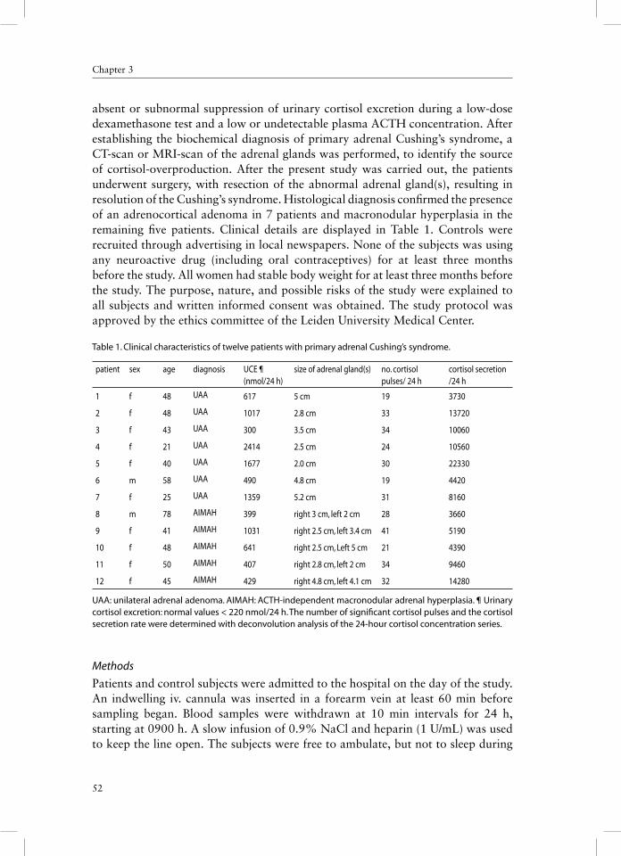
52
Chapter 3
absent or subnormal suppression of urinary cortisol excretion during a low-dose dexamethasone test and a low or undetectable plasma ACTH concentration. After establishing the biochemical diagnosis of primary adrenal Cushing’s syndrome, a CT-scan or MRI-scan of the adrenal glands was performed, to identify the source of cortisol-overproduction. After the present study was carried out, the patients underwent surgery, with resection of the abnormal adrenal gland(s), resulting in resolution of the Cushing’s syndrome. Histological diagnosis confi rmed the presence of an adrenocortical adenoma in 7 patients and macronodular hyperplasia in the remaining fi ve patients. Clinical details are displayed in Table 1. Controls were recruited through advertising in local newspapers. None of the subjects was using any neuroactive drug (including oral contraceptives) for at least three months before the study. All women had stable body weight for at least three months before the study. The purpose, nature, and possible risks of the study were explained to all subjects and written informed consent was obtained. The study protocol was approved by the ethics commit tee of the Leiden University Medical Center.
Table 1. Clinical characteristics of twelve patients with primary adrenal Cushing’s syndrome.
patient sex age diagnosis UCE ¶(nmol/24 h)
size of adrenal gland(s) no. cortisol pulses/ 24 h
cortisol secretion /24 h
1 f 48 UAA 617 5 cm 19 3730
2 f 48 UAA 1017 2.8 cm 33 13720
3 f 43 UAA 300 3.5 cm 34 10060
4 f 21 UAA 2414 2.5 cm 24 10560
5 f 40 UAA 1677 2.0 cm 30 22330
6 m 58 UAA 490 4.8 cm 19 4420
7 f 25 UAA 1359 5.2 cm 31 8160
8 m 78 AIMAH 399 right 3 cm, left 2 cm 28 3660
9 f 41 AIMAH 1031 right 2.5 cm, left 3.4 cm 41 5190
10 f 48 AIMAH 641 right 2.5 cm, Left 5 cm 21 4390
11 f 50 AIMAH 407 right 2.8 cm, left 2 cm 34 9460
12 f 45 AIMAH 429 right 4.8 cm, left 4.1 cm 32 14280
UAA: unilateral adrenal adenoma. AIMAH: ACTH-independent macronodular adrenal hyperplasia. ¶ Urinary cortisol excretion: normal values < 220 nmol/24 h. The number of signifi cant cortisol pulses and the cortisol secretion rate were determined with deconvolution analysis of the 24-hour cortisol concentration series.
Methods
Patients and control subjects were admitted to the hospital on the day of the study. An indwelling iv. cannula was inserted in a forearm vein at least 60 min before sampling began. Blood samples were withdrawn at 10 min intervals for 24 h, starting at 0900 h. A slow infusion of 0.9% NaCl and heparin (1 U/mL) was used to keep the line open. The subjects were free to ambulate, but not to sleep during

53
GH secretion in Cushing’s syndrome
the daytime. Meals were served at 0900, 1230 and 1730 h. Lights were turned off between 2200-2400 h. No sleep monitoring by EEG was used. Plasma for GH and cortisol measurements was collected, centrifuged at 4o C for 10 min, and stored at –20o C until later analysis. The results of the cortisol data were reported in a separate paper (submitted elsewhere); here we use only the 24 h secretion rates in regression analyses.
Assays
Plasma GH concentrations were measured in duplicate using a sensitive time-resolved immunofl uorometric assay (Wallac, Inc., Turku, Finland), specifi c for the 22-kDa GH protein. Human biosynthetic GH (Pharmacia & Upjohn Inc., Uppsala, Sweden) was used as standard calibrated against WHO-IRP 80-505, with a detection limit of 0.03 mU/L and an intra-assay variation coeffi cient of 1.6-8.4% at plasma values between 0.25-40 mU/L (to convert mU/L to μg/L divide by 2.6). All samples from any subject were run in the same assay.
The serum IGF-I was determined by RIA (Incstar Corp., Stillwater, MN.) with a detection limit of 1.5 nmol/L and an interassay variation coeffi cient of less than 11%. Plasma cortisol concentrations were measured by RIA (Sorin Biomedica, Milan, Italy). The detection limit of the assay was 25 nmol/l. The interassay variation varied from 2 – 4 % at the concentrations obtained in this study.
CALCULATIONS AND STATISTICS
Deconvolution analysis
A multiparameter deconvolution technique was used to estimate relevant measures of GH secretion from the 24-h serum GH concentration profi les, as described previously (53). Initial estimates of basal GH secretion rate were calculated to approximate the lowest 5% of all plasma GH concentrations in the time series. Peak detection entailed application of 95% statistical confi dence intervals to two thirds of all GH secretory peaks considered jointly and individual 95% statistical confi dence intervals to the remaining one third smaller pulses, as validated in simulations (12). The following four secretory and clearance measures of interest were estimated: 1) the number and locations of secretory events; 2) the amplitudes of secretory bursts; 3) the durations of randomly dispersed GH secretory bursts; and 4) the endogenous single component subject specifi c plasma half-life of GH. It was assumed the GH distribution volume and half-life were time and concentration invariant. The following parameters were calculated: Half-duration of secretory bursts (duration of the secretory burst at half-maximal amplitude), hormone half-life, burst frequency, amplitude of the secretory burst (maximal secretory rate attained within a burst), mass secreted per burst, basal secretion rate, pulsatile secretion rate (product of burst frequency and mean burst mass) and total secretion (sum of basal and pulsatile).

54
Chapter 3
Approximate Entropy
The univariate approximate entropy (ApEn) statistic was developed to quantify the degree of irregularity, or disorderliness, of a time series (42). Technically, ApEn quantifi es the summed logarithmic likelihood that templates (of length m) of patterns in the data that are similar (within r) remain similar (within the same tolerance r) on next incremental comparison and has been formally defi ned elsewhere (43). The ApEn calculation provides a single non-negative number, which is an ensemble estimate of relative process randomness, wherein larger ApEn values denote greater irregularity, as observed for ACTH in Cushing’s disease, GH in acromegaly, and PRL in prolactinomas (43,51,52). Cross-ApEn (X-ApEn) quantifi es joint pattern synchrony between two separate, but parallel time-series after standardization (z-score transformation) (44, 45). In the present analysis, we calculated cross-ApEn between cortisol (leading) and GH, with r=20% of the SD of the individual time-series and m=1. This parameters choice affords sensitive, valid and statistically well-replicated ApEn and cross-ApEn metrics for assessing hormone time-series of this length (44). ApEn and cross-ApEn results are reported as absolute values and as the ratio of the absolute value to that of the mean of 1000 randomly shuffl ed data series. Ratio values that approach 1.0 thus denote mean empirical randomness.
Copulsatility
Copulsatility between the cortisol and GH time-series was quantifi ed by the hypergeometric (joint binomial) distribution (54). This program calculates the probability that hormone pulses in time-series occur randomly. We used a time-window of 40 min, with cortisol as leading hormone series. The position (time of maximal secretion rate within a pulse) and number of pulses were derived from the deconvolution analyses.
Statistical analysis
Results are expressed as the mean ± SEM. Comparison between groups was done with one-way ANOVA, followed post hoc by Tuckey’s honestly signifi cantly different (HSD) test to contrast means. Derived measures (deconvolution and ApEn) were transformed logarithmically before analysis to limit dispersion of variance. In addition, (stepwise) linear regression was applied to evaluate the relation between relevant variables. Cross-correlation analysis was applied to test for signifi cant time-lagged (linear) synchrony between successive serum concentrations of cortisol and GH, considered pair wise, as described previously (54). Calculations were carried out with Systat (release 11, Systat Software Inc, Richmond, CA). Differences were considered signifi cant for P < 0.05.

55
GH secretion in Cushing’s syndrome
RESULTS
Daily plasma GH in patients and BMI-matched controls
Secretion profi les of the 24 h plasma GH concentration series of the patients are shown in Fig 1. Deconvolution of the GH profi les revealed no differences in basal GH secretion rate, secretory-burst half duration, burst amplitude, burst mass, half-life, basal secretion, pulsatile secretion and total secretion between the patients and the BMI-matched controls (table 2). GH was secreted in a predominantly pulsatile fashion in patients and in BMI-matched controls as displayed in the fi gure. In healthy lean controls GH secretion was two-fold higher than in patients, and was accomplished by a 2.5-fold increase in burst mass (P = 0.001) at similar pulse frequency. Total serum IGF-I concentrations were similar in the groups: patients 16.5 ± 3.4 nmol/L, BMI-matched controls 16.8 ± 0.8, and lean controls 20.1 ± 2.2 (ANOVA, P = 0.44).
9 15 21 3 902468
10Patien t # 2
9 15 21 3 90369
1215
Patien t # 1
9 15 21 3 902468
10Patien t # 6
9 15 21 3 902468
10Patien t # 8
9 15 21 3 90369
1215
P atien t # 9
9 15 21 3 902468
10Patien t # 12
GH
secr
etio
n ra
te (m
U/L
/ 24h
)
T im e (clock hou rs)
9 15 21 3 902468
109 15 21 3 9
02468
10
9 15 21 3 90
20
40
60
80
9 15 21 3 90
20
40
60
80
9 15 21 3 90
10
20
30
9 15 21 3 90369
1215
B M I-contro l
L ean contro l
L ean contro l
L ean contro l
B M I-contro l
B M I-contro l
Fig 1. GH concentration profi les of hypercortisolemic patients, obtained by 10-min blood sampling for 24 h. Sampling started at 0900 h, One mU/L=0.38 µg/L.

56
Chapter 3
Table 2. Secretory parameters of the 24 h GH plasma concentration series in twelve patients with ACTH-independent hypercortisolism and control groups
Patients (n=12) BMI-matched controls (n=12)
Lean controls (n=12)
P-value vs matched C
P-value vs lean C
ANOVA
Basal secretory rate (mU/L/min)
0.00563 ± 0.0012 [0.0044]
0.0067 ± 0.0009 [0.0062]
0.0116 ± 0.0035[0.0074]
NS NS 0.13
Half-life (min) 14.7 ± 0.6 [15.4]
14.2 ± 0.6[14.6]
14.7 ± 0.6[14.8]
NS NS 0.78
Secretory-burst half duration (min)
28.2 ± 2.2 [27.0]
25.5 ± 1.7 [26.6]
27.1 ± 1.8[27.7]
NS NS 0.62
No. of secretory bursts/24 h
20.5 ± 0.9 [21]
17.7 ± 1.4 [17]
17.3 ± 1.2[17.5]
0.10 0.06 0.13
Mean burst interval (min)
71 ± 3 [65]
83 ± 7 [80]
86 ± 6[84]
0.13 0.07 0.15
Secretory burst amplitude (mU/L/min)
0.178 ± 0.025 [0.172]
0.310 ± 0.047 [0.296]
0.490 ± 0.057[0.462]
0.11 0.00009 0.0001
Basal secretion (mU/L/24h)
8.1 ± 1.7 [6.3]
9.6 ± 1.4 [9.1]
16.7 ± 5.1[10.7]
0.90 0.15 0.13
Pulsatile secretion (mU/L/24h)
102 ± 13 [102]
134 ± 26[120]
229 ± 36[190]
0.69 0.006 0.006
Total secretion (mU/L/24h)
110 ± 13 [112]
143 ± 27 [127]
245 ± 40[200]
0.71 0.007 0.007
Statistical comparisons were made by ANOVA, followed by post hoc Tuckey’s HSD test. Data are expressed as mean ± SE. The median value is shown in brackets. One mU/L =0.38 µg/L.
GH and meals
The infl uence of meals on GH concentrations was analyzed by comparing the mean of 10 serial samples preceding lunch and dinner in patients and body weight-matched controls and mean GH in the samples after start of lunch and dinner during 90 min. In patients the mean GH decrease after lunch was 0.48 mU/L (P = 0.03), and in controls 1.16 mU/L (P = 0.006).The mean GH decrease after diner was 1.51 mU/L in patients (P = 0.04) and in controls 2.19 mU/L (P = 0.02). The mean GH decreases in patients and controls were statistically similar.
Approximate entropy
ApEn in patients was increased, denoting an irregular secretion pattern: patients 0.7386 ± 0.044 vs. BMI-matched controls 0.5271 ± 0.0455 (P = 0.04) and vs.
lean controls 0.4492 ± 0.050 (P = 0.001). The ApEn ratio in patients was 0.5102 ± 0.015, 0.4250 ± 0.021 in body weight-matched controls (P=0.016) and 0.3820 ± 0.024 in lean controls (P = 0.0002).
Factors infl uencing GH secretion
In a stepwise linear regression analysis the 24 h GH secretion in patients and BMI-matched controls was signifi cantly negatively correlated with BMI (R = -0.55, P = 0.005), as displayed in Fig 2. However, other parameters including cortisol secretion

57
GH secretion in Cushing’s syndrome
rate, free urinary cortisol excretion, age, estradiol, gender and duration of cortisol excess (in patients only) were non-signifi cant predictors. Thus, the variation in total GH secretion was explained by BMI for 30%. In addition, ApEn was signifi cantly and positively correlated (R = 0.77, P = 0.003) with the cortisol secretion rate, as displayed in Fig 3, but not with BMI (R = 0.03).
Relation between cortisol and GH secretion
Pattern synchrony between cortisol and GH was quantifi ed by cross-ApEn in patients and BMI-matched controls. X-ApEn in patients was 1.648 ± 0.113 and in controls 1.004 ± 0.050 (P < 0.0001). The ApEn ratios were 0.8682 ± 0.054 and 0.6134 ± 0.026, respectively (P< 0.0001), denoting diminished pattern synchrony in patients. Conventional linear cross-correlation between cortisol (leading) and GH concentrations revealed a negative correlation in control subjects (median -0.30, 95% confi dence interval (CI) -0.15 to -.0.39), and a mean time lag of 30 min (95% CI 0-65 min), indicating opposite changes in cortisol concentrations
9 15 21 3 90369
9 15 21 3 902468
10Patien t # 12
T im e (clock hou rs)
9 15 21 3 90
20
40
60
809 15 21 3 9
0
10
L ean contro l
BMI (kg/m2 )
18 20 22 24 26 28 30 32 34 36 38
GH
Sec
retio
n (m
U/L
/24
h)
0
100
200
300
400
500
R = -0.55P = 0.005
Fig 2. Linear regression of GH secretion rate per 24 h, as estimated by deconvolution of the serum GH profi les, on BMI. Closed symbols refl ect hypercortisolemic patients (circles unilateral adenoma, triangles bilateral nodular hyperplasia) and open squares BMI-matched controls.
Cortisol secretion (nmol/L/ 24h)0 5000 10000 15000 20000 25000
GH
ApE
n 1,
20%
0.4
0.5
0.6
0.7
0.8
0.9
1.0
1.1R=0.77P=0.003
Fig 3. Relation between cortisol secretion rate and ApEn in patients. Patients with a unilateral adenoma are shown as circles and patients with bilateral hyperplasia as triangles.

58
Chapter 3
followed by those of GH. Five of the patients had a positive correlation. Median correlation coeffi cient was -0.09 with a 95% CI of -0.15 to +0.16. The mean time lag was 75 min, 95% CI 37-100 min. Co-pulsatility of cortisol and GH pulses was statistically highly signifi cant in all patients and in 10 of 12 control subjects ( P-values between 10-3 to 10-13).
Unilateral vs. bilateral adrenal pathology
BMI, IGF-I and age were comparable in these subgroups. No differences were found in GH secretion parameters as estimated by deconvolution, ApEn and synchrony estimates of GH and cortisol.
DISCUSSION
In this investigation of primary adrenal-cortisol excess, the 24-h GH secretion was comparable to BMI-matched healthy controls, and IGF-I concentrations were similar. However, the regularity of the GH secretory process and the pattern synchrony of cortisol and GH in the patients were clearly diminished.
Stimulated GH release is severely restricted in Cushing’s syndrome, and either no increase or only a small increment is noted after administration of GHRH, GHRP (hexarelin and GHRP-2) and ghrelin (2, 20, 30, 33). Since most of the GH-stimulation studies in Cushing’s syndrome lack body weight-matched controls, the specifi city of this fi nding might be questioned. GH release after reduction of the endogenous somatostatin tonus is also greatly diminished in hypercortisolism, e.g. by pre-treatment with pyridostigmine, arginine infusion, or after abrupt cessation of an iv infusion with somatostatin (13, 28,34). Collectively, these results could point to a (reversible) defect of the pituitary gland, i.c. the somatotropic cell. Indeed, repeated GHRH administration in the hypercortisolemic state leads to potentiation to this hormone (29). Furthermore, administration of acipimox caused a 7-fold increase in GH release after GHRH administration, accompanied by a 3-fold decrease in circulating FFA’s, and almost doubling of spontaneous 24 h GH secretion (31). Finally a hypocaloric diet for 3 days resulted in a 4-fold GH increase after GHRH injection (32).
Similarities with experimental results in obesity are distinct, since it is well-established that GHRH-stimulated GH release is diminished in obesity and increases during caloric restriction and after weight loss (14). Spontaneous 24-h GH secretion is severely restricted in the overweight human and increases or normalizes after weight reduction and during acipimox treatment (23, 41). In other studies, both BMI and abdominal visceral-fat mass predict irregular (disorderly) GH release (12, 50). The basis for this inferred feedback alteration in GH secretion is not known (14).
Reports on spontaneous GH secretion in Cushing’s syndrome, as studied with 24 h blood sampling protocols, are scarce. In one such contribution, Magiakou studied

59
GH secretion in Cushing’s syndrome
15 patients with hypercortisolism (14 pituitary-dependent and one with primary bilateral pigmented nodular hyperplasia), of whom 6 patients were prepubertal. They described severely depressed GH secretion compared with normal-weight controls, mainly caused by decreased pulse amplitude, but with unchanged pulse frequency (35). The intriguing observation was that the expected restoration of GH secretion after curative pituitary surgery failed to occur, notwithstanding signifi cant weight loss and normalization of BMI in the 50% of the patients, who had preoperatively increased values. These observations suggest that (visceral) obesity is an important determinant of GH secretion in Cushing’s syndrome, irrespective of its etiology, but apparently after pituitary surgery other factors play (or still play) a role in the diminished GH secretion.
We established a signifi cant negative relationship between BMI and GH secretion in pituitary-independent hypercortisolism and in the matched controls. BMI, however, explained only 30% of the variability in GH, suggesting that other mechanisms likely contribute to the observed hyposomatotropism, as discussed above. It is unfortunate that we had no data on visceral fat mass in our patients and controls, because most likely, a higher correlation coeffi cient would have been found. Nevertheless, we did not fi nd a relation between the degree of cortisol excess and GH secretion rate, as we previously found for pituitary–dependent hypercortisolism (17). A conspicuous difference in clinical presentation between the two forms of the syndrome was the very high cortisol secretion rate in some of the (male) Cushing’s disease patients, which could explain the divergent results.
Compared with lean controls our patients had a 50 % reduction in pulsatile GH secretion, exclusively caused by secretory-burst amplitude decrement. In the absence of a signifi cant change in basal (non-pulsatile) secretion this observation is compatible with heightened somatostatin inhibition (3), decreased hypothalamic GHRH secretion, a defect in the GHRH/GH secretagogue receptor signalling or direct non-receptor-related GH inhibition. Experimental evidence, mainly obtained in the rat, has demonstrated that high doses of glucocorticoids decrease the expression of hypothalamic GHRH mRNA, and increase that of somatostatin (11, 14, 27). On the other hand, dexamethasone increased mRNA of the GHRH receptor and the GH secretagogue receptor, which certainly explains the dexamethasone-potentiation of GH release after GHRH in the human and in the rat (26, 37, 49), but not the diminished GH response to GHRH/GHS during chronic glucocorticoid excess. Accordingly, the amount and duration of cortisol excess appear to be important.
Other mechanisms might limit GH secretion in chronic hypercortisolism. For instance, in the rat dexamethasone administration decreased mitosis and increased apoptosis of pituitary cells (38). If such a mechanism is also present in the human somatotrope, this might (partly) explain the extended time (one year or more) it takes for restoration of GH secretion in most of the (adult) patients after surgical cure of Cushing’s syndrome (18, 21, 49). Nonetheless, permanent damage to the somatotrope appears to be the rule rather than exception in childhood-onset

60
Chapter 3
Cushing’s disease after surgery and radiation treatment (4). Another mechanism potentially relevant for the inhibitory effect of glucocorticoids on GH secretion is via the action of annexin 1. This peptide is a mediator of the anti-infl ammatory actions of glucocorticoids and has signifi cant effects on cell growth, differentiation, apoptosis, membrane fusion, endocytosis and exocytosis (22). This peptide, widely distributed in the body, is also present in the folliculostellate cells in the pituitary gland, but not in the pituicytes, and exerts its GH-suppressing effect on the somatotrope via a paracrine mechanism at a point distal to the formation of cyclic AMP and Ca ion entry (48). However, the same mediator has also a centrally stimulatory effect on GH (40). Finally, leptin might also be involved in the GH regulation. Circulating leptin concentrations in Cushing’s syndrome are disproportionately increased compared with BMI-matched healthy controls (7, 15, 36). Short-term fasting in Cushing’s syndrome did not restore normal leptin levels, and GH secretion remained blunted (19). However, several recent clinical studies suggest that a direct role for leptin in GH regulation is rather limited. In morbidly obese patients treated by biliopancreatic diversion changes in insulin levels predicted changes in leptin levels and the somatotropic axis (8). Also observations in patients with homozygous and heterozygous leptin gene mutations indicate that GH secretion is correlated with adiposity (39). Finally, r-metHuleptin administration in healthy lean men did not prevent fasting-induced augmentation of GH pulsatility or decline in free IGF-I levels, but restored in part total IGF-I levels (5).
GH concentration fell after meals in patients and in controls. Theoretically, one might expect a diminished inhibitory action in patients, because of decreased hypothalamic GHRH expression, and increased somatostatin expression, as discussed above (11, 14, 27). The differences between patients and controls were not signifi cant (P-values ~ 0.60), suggesting that lack of power was not responsible.
A conspicuous and specifi c observation was the decreased regularity of GH secretion measured using ApEn, as previously described in patients with ACTH-producing pituitary adenomas (17). The degree of irregularity of GH release in patients with adrenal cortisol excess was signifi cantly greater than that estimated in obese controls. The ApEn statistic quantitates the relative orderliness or reproducibility of subordinate (nonpulsatile) secretory patterns in neurohormone time series, which in turn mirrors feedforward and feedback adjustments driven by (patho) physiological changes in interglandular communication. The validity of ApEn to this end has been established in theoretical and experimental contexts (9, 55, 56). In view of the unchanged IGF-I feedback signal in the patients, decreased regularity of GH secretion could refl ect impaired coordinate control of GH secretion by somatostatin, GHRH and ghrelin and/or altered pituitary responsiveness to these peptides (9,10). Available data do not address the reversibility of disorderly GH release due to endogenous adrenal cortisol excess with presumptively normal premorbid hypothalamo-pituitary function.
The 24-hour concentration profi les allowed an appraisal of possible coordinate secretion of cortisol with GH. In normal subjects we found a reciprocal relationship

61
GH secretion in Cushing’s syndrome
between these two hormones, as previously demonstrated in mid-luteal phase-women and in children (1, 6). The inverse relationship might be explained by the known ability of glucocorticoids to suppress GH secretion, possibly via heightened somatostatinergic tone (14). In patients the correlation between the two hormones was smaller, and even positive in 5 subjects. Indeed, abolishment of the cortisol-GH correlation can be induced by fasting in adult healthy women, while a positive correlation is seen in children with congenital adrenal hyperplasia under glucocorticoid substitution therapy (1, 6). Changes of cortisol patterns as observed during the stress of caloric deprivation, and by defi nition non-physiological glucocorticoid substitution therapy lead to desynchronization of hormone secretion patterns, as we now also described for endogenous primary adrenal hypercorticism. The loss of inter-axis synchrony in our patients is corroborated by (lag-independent) cross-ApEn analysis. Disruption of pattern synchrony of GH and cortisol is also seen during fasting in adult women. Interestingly and not previously reported was the loss of synchrony between GH and cortisol in 15 patients with ACTH-dependent hypercorticism (45, 47). In these patients cross-ApEn was 1.640 ± 0.068, thus greatly elevated to a similar degree as the adrenal form of hypercorticism (P = 0.000013 vs. controls, and P = 0.99 vs. adrenal hypercorticism). Collectively, these results indicate that endogenous hypercorticism leads to disruption of cortisol-GH synchrony, irrespective of its cause. Notwithstanding the obvious loss in synchrony, copulsatility of cortisol and GH remained strong. This fi nding is somewhat surprising, since tumoral cortisol secretion in patients with adrenal adenoma is ACTH-independent, and could therefore indicate that cortisol feedback is involved in the temporal timing of GH pulses. At present, no other data in literature are available to support this hypothetical view.
In summary, patients with primary adrenal Cushing’s syndrome exhibit moderate hyposomatotropism, as demonstrated by decreased GH pulsatile secretion, which is only partly ( ~ 30%) explained by adiposity. This observation in combination with disruption of GH pattern regularity and synchrony points to impaired net peptidyl-drive of orderly somatotrope secretion.

62
Chapter 3
REFERENCES
1. Bergendahl M, Iranmanesh A, Pastor C, Evans WS and Veldhuis JD. Homeostatic joint amplifi cation of pulsatile and 24-hour rhythmic cortisol secretion by fasting stress in midluteal phase women: concurrent disruption of cortisol-growth hormone, cortisol-luteinizing hormone, and cortisol-leptin synchrony. J Clin Endocrinol Metab 85: 4028-4035, 2000.
2. Burguera B, Muruais C, Penalva A, Dieguez C and Casanueva FF. Dual and selective actions of glucocorticoids upon basal and stimulated growth hormone release in man. Neuroendocrinology 51: 51-58. 1990.
3. Calabresi E, Ishikawa E, Bartolini L, Delitala G, Fanciulli G, Oliva O, Veldhuis JD and Serio M. Somatostatin infusion suppresses GH secretory burst number and mass in normal men: a dual mechanism of inhibition. Am J Physiol Endocrinol Metab 270: E975-E979, 1996.
4. Carroll PV, Monson JP, Grossman AB, Besser GM, Plowman PN, Afshar F and Savage MO. Successful treatment of childhood-onset Cushing’s disease is associated with persistent reduction in growth hormone secretion. Clin Endocrinol 60:169-174, 2004.
5. Chan JL, Heist K, DePaoli AM, Veldhuis JD and Mantzoros CS. The role of falling leptin levels in the neuroendocrine and metabolic adaptation to short-term starvation in healthy men. J Clin Invest 111:1409-1421, 2003.
6. Charmandari, E, Pincus SM, Mathews DR, Johnston A, Brook CGD and Hindmarsh PC. Oral hydrocortisone administration in children with classic 21-hydroxylase defi ciency leads to more synchronous joint growth hormone and cortisol secretion. J Clin Endocrinol Metab 87: 2238-2244, 2002.
7. Cizza G, Lotsikas AJ, Licinio J, Gold PW and Chrousos GP. Plasma leptin levels do not change in patients with Cushing’s disease shortly after correction of hypercortisolism. J Clin Endocrinol Metab 82: 2747-2750, 1997.
8. De Marinis L, Bianchi A, Mancini A, Gentilella R, Perrella M, Giampietro A, Porcelli T, Tilaro L, Fusco A, Valle D and Tacchino RM. Growth hormone secretion and leptin in morbid obesity before and after biliopancreatic diversion: relationships with insulin and body composition. J Clin Endocrinol Metab 89:174-180, 2004.
9. Fahry LS and Veldhuis JD. Joint pituitary-hypothalamic and intrahypothalamic autofeedback construct of pulsatile growth hormone secretion. Am J Physiol Regul Integr Comp Physiol 285:R1240-R1249, 2003.
10. Fahry LS and Veldhuis JD. Putative GH pulse renewal: periventricular somatostatinergic control of an arcuate-nuclear somatostatin and GH-releasing hormone oscillator. Am J Physiol Regul Integr Comp Physiol 286:R1030-R1042, 2004.
11. Fife SK, Brogan RS, Giustina A and Wehrenberg WB. Immunocytochemical and molecular analysis of the effects of glucocorticoid treatment on the hypothalamic-somatotropic axis in the rat. Neuroendocrinology 64:131-138, 1996.
12. Friend K, Iranmanesh A and Veldhuis JD. The orderliness of growth hormone (GH) release process and the mean mass of GH secreted per burst are highly conserved in individual men on successive days. J Clin Endocrinol Metab 81:3746-3753, 1996.
13. Giustina A, Bossoni S, Bodini C, Ferrari C, Pizzocolo G, Scalvini T, Schettino M and Wehrenberg WB. Pyridostigmine enhances even it does not normalize the growth hormone responses to growth hormone-releasing hormone in patients with Cushing’s disease. Horm Res 35:99-103, 1991.
14. Giustina A and Veldhuis JD. Pathophysiology of the neuroregulation of growth hormone secretion in experimental animals and the human. Endocr Rev 19:717-797, 1998.
15. Groote Veldman R, Frölich M, Pincus SM, Veldhuis JD and Roelfsema F. Hyperleptinemia in women with Cushing’s disease is driven by high-amplitude pulsatile, but orderly and eurhythmic, leptin secretion. Eur J Endocrinol 144:21-27, 2001.
16. Groote Veldman R, Van den Berg G, Pincus SM, Frölich M, Veldhuis JD and Roelfsema F. Increased episodic release and disorderliness of prolactin secretion in both micro- and macroprolactinomas. Eur J Endocrinol 140: 192-200, 1999.

63
GH secretion in Cushing’s syndrome
17. Groote Veldman R, Frölich M, Pincus SM, Veldhuis JD and Roelfsema F. Growth hormone and prolactin are secreted more irregularly in patients with Cushing’s disease. Clin Endocrinol 52:625-635, 2000.
18. Groote Veldman R, Frölich M, Pincus SM, Veldhuis JD and Roelfsema F. Apparently complete restoration of normal daily adrenocorticotropin, cortisol, growth hormone, and prolactin secretory dynamics in adults with Cushing’s disease after clinically successful transsphenoidal adenomectomy. J Clin Endocrinol Metab 85:4039-4046, 2000.
19. Grottolli S, Gauna C, Tassone F, Aimaretti G, Corneli G, Wu Z, Strasburger CJ, Dieguez C, Cassanueva FF, Ghigo E and Maccario M. Both fasting-induced leptin reduction and GH increase are blunted in Cushing’s syndrome and in simple obesity. Clin Endocrinol 58:220-228, 2003.
20. Hotta M, Shibasaki T, Masuda A, Imaki T, Sugino M, Demura H, Ling N and Shizume K. Effect of human growth hormone-releasing hormone on GH secretion in Cushing’s syndrome and non-endocrine disease patients treated with glucocorticoids. Life Sci 42:979-984, 1988.
21. Hughes NR, Lissett CA and Shalet SM. Growth hormone status following treatment for Cushing’s syndrome. Clin Endocrinol 51:61-66, 1999.
22. John CD, Christian HC, Morris JF, Flower RJ, Solito E and Buckingham JC. Annexin 1 and the regulation of endocrine function. Trends in Endocrinol Metab 15:103-109, 2004.
23. Kok P, Buijs MM, Kok SW, van Iersel IHAP, Frölich M, Roelfsema F, Voshol PJ and Meinders AE, Pijl H. Acipimox enhances growth hormone secretion in obese women. Am J Physiol Regul Integr Comp Physiol 286:R693-R698, 2003.
24. Krieger DT and Glick SM. Growth hormone and cortisol responsiveness in Cushing’s syndrome. Relation to a possible central nervous system etiology. Am J Med 52: 25-40, 1972.
25. Lacroix A, Ndiaye N, Tremblay J and Hamet P. Ectopic and abnormal hormone receptors in adrenal Cushing’s syndrome. Endocr Rev 22:75-110, 2001.
26. Lam KS, Lee MF, Tam SP and Srivastava G. Gene expression of the receptor for growth hormone-releasing hormone is physiologically regulated by glucocorticoids and estrogen. Neuroendocrinology 63: 475-480, 1996.
27. Lam KS and Srivastava G. Gene expression of hypothalamic somatostatin and growth hormone-releasing hormone in dexamethasone-treated rats. Neuroendocrinology 66: 2-8, 1997.
28. Leal-Cerro A, Pereira JL, Garcia-Luna PP, Astorga R, Cordido F, Casanueva FF and Dieguez C. Effect of enhancement of endogenous cholinergic tone with pyridostigmine on growth hormone (GH) responses to GH-releasing hormone in patients with Cushing’s syndrome. Clin Endocrinol 33:291-295, 1990.
29. Leal-Cerro A, Pumar A, Villamil F, Astorga R, Dieguez C and Casanueva FF. Growth hormone releasing hormone priming increases growth hormone secretion in patients with Cushing’s syndrome Clin Endocrinol 38:399-403, 1993.
30. Leal-Cerro A, Pumar A, Garcia-Garcia E, Dieguez C and Casanueva FF. Inhibition of growth hormone release after combined administration of GHRH and GHRP-6 in patients with Cushing’s syndrome. Clin Endocrinol 41: 649-654, 1994.
31. Leal-Cerro A, Jimenez LM, Astorga R, Fernandez-Lopez I, Dieguez C and Casanueva FF. Acute pharmacological reduction of plasma free fatty acids enhances the growth hormone (GH)-releasing hormone-mediated GH secretion in patients with Cushing’s syndrome. J Clin Endocrinol Metab 82:3165-3168, 1997.
32. Leal-Cerro A, Venegas E, Garcia-Pesquera F, Jimenez LM, Astorga R, Dieguez C and Casanueva FF. Enhanced growth hormone (GH) responsiveness to GH-releasing hormone after dietary restriction in patients with Cushing’s syndrome. Clin Endocrinol 48:117-121, 1998.
33. Leal-Cerro A, Torres E, Soto A, Dios E, Deghenghi R, Arvat E, Ghigo E, Dieguez C and Casanueva FF. Ghrelin is no longer able to stimulate growth hormone secretion in patients with Cushing’s syndrome but instead induces exaggerated corticotrophin and cortisol responses. Neuroendocrinology 76:390-396, 2002.
34. Leal-Cerro A. Soto A, Martinez MA, Alvarez P, Isidro L, Casanueva FF, Dieguez C and Cordido F. Effect of withdrawal of somatostatin plus growth hormone (GH)-releasing hormone as a stimulus of GH secretion in Cushing’s syndrome. Clin Endocrinol 57: 745-749, 2002.

64
Chapter 3
35. Magiakou MA, Mastorakos G, Gomez MT, Rose SR and Chrousos GH. Suppressed spontaneous and stimulated growth hormone secretion in patients with Cushing’s disease before and after surgical cure. J Clin Endocrinol Metab 78:131-137, 1994.
36. Masuzaki H, Ogawa Y, Hosoda K, Miyawaki T, Hanaoka I, Hiraoka J, Yasuno A, Nishimura H, Yoshimasa Y, Nishi S and Nakao K. Glucocorticoid regulation of leptin synthesis and secretion in humans: elevated plasma leptin levels in Cushing’s syndrome. J Clin Endocrinol Metab 82: 2542-2547, 1997.
37. Miller TL and Mayo KJ. Glucocorticoids regulate pituitary growth hormone-releasing hormone receptor messenger ribonucleic acid expression. Endocrinology 138: 2458-2465, 1997.
38. Nolan LA, Kavanagh E, Lightman SL and Levy A. Anterior pituitary cell population control: basal cell turnover and the effects of adrenalectomy and dexamethasone treatment. J Neuroendocrinol 10:207-215, 1998.
39. Ozata M, Dieguez C and Casanueva FF. The inhibition of growth hormone secretion presented in obesity is not mediated by the high leptin levels: a study in human leptin defi ciency patients. J Clin Endocrinol Metab 88: 312-316, 2003.
40. Philip JG, John CD, Cover PO, Morris JF, Christian HC, Flower RJ and Buckingham JC. Opposing infl uences of glucocorticoids and interleukin-1 on the secretion of growth hormone and ACTH in the rat in vivo: role of hypothalamic annexin 1. Br J Pharmacol 134: 887-895, 2001.
41. Pijl H, Langendonk JG, Burggraaf J,Frölich M, Cohen AF, Veldhuis JD and Meinders AE. Altered neuroregulation of GH secretion in viscerally obese premenopausal women. J Clin Endocrinol Metab 86:5509-5515, 2001.
42. Pincus SM. Approximate entropy as a measure of system complexity. Proc Natl Acad Sci USA 88:2297-2301, 1991.
43. Pincus SM and Goldberger AL. Physiological time-series analysis: what does regularity quantifi es? Am J Physiol 266: H1643-H1656, 1994.
44. Pincus SM, Mulligan T, Iranmanesh A, Gheorghiu S, Godschalk M and Veldhuis JD. Older males secrete luteinizing hormone more irregularly, and jointly more asynchronously than younger males. Proc Natl Acad Sci USA 93: 14100-14105, 1994.
45. Roelfsema F, Pincus SM and Veldhuis JD. Patients with Cushing’s disease secrete adrenocorticotropin and cortisol jointly more asynchronously than healthy subjects. J Clin Endocrinol Metab 83:688-692, 1998.
46. Smals AE, Pieters GF, Smals AG, Benraad TJ and Kloppenborg PJ. Human pancreatic growth hormone releasing hormone fails to stimulate human growth hormone both in Cushing’s disease and in Cushing’s syndrome due to adrenal adenoma. Clin Endocrinol 24:401-407, 1986.
47. Tamura H, Kamegai J, Sugihara H, Kineman RD, Frohman LA and Wakabayashi I. Glucorticoids regulate pituitary growth hormone secretagogue receptor gene expression J Neuroebdocrinol 12:481-485, 2000.
48. Taylor AD, Christian HC, Morris JF, Flower RJ and Buckingham JC. Evidence from immunoneutralization and antisense studies that the inhibitory actions of glucocorticoids on growth hormone release in vitro require annexin 1 (lipocortin 1). Br J Pharmacol 131:1309-1316, 2000.
49. Tzanela M, Karavitaki N, Stylianidou C, Tsagarakis S and Thalassinos NC. Assessment of GH reserve before and after successful treatment of adult patients with Cushing’s syndrome. Clin Endocrinol 60:309-314, 2004.
50. Vahl N, Jorgensen JO, Skjaerback C, Veldhuis JD, Orskov H and Christiansen J.Abdominal adiposity rather than age and sex predicts the mass and pattern regularity of growth hormone secretion in mid-life healthy adults. Am J Physiol Endocrinol Metab 272: E1108-E1116, 1997.
51. Van den Berg, Pincus SM, Veldhuis JD, Frölich M and Roelfsema F. Greater disorderliness of ACTH and cortisol release accompanies pituitary-dependent Cushing’s disease. Eur J Endocrinol 136:394-400, 1997.
52. Van den Berg G, Pincus SM, Frölich M, Veldhuis JD and Roelfsema F Reduced disorderliness of growth hormone release in biochemically inactive acromegaly after pituitary surgery. Eur J Endocrinol 138:164-169, 1998.

65
GH secretion in Cushing’s syndrome
53. Veldhuis JD, Carlson ML and Johnson ML. The pituitary gland secretes in bursts: appraising the nature of glandular secretory impulses by simultaneous multiple-parameter deconvolution of plasma hormone concentrations. Proc Natl Acad Sci USA 84: 7686-7690, 1987.
54. Veldhuis JD, Johnson ML, Faunt LM and Seneta E. Assessing temporal coupling between two, or among three or more, neuroendocrine pulse trains: cross-correlation analysis, simulation methods, and conditional probability testing. Methods Neurosci 20:336-376, 1994.
55. Veldhuis JD, Straume M, Iranmanesh A, Mulligan T, Jaffe C, Barkan A, Johnson ML and Pincus S. Secretory process regularity monitors neuroendocrine feedback and feedforward signalling strength in humans. Am J Physiol Regul Integr Comp Physiol 280:R721-R729, 2001.
56. Veldhuis JD, Johnson ML, Veldhuis OL, Straume M and Pincus SM. Impact of pulsatility on the ensemble orderliness (approximate entropy) of neurohormone secretion. Am J Physiol Regul Integr Comp Physiol 281:R1975-R1985, 2001.


Chapter 4Profound Amplifi cation of Secretory-Burst Mass and Anomalous Regularity of ACTH Secretory Process in Patients with Nelson’s
Syndrome Compared with Cushing’s Disease
Maarten O. van Aken1, Alberto M Pereira1, Gerrit van den Berg 1, Johannes A. Romijn1, Johannes D.Veldhuis 2, Ferdinand Roelfsema1
1Department of Endocrinology and Metabolic Diseases, Leiden University Medical Center, the Netherlands, and 2 Endocrine Research Unit, Department of Internal Medicine, General Clinical Research Center, Mayo Medical School, Mayo Clinic and Foundation, Rochester,
MN 55905.
Clinical Endocrinology 2004:60;765-772

68
Chapter 4
SUMMARY
Objective
As described originally, Nelson’s syndrome is characterized by grossly elevated ACTH concentrations, a sellar mass and skin hyperpigmentation emerging in the course of Cushing’s disease after bilateral adrenalectomy. No detailed studies have defi ned whether the mechanisms directing ACTH secretion differ in Nelson’s syndrome and untreated Cushing’s disease.
Patients and methods
To address this pathophysiological issue, we studied 9 patients fulfi lling the criteria of Nelson’s syndrome receiving glucocorticoid and mineralocorticoid replacement; 9 patients with untreated pituitary-dependent Cushing’s disease and 9 gender- and age-matched controls. ACTH release was appraised by monitoring plasma ACTH concentrations in blood samples collected every 10 min for 24 h. ACTH secretion rates and endogenous decay were quantifi ed by multiparameter deconvolution analysis. The orderliness of the ACTH release process was delineated by the approximate entropy (ApEn) statistic. Diurnal variation in ACTH secretion was appraised by Cosinor analysis.
Results
Basal ACTH secretion was increased 6-fold and pulsatile secretion 9-fold in patients with Nelson’s syndrome compared with Cushing’s disease (P < 0.01 and P<0.001, respectively). The increase in pulsatile secretion was due to an 8-fold augmentation of burst mass. Event frequency was comparable in both patient groups (32 ± 1 vs 28 ± 2 pulses/24h), and higher than in normal controls (22 ± 1 pulses per 24 h, P< 0.0001). Paradoxically, the consistency of subordinate patterns of serial ACTH release, albeit disrupted in active Cushing’s disease, was normal in Nelson’s syndrome (P = 0.014). Normal ACTH secretory-process regularity in Nelson’s syndrome was attributable to a more reproducible (lower ApEn) succession of ACTH secretory-burst mass denoting more uniform amplitude evolution over 24 h (P=0.007, Nelson vs Cushing). On the other hand, the quantifi able regularity of serial interburst intervals (waiting times) was unexpectedly elevated in Nelson’s syndrome (P=0.022). Nelson patients maintained a signifi cant diurnal rhythm in ACTH release, which was marked by a 15-fold greater amplitude (P = 0.0018 vs Cushing’s) and a 4-h acrophase (maximum) delay (P=0.037 vs control).
Conclusion
The present detailed analyses delineate marked ACTH secretory-burst mass amplifi cation and (amplitude-independent) anomalous regularity of successive pulse size and timing in Nelson’s syndrome compared with Cushing’s disease or controls. We postulate that the foregoing novel distinctions are due to unique tumoral

69
Neurohormone output in Nelsons’s syndrome
secretory properties, concurrently required glucocorticoid replacement and/or hypothalamic injury associated with prior radiotherapy in Nelson’s syndrome.
INTRODUCTION
Nelson’s syndrome was fi rst described in 1958 as the constellation of a pituitary macroadenoma, markedly elevated ACTH concentrations, and hyperpigmentation of the skin in a patient after bilateral adrenalectomy for pituitary-dependent hypercortisolism (Cushing’s disease) (Nelson et al., 1958). The syndrome develops in 8 - 38% of adults requiring bilateral adrenalectomy for Cushing’s disease (Nagesser et al., 2000 a, Kemink et al., 2001) and occurs infrequently in patients aged 40 yr or more at the time of bilateral adrenalectomy, in contrast to patients treated at an early age (Kemink et al., 1994). The pathogenetic mechanism’s underlying tumorigenesis and unrestrained ACTH secretion in Nelson’s syndrome are not well understood.
In Cushing’s disease, excessive ACTH production is characterized by a marked elevation of basal (nonpulsatile) secretion and secretory-burst mass in association with marked disruption of orderly release and diurnal rhythmicity (Van den Berg et al., 1995). Transsphenoidal adenomectomy normalises most or all alterations in ACTH secretion (Groote Veldman et al., 2000). Cushing’s disease and Nelson’s syndrome are considered to be distinct pathoaetiological presentations of the same primary biological entity. For example, impaired responsiveness to glucocorticoid enforced negative feedback on ACTH is common to both (Cook et al., 1976). In addition, under in vitro conditions the secretion of POMC-derived peptides was similar in tumoural tissue derived from patients with Cushing’s disease and Nelson’s syndrome (Westphal & Lüdecke, 1984) However, CRH infusion stimulates greater and more prolonged ACTH secretion in patients with Nelson’s syndrome than Cushing’s disease (Oldfi eld et al., 1986). At present, there are relatively few other quantitative comparisons of neurosecretory control of tumoural ACTH secretion in these two clinical pathophysiological entities. The purpose of the present study was to explore and compare the 24-h spontaneous ACTH secretion dynamics in this group of patients with untreated classical Cushing’s disease and healthy controls.
Subjects and Methods
Before the availability of transsphenoidal microsurgery for the treatment of Cushing’s disease, patients usually underwent bilateral adrenalectomy. In our centre, however, patients were treated by unilateral adrenalectomy and pituitary irradiation until 1978, resulting in remission of the disease in 64% (Nagesser et al., 2000 b). Non-cured patients underwent complete adrenalectomy, usually after one year of clinical follow-up. Seven of the patients (see Table I) were treated in this way, but developed clinical symptoms of Nelson’s syndrome, i.e. hyperpigmentation and grossly elevated ACTH concentrations. Two other patients (patients 7 and 8) underwent bilateral adrenalectomy after unsuccessful pituitary surgery and subsequently developed

70
Chapter 4
Nelson’s syndrome. We defi ned Nelson’s syndrome as bilateral adrenalectomy for Cushing’s disease in the past, plasma ACTH-levels of more than 300 ng/L during hydrocortisone replacement therapy (20 mg/day) and hyperpigmentation of the skin. Radiological evidence of a pituitary tumour was found in 7/9 patients. This defi nition of Nelson’s syndrome agrees with a previous report and discussion ( Kasperlik-Zaluska et al., 1996, Kasperlik-Zaluska & Jeske, 2001). In total we studied nine patients with Nelson’s syndrome (7 females, 2 males), nine patients with proven Cushing’s disease and 9 healthy controls matched for gender and BMI.
In order to prevent spurious elevated ACTH concentrations due to low circulating cortisol concentrations under substitution, the medication was switched to dexamethasone. Therefore, starting one day before and during the sampling period, patients with Nelson’s syndrome received a standardized steroid-replacement schedule, consisting of dexamethasone 0.25 mg at 0800 and 1800 h and fl udrocortisone 0.125 mg at 0800 h.
Patients with Cushing’s disease were diagnosed by elevated 24-h urinary excretion of free cortisol, subnormal or absent suppression of plasma cortisol after administration of 1 mg dexamethasone overnight, absent or subnormal suppression of urinary cortisol excretion during a low-dose dexamethasone test, suppression of plasma cortisol by 190 nmol/L or more during a 7-h iv infusion of dexamethasone 1 mg/h (Biemond et al., 1990), positive adenoma immunostaining for ACTH and clinical cortisol dependency for several months after selective removal of the adenoma. The mean 24-h plasma cortisol concentration (mean of 145 samples of each series) was 690 ± 140 nmol/L in Cushing’s disease and 206 ± 20 nmol/L in healthy controls (P=0.008).
Table 1 Clinical characteristics of nine patients with Nelson’s syndrome
Patient Sex (m/f )/ Age (yr)
Primary therapy Interval between ADX and NS (yr)
ACTH (ng/L) (random)*
Pituitary Adenoma Medication other than adrenal steroids
1 M/49 UAPI 28 1500 not identifi ed T4
2 F/57 UAPI 22 356 present T4, DDAVP
3 F/63 UAPI 25 883 present none
4 F/54 UAPI 20 505 present none
5 F/57 UAPI 20 6685 present T4
6 F/49 UAPI 11 372 present T4
7 F/43 TSA 9 640 present none
8 M/28 TSA and RT 1 1017 present T4, testosterone
9 F/39 UAPI 24 1083 not identifi ed none
UAPI: Unilateral adrenalectomy followed by external pituitary irradiation. TSA: transsphenoïdal adenectomy. ADX: bilateral adrenalectomy. NS: Nelson’s syndrome. T4 : thyroxine. GH: growth hormone. DDAVP: desmopressin. *: Blood samples were taken 1-4 hr after hydrocortisone medication. Mean cortisol concentration was 730 nmol/L, range 480-890 nmol/L. Patient 7 was treated initially by TSA, four years later ADX was performed. Patient 8 was treated initially by TSA and subsequently by pituitary irradiation, because of persisting disease, Recurrence of Cushing’s disease occurred 9 years later, after which ADX was performed.

71
Neurohormone output in Nelsons’s syndrome
Methods
Volunteers were admitted to the hospital on the day of the study. An indwelling iv cannula was inserted in a forearm vein at least 60 min before sampling began. Blood samples were withdrawn at 10 min intervals for 24 h, starting at 0900 h. A slow infusion of 0.9% NaCl and heparin (1 U/mL) was used to keep the line open. The subjects were free to ambulate, but not to sleep during the daytime. Meals were served at 0800, 1230 and 1730 h. Lights were turned off between 2200-2400 h. Plasma for ACTH was collected on ice in EDTA-containing tubes, centrifuged at 4o C for 10 min, and stored at –20o C until later assays. The study was approved by the ethical board of the Leiden University Medical Center and informed written consent was obtained from all the patients and control subjects.
Assays
ACTH concentrations were measured in duplicate by two-site monoclonal immunoradiometric assay (Nichol’s Institute, San Clemente, CA) with a detection limit of 2 ng/L. The intraassay coeffi cient of variation was 2.8-7.5% in the concentration range 3-300 ng/L, and 1.0-2.0 % in the concentration range of 300-1800 ng/L.
Deconvolution Analysis
Multiparameter deconvolution analysis is a technique which resolves the serum hormone concentration profi le into its constituent secretory contributions and simultaneously estimates the hormone half-life. This analysis was used to quantify underlying basal and pulsatile ACTH secretion and to estimate the corresponding (endogenous) half-life (Veldhuis et al., 1987). Daily pulsatile secretion is the product of secretory burst (pulse) frequency and the mean mass of hormone released per burst. The mass secreted per burst is the analytical integral of the secretory pulse. The latter is determined by its amplitude (maximal secretory rate) and half-duration (duration of the burst at half-maximal amplitude). Basal secretion was calculated simultaneously as time-invariant interpulse release. Secretory pulse identifi cation for ACTH required that the estimated secretory-burst amplitude exceeded zero by 95% joint statistical confi dence intervals (Veldhuis & Johnson, 1992). Based upon ACTH model simulations (Keenan et al., 2001), this statistical requirement affords 95% sensitivity and 93% specifi city of ACTH pulse detection for 10-min data (Veldhuis & Johnson, 1995).
Approximate Entropy (ApEn) analysis
A sensitive metric of relative disorderliness of hormone concentration profi les, termed approximate entropy (ApEn), was used to quantify objectively the serial regularity or orderliness of ACTH release patterns over 24 h (Pincus, 1991). This statistic is a fi nite positive nonzero real number, developed for any single entire hormone pulse profi le as an ensemble estimate of the ‘point-by-point’ sub-pattern reproducibility within the data. As such, ApEn provides a scale-invariant and

72
Chapter 4
model-independent quantitation of relative disorderliness, whereas higher ApEn values denote greater relative disorderliness or reduced regularity of the release process e.g., as observed for ACTH in Cushing’s disease, GH in acromegaly, and PRL in prolactinoma (Hartman et al, 1994, Groote Veldman et al.,1999,Van den Berg et al., 1997).Technically, ApEn designates the negative logarithm of the probability that a given pattern of successive hormone measurements is repeated upon next incremental comparison within a tolerance r for a data window length m. The parameter r is typically set at 20% of the individual within-series standard deviation to normalise ApEn for unequal mean serum hormone concentrations. For series of lengths < 200, m is typically given as unity. This choice of m and r yields high statistical replicability (Pincus et al., 1999). Thus ApEn is a family of statistics conditional on m and r and relatively insensitive to occasional outliers within the data and to experimental variability (noise) smaller in magnitude than r. Results are presented as absolute ApEn values and normalised ApEn ratios, defi ned by the mean ratio of absolute ApEn to that of 1000 randomly shuffl ed versions of the same series (Veldhuis & Pincus, 1998). Thus ApEn ratios of unity approach mean empirical randomness for any given sequence, whereas values less than 1.0 denote more orderly sequences. In addition, we applied ApEn to the serial interburst interval and burst-mass values from the deconvolution analysis. Thereby, we quantitate relative randomness of serial interburst interval and burst mass values (Veldhuis et al., 2001a, Farhy et al., 2002). For these measures m = 1 and r = 85% are appropriate (Pincus et al., 1999).
Nyctohemeral (24-h) rhythmicity
The twenty-four-hour variations in ACTH concentrations were analysed using a nonlinear unweighted least squares cosine approximation (cosinor analysis), as reported earlier (Veldhuis et al., 1990). Ninety-fi ve percent statistical confi dence intervals were determined for the 24-h cosine amplitude (50% of the nadir-zenith difference), mesor (rhythmic mean) and acrophase (clock-time of maximal value).
Statistical analysis
The primary goal of this investigation was to compare ACTH secretion characteristics with those of patients with proven Cushing disease. Some of the variables of the deconvolution analysis and the cosinor analysis were skewed. Therefore deconvolution and cosinor data were analysed by the Kruskal-Wallis test, followed by the Mann-Whitney test for comparison of groups means. We also used the Kolmogorov-Smirnov test, which gave comparable results. Otherwise, comparison between groups was done with the two-tailed Students t-test for unpaired data. Results are presented as the mean ± SEM. Statistical calculations were performed with Systat, version 10 (SPSS Inc., Chicago, IL). P < 0.05 was considered signifi cant.

73
Neurohormone output in Nelsons’s syndrome
RESULTS
ACTH secretion
Figure 1 illustrates representative profi les of 24 h plasma ACTH concentrations over time in patients with Nelson’s syndrome, Cushing’s disease and controls. Deconvolution analysis was used to quantify specifi c secretory and kinetic features of ACTH output: Table 2. In patients with Nelson’s syndrome, basal ACTH production was increased 6-fold and pulsatile secretion 9-fold compared with values in Cushing’s patients. The increase in pulsatile secretion was attributable to an 8-fold increased mass of ACTH released per event (583 ± 160 ng/L vs 75 ± 20 ng/L, P < 0.001) (fi gure 2), and the 12-fold increased amplitude (maximal rate of secretion, P<0.0001). In contrast, event frequency (32.3 ± 1 vs 28.3 ± 2, P = 0.1) and the apparent half-life of ACTH (18.8 ± 1.4 vs 17.8 ± 2.0 min, P = 0.51) were comparable to estimates in Cushing’s disease. Neither state of ACTH excess was associated with any change in ACTH half-life. The results of the deconvolution analysis of the controls are listed in Table 2.
Table 2 Multiparameter deconvolution of the 24 h ACTH plasma profi les in patients with Nelson’s syndrome, untreated patients with Cushing’s disease and healthy controls
Nelson Cushing Controls P-value
Basal secretion rate (ng/L/min) 13.36±7.0 b 2.10±0.49 0.2043±0.0435 e <0.001
Pulse half duration (min) 24.2±2.1 33.5±3.8 18.9±3.0 0.002
Pulse frequency 32.3±1.0 28.3±2.0 21.9±1.1 e <0.001
Half-life (min) 18.8±1.4 17.8±2.0 20.5±1.4 0.47
Mean pulse interval (min) 44.6±1.7 a 52.6±3.0 65.9±2.7 e 0.002
Mean pulse secretory mass (ng/L) 583±160 c 75.0±19.7 21.1±3.2 e <0.0001
Mean pulse secretory rate (ng/L/min) 24.2±7.25 d 2.06±0.34 1.09±0.12 e < 0.0001
24-h basal secretion (ng/L) 19230±10130 b 3030±710 290±60 e <0.001
24-h pulsatile secretion (ng/L) 18240±4470 c 2000±450 470±90 e < 0.0001
Total secretion/24h (ng/L) 37470±11980 c 5030±970 760±120 e < 0.0001
Data were analysed by the Kruskal-Wallis test (last column). Differences between groups were evaluated by the Mann-Whitney test. Statistical differences between the Nelson and Cushing groups are shown as a: P<0.05, b: P<0.01, c: P<0.001, d <0.0001. Statistical differences between Nelson and control groups are given as: e: P<0.001. Data are shown as mean ± SEM.
ACTH nyctohemeral variation
Cosinor analysis of plasma ACTH concentration time series in Nelson’s syndrome disclosed a 16-fold increase in amplitude and 10-fold elevation in the mesor over values in Cushing’s disease (table 3). The ACTH acrophase in Nelson’s syndrome was delayed compared with controls (1210 ± 113 min vs 0754 ± 20 min, P = 0.037).

74
Chapter 4
Nelson
0 200 400 600 800 1000 1200 1400 1600
AC
TH (n
g/L)
0
400
800
1200
1600
2000
2400
2800
0 200 400 600 800 1000 1200 1400 1600
AC
TH S
ecre
tion
Rat
e (n
g/L/
min
)
0
50
100
150
200
250
300
Control subject
Time (min)
0 200 400 600 800 1000 1200 1400 1600
AC
TH (n
g/L)
0
10
20
30
40
50
60
0 200 400 600 800 1000 1200 1400 1600
AC
TH S
ecre
tion
Rat
e (n
g/L/
min
)
0
1
2
3
4
Cushing
0 200 400 600 800 1000 1200 1400 1600
AC
TH (n
g/L)
0
20
40
60
80
100
120
0 200 400 600 800 1000 1200 1400 1600
AC
TH S
ecre
tion
Rat
e(ng
/L/m
in)
0
2
4
6
8
10
Figure 1. ACTH profi les in a patient with Nelson’s syndrome (upper panel), one patient with Cushing’s disease (middle panel) and a healthy control subject. The ACTH concentrations were monitored by sampling blood every 10-min for 24 h. The vertical bars represent the within-assay dose-dependent standard deviation. The fi tted continuous curve shows the deconvolution-predicted ACTH profi le, the right panels the ACTH secretion rates calculated by multiparameter deconvolution analysis.

75
Neurohormone output in Nelsons’s syndrome
ng/L
/min
0.01
0.1
1
10
100Basal Secretion Rate
ng/L
1
10
100
1000
10000Burst Mass
Nelson Cushing Control Nelson Cushing Control
P < 0.01 P < 0.001
Figure 2. Scatter plots of the basal ACTH secretion rate and ACTH burst mass, calculated by multiparameter deconvolution in 9 patients with Nelson’s syndrome and in the same number of patients with Cushing’s disease (pituitary dependent hypercortisolism) and age-and gender-matched controls. The signifi cance level is shown for the Kruskal-Wallis test. Note that the data are shown on a logarithmic scale.
0.2
0.4
0.6
0.8
1.0
1.2
0.2
0.4
0.6
0.8
1.0
1.2
Serial Burst Mass ApEnSerial Burst Interval ApEn
Controls ControlsCushingCushing NelsonNelson
P=0.04 P=0.03
Figure 3. Scatter plots of Approximate Entropy of the deconvolved plasma ACTH concentration series in 9 patients with Nelson’s syndrome, 9 patients with Cushing’s disease and 9 age- and gender-matched controls. The ApEn statistic was applied to the burst-intervals (left panel) and to the burst-masses of the deconvolved ACTH concentration series. The horizontal lines refl ect the mean. The shown P-value refl ects the ANOVA.

76
Chapter 4
Approximate Entropy
ApEn analysis was applied to the 24-h ACTH concentration profi les to quantitate the regularity of the release process: Table 4. ApEn of ACTH release in Nelson’s syndrome did not differ from that in controls, but was signifi cantly elevated in patients with Cushing’s disease, as reported earlier. The latter denotes the highly irregular minute-to-minute ACTH release. To investigate the (unexpected) preservation of pattern regularity of ACTH release in patients with Nelson’s syndrome, ApEn analysis was also applied to the succession (ordered series) of calculated ACTH burst-mass and interburst-interval values. Statistical comparisons revealed that ApEn of serial interburst intervals was lower in Nelson patients than in controls, indicating heightened regularity of tumour secretory event timing (fi gure 3). In addition, ApEn estimates of sequential ACTH burst mass in Nelson patients was lower than that in Cushing’s patients, but similar to that in controls.
Table 3 Cosinor analysis of the 24 h plasma ACTH concentration series
Nelson M. Cushing Normal Controls P-value
Mesor (ng/L) 1 750 ± 350 a 75 ± 14 12.7 ± 1.4 c <0.001
Amplitude (ng/L) 2 160 ± 70 b 10.8 ± 2.1 5.1 ± 0.7 c 0.004
Acrophase (clock hour ± min) 3 1210 ± 113 1618 ± 132 0754 ± 20 d <0.001
Data were analysed by the Kruskal-Wallis test (last column). Differences between groups were evaluated with the Mann-Whitney test. Differences between the Nelson and Cushing groups are shown as a: P=0.0012, b: P=0.0018. Statistical signifi cant differences between Nelson and control groups are given as: c: P=0.0005, d: P=0.037. Data are shown as mean ± SEM. 1: mean value about which the 24-h rhythm varies. 2 : 50% of the nadir-to-zenith difference in ACTH concentration. 3: time of maximum value.
Table 4 Approximate Entropy analyses of the relative orderliness of ACTH secretion in patients with Nelson’s syndrome, Cushing’s disease and healthy controls
Nelson’s syndrome
Cushing Control P value Nelson vs Cushing
P value Nelson vs Control
ApEn (ACTH) 1.018 ± 0.133 1.420 ± 0.061 0.902 ± 0.049 0.014 0.42
ApEn ratio (ACTH) 0.576 ± 0.069 0.754 ± 0.029 0.507 ± 0.030 0.031 0.37
Serial burst interval (ACTH) 0.797 ± 0.065 0.880 ± 0.047 0.998 ± 0.040 0.34 0.022
Serial burst mass (ACTH) 0.769 ± 0.061 0.974 ± 0.026 0.807 ± 0.066 0.007 0.67
Data were analyzed by the two-tailed Student t-test. Data are shown as the mean ± SEM.

77
Neurohormone output in Nelsons’s syndrome
DISCUSSION
Albeit not established previously to our knowledge, an expected and striking feature in patients with Nelson’s syndrome is the multifold elevation of both basal (time-invariant) and pulsatile (episodic) ACTH secretion. This prediction arises from the combined amplifi cation of basal and pulsatile hormonal release by GH- and prolactin-secreting pituitary tumours (Hartman et al., 1994; Groote Veldman et al., 1999). In contrast, we are unaware of any precedence for (paradoxically) accentuated regularity of adenomatous hormone secretion. Indeed, a cardinal property of neuroendocrine neoplasms is marked deterioration of the quantitative consistency of the release process. Accordingly, the present analytical platform establishes joint secretory mechanisms driving elevated mean plasma ACTH concentrations and unique neuroregulatory contrasts in Nelson’s syndrome and Cushing’s disease.
Two hallmarks of Cushing’s disease are diminished suppressibility of ACTH secretion to glucocorticoids and blunted diurnal rhythmicity. These abnormalities are accompanied by increased basal and pulsatile secretion of ACTH and cortisol and marked deterioration of the individual and joint regularity of the release of both hormones (Van den Berg et al., 1995; Roelfsema et al., 1998). The present data in Nelson’s syndrome identify some similitude with more extensively studied Cushing’s disease; viz., elevated ACTH secretory-burst frequency, amplitude (mass) and basal release.; delayed timing of the daily maximum in ACTH secretion; and normal ACTH elimination half-life.
In as much as Nelson’s syndrome occurs primarily in patients with Cushing’s disease after bilateral adrenalectomy a plausible (but unproven) exacerbating factor is therapeutically incomplete restoration of physiological negative feedback by cortisol or synthetic congeners. In this regard, acute metyrapone administration to healthy individuals induces a 12-fold amplifi cation of ACTH secretory burst mass along with a lesser elevation in basal (non-pulsatile) secretion (1.5-fold) and pulse frequency (1.4-fold) (Veldhuis et al., 2001b). However, 60-fold higher basal ACTH secretion in Nelson’s syndrome than controls and a 6-fold higher release than in patients with Cushing’s disease would not be easily attributable to acutely diminished glucocorticoid feedback. Long-term feedback withdrawal might remain relevant to accentuated basal ACTH release. Mechanistically, the latter in principle could refl ect the total increase in (tumoural) corticotroph cell mass. For example, unpublished observations in 5 male patients with congenital adrenal hyperplasia (CAH) due to 21- hydroxylase defi ciency, who did not use glucocorticoid substitution or were withdrawn from this medication for study purposes, exhibited elevated (8-fold) pulsatile and (6-fold) basal ACTH release. This analogy follows from pituitary cell-specifi c hyperplasia recognized in patients with congenital primary thyroidal or ovarian failure not receiving early or consistent hormone replacement therapy. In addition, although the precise cellular basis of inferentially constitutive basal ACTH release is not known, adenomatous transformation of corticotroph

78
Chapter 4
cells may heighten this marker of unregulated release. The latter concept has been verifi ed in hyperparathyroidism associated with longstanding renal failure (Schmitt et al., 1998).
Our patients were studied while using a standardized steroid-replacement schedule, consisting of dexamethasone 0.25 mg at 0800 and 1800 h and fl udrocortisone 0.125 mg at 0800 h. This schedule was used to obtain stable and approximately physiological systemic glucocorticoid availability. In this regard, Cook et al. (1976) reported that a daily dose of 2 mg dexamethasone does not suppress plasma ACTH concentrations in patients with Nelson’s syndrome, while completely suppressing ACTH secretion in patients with congenital adrenal hyperplasia. Nonetheless, the current data do not explore the potential impact of varying dexamethasone doses on ACTH dynamics. In the latter regard, one patient (of four) with Nelson’s syndrome studied by Karl and colleagues exhibited a mutant glucocorticoid receptor, thereby putatively muting glucocorticoid negative feedback (Karl et al., 1996).
Apparent pulse frequency was elevated comparably in Nelson’s syndrome and Cushing’s disease, as inferred also in patients with somatotropinomas and prolactinomas (Hartman et al., 1994; Groote Veldman et al., 1999). The basis for this general fi nding is not established. However, curative pituitary adenomectomy typically normalizes this feature (Groote Veldman et al., 2000; van den Berg et al., 1994). The latter data could indicate that accelerated event frequency refl ects autonomous properties of adenomatous cells, abnormal tumoural-product feedback on hypothalamic centers, and/or technical overestimation of diminutive release episodes as de facto pulses. Heightened irregularity of tumoural hormone release is an established statistical marker of reduced feedback responsivity (Hartman et al., 1994; Veldhuis et al., 2001c), and would concomitantly accentuate the analytical risk of type I (false positive) pulse enumeration (Veldhuis & Johnson, 1995).
A signifi cant delay in diurnal timing of the ACTH concentration maximum of the 24-h rhythm points toward partial hypothalamic supervision of adenomatous secretion. Although the former shift in ACTH acrophase is not observed in patients with CAH withdrawn from glucocorticoid replacement (unpublished), acute blockade of cortisol synthesis does induce 3-h acrophase delay in healthy adults (Veldhuis et al., 2001b). Acromegaly and tumoural or functional hyperprolactinaemia appear to differ in this regard. In these disorders either no shift in acrophase or only a modest shift is found. In prolactinomas, but also in functional hypothalamic disconnection, no change in acrophase is present, where in active acromegaly an advance shift of about 3 h was observed (Groote Veldman et al., 1999; van den Berg et al., 1994). These divergent observations do not allow ready generalizations at present.
A striking fi nding in the present analysis is signifi cantly greater quantitative regularity of serial ACTH release patterns in Nelson’s syndrome than in untreated Cushing’s disease. In fact, approximate entropy analyses could not discriminate between ACTH secretory orderliness in Nelson’s syndrome and that in age- and gender-matched control subjects. The latter mechanistic distinction in regularity

79
Neurohormone output in Nelsons’s syndrome
control was specifi c to ACTH, since GH output was equivalently irregular in the two hypercorticotropinaemic states (data not shown). Acute reduction in cortisol feedback in healthy adults also signifi cantly increases ACTH orderliness (Veldhuis et al., 2001b). Signifi cantly enhanced ACTH regularity in Nelson’s syndrome compared with Cushing’s disease could therefore refl ect greater resistance to glucocorticoid negative feedback in the former case. In addition, secretagogue infusion studies and simpler reductionist mathematical models predict that reduced feedforward signalling can maintain more regular system output (Veldhuis et al., 2001). According to this analytical framework, lesser endogenous CRH and/or AVP drive (for instance as caused by pituitary irradiation), could facilitate more orderly ACTH secretion in Nelson’s syndrome than in Cushing’s disease. Lastly, the higher ACTH concentrations cannot explain this unique distinction, since the statistically normalized ApEn statistic adjusts analytically for markedly unequal mean hormone measurements (Pincus, 1991; Hartman et al., 1994; Pincus, 1994). In addition, ApEn analyses of sequences of (deconvolved) ACTH secretory-burst mass and interburst-interval times corroborate a paradoxical increase in orderliness in patients with Nelson’s syndrome.
We conclude that Nelson’s syndrome is marked by multifold elevation of both basal and pulsatile modes of ACTH secretion and paradoxical regularity of the ACTH release process. These pathophysiological are consistent with increased corticotroph mass and greater tumoural isolation from both (negative) feedback and (positive) endogenous regulatory signals in Nelson’s disease in comparison with in Cushing’s disease.

80
Chapter 4
REFERENCES
Biemond, P., de Jong, F.H. & Lamberts, S.W. (1990) Continuous dexamethasone infusion for seven hours in patients with the Cushing syndrome. A superior differential diagnostic test. Annals of Internal Medicine, 112,738-742.
Cook, D.M., Kendall, J.W., Allen, J.P. & Lagerquist, L.G. (1976) Nyctohemeral variation and suppressibility of plasma ACTH in various stages of Cushing’s disease. Clinical Endocrinology, 5, 303-312.
Farhy, L.M., Straume, M., Johnson, M.J., Kovatchev, B & Veldhuis, J.D. (2002) Unequal autonegative feedback by growth hormone (GH) models the sexual dimorphism in GH secretory dynamics. American Journal of Physiology, 282, R753-R764
Groote Veldman, R., van den Berg, G., Pincus, S.M., Frölich, M., Veldhuis, J.D. & Roelfsema, F. (1999) Increased episodic release and disorderliness of prolactin secretion in both micro- and macroprolactinomas. European Journal of Endocrinology, 140,192-200.
Groote Veldman, R.G., Frölich, M., Pincus, S.M., Veldhuis, J.D. & Roelfsema, F. ( 2000) Apparently complete restoration of normal daily adrenocorticotropin, cortisol, growth hormone, and prolactin secretory dynamics in adults with Cushing’s disease after clinically successful transsphenoidal adenomectomy. Journal of Clinical Endocrinology and Metabolism, 85, 4039-4046.,
Hartman, M.L., Pincus, S.M., Johnson, M.L., Matthews, D.H., Faunt, L.M., Vance, M.L., Thorner, M.O. & Veldhuis, J.D. (1994) Enhanced basal and disorderly growth hormone secretion distinguish acromegalic from normal pulsatile growth hormone release. Journal of Clinical Investigation, 94,1277-1288.
Karl, M., von Wichert, G., Kempter, E., Katz, D.A., Reincke, M., Mönig, H., Ali, I.U., Stratakis, C.A., Oldfi eld, E.H., Chrousos, G.P. & Schulte, H.M. 1996 Nelson’s syndrome associated with a somatic frame shift mutation in the glucocorticoid receptor gene. Journal of Clinical Endocrinology and Metabolism, 81,124-129.
Kasperlik-Zaluska A.A., Walecki J., Jeske W., Migdalska, B., Janik J., Bonicki W., Brzezinski J., Makowska A.& Brzezinski A.(1996) Early diagnosis of Nelson’s syndrome. Journal of Molecular Neuroscience, 7, 87-90.
Kasperlik-Zaluska A.A. & Jeske W. (2001) Letters to the Editors. Clinical Endocrinology, 55, 819. Keenan, D.M., Licinio, J. & Veldhuis, J.D. (2001) A feedback-controlled ensemble model of the
stress-responsive hypothalamo-pituitary-adrenal axis. Proceedings of the National Academy of Sciences USA, 98, 4028-4033.
Kemink, S.A.G., Pieters, G.F.F.M., Hermus, A.R.M.M., Smals, A.G.H. & Kloppenborg, P. (1994) Patients age is a simple predictive factor for the development of Nelson’s syndrome after total adrenalectomy for Cushing’s disease. Journal of Clinical Endocrinology and Metabolism, 79, 887-889.
Kemink, S.A., Grotenhuis, J.A., De Vries, J., Pieters, G.F., Hermus, A.R. & Smals, A.G. (2001) Management of Nelson’s syndrome: observations in fi fteen patients. Clinical Endocrinology, 54, 45-52.
Nagesser, S.K., van Seters, A.P., Kievit, J., Hermus, J., van Dulken, H., Krans, H.M.J. & van de Velde, C.J.H. (2000 a) Long-term results of total adrenalectomy for Cushing’s disease. World Journal of Surgery 24, 108-113.
Nagesser, S.K., van Seters, A.P., Kievit, J., Hermus, J., van Dulken, H., Krans, H.M.J. & van de Velde, C.J.H. (2000 b) Treatment of pituitary-dependent Cushing’s syndrome: long-term results of unilateral adrenalectomy followed by external irradiation compared to transspenoidal pituitary surgery. Clinical Endocrinology, 52, 427-435.
Nelson, D., Meakin, J., Dealy, J., Matson, D., Emerson, K. & Thorn, G. (1958) ACTH-producing tumor of the pituitary gland. New England Journal of Medicine, 259, 161-164.
Oldfi eld, E.H., Schulte, H.M., Chrousos, G.P., Gold, P.W., Benker, G., Peterson, R.E., Cutler, G.B. & Loriaux, D.L. (1986) Corticotropin-releasing hormone (CRH) stimulation in Nelson’s syndrome: response of adrenocorticotropin secretion to pulse injection and continuous infusion of CRH. Journal of Clinical Endocrinology and Metabolism, 62,1020-1026.

81
Neurohormone output in Nelsons’s syndrome
Pincus, S.M. (1991) Approximate entropy as a measure of system complexity. Proceedings of the National Academy of Sciences USA, 88, 2297-2301.
Pincus, S.M. (1994) Greater signal regularity may indicate increased system isolation. Mathematical Bioscience, 122, 161-181.
Pincus, S.M., Hartman, M.L., Roelfsema, F., Thorner, M.O. & Veldhuis, J.D. (1999) Hormone pulsatility discrimination via coarse and short time sampling. American Journal of Physiology, 277, E948-E957.
Roelfsema, F., Pincus, S.M. & Veldhuis, J.D. (1998) Patients with Cushing’s disease secrete adrenocorticotropin and cortisol jointly more asynchronously than healthy subjects. Journal of Clinical Endocrinology and Metabolism, 83, 688-692.
Schmitt, C.P., Huber, D., Mehls, O., Maiwald, J., Stein, G., Veldhuis, J.D., Ritz, E. & Schaefer, F. (1998) Altered instantaneous and calcium-modulated oscillatory PTH secretion patterns in patients with secondary hyperparathyroidism. Journal of the American Society of Nephrology, 9, 1832-1844.
Van den Berg, G., Frölich, M., Veldhuis, J.D. & Roelfsema, F. (1994) Growth hormone secretion in recently operated acromegalic patients. Journal of Clinical Endocrinology and Metabolism, 79, 1706-1715.
Van den Berg, G., Frölich, M., Veldhuis, J.D. & Roelfsema, F. (1995) Combined amplifi cation of the pulsatile and basal modes of adrenocorticotropin and cortisol secretion in patients with Cushing’s disease: evidence for decreased responsiveness of the adrenal glands. Journal of Clinical Endocrinology and Metabolism, 80, 3750-3757.
Van den Berg, G., Pincus, S.M., Veldhuis, J.D, Frölich, M. & Roelfsema, F. (1997) Greater disorderliness of ACTH and cortisol release accompanies pituitary-dependent Cushing’s disease. European Journal of Endocrinology, 136, 394-400.
Veldhuis, J.D., Carlson, M.L. & Johnson, M.L. (1987) The pituitary gland secretes in bursts: appraising the nature of glandular secretory impulses by simultaneous multiple-parameter deconvolution of plasma hormone concentrations. Proceedings of the National Academy of Sciences USA, 84, 7686-7690.
Veldhuis, J.D. & Johnson, M.L. (1992) Deconvolution analysis of hormone data. Methods in Enzymology, 210, 539-575.
Veldhuis, J.D. & Johnson, M.L. (1995) Specifi c methodological approaches to selected contemporary issues in deconvolution analysis of pulsatile neuroendocrine data. Methods in Neurosciences, 28, 25-92.
Veldhuis, J.D. & Pincus, S.M. (1998) Orderliness of hormone release patterns: a complementary measure to conventional pulsatile and circadian analyses. European Journal of Endocrinology, 138, 358-362.
Veldhuis, J.D., Iranmanesh, A., Johnson, M.L. & Lizarralde, G. (1990) Twenty-four-hour rhythms in plasma concentrations of adenohypophyseal hormones are generated by distinct amplitude and/or frequency modulation of underlying pituitary secretory bursts. Journal of Clinical Endocrinology and Metabolism, 71, 1616-1623.
Veldhuis, J.D., Iranmanesh, A., Naftolowitz, D., Tatham, N., Cassidy, F. & Carroll, B.J. (2001a) Corticotropin secretory dynamics in humans under low glucocorticoid feedback. Journal of Clinical Endocrinology and Metabolism, 86, 5554-5563.
Veldhuis, J.D., Straume, M., Iranmanesh, A., Mulligan, T., Jaffe, C.A., Barkan, A., Johnson, M.L. & Pincus, S.M. (2001 b) Secretory process regularity monitors neuroendocrine feedback and feedforward signalling strength in humans. American Journal of Physiology, 280, R721-R729.
Veldhuis, J.D., Johnson, M.L., Veldhuis, O.L., Straume, M. & Pincus, S. (2001 c) Impact of pulsatility on the ensemble orderliness (approximate entropy) of neurohormone secretion. American Journal of Physiology 281, R1975-R1985.
Westphal,M. & Lüdecke, D.K. (1984) In vitro secretion of pro-opiomelanocortin (POMC) derived peptides in human ACTH-producing pituitary adenomas: evaluation of hormone ratios in different functional states. Molecular and Cellular Endocrinology, 37, 145-151


Chapter 5Salivary cortisol measurement in the diagnosis of Cushing’s
syndrome
M.O. van Aken, J.A. Romijn, J.A. Miltenburg, E.G. LentjesDepartment of Endocrinology and Metabolic Diseases, Leiden University Medical Center,
Leiden, the Netherlands
Published in part in
Clinical Chemistry, 2003;48:1408-1409

84
Chapter 5
ABSTRACT
The diagnostic tests required for the biochemical confi rmation of Cushing’s syndrome (CS) and the underlying cause have been among the most puzzling problems in clinical endocrinology and remain controversial. Distinguishing between CS and situations referred to as pseudo-Cushing states, such as the metabolic syndrome, depression, alcoholism can be diffi cult, with confusing results of biochemical tests. Moreover, with increasing awareness among docters and the widespread availability of biochemical testing, patients are screened in an earlier phase of their disease, in which only minor abnormalities of cortisol secretion are present, making biochemical confi rmation of hypercortisolism even more diffi ccult. In the case of clinical suspicion of CS, biochemical screening for hypercortisolism can be performed. Twenty-four hour urine collection for the measurement of urinary free cortisol and the low-dose dexamethasone suppression test (1 mg) have been used extensively as fi rst line screenings test for CS, but neither test has proven fully capable of distinguishing all cases of CS from other individuals.Since several years, measuring late night salivary cortisol concentration has emerged as another screening test for the diagnosis of Cushing’s syndrome. In the present report, several aspects of salivary cortisol measurement in the diagnosis of Cushing’s syndrome are discussed. Relationship between plasma and salivary cortisol concentration, collection methods, technical aspects of measuring salivary cortisol concentration, especially the validation of an automated assay on the Roche immunoanalyzer, confounders and establishment of reference ranges are described. Furthermore, published data on the clinical use of late-night salivary cortisol measurement in the diagnosis of Cushing’s syndrome are discussed.

85
Automated measurement of salivary cortisol
INTRODUCTION
The diagnostic tests required for the biochemical confi rmation of the underlying cause of Cushing’s syndrome (CS) have been among the most puzzling problems in clinical endocrinology and remain controversial (1;2). Distinguishing between CS and situations referred to as pseudo-Cushing states, such as the metabolic syndrome, depression, alcoholism can be diffi cult, with confusing results of biochemical tests. Moreover, with increasing awareness among docters and the widespread availability of biochemical testing, patients are screened in an earlier phase of their disease, in which only minor abnormalities of cortisol secretion are present, making biochemical discrimination of the different causes of hypercortisolism even more diffi ccult.
The diagnosis of CS should begin with a careful case history and a thorough physical examination, looking for the characteristic features while excluding exogenous intake of oral, parenteral, inhaled or topical glucocorticoids. In the case of clinical suspicion of CS, biochemical screening for hypercortisolism can be performed. Twenty-four hour urine collection for the measurement of urinary free cortisol and the low-dose dexamethasone suppression test (1 mg) have been used extensively as fi rst line screening tests for CS, but neither test has proven fully capable of distinguishing all cases of CS from other individuals.
Twenty-four-hour urinary free cortisol (UFC)
Twenty-four hour urine collection for the measurement of urinary free cortisol (UFC) excretion has been considered a gold standard for the diagnosis of CS, with sensitivity of 100% and specifi city of 98% (3). However, this method has several limitations. Collecting urine for 24 hr is cumbersome, hindering repetitive urinary collection in case of suspected intermittent hypercortisolism. Incorrect or incomplete collection can give false, negative results. In addition, impaired renal function (glomerular fi ltration rate below 30 ml/min.) decreases cortisol excretion, resulting in normal UFC despite the presence of hypercortisolism (4). Conversely, some medications, such as carbamazepin and digoxin, can give false elevations of UFC (5). Mild elevations of urinary cortisol can also be found in pseudo-Cushing’s states and pregnant women (6). Finally, urinary cortisol excretion may be normal, and therefore not identify patients with subclinical CS in which hypercortisolism is still mild. For these reasons, UFC cannot be used as a single screening test for the detection of CS (2).
Low-dose dexamethasone suppression test (DST)
This test is based on the assumption that in patients with CS, negative feedback of glucocorticoids on the hypothalamic-pituitary-adrenal (HPA) axis is diminished. The low-dose DST consists of the oral intake of 1 mg dexamethasone between 2300 and 2400 h, and measurement of fasting plasma cortisol concentration between 0800 and 0900 h the following morning. However, there is no international consensus on the criterion for normal level of suppresion, varying from the

86
Chapter 5
original described 138 nmol/l (6), to the recently advised 50 nmol/l (2;5). The lower cutoff value increases sensitivity, obviously at the cost of lower specifi city (7). Specifi city is further reduced by possible insuffi cient suppression in cases of increased concentrations of cortisol binding globulin, acute and chronic illness and pseudo-Cushing states.Other interfering conditions causing apparent insuffi cient suppression are decreased dexamethasone absorption and drugs enhancing hepatic dexamethasone metabolism (phenytoin, carbamazepin, rifampicine) (8;9). Finally, in a recent series of 103 patients with Cushing’s syndrome, six (8%) patients had suppressed serum cortisol-concentrations after 1 mg dexamethasone below 54 nmol/l, showing that this test should not be used as the sole criterion to exclude the diagnosis of Cushing’s syndrome (7).
Late-night salivary cortisol
The measurement of cortisol-concentrations in saliva has been described since the 70’s (10-12). The study of Laudat et al. (13)was among the fi rst to document the effectiviness of diurnal salivary cortisol sampling to diagnose CS, with an elevated salivary cortisol level in all patients with CS.
Since that study, new assay technologies in measuring cortisol concentration in saliva have emerged and several clinical studies have been performed using salivary cortisol as a fi rst line test in screening for CS. The purpose of the present report is to describe the technical aspects of measuring salivary cortisol concentration, especially the validation of an automated assay on the Roche immunoanalyzer. Furthermore, published data on the clinical use of late-night salivary cortisol measurement in the diagnosis of CS are discussed.
Relationship between plasma and salivary cortisol concentrations
Saliva is produced by three pairs of greater salivary glands (parotideal gland, submandibular gland and sublingeal gland) as well as smaller glands which are spread over the whole oral cavity. Salivary glands are built up of a system of blind ending ducts surrounded by webs of capillary vessels, embedded in connective tissue. In the endpieces of these ducts, saliva is produced by fi ltering blood of the capillaries through the membranes of the acinar cells. There are different transport mechanisms by which molecules pass from blood to saliva (14). Small molecules with a molecular mass <1900 kD may pass through the tight junctions that occupy the intercellular spaces of the acinar cells, as well as through pores of the cell membranes. The membranes of the acinar cells consist of a duplicate layer of lipids with a hydrophylic end at the outer side and a lipophilic end at the innerpart. Therefore, only small lipophilic molecules, like steroids, can pass across the membrane freely by passive diffusion. For larger molecules, specifi c channels exist, e.g. for the transport of proteins. Finally, active transport exists for the transport of enzymes by pinocytosis and for sodium by a specifi c sodium pump. Free serum cortisol has a low molecular weight and is lipophilic. Passage of free cortisol from

87
Automated measurement of salivary cortisol
blood to saliva can occur by passage through the tight junctions and pores in the cell-membrane and by passive diffusion through the acinar membranes.
Consequently, free cortisol in plasma is in equilibrium with cortisol in saliva and is not affected by the rate of saliva production (15). An increase in plasma cortisol is refl ected by a change in salivary cortisol concentration within a few minutes (10). The circadian rhythm of plasma cortisol is similarly refl ected in the diurnal variation of salivary cortisol concentration, with a peak in the early morning and nadir around midnight (13;15;16).
Collection of saliva
The need for expectorating saliva into a test tube has been obviated by the development of different saliva collection devices, from which the Salivette (Sarstedt) has been used extensively in measuring salivary cortisol concentrations. This device consists of a plastic centrifugation tube containing a cotton roll. This roll is placed in the mouth and the subject chews gently on it for 2 – 3 minutes. The roll is then placed back in the plastic tube and transferred to a laboratory. Saliva obtained in this way can be stored at room temperature for at least a week and the salivettes can be transferred to the laboratory by regular post without infl uencing the salivary cortisol concentration (17-19). In the laboratory, the salivette is centrifuged to obtain saliva from the cotton role, and the saliva can be frozen for later measurement of cortisol concentration.
MEASURING CORTISOL CONCENTRATION IN SALIVA
The measurement of cortisol concentrations in saliva offers specifi c problems compared to the measurement of cortisol in plasma.The concentration of cortisol in saliva is about 5% of the total plasma cortisol concentration, making the sensitivity of a salivary cortisol assay a very important issue (20). For the same reason, the standards used in a salivary cortisol assay should be in the low nanomolar concentration-range. When modifi cations of plasma cortisol assays are used, experiments should be performed whether the matrix of saliva is convenient for that specifi c assay, as shown by linear recovery of known amounts of cortisol added to a sample of saliva (21). In recent years, several modifi cations of commercially available serum cortisol radioimmunoassays have been described for measurement of salivary cortisol concentration (16;22-24). In addition, non-isotopic methodologies have been developed, such as solid phase time-resolved fl uoroimmunoassay and enzyme immunoassay (25;26). Recently, a new serum cortisol assay was introduced on the Elecsys (Roche), a random access analyzer, with apparently good performance in the low (nmol/l) concentration range (preliminary data provided by the manufacturer) and with low cross reactivity with cortisone (0.3% at 2.7 µmol/L cortisone) (package insert). This prompted us to evaluate the performance of this new assay for the measurement of salivary cortisol (27).

88
Chapter 5
Validation of an automated salivary cortisol assay
For this study, saliva samples were collected with a Salivette® (Sarstedt), with an insert containing a sterile polyester swab for collection of the saliva, yielding a clear and particle-free sample. The salivettes were used according to the instructions provided by the manufacturer. Samples collected this way are stable at room temperature for at least a week and therefore offer the opportunity to collect samples at home in a patient friendly way (16;19). Salivettes containing saliva were centrifuged at 2000 g for 10 min and the fi ltrates were stored frozen (-20 °C). Prior to analysis the samples were thawed, mixed and placed on the Elecsys analyzer without pretreatment.
The Roche cortisol assay is a competitive electrochemiluminescence immunoassay (ECLIA) using a sheep polyclonal antibody. Endogenous cortisol contained in the sample is liberated from the binding proteins by danazol, and subsequently competes with a cortisol derivative (a cortisol - peptide - Tris bipyridyl ruthenium complex) for the binding sites on the biotinylated antibody. After the addition of streptavidine-coated paramagnetic particles the biotin on the antibody can bind to the streptavidin of the microparticle and form a complex. This complex is then captured on the surface of the magnetic electrode. Electrical stimulation of the ruthenium complex induces chemiluminescent emission which is measured by a photomultiplier. The assay was calibrated against Enzymun-Test-Cortisol which in turn was calibrated via isotope dilution-massspectrometry. The cortisol assay was used as instructed by the manufacturer without modifi cations. The sample volume for the assay was 20 µL. Processing time is 18 minutes.
The linearity of the cortisol determinations in saliva was studied according to the NCCLS EP-6 protocol, using a saliva sample with a low cortisol concentration with added cortisol (Sigma Chemical Co) at a concentration of 240 nmol/L (28). This sample was diluted with the untreated sample. The correlation between added and measured cortisol was linear with slope = 0.99, intercept = -2.9 nmol/L, r2 = 0.998.
A precision profi le was established using saliva samples with or without cortisol additions. Each sample was aliquoted ten times and stored frozen. Analysis of the samples was performed on 10 different days over a time span of 4 months (fi gure 1). The functional sensitivity (20% interassay coeffi cient of variation (CV)) as determined from these measurements is 2.0 nmol/L. The performance of the Elecsys cortisol assay in the low concentration range renders it suitable for the measurement of cortisol concentrations in saliva.
Inter-assay precision evaluated according to the NCCLS EP-5 protocol (29). Two pools of saliva with different cortisol concentration were aliquoted and cortisol was measured on 20 separate days, in each sample twice. Interassay CV was 11.5% at 11 nmol/L and 5% at 50 nmol/L. The somewhat higher CV’s, compared with those found in the former experiment, refl ected by a shift in the concentrations after a calibration.
Cross reactivity by other steroids was investigated by adding increasing amounts of the interfering compound to a saliva sample with a cortisol concentration of

89
Automated measurement of salivary cortisol
5 nmol/L. Apparent cortisol concentration at 10, 100 and 1000 nmol/L of the cross reactant cortisone and 11-deoxycortisol never exceeded respectively 0.6% and 1.8% of the added amount. However, for 6 -hydroxycortisol there was an increasing interference, i.e. apparent cortisol at 10 nmol/L 30 %, at 100 nmol/L 40 % and at 1000 nmol/L 50 % of the added amount. For 21-deoxycortisol at a level of 10, 100, 1000 nmol/L, apparent cortisol concentrations were 44, 24 and 14 % respectively. Importantly, interference by cortisone, an abundant steroid metabolite in saliva, is negligible. There are by contrast no reports on the presence of 6 ß-hydroxycortisol or 21-deoxycortisol in saliva. These steroids are easily excreted in urine (6 ß-hydroxycortisol) or present only at low concentrations in serum, which makes it unlikely that the concentrations in saliva will interfere with the cortisol assay.
We compared the Elecsys cortisol assay with an in-house radioimmunoassay (30)using salivary samples. Correlation of salivary cortisol measured by ECLIA (Elecsys) vs RIA was: Elecsys = 0.92 x RIA – 1.6 (n=34, r = 0.84, SD of slope = 0.1, SD intercept 1.9, Sy.x = 4.3) (Deming regression analysis).
Reference intervals were estimated from an unselected group of healthy individuals, 26 male and 32 female (20-80 yr old). Saliva samples were obtained between 07:00 and 08:00 hr and between 23:00 and 24:00 hr (late night). The mean (SD) for morning salivary cortisol was 13.4 (3.2) and for late night cortisol was 3.55 (0.94). No sex differences were observed. Although these values similar to previously reported reference intervals, they should be established in each laboratory as different assays show different results (31).
This is the fi rst report of a fully automated nonisotopic assay for the measurement of cortisol in saliva. Sensitivity and reproducibility in the low nanomolar concentration range suggest it will be a useful tool in the assessment of the activity of the hypothalamic-pituitary-adrenal axis. This method offers several advantages over isotopic assays and commercially available EIA’s (26;32). It is automated, samples need no pretreatment, results can be obtained within 20 min and there is no need for collecting a certain number of samples for effi cient use. This makes it
precision profile
0
5
10
15
20
25
1 10 100 1000
cortisol concentration (nmol/L)
CV
(%)
Figure 1. Precision profi le of the Elecsys salivary cortisol assay.
(Each point is the mean of 10 measurements)

90
Chapter 5
a suitable test for daily laboratory and clinical use, as recently advocated for the diagnosis of Cushing’s syndrome (33).
Confounding factors
Salivary cortisol measurement has several possible pitfalls. Above all, the contamination of the saliva sample with traces of blood is a risk for falsely elevated salivary cortisol concentration, because of the 20-fold higher cortisol concentrations in plasma compared to in saliva (20). To minimize this risk, patients are advised not to eat, drink or brush their teeth at least 30 minutes before collecting saliva. The possible impact of these factors is uncertain, as one study found no effect of eating or dental care on salivary cortisol levels (19). Visual inspection of the saliva sample for the detection of blood contamination and discarding samples with a slight reddish color is usually suffi cient. For more sensitive assessment, dipsticks designed for the detection of blood in urine can be used.
The concentration of cortisol in saliva is, in theory, independent of changes in cortisol binding globulin (CBG) levels, because salivary cortisol is a refl ection of free plasma cortisol. However, several studies showed that salivary cortisol is elevated in patients with estrogen therapy or in pregnant women, suggesting that estrogen stimulates the activity of the HPA axis in addition to increasing CBG concentration (34;35).
In salivary glands, 11β-Hydroxysteroid dehydrogenase type II (11β-HSD II) is expressed, which converts cortisol into inactive cortisone (36). Substances which interfere with 11β-HSD-II activity, like licorice or chewing tobacco, could infl uence salivary cortisol levels. However, the administration of glycyrrhetinic acid to normotensive or primary hypertensive subjects had no effect on the salivary cortisol/cortisone ratio (37). Two patients were reported having extremely elevated late-night salivary cortisol levels (>500 nmol/l), without other biochemical fi nding’s of Cushing’s syndrome (38). Both patients were on statin therapy. Salivary cortisol/cortisone ratio was elevated in both patients and normalized after discontinuation of statin therapy. Urinary cortisol/cortisone ratio was normal during statin therapy. These fi ndings suggest a mechanism for statin therapy to inhibit 11β-HSD-II activity in salivary glands but not in kidney epithelium. However, there have been no further reports of patients o statins with falsely elevated salivary cortisol levels. In young infants, breast milk might induce false elevated salivary cortisol levels, but for cow's milk this effect has not been demonstrated (31;39). Finally, the use of exogenous steroids, including inhalers and topic agents (contamination of salivette roll) should be avoided before collection of a saliva sample.
Reference ranges
Salivary cortisol concentration has a circadian rhythm, with a peak early morning and nadir around midnight, like plasma cortisol. Interindividual variation of salivary cortisol levels is high in samples collected in the morning, but low in late-night salivary samples (31). Reference ranges are dependent on the assay method

91
Automated measurement of salivary cortisol
and should be established in each laboratory. In healthy elderly subjects, late-night salivary levels have been shown to be slightly increased compared to young subjects, possibly by reduced responsiveness to glucocorticoid feedback inhibition in human aging (40-43).
DIAGNOSIS OF CUSHING’S SYNDROME USING SALIVARY CORTISOL MEASUREMENT
Late-night Salivary Cortisol Concentration
The biochemical diagnosis of Cushing’s syndrome requires the documentation of hypercortisolism. Measurement of elevated UFC and/or insuffi cient suppression of morning cortisol after overnight low-dose dexamathasone testing have been used as the primary markers of hypercortisolism. However, these tests have their limitations, as discussed previously.
Disruption of the normal circadian rhythm of cortisol secretion is considered as one of the characteristics of endogenous CS, with absence of the late night nadir in serum cortisol concentration(44-48). However, more recent studies have shown that the diurnal pattern of cortisol secretion is preserved in certain patients but with levels that are set abnormally high (49-53). (49-53). At midnight, the overlap of serum cortisol levels between patients with Cushing’s syndrome and the normal range was shown to be minimal (54). Assessment of the late night serum or salivary cortisol concentration may therefore be a useful instrument in the biochemical confi rmation of CS.
In two studies, the measurement of midnight serum cortisol concentration in the documentation of hypercortisolism was studied (55;56). In the fi rst study, patients were hospitalized and had to be asleep before taking a midnight blood sample (55). Blood was sampled within 2 minutes of waking the patient. An elevated midnight serum cortisol higher than 50 nmol/l was found in all 150 patients with CS, in contrast to midnight serum cortisol values below 50 nmol/l in 29 healthy subjects. However, as no patients with pseudo-Cushing’s states were included, the specifi city of a midnight serum cortisol concentration greater than 50 nmol/l in the screening for hypercortisolism cannot be calculated from this study. In the second study, patients were also hospitalized, but were not asleep when blood samples were taken via an indwelling venous catheter from 2300 to 0100 h at 30 minutes intervals (56). A midnight serum cortisol value greater than 207 nmol/l correctly identifi ed 225 of 234 patients with CS, in contrast to midnight serum cortisol values below 207 nmol/l in 23 patients with pseudo-Cushing’s syndrome (96% sensitivity at 100% specifi city). However, the need for hospitalization to obtain an unstressed midnight blood sample for the measurement of cortisol makes this a very impracticle test for the primary assessment of hypercortisolism, unsuitable for daily clinical practice.
Collection of an unstressed salivary sample at midnight for measurement of salivary cortisol concentration can obviously be performed at home and might therefore be a useful and practical test in the diagnosis of Cushing’s syndrome.

92
Chapter 5
From halfway the 80’s, several studies on series of less than 10 patients reported on the use of salivary cortisol measurement in the diagnosis of Cushing’s disease (57-59). Subsequently, in the study of Laudat et al, 14 patients were correctly identifi ed having CS with an elevated salivary cortisol concentration greater than 9.1 nmol/l at 20:00 hr, compared to less than 4.3 nmol/l in 58 healthy controls. Limitations of these studies were the small number of patients and the control group without subjects with a cushingoid fenotype or pseudo-Cushing’s state. After these initial reports, seven clinical studies have been published on the use of measuring late-night salivary cortisol concentration in the diagnosis of CS (table 1). In all studies, a high sensitivity and specifi city was found for late-night salivary cortisol-concentration in establishing hypercortisolism. Despite this excellent and similar performance as a diagnostic test, data from different studies vary considerably, mainly in terms of the range of salivary cortisol concentrations found in subjects without CS and thereby in cut-off levels. Several factors could explain these differences. First, the inclusion of subjects without CS varied between studies, with healthy controls, obese subjects or patients referred for suspected CS, in whom CS was ruled out. Therefore, the pretest likelihood of disease varied, which affects sensitivity and specifi city of a test. Ideally, evaluation of late-night salivary cortisol measurement should be performed on a population-based rather than a referral-based sample. However, apart from these differences in “controls”, other factors could play a role. The technique for collection of saliva, direct expectoration or the use of one of the saliva collection devices, might lead to different results. The cotton role in a Salivette may trap cortisol, although in our own experience, pooring the same saliva sample over a cotton role for three times did not lower the salivary cortisol concentration (data not shown and (60)). As shown in table 1, most studies were performed in an in-patient setting, limiting the risk of confounding factors from an out-patient setting, such as wrong timing, exercise or other forms of stress shortly before saliva collection and tooth brushing. In our own experience these disturbing factors occur regularly with out-patients, stressing the importance of adequate instruction for saliva collection in an out-patient setting. All studies use RIA as technique for salivary cortisol measurement, but these assays differ, resulting in different sensitivity and interassay variation. Direct comparison between these assays would certainly reveal disparity in results, as shown by a recently published comparison between a RIA and an enzyme-immunoassay (32). As most assays are adapted serum- or urinary cortisol assays, the characteristics of the assay in a matrix of saliva might play a role in these differences, with different recovery of cortisol from the salivary matrix. Large-scale validation of salivary cortisol assays by commercial laboratories and direct comparisons between assays seem necessary to make them more widely available to clinicians (31). Furthermore, assay-specifi c normative data should be established, especially with results from subjects suspected for CS but proven to be eucortisolemic.

93
Automated measurement of salivary cortisol
Tab
le 1
: Stu
die
s o
n t
he
use
of l
ate-
nig
ht
saliv
ary
cort
iso
l mea
sure
men
t in
th
e d
iag
no
sis
of C
ush
ing’
s sy
nd
rom
e.
Auth
or (R
ef)
Publ
ished
(y
r)Se
ttin
gCo
llect
ion
met
hod
saliv
ary
cort
isol a
ssay
Late
-nig
ht S
aliv
ary
cort
isol c
once
ntra
tion
(nm
ol/l,
mea
n ±
SE) (
Num
ber
of su
bjec
ts)
Cut-
off v
alue
fo
r dia
gosis
CS
(nm
ol/l)
Sens
itiv.
(%)
Spec
ifi c.
(%)
Hea
lthy
Obe
seCu
shin
g’s R
uled
ou
tCu
shin
g’s
Synd
rom
e
Laud
at (1
3)19
88In
-pat
ient
spitt
ing
RIA
3.9
± 0.
2 (1
01)
n.d.
n.d.
35.8
± 5
.0 (1
4)4.
210
010
0
Raff
(64)
1998
Out
-pat
ient
?Sa
livet
teRI
A, C
oat-
a-Co
unt,
Dia
gnos
tic P
rodu
cts,
Los
Ange
les
1.2
± 0.
1 (7
3)n.
d.1.
6 ±
02 (3
9)24
.0 ±
4.5
(39)
3.6
9292
Mar
tinel
li (6
3)19
99In
-pat
ient
, ch
ildre
nsp
ittin
gRI
An.
d.3.
3 ±
2.2
(21)
n.d.
28.3
± 1
3.4
(11)
7.7
100
95.2
Cast
ro (6
2)19
99O
ut-p
atie
ntsp
ittin
gRI
A re
f 22
2.6
± 0.
2 (3
0)3.
7 ±
0.7
(18)
n.d.
25.2
± 2
.6 (3
3)7.
793
.393
.3
Gaf
ni (6
5)20
00O
ut-p
atie
nt,
child
ren
spitt
ing
RIA,
Cov
ance
Lab
orat
orie
s, in
c., V
ienn
a, Va
)3.
6 ±
0.1
(60)
n.d.
n.d.
35.6
± 6
.1 (1
5)7.
593
100
Papa
nico
laou
(3
3)20
02In
-pat
ient
spitt
ing
RIA,
Cov
ance
Lab
orat
orie
s, in
c., V
ienn
a, Va
)7.
2 ±
0.6
(34)
n.d.
Dat
a no
t sta
ted
(22)
Dat
a no
t sta
ted
(122
)15
.293
100
Putig
nano
(6
6)20
03In
-pat
ient
Saliv
ette
RIA,
Byk
-San
gtek
D
iagn
ostik
a, D
ietz
enba
ch,
Ger
man
y
5.0
± 0.
6 (2
7)5.
5 ±
0.3
(199
)6.
3 ±
0.6
(33)
26.7
± 3
.6 (4
1)9.
792
.793
.1
Yane
va (6
7)20
04In
-pat
ient
Saliv
ette
RIA,
CIS
Bio
inte
rnat
iona
l, G
if-su
r-Yve
tte,
Fran
cen.
d.0.
8 ±
0.6
(54)
n.d.
12.3
± 5
6.9
(63)
5.5
100
96

94
Chapter 5
Low-dose dexamethasone testing using salivary cortisol measurement
Failure to suppress morning serum cortisol after taking 1 mg dexamethasone the night before is considered as a second hallmark of CS. The limitations of the low-dose dexamethasone test have been summarized above. Apart from these problems, the stress of drawing blood in the morning can give false positive results. The measurement of morning salivary cortisol concentration after 1 mg dexamethasone the night before might therefore be a rational alternative, making the test an unstressed at-home procedure.
In four studies this approach has been evaluated, all showing a clear separation between unsuppressed salivary cortisol in patients with CS, compared to undetectable or very low levels in controls (table 2). Again, limitations of these studies are the relatively small number of patients, the lack of patients referred for suspected CS but in whom CS was ruled out, and the inpatient setting. Moreover, performing a dexamethasone-test on two separate occasions, using a dose of 0.25 dexamethasone, morning salivary cortisol concentration was shown to be more variable compared to morning serum cortisol (61). This experiment has not yet been performed using the 1 mg dexamethasone test. Finally, a limited interassay variation at very low (1 nmol/l) cortisol concentrations is a prerequisite for reliable measurement of dexamethasone-suppressed salivary cortisol levels.
Combination of late night salivary cortisol and low-dose dexamethasone suppression test using salivary cortisol measurement
With false negative results for both late-night salivary cortisol and for low-dose dexamethasone-test using salivary cortisol measurement, combination of these test might improve diagnostic accuracy. The failure to suppress cortisol secretion after 1 mg dexamethasone and the absence of the late-night cortisol nadir might be independent characteristics of CS. Therefore, combination of evaluation of circadian rhythm by measuring late-night salivary cortisol and a low-dose dexamethasone test using salivary cortisol measurement might be a rational approach to diagnose CS. In two studies this approach has been validated, in adults and children (62;63). In both studies, combination of these two tests improved the ability to differentiate between CS and non-CS patients. However, with a total of 39 patients with CS in both studies, larger studies will be necessary in order to confi rm the validity of this approach.

95
Automated measurement of salivary cortisol
Tab
le 2
: Stu
die
s o
n t
he
low
-do
se d
exam
ath
aso
ne
sup
pre
ssio
n te
st u
sin
g s
aliv
ary
cort
iso
l mea
sure
men
t.
Auth
or (R
ef)
Publ
ished
(yr)
Sett
ing
Colle
ctio
n m
etho
dsa
livar
y co
rtiso
l ass
aySa
livar
y co
rtiso
l con
cent
ratio
n af
ter 1
mg
dex
(nm
ol/l,
mea
n ±
SE) (
Num
ber o
f su
bjec
ts)
Sens
itiv.
(%)
Spec
ifi c.
(%)
Hea
lthy
Obe
seCu
shin
g’s R
uled
out
Cush
ing’
s Syn
drom
e
Laud
at (1
3)19
88In
patie
ntSa
livet
teRI
A2.
1 ±
1.1
(101
)n.
d.n.
d.16
.1 ±
7.8
(14)
100
100
Barr
ou (6
8)19
96O
utpa
tient
2 da
ys 2
mg
dex
Saliv
ette
RIA
n.d.
0.8
– 2.
8 (6
4)n.
d.3.
3 –
34 (2
7)10
010
0
Mar
tinel
li (6
3)19
99In
-pat
ient
, ch
ildre
nsp
ittin
gRI
A n.
d.1.
8 ±
0.3
(21)
n.d.
30.1
± 2
4.4
(11)
100
100
Cast
ro (6
2)19
99O
utpa
tient
spitt
ing
RIA
1.8
± 0.
03 (3
0)3.
0 ±
0.7
(18)
n.d.
28.7
± 3
.5 (3
3)93
100

96
Chapter 5
CONCLUSIONS
Measurement of salivary cortisol concentration using new, sensitive and, recently, automated assay techniques is reliable and simple, making it a suitable test for daily clinical practice. Late-night salivary cortisol measurement has been well-documented as a reliable test for screening for Cushing’s syndrome. However, this has mostly been in an inpatient and research setting, with a limited number of subjects with suspected CS but in whom CS was ruled out. Low-dose dexamethasone testing using salivary cortisol measurement is also a promising, convenient test in the diagnosis of CS. Large-scale validation of salivary cortisol assays by commercial laboratories and direct comparisons between assays seems necessary to make them more widely available to clinicians. Furthermore, assay-specifi c normative data should be established, especially with results from subjects suspected for CS but proven to be normo-cortisolemic. Acceptance by clinicians of this simple, convenient and low-cost test has been slow, but will possibly catch up, stimulated by the recently published guidelines for the diagnosis of CS by a group of infl uential endocrinologists, advocating late-night salivary cortisol measurement as one of three fi rst-line screenings tests (2).

97
Automated measurement of salivary cortisol
REFERENCES
1. Orth DN. Cushing’s syndrome. N Engl J Med 1995; 332(12):791-803. 2. Arnaldi G, Angeli A, Atkinson AB et al. Diagnosis and complications of Cushing’s syndrome: a
consensus statement. J Clin Endocrinol Metab 2003; 88(12):5593-5602. 3. Mengden T, Hubmann P, Muller J, Greminger P, Vetter W. Urinary free cortisol versus 17-
hydroxycorticosteroids: a comparative study of their diagnostic value in Cushing’s syndrome. Clin Investig 1992; 70(7):545-548.
4. Chan KC, Lit LC, Law EL et al. Diminished urinary free cortisol excretion in patients with moderate and severe renal impairment. Clin Chem 2004; 50(4):757-759.
5. Findling JW, Raff H. Newer diagnostic techniques and problems in Cushing’s disease. Endocrinol Metab Clin North Am 1999; 28(1):191-210.
6. Boscaro M, Barzon L, Sonino N. The diagnosis of Cushing’s syndrome: atypical presentations and laboratory shortcomings. Arch Intern Med 2000; 160(20):3045-3053.
7. Findling JW, Raff H, Aron DC. The low-dose dexamethasone suppression test: a reevaluation in patients with Cushing’s syndrome. J Clin Endocrinol Metab 2004; 89(3):1222-1226.
8. Kaye TB, Crapo L. The Cushing syndrome: an update on diagnostic tests. Ann Intern Med 1990; 112(6):434-444.
9. Newell-Price J, Jorgensen JO, Grossman A. The diagnosis and differential diagnosis of Cushing’s syndrome. Horm Res 1999; 51 Suppl 3:81-94.:81-94.
10. Walker RF, Riad-Fahmy D, Read GF. Adrenal status assessed by direct radioimmunoassay of cortisol in whole saliva or parotid saliva. Clin Chem 1978; 24(9):1460-1463.
11. Riad-Fahmy D, Read GF, Walker RF. Salivary steroid assays for screening endocrine function. Postgrad Med J 1980; 56 Suppl 1:75-8.:75-78.
12. Hiramatsu R. Direct assay of cortisol in human saliva by solid phase radioimmunoassay and its clinical applications. Clin Chim Acta 1981; 117(2):239-249.
13. Laudat MH, Cerdas S, Fournier C, Guiban D, Guilhaume B, Luton JP. Salivary cortisol measurement: a practical approach to assess pituitary-adrenal function. J Clin Endocrinol Metab 1988; 66(2):343-348.
14. Vining RF, McGinley RA, Symons RG. Hormones in saliva: mode of entry and consequent implications for clinical interpretation. Clin Chem 1983; 29(10):1752-1756.
15. Vining RF, McGinley RA, Maksvytis JJ, Ho KY. Salivary cortisol: a better measure of adrenal cortical function than serum cortisol. Ann Clin Biochem 1983; 20(Pt 6):329-335.
16. Aardal E, Holm AC. Cortisol in saliva--reference ranges and relation to cortisol in serum. Eur J Clin Chem Clin Biochem 1995; 33(12):927-932.
17. Chen YM, Cintron NM, Whitson PA. Long-term storage of salivary cortisol samples at room temperature. Clin Chem 1992; 38(2):304.
18. Mosnier-Pudar H, Thomopoulos P, Bertagna X, Fournier C, Guiban D, Luton JP. Long-distance and long-term follow-up of a patient with intermittent Cushing’s disease by salivary cortisol measurements. Eur J Endocrinol 1995; 133(3):313-316.
19. Groschl M, Wagner R, Rauh M, Dorr HG. Stability of salivary steroids: the infl uences of storage, food and dental care. Steroids 2001; 66(10):737-741.
20. Vining RF, McGinley RA. The measurement of hormones in saliva: possibilities and pitfalls. J Steroid Biochem 1987; 27(1-3):81-94.
21. Fulton A, Chan S, Coleman G. Effect of salivary proteins on binding curves of three radioimmunoassay kits: Amerlex-M progesterone, Amerlex cortisol, and Biodata testosterone. Clin Chem 1989; 35(4):641-644.
22. Kirschbaum C, Strasburger CJ, Jammers W, Hellhammer DH. Cortisol and behavior: 1. Adaptation of a radioimmunoassay kit for reliable and inexpensive salivary cortisol determination. Pharmacol Biochem Behav 1989; 34(4):747-751.
23. Al Hakiem MH, Abbas SJ. Simple adaptation of a radioimmunoassay kit for serum cortisol used to measure salivary cortisol. Clin Chem 1987; 33(1):191-192.
24. Morineau G, Boudi A, Barka A et al. Radioimmunoassay of cortisone in serum, urine, and saliva to assess the status of the cortisol-cortisone shuttle. Clin Chem 1997; 43(8 Pt 1):1397-1407.

98
Chapter 5
25. Mirasoli M, Deo SK, Lewis JC, Roda A, Daunert S. Bioluminescence immunoassay for cortisol using recombinant aequorin as a label. Anal Biochem 2002; 306(2):204-211.
26. Raff H, Homar PJ, Skoner DP. New enzyme immunoassay for salivary cortisol. Clin Chem 2003; 49(1):203-204.
27. van Aken MO, Romijn JA, Miltenburg JA, Lentjes EG. Automated measurement of salivary cortisol. Clin Chem 2003; 49(8):1408-1409.
28. NCCLS. Evaluation of the linearity of quantitative analytical methods; proposed guideline - second edition. NCCLS document EP6-P2 (ISBN 1-56238-446-5). NCCLS, 940 West Valley Road, Suite 1400, Wayne, Pennsylvania 19087-1898, USA, 2001.
29. NCCLS. Evaluation of Precision Performance of Clinical Chemistry Devices; Approved Guideline. NCCLS document EP5-A (ISBN 1-56238-368-X). NCCLS, 940 West Valley Road, Suite 1400, Wayne, PA 19087-1898, USA, 1999. 2003.
30. Thijssen JH, van den Berg JH, Adlercreutz H et al. The determination of cortisol in human plasma: evaluation and comparison of seven assays. Clin Chim Acta 1980; 100(1):39-46.
31. Raff H. Salivary cortisol: a useful measurement in the diagnosis of Cushing’s syndrome and the evaluation of the hypothalamic-pituirary-adrenal axis. The Endocrinologist 10, 9-17. 2000.
32. Raff H, Homar PJ, Burns EA. Comparison of two methods for measuring salivary cortisol. Clin Chem 2002; 48(1):207-208.
33. Papanicolaou DA, Mullen N, Kyrou I, Nieman LK. Nighttime salivary cortisol: a useful test for the diagnosis of Cushing’s syndrome. J Clin Endocrinol Metab 2002; 87(10):4515-4521.
34. Scott EM, McGarrigle HH, Lachelin GC. The increase in plasma and saliva cortisol levels in pregnancy is not due to the increase in corticosteroid-binding globulin levels. J Clin Endocrinol Metab 1990; 71(3):639-644.
35. Meulenberg PM, Hofman JA. The effect of oral contraceptive use and pregnancy on the daily rhythm of cortisol and cortisone. Clin Chim Acta 1990; 190(3):211-221.
36. Whorwood CB, Mason JI, Ricketts ML, Howie AJ, Stewart PM. Detection of human 11 beta-hydroxysteroid dehydrogenase isoforms using reverse-transcriptase-polymerase chain reaction and localization of the type 2 isoform to renal collecting ducts. Mol Cell Endocrinol 1995; 110(1-2):R7-12.
37. van Uum SH, Walker BR, Hermus AR et al. Effect of glycyrrhetinic acid on 11 beta-hydroxysteroid dehydrogenase activity in normotensive and hypertensive subjects. Clin Sci (Lond) 2002; 102(2):203-211.
38. Raff H, Findling JH, Singh RJ. Salivary cortisol/cortisone measurements: Cushing’s syndrome and statins. Program of the 83rd Annual Meeting of the Endocrine Society, Denver, 2001 (Abstract OR 21-1). 2004. Ref Type: Generic
39. Magnano CL, Diamond EJ, Gardner JM. Use of salivary cortisol measurements in young infants: a note of caution. Child Dev 1989; 60(5):1099-1101.
40. Van Cauter E, Leproult R, Kupfer DJ. Effects of gender and age on the levels and circadian rhythmicity of plasma cortisol. J Clin Endocrinol Metab 1996; 81(7):2468-2473.
41. Raff H, Raff JL, Duthie EH et al. Elevated salivary cortisol in the evening in healthy elderly men and women: correlation with bone mineral density. J Gerontol A Biol Sci Med Sci 1999; 54(9):M479-M483.
42. Wilkinson CW, Peskind ER, Raskind MA. Decreased hypothalamic-pituitary-adrenal axis sensitivity to cortisol feedback inhibition in human aging. Neuroendocrinology 1997; 65(1):79-90.
43. Traustadottir T, Bosch PR, Cantu T, Matt KS. Hypothalamic-pituitary-adrenal axis response and recovery from high-intensity exercise in women: effects of aging and fi tness. J Clin Endocrinol Metab 2004; 89(7):3248-3254.
44. Doe RP, Vennes JA, Flink EB. Diurnal variation of 17-hydroxycorticosteroids, sodium, potassium, magnesium and creatinine in normal subjects and in cases of treated adrenal insuffi ciency and Cushing’s syndrome. J Clin Endocrinol Metab 1960; 20:253-65.:253-265.
45. Krieger DT, Allen W, Rizzo F, Krieger HP. Characterization of the normal temporal pattern of plasma corticosteroid levels. J Clin Endocrinol Metab 1971; 32(2):266-284.

99
Automated measurement of salivary cortisol
46. Sederberg-Olsen P, Binder C, Kehlet H, Neville AM, Nielsen LM. Episodic variation in plasma corticosteroids in subjects with Cushing’s syndrome of differing etiology. J Clin Endocrinol Metab 1973; 36(5):906-910.
47. Hagen C, Kehlet H, Binder C. Diurnal variation in plasma cortisol and prolactin in patients with Cushing’s syndrome. Acta Endocrinol (Copenh) 1978; 88(4):737-743.
48. Boyar RM, Witkin M, Carruth A, Ramsey J. Circadian cortisol secretory rhythms in Cushing’s disease. J Clin Endocrinol Metab 1979; 48(5):760-765.
49. Hellman L, Weitzman ED, Roffwarg H, Fukushima DK, Yoshida K. Cortisol is secreted episodically in Cushing’s syndrome. J Clin Endocrinol Metab 1970; 30(5):686-689.
50. Glass AR, Zavadil AP, III, Halberg F, Cornelissen G, Schaaf M. Circadian rhythm of serum cortisol in Cushing’s disease. J Clin Endocrinol Metab 1984; 59(1):161-165.
51. Van Cauter E, Refetoff S. Evidence for two subtypes of Cushing’s disease based on the analysis of episodic cortisol secretion. N Engl J Med 1985; 312(21):1343-1349.
52. Refetoff S, Van Cauter E, Fang VS, Laderman C, Graybeal ML, Landau RL. The effect of dexamethasone on the 24-hour profi les of adrenocorticotropin and cortisol in Cushing’s syndrome. J Clin Endocrinol Metab 1985; 60(3):527-535.
53. van den BG, Frolich M, Veldhuis JD, Roelfsema F. Combined amplifi cation of the pulsatile and basal modes of adrenocorticotropin and cortisol secretion in patients with Cushing’s disease: evidence for decreased responsiveness of the adrenal glands. J Clin Endocrinol Metab 1995; 80(12):3750-3757.
54. Crapo L. Cushing’s syndrome: a review of diagnostic tests. Metabolism 1979; 28(9):955-977. 55. Newell-Price J, Trainer P, Perry L, Wass J, Grossman A, Besser M. A single sleeping midnight
cortisol has 100% sensitivity for the diagnosis of Cushing’s syndrome. Clin Endocrinol (Oxf) 1995; 43(5):545-550.
56. Papanicolaou DA, Yanovski JA, Cutler GB, Jr., Chrousos GP, Nieman LK. A single midnight serum cortisol measurement distinguishes Cushing’s syndrome from pseudo-Cushing states. J Clin Endocrinol Metab 1998; 83(4):1163-1167.
57. Luthold WW, Marcondes JA, Wajchenberg BL. Salivary cortisol for the evaluation of Cushing’s syndrome. Clin Chim Acta 1985; 151(1):33-39.
58. Evans PJ, Peters JR, Dyas J, Walker RF, Riad-Fahmy D, Hall R. Salivary cortisol levels in true and apparent hypercortisolism. Clin Endocrinol (Oxf) 1984; 20(6):709-715.
59. Guechot J, Fiet J, Passa P et al. Physiological and pathological variations in saliva cortisol. Horm Res 1982; 16(6):357-364.
60. Shirtcliff EA, Granger DA, Schwartz E, Curran MJ. Use of salivary biomarkers in biobehavioral research: cotton-based sample collection methods can interfere with salivary immunoassay results. Psychoneuroendocrinology 2001; 26(2):165-173.
61. Reynolds RM, Bendall HE, Whorwood CB, Wood PJ, Walker BR, Phillips DI. Reproducibility of the low dose dexamethasone suppression test: comparison between direct plasma and salivary cortisol assays. Clin Endocrinol (Oxf) 1998; 49(3):307-310.
62. Castro M, Elias PC, Quidute AR, Halah FP, Moreira AC. Out-patient screening for Cushing’s syndrome: the sensitivity of the combination of circadian rhythm and overnight dexamethasone suppression salivary cortisol tests. J Clin Endocrinol Metab 1999; 84(3):878-882.
63. Martinelli CE, Jr., Sader SL, Oliveira EB, Daneluzzi JC, Moreira AC. Salivary cortisol for screening of Cushing’s syndrome in children. Clin Endocrinol (Oxf) 1999; 51(1):67-71.
64. Raff H, Raff JL, Findling JW. Late-night salivary cortisol as a screening test for Cushing’s syndrome. J Clin Endocrinol Metab 1998; 83(8):2681-2686.
65. Gafni RI, Papanicolaou DA, Nieman LK. Nighttime salivary cortisol measurement as a simple, noninvasive, outpatient screening test for Cushing’s syndrome in children and adolescents. J Pediatr 2000; 137(1):30-35.
66. Putignano P, Toja P, Dubini A, Giraldi FP, Corsello SM, Cavagnini F. Midnight salivary cortisol versus urinary free and midnight serum cortisol as screening tests for Cushing’s syndrome. J Clin Endocrinol Metab 2003; 88(9):4153-4157.
67. Yaneva M, Mosnier-Pudar H, Dugue MA, Grabar S, Fulla Y, Bertagna X. Midnight salivary cortisol for the initial diagnosis of Cushing’s syndrome of various causes. J Clin Endocrinol Metab 2004; 89(7):3345-3351.

100
Chapter 5
68. Barrou Z, Guiban D, Maroufi A et al. Overnight dexamethasone suppression test: comparison of plasma and salivary cortisol measurement for the screening of Cushing’s syndrome. Eur J Endocrinol 1996; 134(1):93-96.

Chapter 6Risk Factors for Meningitis After Transsphenoidal
Surgery
Maarten O. van Aken1, Siem de Marie2, Aart-Jan van der Lely1, Ram Singh3,J. Herbert van den Berge3, Rene M.L. Poublon2, Wytske J. Fokkens2,
Steven W.J. Lamberts1s and Wouter W. de Herder1
1Departments of Internal Medicine, 2Bacteriology, 3Neurosurgery and 4Othorhinolaryngology, University Hospital Rotterdam, Rotterdam, the Netherlands
Clinical Infectious Diseases 1997;25:852-6

102
Chapter 6
ABSTRACT
To evaluate possible risk factors for meningitis, we retrospectively reviewed 228 transsphenoidal operations ( in which a standard regimen of amoxicillin prophylaxis was used) for sellar pathology. The incidence of meningitis was 3.1% (seven of 228 cases). Cultures of preoperative specimens from the anterior nasal vestibule in three of seven patients yielded Staphylococcus aureus, but none of these patients developed S. aureus meningitis. Two of three patients with signifi cant preoperative paranasal sinus abnormalities developed meningitis compared with only fi ve of 225 patients without signifi cant paranasal sinus abnormalities (P < 0.005). Three of 22 patients with intraoperative cerebrospinal fl uid leakage developed meningitis compared with four of 206 patients without intraoperative CSF leakage (P < 0.05). Six of seven patients with postoperative CSF rhinorrhea and only one of 221 patients without postoperative CSF rhinorrhea developed meningitis (P < 0.0001). In conclusion, postoperative CSF leakage is an important risk factor of meningitis after transsphenoidal surgery. Cultures of preoperative specimens from the anterior nasal vestibule did not have any predictive value in our study.

103
Meningitis after transsphenoidal surgery
INTRODUCTION
Transsphenoidal surgery is presently the treatment of choice for lesions in the sellar region. It has replaced craniotomy in most cases, because of the absence of visible scars and because of lower morbidity and mortality rates. Disadvantages of the transsphenoidal approach are a restricted fi eld of surgery, generally absent visualization of the optic nerves, and the risk of postoperative CSF rhinorrhea and meningitis (1). The rol of antimicrobial prophylaxis in neurosurgery has been discussed extensively (2-7). In a recent review, TSS has been classifi ed as a clean- contaminated procedure, since the air-fi lled sphenoidal sinus is crossed; therefore, prophylaxis has been recommended (2). However, meningitis still occurs as a complication of TSS, with its incidence ranging from 0.4% to 9% (8-14). We retrospectively reviewed the results of 228 consecutive transsphenoidal operations in which a standard regimen of amoxicillin prophylaxis was used to evaluate possible risk factors for meningitis. We also studied the value of preoperative nasal cultures in relation to the pathogens isolated from CSF.
Patients and Methods
The medical records of all patients who underwent TSS in our hospital between January 1988 and December 1994 were reviewed. All operations were performed by the same two neurosurgeons. The preoperative workup included culture of a swab from the anterior nasal vestibule for isolation of Staphylococcus aureus and other pathogenic micro organisms and roentgenography of the paranasal sinuses. After TSS, a nasal tampon of oxytetracycline/mL, 10,000 U of polymyxin B/mL, and 5 mg of hydrocortisone/mL (Terra-Cortril, Pfi zer, Rotterdam, the Netherlands) was inserted. The gauzes were removed after 6 postoperative days. All patients received a standard regimen of amoxicillin prophylaxis: 750 mg orally every 8 hours during the day before the operation, a single 1000-mg intramuscular injection on the morning of the operation, and subsequently 750 mg orally every 8 hours until the sixth operative day. Operation records were checked for intraoperative CSF leakage. Postoperative CSF rhinorrhea was established by the presence of 2-transferrin and/or glucose in the nasal fl uid. Onset, duration and management of CSF rhinorrhea were recorded. Cases of meningitis were identifi ed according to the defi nitions of nosocomial infections of the Centers for Disease Control and Prevention (15). Detailed information on clinical presentation, cultures and chemistry analysis of CSF, treatment, and outcome was recorded. The following risk factors were evaluated: diagnosis of TSS, positive culture of preoperative nasal swab specimen, preoperative CSF leakage, postoperative CSF rhinorrhea, and use of lumbar drainage. Statistical analysis was performed by using Fisher’s exact test.

104
Chapter 6
RESULTS
Incidence of Meningitis
In the 7-year period, 228 transsphenoidal operations were performed; the indications of these procedures are listed in table 1. The patients with Nelson’s syndrome were eucortisolemic and received replacement therapy with hydrocortisone (20 – 30 mg daily). Seven operations (3.1%) were complicated by meningitis (table 2). One patient (patient 1) had undergone TSS 5 years previously, and this operation had also been complicated by meningitis due to S. aureus. Four (7.5%) of 53 patients with Cushing’s disease developed postoperative meningitis compared with three (1.7%) of 175 patients treated for other reasons (P = 0.05).
Table 1. Diagnostic reasons for transsphenoidal surgery during a 7-year period.
Diagnostic reason No. of cases
Cushing’s disease 53
Acromegaly 77
Prolactinoma 8
Non-functioning adenoma or gonadotropinoma 70
Nelson’s syndrome 4
Other pathology in the sellar region 16
Total 228
Clinical Presentation and Outcome
The average interval between TSS and the onset of clinical symptoms of meningitis was 12 days (range 4 – 20 days). One patient had already been discharged from the hospital when she presented with a convulsion as the fi rst symptom of meningitis. In one patient (patient 3), meningitis developed while the patient was still receiving antimicrobial prophylaxis. In patients 1-6, at least two of the following three symptoms were present: fever, meningism, and headache. In one patient (patient 7), paralysis of the hands was the only presenting symptom. All patients completely recovered after appropriate antibiotic therapy.
Microbiology
Cultures of CSF specimens from fi ve patients (patients 1, 3, and 5 – 7) yielded gram-positive bacteria (table 2). These organisms were all susceptible to amoxicillin, except for a penicillin-resistant S. aureus isolate from patient 7. Culture of a CSF specimen from one patient (patient 2) yielded Haemophilus infl uenzae. CSF cultures for one patient (patient 4) remained negative, but this patients clinical presentation and high CSF WBC count met the criteria for meningitis.

105
Meningitis after transsphenoidal surgery
Nasal Swab Specimens
Preoperative swab specimens from the anterior nasal vestibule in 211 (92.5%) of 228 patients had been obtained; cultures of 61 (28.9%) of the specimens were positive. Cultures of 54 (25.6%) of the 211 nasal swab specimens yielded S. aureus; 17 (31.5%) of 54 isolates were penicilline-susceptible, and 31 (94%) of 33 isolates were tetratycline-susceptible. Cultures of preoperative nasal swab specimens from three of seven patients who developed meningitis all yielded S. aureus. Only the isolate from patient 4 was susceptible to amoxicillin. S. aureus was not cultured from CSF specimens from any of these three patients during the episodes of meningitis. The two patients with S. aureus meningitis had negative cultures of nasal swab specimens yielded Streptococcus pyogenes (2 patients), Proteus mirabilis (3), Streptococcus pneumoniae (1), Haemophilus parainfl uenzae (1), H. infl uenzae (1), Klebsiella pneumoniae (1), Enterobacter aerogenes (1), and Morganella morganii(1).
Paranasal Sinus Abnormalities
Roentgenograms of paranasal sinuses in 176 (77%) of 228 patients were obtained preoperatively. The paranasal sinuses in 148 (84%) patients were normal. Of the 28 patients with abnormal roentgenograms, only three had clinically signifi cant abnormalities and received treatment for sinusitis; treatment included antibiotics for all three patients and infundibulotomy for one patients (data not shown). TSS was performed 8 to 16 days after this treatment. Two of these three patients (patients 1 and 2) developed meningitis (P < 0.005) (table 2).
CSF Leakage
Intraoperative CSF leakage was observed in 22 patients (9.6%). Three of 22 patients with CSF leakage developed meningitis compared with four of 206 patients without intraoperative CSF leakage (P < 0.05). To prevent CSF rhinorrhea and fi stula formation, nine of 22 patients underwent lumbar drainage immediately after TSS. None of these patients developed meningitis. Of the 13 patients who did not undergo immediate lumbar drainage, three (23%) developed meningitis. Postoperative CSF rhinorrhea occurred in seven patients. In six of these seven patients, meningitis developed. Only one of 221 patients without postoperative CSF rhinorrhea developed meningitis (P < 0.00001).
When CSF rhinorrhea occurred, lumbar drainage could not prevent the development of meningitis (patients 1, 2 and 7). One patient (patient 4) developed meningitis without any signs of intra- or postoperative CSF leakage. Postoperative CSF rhinorrhea in six patients was treated by lumber drainage. Surgical closure of the CSF fi stula in one patient (patient 5) was performed later in the course of meningitis.

106
Chapter 6
Table 2. Characteristics of seven patients with meningitis after transpheniodal surgery with amoxicillin prophylaxis
Patient no.
Characteristic 1 2 3 4 5 6 7
Diagnosis Nelson’s syndrome
Cushing’s disease
Cushing’s disease
Macroprolactinoma
Cushing’s disease Nelson’s syndrome
Cushing’s disease
Preoperative
Abnormalities on paranasal sinus roentgenogram + + + - - - -
Nasal swab culture - - - Staphylococcus aureus
S.aureus - S.aureus
Intraoperative CSF leakage
- + + - - - +
Postoperative CSF rhinorrhea + + + - + + +
Day of onset of CSF rhinorrhea 11 13 25 - 6 11 8
Duration of lumber drainage (d) 11-24 14-28 29-38 -
Surgical closure on day 15 11-18 11-18
Day of onset of symptoms 12 17 20 8 7 4 16
Clinical presentationFeverMeningismHeadacheOther
+++Photophobia
-++-
+++Convulsion
+++-
+-+Vormitin, hemiparesis
+++-
---Paralysis of hands
CSF fi ndings (at the time of diangnosis)
WBC count (/mm3) 2.288 224 4* 1.777 291 4.555 144
Glucose level (mg/dL) ND 110 112 ND 59 23 63
Protein level (mg/L) ND 250 750 ND 1.620 1.590 620
Gram staining result Gram-positive cocci and rods
- - - - - -
Culture result S. aureus Haemophilus infl uenzae
Streptococcus Sanguis II
- Enterococcus species S. aureus Sterptococcus intermedius
Initial therapy Floxacillin, cefotaxime
Floxacillin, cefotaxime
Amoxicillin/clavulanic acid
Floxacillince-fotaxime
Cefuroxime, chloramphenicol
Floxacillin, cefotaxime
Floxacillin,cefotaxime
Outcome Uneventful Uneventful Uneventful Uneventful Uneventful Uneventful Uneventful

107
Meningitis after transsphenoidal surgery
DISCUSSION
TSS for treatment of pituitary lesions continues to be a safe and effective procedure (1). However, this method may be complicated by meningitis. The incidence of meningitis in our series was 3.1%, which is in concordance with rated reported in the literature (8-14). All of our patients with meningitis were cured following appropriate antibiotic therapy.
As in other studies, our study had an overrepresentation of patients with Cushing’s disease who developed meningitis after TSS (four of seven patients) (16). This overrepresentation may be explained by the fact that these patients have some degree of impaired immunity (17). Since two of our three patients with clinically signifi cant sinus abnormalities on roentgenograms developed meningitis, we would recommend the inclusion of a routine roentgenogram of the paranasal sinuses in the preoperative workup for TSS. To avoid contamination of the operative fi eld by micro organisms, sinusitis should be treated adequately, and TSS should be postponed for at least 4 weeks. When urgent TSS is required, appropriate antibiotic therapy should be continued for a longer period after the operation.
The bacterial fl ora of the operative fi eld might be important in the pathogenesis of post surgical meningitis. However in our study, cultures of preoperative nasal swab specimens were not refl ective of the organisms isolated from CSF specimens from patients developing meningitis. Studies of other groups of patients with wound infections have had similar results(18). Therefore, we do not advocate routine culturing of preoperative nasal swab specimens before TSS. The presence of S. Aureus in 25.6% of cultures of nasal swab specimens from our patients in similar to fi ndings of other studies (12). Our prophylactic regimen would not have been expected to prevent meningitis due to the usual ß-lactamase-postitive strains of S. aureus. Nevertheless, since two of the four patients who were not colonized preoperatively developed meningitis due to S. aureus, the possibility of utilizing a prophylactic antibiotic formulation that is active against S. aureus may be considered.
In the literature, the postoperative interval to the appearance of the fi rst symptoms varied from 1 to 42 days (12,19,20). One case of meningitis associated with CSF leakage that occurred 9 years after TSS was reported (6). In a recent study by HaileMariam et al (11), intraoperative contamination through a CSF leak was proposed as the mechanism of meningitis following TSS. In their study, meningitis developed within 4 days after TSS, while the patients were still being treated with prophylaxis for staphylococcal infection.
In our study, the fi rst clinical symptoms of meningitis developed an average of 12 days after TSS. Therefore, in most patients, bacterial contamination of CSF must have taken place >6 days after TSS. In three patients, intraoperative CSF leakage had been observed, but meningitis developed only on the 16th, 17th and 20th postoperative day, respectively. Intraoperative introduction of bacteria is not compatible with this interval. Our data strongly suggest that infection occurred

108
Chapter 6
via a CSF leak in the postoperative period rather than intraoperatively. Once postoperative CSF rhinorrhea had been observed, patients were at a signifi cantly higher risk for meningitis, despite adequate lumbar drainage. However, it seems that immediate lumbar drainage prevented postoperative nasal liquorrhea and consequently meningitis.
Previously, TSS-associated meningitis was reported to be predominantly caused by gram-negative bacteria (11,14). Alteration of the mucosal fl ora to gram-negative species during prolonged hospitalisation and prophylactic use of antibiotics might explain this phenomenon (20,21). In contrast, cultured of CSF samples from fi ve of our seven patients with meningitis yielded gram-positive bacteria, and no nosocomial gram-negative pathogens were found.
CSF leakage with meningitis has been described in association with skull fracture with CSF fi stula and in association with other neurosurgical procedures, including surgery for acoustic neuroma (2,19,22-24). The goal of antimicrobial prophylaxis in the procedures, as in TSS, should be the maintenance of the sterility of CSF when the arachnoidea is ruptured (3,25). However, this goal was not reached with our antibiotic prophylaxis, since amoxicillin doses were not high enough for suffi cient CSF concentrations. Alternatively, resistant strains might emerge because of the selection effect of antimicrobial prophylaxis. A recent consensus report did not recommend antimicrobial prophylaxis for patients with CSF leakage, because no prospective, randomized clinical trials have been performed (2). Because of the fact that our study was not placebo controlled, we cannot draw conclusions on the effi cacy of our regimen for antibiotic prophylaxis. Perhaps the local application of polymyxin B and tertracycline, in addition to systemic amoxicillin, prevented the emergence of gram-negative organisms causing meningitis.
In conclusion, postoperative CSF leakage is an important risk factor for meningitis after TSS. Cultures of preoperative nasal swab specimens did not have any predictive value in our study. Preoperative roentgenograms of the paranasal sinuses seem mandatory. Still, prospective studies are needed to determine preoperative prophylaxis for patients undergoing TSS, especially those with postoperative CSF leakage.

109
Meningitis after transsphenoidal surgery
REFERENCES
1. Laws ER, Jr., Thapar K. Surgical management of pituitary adenomas. Baillieres Clin Endocrinol Metab 1995; 9(2):391-405.
2. Antimicrobial prophylaxis in neurosurgery and after head injury. Infection in Neurosurgery Working Party of the British Society for Antimicrobial Chemotherapy. Lancet 1994; 344(8936):1547-1551.
3. Brown EM. Antimicrobial prophylaxis in neurosurgery. J Antimicrob Chemother 1993; 31 Suppl B:49-63.:49-63.
4. Dempsey R, Rapp RP, Young B, Johnston S, Tibbs P. Prophylactic parenteral antibiotics in clean neurosurgical procedures: a review. J Neurosurg 1988; 69(1):52-57.
5. Djindjian M, Lepresle E, Homs JB. Antibiotic prophylaxis during prolonged clean neurosurgery. Results of a randomized double-blind study using oxacillin. J Neurosurg 1990; 73(3):383-386.
6. Fiad TM, McKenna TJ. Meningitis as a late complication of surgically and medically treated pituitary adenoma. Clin Endocrinol (Oxf) 1991; 35(5):419-422.
7. Shapiro M. Prophylaxis in otolaryngologic surgery and neurosurgery: a critical review. Rev Infect Dis 1991; 13 Suppl 10:S858-68.:S858-S868.
8. Black PM, Zervas NT, Candia GL. Incidence and management of complications of transsphenoidal operation for pituitary adenomas. Neurosurgery 1987; 20(6):920-924.
9. Ciric I, Mikhael M, Stafford T, Lawson L, Garces R. Transsphenoidal microsurgery of pituitary macroadenomas with long-term follow-up results. J Neurosurg 1983; 59(3):395-401.
10. Gransden WR, Wickstead M, Eykyn SJ. Meningitis after trans-sphenoidal excision of pituitary tumours. J Laryngol Otol 1988; 102(1):33-36.
11. Haile-Mariam T, Laws E, Tuazon CU. Gram-negative meningitis associated with transsphenoidal surgery: case reports and review. Clin Infect Dis 1994; 18(4):553-556.
12. Romanowski B, Tyrrell DL, Weir BK, Goldsand G. Meningitis complicating transsphenoidal hypophysectomy. Can Med Assoc J 1981; 124(9):1172-1175.
13. Ross DA, Wilson CB. Results of transsphenoidal microsurgery for growth hormone-secreting pituitary adenoma in a series of 214 patients. J Neurosurg 1988; 68(6):854-867.
14. Wilson CB, Dempsey LC. Transsphenoidal microsurgical removal of 250 pituitary adenomas. J Neurosurg 1978; 48(1):13-22.
15. Garner JS, Jarvis WR, Emori TG, Horan TC, Hughes JM. CDC defi nitions for nosocomial infections, 1988. Am J Infect Control 1988; 16(3):128-140.
16. Landolt AM. Trannssphenoidal surgery of pituitary tumors: its pitfalls and complications. 13, 1-30. 1990. In: Progress in neurological surgery. some pitfalls and problems in neurosurgery, Karger, Basel, Switzerland. Ref Type: Generic
17. Reichlin S. Neuroendocrine-immune interactions. N Engl J Med 1993; 329(17):1246-1253. 18. Weber RS, Callender DL. Antibiotic prophylaxis in clean-contaminated head and neck oncologic
surgery. Ann Otol Rhinol Laryngol Suppl 1992; 155:16-20.:16-20. 19. Blomstedt GC. Infections in neurosurgery: a retrospective study of 1143 patients and 1517
operations. Acta Neurochir (Wien ) 1985; 78(3-4):81-90. 20. Buckwold FJ, Hand R, Hansebout RR. Hospital-acquired bacterial meningitis in neurosurgical
patients. J Neurosurg 1977; 46(4):494-500. 21. Eljamel MS. Antibiotic prophylaxis in unrepaired CSF fi stulae. Br J Neurosurg 1993; 7(5):501-
505. 22. Blomstedt GC. Craniotomy infections. Neurosurg Clin N Am 1992; 3(2):375-385. 23. Bryce GE, Nedzelski JM, Rowed DW, Rappaport JM. Cerebrospinal fl uid leaks and meningitis
in acoustic neuroma surgery. Otolaryngol Head Neck Surg 1991; 104(1):81-87. 24. Mollman HD, Haines SJ. Risk factors for postoperative neurosurgical wound infection. A case-
control study. J Neurosurg 1986; 64(6):902-906. 25. Dellinger EP, Gross PA, Barrett TL et al. Quality standard for antimicrobial prophylaxis in
surgical procedures. Infectious Diseases Society of America. Clin Infect Dis 1994; 18(3):422-427.


Chapter 7Cerebrospinal Fluid Leakage during Transsphenoidal surgery: Postoperative External Lumbar Drainage Reduces the Risk for
Meningitis
M.O. van Aken1, R.A. Feelders2, S. de Marie3, J.H. van de Berge4, A.H.G. Dallenga4, E.J. Delwel4, R.M.L. Poublon5, J.A Romijn1, A.J. van der Lely2, S.W.J. Lamberts2, W.W. de
Herder2
Department of Endocrinology and Metabolism, Leiden University Medical Center, Leiden, the Netherlands1. Department of Internal Medicine, Section of Endocrinology2,
Department of Medical Microbiology and Infectious Diseases3, Department of Neurosurgery4 and Department of Otorhinolaryngology-Head and Neck Surgery5,
Erasmus MC, Rotterdam, the Netherlands.
Pituitary 2004;7:93-97

112
Chapter 7
ABSTRACT
Objective: Postoperative meningitis is a well known complication of transsphenoi-dal surgery (TSS).The objective of this study was to evaluate whether postoperative external cerobro-spinal fl uid (CSF) drainage in case of intraoperative CSF-leakage, reduces the risk of postoperative meningitis. Methods: We retrospectively reviewed a series of 278 consecutive transsphenoidal operations. In all operations with intraoperative CSF leakage, an external lumbar drain (ELD) was inserted directly postoperatively, and removed after at least 5 days. The incidence of postoperative meningitis was compared with that in a previously studied series of 228 consecutive transsphenoidal operations, without insertion of an ELD in cases with intraoperative CSF leakage.Results: In the present series, postoperative meningitis occurred in 2/278 (0.7%) operations, compared to 7/228 (3.1%) operations in the previous study period (P < 0.05). Intraoperative CSF leakage was noted in 70/278 (25.2%) operations. All these patients received an ELD immediately after surgery for at least 5 days. There were no reported complications of ELD insertion. In the present series, 1 of 70 (1.4%) patients with intraoperative CSF leakage developed meningitis, compared to 3 of 22 (13.6%) patients in the previous study (P < 0.05). Conclusion: The present report on 278 consecutive transsphenoidal operations shows that the routine insertion of an ELD in patients in whom intraoperative CSF leakage is observed signifi cantly reduces the incidence of postoperative meningitis. Possibly, diversion of CSF prevents the formation of a CSF fi stula and thereby the risk of infection. The role of prophylactic antibiotic treatment in patients with CSF rhinorrhea after TSS remains to be established.

113
Lumbar drainage after transsphenoidal surgery
INTRODUCTION
Transsphenoidal surgery (TSS) is the treatment of choice for most lesions in the sellar region. In experienced hands, it is a safe procedure with low morbidity and mortality rates. Disadvantages of the transsphenoidal approach are a restricted fi eld of surgery, generally absent visualization of the optic nerves, and the risk of postoperative meningitis(1). Postoperative meningitis is a well known complication of TSS, with an incidence ranging from 0.4% to 9%.
(1). Infection is suggested to occur via a CSF fi stula in the postoperative period. When intraoperative CSF leakage is observed, meticulous, watertight reconstruction of the sellar fl oor should be performed, in order to prevent the formation of a CSF fi stula and CSF rhinorrhea(1-5). In addition, an external lumbar drain (ELD) can be inserted to prevent postoperative rhinorrhea and fi stula formation. However, the effect of ELD insertion on the risk of postoperative meningitis, has not been described yet.
In a previous report, we identifi ed risk factors for meningitis after transsphenoidal surgery(6). In that retrospective study, 228 transsphenoidal operations were reviewed. Postoperative meningitis occurred in 7/228 (3.1%) patients. It was concluded that an abnormal X-ray of the paranasal sinus, indicative of sinusitis, and postoperative CSF rhinorrhea were important risk factors for meningitis after transsphenoidal surgery.
After these fi ndings, our perioperative protocol was changed in two respects, in an attempt to eliminate the previously identifi ed risk factors for meningitis after TSS. First, patients with radiological signs of sinusitis were adequately pre-treated, and only accepted for TSS when the sinus-abnormalities had fi rst been completely resolved. Secondly, when intraoperative CSF leakage was observed, an external lumbar drain (ELD) was inserted directly postoperatively, and removed after 5 days, to prevent postoperative CSF-rhinorrhea.
In the present study we reviewed the results of the consecutive 278 further transsphenoidal operations to evaluate whether adequate preoperative treatment of paranasal sinusitis and postoperative external CSF drainage in case of intraoperative CSF-leakage, reduces the risk of postoperative meningitis.
PATIENTS AND METHODS
The medical records of all patients who underwent TSS in our hospital between January 1996 and October 2003 were reviewed. All operations were performed by the same three neurosurgeons. The preoperative work-up included a roentgenogram of the paranasal sinuses.
In all operations, the sellar fl oor was reconstructed by placement of a portion of the bony nasal septum precisely between the dura and bony openings of the sella, after which the sphenoid sinus is fi lled with tissuecoll to further seal off the sellar fl oor.

114
Chapter 7
After TSS, a nasal tampon with sterile gauze drenched in a suspension containing 5 mg of oxytetracyclin/ml, 10.000 U of polymyxin B/ml, and 5 mg hydrocortisone/ml (Terra-Cortril, Pfi zer) was inserted. The gauzes were removed postoperatively on the 4th or 5th day. The standard perioperative antimicrobial regimen was the same as in the previous study: amoxicillin 750 mg orally every 8 hours during the day before the operation, a single 1000 mg intra-muscular injection on the morning of the operation, and subsequently 750 mg orally every 8 hours until the sixth postoperative day.
Compared to the previous study, the protocol of our perioperative regimen was changed in two respects. First, patients with an abnormal roentgenogram of the paranasal sinus, indicative of sinusitis were adequately pre-treated, and only accepted for TSS when the paranasal sinus X-ray fi rst had been completely normalized. Secondly, when intraoperative CSF leakage was observed, an external lumbar drain (ELD) was inserted directly postoperatively, and removed after at least 5 days, to prevent postoperative CSF-rhinorrhea. Noteworthy, the technique of reconstructing the sellar fl oor in case of intraoperative CSF leakage was the same in both periods. A portion of the bony nasal septum is placed between the dura and the bony opening of the sella. The sphenoid sinus is then fi lled with fi brin glue as to further seal the sellar fl oor.
Postoperative liqorrhea was established by the presence of β-transferrin and/or glucose in the nasal fl uid. Onset, duration and management of CSF rhinorrhea were recorded. Cases of meningitis were identifi ed according to the defi nitions of the Centers for Disease Control and Prevention(7). Detailed information on clinical presentation, culture and chemistry analysis of CSF, treatment and outcome was recorded.
The incidence of postoperative meningitis was compared with the incidence of meningitis reported in the previous study. Statistical analysis was performed by using Fisher’s exact test.
RESULTS
Incidence of meningitis
Over the 7-year period, 278 transsphenoidal operations were performed. The indications of these procedures are listed in table 1. Cases with other pathology in the sellar region included craniopharyngioma, hypophysitis, Rathke’s cleft cyst, meningeoma and chordoma. Postoperative meningitis occurred in 2/278 (0.7%) operations, compared to 7/228 (3.1%) operations in the previous study period (P < 0.05).
Clinical data of the patients with postoperative meningitis are presented in table 2. In one patient (patient 1, table 2), reoperated for a non-functioning pituitary macroadenoma, TSS was complicated by a large CSF-leak, for which an ELD was inserted. On the 5th postoperative day the ELD was removed, according to our

115
Lumbar drainage after transsphenoidal surgery
perioperative protocol. However, on the 7th postoperative day, CSF rhinorrhea was noted, which was treated conservatively, with strict bed-rest. On the 13th postoperative day, the patient developed headache and subfebrile temperature. After a gram stain of the CSF showed gram-negative cocci, antibiotic treatment was started. CSF culture showed Serratia marrescens. Recovery was complete.
The second patient was operated for acromegaly, without intraopaerative CSF leakage. She developed rhinorrhea on the 3rd postoperative day, after a period of acute severe coughing. Because of persisting rhinorrhea, an ELD was inserted on the 5th postoperative day. However, on the 7th day she developed symptoms of meningitis, and antibiotic treatment was started. Cultures of CSF and sputum showed H. infl uenzae. The outcome was uneventful.
Table 1. Diagnostic reasons for transsphenoidal surgery during a 7-year period.
Diagnosis No. of cases
Cushing’s disease 41
Acromegaly 59
Prolactinoma 7
Nonfunctioning adenoma 154
Other pathology in the sellar region 17
Total 278
NOTE. There were 14 reoperations because of Cushing’s disease (2), Prolactinoma (2), Acromegaly (1), Nonfunctioning adenoma (8) and other pathology in the sellar region (1).
Table 2: Characteristics of two patients with meningitis after transsphenoidal surgery.
Patient no.
Characteristic 1 2
Diagnosis Nonfunctioning adenoma Acromegaly
Preoperative abnormalities on sinus roentgenogram - -
Intraoperative CSF leakage + -
Immediate postoperative ELD + -
Day of onset of CSF rhinorrhea 7 3
Duration of lumbar drainage (d) 0 - 5 5 - 15
Day of onset of symptoms 13 12
CSF fi ndings (at the time of diagnosis)
WBC count (/mm3) Not done 3800
Gram staining result gram-negative cocci Not done
Culture result Serratia marrescens Haemophilus infl uenzae
Initial therapy Ceftriaxon, Flucloxacillin Flucloxacillin
Outcome uneventful uneventful

116
Chapter 7
Preoperative sinusitis
The preoperative roentgenograms of the paranasal sinuses showed abnormalities indicative of sinusitis in 8/278 (2.9%) cases. All 8 patients were treated before TSS: 2 patients by Caldwell Luc operation, 2 patients by infundibulotomy and 4 patients by sinus-lavage. After treatment, roentgenograms of the paranasal sinuses were repeated and had normalized in all 8 patients. Only thereafter, patients were accepted for TSS. None of these 8 patients developed postoperative meningitis.
CSF leakage and External Lumbar Drainage
Transsphenoidal surgery was complicated by clear intraoperative CSF leakage in 70/278 (25.2%) operations, compared to 22/221 (9.6%) in the previous study (P < 0.001). All these patients received an ELD immediately after surgery for at least 5 days. There were no reported complications of ELD insertion and their were no drain-related infections. In the present series, 1 of 70 (1.4%) patients with intraoperative CSF leakage developed meningitis, compared to 3 of 22 (13.6%) patients in the previous study (P < 0.05).
Postoperative CSF rhinorrhea occurred in three patients (1.1%). Meningitis developed in two of these three patients (66%). None of 275 patients without postoperative CSF rhinorrhea developed meningitis (P < 0.0001).
DISCUSSION
Postoperative meningitis is a well known complication of TSS. Infection is suggested to occur via a CSF leak in the postoperative period rather than intraoperatively, as meningitis developed on average 12 days after the operation (6). Previously, we demonstrated that preoperative sinusitis and postoperative rhinorrhea are important risk factors for meningitis after TSS. Two of three patients with an abnormal X-ray of the paranasal sinus, indicative for sinusitis, developed meningitis compared with 5 of 225 patients with a normal paranasal sinus X-ray (P < 0.005). Six of seven patients with postoperative cerebrospinal fl uid (CSF) rhinorrhea and only one of 221 patients without postoperative CSF rhinorrhea developed meningitis (P <.0001). When intraoperative CSF leakage is observed, an ELD can be inserted to prevent postoperative rhinorrhea and fi stula formation.
The present study consisting of a series of 278 consecutive patients undergoing TSS suggests that the routine insertion of an ELD in patients in whom intraoperative CSF leakage is observed, considerably reduces the incidence of postoperative meningitis. The incidence of only 0.7% post-TSS meningitis compares favourably with the 3.1% in our previous study and also with the reported incidence of 0.4 - 9% in other series(8-13).
Intraoperative CSF leakage occurred more often in the present series (25.2%) compared to our previous report (9.6%). This observation can be (at least partly) explained by the fact that in the present series more than 55% of the operations

117
Lumbar drainage after transsphenoidal surgery
were cases with non-functioning macro-adenoma’s, needing supra-sellar exploration at an increased risk of rupture of the arachnoidea. Also, more awareness of the neurosurgeons on intraoperative CSF leakage might partly explain the increase in intraoperative CSF leakage. Because of the higher incidence of intraoperative CSF leakage in the present series compared to the previous series, a larger proportion of the patients was at risk of developing a CSF-fi stula and subsequent meningitis. However, despite this increased risk, we still found a decrease in the incidence of meningitis, showing the benefi t of inserting an ELD in case of intraoperative CSF leakage.
In cases of intraoperative CSF leakage, meticulous reconstruction of the sellar fl oor is indicated, for which several techniques have been described (1-5). In addition, a lumbar drain can be inserted to divert the CSF and to prevent postoperative rhinorrhea and fi stula formation. Lumbar drain insertion has been recommended by some authors for every patient with intraoperative CSF leakage, and by others only in those patients with large CSF leaks or in those patients who have had extended cranial base approach with removal of the tuberculum sellae and part of the planum sphenoidale (2,14-16). Recently is has been shown that for small CSF leaks, adequate local repair of the defect may obviate the need for lumbar drain placement (3,17). Similarly, in two series of endoscopic transsphenoidal surgical procedures, a lumbar drain was not routinely necessary for succesful, safe closure of CSF-leaks (2,18). In the present series, the extent of CSF leakage or the size of the dural defect was not well documented in all patients. However, on the basis of the fore-mentioned evidence, insertion of a lumbar drain might be reserved for patients with a large dural defect or patients in which the dural repair is not completely watertight.
In the present series there were no reported complications of ELD insertion and no drain-related infections. In a large series of 530 consecutive transsphenoidal operations with lumbar drainage, no neurologic complications caused by CSF drain placement were observed (16). Bacterial meningitis secondary to the use of lumbar catheters has been studied scarcely, with reported infection-rates between 3 – 10% (19-22). Duration of CSF drainage of more than 5 days has been shown to increase the risk of catheter-related infections (23,24). According to our peroperative protocol, the ELD was removed on the fi fth day in most patients, which may explain why we did not observe any lumbar catheter-related infections. Symptomatic pneumocephalus due to lumbar CSF-drainage is another rare, life-threatening complication, which did not occur in the present series (25,26).
The observation that two of three patients with postoperative rhinorrhea developed meningitis stresses the importance to prevent postoperative CSF leakage. In one patient (patient 1, table 2) rhinorrhea occurred despite previous lumbar drainage for 5 days. This supports the general belief that in order to prevent meningitis additional surgical therapy is warranted in patients with recurrent or persisting CSF rhinorrhea despite adequate lumbar drainage. From the present

118
Chapter 7
data, no conclusions can be drawn on the prophylactic use of antibiotics once CSF rhinorrhea is observed.
Another risk factor for meningitis after TSS identifi ed in our previous study was radiological evidence of sinusitis. In the present series, 8 patients had abnormal roentgenograms, indicative of sinusitis. Noteworthy, none of these patients had clinical signs of sinusitis. All eight patients were treated adequately before TSS, and none of these patients developed meningitis. This fi nding confi rms the importance of a preoperative roentgenogram of the paranasal sinuses followed by local treatment when indicated, even in patients without clinical signs of sinusitis.
In previous series an overrepresentation of patients with Cushing’s disease who developed meningitis after TSS was observed, possibly by a lowered immune response (27). In the present study, none of 35 patients with Cushing’s disease developed meningitis. Since several years, patients in our clinic with Cushing’s syndrome are preoperatively treated with cortisol-lowering drugs, such as ketoconazole and metyrapone This might have (partly) restored immunity, thereby reducing the risk of (postoperative) infectious complications.
In conclusion, after TSS the routine insertion of an ELD in patients in whom intraoperative CSF leakage is observed signifi cantly reduces the incidence of postoperative meningitis. Postoperative rhinorrhea should be treated promptly by adequate lumbar drainage and/or surgical repair. Preoperative roentgenograms of the paranasal sinuses and treatment of cases with signs of sinusitis are mandatory. The role of prophylactic antibiotic treatment in patients with CSF rhinorrhea after TSS remains to be established.

119
Lumbar drainage after transsphenoidal surgery
REFERENCES
1. Laws ER, Jr., Thapar K. Surgical management of pituitary adenomas. Baillieres Clin Endocrinol Metab 1995; 9(2):391-405.
2. Cappabianca P, Cavallo LM, Mariniello G, de Divitiis O, Romero AD, de Divitiis E. Easy sellar reconstruction in endoscopic endonasal transsphenoidal surgery with polyester-silicone dural substitute and fi brin glue: technical note. Neurosurgery 2001; 49(2):473-475.
3. Kitano M, Taneda M. Subdural patch graft technique for watertight closure of large dural defects in extended transsphenoidal surgery. Neurosurgery 2004; 54(3):653-660.
4. Seiler RW, Mariani L. Sellar reconstruction with resorbable vicryl patches, gelatin foam, and fi brin glue in transsphenoidal surgery: a 10-year experience with 376 patients. J Neurosurg 2000; 93(5):762-765.
5. Sonnenburg RE, White D, Ewend MG, Senior B. Sellar reconstruction: is it necessary? Am J Rhinol 2003; 17(6):343-346.
6. van Aken MO, de Marie S, van der Lely AJ et al. Risk factors for meningitis after transsphenoidal surgery. Clin Infect Dis 1997; 25(4):852-856.
7. Garner JS, Jarvis WR, Emori TG, Horan TC, Hughes JM. CDC defi nitions for nosocomial infections, 1988. Am J Infect Control 1988; 16(3):128-140.
8. De P, Rees DA, Davies N et al. Transsphenoidal surgery for acromegaly in wales: results based on stringent criteria of remission. J Clin Endocrinol Metab 2003; 88(8):3567-3572.
9. Abosch A, Tyrrell JB, Lamborn KR, Hannegan LT, Applebury CB, Wilson CB. Transsphenoidal microsurgery for growth hormone-secreting pituitary adenomas: initial outcome and long-term results. J Clin Endocrinol Metab 1998; 83(10):3411-3418.
10. Haile-Mariam T, Laws E, Tuazon CU. Gram-negative meningitis associated with transsphenoidal surgery: case reports and review. Clin Infect Dis 1994; 18(4):553-556.
11. Ross DA, Wilson CB. Results of transsphenoidal microsurgery for growth hormone-secreting pituitary adenoma in a series of 214 patients. J Neurosurg 1988; 68(6):854-867.
12. Black PM, Zervas NT, Candia GL. Incidence and management of complications of transsphenoidal operation for pituitary adenomas. Neurosurgery 1987; 20(6):920-924.
13. Ciric I, Mikhael M, Stafford T, Lawson L, Garces R. Transsphenoidal microsurgery of pituitary macroadenomas with long-term follow-up results. J Neurosurg 1983; 59(3):395-401.
14. Kumar A, Maartens NF, Kaye AH. Reconstruction of the sellar fl oor using Bioglue following transsphenoidal procedures. J Clin Neurosci 2003; 10(1):92-95.
15. Cappabianca P, Cavallo LM, Esposito F, Valente V, de Divitiis E. Sellar repair in endoscopic endonasal transsphenoidal surgery: results of 170 cases. Neurosurgery 2002; 51(6):1365-1371.
16. Grady RE, Horlocker TT, Brown RD, Maxson PM, Schroeder DR. Neurologic complications after placement of cerebrospinal fl uid drainage catheters and needles in anesthetized patients: implications for regional anesthesia. Mayo Perioperative Outcomes Group. Anesth Analg 1999; 88(2):388-392.
17. Kelly DF, Oskouian RJ, Fineman I. Collagen sponge repair of small cerebrospinal fl uid leaks obviates tissue grafts and cerebrospinal fl uid diversion after pituitary surgery. Neurosurgery 2001; 49(4):885-889.
18. Casiano RR, Jassir D. Endoscopic cerebrospinal fl uid rhinorrhea repair: is a lumbar drain necessary? Otolaryngol Head Neck Surg 1999; 121(6):745-750.
19. Coplin WM, Avellino AM, Kim DK, Winn HR, Grady MS. Bacterial meningitis associated with lumbar drains: a retrospective cohort study. J Neurol Neurosurg Psychiatry 1999; 67(4):468-473.
20. Acikbas SC, Akyuz M, Kazan S, Tuncer R. Complications of closed continuous lumbar drainage of cerebrospinal fl uid. Acta Neurochir (Wien ) 2002; 144(5):475-480.
21. Kitchel SH, Eismont FJ, Green BA. Closed subarachnoid drainage for management of cerebrospinal fl uid leakage after an operation on the spine. J Bone Joint Surg Am 1989; 71(7):984-987.
22. Shapiro SA, Scully T. Closed continuous drainage of cerebrospinal fl uid via a lumbar subarachnoid catheter for treatment or prevention of cranial/spinal cerebrospinal fl uid fi stula. Neurosurgery 1992; 30(2):241-245.

120
Chapter 7
23. Mayhall CG, Archer NH, Lamb VA et al. Ventriculostomy-related infections. A prospective epidemiologic study. N Engl J Med 1984; 310(9):553-559.
24. Lyke KE, Obasanjo OO, Williams MA, O’Brien M, Chotani R, Perl TM. Ventriculitis complicating use of intraventricular catheters in adult neurosurgical patients. Clin Infect Dis 2001; 33(12):2028-2033.
25. Candrina R, Galli G, Rossi M, Bollati A. Tension pneumocephalus after transsphenoidal surgery for acromegaly. J Neurosurg Sci 1989; 33(4):311-315.
26. Sawka AM, Aniszewski JP, Young WF, Jr., Nippoldt TB, Yanez P, Ebersold MJ. Tension pneumocranium, a rare complication of transsphenoidal pituitary surgery: Mayo Clinic experience 1976-1998. J Clin Endocrinol Metab 1999; 84(12):4731-4734.
27. Landolt AM. Trannssphenoidal surgery of pituitary tumors: its pitfalls and complications. 13, 1-30. 1990. In: Progress in neurological surgery. some pitfalls and problems in neurosurgery, Karger, Basel, Switzerland.

Chapter 8Postoperative metyrapone test in the early assessment of outcome
of pituitary surgery for Cushing’s disease
Maarten O. van Aken, Wouter W. de Herder, Aart-Jan van der Lely, Frank H. de Jong and Steven W.J. Lamberts
Department of internal Medicine III, University Hospital Rotterdam, Rotterdam, The Netherlands
Clinical Endocrinology 1997:47;145-149

122
Chapter 8
ABSTRACT
Objective: The prediction of relapse during the early months after transsphenoidal surgery for Cushing’s disease remains diffi cult. We have evaluated the usefulness of the postoperative metyrapone test in this situation.Patients: From a retrospective series of 77 consecutive primary pituitary operati-ons for Cushing’s disease 29 patients, who also had a metyrapone test at 14 days postoperatively, were selected. Median follow-up was 35 months (range: 8-118 months).Main outcome measures: Early postoperative: fasting morning serum cortisol, 24-hour urinary cortisol excretion, serum 11-deoxycortisol after 6x 750mg metyrapo-ne. Remission was defi ned as a fasting morning serum cortisol <140 nmol/l and/or 24-hour urinary cortisol excretion <250 nmol. During follow-up: serum cortisol, as well as serum cortisol in the 1 mg overnight dexamethasone-suppression test was measured in order to detect relapse of Cushing’s disease.Results: Twelve of 29 patients were not in remission after surgery. These patients all had serum 11-deoxycortisol levels >350 nmol/l after metyrapone. Seventeen patients met the criteria for early remission. Four of these patients had serum 11-deoxycortisol levels between 150 nmol/l and 350 nmol/l after metyrapone. Three of these 4 patients experienced a relapse of Cushing’s disease during follow-up. In the 13 patients with a serum 11-deoxycortisol <150 nmol/l after metyrapone, no relapse occurred.Conclusions: The metyrapone test is a useful test in the assessment of outcome of pituitary surgery for Cushing’s disease, with a sensitivity of 100% and a specifi city of 75% for the early detection of patients at risk of a relapse. Patients in whom a serum 11-deoxycortisol >150nmol/l is found after metyrapone are at a high risk for relapse of Cushing’s disease.

123
Metyrapone test and recurrent Cushing’s disease
INTRODUCTION
Transsphenoidal selective adenomectomy is the treatment of choice in patients with Cushing’s disease (1-3). With this treatment, immediate remission is achieved in 55-85% of patients (4-7). However, in a signifi cant number of these patients, relapse of Cushing’s disease occurs during follow-up. Reported rates of relapse may vary from 20 to 30% (4,8,9). Several postoperative tests have been evaluated for the prediction of relapse (10). However, no specifi c postoperative tests have been identifi ed. The metyrapone test was introduced 35 years ago to assess the functional capacity of the hypothalamo-pituitary-adrenocortical axis. Since then, it has been used widely for this purpose (11-13). It has also been used for the differential diagnosis of Cushing’s syndrome (14). In our clinic, the metyrapone test is carried out as part of the assessment of the HPA axis after pituitary surgery. The objective of this study was to evaluate the usefulness of the metyrapone test in the assessment of outcome of pituitary surgery for Cushing’s disease. We were especially interested to establish whether the metyrapone test could identify patients at risk of relapse.
PATIENTS AND METHODS
Subjects
From a retrospective series of 77 consecutive primary pituitary operations for Cushing’s disease, 29 patients were selected. A postoperative follow-up of> 11 months was available. In all 29 patients, early postoperative evaluation consisting of determinations of fasting morning serum cortisol levels, 1 or more determinations of 24-hour urinary cortisol excretion and metyrapone testing had been performed. The other 48 patients were excluded from the series because 1 or more of these postoperative parameters had not been determined. The study population included 21 females with a median age of 37 years and 8 males with a median age of 40 years. Preoperatively, all patients demonstrated the typical clinical features of hypercortisolism associated with an increase 24-hour urinary cortisol excretion, loss of diurnal variation in serum cortisol levels, and absence of serum cortisol suppressibility following 1 mg dexamethasone given at 2300h. The diagnosis of Cushing’s disease was suggested by non-suppressed adrenocorticotrophin levels, a decrease of serum cortisol of at least 190 nmol/l in the 7 hours, continuous intravenous dexamethasone suppression test and an increase in serum cortisol of at least 190 nmol/l in the adrenocorticotropin releasing-hormone (CRH) test (15). Neuroradiological investigation consisted of pituitary computer-assisted tomography in 4 patients, and/or magnetic resonance imaging in 25 patients (16). Additionally, bilateral synchronous inferior petrosal sinus sampling for ACTH measurements was performed in 11 patients (15). None of these patients had clinical evidence of cyclical Cushing’s syndrome. None of these patients had received long-term treatment with adrenal enzyme inhibitors prior to surgery

124
Chapter 8
Early postoperative evaluation
Immediately postoperatively, patients were treated with glucocorticoids in a dosage equivalent to 30 mg hydrocortisone daily. Hydrocortisone substitution was reduced by 5 mg hydrocortisone per day to zero, from the seventh to the eleventh postoperative day. Subsequently, early postoperative evaluation was carried out on the fourteenth postoperative day. It included determinations of fasting morning serum cortisol levels and 1 or more determinations of the 24-hours urinary cortisol excretion. In addition, a metyrapone test was performed. On the fi rst day of the test, a fasting serum cortisol level was determined at 0800 h, where after metyrapone was administered in 6 doses of 750 mg orally every 4 hours. On the second day of the test, a fasting blood sample was taken for determination of 11-deoxycortisol and cortisol at 0800h, 4 hours after the last metyrapone dose (14). The criteria for early remission were a fasting serum cortisol less than 140 nmol/l and/or a 24-hour urinary cortisol excretion less than 250 nmol/l (17). Patients who did not meet these criteria were classifi ed as persisting Cushing’s disease or treatment failures.
Long-term evaluation
After metyrapone testing, patients considered in remission were all treated with glucocorticoids for a period of >6 months. After tapering of the glucocortocoid replacement therapy, they were evaluated periodically to detect relapse. The functional recovery of the HPA axis was assessed by metyrapone test as described above. Recurrence of Cushing’s syndrome was excluded by demonstrating suppression of serum cortisol levels to <140 nmol/L in the overnight 1 mg dexamethasone test in combination with non-elevated 24-hour excretion of cortisol in the urine. Relapse was defi ned as the return of the clinical features of Cushing’s syndrome, loss of diurnal rhythm of serum cortisol, increase of 24-hour urinary cortisol excretion and non-suppressibility of the HPA-axis by 1 mg dexamethasone (serum cortisol level > 140 nmol/l) (18). Serum and urinary cortisol were measured by coated tube radioimmunoassay (TIA) (Diagnostis products Corporation, Los Angeles, USA, interassay and intraassay coeffi cients of variation, respectively 12% and 8%). Serum 11-deoxycortisol was measured by RIA (antiserum from Radioassay Systems Labs, Carson, USA, interassay and intraassay coeffi cients of variation, respectively 12% and 13%).
RESULTS
Table 1 shows the clinical an biochemical data of the 29 patients with Cushing’s disease. The median follow-up period for all patients in remission was 46 months (range: 12 – 122 months). Early postoperative evaluation demonstrated remission in 17 patients and persisting disease in 12 patients. During follow-up, relapse of Cushing’s disease occurred in 3 of 17 patients initially in remission at 17 months (patient 21, Table 1), 32 months (patient 22, Table 1) and 80 months (patient 14, Table 1) after surgery respectively.

125
Metyrapone test and recurrent Cushing’s disease
Table 1 Clinical and biochemical data from 29 patients with Cushing’s disease. Early postoperative evaluation
No. Gender/ageSerum cortisol
(nmol/l)Urinary cortisol
(nmol/24 hr)11-deoxycortisol
(nmol/l)Follow-up
period (months)Outcome
1 F/59 394 ND 596 110 Failure
2 F/37 489 ND 475 103 Failure
3 F/40 389 1239 1054 83 Failure
4 F/15 367 2470 940 82 Failure
5 F/58 467 1087 1608 79 Failure
6 F/37 698 1011 850 67 Failure
7 F/25 598 1632 1275 61 Failure
8 F/24 300 800 899 61 Failure
9 F/58 871 1488 944 35 Failure
10 F/49 431 674 864 31 Failure
11 M/51 624 1610 930 24 Failure
12 F/34 440 358 647 23 Failure
13 M/28 72 106 25 122 Cure
14 F/17 123 106 254 115 Relapse
15 F/35 47 23 63 66 Cure
16 M/45 46 38 56 58 Cure
17 M/35 38 12 10 54 Cure
18 F/27 101 22 42 52 Cure
19 F/31 37 44 33 50 Cure
20 F/38 44 684 101 47 Cure
21 M/27 88 88 200 46 Relapse
22 F/44 201 157 250 36 Relapse
23 F/24 99 72 90 31 Cure
24 F/25 7 10 13 30 Cure
25 F/33 192 90 305 28 Cure
26 M/62 16 150 34 19 Cure
27 F/50 392 79 34 18 Cure
28 M/29 13 19 0 12 Cure
29 F/39 83 33 86 12 Cure

126
Chapter 8
Figure 1 (upper panel) shows the results of postoperative 24-hour urinary cortisol excretion in 27 patients. One patient (patient 20, Table 1) was considered cured despite elevated 24-hour urinary cortisol excretion, as her fasting serum cortisol level was below 140 nmol/l. The fi gure shows that 24-hour urinary cortisol excretion did not predict relapse of Cushing’s disease.
Figure 1 (middle panel) shows the results of early postoperative fasting serum cortisol levels. Patients with persisting Cushing’s disease (‘failure’) clearly showed high postoperative fasting serum cortisol levels (> 140 nmol/l). Two patients (patients 25 and 27, Table 1) with fasting serum cortisol levels between 140 and 350 nmol/l were considered cured, as their urinary cortisol excretion was below 250 nmol/24-hour. Fasting serum cortisol levels did not predict relapse, as 2 of 3 patients with relapse of Cushing’s disease had postoperative serum cortisol levels < 140 nmol/l (patients 14 and 21, Table 1).
failure cure relapse0
500
1000
1500
2000 3470
(a)
urin
ary
cort
isol
exc
retio
n (n
mol
/24
h)
Fig 1 a, Urinary cortisol excretion, measured in 27 patients wit Cushing’s disease, 7 – 14 days after pituiatry surgery. Dotted line indicates the cut-off level for cure (< 250 nmol/24 h)
failure cure relapse0
200
400
600
800
1000
(b)
Mor
ning
ser
um c
ortis
ol(n
mol
/l)
Fig 1 b, Fasting early morning serum cortisol, measured in 29 patients with Cushing’s disease, 7 – 14 days after pituitary surgery. Dotted line indicates the cut-off level for cure (140 nmol/l).

127
Metyrapone test and recurrent Cushing’s disease
Figure 1 (lower panel) shows the serum levels of 11-deoxycortisol after metyrapone, 7-14 days postoperatively. In all patients, a serum cortisol of <230 nmol/l after metyrapone was indicative of suffi cient inhibition of 11β-hydroxylase activity (13). Patients considered treatment failures clearly showed elevated serum 11-deoxycortisol levels (serum 11-deoxycortisol levels > 350 nmol/l). Three patients who had serum 11-deoxycortisol levels between 150 and 350 nmol/l, had a relapse of Cushing’s disease. One patient (patient 25, Table 1) with a serum 11-deoxycortisol level between 150 and 350 nmol/l is still in remission after 28 months of follow-up. In 13 patients, the serum 11-deoxycortisol levels after metyrapone were <150 nmol/l and no relapse of Cushing’s syndrome occurred in these patients.
DISCUSSION
We studied the results of the metyrapone test in the assessment of outcome of pituitary surgery for Cushing’s disease. Our results show a relative increase in serum 11-deoxycortisol levels (between 150 and 350 nmol/l) after metyrapone in 4 patients who were initially in remission. Three of these patients experienced a relapse of Cushing’s disease during follow-up at 17, 32 and 80 months postoperative. One of these 4 patients is still in remission after a postoperative follow-up period of 28 months. In the 13 patients with a serum 11-deoxycortisol level <150 nmol/l after metyrapone, no relapse occurred. Consequently, in our series the metyrapone test has a sensitivity of 100% and a specifi city of 75% for the detection of patients at risk for relapse. The arbitrary cut-off point for serum 11-deoxycortisol levels of 150 nmol/l for this purpose will have to prove itself in future studies. Patients with persisting Cushing’s disease after surgery all had a serum 11-deoxycortisol level >350 nmol/l in the metyrapone test, confi rming persisting ACTH production and subsequently persisting stimulation of the HPA-axis.
failure cure relapse0
300
600
900
1200
(c)16081275
11-d
eoxy
cort
isol
afte
r met
yrap
one
(nm
ol/l) Fig 1 c, serum 11-deoxycortisol levels
after 6 x 750 mg metyrapone. Testing was performed in 29 patients with Cushing’s disease, 7 – 14 days after pituitary surgery. Dotted line indicates the lower limit of normal pituitary capacity (350 nmol/l) and an additional cut-off level (150 nmol/l), to predict relapse of Cushing’s disease.

128
Chapter 8
Because of the seriousness of the clinical condition associated with hypercortisolism, early identifi cation of patients at risk for a relapse of Cushing’s disease is important. Early re-operation with hypercortisolism, early identifi cation of patients at risk for a relapse of Cushing’s disease is important. Early re-operation should be considered, not only in cases with persisting disease, but also when a high risk for relapse is suspected (17). Recently, a review of the assessment of cure after pituitary surgery for Cushing’s disease has been presented by McCance et al. (10). In this review, several methods to determine whether surgery has been curative were discussed. First, unmeasurable postoperative fasting serum cortisol levels appear to be valuable indicators for long-term remission. However, with measurable serum cortisol levels, long-term remission is also possible, which can also be concluded from our results. In our series, postoperative fasting serum cortisol did not identify patients at risk for relapse. Similarly, determination of the 24-hour urinary cortisol excretion has not much practical value in the assessment of cure after pituitary surgery for Cushing’s disease (10). In our study, 24-hour urinary cortisol excretion did also not identify patients at risk for relapse. In a number of studies, the role of CRH testing in the early postoperative period for the assessment of cure in Cushing’s disease has been examined. Subjects with a subnormal cortisol and/of ACTH response to CRH generally remain in remission (10,19-21). From the subgroup of patients with a normal to exaggerated ACTH and /or cortisol response to CRH, 5 of 9 patients reported by Vignati an coworkers (21), and 3 of 6 patients reported by Avgerinos and coworkers (19), but only 1 of 5 patients reported by Schrell and coworkers (20). Regretfully, early postoperative CRH testing was not performed in our patients.
In conclusion, in our experience the metyrapone test seems to be a useful test in the assessment of outcome of pituitary surgery for Cushing’s disease. If serum 11-deoxycortisol level >150 nmol/l is measured, patients are at high risk for relapse of Cushing’s disease.

129
Metyrapone test and recurrent Cushing’s disease
REFERENCES
1. Lamberts SW, van der Lely AJ, de Herder WW. Transsphenoidal selective adenomectomy is the treatment of choice in patients with Cushing’s disease. Considerations concerning preoperative medical treatment and the long-term follow-up. J Clin Endocrinol Metab 1995; 80(11):3111-3113.
2. Orth DN. Cushing’s syndrome. N Engl J Med 1995; 332(12):791-803. 3. Tsigos C, Papanicolaou DA, Chrousos GP. Advances in the diagnosis and treatment of Cushing’s
syndrome. Baillieres Clin Endocrinol Metab 1995; 9(2):315-336. 4. Bochicchio D, Losa M, Buchfelder M. Factors infl uencing the immediate and late outcome of
Cushing’s disease treated by transsphenoidal surgery: a retrospective study by the European Cushing’s Disease Survey Group. J Clin Endocrinol Metab 1995; 80(11):3114-3120.
5. Burch W. A survey of results with transsphenoidal surgery in Cushing’s disease. N Engl J Med 1983; 308(2):103-104.
6. Mampalam TJ, Tyrrell JB, Wilson CB. Transsphenoidal microsurgery for Cushing disease. A report of 216 cases. Ann Intern Med 1988; 109(6):487-493.
7. Miller JW, Crapo L. The medical treatment of Cushing’s syndrome. Endocr Rev 1993; 14(4):443-458.
8. Pieters GF, Hermus AR, Meijer E, Smals AG, Kloppenborg PW. Predictive factors for initial cure and relapse rate after pituitary surgery for Cushing’s disease. J Clin Endocrinol Metab 1989; 69(6):1122-1126.
9. Tahir AH, Sheeler LR. Recurrent Cushing’s disease after transsphenoidal surgery. Arch Intern Med 1992; 152(5):977-981.
10. McCance DR, Besser M, Atkinson AB. Assessment of cure after transsphenoidal surgery for Cushing’s disease. Clin Endocrinol (Oxf) 1996; 44(1):1-6.
11. Hartzband PI, Van Herle AJ, Sorger L, Cope D. Assessment of hypothalamic-pituitary-adrenal (HPA) axis dysfunction: comparison of ACTH stimulation, insulin-hypoglycemia and metyrapone. J Endocrinol Invest 1988; 11(11):769-776.
12. Riddick L, Chrousos GP, Jeffries S, Pang S. Comparison of adrenocorticotropin and adrenal steroid responses to corticotropin-releasing hormone versus metyrapone testing in patients with hypopituitarism. Pediatr Res 1994; 36(2):215-220.
13. Steiner H, Bahr V, Exner P, Oelkers PW. Pituitary function tests: comparison of ACTH and 11-deoxy-cortisol responses in the metyrapone test and with the insulin hypoglycemia test. Exp Clin Endocrinol 1994; 102(1):33-38.
14. Avgerinos PC, Yanovski JA, Oldfi eld EH, Nieman LK, Cutler GB, Jr. The metyrapone and dexamethasone suppression tests for the differential diagnosis of the adrenocorticotropin-dependent Cushing syndrome: a comparison. Ann Intern Med 1994; 121(5):318-327.
15. de Herder WW, Uitterlinden P, Pieterman H et al. Pituitary tumour localization in patients with Cushing’s disease by magnetic resonance imaging. Is there a place for petrosal sinus sampling? Clin Endocrinol (Oxf) 1994; 40(1):87-92.
16. de Herder WW, Lamberts SW. Imaging of pituitary tumours. Baillieres Clin Endocrinol Metab 1995; 9(2):367-389.
17. Ram Z, Nieman LK, Cutler GB, Jr., Chrousos GP, Doppman JL, Oldfi eld EH. Early repeat surgery for persistent Cushing’s disease. J Neurosurg 1994; 80(1):37-45.
18. Lamberts SW, Klijn JG, de Jong FH. The defi nition of true recurrence of pituitary-dependent Cushing’s syndrome after transsphenoidal operation. Clin Endocrinol (Oxf) 1987; 26(6):707-712.
19. Avgerinos PC, Chrousos GP, Nieman LK, Oldfi eld EH, Loriaux DL, Cutler GB, Jr. The corticotropin-releasing hormone test in the postoperative evaluation of patients with cushing’s syndrome. J Clin Endocrinol Metab 1987; 65(5):906-913.
20. Schrell U, Fahlbusch R, Buchfelder M, Riedl S, Stalla GK, Muller OA. Corticotropin-releasing hormone stimulation test before and after transsphenoidal selective microadenomectomy in 30 patients with Cushing’s disease. J Clin Endocrinol Metab 1987; 64(6):1150-1159.

130
Chapter 8
21. Vignati F, Berselli ME, Loi P. Early postoperative evaluation in patients with Cushing’s disease: usefulness of ovine corticotropin-releasing hormone test in the prediction of recurrence of disease. Eur J Endocrinol 1994; 130(3):235-241.

Chapter 9Long-term Predictive Value of Postsurgical Cortisol Concentrations
for Cure and Risk of Recurrence in Cushing’s Disease
Alberto M. Pereira1, Maarten O. van Aken1, Hans van Dulken2, Pieter J. Schutte2, Nienke R. Biermasz1, Jan W.A. Smit1, Ferdinand Roelfsema1 and Johannes A. Romijn1
Department of Endocrinology & Metabolism1 and Department of Neurosurgery2, Leiden University Medical Center, Leiden, The Netherlands
Journal of Clinical Endocrinology and Metabolism 2003: 88;5858-5864

132
Chapter 9
ABSTRACT
We assessed the value of postoperative plasma cortisol concentrations to predict cure and recurrence of Cushing’s disease (CD) after transsphenoidal surgery (TS). Seventy-eight out of 80 consecutive patients treated by TS for CD were evaluated. TS cured 72% of the patients. Two weeks after surgery, patients with plasma cortisol levels below 138 nmol/L (n=50, 3 macroadenomas) and 8 (27%) out of 30 patients (9 macroadenomas) with cortisol > 138 nmol/L were cured. Six (5 with a macroadenoma) of these eight patients had cortisol values below 50 nmol/L, 3 months after surgery. Therefore, the optimal cut-off value of cortisol predicting remission was 138 nmol/L, measured 3 months after surgery (positive and negative predictive values 87 and 90 %, respectively). Five patients (9 %) had recurrent CD during a median follow-up of 7 years. Recurrence occurred in 4 of 24 (17%) patients with a follow-up of > 10 years. Therefore, cortisol levels above 138 nmol/L, obtained two weeks after TS, should be repeated, since they do not predict persistent CD in 27% of those patients. Postoperative cortisol levels do not positively predict recurrence of disease during long- term follow up of initially cured patients.

133
Prediction of cure and recurrence in Cushing’s disease
INTRODUCTION
Transsphenoidal microsurgery (TS) is the treatment of choice in patients with Cushing’s disease (1). Although TS allows cure of the disease, the reported success rates vary from 50 to almost 90% (2-6). The skill and experience of the neurosurgeon is a very important factor determining this outcome of TS (7). Additional factors determining the high variability in success rate are differences in criteria used to defi ne remission and differences in duration of follow up, which may result in a low rate of late relapses during short-term follow up. In recent years, several centres for pituitary diseases published their results of TS performed by a single surgeon (8-11) or by different neurosurgeons (12) (Table1). Because they used more or less similar criteria for remission, the variability in long-term success rates decreased substantially, resulting in remission rates ranging from 60 - 75%. However, low post-surgical cortisol levels even when defi ned according to the most stringent criteria, like postoperative serum cortisol levels below 50 nmol/L or adequate suppression on low-dose dexamethasone testing, failed to predict long-term recurrence in 11 to 15% of patients (9, 11). Conversely, a remarkable phenomenon has been observed in two patients who were cured by TS, but in whom unsuppressed postoperative cortisol levels in subsequent weeks decreased to 200 nmol/L in one, and even to undetectable levels in the other patient (13). At the Leiden University Medical Center, TS for Cushing’s disease is performed by a single neurosurgeon (HvD) since 1978. We audited our data retrospectively and report the outcome of TS in 80 consecutive patients with Cushing’s disease, performed between 1978-2002. We focussed on the predictive value of post-operative cortisol levels for cure as well as recurrence. A subgroup of 24 patients, cured by the initial operation and with postoperative follow-up of more than 10 years was analyzed separately. In addition, we investigated the predictive value of other parameters like tumor size.
PATIENTS AND METHODS
Patients and operations: (fi gure 1)
We evaluated 81 consecutive patients who underwent TS for Cushing’s disease between 1978 and 2002, 72 patients as primary treatment and 9 secondary to failure of earlier instituted therapy (unilateral adrenalectomy followed by pituitary irradiation). Eight of 81 patients were operated by TS twice and one patient three times, but in our analysis we focussed on the fi rst operation, unless stated otherwise. One patient died three days after the operation due to cardiorespiratory failure, leaving 80 patients for the fi rst postoperative evaluation. The immediate postoperative follow-up could not be extended because of insuffi cient data in one patient and because of acute cardiac death after three months in another. Postoperative cortisol values in these patients were 70 and below 50 nmol/L,

134
Chapter 9
Tab
le 1
: Sin
gle
cen
ter,
sin
gle
su
rgeo
n s
erie
s: th
e ef
fect
of d
efi n
itio
n o
f po
sto
per
ativ
e cu
re o
n lo
ng
term
rem
issi
on
an
d re
curr
ence
rate
s
Num
ber o
f pat
ient
sFo
llow
up
(mon
ths)
med
ian
(ran
ge)
Post
op co
rtis
ol
mea
sure
men
tCr
iteri
a fo
r cur
ePo
stop
erat
ive
cure
Long
-ter
m
rem
issi
onre
curr
ence
dur
ing
prol
onge
d fo
llow
up
Soni
no e
t al (
1996
)(P
adov
a)
Yap
et a
l (20
02)
(Oxf
ord)
103
89
72 (2
4 –
192)
38 (6 -
348)
5-15
day
s
3-4
days
3 an
d 6
mon
ths
24 h
UFC
< 2
48 n
mol
norm
al lo
w d
ose
dex
test
cort
isol 9
.00
am:
< 50
nm
ol/L
77%
68.5
%
58%
61 %
24%
11.5
%
Chee
, et a
l (20
01)
(New
cast
le)
6188
2,6
wee
ks1
year
cort
isol 9
.00
am a
nd
mid
nigh
t: w
ithin
re
fere
nce
rang
e
79%
, med
ian
9 am
co
rtiso
l: 162
.5 n
mol
/L
(at 2
w)
221
nmol
/L (a
t 6w
)
67 %
12.5
%
Rees
, et a
l (20
02)
(Car
diff
)54
72 (6 -
252)
< 1w
eek
cort
isol 9
.00
am: <
50
nmol
/L if
not
: fur
ther
de
fi nite
ther
apy
77%
74%
5%
Estr
ada,
et a
l (20
01)*
(Mad
rid)
58m
ean
68(6
- 19
8)8-
12 d
ays p
osto
p,ev
ery
3 to
6 m
onth
sco
rtiso
l 9.0
0 am
:<
60.7
+/-
38.
6 nm
ol/L
, if
also
*n
orm
al c
ircad
ian
rhyt
hm +
norm
al re
spon
se to
ITT
71%
57%
*72
%no
t rep
orte
d
pres
ent s
erie
s(L
eide
n)80
86 (12
- 288
)2,
6, 1
2 w
eeks
,6
mon
ths a
nnua
ly
ther
eafte
r
cort
isol 9
.00
am:
< 50
nm
ol/L
<138
nm
ol/L
48%
(2w
)55
% (1
2w)
69%
(<13
8 nm
ol/L
)
65 %
9% (7
yrs
)17
% (1
4.5
yrs)
* p
art
of
a la
rger
ser
ies
of
109
pat
ien
ts o
f w
ho
m 3
4 h
ad p
ersi
sten
t h
yper
cort
ico
lism
. Of
the
rem
ain
ing
75,
58
pat
ien
ts w
ere
sele
cted
fo
r an
ext
ensi
ve p
ost
op
erat
ive
eval
uat
ion
, ref
14

135
Prediction of cure and recurrence in Cushing’s disease
respectively. We therefore present the outcome of 78 patients, which could be evaluated with a follow-up of 12 months to 24 years. Informed consent was obtained from all these patients. The mean age of the patients was 37 years (range 12-81 years) and 80% were female patients. Thirty-two patients with a follow-up duration of more than 10 years after surgery were analyzed separately. The mean age of these patients was 38 years (range 19-68 years) and 81% were female.
Evaluation
The diagnosis of Cushing’s disease was made on clinical grounds together with biochemical confi rmation of Cushing’s disease, based on the following tests: increased 24 h urinary free cortisol excretion (24 h UFC, criterion > 220 nmol), failure of serum cortisol to suppress following low-dose dexamethasone (one evening dose of 1 mg or 2 mg/day for 48 h), suppression of serum cortisol during a 7 h intravenous dexamethasone suppression test as described by Biemond et al (14), and an exaggerated or normal response of serum cortisol and ACTH on intravenous CRH stimulation (15). Pituitary imaging by CT or MRI with intravenous contrast was performed in all patients. In those patients in whom the radiological fi ndings with respect to the visualisation of a pituitary adenoma were inconclusive, bilateral, simultaneous sampling of the inferior petrosal sinuses (IPSS) was performed (23 patients, 28% of cases).
Treatment
Presurgical treatment with cortisol lowering agents, metyrapone or ketoconazole, was given to 40 patients.
At the day of surgery dexamethasone was started (1 mg every six hours). From the fi rst postoperative day dexamethasone was gradually decreased from 1 mg every twelve hours to 0.5 mg per day on the fi fth postoperative day. A hydrocortisone substitution dose (30 mg, in recent years 20 mg per day divided in two doses) was given from the sixth postoperative day until the day prior to endocrinological evaluation. The interval between the last dose of dexamethasone and the fi rst measurement of fasting plasma cortisol was at least 120 hours.
Follow-up
The fi rst postoperative assessment of cortisol and ACTH secretion was performed at 0900 h. in the second postoperative week, 24 hours after the last dose of hydrocortisone. Dynamic stimulation tests were performed with an i.v. bolus of insulin (0.1 IU/kg body weight) or with 10 IU lysin-vasopressin i.m, and since 1983 with an i.v. bolus of CRH (100 µg hCRH). Serum cortisol concentrations (and since 1986 also plasma ACTH) were measured in all tests at baseline, and every fi fteen minutes thereafter for 90 minutes.
Patients with basal serum cortisol concentrations < 138 nmol/L and insuffi cient reaction after stimulation with CRH, ITT or lysin/vasopressin were considered hydrocortisone dependent. These patients were re-evaluated after 6 months by

136
Chapter 9
measurement of fasting morning serum cortisol concentrations after hydrocortisone withdrawal of 24 h. Patients with serum cortisol concentrations > 138 nmol/L and a peak cortisol of > 550 nmol/L after stimulation were considered to be hydrocortisone independent. Patients with basal serum cortisol concentrations > 138 nmol/L were re-evaluated with fasting morning cortisol measurements every two to four weeks within the fi rst three months, 6 months after surgery, and annually thereafter. From the six months after the operation onwards, the biochemical evaluation for all the hydrocortisone independent patients included an annual evaluation with a low dose dexamethasone suppression test as well as two 24 h UFC measurements.
Criteria for cure and relapse
Clinical cure was defi ned six months after surgery by dependency on hydrocortisone substitution according to the above mentioned criteria, or by hydrocortisone independency without any biochemical signs of hypercortisolism and regression of the clinical signs.
Biochemical cure was defi ned as normal suppression to 1 mg oral dexamethasone (cortisol < 100 nmol/L the following morning) and normal 24 h UFC excretion on two consecutive samples. Persistent Cushing’s disease was defi ned as failure to fulfi l clinical and biochemical criteria for remission after the fi rst operation and before a second intervention.
Relapse was defi ned as the recurrence of hypercortisolism, refl ected in insuffi cient suppression of plasma cortisol to 1 mg oral dexamethasone (cortisol >100 nmol/L the following morning) on more than one occasion and/or abnormal 24 h UFC excretion on two consecutive samples, and re-occurrence of clinical signs.
Assays
Cortisol was measured with three different immuno assays over time. Between 1978 and 1986 cortisol was measured by in house RIA with an interassay coeffi cient of variation of 10% and with a detection limit of 50 nmol/L). Between 1986 and 1994 a fl uorescence energy-transfer immunoassay Syva-Advance (Syva Company, Palo Alto, CA) was used, with an interassay variation coeffi cient of 3.6-6.1% and a detection limit of 50 nmol/L. From 1994 cortisol was measured by fl uorescence-
2 wks after surgery (n=80)
3 months after surgery (n=78)
latest evaluation after surgery (n=78)
remission32/37 (86%)
cortisol < 50 nmol/Ln= 37
cortisol < 50 nmol/Ln=38 (48%)
remission11/11 (100%)
cortisol < 138 nmol/Ln=11
cortisol < 138 nmol/Ln=12 (15%)
remission6/6 (100%)
cortisol < 50 nmol/Ln=6
cortisol > 138 nmol/Ln=6 (7%)
remission2/24 (8%)
cortisol > 138nmol/Ln=24
cortisol > 138 nmol/Ln=24 (30%)
80 patients
Figure 1: Biochemical and clinical outcome of Transsphenoidal Surgery (TS)

137
Prediction of cure and recurrence in Cushing’s disease
polarisation assay on a TDx (Abbott, Abbott Park, Ill). The interassay variation coeffi cient is 5-6% above 500 nmol/l and amounts to 12% under 200 nmol/L. The detection limit is 20 nmol/L. The methods correlated well with each other, and therefore no correction factors were introduced for follow-up of patients.
ACTH was measured since 1986 (n= 60 patients), using an immunoradiometric assay (Nichols Institute Diagnostics, San Juan Capistrano, CA) with a detection limit of 3 ng/L. The intra- and interassay average variations ranged from 2.8–7.5% across the sample range observed.
Statistics
Differences between groups were analyzed using the two tailed Student’s t test for unpaired samples, using SPPS for Windows software version 10.0. Receiver operating characteristic (ROC) curves were constructed to describe the relationship between sensitivity and specifi city at various cut off levels, using all postoperative cortisol values measured 2 and 12 weeks after surgery, respectively. The cut off value between 50 to 200 nmol/L was increased, in steps of 2 nmol/l, to determine the optimal combination of sensitivity and specifi city. Uni- and multivariate logistic regression analyses were performed to determine possible independent predictors of remission like adenoma size, preoperative cortisol concentration, pre-treatment with cortisol lowering agents, and postsurgical ACTH concentration. P < 0.05 was considered signifi cant.
RESULTS
Cure of Cushing’s disease
TS cured Cushing’s disease in 56 of the initial 78 patients (72 %), at the evaluation at 6 months postoperatively.
Serum cortisol concentrations (fi gures 1 and 2)
Two weeks postoperatively (n=78), 37/78 (47%) of the patients had fasting plasma cortisol concentrations below 50 nmol/L (two macroadenomas), and 11 patients (14 %) had plasma cortisol concentrations between 50 and 138 nmol/L (one macroadenoma). All these patients appeared later to be cured of Cushing’s disease. In the two weeks postoperatively, a plasma cortisol level < 50 nmol/L identifi ed 66 % of the cured patients (37 out of 56 patients), and a plasma cortisol concentration < 138 nmol/L identifi ed 86 % of the cured patients. The remainder of the patients (n = 30, 38%) had plasma cortisol concentrations > 138 nmol/L (nine macroadenomas), of whom 8 appeared to be cured of Cushing’s disease during long-term follow-up. Thus, with regard to the effect of adenoma size on postoperative cortisol concentrations, 5/8 (63%) cured macroadenomas had postoperative cortisol values at two weeks above 138 nmol/L ( mean 301 ± 95 nmol/L) vs. only 2/48 (4%) of cured microadenomas (mean 61 ± 9 nmol/L)(P < 0.05).

138
Chapter 9
Plasma ACTH concentrations
Two weeks postoperatively ACTH values ranged from < 3 to 226 ng/L (mean ± SEM: 29.1 ± 5.4 ng/L). The ACTH values of the patients who were in remission were signifi cantly lower than those of the failures (16 ± 3 vs 61 ± 15 ng/L, P<0.001).
Three months after the operation, 6 of the 30 patients with initial cortisol concentrations above 138 nmol/L had cortisol concentrations < 50 nmol/L. In these 6 patients initial fasting cortisol levels were 407 ± 95 nmol/L (mean ± SEM), but post absorptive cortisol levels decreased, reaching a nadir below 50 nmol/L, six to twelve weeks after surgery. Remarkably, fi ve of these 6 patients had macroadenomas, whereas one patient had an adenoma of 9 mm. (see Table 2). The remaining 24 patients (31 % of all assessable patients) had persistent cortisol concentrations > 138 nmol/L. Four of these patients had macroadenomas. During prolonged follow-up, two of these 24 patients did not develop any clinical or biochemical sign of Cushing’s disease during a follow-up of 2 and 12 years, respectively. The other 22 patients had both clinical and biochemical signs of persisting Cushing’s disease. The diagnosis of Cushing’s disease in these 22 patients was established by positive ACTH immunostaining in 17 patients, by positive IPSS in one patient, and by documented remission of disease after pituitary irradiation in the remaining four patients.
Thus, a serum cortisol < 50 nmol/L determined three months after surgery, identifi ed 77 % of the cured patients (43 out of 56 cured patients). Serum cortisol levels <138 nmol/L determined three months after surgery, identifi ed 96 % of the cured patients (54 out of 56 cured patients). Postoperative cure rate, defi ned by the disappearance of clinical and biochemical signs of Cushing’s disease at 6 months after surgery, irrespective of postoperative cortisol levels, was 72 % (56 out of 78 assessable patients). In contrast to the results obtained 2 weeks after surgery, there was no difference in cortisol values between cured macro- and microadenomas 3 months after surgery. This is explained by the remarkable pattern of postoperative cortisol concentrations in six of the patients, of whom 5 had macroadenomas.
Sensitivity, specifi city, positive predictive values and negative predictive values
1 - Specificity
1,0,8,6,4,20,0
Sens
itivity
1,0
,8
,6
,4
,2
0,0
Figure 2: Receiver-operating-characteristic curves of postsurgical cortisol concentrations at 2 weeks (in blue) and 3 months (in red) after surgery. Area under the receiver-operating-characteristic curves: at two weeks: 0.846 (95% confi dence interval 0.76-0.93) at 3 months: 0.892 (95% confi dence interval 0.82-0.97). Optimal cut-off value: 138 nmol/L, 3 months after surgery; sensitivity 94%, specifi city 79%, positive predictive value, 87% negative predictive value 90%.

139
Prediction of cure and recurrence in Cushing’s disease
Tab
le 2
: Ch
arac
teri
stic
s o
f pat
ien
ts w
ith
po
sto
per
ativ
e co
rtis
ol l
evel
s> 1
38 n
mo
l/L
afte
r 2 w
eeks
, bu
t co
rtis
ol l
evel
s <
50
nm
ol/
L af
ter 3
mo
nth
s: (n
=6)
Patie
ntTu
mor
size
(Har
dy-W
ilson
)Pr
evio
us th
erap
yPr
eop.
cort
isol
(nm
ol/L
)Po
stop
cort
isol
(n
mol
/L) (
2 w
ks)
Post
op co
rtis
ol(n
adir
)Po
stop
CRH
test
:co
rtis
ol (n
mol
/L)
(bas
al-m
ax le
vel)
Post
op C
RH te
st:
ACTH
(ng/
L)(b
asal
-max
leve
l)
Out
com
eFo
llow
-up
(yrs
)
1IV
-B-E
Non
e71
044
0<5
0 (6
wks
)22
0 - 4
6023
- 84
rem
issio
n2
2IV
-BN
one
1170
760
<50
(6 w
ks)
370
- 820
18- 2
72re
miss
ion
8
3II
–0 (1
2 m
m)
Keto
cona
zole
670
200
<50
(12
wks
)20
0 –
500
25 -
43re
miss
ion
7
4III
-AKe
toco
nazo
le59
057
0<5
0 (6
wks
)57
0 –
920
4 - 1
4re
miss
ion
2.5
5II-
AKe
toco
nazo
le10
0032
0<5
0 (6
wks
)32
0 –
870
10 -
36re
miss
ion
1
6I (
9 m
m)
Non
e10
4015
0<5
0 (8
wks
)15
0 –
200
26 -
100
rem
issio
n4

140
Chapter 9
for cure of cortisol concentrations of 50 nmol/L and 138 nmol/L at 2 weeks and 3 months after surgery, are given in Table 3. The ROC curves and the area under the ROC curves (AUC) are shown in Figure 2. The AUC for cut off values at two weeks was 0.846 (95% confi dence interval 0.76-0.93) and 0.892 (95% confi dence interval 0.82-0.97) for cut off values at 3 months. The optimal cut-off value to detect and predict remission was 138 nmol/L, 3 months after surgery, with a sensitivity of 94% and a specifi city of 79%. The positive predictive value was 87% and the negative predictive value 90%.
Table 3: Sensitivity, specifi city, and predictive values of postoperative cortisol concentrations to predict cure
Cortisol(nmol/L)
Sensitivity(%)
Specifi city(%)
Positive predictive value (%)
Negative predictive value (%)
50 (2 wks) 67 79 74 60
138 (2 wks) 84 79 86 77
50 (12 wks) 76 79 85 68
138 (12 wks) 94 79 87 90
Recurrence of Cushing’s disease in initially cured patients during prolonged follow-up (n = 56)
The recurrence rate of disease in all initially cured patients was 9 % (5 out of 56 patients) during a median period of follow-up of 7 years. Therefore, the long-term cure rate of Cushing’s disease was 65% (51/78) for the whole group studied.
Postoperative plasma cortisol levels did not predict positively long-term recurrence of the disease in initially cured patients. Five of 37 patients (14 %) with initial postoperative cortisol values below 50 nmol/L, who were cured, relapsed after a median of 7.2 years (range 2-20 years). The clinical details of these fi ve patients are described in Table 4. Eleven of the 12 patients with initial postoperative serum cortisol concentrations between 50 and 138 nmol/L, could be evaluated for more than two years after the operation. None of these 11 patients developed a recurrence of the disease. In addition, all six patients with initial postoperative cortisol concentrations above 138 nmol/L, but who reached plasma cortisol levels below 50 nmol/L 6-12 weeks after surgery, remained in remission during prolonged follow- up (1 to 8 years). Finally, the two clinically cured patients, with persistent plasma cortisol levels above 138 nmol/L did not develop recurrence of the disease.
Recurrence rate of Cushing’s disease in patients with a follow up of more than 10 years
Twenty four of the initially cured patients had a postoperative follow up of more than 10 years (median 14.5 yr, range 10-24 yr). Four of these patients developed recurrence of disease (16.7 %). Only one patient developed recurrence of disease more than 10 years after the initial operation (patient 5, Table 4).

141
Prediction of cure and recurrence in Cushing’s disease
Table 4: Characteristics of cases with late relapses (n=5)
Patient Hardy-Wilson classifi cation
Previous therapy
Postop cortisol(nmol/L)
Duration ofremission
Second intervention
Outcome
1 I ketoconazole < 50 6 years radiotherapy remission
2 * I ketoconazole <50 3 years transsphenoidal operation
relapse after 3 years
I ketoconazole <50 3 years transsphenoidal operation
relapse after 3.5 yearsin remission after radiotherapy
3 I none <50 2 years transsphenoidal operation
remission
4 II-0 none <50 4 years transsphenoidal operation
failure; in remission after bilateral adrenalectomy
5 I none <50 20 years transsphenoidal operation
remission
* patient # 2 relapsed twice
Predictors of outcome
Preoperative variables (adenoma size, preoperative cortisol concentration, and pre-treatment with cortisol lowering agents) did not signifi cantly infl uence long-term remission rates in multivariate logistic regression analysis. Postoperative ACTH values did signifi cantly infl uence long-term remission rates. Univariate logistic regression analysis revealed that ACTH was a signifi cant predictor of remission (P = 0.03), but not of relapse.
Outcome in patients with unsuccessful TS
Of the 22 patients with initial failure of surgery, 8 patients (2 macroadenomas) had a second operation and one patient (microadenoma) was operated three times. The median time between the two operations was 3 years. Six of these 10 operations were classifi ed as failures. Thus, long-term cure rates after repeated surgery was 40% in these patients versus 65% after fi rst surgery in the total group. Fifteen patients underwent subsequent radiotherapy (one patient after the third operation), of whom 13 (87 %) are in long- term remission.

142
Chapter 9
DISCUSSION
In our institution, transsphenoidal surgery cured 72 % of the patients with Cushing’s disease. Plasma cortisol levels in the immediate postoperative period are used to predict cured or persistent Cushing’s disease. In accordance with previous publications (4,8,9,12), we found that a low postoperative plasma cortisol level (i.e. below 138 nmol/L), irrespective whether determined 2 weeks or 3 months postoperatively, is a good predictor of cure of the disease. However, the present study also indicates that plasma cortisol levels above 138 nmol/L, obtained two weeks after TS, can not be used indiscriminately to predict persistent Cushing’s disease. The data in our patients demonstrate that the accuracy of a serum cortisol value in postoperative patients to determine disease status is limited. The optimal test would result in a ROC curve with an AUC of 1, whereas our most optimal ROC curve showed an AUC of not more than 0.892. We propose that repeat immediate surgery for persistent postoperative Cushing’s disease, as advocated by some, would have been inappropriate in 27 % of these patients, because they were cured despite detectable postoperative cortisol levels. In other words, if these patients would have had immediate repeat surgery, their cure would have been incorrectly attributed to the second operation. Finally, the current study proves that postoperative cortisol levels do not positively predict recurrence of disease during long-term follow-up of initially cured patients, in accordance with previous observations (e.g. 8).
Several publications indicate that immediate postoperative serum cortisol levels below 50 nmol/L are associated with long-term clinical cure (4,16). However, in our series no differences in cure rates were found between patients with cortisol levels below 50 nmol/L and levels between 50 and 138 nmol/L. Moreover, all patients with serum cortisol concentrations between 50 and 138 nmol/L remained in long-term remission. We found the optimal postoperative cortisol cut-off value for prediction of cure of Cushing’s disease by TS to be 138 nmol/L, measured 6-12 weeks postoperatively.
Six patients were cured despite initial postoperative cortisol levels above 138 nmol/l and showed a remarkable pattern of postoperative cortisol levels (see Table 2 and Figure 1). After initial fasting plasma cortisol levels above 138 nmol/L measured two weeks postoperatively, post absorptive serum cortisol levels decreased, reaching a nadir 6 to 12 weeks after the operation. Interestingly, fi ve of these six patients had macroadenomas, while the sixth patient had a relatively large adenoma diameter of 9 mm. All these patients are still in remission to date during prolonged follow-up. We could not detect any differences with other cured patients in parameters that could predict this sequence of events like pre-treatment with cortisol lowering agents or reaction to postoperative CRH testing. Nevertheless, recent studies indicate differences in biological behaviour between micro- and macroadenomas in Cushing’s disease (17). Although there are no differences in the response of micro- and macroadenomas to CRH, signifi cant differences are present

143
Prediction of cure and recurrence in Cushing’s disease
in the ACTH and cortisol responses to hexarelin, a growth hormone secretagogue (18). When investigating the proliferation and apoptotic indices in ACTH secreting adenomas, a signifi cant difference was found in cell growth fraction, being higher in macroadenomas (19). However, it is unclear to us how these differences between micro- and macroadenomas explain the above mentioned pattern of postoperative cortisol levels in some of these patients with macroadenomas. Another possibility is that (semi)autonomous adrenal nodules were present, which might explain the initial ability to maintain higher cortisol level. Since no ultrasound or CT- or MRI scan of the adrenals was performed, we can not exclude this possibility. However, postoperative ACTH values in these six patients (see Table 2) were comparable to those of the whole group of patients that were in remission (14 ± 4 ng/L, range 4 –26, vs 16 ± 3 ng/L, range <3 –82, P=NS, respectively), which makes the above mentioned possibility less likely. Given these data, we suggest that in patients with macroadenomas and non-suppressed early cortisol concentrations, the measurement of postsurgical plasma ACTH has an additional value in the prediction of cure.
Recurrence of Cushing’s disease developed in 9 % of the initially cured patients during long term follow up of 2-20 years. Remarkably, the recurrences occurred exclusively in the patients with the lowest postoperative plasma cortisol values, according to the most stringent criteria proposed by others (4,16). The rate of recurrence of Cushing’s disease was in accordance with other observations using postoperative cortisol cut-off values of 50 nmol/L: 14% (5/37) in our series vs 11.5% (7/61)(8). However, in our study there were no recurrences in initially cured patients, who had intermediate or even non-suppressed cortisol levels in the fi rst two weeks after the operation. Therefore, cortisol levels obtained during the fi rst few weeks after the operation can not be used to predict recurrence of Cushing’s disease during prolonged follow up.
It can be argued that the administration of dexamethasone during the fi rst few days after surgery might have resulted in falsely low levels of plasma cortisol obtained in the second week after operation, even though the interval between the last dexamethasone administration and the cortisol measurement was at least 120 hours. This could explain the recurrence that occurred only in the 5 patients with the lowest postoperative cortisol levels. However, plasma cortisol levels were also evaluated at additional time points (2, 6, 12 and 26 weeks after surgery as well as annually thereafter) in all patients. The fi ve patients, who had long-term recurrence of Cushing’s disease had undetectable plasma cortisol levels at all these time points, and three of these patients were still hydrocortisone-dependent one year after surgery. The two other patients, who became hydrocortisone- independent, were free of disease, as documented by normal 24h UFC excretion as well as a normal suppression to low dose oral dexamethasone. Therefore, it is highly unlikely that the peri-operative dexamethasone schedule resulted in false negative serum cortisol concentrations in the patients who exhibited recurrence of disease after many years.
In theory, the preoperative use of steroid biosynthesis inhibitors could also have infl uenced the cortisol values obtained in the early postoperative period. However,

144
Chapter 9
analysis of the data according to absence or presence of pre-treatment with ketoconazole and metyrapone did not reveal statistically signifi cant differences between the two groups. Therefore, we think that it is unlikely that our interpretation with respect to the early postoperative cortisol concentrations is infl uenced by preoperative treatment with steroid biosynthesis inhibitors.
Interestingly, pituitary exploration in patients with inconclusive preoperative radiological investigation of the pituitary identifi ed an adenoma in 87 % of cases. This relatively high rate of identifi cation of pituitary adenomas during pituitary exploration is in accordance with published data of other centres (e.g. 7). Moreover, long-term remission rates in these patients did not differ from those with an identifi ed pituitary adenoma on radiological imaging. Apparently, extensive pituitary exploration in experienced hands does not infl uence negatively cure rate.
Previously, we documented a cure rate of 61 % in patients with acromegaly treated by TS by the same pituitary surgeon (20), compared to the surgical cure rate of 72 % of Cushing’s disease in the present series. Moreover, TS cured Cushing’s disease in 8 of the 12 macroadenomas. Therefore, the cure rate of macroadenomas causing Cushing’s disease was not different from that of microadenomas, a fi nding that is consistent with our reported series on acromegalic patients operated by the same neurosurgeon (20). We also compared the long-term recurrence rates of acromegaly in initially cured patients during a follow up of more than 10 years. The incidence of recurrent disease in acromegaly during prolonged follow up was 19 % (20), which compares well with the value of 17 % in patients with recurrent Cushing’s disease during a follow-up of more than 10 years. This is surprising, since it is believed that the long-term recurrence rate of Cushing’s disease is lower than for other hormonally active pituitary adenomas (21).
In conclusion, a postoperative plasma cortisol level below 138 nmol/L is a strong predictor of cure of Cushing’s disease by TS. However, postoperative plasma cortisol levels above 138 nmol/l, obtained two weeks after TS, should be repeated, unless there are other strong indicators of persistence of Cushing’s disease. These unsuppressed postoperative cortisol levels did not predict persistent Cushing’s disease in 27 % of those patients, especially in macroadenomas. Furthermore, postsurgical cortisol levels do not predict positively recurrence of disease during long-term follow-up of initially cured patients, since recurrence of disease occurred in our series only in patients with the lowest postoperative plasma cortisol values. Considering the risk of recurrent disease, all patients with Cushing’s disease cured by surgery require long term follow up.

145
Prediction of cure and recurrence in Cushing’s disease
REFERENCES
1. Fahlbusch R, Buchfelder M 1986 Transsphenoidal surgery for Cushing’s disease. J Royal Soc Med 138:575-579
2. Mampalam TJ, Tyrell JB, Wilson CB 1988 Transsphenoidal microsurgery for Cushing’s disease; a report of 216 cases. Ann Intern Med 109:487-493
3. Swearingen B, Biller BM, Barker FG, Katznelson L, Grinspoon S, Klibanksi A, Zervas NT 1999 Long-term mortality after transsphenoidal surgery for Cushing disease. Ann Intern Med 130:821-824
4. Trainer PJ, Lawrie HS, Verhelst J, Howlett TA, Lowe DG, Grossman AB, Savage MO, Afshar F, Besser GM 1993 Transsphenoidal resection in Cushing’s disease: undetectable serum cortisol as the defi nition of successful treatment. Clin Endocrinol (Oxf) 38:73-78
5. Knappe UJ, Lüdecke DK 1996 Persistent and recurrent hypercorticolism after transsphenoidal surgery for Cushing’s disease. Acta Neurochir [Suppl] 65:31-34
6. Boggan JE, Tyrrell JB, Wilson CB 1983 Transsphenoidal microsurgical management of Cushing’s disease. J Neurosurg 59:1995-2000
7. Lüdecke DK 1991 Transnasal microsurgery of Cushing’s disease 1990. Overview including personal experiences with 256 patients. Path Res Pract 187:608-612
8. Chee GH, Mathias DB, James RA, Kendall-Taylor P 2001 Transsphenoidal pituitary surgery in Cushing’s disease: can we predict outcome? Clin Endocrinol (Oxf) 54:617-626
9. Yap LB, Turner HE, Adams BT, Wass JAH 2002 Undetectable post-operative cortisol does not always predict long term remission in Cushing’s disease, even in good surgical hands. Clin Endocrinol (Oxf) 56:25-31
10. Sonino N, Zielezny, M, Fava GA, Fallo F, Boscaro M 1996 Risk factors and long-term outcome in pituitary-dependent Cushing’s disease. J Clin Endocrinol Metab 81:2647-2652
11. Estrada J, García-Uría J, Lamas C, Alfaro J, Lucas T, Diez S, Salto L, Barceló B 2001 The complete normalization of the adrenocortical function as the criterion of cure after transsphenoidal surgery for Cushing’s disease. J Clin Endocrinol Metab 86:5695-5699
12. Rees DA, Hanna FWF, Davies JS, Mills RG, Vafi dis J, Scanlon MF 2002 Long-term follow-up results of transsphenoidal surgery for Cushing’s disease in a single centre using strict criteria for remission. Clin Endocrinol (Oxf) 56:541-551
13. McDonald SD, Von Hofe SE, Dorfman SG, Jordan RM, LaMorgese S, Young RL 1978 Delayed cure of Cushing’s disease after transsphenoidal surgery of pituitary adenomas. Report of two cases. J Neurosurg 49:593-596
14. Biemond P, de Jong FH, Lamberts SW 1990 Continuous dexamethasone infusion for seven hours in patients with the Cushing syndrome. A superior differential diagnostic test. Ann Intern Med 112(10):738-742
15. Newell-Price J, Trainer P, Besser M, Grossman A 1998 The diagnosis and differential diagnosis of Cushing’s syndrome and pseudo-Cushing’s states. Endocrine Reviews 19:647-672.
16. Newell-Price JDC, Norris J, Afshar F, Plowman PN, Grossmann AB, Besser GM, Trainer PJ 1997 Transsphenoidal hypophysectomy in Cushing’s disease- results and follow-up in 103 patients. J Endocrinol 152, P72
17. Blevins LS Jr, Christy JH, Khajavi M, Tindall GT 1998 Outcomes of therapy for Cushing’s disease due to adrenocorticotropin-secreting pituitary macroadenomas. J Clin Endocrinol Metab 83:63-67
18. Arvat E, Giordano R, Ramunni J Arnaldi G, Colao A, Deghenghi R, Lombardi G, Mantero F, Camanni F, Ghigo E 1998 Adrenocorticotropin and cortisol hyperresponsiveness to hexarelin in patients with Cushing’s disease bearing a pituitary microadenoma, but not in those with macroadenoma. J Clin Endocrinol Metab 4207-4211
19. Losa M, Barzaghi RLA, Mortini P, Franzin A, Mangili F, Terreni MR, Giovanelli M 2000 Determination of the proliferation and apoptotic index in adrenocorticotropin-secreting pituitary tumors. comparison between micro-and macroadenomas. Am J Pathol 156:245-251.
20. Biermasz NR, van Dulken H, Roelfsema F 2000 Ten-year follow-up results of transsphenoidal microsurgery in acromegaly. J Clin Endocrinol Metab 85:4596-4602

146
Chapter 9
21. Buchfelder M, Fahlbusch R, Schott W, Honegger J 1991 Long-term follow-up results in hormonally active pituitary adenomas after primary successful transsphenoidal surgery. Acta Neurochir Suppl (Wien) 53:72-76

Chapter 10Decreased Quality of Life in Patients
Despite Long-term Biochemical Cure of Cushing’s Disease
M.O. van Aken1, A.M Pereira1, N.R. Biermasz1, S.W. van Thiel1, H.C. Hoftijzer1, J.W.A. Smit1, F.Roelfsema1, S.W.J. Lamberts2, J. A. Romijn1
1Departments of Endocrinology, Leiden University Medical Center, Leiden, and 2Erasmus Medical Center, Rotterdam, The Netherlands
Submitted

ABSTRACT
To evaluate the long-term impact of cured Cushing’s disease on subjective well being, we assessed quality of life by validated health-related questionnaires in 58 patients cured from Cushing’s disease by transsphenoidal surgery (n=58), additional radiotherapy (n=11) and/or bilateral adrenalectomy (n=3). The mean duration of remission was 13.4 ± 6.7 yr (range 2 – 25 yr). Patient data were compared with a control group of 98 healthy subjects matched for age and sex, and with age-adjusted reference values available from the literature. General perceived well being, measured by the Nottingham Health Profi le (NHP) and the Short Form (SF-36), was reduced compared with controls for all subscales (P<0.001). Patients with Cushing’s disease had lower scores on fatigue (Multidimensional Fatigue Index: MFI-20), anxiety and depression (Hospital Anxiety and Depression Scale). Compared to reference values from the literature, quality of life was also reduced in the patients according to all questionnaires and all items, except pain (SF-36), sleep (NHP) and reduced activity (MFI-20). Hypopituitarism was an independent predictor of reduced quality of life. Patients without hypopituitarism showed reduced scores on physical items, but normal scores on mental items compared to controls.. In conclusion, despite long term cure of Cushing’s disease, patients experience a considerable decrease in quality of life, with physical and psychosocial impairments, especially in patients with hypopituitarism.

149
Quality of life and Cushing’s disease
INTRODUCTION
Chronic exposure to endogenous glucocorticoid excess in patients with Cushing’s disease has an array of effects on many tissues in the body, such as truncal obesity, facial fullness, gonadal dysfunction, hirsutism (in females), muscle weakness and osteoporosis (1). The brain is another well-recognized target of glucocorticoids. Mood disorders and cognitive impairment occur in 50 to 80% of patients with active Cushing’s disease (2,3). Transsphenoidal selective adenomectomy is the most widely accepted primary therapy for pituitary-dependent Cushing’s disease (4). When performed by a specialist neurosurgeon, long-term remission rates up to 70% can be achieved (5-7). In patients not cured by transsphenoidal surgery, pituitary irradiation and/or bilateral adrenalectomy can eventually normalize cortisol levels (8,9). However, despite succesful treatment of cortisol excess, physical recovery is slow and often incomplete, with residual impairments including osteoporosis, hypertension and pituitary defi ciencies. Similarly, disappearance of psychological distress does not always occur upon proper endocrine treatment (2,10). These persisting physical and psychological impairments may affect quality of life in patients with Cushing’s disease despite long-term biochemical cure. However, with a few exceptions, most studies on treatment of Cushing’s disease have focussed on hard biochemical outcome rather than functional recovery, and the long-term impact of Cushing’s disease on subjective well being after successful treatment of cortisol excess is unclear.
The purpose of the study was to evaluate various physical and psychological aspects of quality of life in patients with long-term cure of Cushing’s disease. Therefore, we assessed in the present study quality of life in patients with Cushing’s disease previously treated in our center by transsphenoidal surgery, and, if necessary, by additional treatment consisting of pituitary irradiation and/or bilateral adrenalectomy. We used four validated health-related quality of life questionnaires and compared the results with a healthy control group with equal age and sex distribution and with literature reference ranges.
PATIENTS AND METHODS
Protocol
To assess quality of life after treatment for Cushing’s disease we identifi ed all living patients diagnosed with Cushing’s disease who have been treated in our center between 1978 and 2002 by transsphenoidal surgery, and, if necessary, by additional treatment. The effect of treatment on biochemical control of Cushing’s disease in these patients have extensively been described previously (5). From a total of 81 patients treated by transsphenoidal surgery, 63 patients were identifi ed who were considered cured according to normal 24-h urinary cortisol excretion of less than < 80 µg/24 h (< 220 nmol/24 h) and normal overnight suppression of

150
Chapter 10
serum cortisol below 3.6 µg/dl (< 0.1 µmol/l) after 1 mg dexamethasone. With these stringent criteria for cure, persistence of (subclinical) Cushing’s disease in these patients seems unlikely. Moreover, these tests were performed regularly during follow-up, in order to detect possible recurrence of Cushing’s disease, which was not found in the present series of patients. Of the 81 patients, 15 patients had died during follow-up and 3 patients were lost to follow-up. All patients were under regular control by an endocrinologist, with adequate evaluation and treatment of possible defi cits of pituitary hormones. Tests for the detection of growth hormone (GH) defi ciency (insulin tolerance test and/or arginine-GHRH test) were performed only in patients under the age of 70 years, and only after at least two years of remission of Cushing’s syndrome. In patients who were glucocorticoid-dependent after treatment for Cushing’s disease, recovery of the pituitary-adrenal axis was tested regularly, by tapering of the glucocorticoid-dosage and metyrapone-tests.
The patients were asked to participate by letter and questionnaires were sent to their homes in prepaid envelopes. After 6 weeks, non-responders received a reminder letter and thereafter they were contacted by telephone to encourage completion and return of the questionnaires.
The data obtained from the patients were compared with two different control populations. First, a control population with the same sex and age distribution was recruited from relatives of patients visiting the outpatient clinic of the department of endocrinology of the Leiden University Medical Center, and thus from the same geographical area as our patients. Secondly, Dutch or West European age-adjusted mean reference values were collected from the literature for all four questionnaires.
Primary study-parameters were the results of four health-related quality of life questionnaires. The outcomes were related to patients characteristics (age and sex), applied treatments (transsphenoidal surgery, radiotherapy), severity of cortisol excess, presence of hypopituitarism defi ned as the need for replacement therapy and duration of cure.
The study protocol was approved by the Medical Ethics Committee of the Leiden University Medical Center, and all patients had given informed consent before enrolment in the study.
Patients and controls
A very high percentage of patients responded to the study. Fifty-nine patients returned the questionnaires (response rate 95%), from which one patient preferred not to participate in the study, resulting in a study population of 58 patients. The patient group consisted of 10 male and 48 female patients with a mean age of 51.7 ± 15.2 years (range 31 – 84 yr). A microadenoma was originally present in 51 cases, a macroadenoma in 7 cases. Patient characteristics are detailed in Table I. The mean age of the control group was 52.5 ± 13.3 year and there were 23 male and 75 female controls. The age and sex were not different between patients and controls (P=0.72 and P=0.42 respectively).

151
Quality of life and Cushing’s disease
Questionnaires
SF (Short Form)-36The SF-36 questionnaire comprises 36 items, and records general wellbeing during
the previous 30 days (11,12). The items are formulated as statements or questions to assess eight health concepts; 1) limitations in physical activities because of health problems; 2) limitations in social activities because of physical or emotional problems; 3) limitations in usual role activities because of physical health problems; 4) bodily pain; 5) general mental health (psychological distress and well-being); 6) limitations in usual role activities because of emotional problems; 7) vitality (energy and fatigue); and 8) general health perceptions and change in health. Because the HADS and the MFI- 20 (see below) are more specifi c questionnaires for mental health, vitality and general mental health were left out in this evaluation. Scores are expressed on a 0 – 100 scale and higher scores are associated with a better quality of life. Age- related Dutch reference values were derived from the Dutch manual (13).
NHP (Nottingham Health Profi le)The Nottingham Health Profi le is frequently used in patients with pituitary
disease to assess general well-being and consists of 38 yes/no questions, which are subdivided in 6 scales assessing impairments, i.e., pain (8 items), energy level (3 items), sleep (5 items), emotional reactions (9 items), social isolation (5 items), and disability/ functioning, i.e. physical mobility (8 items) (14,15). Subscale scores are calculated as a weighted mean of the associated items and are expressed as a value between 0 and 100. The total score is the mean of the 6 subscales. A higher score is associated with a worse quality of life. Age-related West European reference values were derived from the paper by Hinz et al. (16).
MFI-20 (Multidimensional Fatigue Index)MFI –20 comprises 20 statements to assess fatigue, which are measured on a 5-
point scale (17). Five different dimensions of fatigue (four items each) are calculated from these statements; 1) general fatigue; 2) physical fatigue; 3) reduced activity; 4) reduced motivation and 5) mental fatigue. Scores vary from 0 to 20, a high score indicating higher experienced fatigue. Age-related Dutch reference values were derived from the study by Smets et al. (18).
HADS (Hospital Anxiety and Depression Scale)The hospital anxiety and depression scale consists of 14 items pertaining to
anxiety and depression, which are measured on a 4-point scale. Scores for the anxiety and depression subscale range from 0 to 21 and for the total score from 0 to 42. Higher scores indicate more severe anxiety or depressive symptoms, with a total score >13 indicating major depression (19). Dutch reference values of the general population were derived from the paper by Spinhoven et al. (20).
Statistics
SPSS for Windows version 11.0 (SPSS Inc., Chicago, IL) was used to perform data analysis. Data were expressed as mean ± SD unless otherwise mentioned. We used unpaired t-tests and Chi-square tests to compare patient and control data

152
Chapter 10
and different patients groups. Independent variables affecting quality of life were explored by stepwise linear regression analysis. Literature reference data used were weighted means according to the age distribution in our patients cohort.
RESULTS
Patient characteristics (table 1)
Clinical characteristics of the patients are detailed in Table 1. Selective transsphenoidal surgery was performed as an initial treatment in all 58 patients by a single neurosurgeon. Because of persistent postoperative Cushing’s disease, additional treatment was given in the form of radiotherapy in 11 patients, and bilateral adrenalectomy in 3 patients. In the present evaluation all 58 patients were considered cured according to persistently normal 24-h urinary cortisol excretion and normal overnight suppression of serum cortisol after 1 mg dexamethasone. The mean duration of remission was 13.4 ± 6.7 yr (range 2 – 25 yr).
After treatment for Cushing’s disease, hypopituitarism, defi ned as one or more pituitary hormone defi ciencies requiring replacement therapy, occurred in 30 (52%) patients (suppletion of glucocorticoids in 28 (48%) patients, GH in 13
Anxiety Depression Total0
5
10
15
*
*
**
control
patient
HA
DS
genera
l fatig
ue
physica
l fatig
ue
reduce
d activ
ity
reduce
d motiv
ation
mental
fatigue
0
5
10
15
20
* * ****
MFI
-20
Figure 1. Quality of life in patients cured from Cushing’s disease (n = 58, black bars) and healthy controls with the same age and sex distribution (n = 98, white bars), according to HADS, MFI-20, NHP and SF-36.
Comparisons showing signifi cant differences between patients and controls are shown by asterisks: * P < 0.001 patients versus controls, ** P < 0.01 patients versus controls.
energ
ypain
emotio
nal rea
ction
sleep
physica
l abilit
y
socia
l isolat
ion0
10
20
30
40
50
60
*
** ***
**NH
P
physica
l functi
oning
socia
l functi
oning
role
physica
l
role
emotio
nalpain
genera
l hea
lth
health
chan
ge0
25
50
75
100
* ***
** **
SF-3
6

153
Quality of life and Cushing’s disease
(22%) patients, thyroxin in 21 (36%) patients, testosterone in 1 (10%) of 10 male patients, estrogen in 7 (25%) of 28 premenopauzal women, DDAVP in 11 (19%) patients and fl udrocortisone in the 3 (5%) patients after bilateral adrenalectomy). From 11 patients who underwent radiation therapy, 5 (45%) had defi ciency of one or more pituitary hormones and 6 (55%) had normal pituitary function.
Table 1. Characteristics of 58 patients treated for Cushing’s disease and 98 healthy controls
Patients treated for Cushing’s Disease(n=58)
Controls (n=98)
Age (yr) (mean ± SD) 51.7 ± 15.2 52.5 ± 13.3*
Sex (M/F) (n) 10/48 23/75*
Preoperative Urinary 24 hr cortisol excretion (µg/24 hr)$ 518 (58 – 2542 µg/24 hr) NA
Radiotherapy (%) 11 (19 %) NA
Bilateral adrenalectomy 3 (5 %) NA
Hypopituitarism (%) 30 (52 %) NA
Follow-up (yr, mean ± SD) 13.4 ± 6.7 NA
NA = not applicable. * Not signifi cantly different from patients treated for Cushing’s disease. $ reference range < 80 µg/24 hr. To convert to S.I. units (nmol/24 hr) multiply by 2.75
General perceived health in patients treated for Cushing’s disease and controls (fi gure 1 and Table 2)
Compared to our own controls, patients treated for Cushing’s disease had a reduced quality of life at all questionnaires and all assessed items. This fi nding was consistent between the comparable items of different questionnaires, refl ected in highly signifi cant correlations between those items (data not shown). According to the SF-36, we observed reduced physical and social functioning, limitations in role functioning both due to emotional and physical problems, increased pain and a decreased general well being. The corresponding items of the NHP supported these fi ndings and also the sleep score was signifi cantly worse in patients treated for Cushing’s disease compared to controls. All subscales of fatigue as assessed using the MFI-20 were affected, especially general fatigue, physical and mental fatigue and activity level. According to the HADS, both anxiety and depression scores were signifi cantly higher compared to controls.
Comparing the data obtained in our patients cured for Cushing’s disease with Dutch or West European age-adjusted mean reference values available from the literature (table 2), quality of life was reduced in patients treated for Cushing’s disease according to all questionnaires and all items, except pain (SF-36), sleep (NHP) and reduced activity (MFI-20).

154
Chapter 10
Table 2. Summary of Quality of Life assessments between patients treated for Cushing’s disease and age-adjusted reference values from the literature.
Questionnaire Patients treated for Cushing’s disease(n=58)
Age-adjusted reference values from literature
P valueCushing vs literature ref.
SF-36
Physical functioning 68 ± 29 79 ± 22 < 0.05
Social functioning 73 ± 26 87 ± 21 < 0.001
Role limitations due to physical problems 65 ± 41 77 ± 37 < 0.05
Role limitations due to emotional problems 67 ± 42 84 ± 32 < 0.01
Bodily pain 73 ± 28 80 ± 25 n.s.
General health perception 54 ± 25 69 ± 22 < 0.001
Change in health 52 ± 22 51 ± 19 n.s.
NHP
Energy 35 ± 40 14 ± 26 < 0.001
Pain 16 ± 26 8 ± 18 < 0.05
Emotional reaction 21 ± 30 9 ± 16 < 0.01
Sleep 22 ± 32 16 ± 25 n.s.
Physical ability 18 ± 22 7 ± 14 < 0.001
Social isolation 13 ± 24 6 ± 16 < 0.05
MFI-20
General fatigue 13 ± 5 10 ± 5 < 0.01
Physical fatigue 12 ± 5 9 ± 5 < 0.01
Reduced activity 10 ± 5 9 ± 5 n.s.
Reduced motivation 10 ± 5 8 ± 4 < 0.02
Mental fatigue 11 ± 6 8 ± 5 < 0.001
HADS
Anxiety 7 ± 5 5 ± 4 < 0.01
Depression 5 ± 5 4 ± 3 < 0.02
Total 12 ± 9 8 ± 4 < 0.01
Date shown are mean ± SD. Reference values are weighted means according to the age distribution in our patient cohort. Dutch- or West-European reference data were retrieved from the literature
(n= number of subjects). SF-36 : van der Zee et al. (n = 1063)(13). NHP: Hinz et al. (n = 1996) (16). MFI-20: Smets et al. (n = 139)(18). HADS: Spinhoven et al. ( n = 2100) (20).
n.s. : not signifi cant.

155
Quality of life and Cushing’s disease
Factors affecting quality of life in patients treated for Cushing’s disease
GenderFemale patients treated for Cushing’s disease scored worse compared to male
patients on several fatigue scales: reduced activity (11.2 ± 4.8 vs. 5.5 ± 1.4, P = 0.001), reduced motivation (10.9 ± 4.8 vs. 5.5 ± 2.0, P = 0.001) and mental fatigue (12.4 ± 5.6 vs. 6.8 ± 3.8, P = 0.004). Accordingly in the NHP, energy was reduced in female patients (40.0 ± 41 vs. 12.4 ± 21.7, P = 0.006).
AgeIn patients treated for Cushing’s disease, there was an association of increasing
NHP scores (and thus decreased quality of life) with increasing age for sleep (NHP, R = 0.396, P = 0.002) and physical ability (NHP, R = 0.471, P = < 0.001). In the SF-36, a decreasing score (and thus impaired quality of life) with advancing age was seen for physical functioning (SF 36, R = -0.367, P = < 0.001). In the other questionnaires no age trends were observed.
In our control subjects, age was also associated with decreased quality of life for several items, including in the NHP for pain, sleep and physical mobility and in the SF-36 for physical functioning.
Severity of Disease (24 h urinary cortisol excretion)Severity of hypercortisolism, assessed by 24 h urinary cortisol excretion before
treatment, did not correlate to any of the QOL-scales. In addition, we did not fi nd a relationship between the interval since cure of Cushing’s disease and any item of QOL..
Radiotherapy Patients who underwent radiotherapy as a part of treatment of Cushing’s disease
did not report worse QOL-scores compared to patients who had no irradiation. HypopituitarismThe presence of any degree of hypopituitarism did affect quality of life in this
cohort as evidenced by signifi cant differences in several assessed questionnaires (fi gure 2). In the HADS, patients with hypopituitarism showed worse scores for anxiety, depression and total scores, whereas patients without hypopituitarism had similar scores compared to controls. Similarly, in the MFI-20, patients with hypopituitarism had impaired quality of life for all items, whereas patients without hypopituitarism only scored worse for general fatigue. In the NHP, the infl uence of hypopituitarism was less pronounced. Patients with normal pituitary function scored worse on all items except pain and sleep compared with controls. The same picture was also true for the SF-36, in which the presence of hypopituitarism only infl uenced scores for emotional role, pain and general health.
Linear regression analysis
Stepwise univariate linear regression analysis was performed in a model including age, age at time of diagnosis, gender, severity of hypercortisolism (refl ected by 24 h urinary cortisol excretion prior to treatment), applied radiotherapy, treatment for hypopituitarism, duration of cure and presence of depression / anxiety symptoms

156
Chapter 10
as independent variables and the questionnaire items as dependent variable. As shown in Table 3, age was a signifi cant independent predictor of health change (SF-36) and of sleep, physical mobility and total score of the NHP. Age at diagnosis negatively infl uenced physical functioning, physical role limitations (SF-36), with a positive effect on health change (SF-36). Male patients showed a better score on motivation and activation compared to female patients (MFI-20). Patients with hypopituitarism had worse scores on physical function and general health scales (SF-36), physical fatigue and reduced activation scales (MFI-20) and energy and pain scales of the NHP. Remarkably, duration of cure did not affect any of the QOL-parameters. Anxiety and depression scores according to the HADS signifi cantly infl uenced the scores on the other quality of life questionnaires. Thus, age, age at diagnosis, gender, HADS-score and especially hypopituitarism are independent determinants of quality of life after succesful treatment of Cushing’s disease.
Anxiety Depression Total0
5
10
15
20
*
*
*
HypopituitarismNo HypopituitarismControl
HA
DS
genera
l fatig
ue
physica
l fatig
ue
reduce
d activ
ity
reduce
d motiv
ation
mental
fatig
ue0
5
10
15 ** **
***
MFI
-20
energ
ypain
emotio
nal rea
ction
sleep
physica
l abilit
y
socia
l isolat
ion0
10
20
30
40
50
60 *
* * * ****
** ****N
HP
physica
l functi
oning
socia
l functi
oning
role
physica
l
role
emotio
nalpain
genera
l hea
lth
health
chan
ge0
25
50
75
100** **
*
***** * *
SF-3
6
Figure 2. Quality of life in patients cured from Cushing’s disease, with hypopituitarism (n = 28, black bars), without hypopituitarism (n = 30, grey bars) and healthy controls with the same age and sex distribution (n = 98, white bars), according to HADS, MFI-20, NHP, SF-36.
Comparisons showing signifi cant differences between patients and controls are shown by asterisks: * P < 0.05 patients without hypopituitarism versus controls, ** P < 0.05 patients with hypopituitarism compared to controls.

157
Quality of life and Cushing’s disease
Table 3. Linear regression analysis of independent predictive factors of quality of life in patients treated for Cushing’s disease.
Age (yr) Age at diagnosis (yr)
Gender (F/M) Hypopituitarism (no/yes)
HADS (total score)
SF-36
physical function # -1.03±0.17 (<0.001)
# -11.68±5.67 (0.047)
-1.54±0.35 (<0.001)
social function # # # # -1.91±0.36 (<0.001)
role limitations (physical)
- 0.87±0.32 (0.01)
# # -2.39±0.65 (0.001)
role limitations (emotional)
# # # # -2.41±0.67 (0.001)
general health perception
# # # -13.91±5.63 (0.018)
-1.78±0.36 (<0.001)
health change -1.57±0.49 (0.003)
1.67±0.48 (0.001)
# # -1.05±0.34 (0.003)
MVI-20
general fatigue # # # 0.38±0.06 (<0.001)
physical fatigue # # # 2.47±1.06 (0.026)
0.40±0.06 (<0.001)
reduced motivation # # -2.82±1.37 (0.046)
# 0.38±0.06 (<0.001)
reduced activation # # -3.04±1.15 (0.047)
0.23±0.09 (0.018)
0.37±0.07 (<0.001)
mental fatigue # # # # 0.32±0.09 (0.001)
NHP
energy # # # 21.61±7.73 (0.008)
3.06±0.46 (<0.001)
Pain # # # 16.44±6.10 (0.01)
#
emotional reaction # # # # 2.64±0.32 (<0.001)
Sleep 0.87±0.25 (0.001)
# # # 1.68±0.48 (0.001)
physical mobility 0.67±0.12 (<0.001)
# # # 1.62±.0.24 (<0.001)
social isolation # # # # 1.65±.0.27 (<0.001)
Total score 2.02±0.61 (0.002)
# # # 12.94±1.01 (<0.001)
Univariate stepwise regression analysis with the following parameters: age, sex, duration of cure, radiotherapy, hypopituitarism and anxiety/ depression score. Data shown are the unstandardized ß ± SEM (P value) of independent predictive factors for several (sub)scales. Excluded variables are indicated by a “#”. For example: SF-36 General health perception decreases 13.91 points in patients with hypopituitarism.

158
Chapter 10
DISCUSSION
The results of the present study demonstrate, that patients successfully treated for Cushing’s disease have a persistently impaired quality of life compared with the normal population and that patients with hypopituitarism are the most severely affected. The decreased quality of life perception of various health-related aspects contrasts with the successful and long-term elimination of hypercortisolism in all patients in this study. The present data indicate that Cushing’s disease induces persistent, most likely irreversible, limitations in both physical and mental functioning, thereby reducing quality of life.
The response rate of this study was very high, as 92% of patients chose to participate. Therefore, selection bias is not involved in this study, also because the clinical characteristics of few patients, that could not be included, were not different from the participating patients. The use of a control population of relatives of patients from the outpatient clinic of the department of endocrinology, but chosen by these patients, may have introduced a bias as controls with a good quality of life are more likely to being asked. Because of this potential bias we also report age-adjusted reference data from the literature. Our own controls performed indeed signifi cantly better than literature reference data. However, compared to the literature reference populations, patients treated for Cushing’s disease still scored worse on all items, except pain (SF-36), sleep (NHP) and reduced activity (MFI-20).
A control population of patients who underwent transsphenoidal surgery for non-functioning pituitary tumors could add valuable information, offering the opportunity to further explore the separate effects of hypercortisolism and the effect of transssphenoidal surgery per se. However, such a control group would not necessarily have the same pituitary hormone defi cits, hindering direct comparisons.
One might argue that a limitation of our study is the use of questionnaires, that have not specifi cally been developed for the measurement of quality of life in patients with (cured) Cushing’s disease. In contrast to acromegaly, for which recently a disease-specifi c questionnaire has been developed (ACRO-QOL, Acromegaly-Quality of Life) (21), there is no disease-specifi c questionnaire available for Cushing’s syndrome. We used questionnaires regarding different aspects of quality of life (physical and mental), validated for West-European subjects, with West-European reference ranges. Comparable items of different questionnaires showed consistent results, with highly signifi cant correlations between those items. We therefore believe that our study provides a valid assessment of quality of life in our patients treated for Cushing’s disease.
Structured quality of life research in patients with active Cushing’s disease has been subject to study in only few reports up to now, although this important clinical topic receives increasing attention. Hypercortisolism has been reported to seriously compromise health-related quality of life (22). In comparison to patients with other

159
Quality of life and Cushing’s disease
pituitary adenoma’s, quality of life in patients with active Cushing’s disease was most severely affected (23).
Most studies, with a few exceptions, on succesful treatment of Cushing’s disease have focussed on hard biochemical and/or clinical outcome parameters rather than on functional recovery. Recently, Lindholm et al. evaluated quality of life in 45 patients cured for Cushing’s disease, using the SF-36 questionnaire (24). Their results showed signifi cantly impaired quality of health for all items, except for bodily pain and mental health. Similarly, two other studies have shown lower SF-36 scores in patients treated for Cushing’s disease by bilateral adrenalectomy (25, 26). In another survey on 74 patients treated for Cushing’s syndrome, including 43 patients with Cushing’s disease, only 46% reported to feel fully recovered, with 31% not feeling recovered and 23% to be unsure (27). The present study is the fi rst cross-sectional study to evaluate various physical and psychological aspects of quality of life in patients after long-term biochemical cure of Cushing’s disease. Collectively, the data point to the notion that Cushing’s disease induces persistent, most likely irreversible, limitations in both physical and mental functioning.
Previous studies of patients with pituitary insuffi ciency have indicated that these patients suffer from suboptimal well-being and impaired psychological functions despite replacement with adequate doses of conventional hormones, including GH (28-31). Recently, Malik et al. confi rmed signifi cant impairments in multiple aspects of quality of life despite replacement with GH and other pituitary hormones for at least 1 year (mean 3 years) (32). Another recent study focused on the effect of GH replacement in 135 hypopituitary patients previously treated for Cushing’s disease, showing a modest, non-signifi cant increase in quality of life (33). These observations are in agreement with the results in our cohort of patients treated for Cushing’s disease, in which the presence of co-existent hypopituitarism had a negative effect on quality of life. This fi nding might be explained by intrinsic shortcomings of hormone replacement therapy (34) and/or by long-term endocrine withdrawal effects following correction of longstanding hypercortisolism (35). In the absence of hypopituitarism, scores on mental items of QOL were not different from controls. However, patients without hypopituitarism showed reduced QOL on items concerning physical functioning. In view of the general predictable order of pituitary hormone defi ciency, the incidence of TSH-defi ciency in this population is surprisingly high, for which we have no straightforward explanation.
Cushing’s syndrome is associated with signifi cant psychopathology during the course of the disease, as shown by a longitudinal study by Dorn et al.(2). In active Cushing’s disease, 67% of the patients had signifi cant psychopathology. After cure, overall psychopathology decreased signifi cantly to 54% at 3 months, 36% at 6 months, and 24% at 12 months. In our cohort, 26 (45%) patients had a total HADS-score larger than 13, indicating depression (19). The discrepancy between patient and physician assessments of medical comorbidity in chronic depression is of note and may relate to the depressed mood (36). This can explain our fi nding of a signifi cant association between the anxiety and depression scores as assessed with the HADS

160
Chapter 10
and all other quality of life scores, and refl ects the important infl uence of depression and anxiety symptoms on the experience of all other complaints. Alternatively, but less likely, the HADS could be a sensitive measure of quality of life.
Defi cits in cognitive function are another consequence of chronic exposure to elevated glucocorticoid levels in Cushing’s syndrome. Forget et al. studied several aspects of cognitive function in patients one year after treatment for Cushing’s syndrome (37). The results showed little change in performance in tests of attention, visuospatial processing, memory, reasoning and verbal fl uency, suggesting that hypercortisolism can cause long-lasting and possibly irreversible deleterious effects on cognitive function and subsequently quality of life.
The observed long-term effect of hypercortisolism on physical and psychological aspects of quality of life has several possible explanations. The brain is a well-recognized target of glucocorticoids. For instance, Lupien et al. demonstrated that aged humans with signifi cant prolonged cortisol elevations, but without clinical signs of hypercorticolism, showed reduced hippocampal volume and defi cits in hippocampus-dependent memory tasks compared to controls with normal cortisol levels (38). In addition, early postnatal dexamethasone therapy has been shown to induce substantial adverse effects on neuromotor and cognitive function at school age (39). A recent study in patients with Cushing’s syndrome showed that brain volume loss is highly prevalent in Cushing’s syndrome and is at least partially reversible following correction of hypercortisolism (40). Although many brain regions are likely to be affected by hypercortisolemia, the human hippocampus exhibits increased sensitivity to cortisol, affecting both volume loss and recovery (41). Therefore, the impaired quality of life after long-term remission of Cushing’s disease may be explained by irreversible glucocorticoid-induced changes in the central nervous system. Alternatively, persisting physical impairments or psychological distress of living with a previous disease and treatment might play a role. Finally, long-term endocrine withdrawal effects may have led to irreversible alterations in perceived quality of life (35).
In conclusion, quality of life in patients in long-term remission after treatment for Cushing’s disease is reduced compared to controls and literature reference values, assessed by four health-related questionnaires, with both physical and psychological impairments. In addition, hypopituitarism negatively affects quality of life in these patients, despite conventional hormone replacement therapy. This paper documents the discrepancy between the notion of doctors of cure of Cushing’s disease versus the persistence of complaints in patients. Failure to appreciate this discrepancy may inadvertently affect adequate follow-up and doctor-patient relationships.

161
Quality of life and Cushing’s disease
REFERENCES
1. Yanovski JA, Cutler GB, Jr. 1994 Glucocorticoid action and the clinical features of Cushing’s syndrome. Endocrinol Metab Clin North Am 23:487-509
2. Dorn LD, Burgess ES, Friedman TC, Dubbert B, Gold PW, Chrousos GP 1997 The longitudinal course of psychopathology in Cushing’s syndrome after correction of hypercortisolism. J Clin Endocrinol Metab 82:912-919
3. Sonino N, Navarrini C, Ruini C, Ottolini F, Paoletta A, Fallo F, Boscaro M, Fava GA 2004. Persistent psychological distress in patients treated for endocrine disease. Psychother Psychosom 73:78-83
4. Fahlbusch R, Buchfelder M, Muller OA 1986. Transsphenoidal surgery for Cushing’s disease. J R Soc Med 79:262-269
5. Pereira AM, van Aken MO, van Dulken H, Schutte PJ, Biermasz NR, Smit JW, Roelfsema F, Romijn JA 2003 Long-term predictive value of postsurgical cortisol concentrations for cure and risk of recurrence in Cushing’s disease. J Clin Endocrinol Metab 88:5858-5864
6. Yap LB, Turner HE, Adams CB, Wass JA 2002 Undetectable postoperative cortisol does not always predict long-term remission in Cushing’s disease: a single centre audit. Clin Endocrinol 56:25-31
7. Rees DA, Hanna FW, Davies JS, Mills RG, Vafi dis J, Scanlon MF 2002 Long-term follow-up results of transsphenoidal surgery for Cushing’s disease in a single centre using strict criteria for remission. Clin Endocrinol 56:541-551
8. Estrada J, Boronat M, Mielgo M, Magallon R, Millan I, Diez S, Lucas T, Barcelo B 1997 The long-term outcome of pituitary irradiation after unsuccessful transsphenoidal surgery in Cushing’s disease. N Engl J Med 336:172-177.
9. Lanzi R, Montorsi F, Losa M, Centemero A, Manzoni MF, Rigatti P, Cornaggia G, Pontiroli AE, Guazzoni G 1998 Laparoscopic bilateral adrenalectomy for persistent Cushing’s disease after transsphenoidal surgery. Surgery 123:144-150
10. Dorn LD, Burgess ES, Friedman TC, Dubbert B, Gold PW, Chrousos GP 1997 The longitudinal course of psychopathology in Cushing’s syndrome after correction of hypercortisolism. J Clin Endocrinol Metab 82:912-919
11. Ware JE, Jr., Sherbourne CD 1992 The MOS 36-item short-form health survey (SF-36). I. Conceptual framework and item selection. Med Care 30:473-483
12. Brazier JE, Harper R, Jones NM, O’Cathain A, Thomas KJ, Usherwood T, Westlake L 1992 Validating the SF-36 health survey questionnaire: new outcome measure for primary care. BMJ 305:160-164
13. VanderZee KI, Sanderman R, Heyink J 1996 A comparison of two multidimensional measures of health status: the Nottingham Health Profi le and the RAND 36-Item Health Survey 1.0. Qual Life Res 5:165-174
14. Hunt SM, McKenna SP, McEwen J, Backett EM, Williams J, Papp E 1980 A quantitative approach to perceived health status: a validation study. J Epidemiol Community Health 34:281-286
15. Hunt SM, McEwen J 1980 The development of a subjective health indicator. Sociol Health Illn 2:231-246
16. Hinz A, Klaiberg A, Schumacher J, Brahler E 2003 The psychometric quality of the Nottingham Health Profi le (NHP) in the general population. Psychother Psychosom Med Psychol 53:353-358
17. Smets EM, Garssen B, Bonke B, De Haes JC 1995 The Multidimensional Fatigue Inventory (MFI) psychometric qualities of an instrument to assess fatigue. J Psychosom Res 39:315-325
18. Smets EM, Visser MR, Willems-Groot AF, Garssen B, Schuster-Uitterhoeve AL, De Haes JC 1998 Fatigue and radiotherapy: (B) experience in patients 9 months following treatment. Br J Cancer 78:907-912
19. Zigmond AS, Snaith RP 1983 The hospital anxiety and depression scale. Acta Psychiatr Scand 67:361-370

162
Chapter 10
20. Spinhoven P, Ormel J, Sloekers PP, Kempen GI, Speckens AE, Van Hemert AM 1997 A validation study of the Hospital Anxiety and Depression Scale (HADS) in different groups of Dutch subjects. Psychol Med 27:363-370
21. Webb SM, Prieto L, Badia X Albareda M, Catala M, Gaztambide S, Lucas T, Paramo C, Pico A, Lucas A, Halperin I, Obiols G, Astorga R 2002 Acromegaly Quality of Life Questionnaire (ACROQOL) a new health-related quality of life questionnaire for patients with acromegaly: development and psychometric properties. Clin Endocrinol 57:251-258
22. Sonino N, Fava GA 1998 Psychological aspects of endocrine disease. Clin Endocrinol 49:1-723. Johnson MD, Woodburn CJ, Vance ML 2003 Quality of life in patients with a pituitary adenoma.
Pituitary 6:81-87 24. Lindholm J, Juul S, Jorgensen JO, Astrup J, Bjerre P, Feldt-Rasmussen U, Hagen C, Jorgensen J,
Kosteljanetz M, Kristensen L, Laurberg P, Schmidt K, Weeke J 2001 Incidence and late prognosis of cushing’s syndrome: a population-based study. J Clin Endocrinol Metab 86:117-123
25. Hawn MT, Cook D, Deveney C, Sheppard BC 2002 Quality of life after laparoscopic bilateral adrenalectomy for Cushing’s disease. Surgery 132:1064-1068
26. Nagesser SK, van Seters AP, Kievit J, Hermans J, Krans HM, van de Velde CJ 2000 Long-term results of total adrenalectomy for Cushing’s disease. World J Surg 24:108-113
27. Pikkarainen L, Sane T, Reunanen A 1999 The survival and well-being of patients treated for Cushing’s syndrome. J Intern Med 245:463-468
28. Burman P, Deijen JB 1998 Quality of life and cognitive function in patients with pituitary insuffi ciency. Psychother Psychosom 67:154-167
29. Rosen T, Wiren L, Wilhelmsen L, Wiklund I, Bengtsson BA 1994 Decreased psychological well-being in adult patients with growth hormone defi ciency. Clin Endocrinol 40:111-116
30. McGauley GA 1989 Quality of life assessment before and after growth hormone treatment in adults with growth hormone defi ciency. Acta Paediatr Scand Suppl 356:70-72
31. Bulow B, Hagmar L, Orbaek P, Osterberg K, Erfurth EM 2002 High incidence of mental disorders, reduced mental well-being and cognitive function in hypopituitary women with GH defi ciency treated for pituitary disease. Clin Endocrinol 56:183-193
32. Malik IA, Foy P, Wallymahmed M, Wilding JP, MacFarlane IA 2003 Assessment of quality of life in adults receiving long-term growth hormone replacement compared to control subjects. Clin Endocrinol 59:75-81
33. Feldt-Rasmussen U, Abs R, Bengtsson BA Bennmarker H, Bramnert M, Hernberg-Stahl E, Monson JP, Westberg B, Wilton P, Wuster C; KIMS International Study Board on behalf of KIMS Study Groupe 2002 Growth hormone defi ciency and replacement in hypopituitary patients previously treated for acromegaly or Cushing’s disease. Eur J Endocrinol 146:67-74
34. Romijn JA, Smit JW, Lamberts SW 2003 Intrinsic imperfections of endocrine replacement therapy. Eur J Endocrinol 149:91-97
35. Hochberg Z, Pacak K, Chrousos GP 2003 Endocrine withdrawal syndromes. Endocr Rev 24:523-538
36. Schrader GD 1997 Subjective and objective assessments of medical comorbidity in chronic depression. Psychother Psychosom 66:258-260
37. Forget H, Lacroix A, Cohen H 2002 Persistent cognitive impairment following surgical treatment of Cushing’s syndrome. Psychoneuroendocrinology 27:367-383
38. Lupien SJ, de Leon M, de Santi S, Convit A, Tarshish C, Nair NP, Thakur M, McEwen BS, Hauger RL, Meaney MJ 1998 Cortisol levels during human aging predict hippocampal atrophy and memory defi cits. Nat Neurosci 1:69-73
39. Yeh TF, Lin YJ, Lin HC, Huang CC, Hsieh WS, Lin CH, Tsai CH 2004 Outcomes at school age after postnatal dexamethasone therapy for lung disease of prematurity. N Engl J Med 350:1304-1313
40. Bourdeau I, Bard C, Noel B, Leclerc I, Cordeau MP, Belair M, Lesage J, Lafontaine L, Lacroix A 2002 Loss of brain volume in endogenous Cushing’s syndrome and its reversibility after correction of hypercortisolism. J Clin Endocrinol Metab 87:1949-1954
41. Starkman MN, Giordani B, Gebarski SS, Berent S, Schork MA, Schteingart DE 1999 Decrease in cortisol reverses human hippocampal atrophy following treatment of Cushing’s disease. Biol Psychiatry 46:1595-1602

Chapter 11Summary and Conclusions

Endogenous Cushing’s syndrome is a clinical state resulting from prolonged, inappropriate exposure to excessive secretion of cortisol. In general, the clinical picture may vary considerably, with a variety of symptoms associated with hypercortisolemia including weight gain, lethargy, weakness, menstrual irregularities, loss of libido, depression, hirsutism, acne, purplish skin striae, thinned skin and hyperpigmentation. Biochemically, Cushing’s syndrome is characterized by loss of normal feedback regulation of the hypothalamo-pituitary-adrenal (HPA)-axis and of the normal circadian rhythm of cortisol secretion. The etiology of Cushing’s syndrome can be divided in ACTH-dependent and ACTH-independent causes. ACTH-dependent Cushing’s syndrome is usually caused be excessive ACTH production from an adenoma in the pituitary gland, or, more rarely, by ectopic ACTH secretion from a non-pituitary tumor. ACTH-independent Cushing’s syndrome is caused by excessive secretion of cortisol by an adrenocortical adenoma or carcinoma, or, rarely, micro-or macronodular bilateral adrenal hyperplasia.
The diagnosis, treatment and follow-up of patients with Cushing’s syndrome remains a challenge to clinicians. In this thesis, several aspects of Cushing’s syndrome are addressed, especially concerning the characterization of temporal changes in hormonal secretion, diagnostic problems involved in Cushing’s syndrome, treatment of pituitary-dependent Cushing’s disease and its complications, assessment of cure and risk of relapse after treatment of Cushing’s disease and fi nally quality of life after succesfull treatment of Cushing’s disease.
CHARACTERIZATION OF TEMPORAL CHANGES IN HORMONAL SECRETION
Cortisol Secretion in Primary Adrenal Cushing’s Syndrome
From studies in patients with pituitary-dependent Cushing’s disease it appeared that hypercortisolism is characterized by increased basal and increased pulsatile cortisol secretion due to an increased number of pulses and an increased mass secreted per pulse. Furthermore, the cortisol-secretion pattern showed more disorderliness compared to healthy controls. It was unclear to which extent the cortisol secretory patterns are different among the different causes of Cushing’s syndrome. Therefore, we wondered whether the differences between pituitary and adrenal causes of Cushing’s syndrome might also be refl ected in an altered temporal architecture of cortisol concentrations. Therefore, in chapter 2, the secretory profi les of cortisol in primary adrenal Cushing’s syndrome are described. Twelve patients with primary adrenal Cushing’s disease (7 with a unilateral adenoma and 5 with bilateral macronodular hyperplasia), 12 patients with pituitary-dependent hypercortisolism and 12 age- and gender matched controls were studied. Quantitative data were analyzed of basal and pulsatile secretion, diurnal rhythmicity and secretory process regularity, comparing unilateral adenoma and bilateral macronodular hyperplasia. Moreover, neurosecretory control of tumoral cortisol secretion in primary adrenal hypercortisolism was compared with that in pituitary-dependent Cushing’s disease.

165
Summary and conclusions
In patients with primary adrenal Cushing’s syndrome, basal cortisol secretion was increased two-fold and pulsatile secretion was also increased two-fold, attributable to increased pulse frequency. All patients with primary adrenal Cushing’s syndrome showed a signifi cant diurnal rhythm with a delay phase shift of 3h. Approximate entropy ratio was increased compared to controls, denoting loss of autoregulation. Comparing unilateral adrenal versus bilateral macronodular disease (AIMAH) revealed a slightly decreased burst mass in AIMAH and a 2-fold increase in cosinor amplitude in unilateral adenoma’s. No signifi cant differences were found in cortisol profi les between pituitary-dependent Cushing’s disease and primary adrenal hypercortisolism, except from a normal acrophase timing in pituitary-dependent Cushing’s disease. Apparently, the adrenal gland reacts in a stereotypic pattern, irrespective of the pathophysiological mechanism of cortisol excess. The partial preservation of secretory regularity and diurnal rhytmicity in patients with primary adrenal Cushing’s syndrome point to incomplete autonomy of these tumors. Several intra- or extra-adrenal factors might contribute to this (modifi ed) diurnal cortisol rhythm in the absence of the physiological ACTH oscillator. Possible regulatory signals are the effect of non-ACTH hormones and their respective receptors expressed in the adrenal tumors, paracrine actions of neuropeptides, increased leptin concentrations, interactions between adrenal cortex and medulla, or splanchnic neuronal input on the adrenal glands.
These fi ndings challenge the view that cortisol secretion in primary adrenal Cushing’s syndrome is a purely autonomous process. The role of other regulatory signals than ACTH in the (patho-)physiology of adrenal cortisol secretion is an interesting fi eld of further investigation. The identifi cation and exploration of these regulatory factors might lead to new therapeutic options for the control of hypercortisolism in patients with ACTH-independent Cushing’s syndrome. This has already been shown for patients with Cushing’s syndrome due to bilateral macronodular adrenal hyperplasia, with LH-dependent hypercortisolism. In these patients, from which one patient at our institution, the administration of Leuprorelide, an LHRH-agonist, resulted in complete normalisation of cortisol secretion. Also, one patient has been reported with catecholamine-dependent hypercortisolism, which was succesfully treated by the administration of propranolol.
GH Secretion in Primary Adrenal Cushing’s Syndrome
In adult patients, sustained hypercortisolism is known to have adverse catabolic effects on various tissues, leading to muscle atrophy and osteoporosis. Concomitantly,
GH responses to various stimuli, including insulin-induced hypoglycemia, GHRH, growth hormone secretagogues and ghrelin are diminished. In a previous study in patients with pituitary-dependent hypercortisolism the 24 h GH secretion was negatively correlated to urinary cortisol excretion and the GH secretion regularity was signifi cantly decreased. Hypothetically, these GH secretory abnormalities could be the result of the presence of the pituitary adenoma itself, a tumoral product

166
Chapter 11
acting as a paracrine signal on the somatotrops or the result of cortisol excess per se on the somatotropic axis.
In order to further explore these observations, in chapter 3, the dynamics of spontaneous GH secretion in patients with primary adrenal Cushing’s syndrome are described, since these patients lack a pituitary adenoma, but otherwise suffer from chronic endogenous cortisol excess. The prime question was whether such patients display low-amplitude and/or disorderly GH secretion compared with BMI-matched controls, like we previously found in pituitary-dependent hypercortisolism.
We investigated spontaneous 24 h GH secretion in adult patients with ACTH-independent hypercortisolism. Seven patients had a unilateral cortisol-producing adenoma and 5 others bilateral nodular hyperplasia. Plasma GH concentration profi les (10 min samples) were analyzed by deconvolution to reconstruct secretion and approximate entropy to quantitate orderliness of the release process. Comparisons were made with a BMI-, age- and gender-matched control group and an age- and gender-matched group of lean controls.
In patients with primary adrenal Cushing’s syndrome, GH secretion rates did not differ from BMI-matched controls, but was 50 % reduced compared to lean controls, caused by 2.5-fold decrease in burst mass at similar pulse frequency. Approximate entropy was increased in patients compared with both control groups, denoting more irregular GH secretion patterns. Total serum IGF-I concentrations were similar in the 3 groups. The temporal changes of GH secretion in primary adrenal Cushing’s syndrome are in line with the previously reported data on GH secretion in pituitary Cushing’s disease. Therefore, in pituitary Cushing’s disease, the presence of the pituitary adenoma itself or a tumoral product acting as a paracrine signal do not seem to play a role in changing the dynamics of GH secretion. We could not fi nd a relation between the degree of cortisol excess and GH secretion rate, as we previously found for pituitary–dependent hypercortisolism. A conspicuous difference in clinical presentation between the two forms of the syndrome was the very high cortisol secretion rate in some of the (male) pituitary-dependent Cushing’s disease patients, which could explain the divergent results.
We conclude that hyposomatotropism in Cushing’s syndrome is only partly explained (~30%) by increased body weight, and that increased GH secretory irregularity suggests altered coordinated regulation of GH release. In the absence of a signifi cant change in basal (non-pulsatile) secretion this observation is compatible with increased somatostatin inhibition, decreased hypothalamic GHRH secretion, a defect in the GHRH/GH secretagogue receptor signalling or direct non-receptor-related GH inhibition. Other mechanisms might limit GH secretion in chronic hypercortisolism, including decreased mitosis and increased apoptosis of pituitary cells, the inhibitory effect of glucocorticoids on GH secretion via the action of annexin 1, or the infl uence of leptin on GH secretion. In view of the unchanged IGF-I feedback signal in the patients, decreased regularity of GH secretion could refl ect impaired coordinated control of GH secretion by somatostatin, GHRH and ghrelin and/or altered pituitary responsiveness to these peptides. The present

167
Summary and conclusions
data do not address the reversibility of disorderly GH release due to endogenous adrenal cortisol excess with presumably normal premorbid hypothalamo-pituitary function. Studying the 24 h GH secretion in the same patient group with primary adrenal Cushing’s syndrome, after long-term correction of hypercortisolism by (bilateral) adrenalectomy could further clarify the (ir)reversibility of the effects of chronic glucocorticoid excess on the somatotropic axis.
Nelson’s syndrome
Nelson’s syndrome was fi rst described in 1958 as the constellation of a pituitary macroadenoma, markedly elevated ACTH concentrations, and hyperpigmentation of the skin in a patient after bilateral adrenalectomy for pituitary-dependent hypercortisolism (Cushing’s disease). Cushing’s disease and Nelson’s syndrome are considered to be distinct pathoetiological presentations of the same primary biological entity. The pathogenetic mechanism’s underlying tumorigenesis and unrestrained ACTH secretion in Nelson’s syndrome are not well understood. Quantitative comparisons of neurosecretory control of tumoral ACTH secretion in Nelson’s syndrome and Cushing’s disease have not been reported, but could shed light on the pathophysiology of these two clinical entities.
In chapter 4, the issue is addressed whether the mechanisms directing ACTH secretion differ in Nelson’s syndrome and untreated Cushing’s disease, by analyzing 24 h ACTH profi les in these distinct conditions.
Basal ACTH secretion was increased 6-fold and pulsatile secretion 9-fold in patients with Nelson’s syndrome compared with Cushing’s disease. The increase in pulsatile secretion was due to an 8-fold augmentation of burst mass. Event frequency was comparable between both patient groups and higher than in normal controls. Paradoxically, the consistency of subordinate patterns of serial ACTH release, albeit disrupted in active Cushing’s disease, was normal in Nelson’s syndrome. Normal ACTH secretory-process regularity in Nelson’s syndrome was attributable to a more reproducible (lower ApEn) succession of ACTH secretory-burst mass denoting more uniform amplitude evolution over 24 h. On the other hand, the quantifi able regularity of serial interburst intervals (waiting times) was unexpectedly elevated in Nelson’s syndrome. Nelson patients maintained a signifi cant diurnal rhythm in ACTH release, which was marked by a 15-fold greater amplitude and a 4-h acrophase (maximum) delay. The present detailed analyses delineate marked ACTH secretory-burst mass amplifi cation and (amplitude-independent) anomalous regularity of successive pulse size and timing in Nelson’s syndrome compared with Cushing’s disease or controls. We postulate that the foregoing novel distinctions are due to unique tumoral secretory properties, concurrently required glucocorticoid replacement and/or hypothalamic injury associated with prior radiotherapy in Nelson’s syndrome.

168
Chapter 11
DIAGNOSIS OF CUSHING’S SYNDROME: LATE-NIGHT SALIVARY CORTISOL
It can be very diffi cult to distinguish between mild forms of Cushing’s syndrome and situations referred to as pseudo-Cushing states, such as the metabolic syndrome, depression and alcoholism. In these conditions the pretest likelihood of the presence of disease is low and the positive predictive value of the biochemical tests is negatively affected. This may often lead to confusing results of biochemical tests. Since Cushing’s syndrome is associated with considerable morbidity and mortality and correction of hypercortisolism may substantially improve the metabolic consequences of cortisol excess, even a low index of suspicion should mandate at least a screening evaluation for Cushing’s syndrome.
There are several diagnostic tools available for the biochemical screening for hypercortisolism.
Twenty-four hour urine collection for the measurement of urinary free cortisol (UFC) has been considered a gold standard for the diagnosis of CS. However, urinary cortisol excretion may be normal in patients with subclinical CS in whom hypercortisolism is still mild. In addition, mild elevations of urinary cortisol can also be found in pseudo-Cushing’s states and pregnant women, and some medications, such as carbamazepin and digoxin, can give false elevations of UFC. The low-dose dexamethasone suppression test (DST) is the second screening test. The low-dose DST consists of the oral intake of 1 mg dexamethasone between 2300 and 2400 h, and measurement of fasting plasma cortisol concentration between 0800 and 0900 h the following morning. This test has also several limitations, including low specifi city when a low cut-off level (below 50 nmol/l) is used, interference of drugs, and interperson variability of cortisol-suppression after low dose dexametyhasone. Furthermore, in some patients with Cushing’s syndrome, serum cortisol-concentrations below 54 nmol/l after 1 mg dexamethasone have been found.
The third screening test for hypercortisolism is measurement of midnight serum cortisol concentration. In two studies, a single midnight serum cortisol value correctly identifi ed almost all subjects with Cushing’s syndrome. However, the need for hospitalization to obtain an unstressed midnight blood sample for the measurement of serum cortisol makes this a very impractical test for the primary assessment of hypercortisolism, unsuitable for daily clinical practice.
The fourth, recently introduced screening test is the measurement of cortisol-concentrations in saliva. Salivary cortisol is a valid indicator of the plasma free cortisol concentration, and independent of changes in cortisol binding globulin (CBG) concentration and the rate of saliva production. The concentration of cortisol in saliva is about 5% of the total plasma cortisol concentration. In chapter 5, several aspects of salivary cortisol measurement in the diagnosis of Cushing’s syndrome are discussed.
A sample of saliva can easily be collected at home, using a special device, a Salivette (Sarstedt), stored at room temperature for at least a week and transferred to the laboratory by regular post without infl uencing the salivary cortisol concentration.

169
Summary and conclusions
We validated an automated assay for salivary cortisol measurement on the Roche immunoanalyzer, with a functional detection limit of 2 nmol/l, and adequate reproducibility in the low nanomolar range. Possible confounding factors are contamination of the saliva sample with traces of blood, substances interfering with 11β-Hydroxysteroid dehydrogenase type II and the use of exogenous steroids, including inhalers and topic agents. Reference ranges are dependent on the assay method and should be established in each laboratory.
Several clinical studies have been performed using salivary cortisol as a fi rst line test in screening for CS. In all studies, late-night salivary cortisol-concentration performed well in establishing hypercortisolism, with sensitivity and specifi city ranging from 92 to 100%. However, these studies have mostly been performed in a resarch setting, with in-patients, under ideal conditions to accomplish the desired outcome. Also, patient series in all studies were referral-based samples, introducing possible selection bias. Finally, different cut-off levels of salivary cortisol for the diagnosis of CS were reported, at least partly explained by the use of different assays.
In conclusion, measurement of salivary cortisol concentration using new, sensitive and, recently, automated assay techniques is reliable and simple, making it a suitable test for daily clinical practice. Late-night salivary cortisol measurement will have to prove itself in daily clinical practice as a reliable test for screening for Cushing’s syndrome. Careful clinical evaluation and, when Cushing’s syndrome is suspected, the combination of at least two screening tests is the current guideline to diagnose Cushing’s syndrome. The diagnostic evaluation to establish the precise etiology of hypercortisolism should not proceed unless the diagnosis of Cushing’s syndrome is unequivocal. Measurement of salivary cortisol concentration might also be useful in the evaluation and individual optimalisation of glucocorticoid suppletion in patients with adrenal insuffi ciency, in patients using ortho-para DDD, and as a research tool for the assessment of the Hypothalamic-Pituitary-Adrenal axis in other (patho)-physiological states, including critically ill patients.
TREATMENT OF CUSHING’S DISEASE: TRANSSPHENOIDAL SURGERY AND COMPLICATIONS
Risk factors for meningitis after transsphenoidsal surgery
Transsphenoidal surgery (TSS) is the treatment of choice for most lesions in the sellar region, including pituitary-dependent Cushing’s disease. In the literature, the incidence of postoperative meningitis after transsphenoidal surgery ranges from 0.4% to 9%, with an overrepresentation of patients with Cushing’s disease. In chapter 6, we retrospectively reviewed the results of 228 consecutive transsphenoidal operations in which a standard regimen of amoxicillin prophylaxis was used to evaluate possible risk factors for meningitis. We also studied the value of preoperative nasal cultures in relation to the pathogens isolated from the cerebrospinal fl uid (CSF).

170
Chapter 11
The incidence of meningitis was 3.1% (seven of 228 cases). Four out of seven patients had been operated for Cushing’s disease. Postoperative CSF leakage was shown to be an important risk factor of meningitis after transsphenoidal surgery (six of seven patients with postoperative CSF rhinorrhea and only one of 221 patients without postoperative CSF rhinorrhea developed meningitis). Cultures of preoperative specimens from the anterior nasal vestibule did not have any predictive value in our study. Therefore, we do not advocate routine culturing of preoperative nasal swab specimens before TSS. Since two of our three patients with radiological signs of sinusitis developed meningitis, we would recommend the inclusion of routine radiological imaging of the paranasal sinuses in the preoperative workup for TSS. Sinusitis should be treated adequately, and TSS should be postponed for at least 4 weeks. Prospective studies are needed to determine preoperative prophylaxis for patients undergoing TSS, especially those with postoperative CSF leakage.
Postoperative external lumbar drainage and the risk for meningitis
During transsphenoidal surgery, cerebrospinal fl uid (CSF) leakage can occur, especially in cases with suprasellar extending pituitary tumors. The management of intraoperative CSF leakage during transsphenoidal surgery consists primarily of meticulous, watertight reconstruction of the sellar fl oor. Several techniques of sella closure have been described in the literature. In addition, an external lumbar drain (ELD) can be inserted to prevent postoperative rhinorrhea and fi stula formation, a method which is employed by some, but rejected by others. Previously, as described in chapter 5 of this thesis, we identifi ed postoperative CSF leakage as a risk factor for meningitis after transsphenoidal surgery. However, the effect of ELD insertion on the risk of postoperative meningitis, had not been described yet.
In chapter 7, the question is addressed whether routine postoperative external CSF drainage in case of intraoperative CSF-leakage, can reduce the risk of postoperative meningitis.
We retrospectively reviewed a series of 278 consecutive transsphenoidal operations. In all operations with intraoperative CSF leakage, an external lumbar ELD was inserted directly postoperatively, and removed after at least 5 days.
The incidence of postoperative meningitis was compared with that in a previously studied series of 228 consecutive transsphenoidal operations, without insertion of an ELD in cases with intraoperative CSF leakage.
In the present series, postoperative meningitis occurred in 2/278 (0.7%) operations, compared to 7/228 (3.1%) operations in the previous study period (P < 0.05). Intraoperative CSF leakage was noted in 70/278 (25.2%) operations. All these patients received an ELD immediately after surgery for at least 5 days. There were no reported complications of ELD insertion. In the present series, 1 of 70 (1.4%) patients with intraoperative CSF leakage developed meningitis, compared to 3 of 22 (13.6%) patients in the previous study (P < 0.05).
The present study suggests that the routine insertion of an ELD in patients in whom intraoperative CSF leakage is observed, considerably reduces the incidence

171
Summary and conclusions
of postoperative meningitis. The incidence of only 0.7% post-TSS meningitis compares favourably with the 3.1% in our previous study and also with the reported incidence of 0.4 - 9% in other series. Furthermore, there were no reported complications of ELD insertion and no drain-related infections. Recent studies have shown that for small CSF leaks, adequate local repair of the defect may obviate the need for lumbar drain placement, both in conventional transsphenoidal surgery as in endoscopic transsphenoidal surgical procedures. Therefore, insertion of a lumbar drain might be reserved for patients with a large dural defect or patients in whom the dural repair is not completely watertight. This more restrictive approach in the use of postoperative lumbar drainage after transsphenoidal surgery is probably safe, without increasing the incidence of postoperative meningitis. Moreover, this approach is more patient-friendly, obviating postoperative bed rest. This is especially important for patients treated for Cushing’s disease, who are at an increased risk of arterial and venous thrombosis.
POSTOPERATIVE EVALUATION AND FOLLOW-UP
Although transsphenoidal surgery allows cure of Cushing’s disease, the reported success rates vary from 50 to almost 90%. Immediate postoperative assessment of outcome of pituitary surgery is important in order to plan further treatment in patients with persistent hypercortisolism. Early postoperative assessment is also valuable as an early marker of the risk of relapse of Cushing’s syndrome. For the assessment of cure and risk of relapse after pituitary surgery for Cushing’s disease, several tests have been used. We evaluated two of these methods.
Postoperative metyrapone test
Chapter 8 provides data on the use of a postoperative metyrapone test in the early assessment of outcome of pituitary surgery for Cushing’s disease. Metyrapone is a potent inhibitor of 11-ß-hydroxylase, an enzyme involved in the last step of adrenal cortisol synthesis, converting 11-deoxycortisol to cortisol. Under normal circumstances, administration of metyrapone results in lowering of the serum cortisol-concentration followed by an increase in pituitary ACTH-release and subsequent rise in 11-deoxycortisol.
A metyrapone test was performed 14 days postoperatively in 29 patients who had been treated for Cushing’s disease by transshenoidal surgery. Twelve patients were not in remission after surgery. These patients all had 11-deoxycortisol levels > 350 nmol/l. Seventeen patients met the criteria for early remission, defi ned as a fasting serum cortisol less than 140 nmol/l and/or a 24-hour urinary cortisol excretion less than 250 nmol/l. Four of these patients had serum 11-deoxycortisol levels between 150 nmol/l and 350 nmol/l after metyrapone. Three of these 4 patients experienced a relapse of Cushing’s disease during follow-up, after 17, 32 and 80 months. In

172
Chapter 11
the 13 patients with a serum 11-deoxycortisol <150 nmol/l after metyrapone, no relapse occurred during a median follow-up of 35 months.
From this retrospective series is was concluded that the metyrapone test is a useful test in the assessment of outcome of pituitary surgery for Cushing’s disease, with a sensitivity of 100% and a specifi city of 75% for the early detection of patients at risk of a relapse.
Although these results are promising, it is a retrospective study in a limited number of patients and a relative short median follow-up of 35 months. The preoperative use of cortisol-lowering drugs such as ketoconazole and the peri-operative glucocorticoid regime might also infl uence the results of early postoperative tests, and these factors should also be taken into account. Prospective, large studies with long-term follow-up are needed in order to determine the optimal assessment of cure and risk of recurrence in patients treated for Cushing’s disease.
Long-term predictive value of postsurgical cortisol concentrations
In chapter 9, the long-term predictive value of postsurgical cortisol concentrations is described in establishing cure and risk of recurrence in Cushing’s disease in patients treated by transsphenoidal surgery (TS).
TS initially cured 56 (72%) of 78 patients with Cushing’s disease. During a median follow-up of 7 years, 5 (9%) patients had recurrent Cushing’s disease. In patients with a follow-up of more than 10 years, recurrence occurred in 4 (17%) of 24 patients. Considering the risk of recurrent disease, all patients with Cushing’s disease cured by transsphenoidal surgery require long-term follow-up.
In addition, we found that a low postoperative plasma cortisol level (i.e. below 138 nmol/L), irrespective whether determined 2 weeks or 3 months postoperatively, is a good predictor of cure of the disease. However, the present study also indicates that plasma cortisol levels above 138 nmol/L, obtained two weeks after TS, can not be used indiscriminately to predict persistent Cushing’s disease, as 8 (27%) out of 30 patients with cortisol > 138 nmol/L were cured when patients were evaluated at 3 months postoperatively. In these 8 patients, repeat immediate surgery for persistent postoperative Cushing’s disease would have been inappropriate, because they were cured despite detectable postoperative cortisol levels. Finally, the current study proves that postoperative cortisol levels do not positively predict recurrence of disease during long-term follow-up of initially cured patients.
QUALITY OF LIFE AFTER SUCCESFULL TREATMENT OF CUSHING’S DISEASE
TSS allows cure of Cushing’s disease in a large proportion of patients, whereas pituitary irradiation and/or bilateral adrenalectomy can correct hypercortisolism in the remaining patients. Despite this excellent prognosis (from a hormonal perspective), physical recovery is remarkably slow and often incomplete, with residual impairments such as osteoporosis, hypertension and pituitary defi ciencies.

173
Summary and conclusions
These persisting physical and psychological impairments may affect quality of life in patients with Cushing’s disease despite long-term biochemical cure. However, the long-term impact of Cushing’s disease on subjective well-being after successful treatment of cortisol excess is unclear.
In chapter 10 we assessed quality of life in patients with Cushing’s disease treated in our center by transsphenoidal surgery, and, if necessary, by additional treatment consisting of pituitary irradiation and/or bilateral adrenalectomy. We used four validated health-related quality of life questionnaires and compared the results with a healthy control group with equal age and sex distribution and with literature reference ranges. The purpose of the study was to evaluate various physical and psychological aspects of quality of life in cured patients with Cushing’s disease.
General perceived well-being as measured by the Nottingham Health Profi le (NHP) and the Short Form (SF-36) was reduced compared with controls for all subscales. Patients with Cushing’s disease also had lower scores on fatigue (Multidimensional Fatigue Index: MFI-20), anxiety and depression (Hospital Anxiety and Depression Scale: HADS). Compared with age-adjusted mean reference values available from the literature, quality of life was also reduced in patients treated for Cushing’s disease according to all questionnaires and all items, except pain (SF-36), sleep (NHP) and reduced activity (MFI-20).
Hypopituitarism after treatment for Cushing’s disease was associated with decreased quality of life on physical, fatigue and depression scales. Independent predictors of quality of life were age (physical subscales), gender (fatigue subscales) and hypopituitarism (physical and fatigue subscales).
The present data indicate that despite long term biochemical cure Cushing’s disease induces persistent, most likely irreversible, limitations in both physical and mental functioning, thereby reducing quality of life. Several mechanism may explain this observation. The impaired quality of life after long-term remission of Cushing’s disease may be related to irreversible, glucocorticoid-induced changes in the central nervous system. It has been demonstrated in rodents, that cortisol excess results in irreversible changes in the central nervous system. In the hypothalamus, cortisol excess has profound effects on the plasticity of neurons. Therefore, we postulate that irreversible changes may be present in patients cured for Cushing’s disease. The fi nding of reduced quality of life in patients with hypopituitarism might also be explained by intrinsic shortcomings of hormone replacement therapy. In the present study, patients with hypopituitarism did not receive DHEA-suppletion, which has been shown to improve quality of life scores in women with adrenal insuffi ciency. Long-term endocrine withdrawal effects following correction of longstanding hypercortisolism might also explain in part the reduced quality of life scores.
In our cohort, 26 (45%) patients had a total HADS-score larger than 13, indicating depression. This can explain our fi nding of a signifi cant association between the anxiety and depression scores as assessed with the HADS and all other quality of life scores, and refl ects the important infl uence of depression and anxiety symptoms on the experience of all other complaints.

174
Chapter 11
The present study documents the discrepancy between the notion of doctors of cure of Cushing’s disease versus the persistence of complaints in patients. Moreover, the manifestations of reduced quality of life can not readily assessed by clinical or biochemical assessment, which may complicate doctor-patient relationships. Further research on optimal management of patients after correction of hypercortisolism is warranted, including optimalisation of hormone replacement therapy and treatment of coexistent psychological symptoms.

Samenvatting


177
Samenvatting
SAMENVATTING
Het syndroom van Cushing:
Hormonale ritme’s, vaststellen van de diagnose, complicaties en behandelingsresultaten
Het syndroom van Cushing vormt het onderwerp van mijn proefschrift. Na een algemene inleiding worden, in negen hoofdstukken, onderzoeken beschreven naar verschillende aspecten van het syndroom van Cushing. Inmiddels zijn de resultaten van acht van deze studies in internationale tijdschriften gepubliceerd. Ten slotte volgt een samenvatting van de resulaten en een bespreking van de implicaties van de bevindingen.
Deze Nederlandstalige samenvatting is met name bedoeld voor geïnteresseerde, maar niet in medische aangelegenheden ingewijde lezers. Het is dan ook geen vertaling van de voorafgaande “summary and conclusions”. Uit onderstaand overzicht blijkt dat het syndroom van Cushing voor patiënten een beproeving vormt, vanwege de vaak al lang bestaande klachten met een grote impact op het dagelijkse functioneren. De behandelend artsen worden geconfronteerd met moeilijkheden bij het stellen van de juiste diagnose, het instellen van een adequate behandeling en het voorkomen van complicaties en restverschijnselen.
Wat is het syndroom van Cushing?
Het syndroom van Cushing wordt veroorzaakt door een overmaat aan cortisol in bloed. Cortisol wordt geproduceerd in de bijnieren, twee kleine orgaantjes die boven op de nieren liggen. Cortisol-productie door de bijnieren wordt gestimuleerd vanuit de hypofyse, een orgaantje met de afmeting van een druif dat zich onderaan de hersenen bevindt, waarvoor in de schedelbasis een kleine holte is uitgespaard, het zogenaamde turkse zadel (sella turcica).
De hypofyse produceerd een ander hormoon, “ACTH”, wat via de bloedbaan naar de bijnieren wordt getransporteerd en aldaar leidt tot cortisol-afgifte.
Cortisol is een zeer belangrijk hormoon dat onder andere een belangrijke rol speelt bij het regelen van het afweersysteem en van de stofwisseling.
Een overmaat aan cortisol kan leiden tot een scala van verschijnselen, die gezamenlijk het syndroom van Cushing genoemd worden. Harvey Cushing (1856 – 1939) was een beroemde amerikaanse neurochirurg, die in 1932 als eerste dit syndroom heeft beschreven, en later is dan ook zijn naam hieraan verbonden.
Het syndroom van Cushing kan ontstaan door een cortisol-producerend gezwel (veelal goedaardig, soms kwaadaardig) in één of beide bijnieren, of door overstimulatie vanuit een goedaardig gezwel in de hypofyse. Het syndroom van Cushing is een zeldzame ziekte. Het herkennen van de symptomen kan lastig zijn, waardoor de ziekte dikwijls pas na vele jaren ontdekt wordt ontdekt.
Patiënten met het syndroom van Cushing ontwikkelen door de overmaat aan cortisol een aantal uiterlijke kenmerken. Zij krijgen een bol gezicht, met rode

178
Samenvatting
wangen. Achter op de nek kan zich een vetbobbel vormen. Tevens neemt hun gewicht toe, waarbij er vooral toename van de buikomvang optreedt, en de armen en benen relatief dun blijven. De huid wordt dunner en er kunnen spontaan blauwe plekken optreden. Vrouwen kunnen last krijgen van overmatige beharing.
Naast deze uiterlijke kenmerken kunnen een aantal andere problemen onstaan. Veel patiënten hebben last van vermoeidheid en spierzwakte, hetgeen zich bijvoorbeeld uit door moeite met traplopen. Er is een verhoogde vatbaarheid voor infecties en voor de vorming van bloedstolsels. Bij vrouwen is de menstruatie verstoord. Vaak is er sprake van een te hoge bloeddruk en botontkalking. Tenslotte kunnen er psychische klachten zijn, zoals depressiviteit.
Hormonale ritme’s bij het syndroom van Cushing: nieuwe wegen naar behandeling?
Hormonen, waaronder cortisol, worden niet continu, maar in pieken geproduceerd. Dit proces verloopt volgens een vast patroon en ritme, zodat het hormoon aanwezig is op momenten dat dit ook noodzakelijk is. Hormonale ritme’s geven inzicht over de regulatie van een bepaald hormoon. Dit inzicht kan helpen bij de ontwikkeling van behandeling van ziektes waarbij sprake is van overmatige hormoon-productie.
In hoodstuk 2,3 en 4 van dit proefschrift worden door mij verrichte onderzoeken beschreven naar hormonale ritme’s bij het syndroom van Cushing. Het is voor de eerste keer dat de hormoonritme’s van cortisol-producerende bijniergezwellen beschreven is. Voor deze onderzoeken werd bij deze patiënten met het syndroom van Cushing een 24-uurs hormoon profi el bepaald, door gedurende 1 dag en nacht, om de 10 minuten een bloedmonster af te nemen.
Eerder dacht men dat de cortisol-productie door bijniergezwellen volledig zelfstandig is, ofwel autonoom. Onverwachts bleek echter uit het onderzoek dat de cortisol-productie door deze bijniergezwellen nog steeds door bepaalde externe stimuli gereguleerd wordt. Deze stimuli zouden kunnen bestaan uit andere hormonen (geslachtshormonen, adrenaline e.d.) of via zenuwbanen. Het blokkeren van deze stimuli zou mogelijk gebruikt kunnen worden om de cortisol-productie bij deze patiënten af te remmen. Dit onderzoek opent dus nieuwe mogelijke wegen naar de medicamenteuze behandeling van het syndroom van Cushing.
Hoe wordt de diagnose “syndroom van Cushing” gesteld?
Bij patiënten met uitgesproken symptomen van sterk verhoogde cortisol-productie is het stellen van de diagnose meestal eenvoudig. Het meten van de hoeveelheid cortisol in de urine (verzameld gedurende een dag en nacht) levert in deze gevallen de diagnose op..
Wanneer er slechts milde klachten zijn, kan het vaststellen van de diagnose syndroom van Cushing echter zeer moeilijk zijn. Bij personen met overgewicht, depressie of overmatig alcohol-gebruik kunnen er eveneens symptomen optreden die onderdeel uitmaken van het syndroom van Cushing. Bij deze personen kan de hoeveelheid cortisol in de urine ook verhoogd zijn, en deze test is dan ook niet

179
Samenvatting
geschikt. Er is dus behoeft aan een nieuwe test om de diagnose syndroom van Cushing te kunnen stellen.
Cortisol concentratie in speeksel: een nieuwe, eenvoudige en veelbelovende test
Bij gezonde personen varieert de cortisol-spiegel in het bloed gedurende de dag, waarbij ‘s ochtends de hoogste waarde wordt bereikt, en rond middernacht de laagste waarde. Bij patiënten met het syndroom van Cushing is dit dag-nacht ritme afwijkend en is de cortisol-spiegel in het bloed rond middernacht verhoogd. Om dit vast te stellen moesten patiënten echter tot nu toe in het ziekenhuis worden opgenomen, voor het afnemen van een bloedmonster rond middernacht. Dit is een belastende en dure test om vast te stellen of er sprake is van het syndroom van Cushing.
Recent is er echter een nieuwe test ontwikkeld, namelijk het meten van de cortisol-concentratie in speeksel. Cortisol in bloed en speeksel zijn met elkaar in evenwicht. Patiënten kunnen thuis, rond middernacht, op een wattenstaafje kauwen dat doordrenkt wordt met speeksel. Dit wattenstaafje kan vervolgens per post naar het ziekenhuis worden verzonden, om aldaar de cortisol-concentratie te bepalen. Een verhoogde cortisol-concentratie in speeksel rond middernacht past bij het syndroom van Cushing.
In mijn onderzoek in hoofdstuk 5 wordt een nieuwe methode beschreven waarmee op eenvoudige, betrouwbare en volledig geautomatiseerde wijze, in 20 minuten, de cortisol concentratie in speeksel gemeten kan worden. Deze nieuwe test wordt inmiddels in onze kliniek met succes toegepast. Voor deze test hoeven patiënten niet opgenomen te worden, en zelfs niet naar het ziekenhuis te komen.
Deze nieuwe techniek is zeer veelbelovend voor toepassing in de praktijk, om op eenvoudige, patiënt-vriendelijke en snelle wijze te onderzoeken of er bij een patiënt sprake is van het syndroom van Cushing.
Op zoek naar de oorzaak van het syndroom van Cushing
Wanneer eenmaal de diagnose “syndroom van Cushing” is gesteld, volgt het opsporen van de oorzaak hiervan. Allereerst wordt in het bloed de concentratie ACTH (het stimulerende hormoon uit de hypofyse) gemeten. Een verhoogde of normale waarde wijst op een gezwel in de hypofyse. Met een MRI-scan kan dit gezwel worden opgespoord. In uitzonderlijke gevallen kan ACTH geproduceerd worden door een gezwel op een andere plaats dan de hypofyse. De complexe aanpak van dit probleem laat ik buiten beschouwing.
Een verlaagde ACTH-waarde wijst op een gezwel in één of beide bijnieren. Ter bevestiging wordt dan een CT-scan van de bijnieren verricht.
Hoe wordt het syndroom van Cushing behandeld en wat zijn mogelijke complicaties?
Wanneer het syndroom van Cushing veroorzaakt wordt door een gezwel in de hypofyse, kan behandeling plaatsvinden door een operatie via de neus. De hypofyse kan bereikt worden via een centraal gelegen neusbijholte, de sinus sphenoidalis

180
Samenvatting
(vandaar de benaming transsphenoidale operatie). Deze behandeling is over het algemeen veilig uit te voeren. In een beperkt aantal gevallen kunnen echter complicaties optreden, waaronder hersenvliesontsteking (meningitis). In hoofdstuk 6 is mijn onderzoek beschreven naar de risicofactoren voor hersenvliesontsteking na hypofyse-operaties. Uit een grote serie van meer dan 220 transsphenoidale operaties bleek dat indien er tijdens de operatie lekkage optreedt van hersenvocht, er na de operatie een verhoogd risico is op hersenvliesontsteking.
Naar aanleiding van deze resultaten werd het beleid rondom een hypofyse-operatie aangepast. Indien er tijdens de operatie lekkage van hersenvocht optrad, dan werd direct na de operatie een tijdelijke drain (afvoerend slangetje) ter hoogte van de lendenen in het ruggemergsvocht (wat in verbinding staat met het hersenvocht) geplaatst.
Het effect van deze aanpassing blijkt uit mijn onderzoek in hoofdstuk 7. In een volgende serie van ruim 270 hypofyse-operaties bleek dat met de maatregel het risico op meningitis inderdaad sterk werd teruggedrongen. Deze gegevevens hebben bijgedragen aan een veiliger verloop van hypofyse-operaties.
Is de behandeling van het syndroom van Cushing altijd succesvol?
Een hypofyse-operatie via de neus kan in 60-80% van de gevallen leiden tot genezing van het syndroom van Cushing. Indien de operatie niet succesvol verloopt, kan een aanvullende behandeling volgen middels medicijnen, bestraling van de hypofyse of het verwijderen van beide bijnieren.
Het is niet altijd eenvoudig om vast te stellen of de operatie inderdaad succesvol is verlopen. Soms is er nog een klein restant van het hypofyse-gezwel achergebleven. In andere gevallen kan het, ondanks een geslaagde operatie, juist wat langer duren totdat de hormoon-productie normaliseert.
Uit het onderzoek, beschreven in hoofdstuk 8, bleek dat pas 3 maanden na de operatie met zekerheid kan worden vastgesteld of de operatie het gewenste resultaat heeft opgeleverd. Deze nieuwe gegevens betekenen dat de noodzaak van een eventuele aanvullende behandeling pas na 3 maanden vastgesteld kan worden, en niet eerder zoals voorheen werd verondersteld.
Kan het syndroom van Cushing na geslaagde behandeling weer terugkomen?
Helaas blijkt bij 10-20% van de patiënten, die aanvankelijk door een hypofyse-operatie zijn genezen van het syndroom van Cushing, de ziekte later alsnog de kop weer op steekt. Dit is dan ook de reden om patiënten die ooit hiervoor behandeld zijn, gedurende hun verdere leven te volgen. Een test om vast te stellen of bepaalde patiënten een extra hoog risico hebben op het opnieuw krijgen van het syndroom van Cushing zou zeer nuttig zijn. Deze patiënten zouden dan extra zorgvuldig gecontroleeerd kunnen worden, gevolgd door snelle behandeling bij terugkeer van de ziekte.
In hoofdstuk 9 wordt een nieuwe test beschreven die voor dit doel veelbelovend lijkt. Deze test werd uitgevoerd bij een groep patiënten die korte tijd daarvoor

181
Samenvatting
een succesvolle hypofyse-operatie hadden ondergaan. Met deze test konden alle patiënten geïdentifi ceerd worden die in een latere fase de ziekte opnieuw kregen. Momenteel wordt in onze kliniek deze test standaard uitgevoerd na een hypofyse-operatie voor het syndroom van Cushing, met als doel om in een grotere serie de waarde hiervan te bevestigen..
Hoe functioneren patiënten die succesvol werden behandeld voor het syndroom van Cushing?
Het succes van de behandeling van het syndroom van Cushing is het meest onderzocht in de zin van het normaliseren van de cortisol-productie. Minstens zo belangrijk is uiteraard of daarmee ook alle klachten verdwijnen of dat er restverschijnselen blijven bestaan die het lichamelijke en psychische functioneren beïnvloeden. Vreemd genoeg waren er echter tot nu toe geen grote onderzoeken hiernaar gedaan.
Reden te over voor het onderzoek beschreven in hoofdstuk 10, naar de kwaliteit van leven van mensen die vele jaren geleden (gemiddeld ruim 13 jaar) behandeld werden voor het syndroom van Cushing. Uit dit onderzoek bleek dat er bij een aanzienlijk deel van succesvol behandelde patiënten toch nog lichamelijke en geestelijke beperkingen blijven bestaan. Dat gold met name voor patiënten bij wie na de behandeling een tekort aan hypofyse-hormonen was ontstaan, die met medicijnen werd aangevuld. Een mogelijke verklaring hiervoor zou kunnen zijn dat de langdurige blootstelling aan sterk verhoogde cortisol-spiegels ook op lange termijn schadelijke effecten heeft. Daarnaast zou de medicijn-behandeling, ter aanvulling van het tekort aan hypofyse-hormonen, niet afdoende kunnen zijn. In elk geval is hiermee duidelijk dat het normaliseren van de cortisol-productie alleen niet synoniem is aan genezing van het syndroom van Cushing.
Het syndroom van Cushing, complexe diagnose en behandeling
Uit bovenstaand beknopt overzicht blijkt dat het syndroom van Cushing een zeer complex ziektebeeld is. De genoemde moeilijkheden wat betreft het stellen van de diagnose, de behandeling en mogelijke complicaties hiervan en eventuele restverschijnselen geven aan dat patiënten met deze ziekte in gespecialiseerde centra behandeld dienen te worden. Mijn proefschrift levert hopelijk een bijdrage aan het optimaliseren van de behandeling van patiënten met het syndroom van Cushing in de dagelijkse praktijk. Daarnaast is verder onderzoek naar dit intrigerende ziektebeeld uiteraard van wezenlijk belang.


183
Nawoord
NAWOORD
Hoera, het boekje is af! Het schrijven van een proefschrift is bij uitstek een exercitie waarbij de bijdrage van velen onmisbaar is. Hieronder volgt een beknopt overzicht van degenen die direct of indirect bij mijn onderzoek betrokken waren.
Het syndroom van Cushing is een zeldzame ziekte. Onderzoek naar deze ziekte is dan ook alleen mogelijk in centra met een lange historie in de behandeling van endocrinologische ziekten, in het bijzonder het syndroom van Cushing. Ik beschouw het dan ook als een voorrecht dat ik in twee van dergelijke centra heb mogen werken. De eerste periode van onderzoek vond plaats in het Erasmus Medisch Centrum (toenmalig Dijkzigt Ziekenhuis), onder leiding van prof. dr. S.W.J. Lamberts en dr. W.W. de Herder. De combinatie van de aanwezigheid van een grote hoeveelheid, goed gedocumenteerde patiënten gegevens, zeer gedegen adviezen en inspirerend enthousiasme vormde voor mij de ideale voedingsbodem voor mijn eerste studies.
Na een onderbreking, in de vorm van de opleiding tot internist, kreeg ik de gelegenheid het onderzoek voort te zetten in het Leids Universitair Medisch Centrum, op de afdeling endocrinologie, onder leiding van prof. dr. J.A. Romijn. Voortbordurend op een lange traditie van onderzoek naar hypofyse-aandoeningen, in een creatieve, stimulerende sfeer, konden de beschreven onderzoeken in een relatief overzichtelijke periode plaatsvinden. Dat er daarnaast ruimte was voor het dagelijkse klinische werk, leidend tot registratie als endocrinoloog, en tevens voor bredere zelfontplooiing getuigt van een zeer collegiale en moderne werksfeer.
Hormonale secretie patronen vormden een belangrijk deel van dit onderzoek. De analyses en interpretatie van de vele 24-uurs ritme’s zijn de verdienste van dr
F. Roelfsema. De grote voortvarendheid van deze analyses, evenals de revisie van de manuscripten doen vergeten dat hij eigenlijk al met pensioen is.
Alberto Pereira, we delen onze voorliefde voor het syndroom van Cushing. Dank voor het meedelen in je grote werklust en productiviteit.
Nienke Biermasz en Sjoerd van Thiel stonden aan de basis van onderzoek naar de kwaliteit van leven bij patiënten met hypofyse-aandoeningen. Tevens was het goed om de worsteling met een proefschrift te kunnen delen.
De verpleegkundigen en assistentes op de afdeling en de polikliniek endocrinologie in het LUMC ben ik zeer dankbaar voor alle hulp en bijzonder prettige tijd.
Zonder bereidwillige patiënten is er uiteraard geen klinisch onderzoek mogelijk. Graag dank ik alle patiënten die deelnamen aan de intensieve 24-uurs onderzoeken.
Aart-Jan van der Lely, na de “heupfracturen studie” krijgt onze samenwerking een mooi vervolg. Dank voor de geboden mogelijkheden.
Richard Feelders, dank voor de goede start in Rotterdam, in onze “suite”op 4 Noord.
Eliane Leijten, jij verdient het predikaat “de ideale schoonzus”. Toen de interne opleiding in Rotterdam vol bleek te zitten, zette jij mij op het juiste moment ertoe

184
om in Leiden te solliciteren, en dat terwijl jij daar al dezelfde procedure volgde. Zeer terecht werd jij aangenomen, en ik zowaar ook.
Rutger van der Waal en Taco van Witsen, beste paranimfen. Dank voor jullie vriendschap sinds onze gezamenlijke tijd bij de marine.
Zonder de toegewijde zorg van mijn ouders voor onze kinderen, elke dinsdag gedurende de afgelopen 6 jaar, was dit proefschrift nooit afgekomen. Lieve mam en pap, enorm veel dank voor jullie altijd aanwezige steun.
Lieve Pepijn, Jasmine en Friso. De laatste maanden vroegen jullie regelmatig hoeveel bladzijden pappa nu nog moest schrijven. Dat boekje was nu toch wel eens klaar. Nou lieverds, het is dan eindelijk zover. Jullie zijn de schatten van mijn leven.
Tenslotte, lieve Manon, mijn belangrijkste “adviseur”, het boekje is echt af, dus nu weer ’s avonds zeilen op de Kaag.
Nawoord

185
Curriculum Vitae
CURRICULUM VITAE
De auteur van dit proefschrift werd geboren op 11 februari 1968 te Delft. Na het Gymnasium Heilig Hart te Bergen op Zoom, studeerde hij Geneeskunde aan de Erasmus Universiteit te Rotterdam. Het artsexamen werd behaald in 1993. Hierna vervulde de auteur zijn dienstplicht als offi cier-arts bij de Koninklijke Marine. In 1995 werkte hij aan een klinische studie in het Dijkzigt Ziekenhuis te Rotterdam naar het effect van groeihormoon toediening op de revalidatie na een heupfractuur, onder leiding van dr. A.J. van der Lely. In deze periode vond tevens een deel van het in dit proefschrift beschreven onderzoek plaats, onder leiding van dr. W.W. de Herder en prof dr S.W.J. Lamberts.
Vanaf januari 1996 startte hij met de opleiding tot internist in het Rode Kruis Ziekenhuis te Den Haag (opleider dr. R.M. Valentijn). De opleiding werd in 1999 voortgezet in het Leids Universitair Medisch Centrum (LUMC) te Leiden (opleider prof. dr. A.E. Meinders), leidend tot registratie als internist eind 2001.
Vanaf januari 2002 tot september 2004 was hij werkzaam op de afdeling endocrinologie en stofwisselingsziekten in het LUMC, onder leiding van prof. dr. J.A. Romijn, leidend tot registratie als endocrinoloog. Tevens werd in deze periode het in dit proefschrift beschreven onderzoek verricht, onder leiding van prof dr J.A. Romijn.
Vanaf 1 september 2004 werkt hij als stafl id bij de afdeling inwendige geneeskunde, sectie endocrinologie, in het Erasmus MC te Rotterdam.

186
PUBLICATIES
(1) van Aken MO, Singh R, van den Berge JH, Tanghe HL, Pieterman H, de Herder WW: Ziekte van Cushing: succesvollere chirurgie door verbeterde preoperatieve tumorlokalisatie. Ned Tijdschr Geneeskd 1996; 140(28):1455-1459.
(2) van Aken MO, de Herder WW, van der Lely AJ, de Jong FH, Lamberts SW: Postoperative metyrapone test in the early assessment of outcome of pituitary surgery for Cushing’s disease. Clin Endocrinol (Oxf) 1997; 47(2):145-149.
(3) van Aken MO, de MS, van der Lely AJ, Singh R, van den Berge JH, Poublon RM, Fokkens WJ, Lamberts SW, de Herder WW: Risk factors for meningitis after transsphenoidal surgery. Clin Infect Dis 1997; 25(4):852-856.
(4) van Aken MO, de Craen AJ, Gussekloo J, Moghaddam PH, Vandenbroucke JP, Heijmans BT, Slagboom PE, Westendorp RG: No increase in mortality and morbidity among carriers of the C282Y mutation of the hereditary haemochromatosis gene in the oldest old: the Leiden 85-plus study. Eur J Clin Invest 2002; 32(10):750-754.
(5) van Aken MO, Romijn JA, Miltenburg JA, Lentjes EG: Automated measurement of salivary cortisol. Clin Chem 2003; 49(8):1408-1409.
(6) de Craen AJ, van Aken MO, Westendorp RG: [The prioritisation of genetic screening with primary haemochromatosis as an example]. Ned Tijdschr Geneeskd 2003; 147(30):1442-1445.
(7) Pereira AM, van Aken MO, van DH, Schutte PJ, Biermasz NR, Smit JW, Roelfsema F, Romijn JA: Long-term predictive value of postsurgical cortisol concentrations for cure and risk of recurrence in Cushing’s disease. J Clin Endocrinol Metab 2003; 88(12):5858-5864.
(8) van Aken MO, Pereira AM, van den BG, Romijn JA, Veldhuis JD, Roelfsema F: Profound amplifi cation of secretory-burst mass and anomalous regularity of ACTH secretory process in patients with Nelson’s syndrome compared with Cushing’s disease. Clin Endocrinol (Oxf) 2004; 60(6):765-772.
(9) van Aken MO, Pereira AM, Frolich M, Romijn JA, Pijl H, Veldhuis JD, Roelfsema F: Growth hormone secretion in primary adrenal Cushing’s syndrome is disorderly and inversely correlated with body mass index. Am J Physiol Endocrinol Metab 2005; 288(1):E63-E70.
(10) van Aken MO, Pereira AM, van Thiel SW, van den BG, Frolich M, Veldhuis JD, Romijn JA, Roelfsema F: Irregular and Frequent Cortisol Secretory Episodes with Preserved Diurnal Rhythmicity in Primary Adrenal Cushing’s syndrome. J Clin Endocrinol Metab, in press
(11) van Aken MO, Feelders RA, de Marie S, van de Berge JH, Dallenga AHG, Delwel EJ, Poublon RML, Romijn JA, van der Lely AJ, Lamberts SWJ, de Herder WW. Cerebrospinal fl uid leakage during transsphenoidal surgery: postoperative external lumbar drainage reduces the risk for meningitis.
Pituitary 2004;7: 93-97
Publicaties

Related Documents