35 About 100 million people in Central and South America are at risk of being infected with the protozoan Trypano- soma cruzi. This causal agent of Chagas’ disease afflicts more than 18 million people (1). The disease has attracted the attention of researchers due to its obvious relevance to public health but also because of the unusual host-parasite interaction. Infection with T. cruzi stimulates humoral and cellular immune responses (2), but immunity is not neces- sarily beneficial to the host. Severe immunosuppression of the response to specific antigen (3, 4) and polyclonal activa- tion of B cells, which may result in autoimmunity (5, 6), are two obscure aspects of the disease that have not yet been fully elucidated. Once infected, susceptible hosts carry the parasite for the rest of their lives. The disease has two main phases: an acute phase, during which patent parasitemia facilitates diagno- sis, and a chronic phase, when tissular parasitism predomi- nates and blood parasites are seldom detected (7). These phases also exist in infected mice, which are widely used as an experimental model (8). In mice, the outcome of the dis- ease (death or survival during the acute phase) depends on several factors, among which the host’s genetic background plays an important role (9–11). Different parasite strains have variable degrees of patho- genicity (12, 13), and depending on the conditions used to maintain the parasites, stocks with different properties can be obtained. Thus, we isolated two stocks from the Y strain of T. cruzi (14). One of these stocks, known as TC, infects CBA mice but does not kill them. The pathologic changes can be influenced by the conditions used. It is well known that the microbiological status of laboratory animals can in- fluence the results obtained in different areas of biomedical Lymphoid Organ Alterations Enhanced by Sub- Lethal Doses of Coronaviruses in Experimentally Induced Trypanosoma cruzi Infection in Mice Liana Verinaud, 1 * Irineu J. B. Camargo, 1 José Vassallo, 2 Júlia K. Sakurada, 1 and Humberto A. Rangel 1 Department of Microbiology and Immunology, Institute of Biology/Unicamp 1 and Department of Pathology, Medical School/Unicamp, Campinas, São Paulo, Brazil 2 *Address correspondence to Prof. Dr. Liana Verinaud, Departamento de Microbiologia e Imunologia, Instituto de Biologia, UNICAMP, C.P. 6109, CEP: 13081-970, Campinas, São Paulo, Brasil. Abstract The effect of sub-lethal doses of coronaviruses on the course of disease in CBA mice experimen- tally infected with a mildly pathogenic strain of Trypanosoma cruzi was investigated. Mice were inoculated with either T. cruzi, 0.1 median lethal dose (LD 50 ) of coronavirus (mouse hepatitis virus [MHV-3] or virus X), or both pathogens. Levels of parasitemia, mortality, and the extent of pathologic alterations in lymphoid organs were determined. Mice inoculated with T. cruzi had mild alterations in their lymphoid organs and survived infection. In contrast, mice inoculated with both pathogens died, and had significantly higher levels of para- sitemia and profound alterations in lymphoid organs. These results indicate that the pathologic profile of T. cruzi infection can be profoundly altered by subclinical infection with coronaviruses. research (15–18). Thus, axenic animals are more susceptible to infection by T. cruzi (19) than are those with defined intes- tinal flora. To the authors’ knowledge, the importance of in- digenous murine infections in relation to susceptibility to T. cruzi has not yet been defined. Recently, we reported that contamination of parasite stocks with a coronavirus (virus X) resulted in higher parasitemia level and shorter survival time (20). Coronaviruses are distributed worldwide (21–23), with a particularly high prevalence in some geographic ar- eas (24). Furthermore, there is a large probability of con- tamination with this virus when animals are kept under conventional conditions either for breeding or experimental purposes. In the study reported here, we investigated the ef- fect of a sub-lethal dose of coronaviruses (virus X or mouse hepatitis virus [MHV-3]) on the course of disease in mice in- fected with a mildly pathogenic strain of T. cruzi. Materials and Methods Animals: Mice (CBA, 8 to 10 weeks old and BALB/c, 4 weeks old) of both sexes were obtained from specific-patho- gen-free colonies bred under barrier conditions at CEMIB- UNICAMP. These colonies have been periodically screened by CEMIB since 1989, and results have been consistently negative for the following microorganisms: coronavirus (MHV-3), Sendai virus, lymphocytic choriomeningitis virus, mouse rotavirus, pneumonia virus of mice, reovirus 3, Theiler’s mouse encephalomyelitis virus strain (GD-VII), minute virus of mice, K virus, ectromelia virus, mouse aden- ovirus, mouse cytomegalovirus and lactate dehydrogenase- elevating virus, Mycoplasma pulmonis, pathogenic bacteria, ectoparasites, and endoparasites. Mice were housed in ster- ile cages and kept in plastic isolators (25) that were located in a conventional room. Sterile water and feed were provided ad libitum. The houses were maintained at 20 to 25 C under a 12/12-h light/dark cycle. All procedures were carried out in accordance with the guidelines proposed by the Brazilian Council on Animal Care (COBEA). Laboratory Animal Science Copyright 1999 by the American Association for Laboratory Animal Science Vol 49, No 1 February 1999

Welcome message from author
This document is posted to help you gain knowledge. Please leave a comment to let me know what you think about it! Share it to your friends and learn new things together.
Transcript

35
About 100 million people in Central and South Americaare at risk of being infected with the protozoan Trypano-soma cruzi. This causal agent of Chagas’ disease afflictsmore than 18 million people (1). The disease has attractedthe attention of researchers due to its obvious relevance topublic health but also because of the unusual host-parasiteinteraction. Infection with T. cruzi stimulates humoral andcellular immune responses (2), but immunity is not neces-sarily beneficial to the host. Severe immunosuppression ofthe response to specific antigen (3, 4) and polyclonal activa-tion of B cells, which may result in autoimmunity (5, 6), aretwo obscure aspects of the disease that have not yet beenfully elucidated.
Once infected, susceptible hosts carry the parasite for therest of their lives. The disease has two main phases: an acutephase, during which patent parasitemia facilitates diagno-sis, and a chronic phase, when tissular parasitism predomi-nates and blood parasites are seldom detected (7). Thesephases also exist in infected mice, which are widely used asan experimental model (8). In mice, the outcome of the dis-ease (death or survival during the acute phase) depends onseveral factors, among which the host’s genetic backgroundplays an important role (9–11).
Different parasite strains have variable degrees of patho-genicity (12, 13), and depending on the conditions used tomaintain the parasites, stocks with different properties canbe obtained. Thus, we isolated two stocks from the Y strainof T. cruzi (14). One of these stocks, known as TC, infectsCBA mice but does not kill them. The pathologic changescan be influenced by the conditions used. It is well knownthat the microbiological status of laboratory animals can in-fluence the results obtained in different areas of biomedical
Lymphoid Organ Alterations Enhanced by Sub-Lethal Doses of Coronaviruses in Experimentally
Induced Trypanosoma cruzi Infection in Mice
Liana Verinaud,1* Irineu J. B. Camargo,1 José Vassallo,2 Júlia K. Sakurada,1 and Humberto A. Rangel1
Department of Microbiology and Immunology, Institute of Biology/Unicamp1
and Department of Pathology, Medical School/Unicamp, Campinas, SãoPaulo, Brazil2
*Address correspondence to Prof. Dr. Liana Verinaud, Departamento deMicrobiologia e Imunologia, Instituto de Biologia, UNICAMP, C.P. 6109,CEP: 13081-970, Campinas, São Paulo, Brasil.
Abstract � The effect of sub-lethal doses of coronaviruses on the course of disease in CBA mice experimen-tally infected with a mildly pathogenic strain of Trypanosoma cruzi was investigated. Mice were inoculatedwith either T. cruzi, 0.1 median lethal dose (LD50) of coronavirus (mouse hepatitis virus [MHV-3] or virus X), orboth pathogens. Levels of parasitemia, mortality, and the extent of pathologic alterations in lymphoid organswere determined. Mice inoculated with T. cruzi had mild alterations in their lymphoid organs and survivedinfection. In contrast, mice inoculated with both pathogens died, and had significantly higher levels of para-sitemia and profound alterations in lymphoid organs. These results indicate that the pathologic profile of T.cruzi infection can be profoundly altered by subclinical infection with coronaviruses.
research (15–18). Thus, axenic animals are more susceptibleto infection by T. cruzi (19) than are those with defined intes-tinal flora. To the authors’ knowledge, the importance of in-digenous murine infections in relation to susceptibility to T.cruzi has not yet been defined. Recently, we reported thatcontamination of parasite stocks with a coronavirus (virusX) resulted in higher parasitemia level and shorter survivaltime (20). Coronaviruses are distributed worldwide (21–23),with a particularly high prevalence in some geographic ar-eas (24). Furthermore, there is a large probability of con-tamination with this virus when animals are kept underconventional conditions either for breeding or experimentalpurposes. In the study reported here, we investigated the ef-fect of a sub-lethal dose of coronaviruses (virus X or mousehepatitis virus [MHV-3]) on the course of disease in mice in-fected with a mildly pathogenic strain of T. cruzi.
Materials and MethodsAnimals: Mice (CBA, 8 to 10 weeks old and BALB/c, 4
weeks old) of both sexes were obtained from specific-patho-gen-free colonies bred under barrier conditions at CEMIB-UNICAMP. These colonies have been periodically screenedby CEMIB since 1989, and results have been consistentlynegative for the following microorganisms: coronavirus(MHV-3), Sendai virus, lymphocytic choriomeningitis virus,mouse rotavirus, pneumonia virus of mice, reovirus 3,Theiler’s mouse encephalomyelitis virus strain (GD-VII),minute virus of mice, K virus, ectromelia virus, mouse aden-ovirus, mouse cytomegalovirus and lactate dehydrogenase-elevating virus, Mycoplasma pulmonis, pathogenic bacteria,ectoparasites, and endoparasites. Mice were housed in ster-ile cages and kept in plastic isolators (25) that were locatedin a conventional room. Sterile water and feed were providedad libitum. The houses were maintained at 20 to 25�C undera 12/12-h light/dark cycle. All procedures were carried out inaccordance with the guidelines proposed by the BrazilianCouncil on Animal Care (COBEA).
Laboratory Animal ScienceCopyright 1999by the American Association for Laboratory Animal Science
Vol 49, No 1February 1999

Vol 49, No 1Laboratory Animal ScienceFebruary 1999
36
Parasites: Mice were infected with cultured try-pomastigote forms obtained from an avirulent stock of the Ystrain of T. cruzi, labeled TC, maintained in monolayers ofLLC-MK2 cells as indicated (14). This parasite stock inducedactive infection, as indicated by parasitemia that peaked onday 7 after infection, but did not kill CBA mice when 105
parasites were injected subcutaneously.Viruses: Two strains of murine coronavirus, MHV-3 and
virus X, the latter an earlier isolate from our laboratory,were used (20). The MHV-3 was cultured in L-929 cells andstored under liquid nitrogen (26). Virus X was maintainedby serial subcutaneous injection (0.2 ml) into immune-naiveCBA mice of 0.2-�m filtered plasma obtained from mice in-oculated days earlier. The livers of these infected CBA micewere subsequently removed and frozen at -70�C. Whenevervirus X was required, frozen livers were homogenized inglass tissue grinders containing 2 ml of RPMI 1640 medium(Sigma Chemical Co., St. Louis, Mo.), followed by centrifuga-tion at 400 X g for 10 min. The resulting supernatants werepassed through 0.2-�m filters and were used to serially in-fect 4-week-old BALB/c mice to recover viral pathogenicitythat tended to decrease slightly during storage. The viruswas then transferred to immune-naive mice by injecting thefiltered plasma from mice infected 4 to 5 days earlier. Vi-rus X is a coronavirus, because MHV-3 and virus X cross-reacted extensively. Anti-MHV-3 antibodies abrogated thecapacity of virus X to kill CBA mice, and anti-virus X an-tibodies had immunofluorescence reactions with MHV-3-infected cells. This reaction could be inhibited by eitherMHV-3 or virus X preparations.
Other viruses in our mice could not be detected by use ofthe mouse antibody production test (26). However, virus Xhas not been fully characterized until now, and the possibil-ity that virus X is a coronavirus of a type specificity differentfrom MHV-3 cannot be excluded. This is particularly truebecause murine coronaviruses are a large family of relatedviruses that vary in virulence and genetic and antigeniccomposition (27). The median lethal dose (LD50) of the viruspreparations was determined by use of the method of Reed& Muench (28).
Experimental design: Five groups of 25 CBA mice eachwere inoculated subcutaneously in the left hind limb with 105
trypomastigotes of the TC stock of T. cruzi, 0.1 LD50 of murinevirus X, 0.1 LD50 of murine MHV-3, 105 trypo-mastigotes ofthe TC stock and virus X, or 105 trypo-mastigotes of the TCstock and MHV-3. Five mice from each group were killed bycervical dislocation 1, 2, 3, 5, and 7 days after inoculation. Af-ter euthanasia, the ipsilateral inguinal lymph nodes and thespleen and thymus were removed and weighed on an analyti-cal balance. Single-cell suspensions were obtained from theseorgans by gently teasing the tissue in RPMI 1640 medium.Cells were washed twice and suspended in the same medium,after which viability (ranging from 95 to 100%) was deter-mined by trypan blue dye exclusion followed by counting in ahemocytometer. The vertical axes in Figures 2B and 3B rep-resent the total number of cells obtained per organ. Organsfrom clinically normal uninfected mice were used as controls.Additional groups of five mice each were used to determineparasitemia and mortality.
Parasitemia and mortality: To determine level of para-sitemia, 5 �l of blood from the tail vein was smeared on aslide under a 22 x 22-mm coverslip. Parasites were countedin 50 microscopic fields, and the number was converted tothe total number of parasites per ml of blood as described(2). Mortality was recorded over a period of 30 days.
Histologic study: At necropsy, lymphoid organs (lymphnodes, spleen, thymus, and bone marrow) were collected,fixed in phosphate-buffered 10% paraformaldehyde, pro-cessed in routine manner, and embedded in paraffin. Sec-tions (4 �m) for histologic examination were stained withhematoxylin and eosin (H&E) and were examined by lightmicroscopy.
Statistical analysis: Results were presented as meanand SD of five mice in each group. One- or two-way analysisof variance was used together with Student’s t test to evalu-ate differences among experimental groups. For all studies,the criterion for statistical significance was P < 0.01.
ResultsExperiments with MHV-3 and virus X were always con-
ducted simultaneously to allow direct comparison of results.Because significant differences were not observed in re-sponse to the two viruses, results obtained for both strainswill be referred to collectively as coronavirus data. Mice in-fected with 0.1 LD50 of coronavirus did not have clinicalsigns of infection during the 30-day observation period. Miceinfected with 105 parasites survived the infection, althoughthey developed moderate parasitemia that peaked by day 7after infection. In contrast, when mice received parasitesand coronavirus, they were markedly parasitemic (Figure 1)and died by day 15 after infection.
Cell numbers in lymph nodes (data not shown), and spleenand splenic weight (Figure 2) increased significantly by day 5in mice inoculated with either TC or TC and coronavirus. Al-terations were not evident in mice infected only withcoronavirus. Thymus weight and cell numbers were not sig-nificantly different in mice infected only with the parasites,but were significantly reduced in mice inoculated withcoronavirus alone or TC and coronavirus (Figure 3).
Figure 1. Parasitemia in mice infected with Trypanosoma cruzi(TC) or T. cruzi and murine coronavirus (TC + coronavirus). Signifi-cant (P < 0.01) differences between TC and TC + coronavirus weredetected at all points. Data are expressed as mean and SD (n = 10).

37
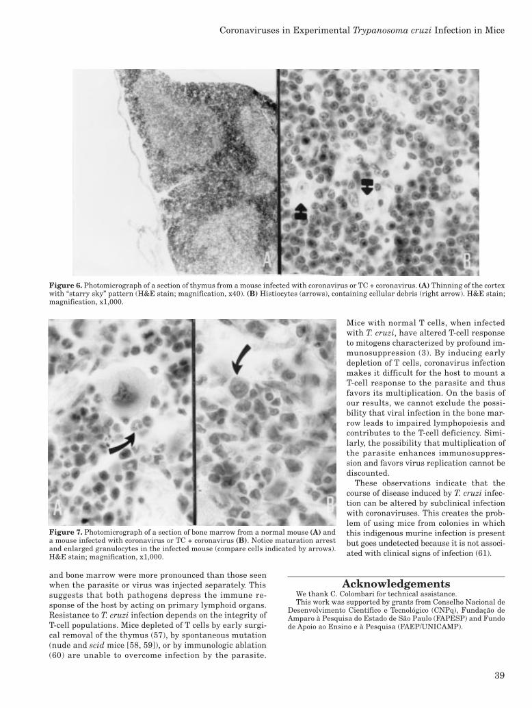
row sections from normal and TC-infected mice. In contrast,mice infected with coronavirus had an arrested maturationpattern of hematopoietic precursors, mainly granulocytic.When mice were infected with TC and coronavirus, cell num-bers generally increased and sinuses largely collapsed. Agreater degree of maturation arrest in granulocytic precur-sors also was observed (Figure 7).
DiscussionCoronaviruses are a highly prevalent and contagious
group of viruses (29) that include several strains differing inantigenicity and biological action (30–33). The ubiquity ofcoronaviruses makes them the most frequent contaminantin immunologic research because lymphotropism is a hall-mark of MHV infection (34–37).
Figure 2. Splenic weight (A) and cell numbers (B) in CBA miceinfected with T. cruzi (TC), coronavirus (virus X and MHV-3), orT. cruzi and coronavirus (TC + coronavirus). The horizontal barsrepresent one SD of the mean for spleens from normal uninfectedmice. Data are expressed as the mean and SD (n = 10). *P < 0.01,TC or TC + coronavirus vs normal or coronavirus (ANOVA).
Figure 3. Thymic weight (A) and cell numbers (B) in CBA miceinfected with T. cruzi (TC), coronavirus (virus X and MHV-3), orT. cruzi and coronavirus (TC + coronavirus). The horizontal barsrepresent one SD of the mean for thymuses from normal uninfectedmice. Data are expressed as the mean and SD (n = 10). *P < 0.01,coronavirus or TC + coronavirus vs normal or TC (ANOVA).
Lymph nodes of mice from the various experimentalgroups had a common microscopic pattern of inflammatoryreaction, including hyperplastic cortex and increased num-bers of phagocytic cells arranged along the sinuses. Spleensfrom mice infected with TC contained hyperplastic lymphoidfollicles surrounded by increased width of marginal zones(Figure 4), whereas those from mice infected with coro-naviruses or both agents had hyperplastic white pulp associ-ated with a pattern of increased hematopoiesis andproliferating megakaryocytes (Figure 5). Alterations werenot detected in the thymus of mice infected with TC; how-ever, mice infected with coronavirus or both agents had thin-ner cortexes with a “starry sky” pattern owing to thepresence of histiocytes, many of which contained cellular de-bris (Figure 6). Alterations were not observed in bone mar-
Coronaviruses in Experimental Trypanosoma cruzi Infection in Mice

Vol 49, No 1Laboratory Animal ScienceFebruary 1999
38
Because coronaviruses are known to alter the immuneresponse of the host (38–40), one would expect that resis-tance to T. cruzi would also be altered. These expectationswere confirmed experimentally in this study by showingthat resistance to T. cruzi was affected by contaminationwith low doses of virus (either the X-strain or the classicalMHV-3 strain). Mice that would usually survive if in-fected with either parasite or virus died with a high levelof parasitemia when both pathogens were present,
thereby indicating a reduced resistance tothe parasite.
An explanation for the mechanism(s) ofthis decrease in resistance can be found inthe biology of the two infections. In agree-ment with previous findings (41), TC stock ofthe parasite induced lymph node and splenichyperplasia. As reported by other investiga-tors (42, 43), up to 65% of all splenic andlymph node lymphocytes are in the mitoticcycle, with large increase of immunoglobulin-secreting cells (10- to 100-fold higher than incontrol mice). Although the acute infectionwith T. cruzi is characterized by a polyclonalactivation of B and T cells, immunosuppres-sion of humoral and cellular responses is al-ways observed (44). However, experiments inour laboratory indicate that this immuno-suppression can be lower in mice infectedwith this stock of parasites (Verinaud et al.,manuscript in preparation). We found no his-topathologic alteration in the primary lym-phoid organs by using this stock of parasites.Although T. cruzi-induced alterations inthe thymus have been reported (45, 46), wewere unable to detect any changes in thisorgan, probably because we used a para-site stock of low pathogenicity and investi-gated only the early phase of infection (i.e.,up to 7 days after infection). Resistance tothe parasite is known to be dependent onT-cell responses (47, 48) and was not al-tered by infection with this parasite stock,because mice survived.
Mice infected with the murine coro-naviruses alone had profound alterations inthymus and bone marrow. Besides, viral in-fection induced hematopoiesis in the second-ary lymphoid organs, probably because of itseffects on the bone marrow (i.e., arrest in thematuration of the hematopoietic precursors).Significant atrophy occurred in the thymus.It is well documented that infection with dif-ferent strains of coronavirus results in im-munosuppression (49, 50) accompanied by invitro alterations in T-cell proliferation andcytokine production (51, 52), as well as invo-lution of the thymus with cellular death byapoptosis (53). The pathogenicity of the vi-rus can be correlated with its lym-
photropism (54, 55). Indeed, in susceptible hosts lyticviral replication occurs in thymic stromal cells, and infec-tion is transferred to complexed thymocytes, which inturn leads to depletion of different T-cell subpopulations(49). In bone marrow, a lytic viral infection of the 14.8+ �+B-lymphocyte subpopulation eventually leads to arrest ofB-cell lymphopoiesis (56).
Experiments of T. cruzi and murine coronavirus adminis-tered simultaneously indicated that the lesions in thymus
Figure 4. Photomicrograph of a section of spleen from a mouse infected with T. cruzi.Notice follicular hyperplasia with increased width of marginal zone (between arrows).H&E stain; magnification, x40.
Figure 5. Photomicrograph of a section of spleen from a mouse infected withcoronavirus or TC + coronavirus. Notice extramedullary hematopoiesis with nests oferythroblasts (short arrows) and proliferating megakaryocytes (long arrows). H&Estain; magnification, x400.
39
and bone marrow were more pronounced than those seenwhen the parasite or virus was injected separately. Thissuggests that both pathogens depress the immune re-sponse of the host by acting on primary lymphoid organs.Resistance to T. cruzi infection depends on the integrity ofT-cell populations. Mice depleted of T cells by early surgi-cal removal of the thymus (57), by spontaneous mutation(nude and scid mice [58, 59]), or by immunologic ablation(60) are unable to overcome infection by the parasite.
Mice with normal T cells, when infectedwith T. cruzi, have altered T-cell responseto mitogens characterized by profound im-munosuppression (3). By inducing earlydepletion of T cells, coronavirus infectionmakes it difficult for the host to mount aT-cell response to the parasite and thusfavors its multiplication. On the basis ofour results, we cannot exclude the possi-bility that viral infection in the bone mar-row leads to impaired lymphopoiesis andcontributes to the T-cell deficiency. Simi-larly, the possibility that multiplication ofthe parasite enhances immunosuppres-sion and favors virus replication cannot bediscounted.
These observations indicate that thecourse of disease induced by T. cruzi infec-tion can be altered by subclinical infectionwith coronaviruses. This creates the prob-lem of using mice from colonies in whichthis indigenous murine infection is presentbut goes undetected because it is not associ-ated with clinical signs of infection (61).
AcknowledgementsWe thank C. Colombari for technical assistance.This work was supported by grants from Conselho Nacional de
Desenvolvimento Científico e Tecnológico (CNPq), Fundação deAmparo à Pesquisa do Estado de São Paulo (FAPESP) and Fundode Apoio ao Ensino e à Pesquisa (FAEP/UNICAMP).
Figure 6. Photomicrograph of a section of thymus from a mouse infected with coronavirus or TC + coronavirus. (A) Thinning of the cortexwith “starry sky” pattern (H&E stain; magnification, x40). (B) Histiocytes (arrows), containing cellular debris (right arrow). H&E stain;magnification, x1,000.
Figure 7. Photomicrograph of a section of bone marrow from a normal mouse (A) anda mouse infected with coronavirus or TC + coronavirus (B). Notice maturation arrestand enlarged granulocytes in the infected mouse (compare cells indicated by arrows).H&E stain; magnification, x1,000.
Coronaviruses in Experimental Trypanosoma cruzi Infection in Mice

Vol 49, No 1Laboratory Animal ScienceFebruary 1999
40
References 1. World Health Organization. 1991. Control of Chagas’ dis-
ease. W.H.O. 187:1–99. 2. Brener, Z. 1980. Immunity to Trypanosoma cruzi. Adv.
Parasitol. 18:247–291. 3. Reed, S. G., J. A. Inverso, and S. B. Roters. 1984. Sup-
pressed antibody responses to sheep erythrocytes in mice withchronic Trypanosoma cruzi infections are restored withinterleukin 2. J. Immunol. 133:3333–3337.
4. Tarleton, R. L. 1988. Trypanosoma cruzi-induced suppres-sion of IL2 production. II. Evidence for a role for suppressorcells. J. Immunol. 140:2763–2769.
5. Minoprio, P., H. Eisen, L. Forni, et al. 1986. Polyclonallymphocytes to murine Trypanosoma cruzi infection. I.Quantitation of both T and B cell responses. Scand. J. Immunol.24:661–668.
6. Ribeiro dos Santos, R., and L. Hudson. 1980. Immuno-logical consequences of parasite modification of host cells. Clin.Exp. Immunol. 40:36–41.
7. Andrade, Z. 1991. Pathogenesis of Chagas’ disease. Res.Immunol. 142:126–129.
8. Chagas, C. 1909. Nova tripanozomiaze humana: estudos sobrea morfologia e o ciclo evolutivo do Schizotrypanum cruzi n.gen., n. sp., agente etiológico de nova entidade mórbida dohomem. Mem. Inst. Oswaldo Cruz. 1:159–218.
9. Nogueira, N., J. Ellis, S. Chaplan, et al. 1981. Trypano-soma cruzi: in vivo and in vitro correlation between T-cell ac-tivation and susceptibility in inbred strains of mice. Exp.Parasitol. 51:325–330.
10. Wrightsman, R., S. Krassner, and J. Watson. 1982. Ge-netic control of responses to Trypanosoma cruzi in mice: mul-tiple genes influencing parasitemia and survival. Infect.Immun. 36:637–644.
11. Trischmann, T. M. 1986. Trypanosoma cruzi: early parasiteproliferation and host resistance in inbred strains of mice.Exp. Parasitol. 62:194–201.
12. Phillips, N. R. 1960. Experimental studies on the quantita-tive transmission of Trypanosoma cruzi: considerations regard-ing the standardization of materials. Ann. Trop. Med. Parasitol.54:60–66.
13. Brener, Z. 1977. Intraspecific variation in Trypanosoma cruzi:two types of parasite populations presenting distinct features.Pan American Health Organization, Scientific Publication347:11–21.
14. Camargo, I. J. B., J. Sakurada, M. R. Zucato, et al. 1989.The early phase of immune response of CBA mice infectedwith Trypanosoma cruzi. Immunol. Lett. 20:213–216.
15. Boorman, G. A., M. L. Luster, J. H. Dean, et al. 1982. Peri-toneal macrophage alterations caused by naturally occurringmouse hepatitis virus. Am. J. Pathol. 106:110–117.
16. Dempsey, W. L., A. L. Smith, and P. S. Morahan. 1986.Effect of inapparent murine hepatitis virus infections on mac-rophages and host resistance. J. Leukocyte Biol. 39:559–565.
17. Welberz, S., H. J. Partke, F. Dagnaes-Hansen, et al. 1991.Persistent MHV infections reduce the incidence of diabetesmellitus in non-obese diabetic mice. Diabetologia 34:2–5.
18. Smith, A. L., J. Casals, and A. J. Main. 1983. Antigeniccharacterization of Tettnang virus: complications caused bypassage of the virus in mice from a colony enzootically in-fected with mouse hepatitis virus. Am. J. Trop. Med. Hyg.32:1172–1176.
19. Silva, M. E., E. A. Evangelista, J. R. Nicoli, et al. 1987.American Trypanosomiasis (Chagas’ disease) in conventionaland germfree rats and mice. Rev. Inst. Med. Trop. São Paulo.29:284–288.
20. Rangel, H. A., L. Verinaud, I. J. B. Camargo, et al. 1994.Murine virus contaminant of Trypanosoma cruzi experimen-tal infection. Rev. Inst. Med. Trop. São Paulo 36:423–431.
21. Suzuki, E., J. Matsubara, and M. Saito. 1982. Serologicalsurvey of laboratory rodents for infection with Sendai virus,mouse hepatitis virus, reovirus type 3 and mouse adenovirus.Jan. J. Med. Sci. Biol. 35:249–254.
22. Lindsey, J. R. 1986. Prevalence of viral and mycoplasmalinfections in laboratory rodents, p. 801–808. In P. N. Bhatt,R. O. Jacoby, H. C. Morse, et al. (ed.). Viral and mycoplasmalinfections of laboratory rodents. Effects on biomedical re-search. Academic Press, Inc., New York.
23. Lussier, G., and J. P. Decoteaux. 1986. Prevalence of natu-ral virus infections in laboratory mice and rats used in Canada.Lab. Anim. Sci. 36:145–148.
24. Gilioli, R., J. Sakurada, L. Andrade, et al. 1996. Virus in-fection in rat and mouse colonies reared in Brazilian animalfacilities. Lab. Anim. Sci. 46:582–584.
25. Trexler, P. C. 1959. The use of plastics in the design of anisolator system. Ann. N.Y. Acad. Sci. 78:29–36.
26. Kraft, V., and B. Meyer. 1986. Diagnosis of murine infec-tions in relation to test methods employed. Lab. Anim. Sci.36:272–276.
27. Compton, S. R., S. W. Barthold, and A. L. Smith. 1993.The cellular and molecular pathogenesis of coronaviruses. Lab.Anim. Sci. 43:15–28.
28. Reed, L. J., and H. Muench. 1938. A simple method of esti-mating fifty percent end points. Am. J. Hyg. 27:493–497.
29. Lussier, G., and J. P. Descoteaux. 1990. Prevalence ratesof natural viral infections in laboratory rats and mice used inCanada: a three year study. CALAS Newsletter 23:6–11.
30. Wege, H., S. Siddell, and V. terMeulen. 1982. The biologyand pathogenesis of coronavirus. Curr. Top. Microbiol.Immunol. 99:162–200.
31. Taguchi, F., A. Yamada, and K. Fujiwara. 1980. Resistanceto highly virulent mouse hepatitis virus acquired by mice af-ter low-virulence infection: enhanced antiviral activity ofmacrophages. Infect. Immun. 29:42–49.
32. Dupuy, J. M., and D. Rodrigues. 1981. Heterogeneity inevolutive pattern of inbred mice infected with a clonedsubstrain of mouse hepatitis virus type 3. Intervirology16:116–123.
33. Hirasawa, T., T. Hirano, and O. Ohara. 1988. Character-ization of low-virulent mouse coronavirus isolated from fecesin a mouse colony. J. Vet. Med. B55:435–442.
34. Lamontagne, L., J. P. Descoteaux, and P. Jolicoeur. 1989.Mouse hepatitis virus 3 replication in T and B lymphocytescorrelate with viral pathogenicity. J. Immunol. 142:4458–4465.
35. Lamontagne, L., C. Dupuy, D. Leray, et al. 1985.Coronavirus-induced immunosuppression: role of mouse hepa-titis virus 3-lymphocyte interaction. Prog. Leukocyte Biol.1:29–35.
36. Lamontagne, L., and J. M. Dupuy. 1984. Persistent in vitroinfection with mouse hepatitis virus type 3 in mouse lymphoidcell lines. Infect. Immun. 44:716–721.
37. Lamontagne, L., D. Décarie, and P. Jolicoeur. 1990. Im-mune cell tropism of attenuated MHV3 viruses isolated frombrains of chronically infected mice. Viral Immunol. 3:1–6.
38. Smith, A. L., D. F. Winograd, and M. S. deSouza. 1991. Invitro splenic T cell responses of diverse mouse genotypes af-ter oronasal exposure to mouse hepatitis virus, strain JHM.Lab. Anim. Sci. 41:106–111.
39. Leray, D., C. Dupuy, and J. M. Dupuy. 1982. Immunopa-thology of mouse hepatitis virus 3 infection. IV. MHV 3 in-duced immunodepression. Clin. Immunol. Immunopathol.23:223–228.
40. Smith, A. L., M. S. deSouza, D. Finzi, et al. 1992. Responsesof mice to murine coronavirus immunization. Arch. Virol.125:39–52.
41. Takasu, N., T. Nishimura, K. Kato, et al. 1990. Increase inCSF in serum and augmentation of CSF responsiveness oflymphoid mononuclear cells by acute Trypanosoma cruzi in-fection in mice. J. Exp. Med. 160:67–73.

41
42. D’Império Lima, M. R., M. Joskowicz, A. Coutinho, etal. 1985. Very large and isotypically atypical polyclonal plaqueforming cell responses in mice infected with T. cruzi. Eur. J.Immunol. 15:201–203.
43. D’Império Lima, M. R., H. Eisen, P. Minóprio, et al. 1986.Persistence of polyclonal B cell activation with undetectableparasitemia in late stages of experimental Chagas’ disease.J. Immunol. 137:353–356.
44. Reed, S. G., S. B. Roters, and E. A. Goidl. 1983. Spleen cellmediated suppression of IgG production to a non-parasiteantigen during chronic Trypanosoma cruzi infection in mice.J. Immunol. 131:1978–1982.
45. Savino, W., M. C. L. Moraes, M. H. Joskowicz, et al. 1989.Studies on the thymus in Chagas’ disease. I. Changes in thethymic microenvironment in mice acutely infected with Try-panosoma cruzi. Eur. J. Immunol. 19:1727–1733.
46. Savino, W., M. C. L. Moraes, S. D. S. Barbosa, et al. 1992.Is the thymus a target organ in infectious diseases? Mem.Inst. Oswaldo Cruz. 87:73–78.
47. Tarleton, R., B. H. Koller, A. Latour, et al. 1992. Suscepti-bility of �2 microglobulin deficient mice to Trypanosoma cruziinfection. Nature 356:338–340.
48. Rottenberg, M. E., M. Bakhiet, T. Olsson, et al. 1993. Dif-ferential susceptibility of mice genomically depleted of CD4and CD8 to infections with Trypanosoma cruzi and Trypano-soma brucei. Infect. Immun. 61:5129–5133.
49. Jolicoeur, P., and L. Lamontagne. 1994. Impaired T and Bcell sub populations involved in a chronic disease induced bymouse hepatitis virus type 3. J. Immunol. 153:1318–1327.
50. Casebolt, D. B., D. M. Spalding, T. R. Schoeb, et al. 1987.Suppression of immune response induction in Peyer’s patchlymphoid cells from mice infected with mouse hepatitis virus.Cell Immunol. 23:539–547.
51. Smith, A. L., S. W. Bottomly, and D. F. Winograd. 1987.Altered splenic T cell function of Balb/cByJ mice infectedwith mouse hepatitis virus or Sendai virus. J. Immunol.138:3426–3430.
52. deSouza, M. S., A. L. Smith, and K. Bottomly. 1991. Infec-tion of BALB/cByJ mice with the JHM strain of mouse hepa-titis virus alters the in vitro splenic T cell proliferation andcytokine production. Lab. Anim. Sci. 41:99–105.
53. Verinaud, L., M. A. Cruz-Hofling, J. K. Sakurada, et al.1998. Immunodepression induced by Trypanosoma cruzi andmouse hepatitis virus type 3 is associated with thymusapoptosis. Clin. Diag. Lab. Immunol. 5:186–191.
54. Lamontagne, L., and P. Jolicoeur. 1991. Mouse hepatitisvirus 3 thymic cell interactions correlating with viral patho-genicity. J. Immunol. 146:3152–3159.
55. Coutelier, J. P., C. Godfraind, G. S. Dveksler, et al. 1994.B lymphocyte and macrophage expression of carcioembryonicantigen-related adhesion molecules that serve as receptorsfor murine coronavirus. Eur. J. Immunol. 24:1383–1390.
56. Jolicoeur, P., and L. Lamontagne. 1989. Mouse hepati-tis virus 3 pathogenicity expressed by a lytic viral infec-tion in bone marrow 14.8+�+ B lymphocyte sub populations.J. Immunol. 143:3722–3730.
57. Schmunis, G. A., S. M. Gonzales-Cappa, O. C. Traversa,et al. 1971. The effect of immuno-depression due to neonatalthymectomy on infections with Trypanosoma cruzi in mice.Trans. R. Soc. Trop. Med. Hyg. 65:89–94.
58. Kierszenbaum, F., and P. Pienkowski. 1979. Thymus-de-pendent control of host defense mechanisms against Trypa-nosoma cruzi. Infect. Immun. 24:117–123.
59. Trischmann, T. M., and B. R. Bloom. 1980. Trypanosomacruzi: ability of T-cell-depleted and -enriched populations topassively protect mice. Exp. Parasitol. 49:225–231.
60. Robertson, E. L., W. L. Hanson, and W. I. Chapman. 1973.Trypanosoma cruzi: effects of anti thymocyte serum in miceand neonatal thymectomy in rats. Exp. Parasitol. 34:168–180.
61. Midoro, K., K. Nakanaga, S. Kyuwa, et al. 1989. Immuno-pathology of chronic progressive hepatitis in nude mice in-fected with low-virulent mouse hepatitis virus. Microbiol.Immunol. 33:669–682.
Coronaviruses in Experimental Trypanosoma cruzi Infection in Mice
Related Documents