HOW I DO IT ARTICLE Open Access How I do it: Lung ultrasound Luna Gargani 1* and Giovanni Volpicelli 2 Abstract In the last 15 years, a new imaging application of sonography has emerged in the clinical arena: lung ultrasound (LUS). From its traditional assessment of pleural effusions and masses, LUS has moved towards the revolutionary approach of imaging the pulmonary parenchyma, mainly as a point-of-care technique. Although limited by the presence of air, LUS has proved to be useful in the evaluation of many different acute and chronic conditions, from cardiogenic pulmonary edema to acute lung injury, from pneumothorax to pneumonia, from interstitial lung disease to pulmonary infarctions and contusions. It is especially valuable since it is a relatively easy-to-learn application of ultrasound, less technically demanding than other sonographic examinations. It is quick to perform, portable, repeatable, non-ionizing, independent from specific acoustic windows, and therefore suitable for a meaningful evaluation in many different settings, both inpatient and outpatient, in both acute and chronic conditions. In the next few years, point-of-care LUS is likely to become increasingly important in many different clinical settings, from the emergency department to the intensive care unit, from cardiology to pulmonology and nephrology wards. Keywords: Lung ultrasound, B-lines, Point-of-care ultrasound, Chest sonography What is lung ultrasound? Assessment of the lung has always been considered off- limits for ultrasound, since it is standard textbook know- ledge that «because ultrasound energy is rapidly dissipated by air, ultrasound imaging is not useful for the evaluation of the pulmonary parenchyma» [1]. The concept that ul- trasound cannot be employed for evaluating the lung is linked to the presence of air, which determines a high acoustic mismatch with the surrounding tissues, causing a complete reflection of the ultrasound beam, preventing the creation of direct imaging of the pulmonary paren- chyma [2]. In a normally aerated lung, the only detectable structure is the pleura, visualized as a hyperechoic hori- zontal line. It is debated whether this line represents an artefact due to a reflection phenomenon at the interface between alveolar air and the soft tissues of the thoracic wall, or it images the real pleura. The pleural line moves synchronously with respiration [3]: this dynamic horizon- tal movement is called lung sliding. In addition, there are some hyperechoic, horizontal lines arising at regular inter- vals from the pleural line: the A-lines. When combined with lung sliding, these reverberation artefacts represent a sign of normal or excessive content of air in the alveolar spaces (Figure 1, Additional file 1). When the air content decreases and lung density increases due to the presence in the lung of exudate, transudate, collagen, blood, etc. the acoustic mismatch between the lung and the surrounding tissues is lowered, and the ultrasound beam can be partly reflected at deeper zones and repeatedly. This phenome- non creates some vertical reverberation artefacts known as B-lines (Figure 2, Additional file 2). B-lines belong to the family of the comet-tail artifacts, well known in the setting of abdominal ultrasound [4]. B-lines have also been addressed as comet-tail artifacts or ultrasound lung co- mets before an expert agreement on nomenclature was obtained [3]. B-lines are defined as discrete laser-like verti- cal hyperechoic reverberation artifacts that arise from the pleural line, extend to the bottom of the screen without fading, and move synchronously with lung sliding [3]. Multiple B-lines are considered the sonographic sign of lung interstitial syndrome, and their number increases along with decreasing air content and increase in lung density [5,6]. When the air content further decreases, such as in lung consolidations, the acoustic window on the lung becomes completely open, and the lung may be directly visualized as a solid parenchyma, like the liver or the spleen (Figure 3). Consolidation of the lung may be the result of an infectious process, an infarction due to * Correspondence: [email protected] 1 Institute of Clinical Physiology, National Research Council, Via Moruzzi, 1, 56124 Pisa, Italy Full list of author information is available at the end of the article CARDIOVASCULAR ULTRASOUND © 2014 Gargani and Volpicelli; licensee BioMed Central Ltd. This is an Open Access article distributed under the terms of the Creative Commons Attribution License (http://creativecommons.org/licenses/by/4.0), which permits unrestricted use, distribution, and reproduction in any medium, provided the original work is properly credited. The Creative Commons Public Domain Dedication waiver (http://creativecommons.org/publicdomain/zero/1.0/) applies to the data made available in this article, unless otherwise stated. Gargani and Volpicelli Cardiovascular Ultrasound 2014, 12:25 http://www.cardiovascularultrasound.com/content/12/1/25

Welcome message from author
This document is posted to help you gain knowledge. Please leave a comment to let me know what you think about it! Share it to your friends and learn new things together.
Transcript

CARDIOVASCULAR ULTRASOUND
Gargani and Volpicelli Cardiovascular Ultrasound 2014, 12:25http://www.cardiovascularultrasound.com/content/12/1/25
HOW I DO IT ARTICLE Open Access
How I do it: Lung ultrasoundLuna Gargani1* and Giovanni Volpicelli2
Abstract
In the last 15 years, a new imaging application of sonography has emerged in the clinical arena: lung ultrasound(LUS). From its traditional assessment of pleural effusions and masses, LUS has moved towards the revolutionaryapproach of imaging the pulmonary parenchyma, mainly as a point-of-care technique. Although limited by thepresence of air, LUS has proved to be useful in the evaluation of many different acute and chronic conditions, fromcardiogenic pulmonary edema to acute lung injury, from pneumothorax to pneumonia, from interstitial lungdisease to pulmonary infarctions and contusions. It is especially valuable since it is a relatively easy-to-learn applicationof ultrasound, less technically demanding than other sonographic examinations. It is quick to perform, portable,repeatable, non-ionizing, independent from specific acoustic windows, and therefore suitable for a meaningfulevaluation in many different settings, both inpatient and outpatient, in both acute and chronic conditions.In the next few years, point-of-care LUS is likely to become increasingly important in many different clinical settings,from the emergency department to the intensive care unit, from cardiology to pulmonology and nephrology wards.
Keywords: Lung ultrasound, B-lines, Point-of-care ultrasound, Chest sonography
What is lung ultrasound?Assessment of the lung has always been considered off-limits for ultrasound, since it is standard textbook know-ledge that «because ultrasound energy is rapidly dissipatedby air, ultrasound imaging is not useful for the evaluationof the pulmonary parenchyma» [1]. The concept that ul-trasound cannot be employed for evaluating the lung islinked to the presence of air, which determines a highacoustic mismatch with the surrounding tissues, causing acomplete reflection of the ultrasound beam, preventingthe creation of direct imaging of the pulmonary paren-chyma [2]. In a normally aerated lung, the only detectablestructure is the pleura, visualized as a hyperechoic hori-zontal line. It is debated whether this line represents anartefact due to a reflection phenomenon at the interfacebetween alveolar air and the soft tissues of the thoracicwall, or it images the real pleura. The pleural line movessynchronously with respiration [3]: this dynamic horizon-tal movement is called lung sliding. In addition, there aresome hyperechoic, horizontal lines arising at regular inter-vals from the pleural line: the A-lines. When combinedwith lung sliding, these reverberation artefacts represent a
* Correspondence: [email protected] of Clinical Physiology, National Research Council, Via Moruzzi, 1,56124 Pisa, ItalyFull list of author information is available at the end of the article
© 2014 Gargani and Volpicelli; licensee BioMeCreative Commons Attribution License (http:/distribution, and reproduction in any mediumDomain Dedication waiver (http://creativecomarticle, unless otherwise stated.
sign of normal or excessive content of air in the alveolarspaces (Figure 1, Additional file 1). When the air contentdecreases and lung density increases due to the presencein the lung of exudate, transudate, collagen, blood, etc. theacoustic mismatch between the lung and the surroundingtissues is lowered, and the ultrasound beam can be partlyreflected at deeper zones and repeatedly. This phenome-non creates some vertical reverberation artefacts knownas B-lines (Figure 2, Additional file 2). B-lines belong tothe family of the comet-tail artifacts, well known in thesetting of abdominal ultrasound [4]. B-lines have also beenaddressed as comet-tail artifacts or ultrasound lung co-mets before an expert agreement on nomenclature wasobtained [3]. B-lines are defined as discrete laser-like verti-cal hyperechoic reverberation artifacts that arise from thepleural line, extend to the bottom of the screen withoutfading, and move synchronously with lung sliding [3].Multiple B-lines are considered the sonographic sign oflung interstitial syndrome, and their number increasesalong with decreasing air content and increase in lungdensity [5,6]. When the air content further decreases, suchas in lung consolidations, the acoustic window on the lungbecomes completely open, and the lung may be directlyvisualized as a solid parenchyma, like the liver or thespleen (Figure 3). Consolidation of the lung may be theresult of an infectious process, an infarction due to
d Central Ltd. This is an Open Access article distributed under the terms of the/creativecommons.org/licenses/by/4.0), which permits unrestricted use,, provided the original work is properly credited. The Creative Commons Publicmons.org/publicdomain/zero/1.0/) applies to the data made available in this
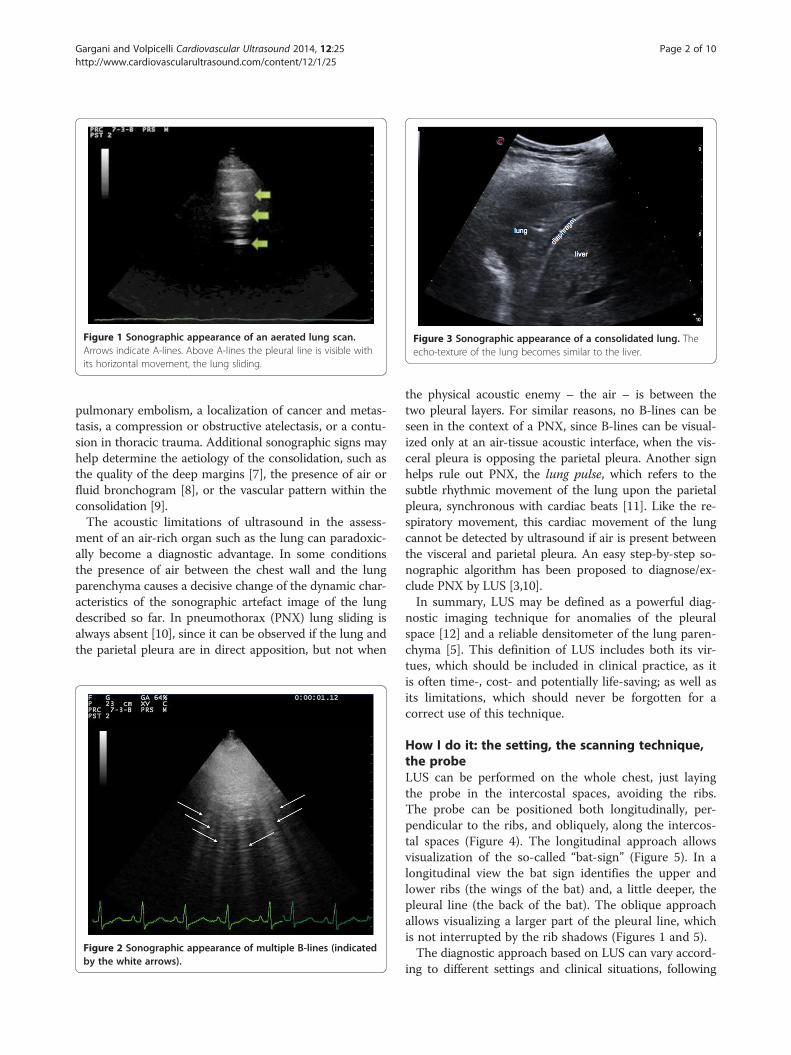
Figure 1 Sonographic appearance of an aerated lung scan.Arrows indicate A-lines. Above A-lines the pleural line is visible withits horizontal movement, the lung sliding.
Figure 3 Sonographic appearance of a consolidated lung. Theecho-texture of the lung becomes similar to the liver.
Gargani and Volpicelli Cardiovascular Ultrasound 2014, 12:25 Page 2 of 10http://www.cardiovascularultrasound.com/content/12/1/25
pulmonary embolism, a localization of cancer and metas-tasis, a compression or obstructive atelectasis, or a contu-sion in thoracic trauma. Additional sonographic signs mayhelp determine the aetiology of the consolidation, such asthe quality of the deep margins [7], the presence of air orfluid bronchogram [8], or the vascular pattern within theconsolidation [9].The acoustic limitations of ultrasound in the assess-
ment of an air-rich organ such as the lung can paradoxic-ally become a diagnostic advantage. In some conditionsthe presence of air between the chest wall and the lungparenchyma causes a decisive change of the dynamic char-acteristics of the sonographic artefact image of the lungdescribed so far. In pneumothorax (PNX) lung sliding isalways absent [10], since it can be observed if the lung andthe parietal pleura are in direct apposition, but not when
Figure 2 Sonographic appearance of multiple B-lines (indicatedby the white arrows).
the physical acoustic enemy – the air – is between thetwo pleural layers. For similar reasons, no B-lines can beseen in the context of a PNX, since B-lines can be visual-ized only at an air-tissue acoustic interface, when the vis-ceral pleura is opposing the parietal pleura. Another signhelps rule out PNX, the lung pulse, which refers to thesubtle rhythmic movement of the lung upon the parietalpleura, synchronous with cardiac beats [11]. Like the re-spiratory movement, this cardiac movement of the lungcannot be detected by ultrasound if air is present betweenthe visceral and parietal pleura. An easy step-by-step so-nographic algorithm has been proposed to diagnose/ex-clude PNX by LUS [3,10].In summary, LUS may be defined as a powerful diag-
nostic imaging technique for anomalies of the pleuralspace [12] and a reliable densitometer of the lung paren-chyma [5]. This definition of LUS includes both its vir-tues, which should be included in clinical practice, as itis often time-, cost- and potentially life-saving; as well asits limitations, which should never be forgotten for acorrect use of this technique.
How I do it: the setting, the scanning technique,the probeLUS can be performed on the whole chest, just layingthe probe in the intercostal spaces, avoiding the ribs.The probe can be positioned both longitudinally, per-pendicular to the ribs, and obliquely, along the intercos-tal spaces (Figure 4). The longitudinal approach allowsvisualization of the so-called “bat-sign” (Figure 5). In alongitudinal view the bat sign identifies the upper andlower ribs (the wings of the bat) and, a little deeper, thepleural line (the back of the bat). The oblique approachallows visualizing a larger part of the pleural line, whichis not interrupted by the rib shadows (Figures 1 and 5).The diagnostic approach based on LUS can vary accord-
ing to different settings and clinical situations, following

Figure 4 Longitudinal and oblique approach to lung ultrasound.
Gargani and Volpicelli Cardiovascular Ultrasound 2014, 12:25 Page 3 of 10http://www.cardiovascularultrasound.com/content/12/1/25
the main principles of what is today known as “point-of-care ultrasound”. Maximum effectiveness of the method isobtained through a clinically-driven and focused assess-ment. If properly driven and correctly interpreted, somesonographic signs become highly accurate for diagnosingspecific pulmonary conditions. For example, in a stablepatient with acute spontaneous pleuritic pain, ultrasound
Figure 5 Longitudinal and oblique lung scanning. A. Longitudinal lungimage that resembles a bat. B. Oblique lung scanning: the pleural line is no
examination will start from the painful chest area [13], fo-cusing on signs of focal pleural and parenchymal abnor-mality. If the pain is caused by a pulmonary conditionwith involvement of the parietal pleura, this will be easilydetected by LUS. Indeed, LUS is a surface imaging tech-nique, highly sensitive in detecting pleural abnormalities.The clinical suspicion and pre-test probability will guide
scanning: the upper rib, the pleural line and the lower rib draw ant interrupted by the ribs, and appears as a horizontal line.

Gargani and Volpicelli Cardiovascular Ultrasound 2014, 12:25 Page 4 of 10http://www.cardiovascularultrasound.com/content/12/1/25
the diagnostic process to rule in or rule out with high ac-curacy several conditions, such as PNX, pleuritis, pneu-monia, lung peripheral infarction [14]. In this setting, ahighly specific sign is the lung point, which represents thetransition point between the typical sonographic patternof PNX (absence of lung sliding and of B-lines) into thenormal pattern of lung sliding, and depicts the physicallimit of PNX as mapped on the chest wall [3]. However,the lung point can be employed to detect the extension ofPNX, but not its volume. Up-to-now, LUS is not recog-nized as a method to differentiate between large and smallPNX.In a patient with acute dyspnea, if cardiogenic pul-
monary edema is in the differential diagnosis, LUS willbe used to examine the anterior and lateral chest to de-tect the diffuse signs of interstitial and alveolar edema,which usually respect three highly specific features: theyare correlated with the severity of the respiratory failure,follow a regular and symmetric spatial distribution, andusually progress from the lateral and inferior (dependentzones) to the anterior upper chest areas. The scanningtechnique that should be employed in the emergency set-ting is the eight-zone examination, consisting of scanningfour chest areas per side (Figure 6): areas 1 and 2 denotethe upper anterior and lower anterior chest, whereas areas3 and 4 denote the upper lateral and basal lateral chest, re-spectively [15]. In the critically ill patient with acute re-spiratory failure, a more rapid anterior two-region scanmay be sufficient to rule out the interstitial syndrome dueto cardiogenic acute pulmonary edema [16]. However, thisfocused anterior scanning, while still highly accurate in the
Figure 6 Eight-zone scanning scheme of the antero-lateralchest (according to Volpicelli G et al. See ref. [15]).
critically ill, may not be sufficient in patients who are notseverely dyspnoic [17], since presence of B-lines on the an-terior chest usually denotes a more severe degree of pul-monary congestion in case of heart failure. Again, this isanother example of adaptation of the LUS technique andsigns to the specific setting and clinical condition.If the main clinical suspicion is the possibility of a
PNX, the LUS examination is started from the non-dependent zones for air collection, corresponding to theanterior-inferior chest in the supine patient. In this case,examining only one hot zone per side will help rule outPNX with high sensitivity both in the extreme emergen-cies and in stable patients. The hot zone examinationwill also be enough to confirm PNX in unstable/cardiacarrest patients [18]. Only in stable patients will examin-ation be extended to the lateral chest to confirm PNX.In the setting of chronic patients, with less time pres-
sure and more borderline cases, the scanning techniqueshould always be more comprehensive. It can include theanterior, lateral and dorsal chest. Different approacheshave been proposed: a detailed scanning scheme hasbeen used in many studies on patients with heart failure[19,20], on dialysis [21-23], and with pulmonary fibrosis[24,25], focused on the assessment of B-lines. These ap-proaches allow accurate examination of the whole chest,which can be applied in several settings in chronic condi-tions. It is particularly useful for quantifying the extent ofthe LUS abnormalities, and for assessing intra-patient var-iations after therapeutic interventions, including dialysis[21,22]. Ultrasound scanning of the anterior and lateralchest is obtained on the right and left hemithorax, fromthe second to the fourth (on the right side to the fifth)intercostal spaces, and from the parasternal line to the ax-illary line (Figure 7). The posterior chest is scanned alongthe paravertebral line, linea scapularis and posterior axil-lary lines (Figure 8). The sum of the B-lines found on eachscanning site yields a score denoting the extent of thepulmonary interstitial syndrome. Zero is defined as acomplete absence of B-lines in the investigated area.When B-lines are a few in a scanning site, they can beeasily counted. When they are more numerous, theytend to be confluent and it is less easy to clearly enu-merate them. To obtain a semiquantification of the sign,you can consider the percentage of the scanning site oc-cupied by B-lines (i.e., the percentage of white screencompared to black screen below the pleural line) andthen divide it by ten (i.e., 30% corresponds to about 3 B-lines, 70% corresponds to about 7 B-lines, and so on) [26].In the case of limited time, even in a chronic setting
the examination can be more focused and should beclinically driven. In patients with heart failure, it is im-portant to scan the dependent zones, i.e., lung posteriorbasis if we are evaluating an out-patient, or along theposterior and mid-axillary lines if we are scanning an
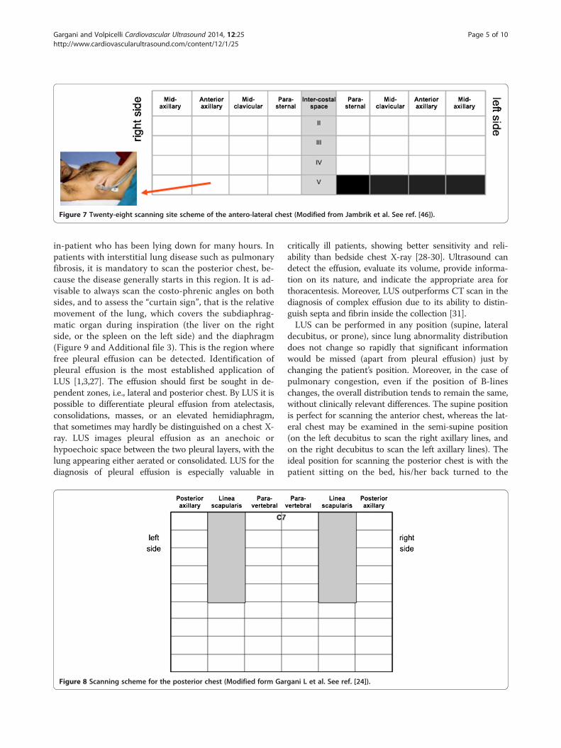
Figure 7 Twenty-eight scanning site scheme of the antero-lateral chest (Modified from Jambrik et al. See ref. [46]).
Gargani and Volpicelli Cardiovascular Ultrasound 2014, 12:25 Page 5 of 10http://www.cardiovascularultrasound.com/content/12/1/25
in-patient who has been lying down for many hours. Inpatients with interstitial lung disease such as pulmonaryfibrosis, it is mandatory to scan the posterior chest, be-cause the disease generally starts in this region. It is ad-visable to always scan the costo-phrenic angles on bothsides, and to assess the “curtain sign”, that is the relativemovement of the lung, which covers the subdiaphrag-matic organ during inspiration (the liver on the rightside, or the spleen on the left side) and the diaphragm(Figure 9 and Additional file 3). This is the region wherefree pleural effusion can be detected. Identification ofpleural effusion is the most established application ofLUS [1,3,27]. The effusion should first be sought in de-pendent zones, i.e., lateral and posterior chest. By LUS it ispossible to differentiate pleural effusion from atelectasis,consolidations, masses, or an elevated hemidiaphragm,that sometimes may hardly be distinguished on a chest X-ray. LUS images pleural effusion as an anechoic orhypoechoic space between the two pleural layers, with thelung appearing either aerated or consolidated. LUS for thediagnosis of pleural effusion is especially valuable in
Figure 8 Scanning scheme for the posterior chest (Modified form Gar
critically ill patients, showing better sensitivity and reli-ability than bedside chest X-ray [28-30]. Ultrasound candetect the effusion, evaluate its volume, provide informa-tion on its nature, and indicate the appropriate area forthoracentesis. Moreover, LUS outperforms CT scan in thediagnosis of complex effusion due to its ability to distin-guish septa and fibrin inside the collection [31].LUS can be performed in any position (supine, lateral
decubitus, or prone), since lung abnormality distributiondoes not change so rapidly that significant informationwould be missed (apart from pleural effusion) just bychanging the patient’s position. Moreover, in the case ofpulmonary congestion, even if the position of B-lineschanges, the overall distribution tends to remain the same,without clinically relevant differences. The supine positionis perfect for scanning the anterior chest, whereas the lat-eral chest may be examined in the semi-supine position(on the left decubitus to scan the right axillary lines, andon the right decubitus to scan the left axillary lines). Theideal position for scanning the posterior chest is with thepatient sitting on the bed, his/her back turned to the
gani L et al. See ref. [24]).

Figure 9 Left costophrenic angle. During inspiration the lungmoves downward and the lung air prevents the visualization of partof the spleen.
Gargani and Volpicelli Cardiovascular Ultrasound 2014, 12:25 Page 6 of 10http://www.cardiovascularultrasound.com/content/12/1/25
operator (Figure 10). Indeed, it is possible to also scanpatients while they are standing, sitting or lying flat,without significant differences in results. The only reallimitation is when LUS needs to be extended to the dor-sal chest of a patient lying down who is intubated in theintensive care unit, or a patient who is unconscious andcannot be moved. In these situations, using small probesthat may be better placed between the bed and the pa-tient may allow the best result.The LUS examination can be performed using any
commercially available 2-D scanner. Different transducershave been used, such as phased array (cardiac), convex(abdominal), microconvex, and linear (vascular) probes.Higher frequencies and macro probes are useful for the
Figure 10 Position of the patient to scan the posterior chest.
evaluation of the pleural line and subpleural space, soshould be preferred for assessing PNX. Phased-arrayprobes can be successfully employed to detect pleuraleffusion, thanks to low frequency and consequent abilityto provide a deeper view of the chest. However, theselatter probes have limitations in detecting PNX and whena detailed examination of the sub-pleural space is needed.The convex and microconvex probes are the most uni-versally used, all-purpose probes for LUS, thanks to theirintermediate frequency values, which allow a reasonablevisualization of the pleural line and subpleural space, with-out losing the overview of the chest. B-lines can be detec-ted by all these different probes, but again low frequencyprobes are probably the best for this application. Althoughthe number of B-lines may be slightly different when usingdifferent probes in a specific chest site, the overall clinicalpicture does not change by changing the transducer [32].The possibility of easily assessing B-lines with any kind oftransducer is one of the advantages of this technique, sono one should give up on scanning a patient just becausethe “ideal” probe is not available.Portable machines and pocket-sized devices have also
been proposed for assessing B-lines, as well as pleural ef-fusion [33-35]. There is no need for a second harmonicor Doppler imaging mode, so even older ultrasound ma-chines can be employed. Visualization of lung consolida-tions is possible with all probes as well. In the case of asmall consolidation, a phased-array transducer may offerless detail, whereas a linear transducer would magnify it.In the case of large consolidations, a linear probe maybe unsuitable for detecting the consolidations’ bordersprecisely, whereas convex and microconvex, and even thephased-array transducers, would be more appropriate.The depth should be tailored to the patient: very thickribcages, large muscles and obese patients need greaterdepths, even to visualize the pleural line. Very thin pa-tients and children may require less depth. Depth shouldalso be adjusted to the target of our examination: if we arelooking for PNX, the depth should be lower, in order tobetter visualize the pleural line and assess the presence orabsence of the sliding sign. If we are looking for a freepleural effusion, the depth should be greater, for a betteroverview of the costo-phrenic angles. Normally the focusshould be positioned at the pleural line level, but it shouldbe moved deeper when our main target is less superficial.
How I do it: interpretationInterpretation of LUS images is usually not very challen-ging. We must keep in mind that LUS is more affectedby lack of specificity than lack of sensitivity. A LUS pat-tern showing absent lung sliding or multiple B-lines or alung consolidation may be not enough to establish a spe-cific diagnosis, since it can be linked to different patho-logic conditions [36]. Indeed, this limitation in specificity

Gargani and Volpicelli Cardiovascular Ultrasound 2014, 12:25 Page 7 of 10http://www.cardiovascularultrasound.com/content/12/1/25
is a common feature of several diagnostic tools that weroutinely interpret in daily clinical practice, from physicalexamination to EKG, from chest X-ray to more sophisti-cated instrumental findings. The power of these tools re-sides in the interpretation of signs when combined witheach other at bedside, together with a consideration of theoverall clinical picture. When all patient characteristicsare taken into account, including history, symptoms, phys-ical examination, setting, comorbidity, medications, etc.,specificity can increase significantly. For example, in a pa-tient with systemic sclerosis and without any known leftheart conditions, presence of multiple B-lines is moreprobably related to pulmonary fibrosis than to extravascu-lar lung water. On the other hand, presence of multiplediffuse bilateral B-lines in a patient with reduced cardiacfunction is more likely to be related to extravascular lungwater than to fibrosis [26].The clinical condition of the patient is probably the
most important feature that helps interpret LUS findingsand influence patient management. For instance, in anunstable patient with signs and symptoms of hemodyna-mic shock or cardiac arrest, the absence of any move-ment of the pleural line, either respiratory (lung sliding)or cardiac (lung pulse), coupled with absence of B-linesraises such a high suspicion of PNX that may lead toplacement of a chest tube, even without the need fora more extensive ultrasound examination or for otherdiagnostic techniques [10]. On the other hand such asimplified protocol is not advisable in a stable patient,where there is time to extend the examination lookingfor adjunctive signs that enhance the specificity of theultrasound diagnostic process. Distribution of B-lines andpleural line characteristics are also crucial to increasingthe specificity of LUS. B-lines due to cardiogenic pul-monary edema are usually bilateral, start appearing in thedependent zones and usually diffusing or recovering sym-metrically. B-lines due to pulmonary fibrosis generally
Figure 11 Multiple B-lines in cardiogenic pulmonary edema and lungedema: the arrow indicates a normal pleural line. B. Multiple B-lines in a paline, which looks irregular.
start at the posterior lung basis, and are often associatedwith irregularity of the pleural line and subpleural smallconsolidations (Figure 11). In contrast to pulmonaryedema due to congestion or overhydration, acute lunginjury/ARDS shows a dishomogeneous and irregularpattern, featuring many subpleural consolidations, highlyfragmented pleural line and intense hyperlucent multipleB-lines alternating with spared areas [37]. This irregulardistribution of B-lines contrasts with that observed incardiogenic pulmonary edema, where B-lines are usuallydetected in more homogenous distribution, which isgravity-related, and it is quite rare visualizing subpleuralconsolidations. The presence of a lung consolidationwith blurred margins in a patient with fever will raisehigh suspicion of pneumonia, whereas a triangular-shapedconsolidation with absence of any color-Doppler signal, ina patient with chest pain and clinical risk factors forthrombo-embolic disease, will raise the suspicion of a per-ipheral pulmonary infarction. Dynamic response to ther-apy can also be useful for increasing the accuracy of LUS.In the case of multiple bilateral B-lines that resolve in daysduring ordinary treatment [38,39], or even in a few hoursby an acute diuretic load [26,40], the cardiogenic or vol-ume overload origin of B-lines is strongly suggested.Similarly, in end-stage renal disease patients, B-lines de-creasing or even disappearing after either a hemodialysis[21,22] or peritoneal dialysis session [41,42], indicatepulmonary congestion due to overload.Additional information deriving from a bedside focused
ultrasound evaluation of other organs may also be helpful.This approach has been recently described in patientswith undifferentiated hypotension, where the integratedpoint-of-care multiorgan ultrasonography of the heart,inferior vena cava, lungs and abdomen significantly agreedwith a final clinical diagnosis obtained by retrospectivechart review [43]. A multiorgan ultrasound approach in-cluding lung, heart and peripheral veins recently showed a
fibrosis. A. Multiple B-lines in a patient with cardiogenic pulmonarytient with pulmonary fibrosis: the arrow indicates the abnormal pleural

Gargani and Volpicelli Cardiovascular Ultrasound 2014, 12:25 Page 8 of 10http://www.cardiovascularultrasound.com/content/12/1/25
better performance also for the diagnosis of pulmonaryembolism than LUS alone [44,45].A controversial issue is quantification of B-lines. In
critically ill patients the assessment can be qualitative,since the ultrasound finding of acute conditions is usu-ally well defined and clear. For instance, in a critically illpatient with acute respiratory failure, if the underlyingcondition is cardiogenic pulmonary edema, the sono-graphic appearance of the lungs will be striking, withmultiple diffuse bilateral B-lines to convey a picture of“sonographic white lung”. In these patients, B-lines canalso be found in the least dependent zones, i.e. the anter-ior chest. On the contrary, finding a limited number ofB-lines (even if bilateral) in a very symptomatic respi-ratory failure patient should lead to excluding the diag-nosis of a cardiogenic origin of the actual condition. Innon-critical patients, a more careful assessment andquantification of B-lines may be useful, especially for thefollow-up. As highlighted above, a semi-quantificationof B-lines has been proposed [46] and subsequentlyused in many papers from different research groups[19-25,35,47-50]. For clinical purposes, the final numberof B-lines can be categorized ranging from mild to se-vere degrees, similar to what is done for most echocar-diographic parameters. This counting approach can beimprecise when considering single scanning sites, butnevertheless provides a reliable overall LUS picture,allowing more accurate monitoring of patients, both inacute conditions - i.e., rapid changes after diuretic ther-apy or dialysis [21-23] – but also in stable outpatients[20]. Moreover, this approach has shown good intraob-server and interobserver variability, consistently < 10%[22,46,51].
How I do it: pediatric patientsLung ultrasound can be very useful in neonates and chil-dren. The advantage in this population is related to thesmall size of the chest, which allows an optimal, al-though still indirect, visualization of the lungs. All LUSsigns and patterns described in the adult are alike in ne-onates and children, in both normal and pathologicalconditions [52]. A number of studies have described theusefulness of LUS in the pediatric population, from tran-sient tachypnea of the newborn [53] to respiratory distresssyndrome [54], from bronchiolitis [55] to post-cardiac sur-gery lung complications [56] and anesthesia-induced atel-ectasis [57]. In the pediatric patients LUS is especiallyvaluable in detecting pneumonia, with a sensitivity evenhigher than that of chest X-ray [58-60]. Given the smallsize of a child’s chest, a linear probe allows the best vis-ualization of the lungs in most cases, irrespective of thedepth of the main target of the examination. Consideringtheir higher radio-sensitivity [61], children may especiallybenefit from a non-ionizing technique such as LUS, above
all in chronic disease or during intensive care unit stay,where the cumulative radiation dose can reach high levels[62,63].
LimitationsLUS limitations are essentially patient-dependent. Obesepatients may be more difficult to examine due to thethickness of their ribcage and soft tissues. The presenceof subcutaneous emphysema or large thoracic dressingsalters or precludes the propagation of the ultrasoundbeams to the subpleural lung parenchyma.It should be emphasized that LUS does not rule out
pulmonary abnormalities that do not reach the pleura.This physical limitation is especially important when rul-ing out consolidations, since some consolidations, espe-cially in the case of tumors, can be medially located andsurrounded by aerated lung, which will prevent theirvisualization by sonography. The pulmonary interstitialsyndrome from different etiologies sometimes may spare(although rarely) the subpleural space. A focal interstitialsyndrome can sometimes be the “peripheral alarm” of amore medial pathological condition, for example in thecase of peri-lesional interstitial edema, due to either in-flammation or impaired lymphatic drainage.
ConclusionsWhile application of ultrasound for the detection ofpleural effusions and masses is well established, the sono-graphic assessment of the lung parenchyma is relativelynew. We can perform LUS for evaluating both lung paren-chyma and pleural space quite easily, after a relatively brieflearning curve that is significantly shorter than for othersonographic techniques, although it still requires propertraining focused on the understanding of the ultrasoundpulmonary semiotics and the correct clinical interpret-ation of the LUS patterns. LUS is very suitable for a cli-nically driven, point-of-care assessment that should betailored to the clinical suspicion and the setting. In thenext few years this technique is likely to become the stan-dard of care in several acute and chronic conditions.
ConsentWritten informed consent was obtained from the pa-tients for the publication of this report and any accom-panying images (Figure 6 and 10).
Additional files
Additional file 1: Sonographic appearance of an aerated lung scan.Arrows indicated A-lines. Above A-lines the pleural line is visible with itshorizontal movement, the lung sliding.
Additional file 2: Sonographic appearance of multiple B-lines(indicated by the white arrows).

Gargani and Volpicelli Cardiovascular Ultrasound 2014, 12:25 Page 9 of 10http://www.cardiovascularultrasound.com/content/12/1/25
Additional file 3: Left costophrenic angle. During inspiration the lungmoves downward and the lung air prevents the visualization of part ofthe spleen.
Competing interestsThe authors declare that they have no competing interests.
Authors’ contributionsLG contributed to conception and design, and wrote the paper. GV;contributed to conception and design, and critically edited and revised thepaper. Both authors read and approved the final manuscript.
AcknowledgementsThe authors would like to thank Alison Frank who revised the English versionof the manuscript.
Author details1Institute of Clinical Physiology, National Research Council, Via Moruzzi, 1,56124 Pisa, Italy. 2Department of Emergency Medicine, San Luigi GonzagaUniversity Hospital, Orbassano, Torino, Italy.
Received: 26 April 2014 Accepted: 25 June 2014Published: 4 July 2014
References1. Longo D, Fauci A, Kasper D, Hauser S, Jameson J, Loscalzo J: Harrison’s
Principles of Internal Medicine. 2008.2. Volpicelli G: Lung Sonography. J Ultrasound Med 2013, 32:165–171.3. Volpicelli G, Elbarbary M, Blaivas M, Lichtenstein DA, Mathis G, Kirkpatrick
AW, Melniker L, Gargani L, Noble VE, Via G, others: International LiaisonCommittee on Lung Ultrasound (ILC-LUS) for Interna tional ConsensusConference on Lung Ultrasound (ICC-LUS). International evidence-basedrecommendations for point-of-care lung ultrasound. Intensive Care Med2012, 38:577–591.
4. Ziskin MC, Thickman DI, Goldenberg NJ, Lapayowker MS, Becker JM: Thecomet tail artifact. J Ultrasound Med 1982, 1(1):1–7.
5. Gargani L, Picano E, Caramella D, Abramo A, Giunta F, Forfori F, Baldi G,D’Errico L: Lung water assessment by lung ultrasonography in intensivecare: a pilot study. Intensive Care Med 2013, 39:74–84.
6. Soldati G, Copetti R, Sher S: Sonographic interstitial syndrome: the soundof lung water. J Ultrasound Med 2009, 28:163–174.
7. Reissig A, Kroegel C: Transthoracic Ultrasound of Lung and Pleura in thediagnosis of Pulmonary Embolism: a novel non-invasive bedsideapproach. Respiration 2003, 70:441–452.
8. Lichtenstein D, Mezière G, Seitz J: The dynamic air bronchogram. A lungultrasound sign of alveolar consolidation ruling out atelectasis. Chest2009, 135:1421–1425.
9. Görg C, Seifart U, Konrad Görg GZ: Color doppler sonographic mapping ofpulmonary lesions. J Ultrasound Med 2013, 22:1033–1039.
10. Volpicelli G: Sonographic diagnosis of pneumothorax. Intensive Care Med2011, 37:224–232.
11. Lichtenstein D a, Lascols N, Prin S, Mezière G: The “lung pulse”: an earlyultrasound sign of complete atelectasis. Intensive Care Med 2003,29:2187–2192.
12. Cardinale L, Ardissone F, Garetto I, Marci V, Volpicelli G, Solitro F, Fava C:Imaging of benign solitary fibrous tumor of the pleura: a pictorial essay.Rare Tumors 2010, 2:e1.
13. Volpicelli G, Frascisco M: Lung ultrasound in the evaluation of patientswith pleuritic pain in the emergency department. J Emerg Med 2008,34:179–186.
14. Volpicelli G, Cardinale L, Berchialla P, Mussa A, Bar F, Frascisco MF: Acomparison of different diagnostic tests in the bedside evaluation ofpleuritic pain in the ED. Am J Emerg Med 2012, 30:317–324.
15. Volpicelli G, Mussa A, Garofalo G, Cardinale L, Casoli G, Perotto F, Fava C,Frascisco M: Bedside lung ultrasound in the assessment of alveolar-interstitial syndrome. Am J Emerg Med 2006, 24:689–696.
16. Lichtenstein D a, Mezière G a: Relevance of lung ultrasound in thediagnosis of acute respiratory failure: the BLUE protocol. Chest 2008,134:117–125.
17. Volpicelli G, Noble VE, Liteplo A, Cardinale L: Decreased sensitivity of lungultrasound limited to the anterior chest in emergency departmentdiagnosis of cardiogenic pulmonary edema: a retrospective analysis.Crit Ultrasound J 2010, 2:47–52.
18. Volpicelli G: Usefulness of emergency ultrasound in nontraumatic cardiacarrest. Am J Emerg Med 2011, 216–223.
19. Gargani L, Frassi F, Soldati G, Tesorio P, Gheorghiade M, Picano E:Ultrasound lung comets for the differential diagnosis of acutecardiogenic dyspnoea: a comparison with natriuretic peptides. Eur JHeart Fail 2008, 10:70.
20. Miglioranza MH, Gargani L, Sant’anna RT, Rover MM, Martins VM, MantovaniA, Weber C, Moraes MA, Feldman CJ, Kalil RAK, Sicari R, Picano E, Leiria TLL:Lung Ultrasound for the Evaluation of Pulmonary Congestion inOutpatients: A Comparison With Clinical Assessment, NatriureticPeptides, and Echocardiography. JACC Cardiovasc Imaging 2013.
21. Noble VE, Murray AF, Capp R, Sylvia-Reardon MH, Steele DJR, Liteplo A:Ultrasound assessment for extravascular lung water in patientsundergoing hemodialysis. Time course for resolution. Chest 2009,135:1433–1439.
22. Mallamaci F, Benedetto F a, Tripepi R, Rastelli S, Castellino P, Tripepi G,Picano E, Zoccali C: Detection of pulmonary congestion by chestultrasound in dialysis patients. JACC Cardiovasc Imaging 2010,3:586–594.
23. Trezzi M, Torzillo D, Ceriani E, Costantino G, Caruso S, Damavandi PT,Genderini A, Cicardi M, Montano N, Cogliati C: Lung ultrasonography forthe assessment of rapid extravascular water variation: evidence fromhemodialysis patients. Intern Emerg Med 2013, 8:409–415.
24. Gargani L, Doveri M, D’Errico L, Frassi F, Bazzichi ML, Delle Sedie A, Scali MC,Monti S, Mondillo S, Bombardieri S, D’Errico L, Caramella D, Picano E:Ultrasound lung comets in systemic sclerosis: a chest sonographyhallmark of pulmonary interstitial fibrosis. Rheumatology 2009,48:1382–1387.
25. Barskova T, Gargani L, Guiducci S, Randone SB, Bruni C, Carnesecchi G,Conforti ML, Porta F, Pignone A, Caramella D, others: Lung ultrasound forthe screening of interstitial lung disease in very early systemic sclerosis.Ann Rheum Dis 2013, 72:390–395.
26. Gargani L: Lung ultrasound: a new tool for the cardiologist. CardiovascUltrasound 2011, 9:6.
27. Maskell N, Butland R: BTS guidelines for the investigation of a unilateralpleural effusion in adults. Thorax 2003, 8–17.
28. Eibenberger K, Dock W, Ammann M: Quantification of pleural effusions:sonography versus radiography. Radiology 1994, 266:681–684.
29. Roch A, Roch A, Bojan M, Michelet P, Romain F, Bregeon F, Papazian L,Auffray J-P: Usefulness of ultrasonography in predicting pleural effusions> 500 mL in patients receiving mechanical ventilation. Chest 2005,127:224–232.
30. Vignon P, Chastagner C, Berkane V, Chardac E, François B, Normand S,Bonnivard M, Clavel M, Pichon N, Preux P-M, Maubon A, Gastinne H:Quantitative assessment of pleural effusion in critically ill patients bymeans of ultrasonography. Crit Care Med 2005, 33:1757–1763.
31. Kurian J, Levin TL, Han BK, Taragin BH, Weinstein S: Comparison ofultrasound and CT in the evaluation of pneumonia complicated byparapneumonic effusion in children. AJR Am J Roentgenol 2009,193:1648–1654.
32. Delle Sedie A, Doveri M, Frassi F, Gargani L, D’Errico G, Pepe P, Bazzichi L,Riente L, Caramella D, Bombardieri S: Letter to the editors: Ultrasoundlung comets in systemic sclerosis: a useful tool to detect lung interstitialfibrosis. Clin Exp Rheumatol 2010, 28.
33. Sicari R, Sicari R, Galderisi M, Voigt J-U, Habib G, Zamorano JL, Lancellotti P,Badano LP: The use of pocket-size imaging devices: a position statementof the European Association of Echocardiography. Eur J Echocardiogr2011, 12:85–87.
34. Neskovic AN, Hagendorff A, Lancellotti P, Guarracino F, Varga A, CosynsB, Flachskampf FA, Popescu BA, Gargani L, Zamorano JL, others:Emergency echocardiography: the European Association ofCardiovascular Imaging recommendations. Eur Heart J CardiovascImaging 2013, 14:1–11.
35. Cogliati C, Antivalle M, Torzillo D, Birocchi S, Norsa A, Bianco R, CostantinoG, Ditto MC, Battellino M, Sarzi Puttini PC, Montano N: Standard andpocket-size lung ultrasound devices can detect interstitial lung diseasein rheumatoid arthritis patients. Rheumatology (Oxford) 2014, 1–7.

Gargani and Volpicelli Cardiovascular Ultrasound 2014, 12:25 Page 10 of 10http://www.cardiovascularultrasound.com/content/12/1/25
36. Picano E, Gargani L: Ultrasound lung comets: the shape of lung water.Eur J Heart Fail 2012, 14:1194–1196.
37. Copetti R, Soldati G, Copetti P: Chest sonography: a useful tool todifferentiate acute cardiogenic pulmonary edema from acute respiratorydistress syndrome. Cardiovasc Ultrasound 2008, 6:16.
38. Volpicelli G, Melniker L a, Cardinale L, Lamorte A, Frascisco MF: Lungultrasound in diagnosing and monitoring pulmonary interstitial fluid.Radiol Med 2013, 118:196–205.
39. Volpicelli G, Caramello V, Cardinale L, Mussa A, Bar F, Frascisco MF: Bedsideultrasound of the lung for the monitoring of acute decompensatedheart failure. Am J Emerg Med 2008, 26:585–591.
40. Picano E, Frassi F, Gligorova S, Gargani L, Mottola G, others, Agricola E:Ultrasound lung comets: a clinically useful sign of extravascular lungwater. J Am Soc Echocardiogr 2006, 19:356–363.
41. Panuccio V, Enia G, Tripepi R, Torino C, Garozzo M, Battaglia GG, MarcantoniC, Infantone L, Giordano G, De Giorgi ML, Lupia M, Bruzzese V, Zoccali C:Chest ultrasound and hidden lung congestion in peritoneal dialysispatients. Nephrol Dial Transplant 2012, 27:3601–3605.
42. Enia G, Torino C, Panuccio V, Tripepi R, Postorino M, Aliotta R, Bellantoni M,Tripepi G, Mallamaci F, Zoccali C: Asymptomatic pulmonary congestionand physical functioning in hemodialysis patients. Clin J Am Soc Nephrol2013, 8:1343–1348.
43. Volpicelli G, Lamorte a, Tullio M, Cardinale L, Giraudo M, Stefanone V, BoeroE, Nazerian P, Pozzi R, Frascisco MF: Point-of-care multiorganultrasonography for the evaluation of undifferentiated hypotension inthe emergency department. Intensive Care Med 2013, 39:1290–1298.
44. Nazerian P, Vanni S, Volpicelli G, Gigli C, Zanobetti M, Bartolucci M,Ciavattone A, Lamorte A, Veltri A, Fabbri A, Grifoni S: Accuracy of point-of-care multiorgan ultrasonography for the diagnosis of pulmonaryembolism. Chest 2013.
45. Koenig S, Chandra S, Alaverdian A, Dibello C, Mayo PH, Narasimhan M:Ultrasound assessment of pulmonary embolism in patients receiving ctpulmonary angiography. CHEST J 2014, 145:818–823.
46. Jambrik Z, Monti S, Coppola V, Agricola E, Mottola G, Miniati M, Picano E:Usefulness of ultrasound lung comets as a nonradiologic sign ofextravascular lung water. Am J Cardiol 2004, 93:1265–1270.
47. Pratali L, Cavana M, Sicari R, Picano E: Frequent subclinical high-altitudepulmonary edema detected by chest sonography as ultrasound lungcomets in recreational climbers. Crit Care Med 2010, 38:1818–1823.
48. Tardella M, Gutierrez M, Salaffi F, Carotti M, Ariani A, Bertolazzi C, FilippucciE, Grassi W: Ultrasound in the assessment of pulmonary fibrosis inconnective tissue disorders: correlation with high-resolution computedtomography. J Rheumatol 2012, 39:1641–1647.
49. Platz E, Lattanzi A, Agbo C, Takeuchi M, Resnic FS, Solomon SDDA: Utility oflung ultrasound in predicting pulmonary and cardiac pressures. Eur JHear Fail 2012, 14.
50. Platz E, Cydulka R, Werner S, Resnick J, Jones R: The effect of pulmonarycontusions on lung sliding during bedside ultrasound. Am J Emerg Med2009, 27:363–365.
51. Basso F, Milan Manani S, Cruz DN, Teixeira C, Brendolan A, Nalesso F,Zanella M, Ronco C: Comparison and Reproducibility of Techniques forFluid Status Assessment in Chronic Hemodialysis Patients. CardiorenalMed 2013, 3:104–112.
52. Lichtenstein D a, Mauriat P: Lung Ultrasound in the Critically Ill Neonate.Curr Pediatr Rev 2012, 8:217–223.
53. Copetti R, Cattarossi L: The “double lung point”: an ultrasound signdiagnostic of transient tachypnea of the newborn. Neonatology 2007,91:203–209.
54. Copetti R, Cattarossi L, Macagno F, Violino M, Furlan R, Paper O: LungUltrasound in Respiratory Distress Syndrome: A Useful Tool for EarlyDiagnosis. Neonatology 2008, 1–8.
55. Caiulo VA, Gargani L, Caiulo S, Fisicaro A, Moramarco F, Latini G, Picano E:Lung ultrasound in bronchiolitis: comparison with chest X-ray. Eur JPediatr 2011, 1427–1433.
56. Vitale V, Ricci Z, Cogo P: Lung ultrasonography and pediatric cardiacsurgery: first experience with a new tool for postoperative lungcomplications. Ann Thorac Surg 2014, 97:e121–e124.
57. Acosta CM, Maidana GA, Jacovitti D, Belaunzarán A, Cereceda S, Rae E,Molina A, Gonorazky S, Bohm SH, Tusman G: Accuracy of TransthoracicLung Ultrasound for Diagnosing Anesthesia-induced Atelectasis inChildren. Anesthesiology. 9000, Online Fir.
58. Caiulo VA, Gargani L, Caiulo S, Fisicaro A, Moramarco F, Latini G, Picano E,Mele G: The role of ultrasound in community-acquired pneumonia.Pediatr Pulmonol 2013, 4:1043–1044.
59. Copetti R, Cattarossi L: Ultrasound diagnosis of pneumonia in children.Radiol Med 2008, 113:190–198.
60. Shah VP, Tunik MG, Tsung JW: Prospective evaluation of point-of-careultrasonography for the diagnosis of pneumonia in children and youngadults. JAMA Pediatr 2013, 167:119–125.
61. Picano E, Vañó E, Rehani M: The appropriate and justified use of medicalradiation in cardiovascular imaging: a position document of the ESCAssociations of Cardiovascular Imaging, Percutaneous CardiovascularInterventions and Electrophysiology. Eur Heart J 2014, 2:665–672.
62. Scott MV, Fujii aM, Behrman RH, Dillon JE: Diagnostic ionizing radiationexposure in premature patients. J Perinatol 2014, 1–4.
63. Iyer NP, Baumann A, Rzeszotarski MS, Ferguson RD, Mhanna MJ: Radiationexposure in extremely low birth weight infants during their neonatalintensive care unit stay. World J Pediatr 2013, 9:175–178.
doi:10.1186/1476-7120-12-25Cite this article as: Gargani and Volpicelli: How I do it: Lung ultrasound.Cardiovascular Ultrasound 2014 12:25.
Submit your next manuscript to BioMed Centraland take full advantage of:
• Convenient online submission
• Thorough peer review
• No space constraints or color figure charges
• Immediate publication on acceptance
• Inclusion in PubMed, CAS, Scopus and Google Scholar
• Research which is freely available for redistribution
Submit your manuscript at www.biomedcentral.com/submit
Related Documents