Lung-enriched Organisms and Aberrant Bacterial and Fungal Respiratory Microbiota after Lung Transplant Emily S. Charlson 1,2 , Joshua M. Diamond 1 , Kyle Bittinger 2 , Ayannah S. Fitzgerald 1 , Anjana Yadav 1 , Andrew R. Haas 1 , Frederic D. Bushman 2 *, and Ronald G. Collman 1,2 * 1 Pulmonary, Allergy and Critical Care Division, Department of Medicine, and 2 Department of Microbiology, University of Pennsylvania School of Medicine, Philadelphia, Pennsylvania Rationale: Long-term survival after lung transplantation is limited by infectious complications and by bronchiolitis obliterans syndrome (BOS), a form of chronic rejection linked in part to microbial triggers. Objectives: To define microbial populations in the respiratory tract of transplant patients comprehensively using unbiased high-density sequencing. Methods: Lung was sampled by bronchoalveolar lavage (BAL) and upper respiratory tract by oropharyngeal wash (OW). Bacterial 16S rDNA and fungal internal transcribed spacer sequencing was used to profile organisms present. Outlier analysis plots defining taxa enriched in lung relative to OW were used to identify bacteria enriched in lung against a background of oropharyngeal carryover. Measurements and Main Results: Lung transplant recipients had higher bacterial burden in BAL than control subjects, frequent appearance of dominant organisms, greater distance between communities in BAL and OW indicating more distinct populations, and decreased respira- tory tract microbial richness and diversity. Fungal populations were typically dominated by Candida in both sites or by Aspergillus in BAL but not OW. 16S outlier analysis identified lung-enriched taxa indicat- ing bacteria replicating in the lower respiratory tract. In some cases this confirmed respiratory cultures but in others revealed enrichment by anaerobic organisms or mixed outgrowth of upper respiratory flora and provided quantitative data on relative abundances of bacteria found by culture. Conclusions: Respiratory tract microbial communities in lung trans- plant recipients differ in structure and composition from healthy sub- jects. Outlier analysis can identify specific bacteria replicating in lung. These findings provide novel approaches to address the relationship between microbial communities and transplant outcome and aid in assessing lung infections. Keywords: microbiome; 16S; ITS; bronchiolitis obliterans syndrome The outcome for lung transplantation is much worse than other solid organ transplants, with only 53 and 30% survival at 5 and 10 years, respectively (1). Infection and primary graft dysfunction are the principal causes of death early after transplantation, whereas the major causes of later death are infection and bron- chiolitis obliterans syndrome (BOS) (1). Lung shares with all or- gan transplants an increased susceptibility to infection resulting from immunosuppressive therapy, but additional factors make it uniquely susceptible. These include direct communication with the external environment and microbe-rich upper respiratory tract (URT), defective lower respiratory tract (LRT) mechanical de- fense due to vagal denervation cough reflex defects and abnormal mucocilliary clearance at anastamotic sites, and a high propensity for microaspiration (2, 3). Microbial triggers are also linked to BOS, the clinical manifestation of chronic rejection, based on epidemiological association with specific bacteria, fungi, or viruses detected in lung (4–8), linkage with genetic polymorphisms in in- nate microbial sensors (9–12), and responsiveness to azithromycin (13–15). Traditional respiratory culture methods focus on individual organisms rather than whole populations, are only semiquanti- tative, and typically exclude organisms normally present in the upper respiratory tract due to the confounding issue of URT ad- mixture in lung-derived specimens. Culture-independent molecular (Received in original form April 16, 2012; accepted in final form July 2, 2012) *These investigators are co–senior authors. This work was supported in part by the Lung HIV Microbiome Project Grant U01 HL098957 from the National Institutes of Health (R.G.C. and F.D.B.), by grant T32 AI060516 (E.S.C.), and grant K12 HL090021 (J.M.D), and by grants from the Penn Genome Frontiers Institute and the Pennsylvania Department of Health; the Department of Health specifically disclaims responsibility for any analyses, inter- pretations, or conclusion. The funders had no role in study design, data collection and analysis, decision to publish, or preparation of the manuscript. Author Contributions: E.S.C, J.M.D., F.D.B., and R.G.C. designed the research; J.M.D., A.R.H., A.S.F., and A.Y. collected the samples; E.S.C. performed research; E.S.C., K.B., F.D.B., and R.G.C. analyzed data; and E.S.C., F.D.B. and R.G.C. wrote the paper. Correspondence and requests for reprints should be addressed to Ronald G. Collman, M.D., University of Pennsylvania School of Medicine, 522 Johnson Pavilion, 3610 Hamilton Walk, Philadelphia, PA 19104. E-mail: [email protected]. upenn.edu This article has an online supplement, which is accessible from this issue’s table of content at www.atsjournals.org Am J Respir Crit Care Med Vol 186, Iss. 6, pp 536–545, Sep 15, 2012 Copyright ª 2012 by the American Thoracic Society Originally Published in Press as DOI: 10.1164/rccm.201204-0693OC on July 12, 2012 Internet address: www.atsjournals.org AT A GLANCE COMMENTARY Scientific Knowledge on the Subject Two of the major factors limiting long-term survival after lung transplantation are pulmonary infections and bron- chiolitis obliterans syndrome, which is also linked to mi- crobial triggers in the lung. Molecular analysis of microbial marker genes now enables comprehensive and quantitative identification of entire bacterial and fungal populations in a body site. Despite the importance of microbial factors in transplant outcome, these approaches have not been applied to define the respiratory microbiome in lung transplant recipients. What This Study Adds to the Field Bacterial and fungal communities in airways of lung transplant recipients differ from those of healthy people in structure, composition and dominant organisms, and higher overall bacterial burden and show greater differences between lower and upper respiratory tract organisms, indicating microbial outgrowth in transplant lungs. The specific bacteria replicating in transplant lungs were identified, independently of culture, by defining lung-enriched organisms in bronchoalveolar lavage against a background of upper respiratory tract flora. These approaches can be used to better understand how lung microbiota influence long-term trans- plant outcome.

Welcome message from author
This document is posted to help you gain knowledge. Please leave a comment to let me know what you think about it! Share it to your friends and learn new things together.
Transcript

Lung-enriched Organisms and Aberrant Bacterial andFungal Respiratory Microbiota after Lung Transplant
Emily S. Charlson1,2, Joshua M. Diamond1, Kyle Bittinger2, Ayannah S. Fitzgerald1,Anjana Yadav1, Andrew R. Haas1, Frederic D. Bushman2*, and Ronald G. Collman1,2*
1Pulmonary, Allergy and Critical Care Division, Department of Medicine, and 2Department of Microbiology, University of Pennsylvania School of
Medicine, Philadelphia, Pennsylvania
Rationale: Long-term survival after lung transplantation is limited byinfectious complications and by bronchiolitis obliterans syndrome(BOS), a formof chronic rejection linked inpart tomicrobial triggers.Objectives: To definemicrobial populations in the respiratory tract oftransplant patients comprehensively using unbiased high-densitysequencing.Methods: Lung was sampled by bronchoalveolar lavage (BAL) andupper respiratory tract by oropharyngeal wash (OW). Bacterial 16SrDNAand fungal internal transcribed spacer sequencingwasused toprofileorganismspresent.Outlieranalysisplotsdefiningtaxaenrichedin lung relative to OWwere used to identify bacteria enriched in lungagainst a background of oropharyngeal carryover.Measurements and Main Results: Lung transplant recipients had higherbacterial burden in BAL than control subjects, frequent appearance ofdominant organisms, greater distance between communities in BALand OW indicating more distinct populations, and decreased respira-tory tract microbial richness and diversity. Fungal populations weretypically dominated by Candida in both sites or by Aspergillus in BALbut notOW. 16S outlier analysis identified lung-enriched taxa indicat-ingbacteria replicatinginthe lowerrespiratorytract. Insomecases thisconfirmed respiratory cultures but in others revealed enrichment byanaerobic organisms or mixed outgrowth of upper respiratory floraand provided quantitative data on relative abundances of bacteriafound by culture.Conclusions: Respiratory tract microbial communities in lung trans-plant recipients differ in structure and composition from healthy sub-jects. Outlier analysis can identify specific bacteria replicating in lung.These findings provide novel approaches to address the relationshipbetween microbial communities and transplant outcome and aid inassessing lung infections.
Keywords: microbiome; 16S; ITS; bronchiolitis obliterans syndrome
The outcome for lung transplantation is much worse than othersolid organ transplants, with only 53 and 30% survival at 5 and 10
years, respectively (1). Infection and primary graft dysfunctionare the principal causes of death early after transplantation,whereas the major causes of later death are infection and bron-chiolitis obliterans syndrome (BOS) (1). Lung shares with all or-gan transplants an increased susceptibility to infection resultingfrom immunosuppressive therapy, but additional factors make ituniquely susceptible. These include direct communication withthe external environment and microbe-rich upper respiratory tract(URT), defective lower respiratory tract (LRT) mechanical de-fense due to vagal denervation cough reflex defects and abnormalmucocilliary clearance at anastamotic sites, and a high propensityfor microaspiration (2, 3). Microbial triggers are also linked toBOS, the clinical manifestation of chronic rejection, based onepidemiological association with specific bacteria, fungi, or virusesdetected in lung (4–8), linkage with genetic polymorphisms in in-nate microbial sensors (9–12), and responsiveness to azithromycin(13–15).
Traditional respiratory culture methods focus on individualorganisms rather than whole populations, are only semiquanti-tative, and typically exclude organisms normally present in theupper respiratory tract due to the confounding issue of URT ad-mixture in lung-derived specimens. Culture-independentmolecular
(Received in original form April 16, 2012; accepted in final form July 2, 2012)
*These investigators are co–senior authors.
This work was supported in part by the Lung HIV Microbiome Project Grant U01
HL098957 from the National Institutes of Health (R.G.C. and F.D.B.), by grant
T32 AI060516 (E.S.C.), and grant K12 HL090021 (J.M.D), and by grants from the
Penn Genome Frontiers Institute and the Pennsylvania Department of Health; the
Department of Health specifically disclaims responsibility for any analyses, inter-
pretations, or conclusion. The funders had no role in study design, data collection and
analysis, decision to publish, or preparation of the manuscript.
Author Contributions: E.S.C, J.M.D., F.D.B., and R.G.C. designed the research; J.M.D.,
A.R.H., A.S.F., and A.Y. collected the samples; E.S.C. performed research; E.S.C., K.B.,
F.D.B., and R.G.C. analyzed data; and E.S.C., F.D.B. and R.G.C. wrote the paper.
Correspondence and requests for reprints should be addressed to Ronald G. Collman,
M.D., University of Pennsylvania School of Medicine, 522 Johnson Pavilion,
3610 Hamilton Walk, Philadelphia, PA 19104. E-mail: [email protected].
upenn.edu
This article has an online supplement, which is accessible from this issue’s table of
content at www.atsjournals.org
Am J Respir Crit Care Med Vol 186, Iss. 6, pp 536–545, Sep 15, 2012
Copyright ª 2012 by the American Thoracic Society
Originally Published in Press as DOI: 10.1164/rccm.201204-0693OC on July 12, 2012
Internet address: www.atsjournals.org
AT A GLANCE COMMENTARY
Scientific Knowledge on the Subject
Two of the major factors limiting long-term survival afterlung transplantation are pulmonary infections and bron-chiolitis obliterans syndrome, which is also linked to mi-crobial triggers in the lung. Molecular analysis of microbialmarker genes now enables comprehensive and quantitativeidentification of entire bacterial and fungal populations ina body site. Despite the importance of microbial factors intransplant outcome, these approaches have not been appliedto define the respiratory microbiome in lung transplantrecipients.
What This Study Adds to the Field
Bacterial and fungal communities in airways of lungtransplant recipients differ from those of healthy people instructure, composition and dominant organisms, and higheroverall bacterial burden and show greater differencesbetween lower and upper respiratory tract organisms,indicating microbial outgrowth in transplant lungs. Thespecific bacteria replicating in transplant lungswere identified,independently of culture, by defining lung-enriched organismsin bronchoalveolar lavage against a background of upperrespiratory tract flora. These approaches can be used to betterunderstand how lung microbiota influence long-term trans-plant outcome.
approaches can identify entire microbial populations present andcan be highly quantitative. Thesemethods are starting to be appliedto the respiratory tract (16–19). Despite the unique susceptibilityof lung transplants to microbial invasion and its importance inoutcome, the lung transplant microbiome has not been studiedby deep sequencing.
We used bacterial 16S rRNA gene and fungal internal tran-scribed spacer (ITS) sequencing to define the microbial popula-tions in lung allografts from transplant recipients sampled bybronchoalveolar lavage (BAL). Because a principal challengeto LRT sampling by bronchoscopy is instrument carryoverfrom the upper tract, lung microbiota in BAL were comparedwith URT populations sampled by oropharyngeal wash (OW).We report here that the transplant recipients’ lung microbial com-munities were notably different from those found in healthy con-trol subjects in composition and structure, were less closelyrelated to matched URT samples, and showed frequent lung-enrichment of specific bacteria. These methods help to defineallograft colonization, may aid in the diagnosis of lung infec-tions, and can provide insight into microbial contributors toBOS.
METHODS
Subjects and Sample Collection
Lung transplant recipients undergoing clinical bronchoscopy provideda 10-ml saline oropharyngeal wash/gargle (OW) (20) followed bytransoral bronchoscopy using standard clinical procedures. BALwas performed using 60 to 120 ml of saline with the bronchoscopewedged in the right middle lobe for bilateral or single-right trans-plants or in the left lingula for single-left transplants. BAL fluid wassubmitted for bacterial and fungal respiratory culture, and an aliquotwas used for microbiome evaluation. To control for microbial DNAthat might be present in instruments or reagents, 10 ml of saline waswashed through the bronchoscope immediately before each procedure.
Nontransplant control subjects included healthy volunteers who un-derwent research bronchoscopy using a two-scope procedure previouslyreported (20). In that protocol, one bronchoscope was used to anesthe-tize the glottis and provide URT samples, and a second clean scope wasused to sample the LRT, to minimize and precisely define potentialcarryover from the URT by a bronchoscope entering the LRT. TheirBAL first return was used to best approximate clinical (single-scope)conditions (20). Another group of healthy volunteers underwent re-search bronchoscopy using a standard single-scope procedure iden-tical to clinical bronchoscopy. Samples were also collected from twonontransplant subjects undergoing diagnostic bronchoscopy for eval-uation of sarcoidosis and a lung nodule found to be adenocarcinoma.
All subjects gave written informed consent under InvestigationalReview Board–approved protocols.
DNA Extraction and 16S rDNA Quantification
For 16S analysis, genomic DNA was isolated from unfractionated OWand BAL using the PowerSoil DNA isolation kit (MoBio, Carlsbad, CA)as previously described (20). For ITS analysis, samples were additionallyincubated at 958C for 10 minutes before beadbeating. Bacterial 16S copynumber was quantified by real-time PCR (21).
Bacterial 16S rDNA and Fungal ITS Gene Amplification
and Sequence Analysis
16S rDNA genes were amplified using barcoded V1V2 primers, andamplicons were purified, pooled, and 454-pyrosequenced as described(22). Using the QIIME pipeline (23), reads were denoised, clusteredinto 97% similar operational taxonomic units (OTUs), aligned to full-length 16S rDNA sequences, assigned taxonomy with RDP Classifier,and used to generate de novo phylogenetic trees with FastTree2 (24).Sequences were removed if unalignable or identified as chimeras (25).ITS genes were amplified using barcoded ITS1F/ITS2 primers (26).Amplicons were purified, pooled, and 454-pyrosequenced, and sequences
were denoised, clustered into OTUs at 95.2% sequence identity, andassigned taxonomy with the BROCC software pipeline as described(S. Dollive, unpublished observations). Additional details are pro-vided in the online supplement.
Statistical Analysis
UniFrac (27) was used to measure b diversity as described by Lozu-pone and colleagues (28) and visualized using principal coordinateanalysis of the distance matrix. We tested for differences in UniFracdistance, within versus between groups, by comparing the T statisticagainst 10,000 random permutations (28). For Procrustes analysis, thegoodness of fit (M2 value) was measured by summing over the residuals,and significance was assessed by the Monte-Carlo label permutationmethod (29, 30). The Wilcoxon rank-sum test was used to compare thenumber of OTUs in healthy and transplant samples, rarefied to 3,000sequence reads per sample. OTUs enriched in BAL versus OW wereidentified by computing the maximum likelihood parameters for aDirichlet-multinomial distribution of OTU counts and then using the mar-ginal form of this distribution to perform a one-sided test of OTU counts inthe BAL sample (31, 32) (E.S. Charlson, unpublished observations).
RESULTS
Lower Respiratory Tract Sampling from Lung
Transplant Recipients
Paired BAL andOWwere obtained from 21 lung transplant sub-jects (Tables 1 and 2 and Table E1 in the online supplement).Seven subjects were transplanted for COPD or emphysema dueto a-1 antitrypsin deficiency. Seven subjects were transplantedfor interstitial diseases including IPF, collagen-vascular associ-ated interstitial lung disease (ILD), or sarcoidosis. One subjectwas transplanted for congenital heart disease with pulmonaryhypertension, and six subjects were transplanted for suppura-tive lung diseases, mainly cystic fibrosis (CF). Most had bilateraltransplants (81%), and two with severe pulmonary hypertensionalso received heart transplants. All bronchoscopies were routinesurveillance procedures performed at regularly scheduled inter-vals during the first 15 months after transplant, except for Tx33,who had nonresolving clinical pneumonia despite treatment forculture-positive Pseudomonas aeruginosa, and Tx38, who wasevaluated for declining FEV1 9.5 years after transplantation(late BOS). Two subjects also had early BOS identified bydecreased FEV1 (Tx25, Tx40). All subjects were on immuno-suppressives and antimicrobials typical of posttransplant man-agement (Table 1). Nontransplant control subjects includedhealthy volunteers who underwent research bronchoscopyusing procedures identical to clinical bronchoscopy or usinga previously described two-bronchoscope procedure (20) andtwo subjects without infection or immunosuppression undergo-ing clinical diagnostic bronchoscopy.
Bacterial Quantification in Lung Transplant
Recipient Airways
Bronchoscope prewashes and control samples demonstrated lev-els of 16S DNA close to or below the lower limit of qPCR quan-tification (LOQ: 725 copies/ml; data not shown) as we previouslydescribed (20). OW showed high numbers of 16S copies, andvalues were similar between subject groups (Figure 1A). Incontrast, BAL of lung transplant subjects had a mean copynumber that was 44-fold higher than control subjects sampled bysingle-scope bronchoscopy and 91-fold higher than BAL firstreturn from healthy subjects sampled with a two-scope protocol(Figure 1B). Bacterial 16S DNA levels in BAL did not differbased on underlying indication for transplant whether grouped bysuppurative versus nonsuppurative or among the nonsuppurativesubgroups (Figures 1C and 1D).
Charlson, Diamond, Bittinger, et al.: Lung Transplant Microbiome 537
Characterization of Bacterial Lineages in the Transplanted
Lung by 454 Pyrosequencing
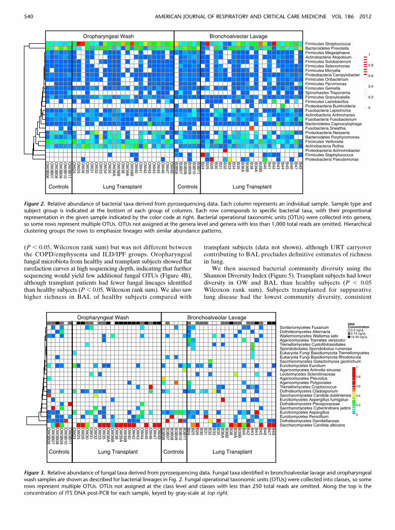
We surveyed bacterial lineages using deep sequencing of 16SrRNA genes (Figure 2). Bronchoscope prewash and control speci-mens, which had low copy numbers, showed bacterial lineagestypical of water and skin organisms (see Figure E1 in the onlinesupplement) as previously described (20).
Oropharyngeal samples contained high abundances of taxacharacteristic of the oral cavity, including Streptococcus, Prevotella,Veillonella, Porphyromonas, Neisseria, and Rothia (22, 33, 34).Most transplant recipients had similar oropharyngeal flora ashealthy subjects, except for four subjects whose OW were dom-inated by Streptococcus spp.
BAL from healthy subjects shared a similar overall bacterialprofile as OW samples, as we previously reported (20). Trans-plant subjects’ BAL largely shared bacterial profiles with OWsamples, but a subset was clearly dominated by specific lineagesthat were absent in OW or detected at much lower abundances.Two BAL samples were dominated by P. aeruginosa (B26 andB33; both transplanted for CF) and revealed P. aeruginosa on cul-ture. A high abundance of Staphylococcus aureus was seen in oneBAL (B24; transplanted for IPF), also detected by culture.Similarly, one BAL showed dominant Achromobacter (B32;transplanted for CF), also identified by culture. In contrast,BAL from the subject with late-onset BOS (B38) was dominated
by the unusual anaerobic lineage Sneathia (35), which was notidentified by culture. Three BAL samples showed high levels ofStreptococcus spp. (B25, B39B, B42; transplanted for COPD ora-1 antitrypsin deficiency) without any corresponding culturedorganism, but Streptococcus spp. were also found at high or mod-erate abundance in OW from these subjects.
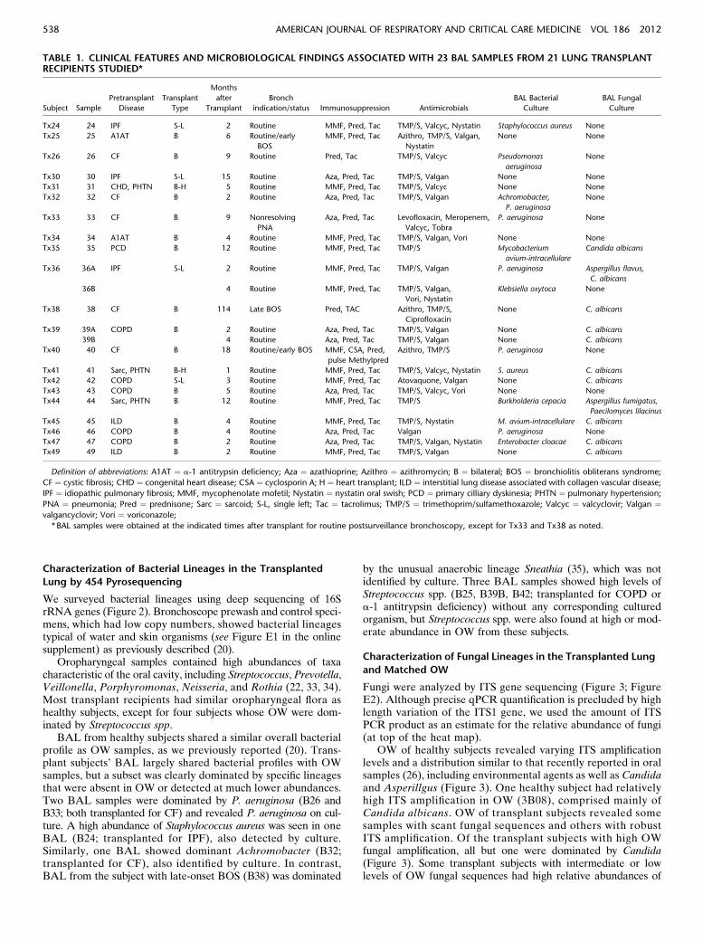
Characterization of Fungal Lineages in the Transplanted Lung
and Matched OW
Fungi were analyzed by ITS gene sequencing (Figure 3; FigureE2). Although precise qPCR quantification is precluded by highlength variation of the ITS1 gene, we used the amount of ITSPCR product as an estimate for the relative abundance of fungi(at top of the heat map).
OW of healthy subjects revealed varying ITS amplificationlevels and a distribution similar to that recently reported in oralsamples (26), including environmental agents as well as Candidaand Asperillgus (Figure 3). One healthy subject had relativelyhigh ITS amplification in OW (3B08), comprised mainly ofCandida albicans. OW of transplant subjects revealed somesamples with scant fungal sequences and others with robustITS amplification. Of the transplant subjects with high OWfungal amplification, all but one were dominated by Candida(Figure 3). Some transplant subjects with intermediate or lowlevels of OW fungal sequences had high relative abundances of
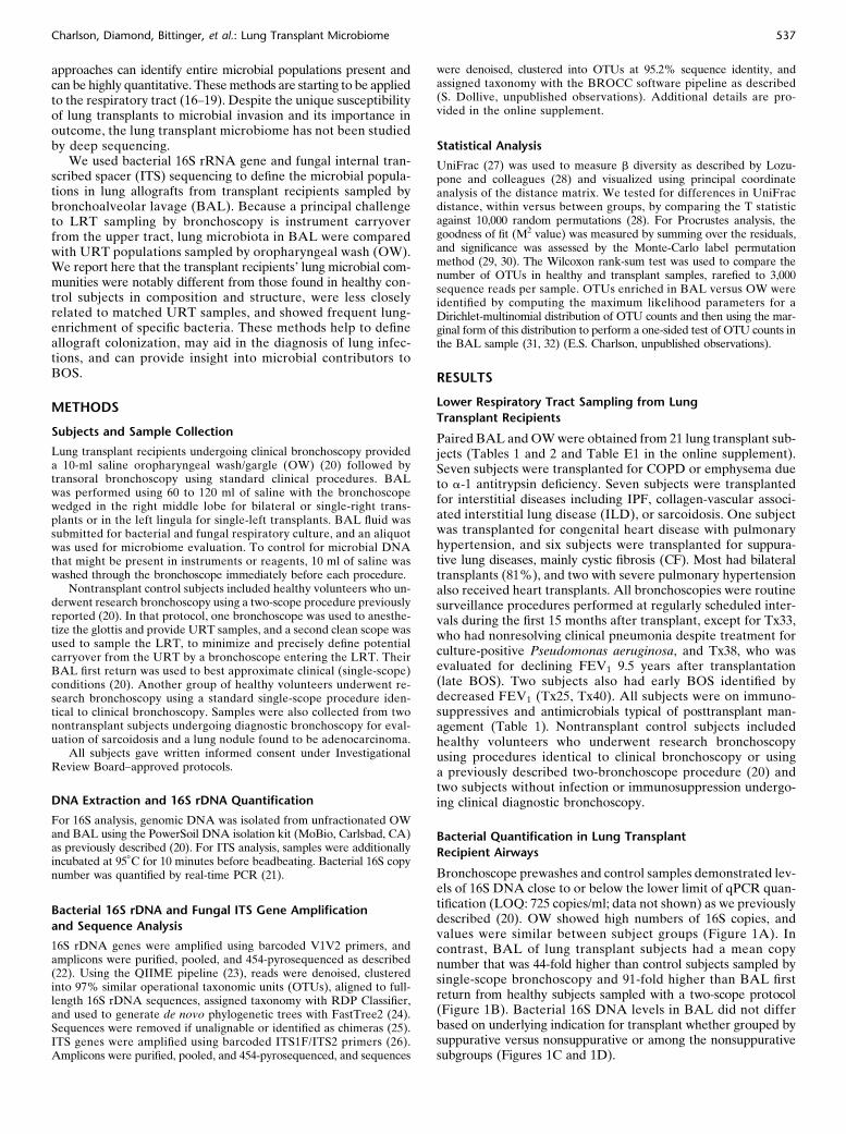
TABLE 1. CLINICAL FEATURES AND MICROBIOLOGICAL FINDINGS ASSOCIATED WITH 23 BAL SAMPLES FROM 21 LUNG TRANSPLANTRECIPIENTS STUDIED*
Subject Sample
Pretransplant
Disease
Transplant
Type
Months
after
Transplant
Bronch
indication/status Immunosuppression Antimicrobials
BAL Bacterial
Culture
BAL Fungal
Culture
Tx24 24 IPF S-L 2 Routine MMF, Pred, Tac TMP/S, Valcyc, Nystatin Staphylococcus aureus None
Tx25 25 A1AT B 6 Routine/early
BOS
MMF, Pred, Tac Azithro, TMP/S, Valgan,
Nystatin
None None
Tx26 26 CF B 9 Routine Pred, Tac TMP/S, Valcyc Pseudomonas
aeruginosa
None
Tx30 30 IPF S-L 15 Routine Aza, Pred, Tac TMP/S, Valgan None None
Tx31 31 CHD, PHTN B-H 5 Routine MMF, Pred, Tac TMP/S, Valcyc None None
Tx32 32 CF B 2 Routine Aza, Pred, Tac TMP/S, Valgan Achromobacter,
P. aeruginosa
None
Tx33 33 CF B 9 Nonresolving
PNA
Aza, Pred, Tac Levofloxacin, Meropenem,
Valcyc, Tobra
P. aeruginosa None
Tx34 34 A1AT B 4 Routine MMF, Pred, Tac TMP/S, Valgan, Vori None None
Tx35 35 PCD B 12 Routine MMF, Pred, Tac TMP/S Mycobacterium
avium-intracellulare
Candida albicans
Tx36 36A IPF S-L 2 Routine MMF, Pred, Tac TMP/S, Valgan P. aeruginosa Aspergillus flavus,
C. albicans
36B 4 Routine MMF, Pred, Tac TMP/S, Valgan,
Vori, Nystatin
Klebsiella oxytoca None
Tx38 38 CF B 114 Late BOS Pred, TAC Azithro, TMP/S,
Ciprofloxacin
None C. albicans
Tx39 39A COPD B 2 Routine Aza, Pred, Tac TMP/S, Valgan None C. albicans
39B 4 Routine Aza, Pred, Tac TMP/S, Valgan None C. albicans
Tx40 40 CF B 18 Routine/early BOS MMF, CSA, Pred,
pulse Methylpred
Azithro, TMP/S P. aeruginosa None
Tx41 41 Sarc, PHTN B-H 1 Routine MMF, Pred, Tac TMP/S, Valcyc, Nystatin S. aureus C. albicans
Tx42 42 COPD S-L 3 Routine MMF, Pred, Tac Atovaquone, Valgan None C. albicans
Tx43 43 COPD B 5 Routine Aza, Pred, Tac TMP/S, Valcyc, Vori None None
Tx44 44 Sarc, PHTN B 12 Routine MMF, Pred, Tac TMP/S Burkholderia cepacia Aspergillus fumigatus,
Paecilomyces lilacinus
Tx45 45 ILD B 4 Routine MMF, Pred, Tac TMP/S, Nystatin M. avium-intracellulare C. albicans
Tx46 46 COPD B 4 Routine Aza, Pred, Tac Valgan P. aeruginosa None
Tx47 47 COPD B 2 Routine Aza, Pred, Tac TMP/S, Valgan, Nystatin Enterobacter cloacae C. albicans
Tx49 49 ILD B 2 Routine MMF, Pred, Tac TMP/S, Valgan None C. albicans
Definition of abbreviations: A1AT ¼ a-1 antitrypsin deficiency; Aza ¼ azathioprine; Azithro ¼ azithromycin; B ¼ bilateral; BOS ¼ bronchiolitis obliterans syndrome;
CF ¼ cystic fibrosis; CHD ¼ congenital heart disease; CSA ¼ cyclosporin A; H ¼ heart transplant; ILD ¼ interstitial lung disease associated with collagen vascular disease;
IPF ¼ idiopathic pulmonary fibrosis; MMF, mycophenolate mofetil; Nystatin ¼ nystatin oral swish; PCD ¼ primary cilliary dyskinesia; PHTN ¼ pulmonary hypertension;
PNA ¼ pneumonia; Pred ¼ prednisone; Sarc ¼ sarcoid; S-L, single left; Tac ¼ tacrolimus; TMP/S ¼ trimethoprim/sulfamethoxazole; Valcyc ¼ valcyclovir; Valgan ¼valgancyclovir; Vori ¼ voriconazole;
* BAL samples were obtained at the indicated times after transplant for routine postsurveillance bronchoscopy, except for Tx33 and Tx38 as noted.
538 AMERICAN JOURNAL OF RESPIRATORY AND CRITICAL CARE MEDICINE VOL 186 2012

Candida, whereas others revealed no dominant lineage orenvironmental fungi. One transplant OW with an intermediatelevel of ITS amplification was dominated by Cryptococcus(OW46).
Healthy volunteers’ BAL yielded scant fungal amplification.Although Candida was identified in several healthy subjects’OW, it was absent in their BAL. The few fungal reads presentin healthy BAL were comprised largely of environmental agentssuch as Davidiellaceae and Cladosporium, also present in pre-wash control samples (Figure E2), as well as low abundancesof Aspergillus.
BAL from lung transplant recipients showed a markedly dif-ferent pattern. Four yielded high levels of ITS amplification;three of these were dominated by Candida (B38, B47, and B49),and one (B44) was dominated by Aspergillus. In each case, thecorresponding fungus was found by culture. Several subjects withintermediate ITS levels revealed high abundance of Candida (B41,B25, B26, B39, B39B, and B42), and one was dominated by As-pergillus (B36, also found by culture). When Candida was seen inBAL, it was also always found at high abundance in OW, whereasfor subjects with a high abundance of Aspergillus in BAL, thisfungus was absent or at markedly lower abundance in OW. Cryp-tococcus was present in six transplant patients’ BAL but atrelatively low abundance or in samples with low total ITS readnumbers. Cryptococcus was not seen in BAL from the trans-plant subject who had this fungus at high abundance in OW(Tx46).
Community Richness, Completeness of Microbial
Identification, and Species Diversity
To determine whether transplant and healthy subjects’ microbialcommunities differed in species richness, we calculated thenumber of observed OTUs at various sequencing depths forbacteria and fungi in OW and BAL specimens. Rarefactionanalysis of bacterial oropharyngeal OTUs (Figure 4A) indicatedthat richness was lower in transplant than healthy OW (P , 0.05,Wilcoxon rank sum) and that sampling was near saturation intransplant subjects but not in healthy subjects. Richness was low-est in the subjects transplanted for suppurative lung disease andintermediate in those transplanted for nonsuppurative disease
TABLE 2. TRANSPLANT SUBGROUPS USED FOR ANALYSIS
Nonsuppurative (n ¼ 15)
Age, yr (mean 6 SD) 57.5 6 7.2
Single/bilateral 4/11
Heart (n) 2
Suppurative (n ¼ 6)
Age, yr (mean 6 SD) 35.0 6 11.3
Single/bilateral 0/6
Heart (n) 0
ILD/IPF (n ¼ 7)
IPF (n) 3
Other ILD (n) 4
Age, yr (mean 6 SD) 60.7 6 6.2
Single/bilateral 3/4
Heart (n) 1
COPD/emphysema (n ¼ 7)
COPD (n) 5
A1AT deficiency (n) 2
Age, yr (mean 6 SD) 54.3 6 7.0
Single/bilateral 1/6
Heart (n) 0
Suppurative (n ¼ 6)
Cystic fibrosis (n) 5
Primary cilliary dyskinesia (n) 1
Age, yr (mean 6 SD) 35.0 6 11.3
Single/bilateral 0/6
Heart (n) 0
Definition of abbreviations: A1AT ¼ a-1 antitrypsin deficiency; COPD ¼ chronic
obstructive pulmonary disease; ILD ¼ interstitial lung disease; IPF ¼ idiopathic
pulmonary fibrosis.
The top set of data shows group characteristics of subjects transplanted for
nonsuppurative and suppurative indications. The lower data set shows underly-
ing disease for the major subgroups of transplant subjects. One subject with
congenital heart disease and pulmonary hypertension is included in the non-
suppurative group but not included in further subgroup analysis.
Figure 1. Quantification of bacterial 16S rDNA gene cop-
ies in oropharyngeal wash (OW) and bronchoalveolar la-vage (BAL) from control and lung transplant subjects. 16S
copy number in oropharyngeal (A) and BAL (B) samples of
control and transplant subjects. Copy number was also
examined in BAL samples of major transplant subgroups(C, D) as described in Table 2. The y axis indicates the 16S
rRNA gene copy number by quantitative PCR. Each sam-
ple was analyzed in triplicate. *P , 0.05; **P , 0.01(Mann-Whitney test).
Charlson, Diamond, Bittinger, et al.: Lung Transplant Microbiome 539
(P , 0.05, Wilcoxon rank sum) but was not different betweenthe COPD/emphysema and ILD/IPF groups. Oropharyngealfungal microbiota from healthy and transplant subjects showed flatrarefaction curves at high sequencing depth, indicating that furthersequencing would yield few additional fungal OTUs (Figure 4B),although transplant patients had fewer fungal lineages identifiedthan healthy subjects (P, 0.05, Wilcoxon rank sum). We also sawhigher richness in BAL of healthy subjects compared with
transplant subjects (data not shown), although URT carryovercontributing to BAL precludes definitive estimates of richnessin lung.
We then assessed bacterial community diversity using theShannon Diversity Index (Figure 5). Transplant subjects had lowerdiversity in OW and BAL than healthy subjects (P , 0.05Wilcoxon rank sum). Subjects transplanted for suppurativelung disease had the lowest community diversity, consistent
Figure 2. Relative abundance of bacterial taxa derived from pyrosequencing data. Each column represents an individual sample. Sample type and
subject group is indicated at the bottom of each group of columns. Each row corresponds to specific bacterial taxa, with their proportional
representation in the given sample indicated by the color code at right. Bacterial operational taxonomic units (OTUs) were collected into genera,so some rows represent multiple OTUs. OTUs not assigned at the genera level and genera with less than 1,000 total reads are omitted. Hierarchical
clustering groups the rows to emphasize lineages with similar abundance patterns.
Figure 3. Relative abundance of fungal taxa derived from pyrosequencing data. Fungal taxa identified in bronchoalveolar lavage and oropharyngeal
wash samples are shown as described for bacterial lineages in Fig. 2. Fungal operational taxonomic units (OTUs) were collected into classes, so some
rows represent multiple OTUs. OTUs not assigned at the class level and classes with less than 250 total reads are omitted. Along the top is theconcentration of ITS DNA post-PCR for each sample, keyed by gray-scale at top right.
540 AMERICAN JOURNAL OF RESPIRATORY AND CRITICAL CARE MEDICINE VOL 186 2012

with outgrowth of particular bacteria. Other transplant sub-jects were intermediate between the healthy and suppurativegroups (P , 0.05, Wilcoxon rank sum).
Global Relationship between Lung and URT Bacterial
Communities in Transplant and Healthy Subjects
We next investigated the relationship of lung and URT communi-ties, following the hypothesis that due to the growth of microbes intransplanted lung,BALwouldbemoredifferent fromURTsamplesin transplant subjects than in healthy subjects. For this comparisonwe usedUniFrac, whichmeasures the similarity of bacterial commu-nities based on the extent to whichmembers of the communities arephylogenetically related (27). UniFrac distances were calculated forall pairs of samples, and then relationships among communitieswere plotted using principal coordinate analysis (PCoA).
UniFrac analysis showed that each subject’s BAL was moreclosely related to their own OW than to the BAL of different
individuals (P , 0.05 for abundance-weighted and unweightedUniFrac). An additional nonphylogenetic measure of sharedOTUs, the Jaccard Index (36, 37), also revealed that BAL fromeach individual was most tightly linked to their own OW (P, 0.05binary/abundance Jaccard), consistent with BAL communitiesoriginating largely in the URT (20).
We then quantified the resemblance between lung and URTcommunities. The BAL and OW weighted UniFrac data wereplotted using PCoA, and then the two PCoA plots were alignedusing Procrustes (29, 30), which rotates and scales two matricesto maximize overlap (Figure 6). For healthy subjects, BAL andOW formed a tight cluster, separate from transplant subjects (P,0.05 UniFrac and Jaccard), with small distances between commu-nities from the same individual. In contrast, transplant subjects’BAL and OW microbiota were often divergent and separated bygreater distances. Thus, healthy subjects show close similaritybetween their BAL and OW microbiota, whereas distances aregreater for lung transplant subjects, consistent with outgrowth ofbacterial lineages in transplanted lungs.
Figure 4. Rarefaction analysis of bacterial and fungal operational taxo-nomic units (OTUs) in the oropharynx of healthy and lung transplant
subjects. The y axis denotes the number of OTUs detected by pyrose-
quencing of bacterial 16S genes (A) or fungal internal transcribed
spacer genes (B) at the corresponding sequencing depths shown alongthe x axis. The subject group is indicated by the color key on the right.
Error bars denote the standard deviation within subject groups.
Figure 5. Alpha diversity of bacterial communities in healthy and trans-plant subjects. Diversity was calculated using the Shannon Index for
oropharyngeal wash (A) and bronchoalveolar lavage (B). *P , 0.05;
**P , 0.01 (Wilcoxon rank sum).
Figure 6. Relationship between bronchoalveolar lavage (BAL) and oro-
pharyngeal bacterial communities within individuals. Weighted UniFrac
distances were calculated between all pairs of samples within BAL ororopharyngeal wash (OW), and then each sample type was plotted
separately in 3D space by principal coordinate analysis. The two plots
(BAL and OW) were then transformed by Procrustes analysis to achievemaximum alignment. Each point corresponds to a bacterial commu-
nity, with transplant subjects’ communities shown in blue, healthy sub-
jects’ communities shown in red, and the two communities from each
subject connected by a bar. The orange end of each bar connects to theOW sample data; the black end connects to the BAL sample data from
the same individual. If BAL and OW plots are similar, then the relative
distance between connected points (residuals) will be small. The overall
similarity is summarized by the M2 value, and statistical goodness of fitis measured by a Monte Carlo label permutation approach (10,000
iterations).
Charlson, Diamond, Bittinger, et al.: Lung Transplant Microbiome 541
We then determined whether the relationship between URTand BAL communities differed based on underlying indicationsfor transplant.WhenBAL-OWdistances within individuals werecompared across groups, there was a trend for greater BAL-OWdistances in the suppurative transplant group compared withall nonsuppurative or COPD/emphysema and ILD/IPF groups(Figure E3), suggesting that the greater BAL-OWdistance amongtransplant subjects was driven mainly by the subgroup of subjectstransplanted for suppurative diseases.
Comparing BAL and Oropharyngeal Bacterial Lineages
to Identify Taxa Enriched in Lung
All bacterial and fungal organisms found by BAL culture wereidentified via 16S and ITS sequencing, except forMycobacteriumavium-intracellulare, which was found by culture in two subjects(Tx35, Tx45) but not by 16S sequencing (Tables 1 and E1).Respiratory pathogens identified by culture were sometimesthe most abundant lineage identified by sequencing (B24, B26,B32, B33, B40, B44), but in many cases they were exceeded inabundance by taxa typical of upper respiratory tract flora (B36,B41, B46, B47, B50). Thus, potentially significant pathogens in thelung often represent a minority population against the background
of oropharyngeal organisms in BAL. Furthermore, lung infectioncan result from bacteria that are normal inhabitants of the URT.
Therefore, to identify sequences that represent organismsgenuinely replicating in lung, we developed a single-sided outliertest to compare each taxon’s abundance in BALwith its abundancein OW. This strategy assumes that microbes replicating in the LRTwill be enriched in BAL compared with OW, whereas URT taxapresent in BAL due to passive admixture such as bronchoscopiccarryover or aspiration will be represented at a relative abundanceno greater than that in OW. The model also accounts for uncer-tainty due to stochastics of sampling (E.S. Charlson, unpublishedobservations).
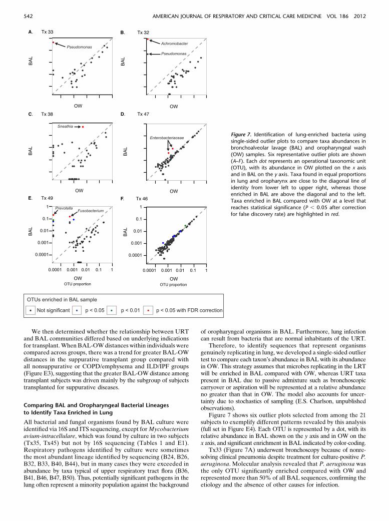
Figure 7 shows six outlier plots selected from among the 21subjects to exemplify different patterns revealed by this analysis(full set in Figure E4). Each OTU is represented by a dot, with itsrelative abundance in BAL shown on the y axis and in OW on thex axis, and significant enrichment in BAL indicated by color-coding.
Tx33 (Figure 7A) underwent bronchoscopy because of nonre-solving clinical pneumonia despite treatment for culture-positive P.aeruginosa. Molecular analysis revealed that P. aeruginosa wasthe only OTU significantly enriched compared with OW andrepresented more than 50% of all BAL sequences, confirming theetiology and the absence of other causes for infection.
Figure 7. Identification of lung-enriched bacteria usingsingle-sided outlier plots to compare taxa abundances in
bronchoalveolar lavage (BAL) and oropharyngeal wash
(OW) samples. Six representative outlier plots are shown
(A–F). Each dot represents an operational taxonomic unit(OTU), with its abundance in OW plotted on the x axis
and in BAL on the y axis. Taxa found in equal proportions
in lung and oropharynx are close to the diagonal line of
identity from lower left to upper right, whereas thoseenriched in BAL are above the diagonal and to the left.
Taxa enriched in BAL compared with OW at a level that
reaches statistical significance (P , 0.05 after correction
for false discovery rate) are highlighted in red.
542 AMERICAN JOURNAL OF RESPIRATORY AND CRITICAL CARE MEDICINE VOL 186 2012

Tx32 (Figure 7B) grew Achromobacter and P. aeruginosa inBAL culture. The outlier plot showed highly significant enrich-ment ofAchromobacter, whereas Pseudomonas was above the lineof identity but did not reach statistical significance.Achromobactercomprised more than 80% of bacteria in BAL and was 17-foldmore abundant than Pseudomonas, indicating the relative quantityof each organism in lung.
Tx38 (Figure 7C) had late-onset BOS. Outlier analysisrevealed high-level BAL enrichment of the anaerobic bacteriumSneathia, which represented more than 50% of all sequences inBAL but less than 0.5% in the oropharynx and indicates ro-bust lung growth. No respiratory pathogens were identified inBAL culture, which would not detect anaerobes such as Sneathia.
Tx47 (Figure 7D) yielded Enterobacter cloacae in BAL cul-ture. The outlier plot revealed clustering of many taxa along theline of identity, suggesting abundant oropharyngeal admixturewith BAL. However, even in the background of high carryover,two Enterobacteriaceae taxa are significantly enriched in BALrelative to OW, confirming outgrowth in lung.
Tx49 (Figure 7E) was negative for respiratory pathogens byculture. However, outlier analysis revealed significant enrichmentin BAL of several taxa normally found in the URT (Prevotella,Fusobacterium), suggesting outgrowth of mixed oropharyngealflora not detected by standard respiratory culture.
Tx46 (Figure 7F) BAL culture revealed P. aeruginosa. How-ever, outlier analysis revealed no taxa enriched in lung comparedwith URT. P. aeruginosa was present in BAL but representedonly 0.01% of sequences and had a similar abundance in OW.This result suggests detection by culture in BAL likely from bron-choscopic carryover or passive aspiration without replication andenrichment in the lung.
Additional examples of clinical utility (Figure E4) include ro-bust single-taxa lung enrichment (Tx24, Tx26, Tx44), lung outgrowthof mixed URT/anaerobic bacteria or other organisms not found onculture (Tx25, Tx41), and lack of outliers indicating absence ofsignificant lung-enriched taxa (Tx30, Tx31).
Figure E4 shows a concise clinical report developed to sum-marize molecular microbiome findings, including the outlier analy-sis, culture, and clinical data for each subject. Statistical and softwaretools for report generation are embedded in a “reproducible ”format, which allows repeated analysis over user-specified datasets (E. S. Charlson, unpublished observations).
DISCUSSION
Approximately 24% of lung transplant recipients die from post-transplant infections, most of which are bacterial or fungal pul-monary infections (1, 38, 39). The major cause of later death isBOS (1, 39), a form of chronic rejection that is not well under-stood but is linked to microbial triggers within the allograft (4–12,40). Here we applied broad-range 16S bacterial and ITS fungalrRNA gene profiling to define microbial populations in the lungsof 21 transplant recipients. Transplant subjects showed markeddifferences from healthy subjects in airway microbial burden,community composition and structure, lower richness, and greaterdistances between lung and upper respiratory communities. Wethen used a novel approach to visualize taxa enriched in bron-choscopic specimens relative to oropharyngeal samples, allow-ing identification of specific bacteria replicating in allografts ofindividual subjects.
The richness and diversity of airway communities were re-duced in transplant subjects compared with control subjects. Al-though richness and diversity was most reduced in subjects withCF, it was also reduced in subjects transplanted for other indications(Figure 5). Decreased airway community richness and diversity inCF has been correlated with deteriorating lung function (41, 42)
but has not been investigated in relation to outcome from lungtransplant either among subjects with CF or subjects with non-suppurative lung disease.
Although molecular profiling can identify all taxa present andcan quantify abundances, the source of those sequences is criticalin determining their significance (43). By comparing the relativeabundance of each bacterial taxon in BAL and oropharyngealsamples, we can identify enrichment in lung (E.S. Charlson,unpublished observations). Enrichment in BAL compared withthe URT suggests replication within the lung and thus can dis-tinguish such organisms from those entering BAL through bron-choscopic carryover or passive aspiration. A large proportion oftransplant recipients had specific lung-enriched bacteria. Enrichedtaxa often confirmed organisms found by culture but also identi-fied bacteria not amenable to culture, such as unusual anaerobesor normal oropharyngeal residents. Molecular profiling also de-termined proportional abundances of organisms, adding quantita-tive information to culture results. In some cases we showed thatbacteria identified by culture were equally represented in BALand URT, providing evidence against authentic LRT replication.
Transplant subjects had markedly higher levels of bacterial16S DNA in BAL than controls subjects. This finding was trueregardless of underlying indication for transplant, with no differ-ence between subjects transplanted for COPD/emphysema, inter-stitial lung disease, or CF/suppurative lung disease. Although taxapresent in BAL at abundances similar to the URTmay result frombronchoscopic carryover, they may also reflect bacteria genuinelypresent in lung due to aspiration without replication and enrich-ment. The high 16S DNA levels seen even in subjects without out-liers (or where outliers reflect a minority of BAL sequences) isconsistent with such a source, which may result from increasedpropensity for aspiration or inability to clear aspirated bacteriaeffectively due to cough and mucocilliary clearance defects (3, 44,45) Passively aspirated or even dead bacteria can be potentimmunological activators, and aspiration has been implicatedin BOS (46).
BOS is associated with polymorphisms in genes that regulateinnate microbial responses (9–12), specific bacteria, fungi, orviruses in the LRT (4–8) and may be more responsive to theantibiotic azithromycin than to intensified immunosuppression(13–15). It is hypothesized that aberrant microbial populationsin the lung, among other factors, trigger inflammatory pathways,resulting in graft injury (40). Our study was not designed toaddress BOS specifically. However, one subject had late-onsetBOS, and 16S sequencing revealed high-level lung enrichmentof the anaerobic lineage Sneathia. This organism is a componentof the vaginal flora and a rare cause of obstetrical infections (35,47–49) but has not previously been reported in the respiratorytract and would not be identified by standard culture. Two sub-jects with early BOS also had taxa enriched in lung that were notdetected by culture (Tx25, Tx40). Longitudinal studies usingbroad-range profiling are needed to determine whether over-all microbial burden or specific lung microbiome features, eitherearly after transplant or at the time of BOS, have a role in lategraft failure.
Our study has several limitations. (1) To not “over-call” lung-enriched taxa, we used a 95% confidence threshold (E.S. Charlson,unpublished observations). There may be organisms authenticallyreplicating in the lung that do not reach statistical significance forenrichment, although they are likely to be present at low abun-dance and/or with modest fold-increase over OW. (2) AlthoughOW is a clinically tractable way to sample the URT, there maybe subtle differences in outliers identified when BAL is comparedwith the most accurate representation of potential carryover,direct peri-glottic specimens obtained with high stringency two-scope sampling (20; E.S. Charlson, unpublished observations). (3)
Charlson, Diamond, Bittinger, et al.: Lung Transplant Microbiome 543

Although most pathogens found by culture were detected bymolecular analysis, two subjects hadM. avium-intracellulare infec-tions on culture that were not found by 16S sequencing. AlthoughMAI is occasionally isolated as a laboratory contaminant, thisresult suggests that mycobacterial detection may be a limitationof 16S rDNA surveys with the primers used (50). (4) A relativelysmall number of subjects was included in each transplant grouphere, which should be expanded to larger numbers of subjectsand serial specimens.
In summary, aberrant microbial communities and specificlung-enriched taxa are common in lung after transplantation. Fu-ture studies are needed to determinewhether particular communitystructures or individual microorganisms identified by microbiomeanalysis are associated with late graft failure and BOS and whetherbroad-range 16S bacterial or ITS fungal profiling added to standardculture improves diagnosis and management of lung colonizationand infection in transplant and other lung diseases.
Author disclosures are available with the text of this article at www.atsjournals.org.
Acknowledgment: The authors thank the research subjects who volunteered for thisstudy and the clinicians who assisted with specimen collection; W. Russell and D.Frame for critical study assistance; the Penn Center for AIDS Research (P30-AI045008) for technical support; J. Christie for valuable discussion and insights;and members of the Bushman and Collman laboratories for helpful discussions.
References
1. Christie JD, Edwards LB, Kucheryavaya AY, Benden C, Dobbels F, Kirk R,
Rahmel AO, Stehlik J, Hertz MI. The registry of the international society
for heart and lung transplantation: twenty-eighth adult lung and heart-
lung transplant report–2011. J Heart Lung Transplant 2011;30:1104–1122.
2. Robertson AG, Griffin SM, Murphy DM, Pearson JP, Forrest IA, Dark
JH, Corris PA, Ward C. Targeting allograft injury and inflammation in
the management of post-lung transplant bronchiolitis obliterans syn-
drome. Am J Transplant 2009;9:1272–1278.
3. Atkins BZ, Trachtenberg MS, Prince-Petersen R, Vess G, Bush EL,
Balsara KR, Lin SS, Davis RD Jr. Assessing oropharyngeal dysphagia
after lung transplantation: altered swallowing mechanisms and in-
creased morbidity. J Heart Lung Transplant 2007;26:1144–1148.
4. Botha P, Archer L, Anderson RL, Lordan J, Dark JH, Corris PA, Gould
K, Fisher AJ. Pseudomonas aeruginosa colonization of the allograft
after lung transplantation and the risk of bronchiolitis obliterans
syndrome. Transplantation 2008;85:771–774.
5. Khalifah AP, Hachem RR, Chakinala MM, Schechtman KB, Patterson
GA, Schuster DP, Mohanakumar T, Trulock EP, Walter MJ. Respi-
ratory viral infections are a distinct risk for bronchiolitis obliterans
syndrome and death. Am J Respir Crit Care Med 2004;170:181–187.
6. Weigt SS, Elashoff RM, Huang C, Ardehali A, Gregson AL, Kubak B,
Fishbein MC, Saggar R, Keane MP, Lynch JP III, et al. Aspergillus
colonization of the lung allograft is a risk factor for bronchiolitis
obliterans syndrome. Am J Transplant 2009;9:1903–1911.
7. Kotsimbos TC, Snell GI, Levvey B, Spelman DW, Fuller AJ, Wesselingh
SL, Williams TJ, Ostergaard L. Chlamydia pneumoniae serology in
donors and recipients and the risk of bronchiolitis obliterans syndrome
after lung transplantation. Transplantation 2005;79:269–275.
8. Heng D, Sharples LD, McNeil K, Stewart S, Wreghitt T, Wallwork J.
Bronchiolitis obliterans syndrome: incidence, natural history, prog-
nosis, and risk factors. J Heart Lung Transplant 1998;17:1255–1263.
9. Munster JM, van der Bij W, Breukink MB, van der Steege G, Zuurman
MW, Hepkema BG, Verschuuren EA, van Son WJ, Seelen MA. As-
sociation between donor MBL promoter haplotype and graft survival
and the development of BOS after lung transplantation. Transplantation
2008;86:1857–1863.
10. Palmer SM, Burch LH, Trindade AJ, Davis RD, Herczyk WF, Reinsmoen
NL, Schwartz DA. Innate immunity influences long-term outcomes after
human lung transplant. Am J Respir Crit Care Med 2005;171:780–785.
11. Palmer SM, Klimecki W, Yu L, Reinsmoen NL, Snyder LD, Ganous
TM, Burch L, Schwartz DA. Genetic regulation of rejection and
survival following human lung transplantation by the innate immune
receptor CD14. Am J Transplant 2007;7:693–699.
12. Kastelijn EA, van Moorsel CH, Rijkers GT, Ruven HJ, Karthaus V,
Kwakkel-van Erp JM, van de Graaf EA, Zanen P, van Kessel DA,
Grutters JC, et al. Polymorphisms in innate immunity genes associated
with development of bronchiolitis obliterans after lung transplantation.
J Heart Lung Transplant 2010;29:665–671.
13. Gerhardt SG, McDyer JF, Girgis RE, Conte JV, Yang SC, Orens JB.
Maintenance azithromycin therapy for bronchiolitis obliterans syndrome:
results of a pilot study. Am J Respir Crit Care Med 2003;168:121–125.
14. Vos R, Vanaudenaerde BM, Verleden SE, De Vleeschauwer SI, Willems-
Widyastuti A, Van Raemdonck DE, Schoonis A, Nawrot TS, Dupont LJ,
Verleden GM. A randomised controlled trial of azithromycin to prevent
chronic rejection after lung transplantation.Eur Respir J 2011;37:164–172.
15. Jain R, Hachem RR, Morrell MR, Trulock EP, Chakinala MM, Yusen
RD, Huang HJ, Mohanakumar T, Patterson GA, Walter MJ.
Azithromycin is associated with increased survival in lung trans-
plant recipients with bronchiolitis obliterans syndrome. J Heart
Lung Transplant 2010;29:531–537.
16. Erb-Downward JR, Thompson DL, Han MK, Freeman CM, McCloskey
L, Schmidt LA, Young VB, Toews GB, Curtis JL, Sundaram B, et al.
Analysis of the lung microbiome in the “healthy” smoker and in
COPD. PLoS ONE 2011;6:e16384.
17. Flanagan JL, Brodie EL, Weng L, Lynch SV, Garcia O, Brown R,
Hugenholtz P, DeSantis TZ, Andersen GL, Wiener-Kronish JP, et al.
Loss of bacterial diversity during antibiotic treatment of intubated
patients colonized with Pseudomonas aeruginosa. J Clin Microbiol
2007;45:1954–1962.
18. Harris JK, De Groote MA, Sagel SD, Zemanick ET, Kapsner R, Penvari C,
Kaess H, Deterding RR, Accurso FJ, Pace NR. Molecular identifica-
tion of bacteria in bronchoalveolar lavage fluid from children with
cystic fibrosis. Proc Natl Acad Sci USA 2007;104:20529–20533.
19. Hilty M, Burke C, Pedro H, Cardenas P, Bush A, Bossley C, Davies J,
Ervine A, Poulter L, Pachter L, et al. Disordered microbial commu-
nities in asthmatic airways. PLoS ONE 2010;5:e8578.
20. Charlson ES, Bittinger K, Haas AR, Fitzgerald AS, Frank I, Yadav A,
Bushman FD, Collman RG. Topographical continuity of bacterial
populations in the healthy human respiratory tract. Am J Respir Crit
Care Med 2011;184:957–963.
21. Hill DA, Hoffmann C, Abt MC, Du Y, Kobuley D, Kirn TJ, Bushman FD,
Artis D. Metagenomic analyses reveal antibiotic-induced temporal and
spatial changes in intestinal microbiota with associated alterations in
immune cell homeostasis. Mucosal Immunol 2010;3:148–158.
22. Charlson ES, Chen J, Custers-Allen R, Bittinger K, Li H, Sinha R, Hwang
J, Bushman FD, Collman RG. Disordered microbial communities in the
upper respiratory tract of cigarette smokers. PLoS ONE 2010;5:e15216.
23. Caporaso JG, Kuczynski J, Stombaugh J, Bittinger K, Bushman FD,
Costello EK, Fierer N, Pena AG, Goodrich JK, Gordon JI, et al.
Qiime allows analysis of high-throughput community sequencing data.
Nat Methods 2010;7:335–336.
24. Price MN, Dehal PS, Arkin AP. Fasttree 2: approximately maximum-
likelihood trees for large alignments. PLoS ONE 2010;5:e9490.
25. Haas BJ, Gevers D, Earl AM, Feldgarden M, Ward DV, Giannoukos G,
Ciulla D, Tabbaa D, Highlander SK, Sodergren E, et al. Chimeric 16s
rRNA sequence formation and detection in sanger and 454-pyrose-
quenced pcr amplicons. Genome Res 2011;21:494–504.
26. GhannoumMA, Jurevic RJ, Mukherjee PK, Cui F, Sikaroodi M, Naqvi A,
Gillevet PM. Characterization of the oral fungal microbiome
(mycobiome) in healthy individuals. PLoS Pathog 2010;6:e1000713.
27. Lozupone C, Knight R. UniFrac: a new phylogenetic method for com-
paring microbial communities. Appl Environ Microbiol 2005;71:8228–
8235.
28. Lozupone CA,HamadyM, Kelley ST, Knight R. Quantitative and qualitative
beta diversity measures lead to different insights into factors that structure
microbial communities. Appl Environ Microbiol 2007;73:1576–1585.
29. Gower JC. Generalized Procrustes analysis. Psychometrika 1975;40:33–51.
30. Hurley JR, Cattell RB. The Procrustes program: producing direct rota-
tion to test a hypothesized factor structure. Behav Sci 1962;7:258–262.
31. Weir BS, Hill WG. Estimating f-statistics. Annu Rev Genet 2002;36:721–750.
32. Ng KWTG, Tang ML. Dirichlet and related distributions. Chichester,
UK: Wiley; 2011.
33. Costello EK, Lauber CL, Hamady M, Fierer N, Gordon JI, Knight R.
Bacterial community variation in human body habitats across space
and time. Science 2009;326:1694–1697.
544 AMERICAN JOURNAL OF RESPIRATORY AND CRITICAL CARE MEDICINE VOL 186 2012
34. Lemon KP, Klepac-Ceraj V, Schiffer HK, Brodie EL, Lynch SV, Kolter R.
Comparative analyses of the bacterial microbiota of the human nostril
and oropharynx. MBio 2010;1. doi: 10.1128/mBio.00129-10
35. Collins MD, Hoyles L, Tornqvist E, von Essen R, Falsen E. Charac-
terization of some strains from human clinical sources which
resemble “Leptotrichia sanguinegens”: description of sneathia san-
guinegens sp. Nov., gen. Nov. Syst Appl Microbiol 2001;24:358–
361.
36. Chao A, Chazdon RL, Colwell RK, Shen T-J. A new statistical approach
for assessing similarity of species composition with incidence and
abundance data. Ecol Lett 2005;8:148–159.
37. Jaccard P. The distribution of the flora in the alpine zone. New Phytol
1912;11:37–50.
38. Valentine VG, Bonvillain RW, Gupta MR, Lombard GA, LaPlace SG,
Dhillon GS, Wang G. Infections in lung allograft recipients: ganci-
clovir era. J Heart Lung Transplant 2008;27:528–535.
39. Christie JD, Edwards LB, Kucheryavaya AY, Aurora P, Dobbels F, Kirk R,
Rahmel AO, Stehlik J, Hertz MI. The registry of the international society
for heart and lung transplantation: twenty-seventh official adult lung and
heart-lung transplant report–2010. J Heart Lung Transplant 2010;29:
1104–1118.
40. Nakajima T, Palchevsky V, Perkins DL, Belperio JA, Finn PW. Lung
transplantation: infection, inflammation, and the microbiome. Semin
Immunopathol 2011;33:135–156.
41. Cox MJ, Allgaier M, Taylor B, Baek MS, Huang YJ, Daly RA, Karaoz U,
Andersen GL, Brown R, Fujimura KE, et al. Airway microbiota and
pathogen abundance in age-stratified cystic fibrosis patients. PLoS ONE
2010;5:e11044.
42. Klepac-Ceraj V, Lemon KP, Martin TR, Allgaier M, Kembel SW, Knapp
AA, Lory S, Brodie EL, Lynch SV, Bohannan BJ, et al. Relationship
between cystic fibrosis respiratory tract bacterial communities and age,
genotype, antibiotics and Pseudomonas aeruginosa. Environ Microbiol
2010;12:1293–1303.
43. Bartlett JG, Alexander J, Mayhew J, Sullivan-Sigler N, Gorbach SL.
Should fiberoptic bronchoscopy aspirates be cultured? Am Rev Respir
Dis 1976;114:73–78.
44. Herve P, Silbert D, Cerrina J, Simonneau G, Dartevelle P. Impairment
of bronchial mucociliary clearance in long-term survivors of heart/
lung and double-lung transplantation. The Paris-Sud lung transplant
group. Chest 1993;103:59–63.
45. Higenbottam T, Jackson M, Woolman P, Lowry R, Wallwork J. The
cough response to ultrasonically nebulized distilled water in heart-
lung transplantation patients. Am Rev Respir Dis 1989;140:58–61.
46. D’Ovidio F, Mura M, Tsang M, Waddell TK, Hutcheon MA, Singer LG,
Hadjiliadis D, Chaparro C, Gutierrez C, Pierre A, et al. Bile acid
aspiration and the development of bronchiolitis obliterans after lung
transplantation. J Thorac Cardiovasc Surg 2005;129:1144–1152.
47. Ling Z, Kong J, Liu F, Zhu H, Chen X, Wang Y, Li L, Nelson KE, Xia
Y, Xiang C. Molecular analysis of the diversity of vaginal microbiota
associated with bacterial vaginosis. BMC Genomics 2010;11:488.
48. Ravel J, Gajer P, Abdo Z, Schneider GM, Koenig SS, McCulle SL,
Karlebach S, Gorle R, Russell J, Tacket CO, et al. Vaginal microbiome of
reproductive-age women. Proc Natl Acad Sci USA 2011;108:4680–4687.
49. De Martino SJ, Mahoudeau I, Brettes JP, Piemont Y, Monteil H,
Jaulhac B. Peripartum bacteremias due to leptotrichia amnionii and
sneathia sanguinegens, rare causes of fever during and after delivery.
J Clin Microbiol 2004;42:5940–5943.
50. Bogner JR, Rusch-Gerdes S, Mertenskotter T, Loch O, Emminger C,
Baumgarten R, Brockmeyer NH, Brockhaus W, Jablonowski H, Stoehr
A, et al. Patterns of Mycobacterium avium culture and PCR positivity
in immunodeficient HIV-infected patients: progression from localized
to systematic disease, German AIDS Study Group (GASG/IDKF).
Scand J Infect Dis 1997;29:579–584.
Charlson, Diamond, Bittinger, et al.: Lung Transplant Microbiome 545
Related Documents