Editing file Respiratory block-Anatomy-Lecture 4 Lung and Pleura

Welcome message from author
This document is posted to help you gain knowledge. Please leave a comment to let me know what you think about it! Share it to your friends and learn new things together.
Transcript
Editing file
Respiratory block-Anatomy-Lecture 4
Lung and Pleura
ObjectivesColor guide :Only in boys slides in GreenOnly in girls slides in Purpleimportant in RedDoctor note in BlueExtra information in Grey
At the end of the lecture, students should:✓ Describe the anatomy of the pleura:
subdivisions into parietal and visceral pleura, nerve supply of each of them.
✓ List the parts of parietal pleura and its recesses. ✓ Describe the surface anatomy of both pleura and lungs.✓ Describe the anatomy of lungs : shape, relations, nerve supply & blood
supply.✓ Describe the difference between right & left lungs.✓ Describe the formation of bronchopulmonary segments and the main
characteristics of each segment in the lung.
Click here for helpful short video ;)
3
Pleura
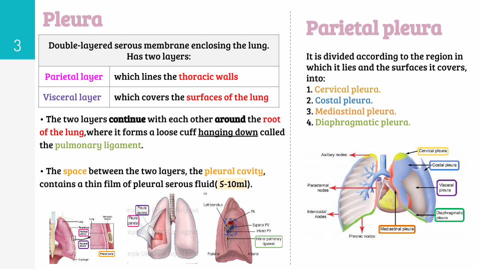
• The two layers continue with each other around the root of the lung,where it forms a loose cuff hanging down called the pulmonary ligament.
• The space between the two layers, the pleural cavity, contains a thin film of pleural serous fluid( 5-10ml).
Double-layered serous membrane enclosing the lung. Has two layers:
Parietal layer which lines the thoracic walls
Visceral layer which covers the surfaces of the lung
It is divided according to the region in which it lies and the surfaces it covers, into:1. Cervical pleura.2. Costal pleura.3. Mediastinal pleura.4. Diaphragmatic pleura.
Parietal pleura
Parietal pleura
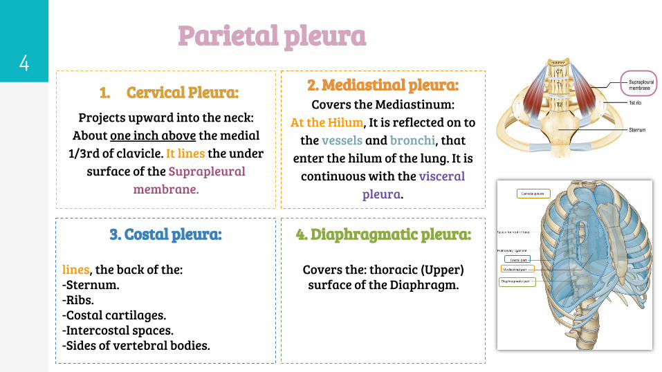
1. Cervical Pleura:Projects upward into the neck:
About one inch above the medial 1/3rd of clavicle. It lines the under
surface of the Suprapleural membrane.
4
3. Costal pleura:
lines, the back of the:-Sternum.-Ribs.-Costal cartilages.-Intercostal spaces.-Sides of vertebral bodies.
2. Mediastinal pleura:Covers the Mediastinum:
At the Hilum, It is reflected on to the vessels and bronchi, that
enter the hilum of the lung. It is continuous with the visceral
pleura.
4. Diaphragmatic pleura:
Covers the: thoracic (Upper) surface of the Diaphragm.
5Pa
riet
al
Pleura nerve supply
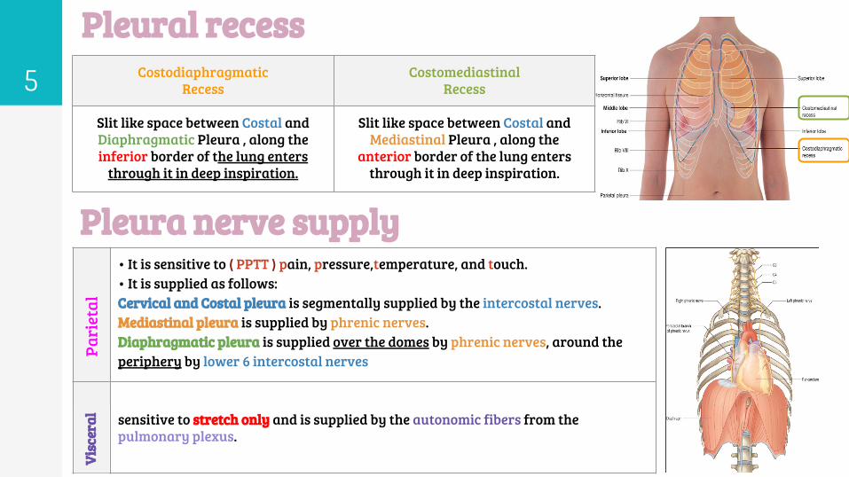
Pleural recessCostodiaphragmatic
RecessCostomediastinal
Recess
Slit like space between Costal and Diaphragmatic Pleura , along the inferior border of the lung enters
through it in deep inspiration.
Slit like space between Costal and Mediastinal Pleura , along the
anterior border of the lung enters through it in deep inspiration.
• It is sensitive to ( PPTT ) pain, pressure,temperature, and touch.• It is supplied as follows:Cervical and Costal pleura is segmentally supplied by the intercostal nerves.Mediastinal pleura is supplied by phrenic nerves.Diaphragmatic pleura is supplied over the domes by phrenic nerves, around the periphery by lower 6 intercostal nerves
sensitive to stretch only and is supplied by the autonomic fibers from the pulmonary plexus.
Visc
eral
6Surface anatomy of pleura
66
4
Note: This slide is important, especially the numbers
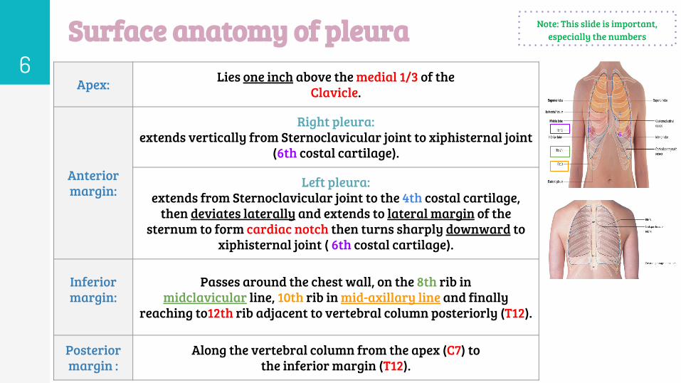
Apex: Lies one inch above the medial 1/3 of theClavicle.
Anterior margin:
Right pleura:extends vertically from Sternoclavicular joint to xiphisternal joint
(6th costal cartilage).
Left pleura:extends from Sternoclavicular joint to the 4th costal cartilage,
then deviates laterally and extends to lateral margin of the sternum to form cardiac notch then turns sharply downward to
xiphisternal joint ( 6th costal cartilage).
Inferior margin:
Passes around the chest wall, on the 8th rib inmidclavicular line, 10th rib in mid-axillary line and finally
reaching to12th rib adjacent to vertebral column posteriorly (T12).
Posterior margin :
Along the vertebral column from the apex (C7) tothe inferior margin (T12).
7Surface anatomy of lung
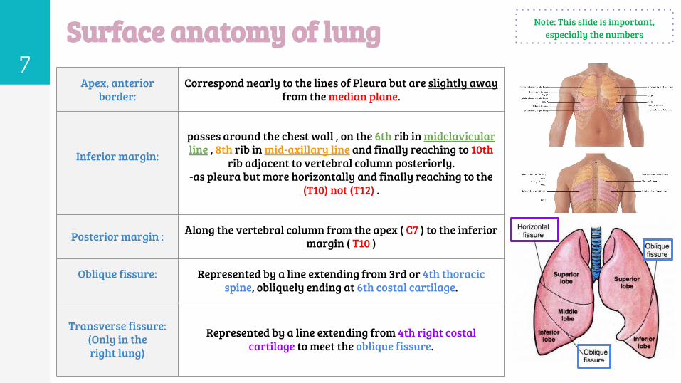
Apex, anterior border:
Correspond nearly to the lines of Pleura but are slightly away from the median plane.
Inferior margin:
passes around the chest wall , on the 6th rib in midclavicular line , 8th rib in mid-axillary line and finally reaching to 10th
rib adjacent to vertebral column posteriorly.-as pleura but more horizontally and finally reaching to the
(T10) not (T12) .
Posterior margin : Along the vertebral column from the apex ( C7 ) to the inferior margin ( T10 )
Oblique fissure: Represented by a line extending from 3rd or 4th thoracic spine, obliquely ending at 6th costal cartilage.
Transverse fissure: (Only in the right lung)
Represented by a line extending from 4th right costal cartilage to meet the oblique fissure.
Note: This slide is important, especially the numbers
8Pleural effusion
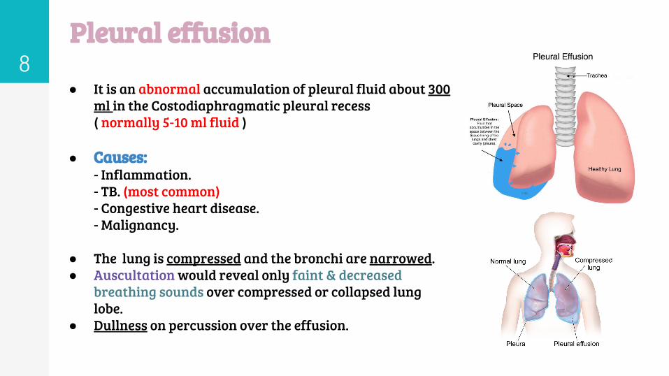
● It is an abnormal accumulation of pleural fluid about 300 ml in the Costodiaphragmatic pleural recess
( normally 5-10 ml fluid )
● Causes:- Inflammation.- TB. (most common)- Congestive heart disease.- Malignancy.
● The lung is compressed and the bronchi are narrowed. ● Auscultation would reveal only faint & decreased
breathing sounds over compressed or collapsed lung lobe.
● Dullness on percussion over the effusion.
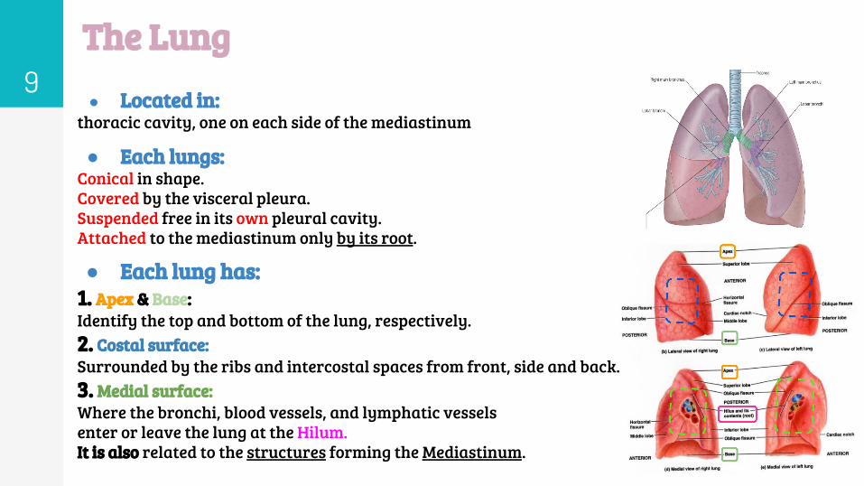
9The Lung● Located in:
thoracic cavity, one on each side of the mediastinum
● Each lungs:Conical in shape.Covered by the visceral pleura.Suspended free in its own pleural cavity.Attached to the mediastinum only by its root.
● Each lung has:1. Apex & Base:Identify the top and bottom of the lung, respectively. 2. Costal surface:Surrounded by the ribs and intercostal spaces from front, side and back.3. Medial surface:Where the bronchi, blood vessels, and lymphatic vesselsenter or leave the lung at the Hilum.It is also related to the structures forming the Mediastinum.
10
The Lung
Borders
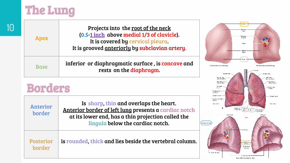
Apex
Projects into the root of the neck (0.5-1 inch above medial 1/3 of clavicle).
It is covered by cervical pleura.It is grooved anteriorly by subclavian artery.
Base inferior or diaphragmatic surface , is concave and rests on the diaphragm.
Anterior border
Is sharp, thin and overlaps the heart.Anterior border of left lung presents a cardiac notch
at its lower end, has a thin projection called the lingula below the cardiac notch.
Posterior border
is rounded, thick and lies beside the vertebral column.
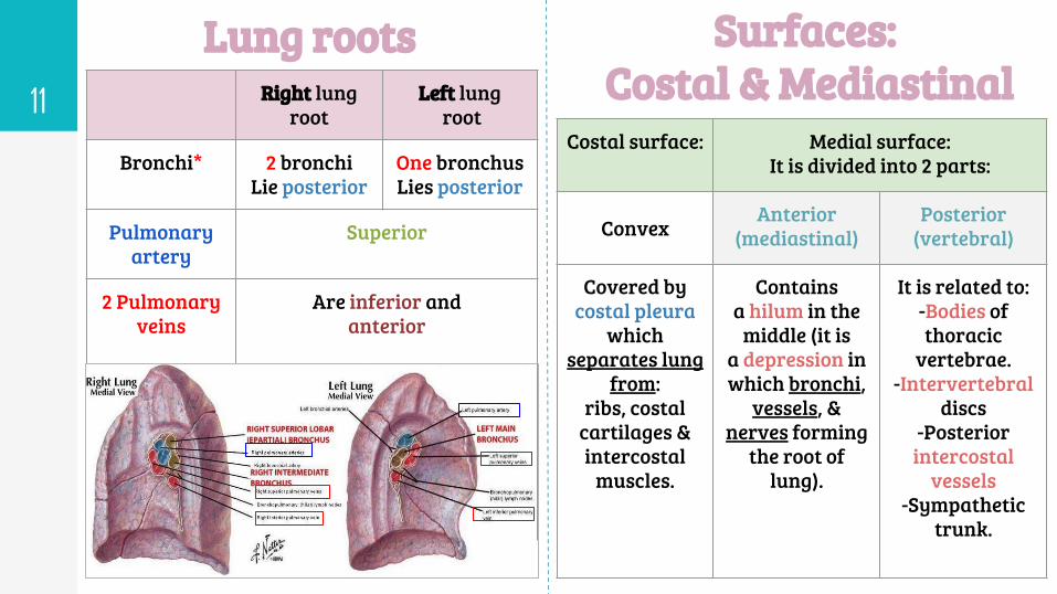
Lung roots11 Right lung
rootLeft lung
root
Bronchi* 2 bronchi Lie posterior
One bronchusLies posterior
Pulmonaryartery
Superior
2 Pulmonaryveins
Are inferior andanterior
Surfaces: Costal & Mediastinal
Costal surface:
Convex
Covered by costal pleura
which separates lung
from:ribs, costal
cartilages &intercostal
muscles.
Medial surface:It is divided into 2 parts:
Anterior (mediastinal)
Posterior (vertebral)
Contains a hilum in the
middle (it is a depression inwhich bronchi,
vessels, &nerves forming
the root oflung).
It is related to:-Bodies of thoracic
vertebrae.-Intervertebral
discs-Posterior
intercostal vessels
-Sympathetic trunk.
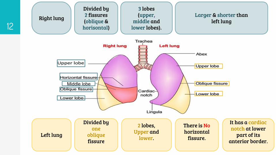
12Right lung Larger & shorter than
left lung
Divided by2 fissures(oblique &
horisontal)
3 lobes(upper,
middle andlower lobes).
Left lung
Divided by one
oblique fissure
There is Nohorizontal
fissure.
2 lobes,Upper and
lower.
It has a cardiacnotch at lower
part of itsanterior border.
Middle lobe
Mediastinal surface
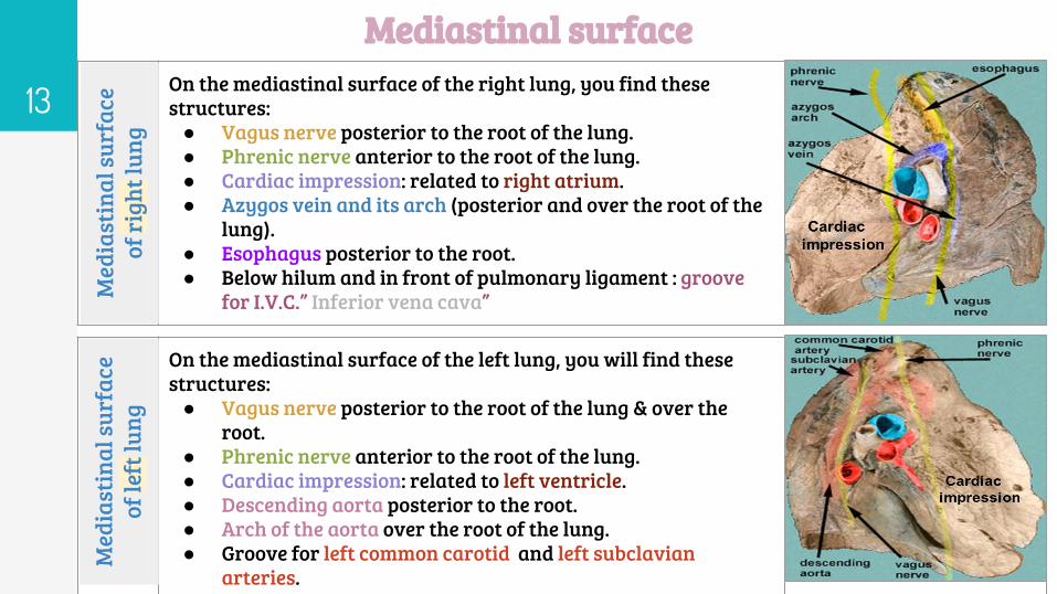
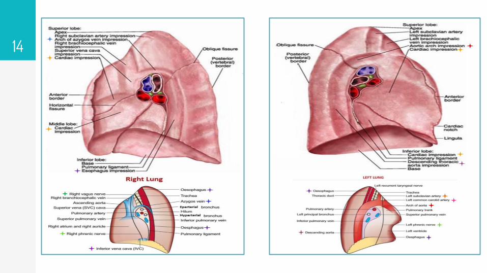
13On the mediastinal surface of the right lung, you find these structures:
● Vagus nerve posterior to the root of the lung.● Phrenic nerve anterior to the root of the lung.● Cardiac impression: related to right atrium.● Azygos vein and its arch (posterior and over the root of the
lung).● Esophagus posterior to the root.● Below hilum and in front of pulmonary ligament : groove
for I.V.C.” Inferior vena cava”Med
iast
inal
surf
ace
of ri
ght l
ung
On the mediastinal surface of the left lung, you will find these structures:
● Vagus nerve posterior to the root of the lung & over the root.
● Phrenic nerve anterior to the root of the lung.● Cardiac impression: related to left ventricle.● Descending aorta posterior to the root.● Arch of the aorta over the root of the lung.● Groove for left common carotid and left subclavian
arteries.
Med
iast
inal
surf
ace
of le
ft lu
ng
14
15
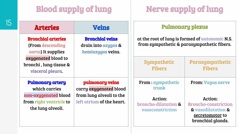
Blood supply of lung Nerve supply of lung
Arteries Veins Bronchial arteries (From descending aorta) It supplies
oxygenated blood to bronchi , lung tissue &
visceral pleura.
Bronchial veins drain into azygos & hemiazygos veins.
Pulmonary artery which carries
non-oxygenated blood from right ventricle to
the lung alveoli.
pulmonary veins carry oxygenated blood from lung alveoli to the left atrium of the heart.
Pulmonary plexus
at the root of lung is formed of autonomic N.S. from sympathetic & parasympathetic fibers.
Sympathetic Fibers
Parasympathetic Fibers
From : sympathetic trunk
Action: broncho-dilatation &
vasoconstriction
From: Vagus nerve
Action: Broncho-constriction
& vasodilatation & secretomotor to
bronchial glands.
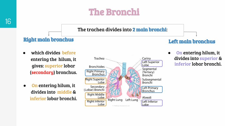
The Bronchi16
● which divides before entering the hilum, it gives: superior lobar
(secondary) bronchus.
● On entering hilum, it divides into middle &
inferior lobar bronchi.
● On entering hilum, it divides into superior & inferior lobar bronchi.
Right main bronchus Left main bronchus
The trachea divides into 2 main bronchi:
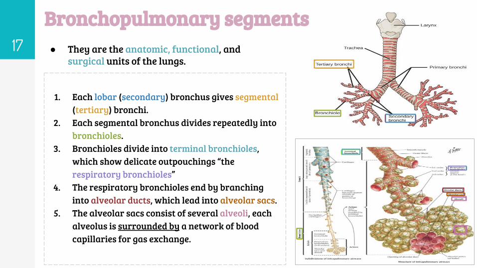
Bronchopulmonary segments17 ● They are the anatomic, functional, and
surgical units of the lungs.
1. Each lobar (secondary) bronchus gives segmental (tertiary) bronchi.
2. Each segmental bronchus divides repeatedly into bronchioles.
3. Bronchioles divide into terminal bronchioles, which show delicate outpouchings “the respiratory bronchioles”
4. The respiratory bronchioles end by branching into alveolar ducts, which lead into alveolar sacs.
5. The alveolar sacs consist of several alveoli, each alveolus is surrounded by a network of blood capillaries for gas exchange.
Bronchopulmonary segments18
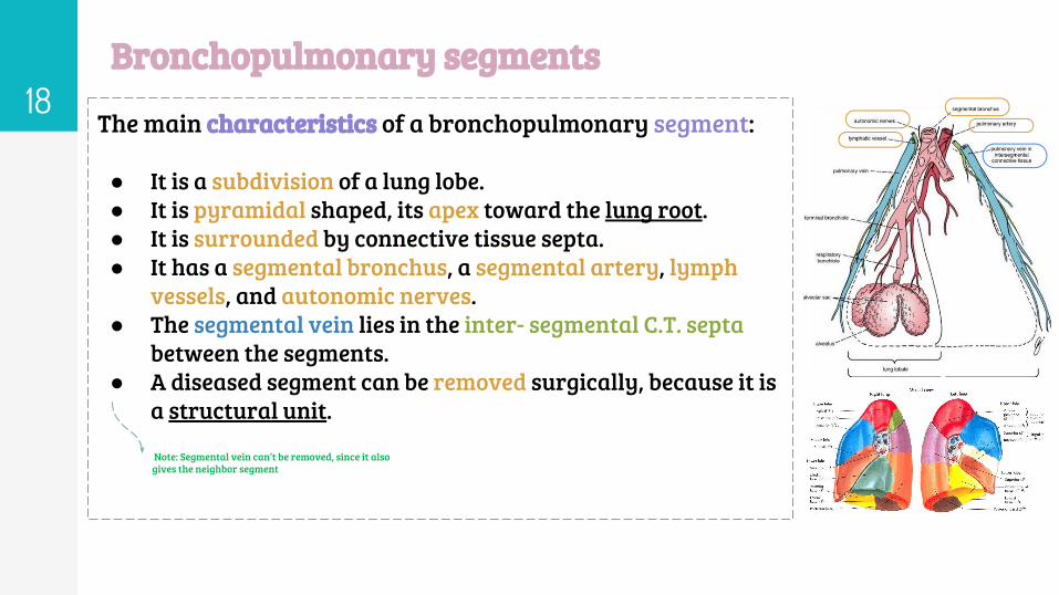
The main characteristics of a bronchopulmonary segment:
● It is a subdivision of a lung lobe.● It is pyramidal shaped, its apex toward the lung root.● It is surrounded by connective tissue septa.● It has a segmental bronchus, a segmental artery, lymph
vessels, and autonomic nerves.● The segmental vein lies in the inter- segmental C.T. septa
between the segments.● A diseased segment can be removed surgically, because it is
a structural unit.
Note: Segmental vein can’t be removed, since it also gives the neighbor segment
19
MCQsQuestion 1: Which feature is found only in the left lung ?
A. Oblique fissure
B. Cardiac notch
C. Transverse fissure
D. Both A and C
Question 2:The lung is innervated by :
A. Sympathetic fibers
B. Parasympathetic fibers
C. Both sympathetic and parasympathetic fibers
D. Motor fibers
Question 3: The pleural cavity, contains a thin film of pleural serous fluid. what
is the normal value of it ?
A. 25-30 ml
B. 15-20 ml
C. 20-25 ml
D. 5-10 ml
Question 4: which one of the following is not a characteristic of the left lung ?
A. contains 2 lobes
B. Has one fissure
C. Has lingual projection
D.shorter than the right lung
Question 5: Mediastinal pleura is supplied by :
A. Phrenic nerves
B. Intercostal nerves
C. Autonomic fibers
D. Both A and B
Question 6: The pulmonary artery carries …… blood from……:
A.Oxygenated / Left ventricle
B. Oxygenated / Left atrium
C. Deoxygenated / Right ventricle
D. Deoxygenated / Right atrium
Question 7: Visceral pleura is supplied by :
A. Autonomic fibers.
B. thoracic nerve.
C. intercostal nerves.
D. Phrenic nerves.
Question 8: The phrenic nerve is found (......) to the root of the lung :
A. Superior
B. Anterior
C. Inferior
D.posterior
Answers: Q1.B- Q2.C -Q3.D -Q4.D- Q5.A- Q6.C-Q7.A- Q8.B

Boys team:
● Khalid AL-Dossari● Naif Al-Dossari● Faisal Alqifari ● Salman Alagla● Ziyad Al-jofan● Suhail Basuhail● Ali Aldawood● Khalid Nagshabandi● Mohammed Al-huqbani● Jehad Alorainy● Khalid AlKhani● Omar Alammari
Team leaders
● Abdulrahman Shadid● Ateen Almutairi
Team membersGirls team :
● Ajeed Al Rashoud● Taif Alotaibi● Noura Al Turki● Amirah Al-Zahrani● Alhanouf Al-haluli● Sara Al-Abdulkarem● Rawan Al Zayed● Renad Al Haqbani● Nouf Al Humaidhi● Jude Al Khalifah● Nouf Al Hussaini● Alwateen Al Balawi● Rahaf Al Shabri● Danah Al Halees● Rema Al Mutawa● Amirah Al Dakhilallah● Maha Al Nahdi ● Ghaida Al Braithen
Don't forget to leave your feedback:
Best wishes
Related Documents


