Job/Unit: I43116 /KAP1 Date: 24-02-15 15:47:36 Pages: 11 FULL PAPER DOI:10.1002/ejic.201403116 Luminescence Matching with the Sensitivity Curve of the Human Eye: Optical Ceramics Mg 8–x M x (BN 2 ) 2 N 4 with M = Al (x = 2) and M = Si (x = 1) Markus Ströbele, [a] Konstantin Dolabdjian, [a] David Enseling, [b] Danuta Dutczak, [a] Boriana Mihailova, [c] Thomas Jüstel, [b] and H.-Jürgen Meyer* [a] Keywords: Ceramics / Solid-state reactions / Solid-state structures / Doping / Luminescence Compounds Mg 8–x M x (BN 2 ) 2 N 4 were prepared by solid-state reaction of MgCl 2 , Li 3 (BN 2 ), and AlN for M = Al, and of Mg 3 N 2 , BN, and Si 3 N 4 for M = Si. Their crystal structures were refined from powder X-ray diffraction data with the space group R3 ¯ m for M = Al, Si, and P2 1 /c for M = Si. The structures of Mg 8–x M x (BN 2 ) 2 N 4 are characterized by layered arrangements of wurtzite-related block layers alternating Introduction The development of photoluminescent materials is a topic of growing interest. Current LED light sources use an electroluminescent (In 1–x Ga x )N-based chip to generate blue light, which is then converted into white light. Prominent converter materials involve YAG:Ce [1] as a yellow emitter, and (Ca,Sr)SiAlN 3 :Eu [2] or (Ca,Sr) 2 Si 5 N 8 :Eu [3] as a red emitter to allow an additive color mixing for the generation of warm white light. Phosphors such as these are required to fulfill high quality standards with respect to their chemi- cal and optical properties. The yellow (or green) emitter in LED light sources is usually a broad-band emitting material (e.g., Ce 3+ activated), whereas the red emitter can be either a broad-band or line emitter, to generate warm white light. In this way, the emission spectra of all these materials are superimposed to produce white light, covering the sensitiv- ity curve of the human eye, preferably for a photopic illumi- nation level. An alternative to a three-color mixture could be a broad emitting material that covers the sensitivity curve of the human eye as a whole. [a] Section for Solid State and Theoretical Inorganic Chemistry, Institute of Inorganic Chemistry, University of Tübingen, Auf der Morgenstelle 18, 72076 Tübingen, Germany E-mail: [email protected] http://www.mnf.uni-tuebingen.de/fachbereiche/chemie/institute/ anorganische-chemie/institut/ag-meyer/startseite.html [b] Department of Chemical Engineering, Münster University of Applied Sciences, Stegerwaldstraße 39, 48565 Steinfurt, Germany [c] Mineralogisch-Petrographisches Institut, University of Hamburg, Grindelallee 48, 20146 Hamburg, Germany Eur. J. Inorg. Chem. 0000, 0–0 © 0000 Wiley-VCH Verlag GmbH & Co. KGaA, Weinheim 1 with a layer of di-nitridoborate ions along the c-axis direc- tion. Cations in each structure are surrounded tetrahedrally by nitride and di-nitridoborate ions. Europium(II)-doped powders of Mg 8–x M x (BN 2 ) 2 N 4 show exceptional broad band emissions in the visible region of the spectrum, on excitation with UV radiation at λ ex = 320 nm. Nitride- and nitridosilicate-containing compounds have attracted much of attention because such compounds have shown potential for applications in the field of lumines- cence. Among these, europium-doped compounds (Ca,Sr)- SiAlN 3 :Eu and (Ca,Sr) 2 Si 5 N 8 :Eu currently belong to the most prominent red-emitting materials for LED applica- tions. Hence, there is growing interest in discovering more nitride materials, although many combinations between metal and nitride or nitridosilicate have been already ex- plored. In this context, the development of new nitride-rich nitridoborates, in which the [BN 2 ] 3– could act as a pseudo- nitride could be an interesting task and an extension of ni- tride chemistry in a broader sense. A substantial number of nitridoborates (boro-nitrides) have been developed by solid- state metathesis reactions with ions such as [BN] n– , [BN 2 ] 3– , [BN 3 ] 6– , [B 2 N 4 ] 8– , and [B 3 N 6 ] 9– . [4] The [BN] n– ion was characterized in ternary compounds [e.g., MNi(BN) [5–8] and RE 3 Ni 2 (BN) 2 N [9–12] ], which show electrical conductiv- ity and superconducting behavior. Compounds containing the linear di-nitridoborate ion ([N=B=N] 3– ) have been es- tablished for various alkali and alkali earth compounds [e.g., Li 3 (BN 2 ) [13] Ca 3 (BN 2 ) 2 , [14,15] and LiCa 4 (BN 2 ) 3 [16] ] af- ter the fundamental work of Goubeau and Anselment. [17] When Li 3 (BN 2 ) is reacted with a trivalent rare-earth chlor- ide, the di-nitridoborate ions can undergo cyclotrimeriz- ation to yield the cyclic [B 3 N 6 ] 9– ion at temperatures be- tween 500 and 600 °C; see Equation (1). 3 LaCl 3 + 3 Li 3 (BN 2 ) La 3 B 3 N 6 + 9 LiCl (1) This type of cyclotrimerization is well known for cyan- ates in organic chemistry and was utilized very recently for

Welcome message from author
This document is posted to help you gain knowledge. Please leave a comment to let me know what you think about it! Share it to your friends and learn new things together.
Transcript

Job/Unit: I43116 /KAP1 Date: 24-02-15 15:47:36 Pages: 11
FULL PAPER
DOI:10.1002/ejic.201403116
Luminescence Matching with the Sensitivity Curve of theHuman Eye: Optical Ceramics Mg8–xMx(BN2)2N4 withM = Al (x = 2) and M = Si (x = 1)
Markus Ströbele,[a] Konstantin Dolabdjian,[a] David Enseling,[b]
Danuta Dutczak,[a] Boriana Mihailova,[c] Thomas Jüstel,[b] andH.-Jürgen Meyer*[a]
Keywords: Ceramics / Solid-state reactions / Solid-state structures / Doping / Luminescence
Compounds Mg8–xMx(BN2)2N4 were prepared by solid-statereaction of MgCl2, Li3(BN2), and AlN for M = Al, and ofMg3N2, BN, and Si3N4 for M = Si. Their crystal structureswere refined from powder X-ray diffraction data with thespace group R3̄m for M = Al, Si, and P21/c for M = Si. Thestructures of Mg8–xMx(BN2)2N4 are characterized by layeredarrangements of wurtzite-related block layers alternating
Introduction
The development of photoluminescent materials is atopic of growing interest. Current LED light sources use anelectroluminescent (In1–xGax)N-based chip to generate bluelight, which is then converted into white light. Prominentconverter materials involve YAG:Ce[1] as a yellow emitter,and (Ca,Sr)SiAlN3:Eu[2] or (Ca,Sr)2Si5N8:Eu[3] as a redemitter to allow an additive color mixing for the generationof warm white light. Phosphors such as these are requiredto fulfill high quality standards with respect to their chemi-cal and optical properties. The yellow (or green) emitter inLED light sources is usually a broad-band emitting material(e.g., Ce3+ activated), whereas the red emitter can be eithera broad-band or line emitter, to generate warm white light.In this way, the emission spectra of all these materials aresuperimposed to produce white light, covering the sensitiv-ity curve of the human eye, preferably for a photopic illumi-nation level. An alternative to a three-color mixture couldbe a broad emitting material that covers the sensitivitycurve of the human eye as a whole.
[a] Section for Solid State and Theoretical Inorganic Chemistry,Institute of Inorganic Chemistry, University of Tübingen,Auf der Morgenstelle 18, 72076 Tübingen, GermanyE-mail: [email protected]://www.mnf.uni-tuebingen.de/fachbereiche/chemie/institute/anorganische-chemie/institut/ag-meyer/startseite.html
[b] Department of Chemical Engineering, Münster University ofApplied Sciences,Stegerwaldstraße 39, 48565 Steinfurt, Germany
[c] Mineralogisch-Petrographisches Institut, University ofHamburg,Grindelallee 48, 20146 Hamburg, Germany
Eur. J. Inorg. Chem. 0000, 0–0 © 0000 Wiley-VCH Verlag GmbH & Co. KGaA, Weinheim1
with a layer of di-nitridoborate ions along the c-axis direc-tion. Cations in each structure are surrounded tetrahedrallyby nitride and di-nitridoborate ions. Europium(II)-dopedpowders of Mg8–xMx(BN2)2N4 show exceptional broad bandemissions in the visible region of the spectrum, on excitationwith UV radiation at λex = 320 nm.
Nitride- and nitridosilicate-containing compounds haveattracted much of attention because such compounds haveshown potential for applications in the field of lumines-cence. Among these, europium-doped compounds (Ca,Sr)-SiAlN3:Eu and (Ca,Sr)2Si5N8:Eu currently belong to themost prominent red-emitting materials for LED applica-tions. Hence, there is growing interest in discovering morenitride materials, although many combinations betweenmetal and nitride or nitridosilicate have been already ex-plored. In this context, the development of new nitride-richnitridoborates, in which the [BN2]3– could act as a pseudo-nitride could be an interesting task and an extension of ni-tride chemistry in a broader sense. A substantial number ofnitridoborates (boro-nitrides) have been developed by solid-state metathesis reactions with ions such as [BN]n–,[BN2]3–, [BN3]6–, [B2N4]8–, and [B3N6]9–.[4] The [BN]n– ionwas characterized in ternary compounds [e.g., MNi(BN)[5–8]
and RE3Ni2(BN)2N[9–12]], which show electrical conductiv-ity and superconducting behavior. Compounds containingthe linear di-nitridoborate ion ([N=B=N]3–) have been es-tablished for various alkali and alkali earth compounds[e.g., Li3(BN2)[13] Ca3(BN2)2,[14,15] and LiCa4(BN2)3
[16]] af-ter the fundamental work of Goubeau and Anselment.[17]
When Li3(BN2) is reacted with a trivalent rare-earth chlor-ide, the di-nitridoborate ions can undergo cyclotrimeriz-ation to yield the cyclic [B3N6]9– ion at temperatures be-tween 500 and 600 °C; see Equation (1).
3 LaCl3 + 3 Li3(BN2) � La3B3N6 + 9 LiCl (1)
This type of cyclotrimerization is well known for cyan-ates in organic chemistry and was utilized very recently for
Job/Unit: I43116 /KAP1 Date: 24-02-15 15:47:36 Pages: 11
www.eurjic.org FULL PAPER
the preparation of inorganic cyanurates (O3C3N3)3– by re-acting alkali earth halides with Li(OCN).[18]
Compounds with larger nitridoborate anions such as[BN3]6–, [B2N4]8–, and [B3N6]9– typically occur with tri-valent rare-earth cations. These compounds can be pre-pared in reactions related to (1), in which the amount ofnitride is controlled by employing Li3N as an additionalreaction partner. However, the preparation of nitride-richnitridoborates has not been explored to a significant extent.Hence, only a relatively small number of nitridoborate-nitrides [e.g., Mg3(BN2)N,[19] La4(B2N4)N, La5(B2N4)N2
[20]]have been reported so far.
The existence of the mixed nitridoborate-silicateMg7Si(BN2)2N4 was first reported in 1994, and its structurewas ascribed as rhombohedral, but without any crystalstructure data given.[21] Two years later, Mg3Al(BN2)N2 wasreported from explorations of the Mg3N2-AlN-BN systemby the same group of researchers.[22] A hexagonally indexedX-ray powder pattern revealed the lattice parameters a =3.439(1) Å and c = 31.43(1) Å for Mg3Al(BN2)N2, as-suming a rhombohedral crystal system. A similar powderdiffraction pattern appeared in 1996, which was assigned as“Mg4SiN4” in a “Private Communication” by Liddell in theCOD Database.
Following our previous attempts at the preparation ofnitridoborates by solid-state metathesis reactions, we haveexplored a straightforward approach to synthesize the ter-nary nitrides Mg8–xMx(BN2)2N4, which are described herewith their crystal structures. Rhombohedral Mg8–xMx-(BN2)2N4 structures are studied by NMR spectroscopy, vi-brational (infrared and Raman) spectroscopy, as well as lu-minescence properties. Monoclinic Mg7Si(BN2)2N4 is pre-sented only with its crystal structure.
Results and Discussion
Synthesis: The preparation of a robust ceramic materialusually requires rigorous reaction conditions such as hightemperature and long reaction time, as previously appliedin the Mg3N2-AlN-BN system.
A concept for the preparation of nitridoborates undermoderate heating conditions by solid-state metathesis(SSM) has been described previously, as shown in reaction(1). A key feature of this type reaction is the employmentof reactive sources. This method of synthesis was previouslyused by us for the preparation of Mg3(BN2)N from an ap-propriate mixture of MgCl2, Li3(BN2), and Li3N at550°C.[23] The coproduced metathesis salt (LiCl) can be re-moved from the reaction product by extraction with water.
Departing from the synthesis of Mg3(BN2)N, a sourcefor aluminum was needed for the preparation ofMg6Al2(BN2)2N4. A straightforward way is to employ AlN,accepting that AlN is the less reactive compound in thegiven reaction mixture. Alternatively AlCl3 could be em-ployed, but this reagent was considered to be too volatileduring the reaction. The employment of AlF3 would pro-duce LiF as a metathesis salt, which is difficult to separate
Eur. J. Inorg. Chem. 0000, 0–0 © 0000 Wiley-VCH Verlag GmbH & Co. KGaA, Weinheim2
from the reaction mixture because of its low solubility inwater.
The synthesis of Mg6Al2(BN2)2N4 was successfully per-formed by following a straightforward metathesis reactionwith additional AlN. Due to its inert nature, AlN shouldproduce a heat sink, which generally slows down a SSMreaction. Since a heat sink is not desired in this reaction, itis compensated by higher firing at 1100 °C; see Equa-tion (2).
6 MgCl2 + 2 AlN + 2 Li3(BN2) + 2 Li3N�Mg6Al2(BN2)2N4 + 12 LiCl(2)
Mg6Al2(BN2)2N4 is stable in air, water, and mineralacids. The metathesis salt (LiCl) can be easily removed fromthe reaction product by extraction with water to yield awhite crystalline powder. Samples doped with (2 mol-%)Eu2+ appear slightly yellowish-green.
The preparation of Mg7Si(BN2)2N4 was achieved directlyfrom nitrides as given in Equation (3).
14 Mg3N2 + 2 Si3N4 + 12 BN � 6 Mg7Si(BN2)2N4 (3)
Small deviations in the proportions of reactants, or thepresence of a small amount of EuCl2 as a dopant revealedthe formation of two closely related structures ofMg7Si(BN2)2N4, which could be structurally identified asrhombohedral and monoclinic phases when both formswere prepared separately.
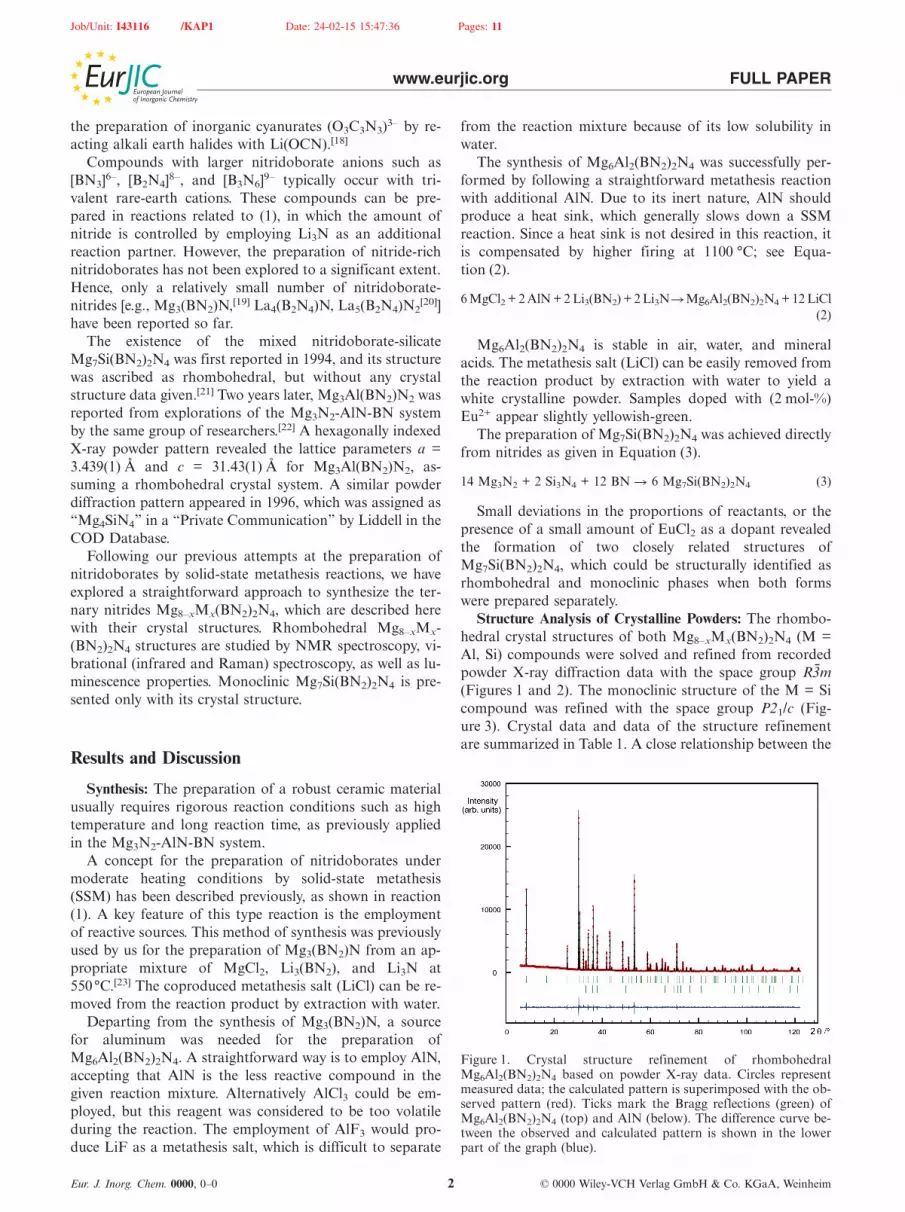
Structure Analysis of Crystalline Powders: The rhombo-hedral crystal structures of both Mg8–xMx(BN2)2N4 (M =Al, Si) compounds were solved and refined from recordedpowder X-ray diffraction data with the space group R3̄m(Figures 1 and 2). The monoclinic structure of the M = Sicompound was refined with the space group P21/c (Fig-ure 3). Crystal data and data of the structure refinementare summarized in Table 1. A close relationship between the
Figure 1. Crystal structure refinement of rhombohedralMg6Al2(BN2)2N4 based on powder X-ray data. Circles representmeasured data; the calculated pattern is superimposed with the ob-served pattern (red). Ticks mark the Bragg reflections (green) ofMg6Al2(BN2)2N4 (top) and AlN (below). The difference curve be-tween the observed and calculated pattern is shown in the lowerpart of the graph (blue).

Job/Unit: I43116 /KAP1 Date: 24-02-15 15:47:36 Pages: 11
www.eurjic.org FULL PAPER
Figure 2. Crystal structure refinement of rhombohedralMg7Si(BN2)2N4 based on powder X-ray data. Circles representmeasured data; the calculated pattern is superimposed with the ob-served pattern (red). Ticks mark the Bragg reflections (green) ofMg7Si(BN2)2N4 (top). The difference curve between the observedand calculated pattern is shown in the lower part of the graph(blue).
rhombohedral and monoclinic structure of Mg7Si(BN2)2N4
can be assumed by comparison of the X-ray diffraction pat-terns shown in Figures 2 and 3.
A clear distinction in these patterns is evidenced for thereflection pattern near 2Θ = 53° appearing as a single peakin the rhombohedral and as double peak in the monoclinicphase.
Temperature-dependent X-ray diffraction studies de-parting from the rhombohedral modification between roomtemperature and 1200 °C revealed no structural transfor-mation; only a breathing behavior towards increased latticeparameters.
The crystal structures of rhombohedral Mg8–xMx-(BN2)2N4 compounds can be considered isotypic. Thestructures can be described as a layered arrangement in
Table 1. Crystal data and structure refinement for Mg8–xMx(BN2)2N4.
Empirical formula Mg5.11(3)Al2(BN2)2N4 Mg6.42(4)Si(BN2)2N4 Mg6.12(2)Si(BN2)2N4
Formula weight [gmol–1] 311.93 312.22 310.31Temperature [K] 298(2)Wavelength (Cu-Kα1) [Å] 1.54060Crystal system rhombohedral monoclinicSpace group R3̄m (no. 166) P21/c (no. 14)Unit cell dimensions [Å] a = b = 3.4318(1) a = b = 3.4383(1) a = 10.5598(2)
b = 6.8994(1)c = 31.436(1) c = 31.107(1) c = 5.9082(1)
β = 100.983(1)Volume [Å3] 320.64(1) 318.47(1) 422.56(1)Z 1.5 2μ (Cu-Kα1) [mm–1] 6.614 6.782 6.757Density (calculated) [gcm–3] 2.423 2.442 2.43872Theta range for data collection [°] 3 to 65.0 2.5 to 50.0 3.5 to 50.0Total number of reflections 99 71 484Refined structural parameters 11 41Rp, Rwp 4.8985, 6.4574 4.8036, 6.3503 4.4207, 5.7592RBragg 3.5281 3.729 4.8067χ2 1.8980 2.1035 2.9267
Eur. J. Inorg. Chem. 0000, 0–0 © 0000 Wiley-VCH Verlag GmbH & Co. KGaA, Weinheim3
Figure 3. Crystal structure refinement of Mg7Si(BN2)2N4 (mono-clinic phase) based on powder X-ray data. Circles represent mea-sured data; the calculated pattern is superimposed with the ob-served pattern (red). Ticks mark the Bragg reflections (green) ofMg7Si(BN2)2N4 (top). The difference curve between the observedand calculated pattern is shown in the lower part of the graph(blue).
which wurtzite-like [(Mg,M)N] blocks alternate with di-nitridoborate ions along the c-axis direction (Figure 4).Structure refinements revealed two distinct cation positionsin the structure, being occupied in a disordered fashion withtwo types of cations in each structure with the space groupR3̄m (Table 2). Selected bond lengths [Å] and multiplicitiesfor Mg8–xMx(BN2)2N4 (rhombohedral modification) areshown in Table 3.
Refinements carried out in the noncentrosymmetricspace group R3m revealed the same kind of cation disorder.In addition, nonlinear optical property (NLO) measure-ments did not provide evidence for the presence of a non-centrosymmetric space group. Hence, magnesium and Mions in the structures were refined to occupy the same crys-tallographic sites.This is true for therhombohedralMg8–xMx-

Job/Unit: I43116 /KAP1 Date: 24-02-15 15:47:36 Pages: 11
www.eurjic.org FULL PAPER
Table 2. Atomic coordinates and equivalent isotropic displacement parameters (pm2) for Mg8–xMx(BN2)2N4 (rhombohedral phase).
x y z z U(eq) U(eq) SOF SOF(M = Al) (M = Si) (M = Al) (M = Si) (M = Al) (M = Si)
Mg1 0 0 0.36315(3) 0.36361(5) 0.0252(4) 0.0334(8) 0.656(4) 0.833(5)M1 0 0 0.36315(3) 0.36361(5) 0.0252(4) 0.0334(8) 0.25 0.125Mg2 0 0 0.23045(3) 0.23100(5) 0.0220(4) 0.022(1) 0.602(5) 0.714(6)M2 0 0 0.23045(3) 0.23100(5) 0.0220(4) 0.022(1) 0.25 0.125B1 0 0 1/2 1/2 0.032(1) 0.022(3)N1 0 0 0.4573(1) 0.4571(1) 0.033(1) 0.039(2)N2 0 0 0.2964(1) 0.2973(1) 0.033(1) 0.035(2)
Figure 4. Projected section of the crystal structure of Mg8–xMx-(BN2)2N4 with M = Al (x = 2) and M = Si (x = 1). All cationswere refined to share the same crystallographic 6c site of the spacegroup R3̄m.
Table 3. Selected bond lengths [Å] and multiplicities forMg8–xMx(BN2)2N4 (rhombohedral modification).
Distance[a] M = Al, x = 2 M = Si, x = 1
Mg1–N1 2.961(2) 2.908(4) (1 �)Mg1–N2 1.994(1) 1.993(4) (3 �)Mg1–N2 2.100(3) 2.062(4) (1 �)Mg2–N1 2.090(1) 2.094(1) (3 �)Mg2–N2 2.072(3) 2.063(4) (1 �)B1–N1 1.341(2) 1.334(3) (2 �)
[a] Magnesium sites are partially occupied with aluminum or sili-con.
(BN2)2N4 structures shown in Figure 4, and for the mono-clinic structure, which can be considered as a substructureof rhombohedral Mg7Si(BN2)2N4 displayed in Figure 5.
The two distinct mixed Mg/M (6c) sites in rhombohedralMg8–xMx(BN2)2N4, assigned as Mg1 and Mg2 in Figure 6,are surrounded by nitride ions in two slightly distinct tetra-hedral fashions. The site assigned as Mg1 is surrounded byfour nitride ions forming a trigonal pyramid, with one (ax-ial, N2) nitride ion being further away from the Mg2+ cen-
Eur. J. Inorg. Chem. 0000, 0–0 © 0000 Wiley-VCH Verlag GmbH & Co. KGaA, Weinheim4
Figure 5. Rhombohedral and monoclinic crystal structures ofMg7Si(BN2)2N4 projected on the ac-plane. The monoclinic structurewas transformed into P21/a in this drawing for better comparison.
ter. The consideration of another remote (axial, N2) nitrideion introduces a distorted trigonal bipyramidal environ-ment. Mg2 is surrounded by four nitrogen atoms, of whichone is a nitride and three are nitrogen atoms that belong tothree distinct di-nitridoborate ions.
Figure 6. Environments of mixed Mg/M cation sites denoted asMg1 and Mg2 (left, center), and of the di-nitridoborate ion (right)in rhombohedral crystal structures of Mg8–xMx(BN2)2N4.
This type of tetrahedral coordination pattern and the cal-culated distances are in good agreement with the situationof Mg2+ ions in the crystal structure of Mg3(BN2)N.Mg3(BN2)N contains one magnesium ion in a tetrahedralenvironment having Mg–N distances of 1�2.040(1) Å

Job/Unit: I43116 /KAP1 Date: 24-02-15 15:47:36 Pages: 11
www.eurjic.org FULL PAPER
(with N3–) and 3� 2.134(1) Å (with N of BN23–), and one
magnesium ion in a trigonal bipyramidal environment hav-ing three equivalent Mg–N distances of 2.046(1) Å withnitride ions and two axial distances with nitrogen atomsof two di-nitridoborate ions at Mg–N distances of2.64.4(3) Å.[24] The significantly shorter distances obtainedin the structure of AlN [1� 1.927(3) Å, 3 �1.8828(7) Å] arein line with the smaller ionic radius of Al3+ (0.39 Å) com-pared with that of Mg2+ (0.57 Å) for coordination numbersfour.
The powder diffraction data on Mg8–xMx(BN2)2N4 can-not be stressed to allow a reliable, clear picture regardingthe disordering of Mg and Al on opposite lattice sites. Inaddition, a cation disordering over all Mg and M sitescould allow a change in the given Mg/M ratio in Mg8–xMx-(BN2)2N4, for example when oxide substitutions would bepresent on nitride sites. However, according to our chemicalanalyses performed for Mg7Si(BN2)2N4 phases, the oxygencontent is negligible (� 2 %). A cation deficiency has beenreported for Mg3–x(BN2)N based on neutron diffractionstudies.[25] This can be a result of magnesium evaporationfrom solid material at elevated temperatures, as has beenshown for MgB2 crystals for which the average Mg contentis systematically decreased when temperature is increasedfrom 800 to 1000 °C. Based on this probability, we have notonly refined the site occupancy but also the cation occu-pancies in our structures, as summarized in Table 2.
Solid-State NMR Studies: The presence of M = Al and Siin the rhombohedral structures of Mg8–xMx(BN2)2N4 wasevidenced by solid-state NMR studies. The 27Al MASNMR spectrum reveals two signals, at chemical shifts ofδiso = 5.2 and 107 ppm, which can be assigned toMg6Al2(BN2)2N4 and the AlN obtained by XRD as side-phase in the sample (Figure 1). A separate MAS NMRmeasurement of our “as used” AlN confirms this assign-ment with δiso = 106 ppm. A solid-state 29Si MAS NMRspectrum recorded for the Mg7Si(BN2)2N4 sample revealeda single signal at δiso = –32 ppm, which appears in the rangeof tetrahedral [SiN4] units of nitridosilicates. Correspondingvalues were reported between –28 ppm for nitridosilicates(SrSi6N8)[26] and –68 ppm for oxo-nitridosilicates(Ba3Si6O9N4).[27] A signal for Si3N4 expected at δiso =48 ppm[28] was absent, as expected from the XRD results(Figure 2).
Infrared and Raman Spectroscopy: The di-nitridoborateion in the rhombohedral Mg8–xMx(BN2)2N4 structure hasD�h symmetry and refined N=B=N distances of 1.341(2)(M = Al) and 1.334(3) Å (M = Si), with the boron atomlying in the mirror plane. These distances appear slightlyshorter than distances of the symmetrical [N=B=N]3– ionsin Mg3(BN2)N reported at 1.365(3) Å. The infrared spectraof Mg8–xMx(BN2)2N4 compounds exhibit most characteris-tic infrared peaks resulting from [N=B=N]3– ions: the anti-symmetric stretching vibration modes (ν̃ = 1799 cm–1 for M= Al, and ν̃ = 1827 cm–1 for M = Si) and bending vibrationsof di-nitridoborate ions and signals, which are superim-posed with stretching and bending vibrations of tetrahedral[AlN4] and [SiN4] units below 800 cm–1. These results con-
Eur. J. Inorg. Chem. 0000, 0–0 © 0000 Wiley-VCH Verlag GmbH & Co. KGaA, Weinheim5
firm the presence of di-nitridoborate ions in our structuresbut do not allow much further insight into structural detailsof the structures, such as cation disordering.
The most intense peak in the Raman spectra ofMg8–xMx(BN2)2N4 is near 1080 cm–1 (see Figure 7), whichmost probably arises from the A1g symmetric stretchingmode of [N=B=N]3– ions, involving in-phase vibrations ofthe N atoms adjacent to B. For undoped Mg3(BN2)N, thesymmetric N=B=N stretching mode generates a single Ra-man peak, whereas for both rhombohedral Mg8–xMx-(BN2)2N4 structures, a splitting of this mode is observed.Three peaks are resolved for Mg6Al2(BN2)2N4, whereas twopeaks are observed for Mg7Si(BN2)2N4. The splitting canbe due to the lower symmetry of Mg8–xMx(BN2)2N4 (R3̄m
Figure 7. Raman scattering generated by the symmetrical N=B=Nstretching mode in Mg3(BN2)N, Mg6Al2(BN2)2N4, andMg7Si(BN2)2N4. Black solid lines are experimental spectra, greenlines are the fitting Lorentzian functions, red dashed lines the re-sultant spectrum profiles; ω and In are respectively the peak posi-tions and fractional intensities obtained from fits.

Job/Unit: I43116 /KAP1 Date: 24-02-15 15:47:36 Pages: 11
www.eurjic.org FULL PAPER
or P21/c) as compared with Mg3(BN2)N (P63/mmc). How-ever, site symmetry analysis[29] revealed that all three spacegroups mentioned above allow only one fully symmetricalirreducible representation A1g in N atoms adjacent to B in-volving N vibrations along the N–B bonds.
Hence, splitting of the symmetric N=B=N stretchingmode due to structural differences should be ruled out. An-other explanation for the observed Raman profiles is two-mode behavior of the symmetric N=B=N stretching as aresult of different chemical surroundings (N3A)3–N=B=N(AN3)3, where A = Mg, Si, Al. In Mg3(BN2)N, allN atoms bonded to B are surrounded only by Mg cations;that is, the existing chemical species are of type MgMgMg–N=B=N–MgMgMg. In Mg6Al2(BN2)2N4 and Mg7Si-(BN2)2N4, one or more Mg cations can be substituted byAl and Si, respectively, and thus several distinct chemicalsurroundings of the [N=B=N]3– ion are expected. Due tothe difference in the electronegativities, the Si–N interactionis expected to be stronger than the Al–N interaction andthey should both be considerably stronger than Mg–N in-teractions. Stronger interactions between N and the sur-rounding A cations will weaken the N=B bonding and con-sequently the wavenumber of the N=B=N stretching willdecrease. Hence, the Raman peak position should dependon the type of the surrounding A cations. Because of thecovalent character of the chemical N–B bonds, two-modebehavior for the N=B=N stretching mode is expected; thatis, each chemically distinguished surrounding will generatea separate peak with a fractional integrated intensity pro-portional to the concentration of the corresponding chemi-cal species. If we consider possible atomic linkages and mul-tiply the number of atoms by the fractional intensity, weshould obtain values close to the nominal chemical compo-sition. In fact, assuming that the two peaks in the Ramanspectrum of Mg7Si(BN2)2N4 are due to MgMgMg–N=B=N–MgMgMg and MgSiSi–N=B=N–MgMgSi (seeFigure 7), we obtain Si/(Si+Mg) = 3�0.22/[3�0.22+(3�0.22+6�0.78)] = 0.11, which is very close tothe nominal chemical ratio Si/(Si+Mg) = 0.125. Similarly,for Mg6Al2(BN2)2N4 we assume three chemically distin-guished surroundings MgMgMg–N=B=N–MgMgMg,MgMgAl–N=B=N–MgMgMg, and AlAlAl–N=B=N–MgAlAl (see Figure 8) and the ratio Al/(Al+Mg) obtainedfrom the Raman data is 0.22, which is very close to thenominal chemistry giving Al/(Al+Mg) = 0.25.
Furthermore, for Mg8–xMx(BN2)2N4, the trend in theobserved Raman peak positions corresponds well to thenumber and type of cations substituting for Mg. The posi-tion of the major Raman peak, i.e., the wavenumber of thepredominant chemical species, is also in good accordancewith the average N–B bond length determined from Riet-veld refinements on powder X-ray diffraction data.
Luminescence: The Mg6Al2(BN2)2N4 and Mg7Si-(BN2)2N4 samples show intense Eu2+ yellow photolumines-cence (see Figures 8 and 9). It is essential to determine thelocation of Eu2+ ions within the crystal structure. In mosthost materials comprising alkaline-earth metals, Eu2+ ionsare supposed to occupy the alkaline-earth sites.[30,31] How-
Eur. J. Inorg. Chem. 0000, 0–0 © 0000 Wiley-VCH Verlag GmbH & Co. KGaA, Weinheim6
Figure 8. Excitation and emission spectra of Mg6Al2(BN2)2N4:5%Eu2+ at room temperature.
ever, the ionic radius of Mg2+ is much too small to haveEu2+ on a Mg2+ site without a major site expansion. Forexample, the ionic radius for the sixfold coordinated Mg2+
site is 0.72 Å, whereas the ionic radius of Eu2+ in the samecoordinated site is 1.17 Å. The occupancy of Eu2+ on theAl3+ or Si4+ sites are also unexpected, because of differentvalence states and because these sites are even smaller thanthat at Mg2+ (Al3+ 0.535 Å and Si4+ 0.4 Å; for the sixfoldcoordinated site). Therefore, it can be assumed that Eu2+
ions occupy interstitial sites in the Mg6Al2(BN2)2N4 andMg7Si(BN2)2N4 crystal structures. The occupancy of Eu2+
on an interstitial position was already reported in relatednitride-based host lattices, such as Mg3GaN3 andMg3N2.[32]
Figure 9. Excitation and emission spectra of Mg7Si(BN2)2N4:1%Eu2+ at room temperature.
The room temperature emission and excitation spectraof the Mg6Al2(BN2)2N4:5%Eu2+ are presented in Figure 8.Upon 330 nm excitation, the Mg6Al2(BN2)2N4:Eu2+ phos-phor shows a broad emission band peaking at approxi-mately 570 nm, with a full-width-half-maximum (FWHM)

Job/Unit: I43116 /KAP1 Date: 24-02-15 15:47:36 Pages: 11
www.eurjic.org FULL PAPER
of 4830 cm–1. The emission corresponds to the allowed elec-tric-dipole transition 4f65d–4f7 of Eu2+. The 5d orbitals in-teract strongly with the surroundings and therefore the 4f7–4f65d1 transitions are strongly influenced by the chemicalenvironment of Eu2+.
As a result, the emission band maximum of divalent eu-ropium can vary from the UV-A to red spectral range, de-pending on the host material.[33–35] In hosts with highlycovalent anions, such as nitrides, long wavelength emissionis commonly observed. The Eu2+ yellow luminescence ofMg6Al2(BN2)2N4:Eu2+ and Mg7Si(BN2)2N4:Eu2+ phos-phors are in line with this trend.
The excitation spectrum of Mg6Al2(BN2)2N4:Eu2+ re-corded for 570 nm emission at room temperature shows abroad band ranging from 250 to 420 nm, which is attributedto the electric-dipole transition from the 8S7/2 ground stateof the [Xe]4f7 configuration to the crystal-field componentsof the [Xe]4f65d1 excited state of Eu2+.
The Mg7Si(BN2)2N4 exists in two crystallographic modi-fications: monoclinic and rhombohedral. The formation ofdifferent modifications can be controlled by the synthesisconditions such as annealing time. The rhombohedralMg7Si(BN2)2N4:Eu2+ shows yellow luminescence with amaximum at approximately 560 nm (Figure 9). The slightshift of emission band towards shorter wavelengths ofMg7Si(BN2)2N4:1 %Eu2+ compared with Mg6Al2(BN2)2N4:5%Eu2+ can be observed.
Similar to Mg6Al2(BN2)2N4:Eu2+, the excitation spec-trum of Mg7Si(BN2)2N4:Eu2+ shows a broad band rangingfrom 250 to 420 nm attributed to Eu2+ transition from the8S7/2 ground state of the [Xe]4f7 configuration to the crys-tal-field components of the [Xe]4f65d1 excited state.
The rhombohedral Mg7Si(BN2)2N4 can only be obtainedby using flux. The effect of Li3N, BN, LiCl, NaCl as a fluxon phase formation and on optical properties ofMg7Si(BN2)2N4:Eu2+ was studied. It was found that theformation of phase-pure Mg7Si(BN2)2N4:Eu2+ is achievedby addition of BN or Li3N as a flux. The phase pureMg7Si(BN2)2N4:Eu2+ shows yellow photoluminescencepeaking at 560 nm. The same material prepared with LiClas a flux contains about 25% of a secondary luminescentphase Mg3(BN2)N:Eu2+ and shows orange luminescence.Furthermore, the Mg7Si(BN2)2N4:Eu2+ prepared withNaCl comprised about 40 % of Mg3(BN2)N and shows redluminescence. The existence of a secondary luminescentphase Mg3(BN2)N:Eu2+ explains the shift of its emissionmaximum towards longer wavelengths. The Eu2+ dopedMg3(BN2)N shows red luminescence peaking at around700 nm.[23] The emission bands of Mg7Si(BN2)2N4:Eu2+
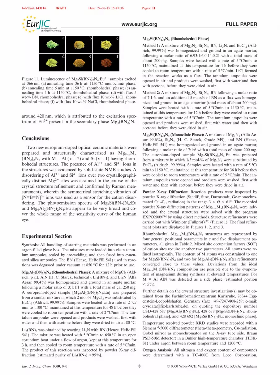
and Mg3(BN2)N show significant spectral overlap and arethus merged to one broad band. A redshift is observed withincreasing content of Mg3(BN2)N:Eu2+. The selected spec-tra of Mg7Si(BN2)2N4:Eu2+ with different fluxes are pre-sented in Figure 10 (a–c) (see also photographs in Fig-ure 11).
The excitation spectrum of Mg7Si(BN2)2N4:Eu2+ withaddition of BN, recorded by monitoring the emission bandat 560 nm, consists of a broad band with a maximum at
Eur. J. Inorg. Chem. 0000, 0–0 © 0000 Wiley-VCH Verlag GmbH & Co. KGaA, Weinheim7
Figure 10. Room temperature excitation and emission spectra ofMg7Si(BN2)2N4:Eu2+ samples made with different fluxes: (a) BN,(b) LiCl, and (c) NaCl.
300 nm. The excitation spectra of Mg7Si(BN2)2N4:Eu2+
with LiCl and NaCl as a flux show an additional band at
Job/Unit: I43116 /KAP1 Date: 24-02-15 15:47:36 Pages: 11
www.eurjic.org FULL PAPER
Figure 11. Luminescence of Mg7Si(BN2)2N4:Eu2+ samples excitedat 366 nm (a) annealing time 36 h at 1150 °C monoclinic phase;(b) annealing time 5 min at 1150 °C, rhombohedral phase; (c) an-nealing time 1 h at 1150 °C, rhombohedral phase; (d) with flux 5wt-% BN, rhombohedral phase; (e) with flux 10 wt-% LiCl, rhom-bohedral phase; (f) with flux 10 wt-% NaCl, rhombohedral phase.
around 420 nm, which is attributed to the excitation spec-trum of Eu2+ present in the secondary phase Mg3(BN2)N.
Conclusions
Two new europium-doped optical ceramic materials wereprepared and structurally characterized as Mg8–xMx
(BN2)2N4 with M = Al (x = 2) and Si (x = 1) having rhom-bohedal structures. The presence of Al3+ and Si4+ ions inthe structures was evidenced by solid-state NMR studies. Adisordering of Al3+ and Si4+ ions over two crystallographi-cally distinct Mg2+ sites was assumed in the course of thecrystal structure refinement and confirmed by Raman mea-surements, wherein the symmetrical stretching vibration of[N=B=N]3– ions was used as a sensor for the cation disor-dering. The photoemission spectra of Mg7Si(BN2)N4:Euand Mg6Al2(BN2)2N4:Eu appear to be very broad and co-ver the whole range of the sensitivity curve of the humaneye.
Experimental SectionSynthesis: All handling of starting materials was performed in anargon-filled glove box. The mixtures were loaded into clean tanta-lum ampoules, sealed by arc-welding, and then fused into evacu-ated silica ampoules. The BN (Henze, HeBoFill 541) used in reac-tions was degassed under vacuum at 850 °C for 12 h prior to use.
Mg6Al2(BN2)2N4 (Rhombohedral Phase): A mixture of MgCl2 (Ald-rich, p.a.), AlN (H. C. Starck, technical), Li3(BN2), and Li3N (AlfaAesar, 99.4%) was homogenized and ground in an agate mortar,following a molar ratio of 3:1:1:1 with a total mass of ca. 250 mg.A europium-doped sample [Mg6Al2(BN2)2N4:Eu] was preparedfrom a similar mixture in which 2 mol-% MgCl2 was substituted byEuCl2 (Aldrich, 99.99%). Samples were heated with a rate of 2 °C/min to 1100 °C, maintained at this temperature for 48 h before theywere cooled to room temperature with a rate of 2 °C/min. The tan-talum ampoules were opened and products were washed, first withwater and then with acetone before they were dried in air at 80 °C.
Li3(BN)2 was obtained by reacting Li3N with BN (Henze, HeBoFill541). The mixture was heated with 5 °C/min to 650 °C in an opencorundum boat under a flow of argon, kept at this temperature for3 h, and then cooled to room temperature with a rate of 5 °C/min.The product of this reaction was inspected by powder X-ray dif-fraction [estimated purity of Li3(BN2) �95%].
Eur. J. Inorg. Chem. 0000, 0–0 © 0000 Wiley-VCH Verlag GmbH & Co. KGaA, Weinheim8
Mg7Si(BN2)2N4 (Rhombohedral Phase)
Method 1: A mixture of Mg3N2, Si3N4, BN, Li3N, and EuCl2 (Ald-rich, 99.99%) was homogenized and ground in an agate mortar,following a molar ratio of 6.93:1:6:0.14:0.21 with a total mass ofabout 200 mg. Samples were heated with a rate of 5 °C/min to1150 °C, maintained at this temperature for 1 h before they werecooled to room temperature with a rate of 5 °C/min. LiCl formedin the reaction works as a flux. The tantalum ampoules wereopened in air and products were washed, first with water and thenwith acetone, before they were dried in air.
Method 2: A mixture of Mg3N2, Si3N4, BN following a molar ratioof 7:1:6, and an additional 5 mass% of BN as a flux was homoge-nized and ground in an agate mortar (total mass of about 200 mg).Samples were heated with a rate of 5 °C/min to 1150 °C, main-tained at this temperature for 12 h before they were cooled to roomtemperature with a rate of 5 °C/min. The tantalum ampoules wereopened and products were washed, first with water and then withacetone, before they were dried in air.
Mg7Si(BN2)2N4 (Monoclinic Phase): A mixture of Mg3N2 (Alfa Ae-sar 99.6%), Si3N4 (H. C. Starck, Grade M9), and BN (Henze,HeBoFill 541) was homogenized and ground in an agate mortar,following a molar ratio of 7:1:6 with a total mass of about 200 mg.The europium-doped sample Mg7Si(BN2)2N4:Eu was preparedfrom a mixture in which 1/3 mol-% of Mg3N2 were substituted byEuCl2 (Aldrich, 99.99%). Samples were heated with a rate of 5 °C/min to 1150 °C, maintained at this temperature for 36 h before theywere cooled to room temperature with a rate of 5 °C/min. The tan-talum ampoules were opened and products were washed, first withwater and then with acetone, before they were dried in air.
Powder X-ray Diffraction: Reaction products were inspected bypowder X-ray diffraction (StadiP, Stoe, Darmstadt, Ge-monochro-mated Cu-Kα1 radiation) in the range 3 � Θ � 61°. The recordedpowder X-ray diffraction patterns of Mg8–xMx(BN2)2N4 were inde-xed and the crystal structures were solved with the programEXPO2009[36] by using direct methods. Structure refinements werecarried out with Winplotr (Fullprof)[37] (Figure 1). The final refine-ment plots are displayed in Figures 1, 2, and 3.
Rhombohedral Mg8–xMx(BN2)2N4 structures are represented byfour general positional parameters in z and five displacement pa-rameters, all given in Table 2. Mixed site occupation factors (SOF)of cation sites require another two parameters. All atoms were re-fined isotropically. The content of M atoms was constrained to onefor Mg7Si(BN2)2N4 and two for Mg6Al2(BN2)2N4 after refinementsconverged close to these values. Deviations from the idealMg8–xMx(BN2)2N4 composition are possible due to the evapora-tion of magnesium during synthesis at elevated temperatures. ForM = Al AlN was detected as a side phase (estimated portion� 5%).
Further details on the crystal structure investigation(s) may be ob-tained from the Fachinformationszentrum Karlsruhe, 76344 Egg-enstein-Leopoldshafen, Germany (fax: +49-7247-808-259; e-mail:[email protected]), on quoting the depository numberCSD-428 687 [Mg6Al2(BN2)2N4], 428 688 [Mg7Si(BN2)2N4; rhom-bohedral phase], and 428 692 [Mg7Si(BN2)2N4; monoclinic phase].
Temperature resolved powder XRD studies were recorded with aSiemens d-5000 diffractometer (theta-theta-geometry, Cu-radiation,Göbel mirror as monochromator on the X-ray tube side, BraunPSD-50M detector) in a Bühler high-temperature chamber (HDK-S1) under argon between room temperature and 1200 °C.
Oxygen Analysis: All nitrogen and oxygen content of compoundswere determined with a TC-400C from Leco Corporation,
Job/Unit: I43116 /KAP1 Date: 24-02-15 15:47:36 Pages: 11
www.eurjic.org FULL PAPER
equipped with TCWin Analysis software, version 4.03, and helium4.6 (Westfalen) as a purge gas. Due to air- and moisture-sensitivityof the materials, all handling took place in a glove box (Braun)under dry Ar. For every analysis, 10–20 mg of the material wereweighed, transferred into tin capsules and sealed therein. For amore homogeneous combustion, three scraps of highly pure nickelwire were added. The subsequent analysis was conducted at 1000 Ain high purity carbon crucibles and repeated three times. N/Oanalyses for rhombohedral and monoclinic Mg7Si(BN2)2N4 samplerevealed small amounts of oxygen [rhombohedral: 1.0 (weight)%oxygen, monoclinic: 0.28% oxygen].
NMR Spectroscopy: Solid-state 27Al magic-angle spinning (MAS)NMR spectra were obtained at a spinning rate of 7.5 kHz with aBruker AVII+500 standard bore NMR spectrometer using 4 mm(o.d.) HR-MAS probe head. Solid-state 29Si magic-angle spinningNMR spectra were obtained with a Bruker ASX-300 wide-borespectrometer using a 7 mm (o.d.) double-bearing MAS probe headand a spinning rate of 4 kHz, referenced with respect to TMS usingQ8M8 as secondary reference.
Infrared Spectroscopy: Infrared transmittance spectra were re-corded with a Bruker Tensor 27 FTIR spectrometer within therange of 400–4000 cm–1 using the KBr-pellet technique. The instru-mental resolution was 4 cm–1.
Raman Spectroscopy: Raman scattering in the range 120–4000 cm–1
was collected from powder in back scattering geometry with a Hor-iba Jobin–Yvon Raman spectrometer HR800 equipped with a con-focal Olympus BX41 microscope, a dispersive grating with 600grooves/mm, and a Peltier cooled (–70 °C) CCD camera with1024 �256 array and a pixel size 26 μm. The spectra were collectedwith an objective of 50 � magnification and 0.5 numerical aperture.As excitation source, a frequency doubled Nd:YAG laser emittingat 532.1 nm with output laser power of 30 mW was used. Energycalibration was performed by using the Raman peak at 520 cm–1
of a Si(111)-wafer. Calibration and confocality was performed byan internal LED. The achieved spectral resolution was between 1.5and 3.0 cm–1.
Luminescence Spectroscopy: Excitation and emission spectra werecollected with a fluorescence spectrometer FLS920 (Edinburgh In-struments) equipped with a 450 W ozone-free xenon arc lamp (OS-RAM) and a sample chamber installed with a mirror optic for pow-der samples. For detection, a R2658P single-photon counting pho-tomultiplier tube (Hamamatsu) was used. All luminescence spectrawere recorded with a spectral resolution of 1 nm, a dwell time of0.4 s in 1 nm steps, and three repeats. Reflection spectra were moni-tored by placing the sample into an integrating sphere coated withbarium sulfate and using a synchron scan, i.e., the excitation andemission monochromator were adjusted to the same wavelengthand tuned synchronously. These reflection spectra were recordedwith an Edinburgh Instruments FS900 spectrometer equipped witha 450 W Xe arc lamp and cooled single-photon counting photo-multiplier (Hamamatsu R928). BaSO4 (99%, Sigma–Aldrich) wasused as a reflectance standard.
Acknowledgments
Support of this research by the Deutsche Forschungsgemeinschaft(DFG) through the project Solid State Metathesis Reactions(ME:25-1) is gratefully acknowledged from scientists of the Eber-hard Karls University in Tübingen. The authors gratefully acknow-ledge the support of our colleagues from the University of Tübin-gen, namely Dr. Klaus Eichele for recording NMR spectra, Dipl.-
Eur. J. Inorg. Chem. 0000, 0–0 © 0000 Wiley-VCH Verlag GmbH & Co. KGaA, Weinheim9
Chem. Jens Kaiser and Prof. Dr. Eberhard Schweda for high-tem-perature X-ray studies, Dipl.-Chem. Johannes Uihlein and Prof.Dr. Thomas Chassé for recording Raman spectra.
[1] S. Nakamura, G. Fasol, The Blue Laser Diode: GaN BasedLight Emitters and Lasers; Springer: Berlin, Germany, 1997, p.216–221.
[2] Y. W. Jung, B. Lee, S. P. Singh, K.-S. Sohn, Opt. Express 2010,18, 17805–17818.
[3] T. Schlieper, W. Milius, W. Schnick, Z. Anorg. Allg. Chem.1995, 621, 1380–1384.
[4] B. Blaschkowski, H. Jing, H.-J. Meyer, Angew. Chem. Int. Ed.2002, 41, 3322–3336; Angew. Chem. 2002, 114, 3468.
[5] B. Blaschkowski, H.-J. Meyer, Z. Anorg. Allg. Chem. 2002, 628,1249–1254.
[6] B. Blaschkowski, Dissertation, University of Tübingen, Ger-many, 2003.
[7] M. Neukirch, B. Blaschkowski, M. Häberlen, H.-J. Meyer, Z.Anorg. Allg. Chem. 2006, 632, 1799–1803.
[8] R. J. Cava, H. W. Zandbergen, B. Batlogg, H. Eisaki, H. Tak-agi, J. J. Krajewski, W. F. Peck Jr., E. M. Gyorgy, S. Uchida,Nature 1994, 372, 245–247.
[9] H. W. Zandbergen, V. Jansen, R. J. Cava, J. J. Krajewski, W. F.Peck Jr., Nature 1994, 372, 759–761.
[10] H. Michor, R. Krendelsberger, G. Hilscher, E. Bauer, C. Dusek,R. Hauser, L. Naber, D. Werner, P. Rogl, H. W. Zandbergen,Phys. Rev. B 1996, 54, 9408–9420.
[11] B. Blaschkowski, H.-J. Meyer, Z. Anorg. Allg. Chem. 2003, 629,129–132.
[12] J. Glaser, T. Mori, H.-J. Meyer, Z. Anorg. Allg. Chem. 2008,634, 1067–1073.
[13] H. Yamane, S. Kikkawa, H. Horiuchi, M. Koizumi, J. SolidState Chem. 1986, 65, 6–12.
[14] H. Womelsdorf, H. J. Meyer, Z. Anorg. Allg. Chem. 1994, 620,262–267.
[15] M. Häberlen, J. Glaser, H.-J. Meyer, J. Solid State Chem. 2005,178, 1478–1487.
[16] M. Somer, U. Herterich, J. Curda, K. Peters, H. G. von Schner-ing, Z. Kristallogr. 1994, 209, 182–182.
[17] J. Goubeau, W. Anselment, Z. Anorg. Allg. Chem. 1961, 310,248–260.
[18] M. Kalmutzki, M. Ströbele, H.-J. Meyer, Dalton Trans. 2013,42, 12934–12939.
[19] H. Hiraguchi, H. Hashizume, O. Fukunaga, A. Takenaka, M.Sakata, J. Appl. Crystallogr. 1991, 24, 286–292.
[20] H. Jing, O. Reckeweg, B. Blaschkowski, H.-J. Meyer, Z. Anorg.Allg. Chem. 2001, 627, 774–778.
[21] A. N. Zhukov, K. P. Burdina, K. N. Semenenko, Russ. J. Gen.Chem. 1994, 8, 1117–1121.
[22] A. N. Zhukov, K. P. Burdina, K. N. Semenenko, Zh. Obshch.Khim. 1996, 66, 1070–1072.
[23] J. Schölch, T. Dierkes, D. Enseling, M. Ströbele, T. Jüstel, H.-J. Meyer, Z. Allg. Anorg. Chem. 2015, in press.
[24] H. Hiraguchi, H. Hashizume, S. Sasaki, S. Nakano, O. Fukun-aga, Acta Crystallogr., Sect. B 1993, 49, 478–483.
[25] H. Hiraguchi, H. Hashizume, J. Appl. Crystallogr. 1991, 24,286–292.
[26] F. Stadler, O. Oekler, J. Senker, H. A. Höppe, P. Knoll, W.Schnick, Angew. Chem. Int. Ed. 2005, 44, 567–570; Angew.Chem. 2005, 117, 573.
[27] P. Kempgens, R. K. Robin, D. P. Thompson, Solid State Nucl.Magn. Reson. 1999, 15, 109–118.
[28] K. J. D. Mackenzie, R. H. Meinhold, G. V. White, C. M. Shep-pard, B. L. Sherriff, J. Mater. Sci. 1994, 29, 2611–2619.
[29] E. Kroumova, M. I. Aroyo, J. M. Perez Mato, J. M. Kirov,C. A. Capillas, S. Ivantchev, H. Wondratschek, Bilbao Crystal-lographic Server: Useful databases and tools for phase transitionsstudies, Phase Transitions 2003, 76, 155–170.
[30] M. Zeuner, S. Pagano, W. Schnick, Angew. Chem. 2011, 123,7898–7820.

Job/Unit: I43116 /KAP1 Date: 24-02-15 15:47:36 Pages: 11
www.eurjic.org FULL PAPER
[31] F. Hintze, F. Hummel, P. J. Schmidt, D. Wiechert, W. Schnick,Chem. Mater. 2012, 24, 402–407.
[32] F. Hintze, N. W. Johnson, M. Seibald, D. Muir, A. Moewes, W.Schnick, Chem. Mater. 2013, 25, 4044–4052.
[33] P. Dorenbos, J. Lumin. 2003, 104, 239–260.[34] K. Van den Eeckhout, P. F. Smet, D. Poelman, Materials 2010,
3, 2536–2566.[35] D. Ravichandran, S. T. Johnson, S. Erdei, R. Roy, W. B. White,
Displays 1999, 19, 197–203.
Eur. J. Inorg. Chem. 0000, 0–0 © 0000 Wiley-VCH Verlag GmbH & Co. KGaA, Weinheim10
[36] A. Altomare, M. Camalli, C. Cuocci, C. Giacovazzo, A. Molit-erni, R. Rizzi, J. Appl. Crystallogr. 2009, 42, 1197–1202.
[37] T. Roisnel, J. Rodriguez-Carvajal, WinPLOTR: A Windows toolfor powder diffraction patterns analysis. Proceedings of the Sev-enth European Powder Diffraction Conference (EPDIC 7)(Eds.: R. Delhez, E. J. Mittenmeijer), 2000, 118–123.
Received: November 24, 2014Published Online: �

Job/Unit: I43116 /KAP1 Date: 24-02-15 15:47:36 Pages: 11
www.eurjic.org FULL PAPER
Luminescent Ceramics
M. Ströbele, K. Dolabdjian, D. Enseling, Compounds Mg8–xMx(BN2)2N4 were pre-D. Dutczak, B. Mihailova, T. Jüstel, pared by solid-state reaction of MgCl2,H.-J. Meyer* .................................. 1–11 Li3(BN2), and AlN for M = Al, and of
Mg3N2, BN, and Si3N4 for M = Si. TheirLuminescence Matching with the Sensi- crystal structures were refined from powdertivity Curve of the Human Eye: Optical X-ray diffraction data with the space groupCeramics Mg8–xMx(BN2)2N4 with M = Al R3̄m for M = Al, Si, and P21/c for M =(x = 2) and M = Si (x = 1) Si. Eu(II)-doped Mg8–xMx(BN2)2N4 show
broad band emissions in the visible regionKeywords: Ceramics / Solid-state reactions / of the spectrum (λex = 320 nm).Solid-state structures / Doping / Lumi-nescence
Eur. J. Inorg. Chem. 0000, 0–0 © 0000 Wiley-VCH Verlag GmbH & Co. KGaA, Weinheim11
Related Documents