Lumbar disc disease Lumbar disc disease

Lumbar disc disease. Very Important Talk!! -- LBP A major public health problem The leading cause of disability for people < 45 2 nd leading cause for.
Dec 29, 2015
Welcome message from author
This document is posted to help you gain knowledge. Please leave a comment to let me know what you think about it! Share it to your friends and learn new things together.
Transcript

Lumbar disc diseaseLumbar disc disease

Very Important Talk!! -- LBPVery Important Talk!! -- LBP
• A major public health problem
• The leading cause of disability for people < 45
• 2nd leading cause for physician visits
• 3rd most common cause for surgical procedures
• 5th most common reason for hospitalizations
• Lifetime prevalence: 49%–80%
Pai et al. 2004, Pai et al. 2004, Orthop Clin N AmOrthop Clin N Am

EpidemiologyEpidemiology• 60 – 90% of adults experience back pain at
some point in their life. - incidence age 35- 55 y.o. - 90% resolve in 6 weeks - 7% become chronic
- M/ F equally affectedMost patients with LBP improve on their own
in time (even without treatment).

Types of LBPTypes of LBP
1. Non-specific “idiopathic”: 85%
2. Degenerative disc disease: discogenic pain, disk herniation, degenerative scoliosis
3. Developmental: spondylolisthesis, idiopathic scoliosis
4. Congenital: scoliosis
5. Traumatic6. Infectious7. Inflammatory8. Neoplastic9. Metabolic 10.Referred

Low Back PainLow Back Pain
• Most episodes of LBP are self limited
• These episodes become more frequent with age
• LBP is usually due to repeated stress on the lumbar spine over many years (“degeneration”), although an acute injury may cause the initiation of pain

Degenerative Disc Disease Degenerative Disc Disease (DJD)(DJD)
• Unfortunately, DJD seems to be sort of a “wastebasket term” that is often used to describe age-related changes on MRI, etc.– While these changes are indeed
“degenerative,” this happens as we age and is not necessarily indicative of any significant underlying pathology or condition.
– The majority of individuals > 60 will show some type of degenerative change(s) on lumbar imaging.

DJDDJD
• Degeneration of an individual disc space typically refers to loss of disc height, loss of water content, fibrosis, end plate sclerosis/defects, osteophyte complexes, etc.

Lumbar Spine Motion SegmentLumbar Spine Motion Segment• Three joint complex• Intervertebral disc + 2 facet joint• Ligamentous structure, vertebral body

Sagittal Section through the Sagittal Section through the Spinal CordSpinal Cord
1. Intervertebral disc
2. Vertebral body
3. Dura mater
4. Extradural or epidural space
5. Spinal cord
6. Subarachnoid space

Intervertebral DiscIntervertebral Disc
• Hydrostatic, load bearing structure between the vertebral bodies
• Nucleus pulposus + annulus fibrosus• No blood supply• L4-5, largest avascular structure in the body

Nucleus PulposusNucleus Pulposus
• Type II collagen strand + hydrophilic proteoglycan
• Water content 70 ~ 90%• Confine fluid within the annulus• Convert load into tensile strain on the annular
fibers and vertebral end-plate

Annulus FibrosusAnnulus Fibrosus
• Outer boundary of the disc• Type I collagen

Vertebral End-PlateVertebral End-Plate
• Cartilaginous and osseous component• Nutritional support for the nucleus• Passive diffusion

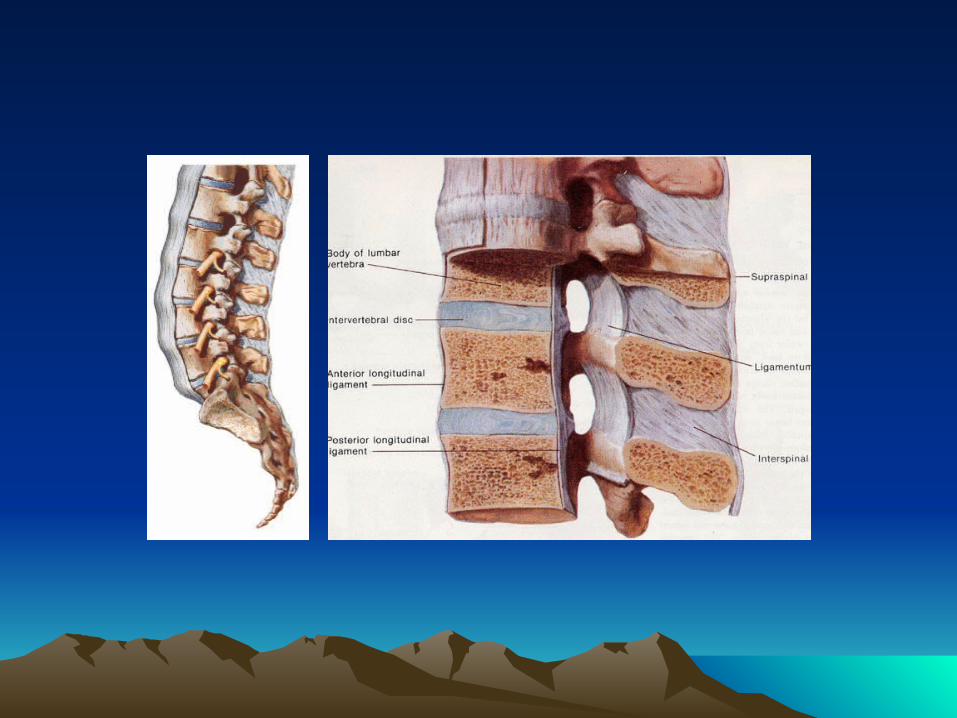
Facet JointFacet Joint
• Synovial joint• Rich innervation with sensory nerve fiber• Load share 18% of the lumbar spine
Facet JointFacet Joint
** Primary disc degeneration Secondary change in the posterior facet joint and soft tissue


Important QuestionsImportant Questions
1. Is systemic disease the cause?
2. Is there social or psycological distress that prolongs or amplifies symptoms?
3. Is there neurologic compromise that requires surgical intervention?

To Answer These Important To Answer These Important Questions Questions
1. Careful History and Physical Exam
2. Imaging and Labs WHEN indicated

Differential Diagnosis of Low Back PainDifferential Diagnosis of Low Back Pain

Evaluation in older adultsEvaluation in older adults
• Cancer, compression fractures, spinal stenosis, aortic aneurysms more common
• Osteoporotic fractures without trauma• Spinal Stenosis secondary to
degenerative processes and spondylolisthesis more common
• Increased AAA associated with CAD• Early radiography recommended

Clues To Systemic DiseaseClues To Systemic Disease
• Age• History of Cancer• Fever• Unexplained Weight Loss• Injection Drug Use• Chronic Infection Elsewhere• Duration and Quality of Pain
-Infection and Cancer not relieved supine• Response to previous therapy• h/o inflammatory arthritis elsewhere

Things that should raise a “red flag”Things that should raise a “red flag”
• Previous hx of cancer, unexplained weight loss• Immunosuppression, hx of steroid use, hx of IV
drug abuse, hx of skin/other infection(s)• Hx of recent falls or trauma (including surgery)• Bladder dysfunction (usually urinary retention
or overflow incontinence) or fecal incontinence, “saddle anesthesia”, leg weakness
• Pain that doesn’t improve with rest; failure to improve after 4 weeks conservative management

Lumbar Disc DiseaseLumbar Disc Disease
Discogenic Back Pain
A. Internal Disc Disruption (IDD)B. Degenerative Disc Disease (DDD)C. Segmental Instability
Lumbar Disc Herniation and Radiculopathy

Lumbar Disc HerniationLumbar Disc Herniation
How pain is generated?
• Inflammatory• Biochemical• Vascular• Mechanical compression

HistoryHistory
• symptom of disc herniation : acute or gradual • after trauma or without and inciting event • most common 3rd and 4th decade
Chief Complain • Pain, radiating from the back or buttock into the leg• Numbness and weakness • Sharp, lancinating, shooting/radiating down the leg
posteriorly below the knee • Coughing, Valsalva maneuver increase intracecal
pressure increase pain • Sitting position, driving out of lordosis increase
intradiscal pressure increase pain

Natural HistoryNatural History
• Recovery from nonspecific LBP generally rapid – 90% within 2 weeks – some studies less rapid (2/3 at 7 weeks)
• Herniated Discs – slower to improve – only about 10% considered for surgery after 6 weeks
• With surgery, no earlier return to work – symptomatic and functional outcome sometimes better

Posture and intradiscal pressurePosture and intradiscal pressure

ConceptConcept
• Intervertebral discs can be thought of, conceptually, kind of like a “jelly donut.” The outside is the annulus fibrosus, and the inside “jelly” is the more watery nucleus pulposus.– Intervertebral discs act as shock absorbers
between the vertebral bodies.– Just like jelly donuts have a “weak spot” where
the jelly squirts out if you squeeze them, the annulus of discs is weak posteriorly where the nucleus pulposus can herniate through, causing symptoms.

• The most common sites for a herniated lumbar disc are L4-5 and L5-S1, resulting in back pain and pain radiating down the posterior and lateral leg, to below the knee
• Back pain caused by a herniated lumbar disc is exacerbated by sitting and bending; conversely, the pain of lumbar muscular strain is aggravated by standing and twisting movements.
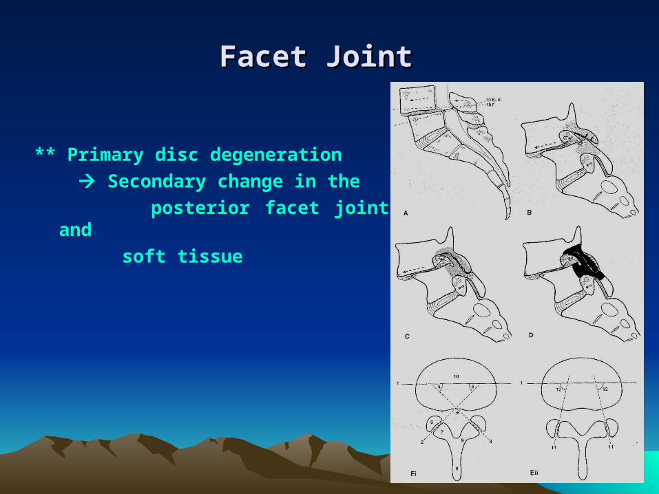
Sciatic nerveSciatic nerve


Disc Degeneration – PhysiologyDisc Degeneration – Physiology
• With age and repeated efforts, the lower lumbar discs lose their height and water content (“bone on bone”)
• Abnormal motion between the bones leads to pain

Disc Disc
• Nucleus pulposus-water rich, gelatinous,axial load, pivotal point,binds vertebrae together
• Annulus fibrosus-fibrous and tougher, less water content,contained the nucleus pulposus
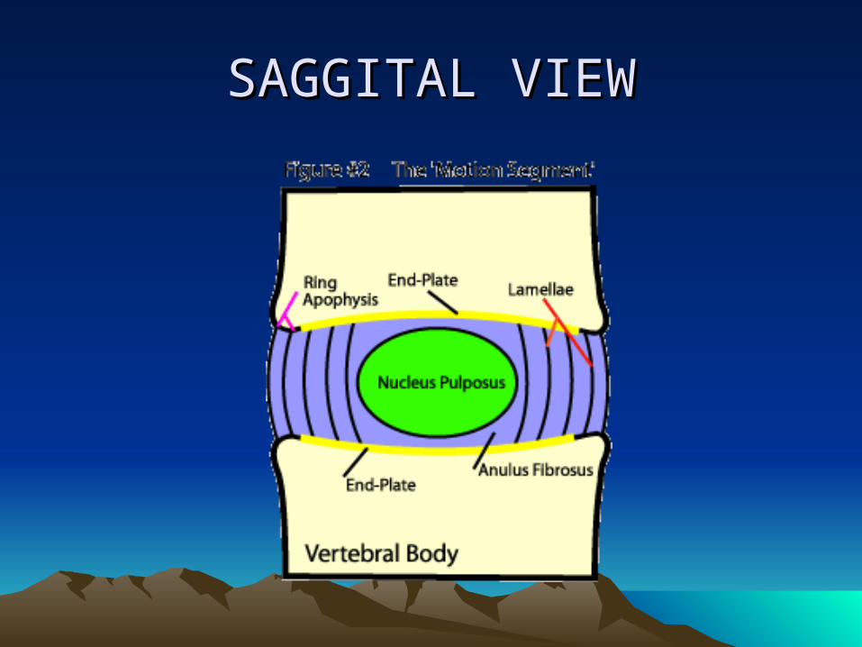
SAGGITAL VIEWSAGGITAL VIEW

DISC PHYSIOLOGYDISC PHYSIOLOGY

DISC NUTRITIONDISC NUTRITION

DIURNAL CHANGEDIURNAL CHANGE
• During day time- disc shrinks by 20%
• Body height reduced by 15 – 25 mm
• In night- body height is increased.


MRI appearance MRI appearance
• T-2 weighted image• Black disc –
dessication

Natural disc ageing Natural disc ageing
• Loss of the proteoglycan molecule from the nucleus of the disc.
• Progressive dehydration.
• Progressive thickening.
• Brown pigmentation formation.
• Increased brittleness of the tissue of the disc.

FACTORS CONTRIBUTING FACTORS CONTRIBUTING TO DISC AGEINGTO DISC AGEING

IDIOPATHIC BLOOD VESSEL/NUTRIENT LOSS AND IDIOPATHIC BLOOD VESSEL/NUTRIENT LOSS AND DEHYDRATION/DECREASED PROTEOGLYCANS DEHYDRATION/DECREASED PROTEOGLYCANS
PRODUCTIONPRODUCTION

Other factorsOther factors
• Vertebral end plate calcification
• Arterial stenosis
• Smoking
• DM
• Exposure to vibration.

Disc degenerationDisc degeneration

Steps of disc herniationSteps of disc herniation


DISC HERNIATION OR DISC HERNIATION OR PROLAPSEPROLAPSE
• Protrusion ( contained or subligamentous herniation )
• Extrusion ( non-contained or transligamentous herniation )
• Sequestration ( freek fragment )

Internal disc disruption/grade -3 Internal disc disruption/grade -3 radial annual tearradial annual tear

Disc protrusion/PLL is still intactDisc protrusion/PLL is still intact

Disc extrusion/ PLL is rupturedDisc extrusion/ PLL is ruptured

MRI disc extrusionMRI disc extrusion

Disc sequestration/final end stage Disc sequestration/final end stage of disc diseaseof disc disease

Physical ExaminationPhysical Examination• Fever – possible infection• Vertebral tenderness - not specific and not
reproducible between examiners• Limited spinal mobility – not specific (may help in
planning P.T.• If sciatica or pseudoclaudication present – do straight
leg raise• Positive test reproduces the symptoms of sciatica –
pain that radiates below the knee (not just back or hamstring)
• Ipsilateral test sensitive – not specific: crossed leg is insensitive but highly specific
• L-5 / S-1 nerve roots involved in 95% lumbar disc herniations

PresentationPresentation
• The classic presentation of Herniated Nucleus Pulposus (HNP), both for cervical and lumbar spine, is radiculopathy.– The disc herniation impinges upon a nerve
root, causing characteristic pain.– Thoracic disc hernations are much, much
rarer.

Lumbar HNPLumbar HNP
• 90% of herniated discs are paracentral (slightly off to one side) and affect the nerve root that corresponds to the lower vertebral level.– Example: a typical L4/5 disc herniation would
cause symptoms referrable to the L5 nerve root.

Assessment of FunctionAssessment of Function
• 98% disc herniations: L4-5; L5-S1
• Impairment: Motor and Sensory L5-S1– L5: Weakness of ankle and great toe
dorsiflexion– S1: Decrease ankle reflex– L5 & S1: Sensory loss in the feet

STRAIGHT LEG RAISE TESTSTRAIGHT LEG RAISE TEST
The straight leg raise test is positive if pain in the sciatic distribution is reproduced between 30° and 70° passive flexion of the straight leg. Dorsiflexion of the foot exacerbates the pain
STRAIGHT LEG RAISE TEST

• Straight-leg raising : L5, S1 root• Contralateral SLR : sequestrated or extruded
disc• Femoral stretching, reverse SLR : L3, L4 root
Root Tension SignsRoot Tension Signs

Sciatica - radiating pain down the leg
Radiculopathy- radiating pain down the leg as a result of nerve root
irritation
Back Pain• change in disc loading and shape, biomechanics • loss of viscoelasticity. • 90% of radiating pain have long-standing prior
episodic low back pain

Differential DiagnosisDifferential Diagnosis
Vascular claudication• Vascular assessment and flow study• Dorsalis pedis palpation
Spinal stenosis• leg pain, dysesthesia, paresthesia, often not
dermatomal • pain d/t mechanical compression of spinal canal and
foramen • lordosis and axial loading • symptomatic on walking, relief by sitting
ThrombophlebitisMetabolic and peripheral neuropathy

Neurologic ExaminationNeurologic Examination


Imaging StudiesImaging Studies
• Progressive Neurologic Defecits
• Failure to Improve
• Hx of Trauma
• Risk for Malignancy or infection
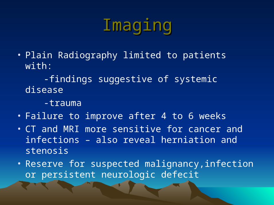
ImagingImaging
• Plain Radiography limited to patients with:
-findings suggestive of systemic disease
-trauma• Failure to improve after 4 to 6 weeks• CT and MRI more sensitive for cancer and
infections – also reveal herniation and stenosis• Reserve for suspected malignancy,infection or
persistent neurologic defecit

MRIMRI
• The gold standard for imaging of the herniated lumbar disc is magnetic resonance imaging

WHAT TO LOOK IN MRIWHAT TO LOOK IN MRI

T-1 AXIAL VIEWT-1 AXIAL VIEW

PROTON DENSITY IMAGEPROTON DENSITY IMAGE

ZONES OF ANTERIOR EPIDURAL ZONES OF ANTERIOR EPIDURAL SPACE / HERNIATION ZONESSPACE / HERNIATION ZONES
• Central region• Paracentral region or
lateral recess• Intraforaminal zone or
subarticular zone• Extraforaminal zone


Lumbar Disc Herniation – Lumbar Disc Herniation – TreatmentTreatment
Conservative Tx.– Moderate bed rest– Spinal manipulation – Physical therapy– Medication
• NSAIDs• Muscle relaxants• Rarely narcotics
Surgical Tx.• “Microdiscectomy”• Less than half of an
inch incision• Go home the same or
next day• Good results in up to
90% of cases

Indication of SurgeryIndication of SurgeryAbsolute surgical indication • cauda equina syndrome• acute urinary retension/incontinence, saddle anesthesia, back/ buttock/ leg pain, weakness,
difficulty walking
Relative indication• progressive weakness • no response to conservative treatment
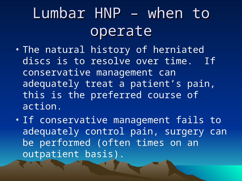
Lumbar HNP – when to operateLumbar HNP – when to operate
• The natural history of herniated discs is to resolve over time. If conservative management can adequately treat a patient’s pain, this is the preferred course of action.
• If conservative management fails to adequately control pain, surgery can be performed (often times on an outpatient basis).

Results of Surgical TreatmentResults of Surgical Treatment
• Good outcome in 80-90% of cases• Residual pain may last up to 6 months postop• Results are worse if pain was present for over 8
months before the operation (permanent nerve damage?)

Disc prolapse commonly occurs at:
L4-L5 L5-S1 C5-C6 C4-C5 C3-C4
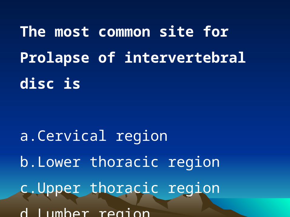
The most common site for Prolapse of
intervertebral disc is
a.Cervical region
b.Lower thoracic region
c.Upper thoracic region
d.Lumber region

After L4 – S1 the next commonest site of
intervertebral disc prolapse is
a.C6- C7
b.T12 – L1
c.L1 - L2
d.L2 -L3

The most common cause of acute sciatica is
due to
a.Trauma
b.Secondaries of spine
c.Acute prolapsed Intervertebral disc
d.Tuberculosis of spine

A building contractor suddenly complains of
lower backache which increase on bending
down He has
a.Renal colic
b.Tuberculosis of spine
c.Disc prolapse
d.Fibrositis'

The most important single special investigation
in lumbar disc prolapse is
a.Epidurography
b.Myelography
c.MRI
d.Discography
e.Spinal venography

Management in case of rupture of disc at L5, S1 is
a.Emergency removal of disc
b.Joint fusion
c.Immobilization for 2 weeks with spinal brace
d.Traction

A 44-year-old man presented with acute onset of low backache
radiating to the right lower limb. Examination revealed SLRT
<40 on the right side, weakness of extensor hallucis longus on
the right side, sensory loss in the first web space of the right
foot and brisk knee jerk. Which of the following is the most
likely diagnosis:
a.Prolapsed intervertebral disc L4-5
b.Spondylolysis L5-S1
c.Lumbar canal stenosis
d.Spondylolisthesis L4-5

A previously healthy 45 yrs old laborer suddenly develops acute lower
back pain with right-leg pain & weakness of dorsiflexion of the right
great toe. Which of the following is true:
a.Immediate treatment should include analgesics muscle relaxants & back
strengthening exercises
b.The appearance of the foot drop indicate early surgical intervention
c.If the neurological sign resolve within 2 to 3 weeks but low back pain
persists, the proper treatment would include fusion of affected Lumbar
vertebra.
d.If the neurological signs fail to resolve within 1 week, Lumbar
laminectomy and exscision of any herniated nucleus pulposus should be
done.

Feature of L2-L3 prolapsed disc is/are
a.Low back pain
b.Straight leg raising test +ve
c.Reversed straight leg raising test +ve
d.Quadriceps weakness
e.Loss of sensation on anteromedial thigh

A middle aged lady presents with complaints of lower back
pain. ON examination there is weakness of extension of
right great toe with no sensory impairment. An MRI of the
lumbosacral spine would most probably reveal a prolapsed
intervertebral disc at what level?
a.L3 - L4
b.L4-L5
c.L5-S1
d.S1-S2

Which of the following is not recommended
in the treatment of Chronic Low Back Pain:
a.NSAIDs
b.Bed Rest for 3 months
c.Exercises
d.Epidural steroid Injection

All of the following are included as yellow flag
signs of low back pain, except:
a.History of systemic steroids use
b.Reliance on Passive Treatment
c.Social Isolation
d.Belief that back pain is severely disabling

A-year-old previously healthy man has had backache with muscle
spasms, weakness, and pain felt in the right hip radiating all the
way to his toes for the past 8 months. He does not have headaches
or other neurologic problems. Physical examination reveals that
the circumference of his right leg is smaller than the left, and he
has paresthesias in an L5 distribution in the right leg. Which of
the following conditions is he most likely to have?
A Spondylolisthesis
B Spina bifida
C Herniated nucleus pulposus
D Osteoporosis
E Paget disease of bone

A 15-year-old girl is noted to have an odd, twisted appearance to her back while she is out swimming with her friends. She is tall and thin. A radiograph reveals an abnormal lateral bowing of the spine, with 20 degrees of lateral curvature in the mid-thoracic region. Which of the following is most likely to produce these findings?
A Asymmetric cartilage growth of vertebral body end platesB Multiple osteochondromas of the vertebral bodiesC Vitamin D deficiency with ricketsD A disorder of procollagen synthesis with multiple compressed fracturesE Trauma

MessagesMessages
• Inflamed discs can cause referred leg pain without neural compression by irritating the sinu -vertebral nerve
• Mild disc degeneration can result in quite severe pain- because of inflammatory chemicals in the disc space- not seen on MRI scans
• Analgesic Discography- a new technique – offers a simple way to confirm the relevant disc as the pain generator
• Interbody fusion can then be used to treat the problem definitively.

Take Home MessagesTake Home Messages
• Know the natural history of the disease
• Know your patient
• Correlate clinical findings, MRI and discograms if needed
• Until definitive evidence available, choose the most cost-effective available treatment option: cognitive therapy, exercise, fusion, arthroplasty, dynamic stabilization

THANK YOUTHANK YOU
Related Documents


![eprints.soton.ac.uk20beliefs... · Web viewLow back pain (LBP) is a leading cause of disability world-wide [42] and is managed mostly within primary care. Most patients have non-specific](https://static.cupdf.com/doc/110x72/5e61bf7753e7510eb6623ead/20beliefs-web-view-low-back-pain-lbp-is-a-leading-cause-of-disability-world-wide.jpg)