LSHTM Research Online Dunne, EM; Murad, C; Sudigdoadi, S; Fadlyana, E; Tarigan, R; Indriyani, SAK; Pell, CL; Watts, E; Satzke, C; Hinds, J; +5 more... Dewi, NE; Yani, FF; Rusmil, K; Mulholland, EK; Kartasasmita, C; (2018) Carriage of Streptococcus pneumoniae, Haemophilus influenzae, Moraxella catarrhalis, and Staphylococcus aureus in Indonesian children: A cross-sectional study. PloS one, 13 (4). e0195098. ISSN 1932-6203 DOI: https://doi.org/10.1371/journal.pone.0195098 Downloaded from: http://researchonline.lshtm.ac.uk/4647317/ DOI: https://doi.org/10.1371/journal.pone.0195098 Usage Guidelines: Please refer to usage guidelines at https://researchonline.lshtm.ac.uk/policies.html or alternatively contact [email protected]. Available under license: http://creativecommons.org/licenses/by/2.5/ https://researchonline.lshtm.ac.uk

Welcome message from author
This document is posted to help you gain knowledge. Please leave a comment to let me know what you think about it! Share it to your friends and learn new things together.
Transcript

LSHTM Research Online
Dunne, EM; Murad, C; Sudigdoadi, S; Fadlyana, E; Tarigan, R; Indriyani, SAK; Pell, CL; Watts,E; Satzke, C; Hinds, J; +5 more... Dewi, NE; Yani, FF; Rusmil, K; Mulholland, EK; Kartasasmita,C; (2018) Carriage of Streptococcus pneumoniae, Haemophilus influenzae, Moraxella catarrhalis, andStaphylococcus aureus in Indonesian children: A cross-sectional study. PloS one, 13 (4). e0195098.ISSN 1932-6203 DOI: https://doi.org/10.1371/journal.pone.0195098
Downloaded from: http://researchonline.lshtm.ac.uk/4647317/
DOI: https://doi.org/10.1371/journal.pone.0195098
Usage Guidelines:
Please refer to usage guidelines at https://researchonline.lshtm.ac.uk/policies.html or alternativelycontact [email protected].
Available under license: http://creativecommons.org/licenses/by/2.5/
https://researchonline.lshtm.ac.uk
RESEARCH ARTICLE
Carriage of Streptococcus pneumoniae,
Haemophilus influenzae, Moraxella catarrhalis,
and Staphylococcus aureus in Indonesian
children: A cross-sectional study
Eileen M. Dunne1,2☯, Chrysanti Murad3☯, Sunaryati Sudigdoadi3, Eddy Fadlyana4,
Rodman Tarigan4, Sang Ayu Kompiyang Indriyani5, Casey L. Pell1, Emma Watts1,
Catherine Satzke1,2,6, Jason Hinds7,8, Nurhandini Eka Dewi9, Finny Fitry Yani10,
Kusnandi Rusmil4, E. Kim Mulholland2,11, Cissy Kartasasmita4*
1 Pneumococcal Research, Murdoch Children’s Research Institute, Parkville, Victoria, Australia,
2 Department of Paediatrics, The University of Melbourne, Parkville, Victoria, Australia, 3 Department of
Biomedical Sciences, Division of Microbiology, Faculty of Medicine, Universitas Padjadjaran, Bandung, West
Java, Indonesia, 4 Department of Child Health, Universitas Padjadjaran/Hasan Sadikin General Hospital,
Bandung, West Java, Indonesia, 5 West Nusa Tenggara Province General Hospital, Mataram, West Nusa
Tenggara, Indonesia, 6 Department of Microbiology and Immunology, The University of Melbourne at the
Peter Doherty Institute for Infection and Immunity, Parkville, Victoria, Australia, 7 Institute for Infection and
Immunity, St. George’s University of London, London, United Kingdom, 8 BUGS Bioscience, London
Bioscience Innovation Centre, London, United Kingdom, 9 District Health Office of Central Lombok, Praya,
West Nusa Tenggara, Indonesia, 10 Department of Child Health, Universitas Andalas, Padang, West
Sumatra, Indonesia, 11 Department of Infectious Disease Epidemiology, London School of Hygiene and
Tropical Medicine, London, United Kingdom
☯ These authors contributed equally to this work.
Abstract
Streptococcus pneumoniae is an important cause of infection and commonly colonizes the
nasopharynx of young children, along with other potentially pathogenic bacteria. The objec-
tives of this study were to estimate the carriage prevalence of S. pneumoniae, Haemophilus
influenzae, Moraxella catarrhalis, and Staphylococcus aureus in young children in Indone-
sia, and to examine interactions between these bacterial species. 302 healthy children aged
12–24 months were enrolled in community health centers in the Bandung, Central Lombok,
and Padang regions. Nasopharyngeal swabs were collected and stored according to World
Health Organization recommendations, and bacterial species detected by qPCR. Pneumo-
coccal serotyping was conducted by microarray and latex agglutination/Quellung. Overall
carriage prevalence was 49.5% for S. pneumoniae, 27.5% for H. influenzae, 42.7% for M.
catarrhalis, and 7.3% for S. aureus. Prevalence of M. catarrhalis and S. pneumoniae, as
well as pneumococcal serotype distribution, varied by region. Positive associations were
observed for S. pneumoniae and M. catarrhalis (OR 3.07 [95%CI 1.91–4.94]), and H. influ-
enzae and M. catarrhalis (OR 2.34 [95%CI 1.40–3.91]), and a negative association was
found between M. catarrhalis and S. aureus (OR 0.06 [95%CI 0.01–0.43]). Densities of S.
pneumoniae, H. influenzae, and M. catarrhalis were positively correlated when two of these
species were present. Prior to pneumococcal vaccine introduction, pneumococcal carriage
prevalence and serotype distribution varies among children living in different regions of
PLOS ONE | https://doi.org/10.1371/journal.pone.0195098 April 12, 2018 1 / 12
a1111111111
a1111111111
a1111111111
a1111111111
a1111111111
OPENACCESS
Citation: Dunne EM, Murad C, Sudigdoadi S,
Fadlyana E, Tarigan R, Indriyani SAK, et al. (2018)
Carriage of Streptococcus pneumoniae,
Haemophilus influenzae, Moraxella catarrhalis, and
Staphylococcus aureus in Indonesian children: A
cross-sectional study. PLoS ONE 13(4): e0195098.
https://doi.org/10.1371/journal.pone.0195098
Editor: Daniela Flavia Hozbor, Universidad Nacional
de la Plata, ARGENTINA
Received: December 18, 2017
Accepted: March 18, 2018
Published: April 12, 2018
Copyright: © 2018 Dunne et al. This is an open
access article distributed under the terms of the
Creative Commons Attribution License, which
permits unrestricted use, distribution, and
reproduction in any medium, provided the original
author and source are credited.
Data Availability Statement: Data from this study
are not publicly available due to ethical reasons.
Requests for data may be sent to the Health
Research Ethics Committee, Universitas
Padjadjaran Faculty of Medicine, via the chair of the
committee, Dr Meita Dhamayanti (meita.
[email protected]) and/or the Institutional
Review Board ([email protected]). Please
note that additional analyses not included in the
study protocol will require ethical approval from
the Health Research Ethics Committee, Universitas
Indonesia. Positive associations in both carriage and density identified among S. pneumo-
niae, H. influenzae, and M. catarrhalis suggest a synergistic relationship among these spe-
cies with potential clinical implications.
Introduction
Streptococcus pneumoniae (pneumococcus) is a Gram-positive bacterium that causes a wide
range of infections including otitis media, pneumonia, sepsis, and meningitis. Pneumococcal
disease is a major cause of pediatric morbidity and mortality worldwide, with disease primarily
occurring in low and middle income countries [1]. The primary reservoir for pneumococci is
the human nasopharynx, and nasopharyngeal carriage rates are highest among young chil-
dren, ranging from 19–86% in different epidemiological settings [2]. Colonization of the naso-
pharynx is typically asymptomatic although can also be associated with mild respiratory
symptoms such as runny nose [3]. Importantly, carriage is considered a prerequisite to pneu-
mococcal disease and serves as the source of pneumococcal transmission [4]. High pneumo-
coccal carriage density has been linked to respiratory infection and pneumonia in children,
and implicated in transmission in animal studies [5, 6].
The nasopharynx is a complex and dynamic environment, where pneumococci interact
with the host immune system and other colonizing bacteria, including potential pathogens
such as Haemophilus influenzae, Moraxella catarrhalis, and Staphylococcus aureus. In some
cases, these interactions are mutually beneficial, whereas in others there is evidence for compe-
tition [7]. A negative association between S. pneumoniae and S. aureus in children has been
well-documented [8, 9]. In contrast, positive associations have been identified among S. pneu-moniae, H. influenzae and M. catarrhalis [7]. Densities of S. pneumoniae and non-typeable H.
influenzae (NTHi) are positively associated during co-colonization [10, 11].
Indonesia is a lower-middle income country consisting of over 10,000 islands and has the
fourth highest population in the world, estimated at over 260 million (www.worldbank.org).
Pentavalent vaccine, which protects against H. influenzae type B (Hib), was introduced in
Indonesia in 2013. Pneumococcal conjugate vaccines are not part of the national immuniza-
tion program, although regional introduction commenced in late 2017. Previous studies con-
ducted in Indonesia have reported a 43–48% pneumococcal carriage prevalence among
healthy young children, with serotypes 6A/B, 15B/C, 11A, 19F, and 23F most commonly iden-
tified [12–14]. In Lombok, a H. influenzae carriage rate of 32% was found in children under
two years old prior to Hib vaccine introduction [15]. There are no published data on carriage
of S. aureus or M. catarrhalis in healthy children in Indonesia. The objectives of the current
study were to investigate the carriage of S. pneumoniae, H. influenzae, M. catarrhalis, and S.
aureus in healthy young children in three diverse regions of Indonesia, and to examine poten-
tial interactions between these bacterial species. These aims were achieved using a cross-sec-
tional study design.
Methods
Study design and participants
This cross-sectional study was conducted between February-March 2016 in six health centers
(puskesmas) located in three regions of Indonesia: Bandung, West Java (Puskesmas Puter,
Bandung city; Puskesmas Jaya Mekar, Padalarang district of Bandung); Central Lombok, West
Nasopharyngeal bacterial carriage in Indonesian children
PLOS ONE | https://doi.org/10.1371/journal.pone.0195098 April 12, 2018 2 / 12
Padjadjaran Faculty of Medicine, and that due to
Indonesian governmental regulations, sharing of
Indonesian data with international parties may
require a Data Transfer Agreement.
Funding: JH is affiliated with BUGS Bioscience,
London Bioscience Innovation Centre but is not
employed by and receives no personal income
from BUGS Bioscience. This study was funded by
PATH Vaccine Solutions https://www.path.org/.
Murdoch Children’s Research Institute was
supported by the Victorian Government’s
Operational Infrastructure Support Program. PATH
Vaccine Solutions was involved in discussions
relating to study design and provided salary
support for EMD, CM, SS, RT, SAKI, CLP, EW,
NED, FFY, KR, EKM, and CK. The specific roles of
these authors are articulated in the ‘author
contributions’ section. The funders had no role in
data collection and analysis, decision to publish, or
preparation of the manuscript.
Competing interests: I have read the journal’s
policy and the authors of this manuscript have the
following competing interests: EF, RT, KR, and CK
have been investigators on clinical trials funded by
Biofarma. JH is affiliated with BUGS Bioscience,
London Bioscience Innovation Centre. BUGS
Bioscience is a not-for-profit spin-out of St
George’s, University of London (SGUL) founded to
support molecular serotyping services and develop
associated software. JH is employed by SGUL and
not BUGS Bioscience. JH is co-founder, board
member and shareholder of BUGS Bioscience but
receives no personal income. JH is an investigator
on studies conducted on behalf SGUL or BUGS
Bioscience that are sponsored and/or funded by
vaccine manufacturers including Pfizer,
GlaxoSmithkline, Sanofi Pasteur and PATH. This
does not alter our adherence to PLOS ONE policies
on sharing data and materials.
Nusa Tenggara (Puskesmas Praya, Praya city; Puskesmas Ubung, Central Lombok); and
Padang, West Sumatra (Puskesmas Padang Timur in Padang city; Puskesmas Bungus, outside
Padang city). A sample size of 100 participants per study region was selected based on an esti-
mated pneumococcal carriage prevalence of 50% at 12–23 months of age [13]. This sample size
would provide a precision (estimated by widths of 95% confidence intervals) for the prevalence
rate of approximately 10% for each study region and 6% overall.
Health center staff identified and invited eligible children, with their parent or guardian, to
the health center on designated study days. Written informed consent was obtained from the
parents/guardians prior to the conduct of any study procedures. Study protocols and consent
forms were approved by the Health Research Ethics Committee, Universitas Padjadaran Fac-
ulty of Medicine (Indonesia) and the Royal Children’s Hospital Human Research Ethics Com-
mittee (Australia; RCH HREC reference number 35258). Inclusion criteria were being 12–24
(inclusive) months of age and residing within the health center jurisdiction. Exclusion criteria
were moderate or severe acute illness, temperature� 38 ˚C, antibiotic use within the previous
14 days, or previous receipt of pneumococcal conjugate vaccine.
Study procedures and laboratory analyses
Data on demographic characteristics, living conditions, and medical history were recorded on
a case report form. A nasopharyngeal swab was collected and stored according to World
Health Organization recommendations using a flocked, nylon swab [16]. Swabs were placed
into 1 ml skim milk tryptone glucose glycerol media (STGG), stored in a cool box, and trans-
ported to the local laboratory within 6 hours of collection. Samples were then vortexed, ali-
quoted, and stored at ultra-low temperature (ULT). Samples from the Padang and Central
Lombok sites were shipped to the central laboratory (Microbiology Laboratory, Faculty of
Medicine, Universitas Padjadaran, Bandung) on dry ice and stored at ULT until analysis. Stor-
age in STGG at -70˚C is optimal for recovery of S. pneumoniae from nasopharyngeal samples
[17].
STGG DNA extraction and qPCR. For DNA extraction, 200 μl of the STGG sample was
thawed and bacteria pelleted by centrifugation at 5,500 x g for 8 min. Bacterial lysis was con-
ducted by a 60 min incubation at 37˚C in a 20 mM Tris-HCl, 2 mM sodium EDTA buffer con-
taining 20 mg/ml lysozyme, 1% (v/v) Triton X-100, 0.075 mg/ml mutanolysin and 2 mg/ml
RNase A, followed by the addition of proteinase K and Buffer AL from the QIAamp 96 DNA
QIAcube HT Kit (Qiagen) and 30 min incubation at 56˚C. Lysates were transferred onto the
QIAcube HT instrument (Qiagen) and DNA extraction performed according to the manufac-
turer’s instructions. Pneumococci were detected using real-time quantitative PCR (qPCR) tar-
geting the lytA gene [18]. qPCR was conducted in 25 μl reactions containing 5 μl of template
DNA on an Applied Biosystems 7500 real-time PCR machine using TaqMan GeneExpression
Mastermix (Applied Biosystems). H. influenzae, M. catarrhalis, and S. aureus were detected
using the FTD Bacterial Pneumonia CAP qPCR kit (Fast-Track Diagnostics). Standard curves
for quantification were prepared using plasmid standards containing a single copy of the target
gene (Fast-Track Diagnostics). Results are reported as genome equivalents/ml (GE/ml), which
approximates bacterial density assuming each bacterial cell contains one genome with a single
copy of the target gene. For H. influenzae, M. catarrhalis, and S. aureus, samples with a Ct
value < 35 were considered positive and 35–40 considered negative. A Hib-specific qPCR tar-
geting the bscB gene was conducted on H. influenzae-positive samples [19]. For pneumococ-
cus, samples with a Ct value< 35 were considered positive, and those from 35–40 considered
equivocal and confirmed by culture. Limits of detection were 40 GE/ml for S. pneumoniae,
3,890 GE/ml for H. influenzae, 8,700 GE/ml for M. catarrhalis, and 3,570 GE/ml for S. aureus.
Nasopharyngeal bacterial carriage in Indonesian children
PLOS ONE | https://doi.org/10.1371/journal.pone.0195098 April 12, 2018 3 / 12

Culture and pneumococcal serotyping. Pneumococcal positive and equivocal samples
were cultured overnight at 37˚C in 5% CO2, on sheep or horse blood agar containing 5 μg/ml
gentamicin. For all samples containing α-hemolytic growth, a representative colony was sub-
cultured on non-selective blood agar with an optochin disk, and if optochin susceptible, stored
in STGG at ULT. The remaining growth from the original culture plate was emulsified into 1
ml phosphate-buffer saline, bacteria pelleted by centrifugation at 11,300 x g for 5 min, and
stored at ULT. DNA was extracted from culture pellets for microarray as described above,
without the addition of mutanolysin. Molecular serotyping by microarray was conducted
using Senti-SP v1.6 (BUGS Bioscience) as previously described [20]. Stored isolates were sero-
typed by latex agglutination as previously described [21, 22]. If a serotype could not be deter-
mined by latex agglutination, Quellung was conducted using commercially available
pneumococcal antisera (Statens Serum Institut, Copenhagen, Denmark) [23]. Latex/Quellung
results were used for samples with growth of only one alpha-hemolytic colony or if DNA con-
centrations were too low to determine serotype by microarray. Serotyping results were consis-
tent between the two methods unless otherwise noted. Serotypes 15B and 15C were reported
as 15B/C as this serotype is known to interconvert [24].
Statistical analysis. Statistical analyses were conducted using Stata version 14.2 (Stata-
Corp LLC) and GraphPad Prism version 7.03 for Windows (GraphPad Software). The chi-
squared test was used to analyze categorical data. Continuous data were assessed for normality
and the t-test or Mann-Whitney test were used to compare two groups and ANOVA or Krus-
kall-Wallis for multiple groups, as appropriate. Bacterial density data were log10 transformed
prior to analysis. Logistic regression models were used to examine differences in carriage prev-
alence among sites and relationships between bacterial species. Potential risk factors shown in
Table 1 were tested by univariable analysis and those found to be significantly associated with
odds of carriage were included in a multivariable model to calculate adjusted odds ratios. Eth-
nicity was not included in the model due to co-linearity with site. P < 0.05 was considered sig-
nificant. Spearman’s correlation was used to examine the relationship between densities of
bacterial species.
Results
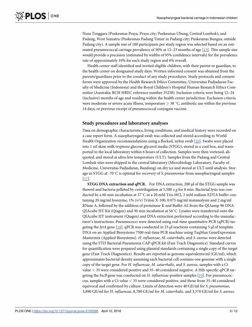
A total of 302 children were included in this study, with participant characteristics shown in
Table 1. Overall, pneumococcal carriage prevalence was 49.5% (95%CI 43.7–55.3), followed by
42.7% (95%CI 37.1–48.5) for M. catarrhalis, 27.5% (95%CI 22.5–32.9) for H. influenzae, and
7.3% (95%CI 4.6–10.8) for S. aureus. Carriage prevalence by region is shown in Fig 1. Carriage
differed significantly by region for S. pneumoniae and M. catarrhalis (p< 0.001 and p = 0.015,
respectively, chi-squared test). To evaluate whether differences in carriage prevalence were due
to differences in risk factors among regions, adjusted odds ratios (aOR) were determined by
logistic regression models that included income, the presence of two or more children under
five in the household, maternal education level, and presence of upper respiratory tract infec-
tion (URTI) symptoms. The difference in carriage prevalence of S. pneumoniae and M. catar-rhalis among regions remained significant. For S. pneumoniae, aOR compared to Padang
(reference) was 3.47 (95%CI 1.87–6.53) for Bandung and 1.78 (95%CI 0.93–3.42) for Central
Lombok (p = 0.0006, chi-squared test). For M. catarrhalis, aOR compared to Padang was 1.60
(95%CI 0.86–2.97) for Bandung and 2.60 (95%CI 1.36–4.97) for Central Lombok (p = 0.015,
chi-squared test).
A total of 164 pneumococci belonging to 32 capsular serotypes and one genetic lineage of
acapsular pneumococci (NT2) were identified in this study [25]. Five lytA-positive samples
were culture negative and therefore not serotyped, and one sample was excluded for technical
Nasopharyngeal bacterial carriage in Indonesian children
PLOS ONE | https://doi.org/10.1371/journal.pone.0195098 April 12, 2018 4 / 12
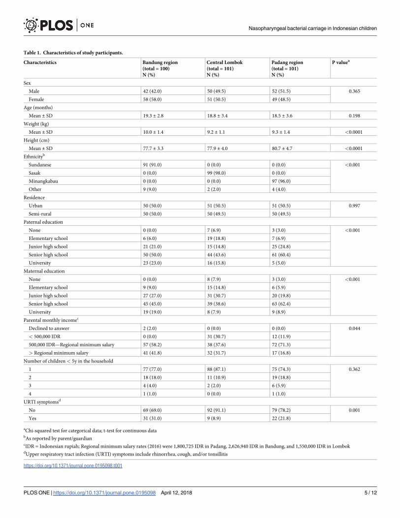
Table 1. Characteristics of study participants.
Characteristics Bandung region
(total = 100)
N (%)
Central Lombok
(total = 101)
N (%)
Padang region
(total = 101)
N (%)
P valuea
Sex
Male 42 (42.0) 50 (49.5) 52 (51.5) 0.365
Female 58 (58.0) 51 (50.5) 49 (48.5)
Age (months)
Mean ± SD 19.3 ± 2.8 18.8 ± 3.4 18.5 ± 3.6 0.198
Weight (kg)
Mean ± SD 10.0 ± 1.4 9.2 ± 1.1 9.3 ± 1.4 <0.0001
Height (cm)
Mean ± SD 77.7 ± 3.3 77.9 ± 4.0 80.7 ± 4.7 <0.0001
Ethnicityb
Sundanese 91 (91.0) 0 (0.0) 0 (0.0) <0.001
Sasak 0 (0.0) 99 (98.0) 0 (0.0)
Minangkabau 0 (0.0) 0 (0.0) 97 (96.0)
Other 9 (9.0) 2 (2.0) 4 (4.0)
Residence
Urban 50 (50.0) 51 (50.5) 51 (50.5) 0.997
Semi-rural 50 (50.0) 50 (49.5) 50 (49.5)
Paternal education
None 0 (0.0) 7 (6.9) 3 (3.0) <0.001
Elementary school 6 (6.0) 19 (18.8) 7 (6.9)
Junior high school 21 (21.0) 15 (14.8) 25 (24.8)
Senior high school 50 (50.0) 44 (43.6) 61 (60.4)
University 23 (23.0) 16 (15.8) 5 (5.0)
Maternal education
None 0 (0.0) 8 (7.9) 3 (3.0) <0.001
Elementary school 9 (9.0) 15 (14.8) 6 (5.9)
Junior high school 27 (27.0) 31 (30.7) 20 (19.8)
Senior high school 45 (45.0) 39 (38.6) 63 (62.4)
University 19 (19.0) 8 (7.9) 9 (8.9)
Parental monthly incomec
Declined to answer 2 (2.0) 0 (0.0) 0 (0.0) 0.044
< 500,000 IDR 0 (0.0) 31 (30.7) 12 (11.9)
500,000 IDR—Regional minimum salary 57 (58.2) 38 (37.6) 72 (71.3)
> Regional minimum salary 41 (41.8) 32 (31.7) 17 (16.8)
Number of children < 5y in the household
1 77 (77.0) 88 (87.1) 75 (74.3) 0.362
2 18 (18.0) 11 (10.9) 19 (18.8)
3 4 (4.0) 2 (2.0) 6 (5.9)
4 1 (1.0) 0 (0.0) 1 (1.0)
URTI symptomsd
No 69 (69.0) 92 (91.1) 79 (78.2) 0.001
Yes 31 (31.0) 9 (8.9) 22 (21.8)
aChi-squared test for categorical data; t-test for continuous databAs reported by parent/guardiancIDR = Indonesian rupiah; Regional minimum salary rates (2016) were 1,800,725 IDR in Padang, 2,626,940 IDR in Bandung, and 1,550,000 IDR in LombokdUpper respiratory tract infection (URTI) symptoms include rhinorrhea, cough, and/or tonsillitis
https://doi.org/10.1371/journal.pone.0195098.t001
Nasopharyngeal bacterial carriage in Indonesian children
PLOS ONE | https://doi.org/10.1371/journal.pone.0195098 April 12, 2018 5 / 12

issues. There were four pneumococci for which serotype could not be determined: three were
reported as 35A/10B-like by microarray (two of these were serogroup 33 by latex agglutina-
tion/Quellung whilst the third did not undergo phenotypic typing), and the fourth was 39/6C-
like by microarray and non-typeable by latex. Two of the six serotype 11A pneumococci iden-
tified were typed as 11F-like by microarray, as previously described [26]. Multiple serotype car-
riage was found in 18/296 (6.1%) of samples overall, and in 18/144 (12.5%) of pneumococcal-
positive samples. Serotypes 15B/C, 23F, NT2, 19F, and 6A were the most commonly identified.
Fig 2 depicts the 20 most common serotypes shown by region. Overall, 76/164 (46.3%) of
pneumococci belonged to PCV13 serotypes, and 72/296 (24.3%) of participants carried a
PCV13 serotype. For PCV10, these proportions were 53/164 (32.3%) and 52/296 (17.6%),
Fig 1. Nasopharyngeal carriage prevalence (%) of S. pneumoniae, H. influenzae, M. catarrhalis, and S. aureus in
Indonesian children aged 12–24 months. Results are shown by region (Bandung, Padang, and Lombok). Error bars
represent 95%CI.
https://doi.org/10.1371/journal.pone.0195098.g001
Fig 2. The twenty most common pneumococcal serotypes identified in nasopharyngeal swabs collected from
Indonesian children aged 12–24 months, shown by region (Bandung, Padang, and Lombok). � indicates serotypes
included in PCV10 and + indicates the additional three serotypes included in PCV13.
https://doi.org/10.1371/journal.pone.0195098.g002
Nasopharyngeal bacterial carriage in Indonesian children
PLOS ONE | https://doi.org/10.1371/journal.pone.0195098 April 12, 2018 6 / 12

respectively. Serotype distribution varied significantly by region, with 26/73 (36%) of pneumo-
cocci in Bandung, 28/53 (53%) in Central Lombok, and 22/38 (58%) in Padang belonging to
PCV13 serotypes (p = 0.043, chi-squared test). None of the 83 samples positive for H. influen-zae were type B; further typing of H. influenzae was not conducted.
Relationships between colonizing species were examined by calculating odds ratios (OR)
and aOR adjusting for income, the presence of two or more children under five in the house-
hold, maternal education level, and presence of URTI symptoms to minimize potential con-
founding. A positive association was observed between carriage of S. pneumoniae and M.
catarrhalis (OR 3.07 [95%CI 1.91–4.94], aOR 2.85 [95%CI 1.72–4.72]) and H. influenzae and
M. catarrhalis (OR 2.34 [95%CI 1.40–3.91], aOR 2.18 [95%CI 1.28–3.72]), and a negative asso-
ciation was found between M. catarrhalis and S. aureus (OR 0.06 [95%CI 0.01–0.43], aOR 0.06
[95%CI 0.01–0.50]). No significant associations were observed between S. pneumoniae and H.
influenzae (OR 1.58 [95%CI 0.95–2.65], aOR 1.64 [95%CI 0.95–2.83]), S. pneumoniae and S.
aureus (OR 0.49 [95%CI 0.19–1.24], aOR 0.58 [95%CI 0.22–1.52]), or H. influenzae and S.
aureus (OR 0.25 [95%CI 0.06–1.07], aOR 0.27 [95%CI 0.06–1.21]).
In children positive for carriage, the median density of carriage was 5.1 log10 GE/ml (range
2.2–7.3) for S. pneumoniae, 5.6 log10 GE/ml (range 3.6–7.8) for H. influenzae, 6.2 log10 GE/ml
(range 3.9–8.5) for M. catarrhalis, and 4.3 log10 GE/ml (range 3.6–7.0) for S. aureus. As seen in
Table 2, S. pneumoniae densities were higher when co-colonizing with H. influenzae or M. cat-arrhalis. Similarly, H. influenzae density was higher when it co-occurred with M. catarrhalis,and M. catarrhalis density was significantly higher when S. pneumoniae was a co-colonizer.
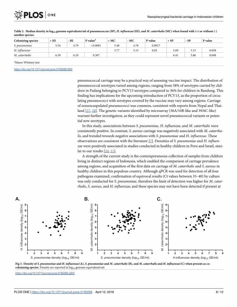
During co-colonization, densities of S. pneumoniae, H. influenzae, and M. catarrhalis were pos-
itively correlated (Fig 3): S. pneumoniae and H. influenzae Spearman ρ = 0.430, p = 0.002; S.
pneumoniae and M. catarrhalis Spearman ρ = 0.400, p = 0.0002; H. influenzae and M. catarrha-lis Spearman ρ = 0.571, p<0.0001. An opposite trend was observed for S. pneumoniae and S.
aureus, as the median carriage density of S. pneumoniae was 4.61 log10 GE/ml with S. aureuscompared to 5.18 log10 GE/ml without S. aureus, although the difference was not statistically
significant (p = 0.09, Mann-Whitney test).
Discussion
We examined nasopharyngeal carriage of four clinically-relevant bacterial pathogens in Indo-
nesian children living in three diverse regions. The carriage prevalence of H. influenzae and S.
aureus were similar among the three regions, whereas carriage of S. pneumoniae and M. catar-rhalis varied. The reasons for regional differences are unclear. Samples were collected during
the same time frame, limiting seasonal effects, and differences remained after adjusting for risk
factors related to socioeconomic status, exposure, and presence of URTI symptoms. Regional
differences in carriage may relate to other risk factors not assessed in this study, ethnic differ-
ences, or environmental differences. The overall pneumococcal carriage prevalence in this
study (49.5%) is consistent with previous carriage studies of children under five conducted in
Lombok (46%) and Semarang, Java (43%) [12, 14]. The 27.5% carriage rate of H. influenzae is
similar to the 32% prevalence previously reported in Lombok in children aged 0–24 months
[15]. In the 1998 Lombok study, carriage prevalence of Hib was 4.5%, whereas no Hib was
detected in the current study, likely due to widespread use of Hib vaccine following its intro-
duction in 2013. Carriage prevalence of S. aureus was low (7.3%), however this is expected as S.
aureus carriage is highest in neonates, older children and adults, and low in children aged 12–
24 months [8, 9].
Overall, 46% of pneumococci belonged to PCV13 serotypes. Due to limited serotyping data
available from invasive pneumococcal disease in Indonesia, monitoring serotype changes in
Nasopharyngeal bacterial carriage in Indonesian children
PLOS ONE | https://doi.org/10.1371/journal.pone.0195098 April 12, 2018 7 / 12
pneumococcal carriage may be a practical way of assessing vaccine impact. The distribution of
pneumococcal serotypes varied among regions, ranging from 58% of serotypes carried by chil-
dren in Padang belonging to PCV13 serotypes compared to 36% for children in Bandung. This
finding has implications for the upcoming introduction of PCV13, as the proportion of circu-
lating pneumococci with serotypes covered by the vaccine may vary among regions. Carriage
of nonencaspulated pneumococci was common, consistent with reports from Nepal and Thai-
land [27, 28]. The genetic variants identified by microarray (36A/10B-like and 39/6C-like)
warrant further investigation, as they could represent novel pneumococcal variants or poten-
tial new serotypes.
In this study, associations between S. pneumoniae, H. influenzae, and M. catarrhalis were
consistently positive. In contrast, S. aureus carriage was negatively associated with M. catarrha-lis, and trended towards negative associations with S. pneumoniae and H. influenzae. These
observations are consistent with the literature [7]. Densities of S. pneumoniae and H. influen-zae were positively associated in studies conducted in healthy children in Peru and Israel, simi-
lar to our results [10, 11].
A strength of the current study is the contemporaneous collection of samples from children
living in distinct regions of Indonesia, which enabled the comparison of carriage prevalence
among regions, and acquisition of the first data on carriage of M. catarrhalis and S. aureus in
healthy children in this populous country. Although qPCR was used for detection of all four
pathogens examined, confirmation of equivocal results (Ct values between 35–40) by culture
was only conducted for S. pneumoniae, therefore the limit of detection was higher for M. catar-rhalis, S. aureus, and H. influenzae, and these species may not have been detected if present at
Table 2. Median density in log10 genome equivalents/ml of pneumococcus (SP), H. influenzae (HI), and M. catarrhalis (MC) when found with (+) or without (-)
another species.
Colonizing species + HI - HI P valuea + MC - MC P value + SP - SP P value
S. pneumoniae 5.54 4.79 <0.0001 5.48 4.78 0.0017
H. influenzae 5.77 5.15 0.02 5.69 5.15 0.058
M. catarrhalis 6.39 6.10 0.367 6.41 5.86 0.048
aMann-Whitney test
https://doi.org/10.1371/journal.pone.0195098.t002
Fig 3. Density of S. pneumoniae and H. influenzae (A), S. pneumoniae and M. catarrhalis (B), and M. catarrhalis and H. influenzae (C) when present as co-
colonizing species. Density are reported in log10 genome equivalents/ml.
https://doi.org/10.1371/journal.pone.0195098.g003
Nasopharyngeal bacterial carriage in Indonesian children
PLOS ONE | https://doi.org/10.1371/journal.pone.0195098 April 12, 2018 8 / 12

low densities. Another limitation of the study is that respiratory viruses, an important compo-
nent of the microbial community of the upper respiratory tract, were not assessed [29].
Bacterial density, and presence of multiple pathogens, has been linked to disease in several
studies, however the mechanisms underlying these interactions are not well understood. In a
study on children under 10 years of age in Tanzania [30], density of S. pneumoniae, H. influen-zae, and M. catarrhalis was significantly higher in children with severe pneumonia compared
to those with mild UTRI, and carriage of multiple species was more common in children with
clinical pneumonia compared to those without respiratory infection. In Vietnamese children
under five, nasopharyngeal density of pneumococcus was higher in children with radiologi-
cally confirmed pneumonia compared to healthy controls or children with other lower respira-
tory tract infection [5]. In both the Tanzanian and Vietnamese studies, nasopharyngeal
sampling was conducted during acute illness and prior to antibiotic treatment. Co-coloniza-
tion of S. pneumoniae and H. influenzae was significantly associated with radiologically con-
firmed pneumonia, whereas co-colonization of H. influenzae and M. catarrhalis was associated
with other lower respiratory tract infection. S. pneumoniae, H. influenzae, and M. catarrhalisare the three most common bacterial causes of otitis media, which is increasingly recognized
as a polymicrobial disease [31]. Co-colonization of S. pneumoniae or H. influenzae with M. cat-arrhalis has been associated with increased risk of otitis media [32]. Using in vivo models,
mixed species biofilms have been found to increase persistence in ear disease [33]. Other pro-
posed mechanisms for positive associations between bacterial species include interspecies quo-
rum sensing and passive antimicrobial resistance, which have been observed in experimental
models of otitis media [34, 35]. Host susceptibility may also be an underlying factor for posi-
tive associations observed between certain bacterial species. For example, a study in Fiji found
that that co-colonization by multiple pathogens was associated with ethnicity [36].
Continued surveillance of pneumococcal carriage and serotype distribution is warranted in
Indonesia, especially in light of the upcoming introduction of pneumococcal conjugate vac-
cine, as our results suggest that vaccine impact may vary by region. Future studies on microbial
interactions in the respiratory tract, ideally including viruses as well as bacterial pathogens,
will help to shed light on the underlying mechanisms, as well as how nasopharyngeal microbi-
ology relates to the development of respiratory infections such as pneumonia and otitis media.
Acknowledgments
The authors thank the study participants and their families and the health center staff from the
six participating puskesmas. We acknowledge the laboratory team from the Microbiology Lab-
oratory at Universitas Padjadjaran for laboratory analyses, the Pneumococcal Research group
at Murdoch Children’s Research Institute for provision of latex serotyping reagents, and Dan
Belluoccio from Agilent Technologies and Kate Gould from BUGS Bioscience for microarray
technical support and advice. We thank the Integrated Laboratory, Faculty of Medicine, Uni-
versitas Indonesia in Jakarta, Indonesia for provision of laboratory space and equipment for
microarray analysis. We thank the Growth and Development clinical trial team from Hasan
Sadikin General Hospital for study support. The Murdoch Children’s Research Institute was
supported by the Victorian Government’s Operational Infrastructure Support Program.
Author Contributions
Conceptualization: Eileen M. Dunne, Chrysanti Murad, Sunaryati Sudigdoadi, Eddy
Fadlyana, Rodman Tarigan, Sang Ayu Kompiyang Indriyani, Emma Watts, Catherine
Satzke, Nurhandini Eka Dewi, Finny Fitry Yani, Kusnandi Rusmil, E. Kim Mulholland,
Cissy Kartasasmita.
Nasopharyngeal bacterial carriage in Indonesian children
PLOS ONE | https://doi.org/10.1371/journal.pone.0195098 April 12, 2018 9 / 12
Data curation: Jason Hinds.
Formal analysis: Eileen M. Dunne, Chrysanti Murad.
Investigation: Eileen M. Dunne, Chrysanti Murad, Sunaryati Sudigdoadi, Eddy Fadlyana,
Rodman Tarigan, Sang Ayu Kompiyang Indriyani, Casey L. Pell, Nurhandini Eka Dewi,
Finny Fitry Yani, Kusnandi Rusmil, Cissy Kartasasmita.
Project administration: Emma Watts.
Resources: Catherine Satzke, Jason Hinds.
Writing – original draft: Eileen M. Dunne, Chrysanti Murad, Casey L. Pell, Cissy
Kartasasmita.
Writing – review & editing: Eddy Fadlyana, Rodman Tarigan, Sang Ayu Kompiyang
Indriyani, Casey L. Pell, Emma Watts, Catherine Satzke, Jason Hinds, Finny Fitry Yani, E.
Kim Mulholland.
References1. O’Brien K, Wolfson L, Watt J, Henkle E, Delport S, Deloria-Knoll M, et al. Burden of disease caused by
Streptococcus pneumoniae in children younger than 5 years: global estimates. Lancet. 2009; 374:893–
902. https://doi.org/10.1016/S0140-6736(09)61204-6 PMID: 19748398
2. Adegbola RA, DeAntonio R, Hill PC, Roca A, Usuf E, Hoet B, et al. Carriage of Streptococcus pneumo-
niae and other respiratory bacterial pathogens in low and lower-middle income countries: a systematic
review and meta-analysis. PLoS ONE. 2014; 9(8):e103293. https://doi.org/10.1371/journal.pone.
0103293 PMID: 25084351
3. Mackenzie G, Leach A, Carapetis J, Fisher J, Morris P. Epidemiology of nasopharyngeal carriage of
respiratory bacterial pathogens in children and adults: cross-sectional surveys in a population with high
rates of pneumococcal disease. BMC Infect Dis. 2010; 10(1):304. https://doi.org/10.1186/1471-2334-
10-304 PMID: 20969800
4. Simell B, Auranen K, Kayhty H, Goldblatt D, Dagan R, O’Brien KL, et al. The fundamental link between
pneumococcal carriage and disease. Expert Rev Vaccines. 2012; 11. https://doi.org/10.1586/erv.12.53
PMID: 22913260
5. Vu HTT, Yoshida LM, Suzuki M, Nguyen HAT, Nguyen CDL, Nguyen ATT, et al. Association between
nasopharyngeal load of Streptococcus pneumoniae, viral coinfection, and radiologically confirmed
pneumonia in Vietnamese children. Pediatr Infect Dis J. 2011; 30(1):11–8 https://doi.org/10.1097/INF.
0b013e3181f111a2 PMID: 20686433
6. Short KR, Reading PC, Wang N, Diavatopoulos DA, Wijburg OL. Increased nasopharyngeal bacterial
titers and local inflammation facilitate transmission of Streptococcus pneumoniae. MBio. 2012; 3(5).
7. Dunne EM, Smith-Vaughan HC, Robins-Browne RM, Mulholland EK, Satzke C. Nasopharyngeal micro-
bial interactions in the era of pneumococcal conjugate vaccination. Vaccine. 2013; 31(19):2333–42.
https://doi.org/10.1016/j.vaccine.2013.03.024 PMID: 23523773
8. Regev-Yochay G, Dagan R, Raz M, Carmeli Y, Shainberg B, Derazne E, et al. Association between car-
riage of Streptococcus pneumoniae and Staphylococcus aureus in children. JAMA. 2004; 292(6):716–
20. https://doi.org/10.1001/jama.292.6.716 PMID: 15304469
9. Bogaert D, van Belkum A, Sluijter M, Luijendijk A, de Groot R, Rumke HC, et al. Colonisation by Strepto-
coccus pneumoniae and Staphylococcus aureus in healthy children. Lancet. 2004; 363(9424):1871–2.
https://doi.org/10.1016/S0140-6736(04)16357-5 PMID: 15183627
10. Lewnard JA, Huppert A, Givon-Lavi N, Pettigrew MM, Regev-Yochay G, Dagan R, et al. Density, sero-
type diversity, and fitness of Streptococcus pneumoniae in upper respiratory tract cocolonization with
nontypeable Haemophilus influenzae. J Infect Dis. 2016; 214(9):1411–20. https://doi.org/10.1093/
infdis/jiw381 PMID: 27540112
11. Chien Y-W, Vidal JE, Grijalva CG, Bozio C, Edwards KM, Williams JV, et al. Density Interactions
between Streptococcus pneumoniae, Haemophilus influenzae and Staphylococcus aureus in the naso-
pharynx of young Peruvian Children. Pediatr Infect Dis J. 2013; 32(1):72–7.
12. Farida H, Severin JA, Gasem MH, Keuter M, Wahyono H, van den Broek P, et al. Nasopharyngeal car-
riage of Streptococcus pneumoniae in pneumonia-prone age groups in Semarang, Java Island, Indone-
sia. PLoS ONE. 2014; 9(1):e87431. https://doi.org/10.1371/journal.pone.0087431 PMID: 24498104
Nasopharyngeal bacterial carriage in Indonesian children
PLOS ONE | https://doi.org/10.1371/journal.pone.0195098 April 12, 2018 10 / 12

13. Soewignjo S, Gessner BD, Sutanto A, Steinhoff M, Prijanto M, Nelson C, et al. Streptococcus pneumo-
niae nasopharyngeal carriage prevalence, serotype distribution, and resistance patterns among chil-
dren on Lombok Island, Indonesia. Clin Infect Dis. 2001; 32(7):1039–43. https://doi.org/10.1086/
319605 PMID: 11264032
14. Hadinegoro SR, Prayitno A, Khoeri MM, Djelantik IG, Dewi NE, Indriyani SA, et al. Nasopharyngeal car-
riage of Streptococcus pneumoniae in healthy children under five years old in Central Lombok Regency,
Indonesia. Southeast Asian J Trop Med Public Health. 2016; 47(3):485–93. PMID: 27405132
15. Gessner BD, Sutanto A, Steinhoff M, Soewignjo S, Widjaya A, Nelson C, et al. A population-based sur-
vey of Haemophilus influenzae type b nasopharyngeal carriage prevalence in Lombok Island, Indone-
sia. Pediatr Infect Dis J. 1998; 17(9):S179–S82.
16. Satzke C, Turner P, Virolainen-Julkunen A, Adrian PV, Antonio M, Hare KM, et al. Standard method for
detecting upper respiratory carriage of Streptococcus pneumoniae: updated recommendations from
the World Health Organization Pneumococcal Carriage Working Group. Vaccine. 2013; 32(1):165–79.
https://doi.org/10.1016/j.vaccine.2013.08.062 PMID: 24331112
17. O’Brien KL, Bronsdon MA, Dagan R, Yagupsky P, Janco J, Elliott J, et al. Evaluation of a medium
(STGG) for transport and optimal recovery of Streptococcus pneumoniae from nasopharyngeal secre-
tions collected during field studies. J Clin Microbiol. 2001; 39(3):1021–4. https://doi.org/10.1128/JCM.
39.3.1021-1024.2001 PMID: 11230421
18. Carvalho Mda G, Tondella ML, McCaustland K, Weidlich L, McGee L, Mayer LW, et al. Evaluation and
improvement of real-time PCR assays targeting lytA, ply, and psaA genes for detection of pneumococ-
cal DNA. J Clin Microbiol. 2007; 45(8):2460–6. https://doi.org/10.1128/JCM.02498-06 PMID: 17537936
19. Laboratory Methods for the Diagnosis of Meningitis Caused by Neisseria meningitidis, Streptococcus
pneumoniae, and Haemophilus influenzae. 2nd ed: WHO Press; 2011.
20. Satzke C, Dunne EM, Porter BD, Klugman KP, Mulholland EK, PneuCarriage project group. The Pneu-
Carriage Project: a multi-centre comparative study to identify the best serotyping methods for examining
pneumococcal carriage in vaccine evaluation studies. PLoS Med. 2015; 12. https://doi.org/10.1371/
journal.pmed.1001903 PMID: 26575033
21. Porter BD, Ortika BD, Satzke C. Capsular serotyping of Streptococcus pneumoniae by latex agglutina-
tion. J Vis Exp. 2014;(91):51747. https://doi.org/10.3791/51747 PMID: 25285991
22. Ortika BD, Habib M, Dunne EM, Porter BD, Satzke C. Production of latex agglutination reagents for
pneumococcal serotyping. BMC Res Notes. 2013; 6:49. https://doi.org/10.1186/1756-0500-6-49 PMID:
23379961
23. Habib M, Porter BD, Satzke C. Capsular serotyping of Streptococcus pneumoniae using the Quellung
reaction. J Vis Exp. 2014;(84):e51208. https://doi.org/10.3791/51208 PMID: 24637727
24. van Selm S, van Cann LM, Kolkman MAB, van der Zeijst BAM, van Putten JPM. Genetic Basis for the
structural difference between Streptococcus pneumoniae serotype 15B and 15C capsular polysaccha-
rides. Infect Immunity. 2003; 71(11):6192–8. https://doi.org/10.1128/iai.71.11.6192-6198.2003
25. Salter SJ, Hinds J, Gould KA, Lambertsen L, Hanage WP, Antonio M, et al. Variation at the capsule
locus, cps, of mistyped and non-typable Streptococcus pneumoniae isolates. Microbiol. 2012; 158
(6):1560–9. https://doi.org/10.1099/mic.0.056580-0
26. Manna S, Ortika BD, Dunne EM, Holt KE, Kama M, Russell FM, et al. A novel genetic variant of Strepto-
coccus pneumoniae serotype 11A discovered in Fiji. Clin Microbiol Infection. 2017. https://doi.org/10.
1016/j.cmi.2017.06.031 PMID: 28736074
27. Turner P, Turner C, Jankhot A, Helen N, Lee SJ, Day NP, et al. A longitudinal study of Streptococcus
pneumoniae carriage in a cohort of infants and their mothers on the Thailand-Myanmar border. PLoS
ONE. 2012; 7(5):e38271. https://doi.org/10.1371/journal.pone.0038271 PMID: 22693610
28. Kandasamy R, Gurung M, Thapa A, Ndimah S, Adhikari N, Murdoch DR, et al. Multi-serotype pneumo-
coccal nasopharyngeal carriage prevalence in vaccine naïve Nepalese children, assessed using molec-
ular serotyping. PLoS ONE. 2015; 10(2):e0114286. https://doi.org/10.1371/journal.pone.0114286
PMID: 25643355
29. Bosch AATM, Biesbroek G, Trzcinski K, Sanders EAM, Bogaert D. Viral and bacterial interactions in the
upper respiratory tract. PLoS Pathogens. 2013; 9(1):e1003057. https://doi.org/10.1371/journal.ppat.
1003057 PMID: 23326226
30. Chochua S, D’Acremont V, Hanke C, Alfa D, Shak J, Kilowoko M, et al. Increased nasopharyngeal den-
sity and concurrent carriage of Streptococcus pneumoniae, Haemophilus influenzae, and Moraxella
catarrhalis are associated with pneumonia in febrile children. PLoS ONE. 2016; 11(12):e0167725.
https://doi.org/10.1371/journal.pone.0167725 PMID: 27907156
31. Vergison A. Microbiology of otitis media: A moving target. Vaccine. 2008; 26(Supplement 7):G5–G10.
https://doi.org/10.1016/j.vaccine.2008.11.006 PMID: 19094935
Nasopharyngeal bacterial carriage in Indonesian children
PLOS ONE | https://doi.org/10.1371/journal.pone.0195098 April 12, 2018 11 / 12
32. Ruohola A, Pettigrew MM, Lindholm L, Jalava J, Raisanen KS, Vainionpaa R, et al. Bacterial and viral
interactions within the nasopharynx contribute to the risk of acute otitis media. J Infect. 2013; 66
(3):247–54. https://doi.org/10.1016/j.jinf.2012.12.002 PMID: 23266462
33. Perez AC, Pang B, King LB, Tan L, Murrah KA, Reimche JL, et al. Residence of Streptococcus pneu-
moniae and Moraxella catarrhalis within polymicrobial biofilm promotes antibiotic resistance and bacte-
rial persistence in vivo. Pathog Dis. 2014; 70(3):280–8. https://doi.org/10.1111/2049-632X.12129
PMID: 24391058
34. Armbruster CE, Hong W, Pang B, Weimer KED, Juneau RA, Turner J, et al. Indirect pathogenicity of
Haemophilus influenzae and Moraxella catarrhalis in polymicrobial otitis media occurs via interspecies
quorum signaling. mBio. 2010; 1(3):e00102-10-e-19. https://doi.org/10.1128/mBio.00102-10 PMID:
20802829
35. Weimer KED, Juneau RA, Murrah KA, Pang B, Armbruster CE, Richardson SH, et al. Divergent mecha-
nisms for passive pneumococcal resistance to β-Lactam antibiotics in the presence of Haemophilus
influenzae. J Infect Dis. 2011; 203(4):549–55. https://doi.org/10.1093/infdis/jiq087 PMID: 21220774
36. Dunne EM, Manning J, Russell FM, Robins-Browne RM, Mulholland EK, Satzke C. Effect of pneumo-
coccal vaccination on nasopharyngeal carriage of Streptococcus pneumoniae, Haemophilus influen-
zae, Moraxella catarrhalis, and Staphylococcus aureus in Fijian children. J Clin Microbiol. 2012; 50
(3):1034–8. https://doi.org/10.1128/JCM.06589-11 PMID: 22170924
Nasopharyngeal bacterial carriage in Indonesian children
PLOS ONE | https://doi.org/10.1371/journal.pone.0195098 April 12, 2018 12 / 12
Related Documents