Taylor Tirahardjo Lower Limb Arterial Ultrasound

Welcome message from author
This document is posted to help you gain knowledge. Please leave a comment to let me know what you think about it! Share it to your friends and learn new things together.
Transcript
Taylor Tirahardjo
Lower Limb Arterial Ultrasound

Learning Objectives
3. RISK FACTORS 1. ANATOMY
7. DOCUMENTATION
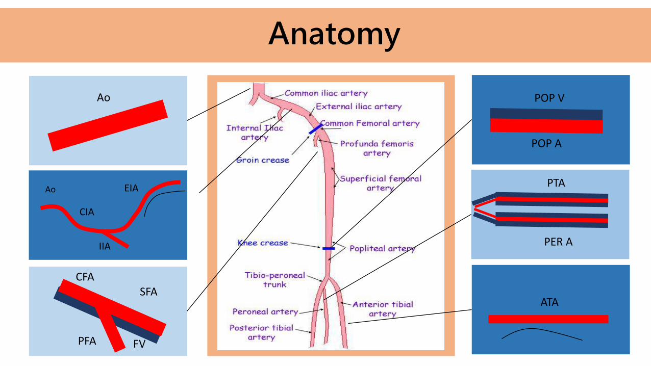
2. PATHOLOGY
5. SCAN TECHNIQUE 6. WHAT TO LOOK FOR
4. CLINICAL INDICATIONS
8. CASES
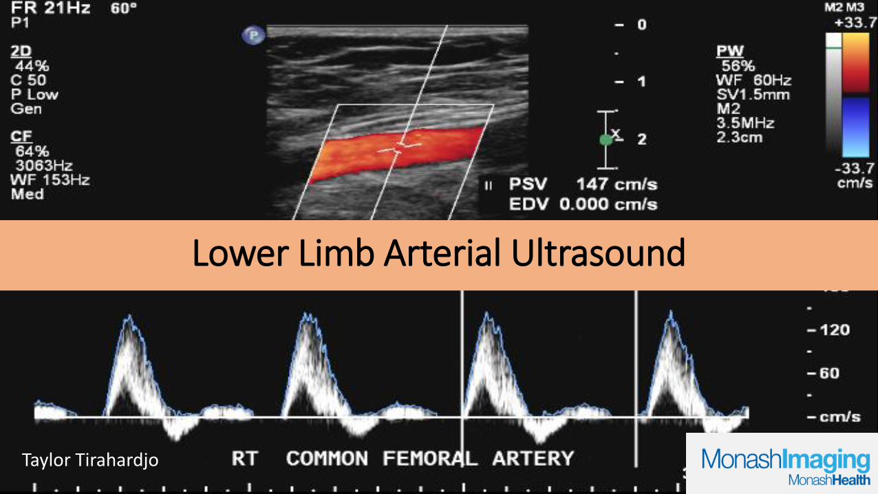
Anatomy
Ao POP V
POP A
PTA
PER A
ATA
Ao
CIA
IIA
EIA
FV
SFACFA
PFA FV
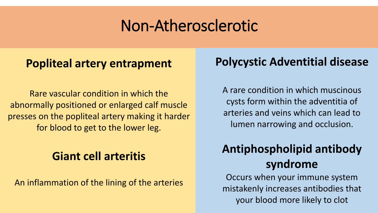
• Popliteal artery entrapment• Giant cell arteritis• Antiphospholipid antibody
syndrome• Polycystic Adventitial
disease
ATHEROSCLEROTIC
NON-ATHEROSCLEROTIC
• Stenosis/occlusion• Aneurysm• Embolism
Pathology

Over time fatty deposits (plaque) made of cholesterol and inflammatory cells also build up at the site and harden and narrow
the artery.
Atherosclerosis
The exact cause is unknown however it is a slow and complex process.
Can begin as early as childhood and progresses more rapidly with age.
Damage to the endothelium – inner lining of the artery.
Blood cells and other substances clump to the injury site.

Atherosclerosis
• Embolic event• If the lining covering the plaque ruptures
then it stimulates blood clot formation.
• With the high velocities in arteries this clot easily flicks off and enters the blood stream – this can then lodge in a new location and block the artery – like brain –stroke.
• Aneurysm • Caused by weakened artery walls.
• Usually atherosclerosis or high blood pressure.
Non-Atherosclerotic
Popliteal artery entrapment
Rare vascular condition in which the abnormally positioned or enlarged calf muscle
presses on the popliteal artery making it harder for blood to get to the lower leg.
Giant cell arteritis
An inflammation of the lining of the arteries
Polycystic Adventitial disease
A rare condition in which muscinouscysts form within the adventitia of
arteries and veins which can lead to lumen narrowing and occlusion.
Antiphospholipid antibody syndrome
Occurs when your immune system mistakenly increases antibodies that
your blood more likely to clot
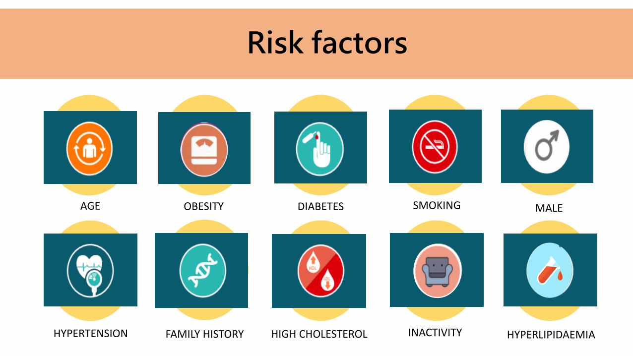
Risk factors
SMOKINGDIABETESAGE OBESITY MALE
HYPERTENSION FAMILY HISTORY HIGH CHOLESTEROL INACTIVITY HYPERLIPIDAEMIA

ClinicalIndications
Leg or foot wounds that are slow to heal.
Unexplained leg pain or cramping especially during exercise or walking.
Skin problems or discoloration on legs and feet.
Poor nail growth.

ClinicalIndications
Reduced peripheral pulses- cold/numb.
Acute or chronic ischemia.
AAA- ?popliteal aneursym
Embolic event to distal vessels.
Monitoring of disease and intervention-angioplasty, stents, by pass grafts.

ScanningGetting Started
Clear patient history
Transducer• Curvi-linear (C5-1MHz) for aorta and iliacs
• Linear (9-3MHz) for legs
Gel and towel
Triangle sponge for iliacs
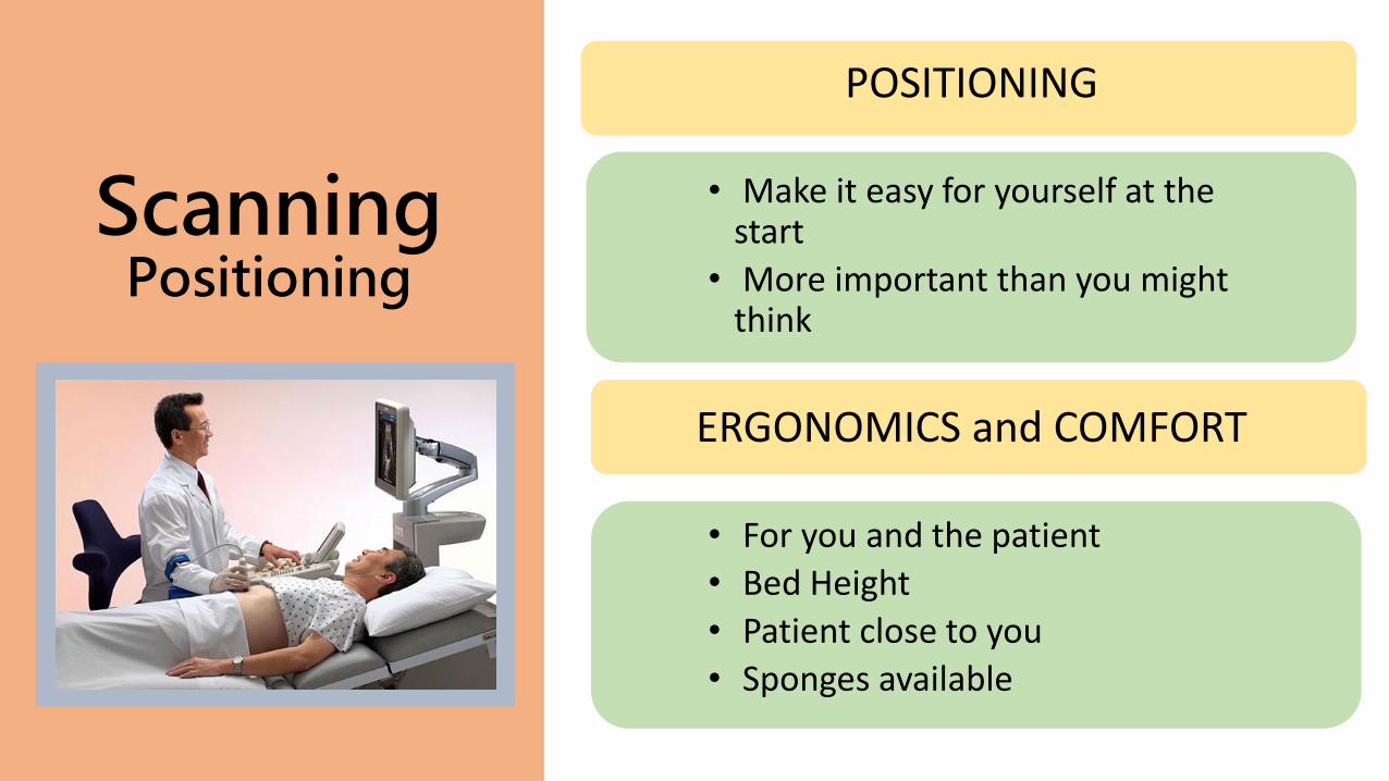
ScanningPositioning
POSITIONING
• Make it easy for yourself at the start
• More important than you might think
ERGONOMICS and COMFORT
• For you and the patient
• Bed Height
• Patient close to you
• Sponges available

ScanningDistal Aorta
Supine
Arms by side
Sustained compression
AoIVC
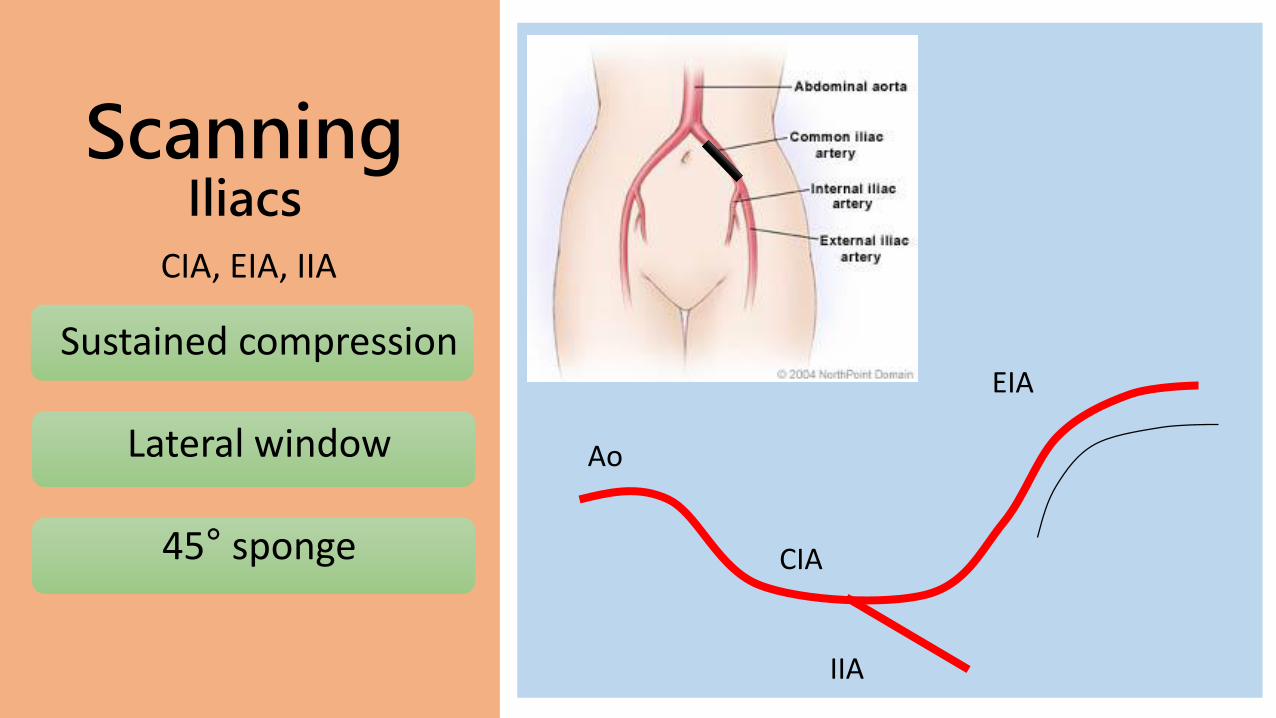
ScanningIliacs
CIA, EIA, IIA
Sustained compression
Lateral window
45° sponge
Ao
CIA
IIA
EIA

ScanningFemoral arteries
CFA, SFA, PFA
Start at groin crease
Turned out leg
Medial window
FV
SFA
CFA
PFA FV
SFA
FV

ScanningPopliteal Artery
POP A, TPT
Patient on side
Posterior window
POP A
TPT
PTA
PER AATA

ScanningCalf Arteries
(medial)PTA, PER A
Patient on side
Medial/DVT window
TPT
PTA
PER A
PTA
PER A
F
T

ScanningCalf Arteries
(lateral)ATA, DPA
Lateral window
ATA
DPA
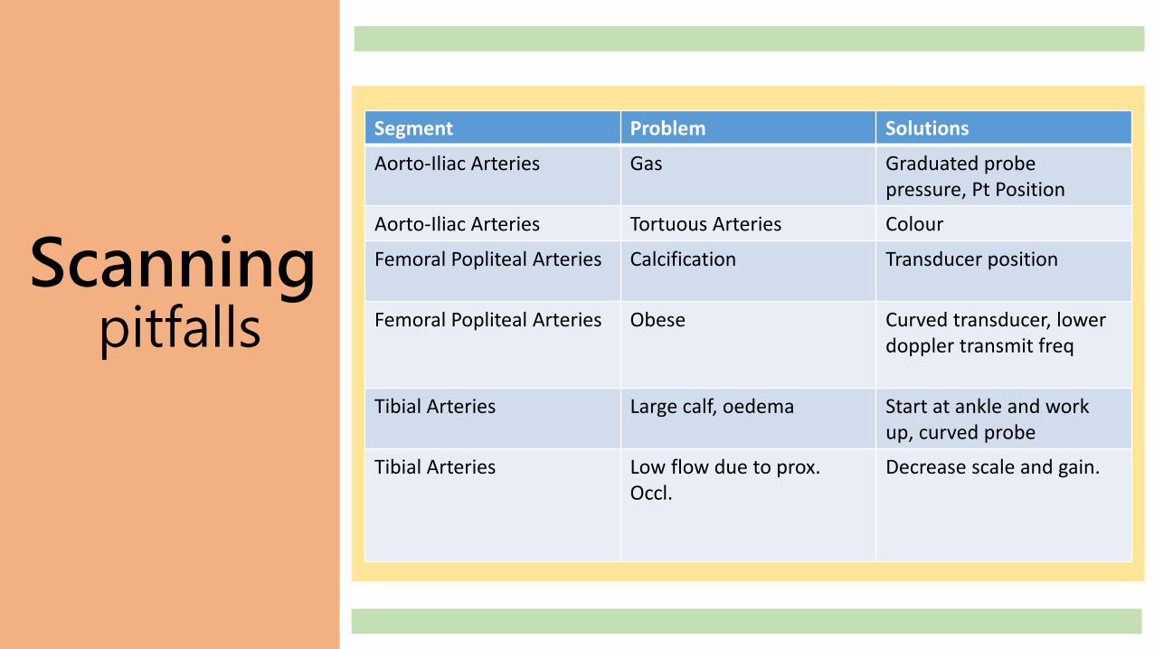
Segment Problem Solutions
Aorto-Iliac Arteries Gas Graduated probepressure, Pt Position
Aorto-Iliac Arteries Tortuous Arteries Colour
Femoral Popliteal Arteries Calcification Transducer position
Femoral Popliteal Arteries Obese Curved transducer, lower doppler transmit freq
Tibial Arteries Large calf, oedema Start at ankle and work up, curved probe
Tibial Arteries Low flow due to prox. Occl.
Decrease scale and gain.
Scanningpitfalls

What we look for
Assess from distal aorta- ankle
Locate and quantify arterial diseaseB-mode, color and spectral
Document on worksheet any
velocity increases, narrowing
or occlusion.
Highlight limitations

• B-mode• Anatomy• Plaque/ calcification
• Colour Doppler• Calibre• Aliasing
• Spectral Doppler• PSV• Waveform
Interpretation

Normal

Stenosis - B-mode and Colour Doppler

StenosisSpectral
At stenosis
↑ PSV
Spectral broadening
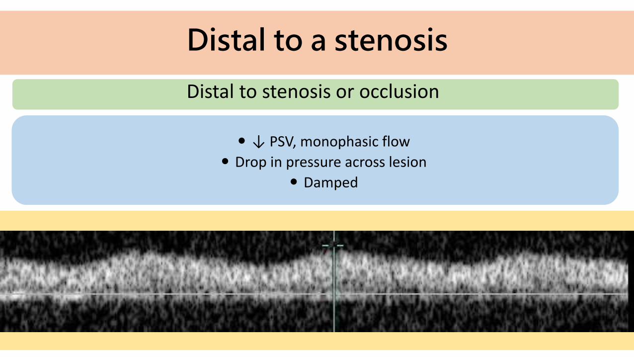
Distal to a stenosis
Distal to stenosis or occlusion
↓ PSV, monophasic flow
Drop in pressure across lesion
Damped

Diagnosing Stenoses
Velocity Criteria
PRE STENSOSIS
AT STENSOSIS
50-75%
>75%
OR
OR
Ratio 2:1-4:1
Ratio >4:1
PSV >200cm/s
PSV > 400cm/s

RecognisingOcclusion
At Occlusion
No flow
Possible collaterals
Distal recanalization
COLLATERALS
If an arterial segment is severely diseased or occluded, there are often alternative pathways that are able to carry blood around the diseased segment.
In some situations, reverse flow is observed in major branches of arteries just distal to an occlusion, where it may help resupply blood to the rest of the leg.
In chronic situations the body may create new pathways to recanalize a distal vessel.
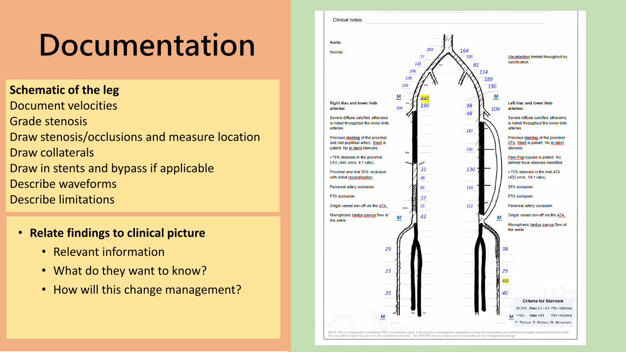
Documentation
• Relate findings to clinical picture
• Relevant information
• What do they want to know?
• How will this change management?
Schematic of the legDocument velocitiesGrade stenosisDraw stenosis/occlusions and measure locationDraw collaterals Draw in stents and bypass if applicableDescribe waveformsDescribe limitations

Summary
• Know the anatomy and pathology
• History
• Good technique
• Manage time
• Document well
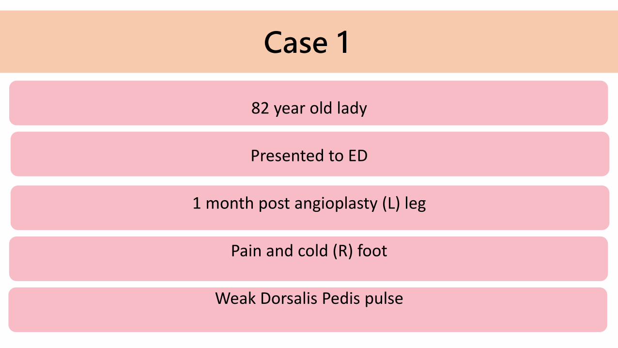
Case 1
82 year old lady
Presented to ED
1 month post angioplasty (L) leg
Pain and cold (R) foot
Weak Dorsalis Pedis pulse
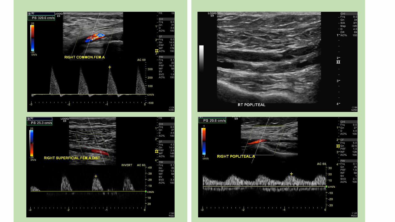
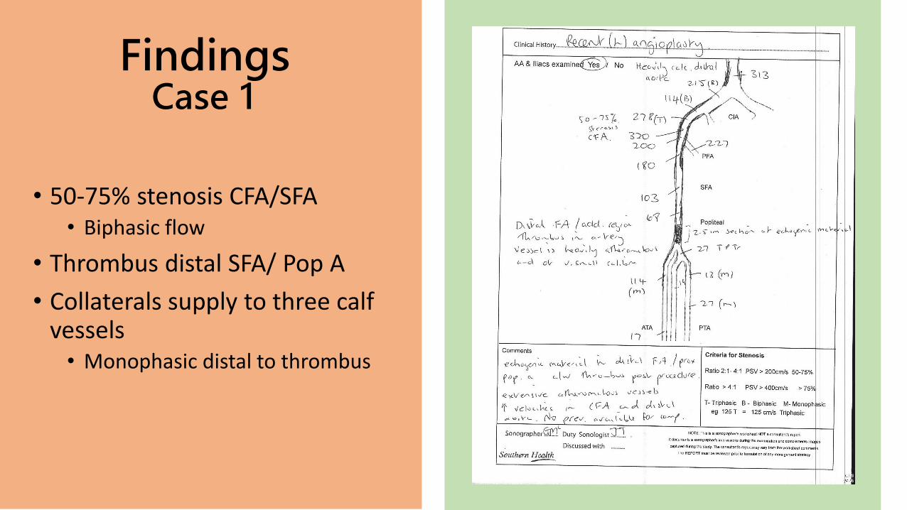
FindingsCase 1
• 50-75% stenosis CFA/SFA• Biphasic flow
• Thrombus distal SFA/ Pop A
• Collaterals supply to three calf vessels• Monophasic distal to thrombus
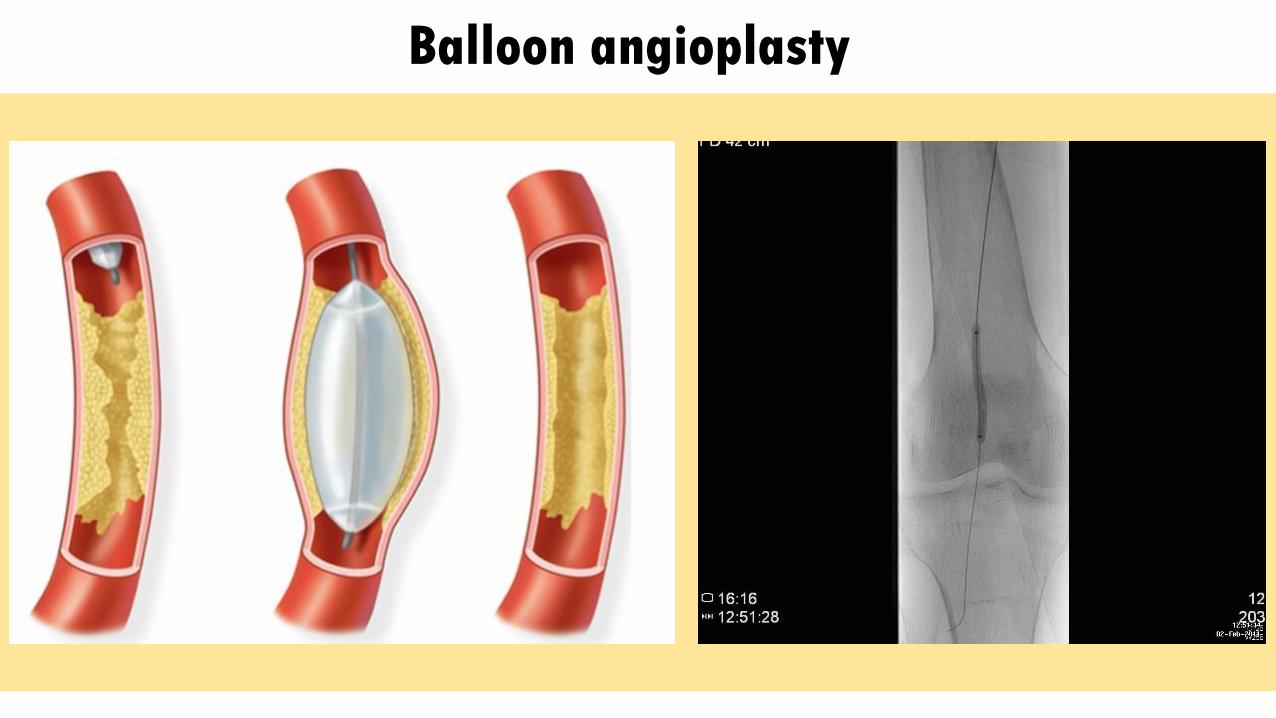
Balloon angioplasty


Case 2
93 year old male (Fit)
Hx of Lt-Rt Fem-Fem bypass graft (2007)
Bilateral mixed venous and arterial ulcers
R>L, worsening, painful
? Arterial insult
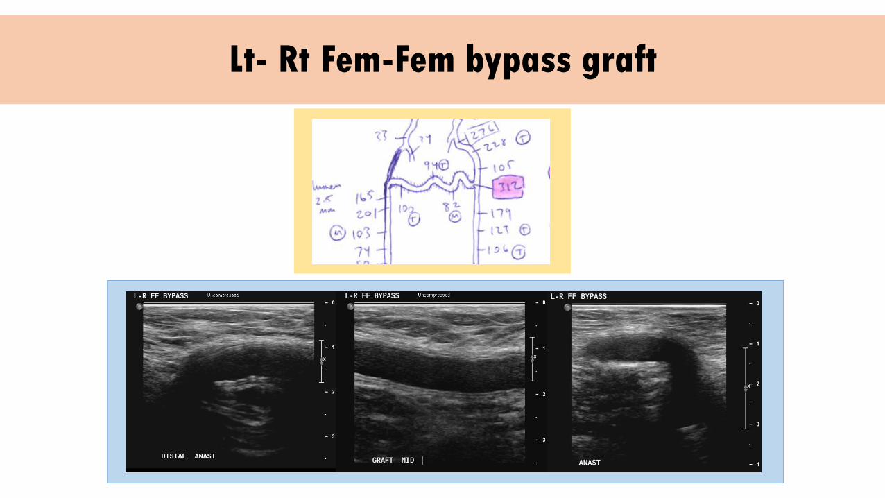
Lt- Rt Fem-Fem bypass graft

? Stenosis
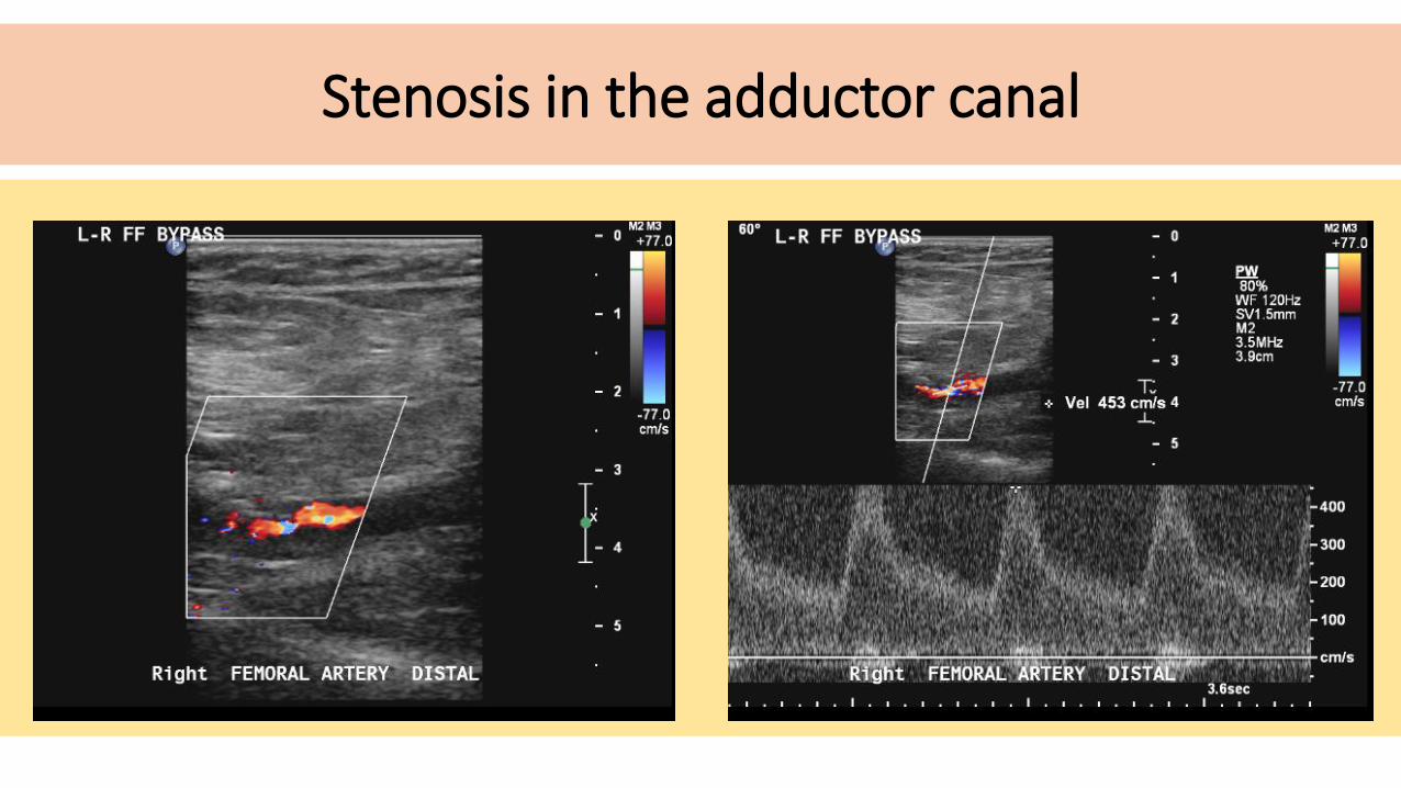
Stenosis in the adductor canal

Case 3
18 year old female presented with Achilles and calf pain with exercise
Right > left
Normal MRI and X-rays
Non-smoker
Not diabetic

Normal leg artery ultrasound
3 vessel run off
Triphasic waveforms

Ankle – Brachial Index
• Objective test for presence of PAD
• Ratio of BP from Arm/leg
•Eg Brachial 150, Ankle 110
110/150 = 0.73
• Rest and Exercise
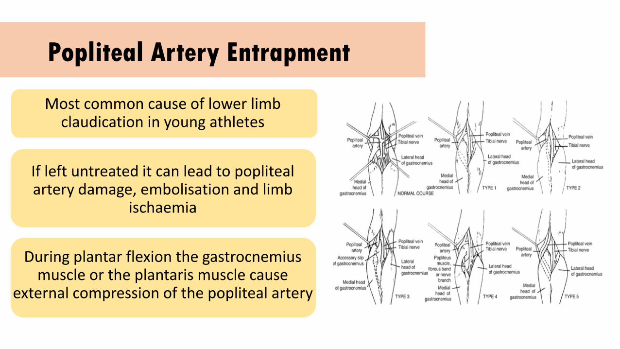
Popliteal Artery Entrapment
Most common cause of lower limb claudication in young athletes
If left untreated it can lead to popliteal artery damage, embolisation and limb
ischaemia
During plantar flexion the gastrocnemius muscle or the plantaris muscle cause
external compression of the popliteal artery

• a) Popliteal artery longitudinal duplex scan in neutral position
• b) Same popliteal longitudinal scan with plantar flexion
• c) Reactive hyperaemic response following resumption of normal position

THANKS FOR LISTENING
Related Documents