mechanism for mutations to be accommo- dated during the coevolution of high-af- finity binding partners. REFERENCES AND NOTES ___________________________ 1. J. Janin and C. Chothia, J. Biol. Chem. 265, 16027 (1990); S. Jones and J. Thornton, Proc. Natl. Acad. Sci. U.S.A. 93, 13 (1996). 2. J. A. Wells and A. M. De Vos, Annu. Rev. Biochem. 65, 609 (1996). 3. A. M. De Vos, M. Ultsch, A. A. Kossiakoff, Science 255, 306 (1992); M. Sundstrom et al., J. Biol. Chem. 271, 32197 (1996). The wild-type complex coordi- nates used in our study are from a 1:1 complex between G120R-hGH and hGHbp determined at 2.6 Å resolution ( T. Clackson, M. Ultsch, J. Wells, A. M. De Vos, in preparation). 4. B. C. Cunningham and J. A. Wells, Science 244, 1081 (1989); J. Mol. Biol. 234, 554 (1993). 5. S. H. Bass, M. G. Mulkerrin, J. A. Wells, Proc. Natl. Acad. Sci. U.S.A. 88, 4498 (1991); T. Clackson and J. A. Wells, Science 267, 383 (1995). 6. The single letter abbreviations for amino acid resi- dues are as follows: A, Ala; C, Cys; D, Asp; E, Glu; F, Phe; G, Gly; H, His; I, Ile; K, Lys; L, Leu; M, Met; N, Asn; P, Pro; Q, Gln; R, Arg; S, Ser; T, Thr; V, Val; W, Trp; and Y, Tyr. Mutants are identified by the wild- type residue, followed by its position, and the mutant residue. Multiple mutants are indicated by a series of single mutants separated by commas. 7. The library was constructed by site-directed mu- tagenesis [ T. A. Kunkel, K. Bebenek, J. McClary, Methods Enzymol. 204, 125 (1991)] by substituting NNS (N 5 A, G, C, or T; S 5 G or C) for the codon to be randomized. The library that randomized hGH at codons 168, 171, 172, 175, and 176 was made by mutagenizing the plasmid pH0753 with the oligonu- cleotide 59-CTG CTC TAC TGC T TC AGG NNS GAC ATG NNS NNS GTC GAG NNS NNS CT T CGA ATC GTG CAG TGC CGC TCT-39. The initial library size was 2 3 10 7 . The plasmid pH0753 was derived from phGHam-g3 [H. B. Lowman, S. H. Bass, N. Simp- son, J. A. Wells, Biochemistry 30, 10832 (1991)] by mutation of the base at position 419 from C to G in order to eliminate an Xba I restriction site. 8. Phage displaying mutants of hGH were selected for binding to W104A-hGHbp immobilized on an immu- nosorb plate (at a concentration of 50 mg/ml) by incubating overnight at 4°C in phosphate-buffered saline solution. Sorting against W104A-hGHbp was continued for seven rounds before individual phage- mids were sequenced (9). 9. S. Atwell and J. Wells, in preparation. 10. B. C. Cunningham, D. L. Lowe, B. Li, B. D. Bennett, J. A. Wells, EMBO J. 13, 2508 (1994); B. Li et al., Science 270, 1657 (1995). 11. H. B. Lowman and J. A. Wells, J. Mol. Biol. 234, 564 (1993). 12. A1-hGH and the single-site revertants were con- structed by mutagenizing pB0720, a G120R-hGH expression plasmid (7 ). These were expressed as soluble proteins [C. N. Chang, M. Rey, B. Bochner, H. Heyneker, G. Gray, Gene 55, 189 (1987)] in the strain 34B8, a nonsuppressor strain of Escherichia coli. Each mutant was isolated from a 50-ml culture in 250-ml shake flasks by pelleting the cells and freezing them at –20°C overnight. Cells were thawed and osmotically shocked in 10 mM tris (pH 8.0). The concentration of hormone was determined by den- sitometry of Coomassie blue–stained gels (4). Initial characterization of binding was measured on a BIA- core instrument by coupling W104A-hGHbp through lysine residues to 2500 resonance units [(4); B. Johnsson, S. Lofas, G. Lindquist, Anal. Biochem. 198, 268 (1991)]. Radioimmunoassay was per- formed for binding of hGH variants to hGHbp or W104A-hGHbp as described [S. A. Spencer et al., J. Biol. Chem. 263, 7862 (1988)]. In the case of wild-type hGHbp, 125 I-labeled hGH was used as competitor; for W104A-hGHbp, an affinity-optimized variant of hGH (11) labeled with 125 I was used. This variant bound with an affinity of 1.6 (60.7) nM to W104A-hGHbp. Standard errors in the K d measure- ments ranged from 630 to 650% of the value indi- cated in the text and were higher near the detection limits of the assay (;1 mM). 13. We purified A1-hGH and W104A-hGHbp from an E. coli fermentation culture using ion exchange and hy- drophobic interaction columns. The two proteins were combined and purified as a complex on a gel filtration column. Crystals were induced by streak seeding with crystals of the F25A, Y42A, Q46A- hGH:hGHbp complex (15)(;5 mg/ml) in 50 mM bis-tris (pH 6.5) and 17 to 20% saturated ammonium sulfate. Crystals were frozen in liquid nitrogen for data collection. 14. A. E. Ericksson et al., Science 255, 178 (1992); A. E. Ericksson, W. A. Baase, B. W. Matthews, J. Mol. Biol. 229, 747 (1993). 15. K. H. Pearce, M. H. Ultsch, R. F. Kelley, A. M. De Vos, J. A. Wells, Biochemistry 35, 10300 (1996). 16. W. Somers, M. Ultsch, A. M. De Vos, A. A. Kossia- koff, Nature 372, 478 (1994); M. R. Walter et al., ibid. 376, 230 (1995); O. Livnah et al., Science 273, 464 (1996). 17. F. M. Richards and W. A. Lim, Q. Rev. Biophys. 26, 423 (1994). 18. I. R. Vetter et al., Protein Sci. 5, 2399 (1996). 19. L. J. Keefe, J. Sondek, D. Shortle, E. E. Lattman, Proc. Natl. Acad. Sci. U.S.A. 90, 3275 (1993). 20. Z. Otwinowski, in Data Collection and Processing, L. Sawyer, N. Isaacs, S. Bailey, Eds. (SERC Daresbury Laboratory, Warrington, UK, 1993), p. 56 – 62. 21. A. T. Brunger, J. Kuriyan, M. Karplus, Science 235, 458 (1987). 22. G. Murshudov, A. Vagin, E. Dodson, Acta Crystal- logr. D 53, 240 (1997). 23. S.A. was supported in part by a NIH Biotechnology Training Grant. We thank the oligonucleotide synthe- sis and fermentation groups at Genentech; T. Clack- son for providing samples of hGHbp and W104A- hGHbp; M. Randal and T. Kossiakoff for help with x-ray data collection; and W. DeLano for help with intermolecular distance calculations. X-ray coordi- nates have been deposited in the Protein Data Bank with access number 1A XI. 20 March 1997; accepted 19 September 1997 A Low-Barrier Hydrogen Bond in the Catalytic Triad of Serine Proteases? Theory Versus Experiment Elissa L. Ash, James L. Sudmeier, Edward C. De Fabo, William W. Bachovchin* Cleland and Kreevoy recently advanced the idea that a special type of hydrogen bond (H-bond), termed a low-barrier hydrogen bond (LBHB), may account for the “missing” transition state stabilization underlying the catalytic power of many enzymes, and Frey et al. have proposed that the H-bond between aspartic acid 102 and histidine 57 in the catalytic triad of serine proteases is an example of a catalytically important LBHB. Experimental facts are here considered regarding the aspartic acid– histidine and cis– urocanic H-bonds that are inconsistent with fundamental tenets of the LBHB hypothesis. The inconsistencies between theory and experiment in these paradigm systems cast doubt on the existence of LBHBs, as currently defined, within enzyme active sites. The H-bond inherently involves the shar- ing of hydrogen atoms to varying extents with other atoms (1). This sharing is often depicted as a chemical equilibration or res- onance hybridization of structures such as 1 and 2 (Eq. 1). Proton sharing can also be depicted as a lengthening of the A-H bond of the donor, 1, as if the proton were in an intermediate stage of transfer to B (2). In conventional H-bonds the H atom is asso- ciated more with one heteroatom than the other. LBHBs are distinguished from conven- tional H-bonds by equal proton sharing be- tween the heteroatoms. LBHBs can be dou- ble-welled, depicted as 1 and 2 contributing equally to the system; or single-welled, de- picted as a single structure with the proton residing at a point equidistant between A and B, as in 3 (Eq. 1). LBHBs were first observed in the gas phase (1, 3), where evidence for their exis- tence and strength is persuasive. Schowen first proposed that LBHBs may exist within the protected interior of proteins (4). Pro- motion of this idea by Cleland and Kreevoy (5) and Frey et al. (6) has led to its substan- tial (7), though not universal (8), accep- tance. Physicochemical parameters used to identify LBHBs include (i) extreme low-field 1 H nuclear magnetic resonance (NMR) chemical shifts [d. 16 parts per million (ppm)]; (ii) deuterium isotope effects on low-field 1 H resonances; (iii) low (,1.0) isotopic fractionation factors; and (iv) deu- terium isotope effects on infrared and Raman frequencies (1)—the most unambiguous of E. L. Ash, J. L. Sudmeier, W. W. Bachovchin, Depart- ment of Biochemistry, Tufts University School of Medi- cine, Boston, MA 02111, USA. E. C. De Fabo, Department of Dermatology, George Washington University School of Medicine, Washington, DC 20037, USA. * To whom correspondence should be addressed. SCIENCE z VOL. 278 z 7 NOVEMBER 1997 z www.sciencemag.org 1128 on September 22, 2015 www.sciencemag.org Downloaded from on September 22, 2015 www.sciencemag.org Downloaded from on September 22, 2015 www.sciencemag.org Downloaded from on September 22, 2015 www.sciencemag.org Downloaded from on September 22, 2015 www.sciencemag.org Downloaded from

Welcome message from author
This document is posted to help you gain knowledge. Please leave a comment to let me know what you think about it! Share it to your friends and learn new things together.
Transcript

mechanism for mutations to be accommo-dated during the coevolution of high-af-finity binding partners.
REFERENCES AND NOTES___________________________
1. J. Janin and C. Chothia, J. Biol. Chem. 265, 16027(1990); S. Jones and J. Thornton, Proc. Natl. Acad.Sci. U.S.A. 93, 13 (1996).
2. J. A. Wells and A. M. De Vos, Annu. Rev. Biochem.65, 609 (1996).
3. A. M. De Vos, M. Ultsch, A. A. Kossiakoff, Science255, 306 (1992); M. Sundstrom et al., J. Biol. Chem.271, 32197 (1996). The wild-type complex coordi-nates used in our study are from a 1:1 complexbetween G120R-hGH and hGHbp determined at 2.6Å resolution ( T. Clackson, M. Ultsch, J. Wells, A. M.De Vos, in preparation).
4. B. C. Cunningham and J. A. Wells, Science 244,1081 (1989); J. Mol. Biol. 234, 554 (1993).
5. S. H. Bass, M. G. Mulkerrin, J. A. Wells, Proc. Natl.Acad. Sci. U.S.A. 88, 4498 (1991); T. Clackson andJ. A. Wells, Science 267, 383 (1995).
6. The single letter abbreviations for amino acid resi-dues are as follows: A, Ala; C, Cys; D, Asp; E, Glu; F,Phe; G, Gly; H, His; I, Ile; K, Lys; L, Leu; M, Met; N,Asn; P, Pro; Q, Gln; R, Arg; S, Ser; T, Thr; V, Val; W,Trp; and Y, Tyr. Mutants are identified by the wild-type residue, followed by its position, and the mutantresidue. Multiple mutants are indicated by a series ofsingle mutants separated by commas.
7. The library was constructed by site-directed mu-tagenesis [T. A. Kunkel, K. Bebenek, J. McClary,Methods Enzymol. 204, 125 (1991)] by substitutingNNS (N 5 A, G, C, or T; S 5 G or C) for the codon tobe randomized. The library that randomized hGH atcodons 168, 171, 172, 175, and 176 was made bymutagenizing the plasmid pH0753 with the oligonu-cleotide 59-CTG CTC TAC TGC TTC AGG NNS GACATG NNS NNS GTC GAG NNS NNS CTT CGA ATCGTG CAG TGC CGC TCT-39. The initial library sizewas 2 3 107. The plasmid pH0753 was derived fromphGHam-g3 [H. B. Lowman, S. H. Bass, N. Simp-son, J. A. Wells, Biochemistry 30, 10832 (1991)] bymutation of the base at position 419 from C to G inorder to eliminate an Xba I restriction site.
8. Phage displaying mutants of hGH were selected forbinding to W104A-hGHbp immobilized on an immu-nosorb plate (at a concentration of 50 mg/ml) byincubating overnight at 4°C in phosphate-bufferedsaline solution. Sorting against W104A-hGHbp wascontinued for seven rounds before individual phage-mids were sequenced (9).
9. S. Atwell and J. Wells, in preparation.10. B. C. Cunningham, D. L. Lowe, B. Li, B. D. Bennett,
J. A. Wells, EMBO J. 13, 2508 (1994); B. Li et al.,Science 270, 1657 (1995).
11. H. B. Lowman and J. A. Wells, J. Mol. Biol. 234, 564(1993).
12. A1-hGH and the single-site revertants were con-structed by mutagenizing pB0720, a G120R-hGHexpression plasmid (7 ). These were expressed assoluble proteins [C. N. Chang, M. Rey, B. Bochner,H. Heyneker, G. Gray, Gene 55, 189 (1987)] in thestrain 34B8, a nonsuppressor strain of Escherichiacoli. Each mutant was isolated from a 50-ml culturein 250-ml shake flasks by pelleting the cells andfreezing them at –20°C overnight. Cells were thawedand osmotically shocked in 10 mM tris (pH 8.0). Theconcentration of hormone was determined by den-sitometry of Coomassie blue–stained gels (4). Initialcharacterization of binding was measured on a BIA-core instrument by coupling W104A-hGHbp throughlysine residues to 2500 resonance units [(4); B.Johnsson, S. Lofas, G. Lindquist, Anal. Biochem.198, 268 (1991)]. Radioimmunoassay was per-formed for binding of hGH variants to hGHbp orW104A-hGHbp as described [S. A. Spencer et al.,J. Biol. Chem. 263, 7862 (1988)]. In the case ofwild-type hGHbp, 125I-labeled hGH was used ascompetitor; for W104A-hGHbp, an affinity-optimizedvariant of hGH (11) labeled with 125I was used. Thisvariant bound with an affinity of 1.6 (60.7) nM to
W104A-hGHbp. Standard errors in the Kd measure-ments ranged from 630 to 650% of the value indi-cated in the text and were higher near the detectionlimits of the assay (;1 mM).
13. We purified A1-hGH and W104A-hGHbp from an E.coli fermentation culture using ion exchange and hy-drophobic interaction columns. The two proteinswere combined and purified as a complex on a gelfiltration column. Crystals were induced by streakseeding with crystals of the F25A, Y42A, Q46A-hGH:hGHbp complex (15) (;5 mg/ml) in 50 mMbis-tris (pH 6.5) and 17 to 20% saturated ammoniumsulfate. Crystals were frozen in liquid nitrogen fordata collection.
14. A. E. Ericksson et al., Science 255, 178 (1992); A. E.Ericksson, W. A. Baase, B. W. Matthews, J. Mol.Biol. 229, 747 (1993).
15. K. H. Pearce, M. H. Ultsch, R. F. Kelley, A. M. DeVos, J. A. Wells, Biochemistry 35, 10300 (1996).
16. W. Somers, M. Ultsch, A. M. De Vos, A. A. Kossia-koff, Nature 372, 478 (1994); M. R. Walter et al., ibid.376, 230 (1995); O. Livnah et al., Science 273, 464(1996).
17. F. M. Richards and W. A. Lim, Q. Rev. Biophys. 26,423 (1994).
18. I. R. Vetter et al., Protein Sci. 5, 2399 (1996).19. L. J. Keefe, J. Sondek, D. Shortle, E. E. Lattman,
Proc. Natl. Acad. Sci. U.S.A. 90, 3275 (1993).20. Z. Otwinowski, in Data Collection and Processing, L.
Sawyer, N. Isaacs, S. Bailey, Eds. (SERC DaresburyLaboratory, Warrington, UK, 1993), p. 56–62.
21. A. T. Brunger, J. Kuriyan, M. Karplus, Science 235,458 (1987).
22. G. Murshudov, A. Vagin, E. Dodson, Acta Crystal-logr. D 53, 240 (1997).
23. S.A. was supported in part by a NIH BiotechnologyTraining Grant. We thank the oligonucleotide synthe-sis and fermentation groups at Genentech; T. Clack-son for providing samples of hGHbp and W104A-hGHbp; M. Randal and T. Kossiakoff for help withx-ray data collection; and W. DeLano for help withintermolecular distance calculations. X-ray coordi-nates have been deposited in the Protein Data Bankwith access number 1AXI.
20 March 1997; accepted 19 September 1997
A Low-Barrier Hydrogen Bond in the CatalyticTriad of Serine Proteases?Theory Versus Experiment
Elissa L. Ash, James L. Sudmeier, Edward C. De Fabo,William W. Bachovchin*
Cleland and Kreevoy recently advanced the idea that a special type of hydrogen bond(H-bond), termed a low-barrier hydrogen bond (LBHB), may account for the “missing”transition state stabilization underlying the catalytic power of many enzymes, and Freyet al. have proposed that the H-bond between aspartic acid 102 and histidine 57 in thecatalytic triad of serine proteases is an example of a catalytically important LBHB.Experimental facts are here considered regarding the aspartic acid–histidine and cis–urocanic H-bonds that are inconsistent with fundamental tenets of the LBHB hypothesis.The inconsistencies between theory and experiment in these paradigm systems castdoubt on the existence of LBHBs, as currently defined, within enzyme active sites.
The H-bond inherently involves the shar-ing of hydrogen atoms to varying extentswith other atoms (1). This sharing is oftendepicted as a chemical equilibration or res-onance hybridization of structures such as 1and 2 (Eq. 1). Proton sharing can also bedepicted as a lengthening of the A-H bondof the donor, 1, as if the proton were in anintermediate stage of transfer to B (2). Inconventional H-bonds the H atom is asso-ciated more with one heteroatom than theother.
LBHBs are distinguished from conven-
tional H-bonds by equal proton sharing be-tween the heteroatoms. LBHBs can be dou-ble-welled, depicted as 1 and 2 contributingequally to the system; or single-welled, de-picted as a single structure with the protonresiding at a point equidistant between Aand B, as in 3 (Eq. 1).
LBHBs were first observed in the gasphase (1, 3), where evidence for their exis-tence and strength is persuasive. Schowenfirst proposed that LBHBs may exist withinthe protected interior of proteins (4). Pro-motion of this idea by Cleland and Kreevoy(5) and Frey et al. (6) has led to its substan-tial (7), though not universal (8), accep-tance. Physicochemical parameters used toidentify LBHBs include (i) extreme low-field1H nuclear magnetic resonance (NMR)chemical shifts [d . 16 parts per million(ppm)]; (ii) deuterium isotope effects onlow-field 1H resonances; (iii) low (,1.0)isotopic fractionation factors; and (iv) deu-terium isotope effects on infrared and Ramanfrequencies (1)—the most unambiguous of
E. L. Ash, J. L. Sudmeier, W. W. Bachovchin, Depart-ment of Biochemistry, Tufts University School of Medi-cine, Boston, MA 02111, USA.E. C. De Fabo, Department of Dermatology, GeorgeWashington University School of Medicine, Washington,DC 20037, USA.
*To whom correspondence should be addressed.
SCIENCE z VOL. 278 z 7 NOVEMBER 1997 z www.sciencemag.org1128
on
Sep
tem
ber
22, 2
015
ww
w.s
cien
cem
ag.o
rgD
ownl
oade
d fr
om
on
Sep
tem
ber
22, 2
015
ww
w.s
cien
cem
ag.o
rgD
ownl
oade
d fr
om
on
Sep
tem
ber
22, 2
015
ww
w.s
cien
cem
ag.o
rgD
ownl
oade
d fr
om
on
Sep
tem
ber
22, 2
015
ww
w.s
cien
cem
ag.o
rgD
ownl
oade
d fr
om
on
Sep
tem
ber
22, 2
015
ww
w.s
cien
cem
ag.o
rgD
ownl
oade
d fr
om
which is claimed to be (i) (6). The proposalfor an LBHB between Asp102 and His57 isbased on criteria (i) and (ii) in studies ofseveral serine proteases, and on the percep-tion that cis–urocanic acid mimics the Asp-His H-bond only in nonaqueous solvents.
The LBHB hypothesis states that LBHBformation requires the absence of H-bond-ing solvents, notably water, and requiresmatched pKa values (where Ka is the acidconstant) for the heteroatoms involved (6).Here we show experimental facts regardingthe Asp-His and cis–urocanic acid systemsthat are inconsistent with these and otherkey tenets of the LBHB hypothesis as cur-rently framed, namely: (i) the H-bonded(Nd1-H) proton is not equally shared be-tween the carboxylate of Asp102 and Nd1 ofHis57, but is localized on Nd1; (ii) theAsp102- His57 H-bond is not sequesteredfrom water, but is surrounded by freely dif-fusable water—even in the presence oftightly bound inhibitors; (iii) cis–urocanicacid mimics the Asp102- His57 H-bond notonly in organic solvents, but also in water;(iv) the pKa values of the carboxylic acidand imidazolium groups of cis–urocanic acidare not matched under conditions wherecis–urocanic acid exhibits putative LBHBbehavior, but differ by ;4 pKa units; and(v) the bond energies of the Asp-His andcis–urocanic acid H-bonds are not .10kcal/mol, but are ;5 kcal/mol.
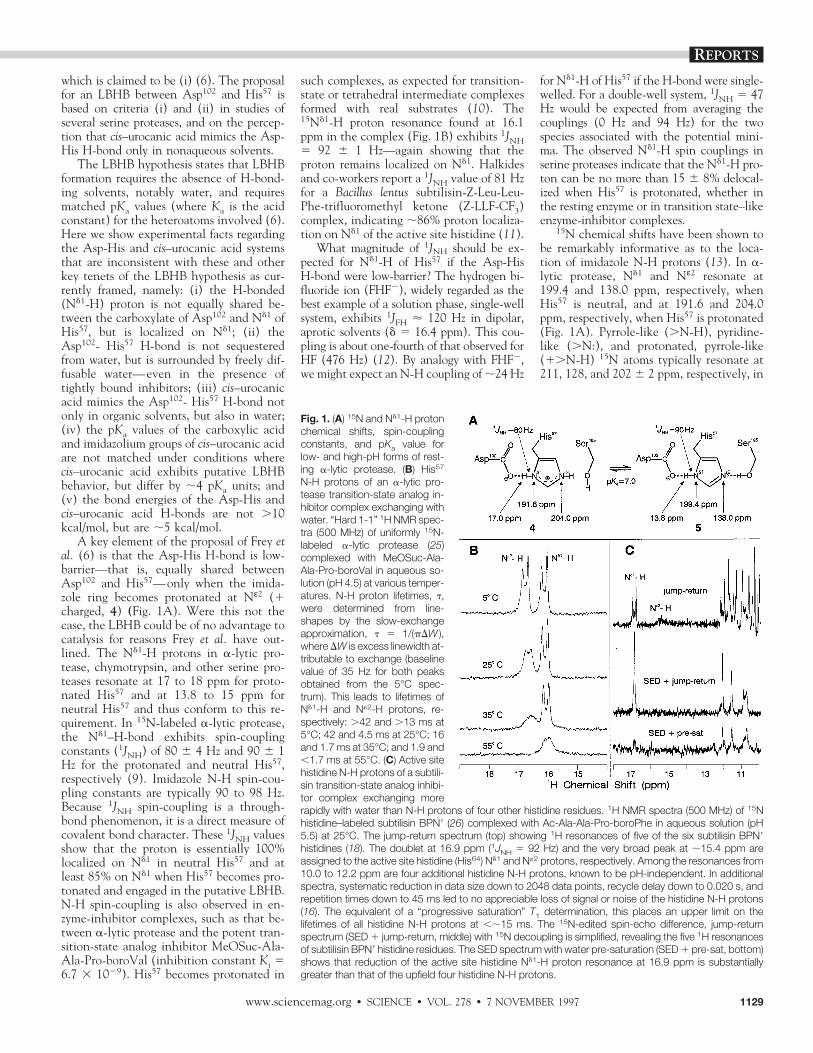
A key element of the proposal of Frey etal. (6) is that the Asp-His H-bond is low-barrier—that is, equally shared betweenAsp102 and His57—only when the imida-zole ring becomes protonated at Nε2 (1charged, 4) (Fig. 1A). Were this not thecase, the LBHB could be of no advantage tocatalysis for reasons Frey et al. have out-lined. The Nd1-H protons in a-lytic pro-tease, chymotrypsin, and other serine pro-teases resonate at 17 to 18 ppm for proto-nated His57 and at 13.8 to 15 ppm forneutral His57 and thus conform to this re-quirement. In 15N-labeled a-lytic protease,the Nd1–H-bond exhibits spin-couplingconstants (1JNH) of 80 6 4 Hz and 90 6 1Hz for the protonated and neutral His57,respectively (9). Imidazole N-H spin-cou-pling constants are typically 90 to 98 Hz.Because 1JNH spin-coupling is a through-bond phenomenon, it is a direct measure ofcovalent bond character. These 1JNH valuesshow that the proton is essentially 100%localized on Nd1 in neutral His57 and atleast 85% on Nd1 when His57 becomes pro-tonated and engaged in the putative LBHB.N-H spin-coupling is also observed in en-zyme-inhibitor complexes, such as that be-tween a-lytic protease and the potent tran-sition-state analog inhibitor MeOSuc-Ala-Ala-Pro-boroVal (inhibition constant Ki 56.7 3 1029). His57 becomes protonated in
such complexes, as expected for transition-state or tetrahedral intermediate complexesformed with real substrates (10). The15Nd1-H proton resonance found at 16.1ppm in the complex (Fig. 1B) exhibits 1JNH5 92 6 1 Hz—again showing that theproton remains localized on Nd1. Halkidesand co-workers report a 1JNH value of 81 Hzfor a Bacillus lentus subtilisin-Z-Leu-Leu-Phe-trifluoromethyl ketone (Z-LLF-CF3)complex, indicating ;86% proton localiza-tion on Nd1 of the active site histidine (11).
What magnitude of 1JNH should be ex-pected for Nd1-H of His57 if the Asp-HisH-bond were low-barrier? The hydrogen bi-fluoride ion (FHF2), widely regarded as thebest example of a solution phase, single-wellsystem, exhibits 1JFH ' 120 Hz in dipolar,aprotic solvents (d 5 16.4 ppm). This cou-pling is about one-fourth of that observed forHF (476 Hz) (12). By analogy with FHF2,we might expect an N-H coupling of ;24 Hz
for Nd1-H of His57 if the H-bond were single-welled. For a double-well system, 1JNH 5 47Hz would be expected from averaging thecouplings (0 Hz and 94 Hz) for the twospecies associated with the potential mini-ma. The observed Nd1-H spin couplings inserine proteases indicate that the Nd1-H pro-ton can be no more than 15 6 8% delocal-ized when His57 is protonated, whether inthe resting enzyme or in transition state–likeenzyme-inhibitor complexes.
15N chemical shifts have been shown tobe remarkably informative as to the loca-tion of imidazole N-H protons (13). In a-lytic protease, Nd1 and Nε2 resonate at199.4 and 138.0 ppm, respectively, whenHis57 is neutral, and at 191.6 and 204.0ppm, respectively, when His57 is protonated(Fig. 1A). Pyrrole-like (.N-H), pyridine-like (.N:), and protonated, pyrrole-like(1.N-H) 15N atoms typically resonate at211, 128, and 202 6 2 ppm, respectively, in
Fig. 1. (A) 15N and Nd1-H protonchemical shifts, spin-couplingconstants, and pKa value forlow- and high-pH forms of rest-ing a-lytic protease. (B) His57
N-H protons of an a-lytic pro-tease transition-state analog in-hibitor complex exchanging withwater. “Hard 1-1” 1H NMR spec-tra (500 MHz) of uniformly 15N-labeled a-lytic protease (25)complexed with MeOSuc-Ala-Ala-Pro-boroVal in aqueous so-lution (pH 4.5) at various temper-atures. N-H proton lifetimes, t,were determined from line-shapes by the slow-exchangeapproximation, t 5 1/(pDW ),where DW is excess linewidth at-tributable to exchange (baselinevalue of 35 Hz for both peaksobtained from the 5°C spec-trum). This leads to lifetimes ofNd1-H and N«2-H protons, re-spectively: .42 and .13 ms at5°C; 42 and 4.5 ms at 25°C; 16and 1.7 ms at 35°C; and 1.9 and,1.7 ms at 55°C. (C) Active sitehistidine N-H protons of a subtili-sin transition-state analog inhibi-tor complex exchanging morerapidly with water than N-H protons of four other histidine residues. 1H NMR spectra (500 MHz) of 15Nhistidine–labeled subtilisin BPN9 (26) complexed with Ac-Ala-Ala-Pro-boroPhe in aqueous solution (pH5.5) at 25°C. The jump-return spectrum (top) showing 1H resonances of five of the six subtilisin BPN9histidines (18). The doublet at 16.9 ppm (1JNH 5 92 Hz) and the very broad peak at ;15.4 ppm areassigned to the active site histidine (His64) Nd1 and N«2 protons, respectively. Among the resonances from10.0 to 12.2 ppm are four additional histidine N-H protons, known to be pH-independent. In additionalspectra, systematic reduction in data size down to 2048 data points, recycle delay down to 0.020 s, andrepetition times down to 45 ms led to no appreciable loss of signal or noise of the histidine N-H protons(16). The equivalent of a “progressive saturation” T1 determination, this places an upper limit on thelifetimes of all histidine N-H protons at ,;15 ms. The 15N-edited spin-echo difference, jump-returnspectrum (SED 1 jump-return, middle) with 15N decoupling is simplified, revealing the five 1H resonancesof subtilisin BPN9 histidine residues. The SED spectrum with water pre-saturation (SED 1 pre-sat, bottom)shows that reduction of the active site histidine Nd1-H proton resonance at 16.9 ppm is substantiallygreater than that of the upfield four histidine N-H protons.
REPORTS
www.sciencemag.org z SCIENCE z VOL. 278 z 7 NOVEMBER 1997 1129

aqueous solution (14). H-bonding perturbsthese shifts by up to 10 ppm—downfield for.N-H atoms acting as proton donors tocarboxylate groups, and upfield for .N:acceptors (13). For a-lytic protease at pH .9, His57 15N shifts have been shown toreflect the exclusive presence of the Nd1-Htautomer, 5, with both ring N atoms en-gaged in H-bonding; the H-bond to Asp102
moves Nd1 downfield ;10 ppm, whereasthe H-bond from Ser195 moves Nε2 upfield;10 ppm from 15N shift values expected forthe pure Nd1-H tautomer. At pH , 5,where the imidazole ring of His57 is proto-nated, 4, the H-bond to Asp102 is reflectedin the ;10 ppm downfield displacement ofthe Nd1 resonance from ;202 to 191.6ppm. Because H-bonding induces chemicalshift changes in the same direction, butlower in magnitude, as those induced byprotonation or deprotonation, H-bondingeffects can be interpreted in terms of partialproton transfer. Thus, the ;10 ppm dis-placement of Nd1, relative to that for fulldeprotonation, represents ;14 6 4% delo-calization of the Nd1-H proton (15), a valuein good agreement with that determinedfrom 1JNH.
The LBHB hypothesis requires theserine protease catalytic triad to be seques-tered from solvent. Solvent accessibility ofN-H groups is often gauged by measuringthe rate at which deuterium replaces pro-tons upon dissolution of the protein inD2O. The Nd1-H proton, however, becomesfully deuterated before an NMR spectrumcan be recorded in a-lytic protease andother serine proteases, placing the exchangehalf-life at ,;1.0 min (16). Longitudinalnuclear relaxation time (T1) measurementson resting a-lytic protease indicate anNd1-H proton exchange lifetime of ,9 ms(16). Transverse nuclear relaxation time(T2) measurements (lineshape analysis) on
chymotrypsinogen A indicate an Nd1-Hproton exchange lifetime in the millisecondrange at 1° to 19°C (17). Such rapid ex-change demonstrates the His57 Nd1-H pro-ton is highly accessible to solvent in restingserine proteases.
However, it could be argued that occu-pancy of the active site by substrates orinhibitors might block solvent access suffi-ciently for LBHB formation. MeOSuc-Ala-Ala-Pro-boroVal, a potent transition-stateanalog inhibitor of a-lytic protease, occu-pies subsites S1 to S4, as would a specificsubstrate. In addition to the Asp-His H-bond (d 5 16.1 ppm), a second putativeLBHB (d 5 16.5 ppm) is formed betweenNε2-H and the boronyl group in this com-plex (10). The inhibitor does reduce sol-vent access, as demonstrated by the narrow-er linewidth of the Nd1-H proton signalrelative to that of resting enzyme (Fig. 1B).Nevertheless, lineshape analysis indicatesthat the Nd1-H and Nε2-H proton lifetimesare still on the order of milliseconds. Incontrast, the lifetime of the bound inhibi-tor, with a Ki value of 6.7 3 1029, is on theorder of minutes. Thus, even in this tightlybound inhibitor complex, both imidazoleN-H protons of His57 are readily accessibleto solvent.
The subtilisin BPN9–Ac-Ala-Ala-Pro-boroPhe (Ki 5 1 3 10210) complex ex-hibits features similar to those of thea-lytic protease–MeOSuc-Ala-Ala-Pro-boro-Val complex, except that exchange of theNε2-H proton signal with water is morerapid, because its resonance is barely ob-servable at 25°C (Fig. 1C, top) (18). Fiveof subtilisin’s six histidines, the catalyticHis and four others, give rise to low-field1H signals, an observation aided by 15N
spectral editing and 15N decoupling (Fig.1C, middle). Upon selective irradiation(presaturation) of water (Fig. 1C, bot-tom), the active site histidine N-H protonresonance essentially disappears, whereasthe other four remain visible. Thus, of thefive observable histidine residues, the cat-alytic histidine is the most solvent acces-sible— even in a complex with a tight-binding transition-state analog inhibitor.
The perception that cis–urocanic acid, 6(Fig. 2), forms an LBHB similar to that ofthe Asp-His diad only in organic solvents isbased on reports that a low-field protonsignal is observed in these solvents (17.5ppm in dimethylsulfoxide, 17.2 ppm in ace-tonitrile, and 16.9 ppm in acetone), but notin water (6). However, the ability of cis–urocanic acid to mimic the Asp-His H-bond is not confined to organic solvents(Fig. 2). The 15N chemical shift behavior ofcis–urocanic acid in 100% water at roomtemperature (19) closely resembles that ofHis57 in a-lytic protease (20) over the en-tire range of pH stability for the enzyme(;4 to 10). Particularly from pH 4 to 5, the15N chemical shifts of cis–urocanic acidshow the same asymmetry exhibited byHis57 of a-lytic protease and attributed tothe H-bond with Asp102—namely, that Nd1
resonates ;10 ppm downfield from Nε2 andfrom chemical shifts characteristic of1.N-H–type nitrogens.
The above results, showing that aqueouscis–urocanic acid is as strongly H-bonded asthe Asp-His diad, predict that a low-fieldNd1-H proton resonance should be presentand suggest that the inability to detect it isdue to rapid exchange with solvent. Uponcooling an 85% acetone-d6, 15% H2O so-lution of cis–urocanic acid at pH 4.4 to
Fig. 2. 15N chemical shifts versus pH of cis–uro-canic acid, 6, are similar to those of His57 of a-lyticprotease, both in aqueous solution. 15N shifts forcis–urocanic acid (solid line) are given over the pHrange 2 to 10, and those of a-lytic protease (dot-ted line) over the pH range 4 to 10. [Reprinted withpermission of Roberts et al. (19). Copyright Amer-ican Chemical Society (1982)]
Fig. 3. Low temperatures required for cis–uro-canic acid to exhibit a low-field Nd1-H proton res-onance in water-containing solution. Low-field400-MHz 1H NMR spectra are shown for ;0.2 Mcis–urocanic acid versus pH and temperature invarious acetone-d6, water cryosolvent mixtures.cis-Urocanic acid was produced by photoisomer-ization of the trans isomer as described (27 ). pHwas measured by immersion of a combinationglass electrode and calomel reference (Cole-Parmer, Vernon Hills, IL), standardized with Na-tional Bureau of Standards buffers at 25°C. (A) 1HNMR spectra of cis–urocanic acid in an 85% ac-etone-d6, 15% H2O cryosolvent (pH 4.4) showsthe temperature-dependent linewidth of theNd1-H proton resonance at 18.5 ppm. Upon sam-ple cooling to 248°C, the Nd1-H proton is observ-able in the aqueous-based solution. With further sample cooling (258°C) the low-field resonancelinewidth decreases, but its chemical shift remains at 18.5 ppm. (B) pH dependence of the low-fieldresonance chemical shift in cis–urocanic acid in 90% acetone-d6, 10% H2O. The Nd1-H proton signalmoves from 15.0 ppm at pH 10.0 to 18.5 ppm at pH 4.6, and back to 15.2 ppm at pH 2.4. 1H spectrawere recorded at various temperatures from 243° to 260°C and showed no temperature dependenceof chemical shift when referenced to sodium 2,2-dimethyl-2-silapentane-5-sulfonate (DSS) as an inter-nal standard.
SCIENCE z VOL. 278 z 7 NOVEMBER 1997 z www.sciencemag.org1130
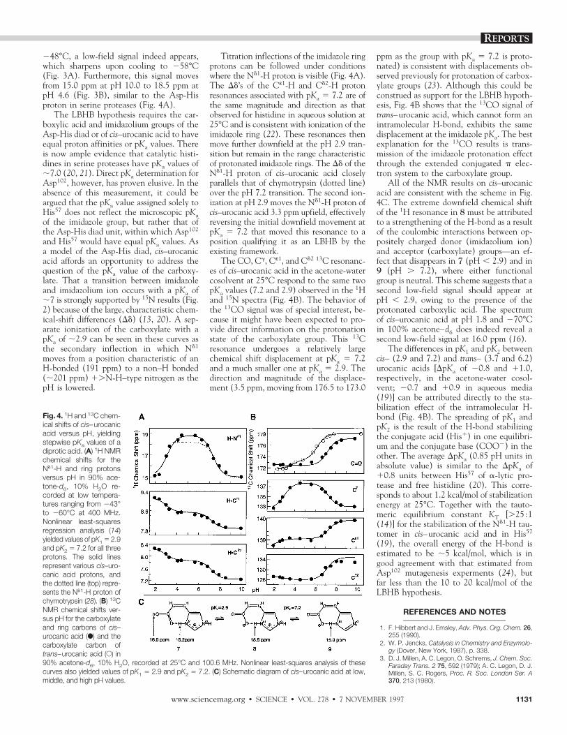
248°C, a low-field signal indeed appears,which sharpens upon cooling to 258°C(Fig. 3A). Furthermore, this signal movesfrom 15.0 ppm at pH 10.0 to 18.5 ppm atpH 4.6 (Fig. 3B), similar to the Asp-Hisproton in serine proteases (Fig. 4A).
The LBHB hypothesis requires the car-boxylic acid and imidazolium groups of theAsp-His diad or of cis–urocanic acid to haveequal proton affinities or pKa values. Thereis now ample evidence that catalytic histi-dines in serine proteases have pKa values of;7.0 (20, 21). Direct pKa determination forAsp102, however, has proven elusive. In theabsence of this measurement, it could beargued that the pKa value assigned solely toHis57 does not reflect the microscopic pKaof the imidazole group, but rather that ofthe Asp-His diad unit, within which Asp102
and His57 would have equal pKa values. Asa model of the Asp-His diad, cis–urocanicacid affords an opportunity to address thequestion of the pKa value of the carboxy-late. That a transition between imidazoleand imidazolium ion occurs with a pKa of;7 is strongly supported by 15N results (Fig.2) because of the large, characteristic chem-ical-shift differences (Dd) (13, 20). A sep-arate ionization of the carboxylate with apKa of ;2.9 can be seen in these curves asthe secondary inflection in which Nd1
moves from a position characteristic of anH-bonded (191 ppm) to a non–H bonded(;201 ppm) 1.N-H–type nitrogen as thepH is lowered.
Titration inflections of the imidazole ringprotons can be followed under conditionswhere the Nd1-H proton is visible (Fig. 4A).The Dd’s of the Cε1-H and Cd2-H protonresonances associated with pKa 5 7.2 are ofthe same magnitude and direction as thatobserved for histidine in aqueous solution at25°C and is consistent with ionization of theimidazole ring (22). These resonances thenmove further downfield at the pH 2.9 tran-sition but remain in the range characteristicof protonated imidazole rings. The Dd of theNd1-H proton of cis–urocanic acid closelyparallels that of chymotrypsin (dotted line)over the pH 7.2 transition. The second ion-ization at pH 2.9 moves the Nd1-H proton ofcis–urocanic acid 3.3 ppm upfield, effectivelyreversing the initial downfield movement atpKa 5 7.2 that moved this resonance to aposition qualifying it as an LBHB by theexisting framework.
The CO, Cg, Cε1, and Cd2 13C resonanc-es of cis–urocanic acid in the acetone-watercosolvent at 25°C respond to the same twopKa values (7.2 and 2.9) observed in the 1Hand 15N spectra (Fig. 4B). The behavior ofthe 13CO signal was of special interest, be-cause it might have been expected to pro-vide direct information on the protonationstate of the carboxylate group. This 13Cresonance undergoes a relatively largechemical shift displacement at pKa 5 7.2and a much smaller one at pKa 5 2.9. Thedirection and magnitude of the displace-ment (3.5 ppm, moving from 176.5 to 173.0
ppm as the group with pKa 5 7.2 is proto-nated) is consistent with displacements ob-served previously for protonation of carbox-ylate groups (23). Although this could beconstrued as support for the LBHB hypoth-esis, Fig. 4B shows that the 13CO signal oftrans–urocanic acid, which cannot form anintramolecular H-bond, exhibits the samedisplacement at the imidazole pKa. The bestexplanation for the 13CO results is trans-mission of the imidazole protonation effectthrough the extended conjugated p elec-tron system to the carboxylate group.
All of the NMR results on cis–urocanicacid are consistent with the scheme in Fig.4C. The extreme downfield chemical shiftof the 1H resonance in 8 must be attributedto a strengthening of the H-bond as a resultof the coulombic interactions between op-positely charged donor (imidazolium ion)and acceptor (carboxylate) groups—an ef-fect that disappears in 7 (pH , 2.9) and in9 (pH . 7.2), where either functionalgroup is neutral. This scheme suggests that asecond low-field signal should appear atpH , 2.9, owing to the presence of theprotonated carboxylic acid. The spectrumof cis–urocanic acid at pH 1.8 and 270°Cin 100% acetone–d6 does indeed reveal asecond low-field signal at 16.0 ppm (16).
The differences in pK1 and pK2 betweencis– (2.9 and 7.2) and trans– (3.7 and 6.2)urocanic acids [DpKa of 20.8 and 11.0,respectively, in the acetone-water cosol-vent; 20.7 and 10.9 in aqueous media(19)] can be attributed directly to the sta-bilization effect of the intramolecular H-bond (Fig. 4B). The spreading of pK1 andpK2 is the result of the H-bond stabilizingthe conjugate acid (His1) in one equilibri-um and the conjugate base (COO2) in theother. The average DpKa (0.85 pH units inabsolute value) is similar to the DpKa of10.8 units between His57 of a-lytic pro-tease and free histidine (20). This corre-sponds to about 1.2 kcal/mol of stabilizationenergy at 25°C. Together with the tauto-meric equilibrium constant KT [.25 :1(14)] for the stabilization of the Nd1-H tau-tomer in cis–urocanic acid and in His57
(19), the overall energy of the H-bond isestimated to be ;5 kcal/mol, which is ingood agreement with that estimated fromAsp102 mutagenesis experments (24), butfar less than the 10 to 20 kcal/mol of theLBHB hypothesis.
REFERENCES AND NOTES___________________________
1. F. Hibbert and J. Emsley, Adv. Phys. Org. Chem. 26,255 (1990).
2. W. P. Jencks, Catalysis in Chemistry and Enzymolo-gy (Dover, New York, 1987), p. 338.
3. D. J. Millen, A. C. Legon, O. Schrems, J. Chem. Soc.Faraday Trans. 2 75, 592 (1979); A. C. Legon, D. J.Millen, S. C. Rogers, Proc. R. Soc. London Ser. A370, 213 (1980).
Fig. 4. 1H and 13C chem-ical shifts of cis–urocanicacid versus pH, yieldingstepwise pKa values of adiprotic acid. (A) 1H NMRchemical shifts for theNd1-H and ring protonsversus pH in 90% ace-tone-d6, 10% H2O re-corded at low tempera-tures ranging from 243°to 260°C at 400 MHz.Nonlinear least-squaresregression analysis (14)yielded values of pK1 5 2.9and pK2 5 7.2 for all threeprotons. The solid linesrepresent various cis–uro-canic acid protons, andthe dotted line (top) repre-sents the Nd1-H proton ofchymotrypsin (28). (B) 13CNMR chemical shifts ver-sus pH for the carboxylateand ring carbons of cis–urocanic acid (F) and thecarboxylate carbon oftrans–urocanic acid (E) in90% acetone-d6, 10% H2O, recorded at 25°C and 100.6 MHz. Nonlinear least-squares analysis of thesecurves also yielded values of pK1 5 2.9 and pK2 5 7.2. (C) Schematic diagram of cis–urocanic acid at low,middle, and high pH values.
REPORTS
www.sciencemag.org z SCIENCE z VOL. 278 z 7 NOVEMBER 1997 1131
4. R. L. Schowen, in Mechanistic Principles of EnzymeActivity, J. F. Liebman and A. Greenburg, Eds. ( VCH,New York, 1988), p. 119.
5. W. W. Cleland and M. M. Kreevoy, Science 264,1887 (1994).
6. P. A. Frey, S. A. Whitt, J. B. Tobin, ibid., p. 1927.7. J. A. Gerlt and P. G. Gassman, Biochemistry 32,
11943 (1993); N. S. Golubev, V. A. Grindin, S. S.Ligai, S. N. Smirnov, Biochemistry (Moscow) 59, 447(1994); S. N. Smirnov et al., J. Am. Chem. Soc. 118,4094 (1996); Q. Zhao, C. Abeygunawardana, P. Ta-lalay, A. S. Mildvan, Proc. Natl. Acad. Sci. U.S.A. 93,8220 (1996); C. S. Cassidy, J. Lin, P. A. Frey, Bio-chemistry 36, 4576 (1997).
8. Warshel has argued that LBHBs would be anticata-lytic in enzymes, whereas Shan et al. have examinedthe energetics of LBHB model systems and failed tofind additional stabilization under conditions predict-ed by LBHB theory [A. Warshel, A. Papazyan, P. A.Kollman, Science 269, 102 (1995); S. O. Shan, S.Loh, D. Herschlag, ibid. 272, 97 (1996); J. P. Guthrie,Chem. Biol. 4, 259 (1996)].
9. W. W. Bachovchin, Proc. Natl. Acad. Sci. U.S.A. 82,7948 (1985).
10. iiii, W. Y. Wong, S. Farr-Jones, A. B. Shenvi,C. A. Kettner, Biochemistry 27, 7689 (1988); C. A.Kettner, R. Bone, D. A. Agard, W. W. Bachovchin,ibid., p. 7682.
11. C. J. Halkides, Y. Q. Wu, C. J. Murray, ibid. 35,15941 (1996).
12. F. Y. Fujiwara and J. S. Martin, J. Am. Chem. Soc.96, 7625 (1974); ibid., p. 7632.
13. W. W. Bachovchin, Biochemistry 25, 7751 (1986).14. S. Farr-Jones, W. Y. L. Wong, W. G. Gutheil, W. W.
Bachovchin, J. Am. Chem. Soc. 115, 6813 (1993).15N chemical shifts are reported in parts per millionupfield of 1.0 M HNO3, with larger values more up-field. Referencing to liquid NH3 is obtained by sub-traction from 377.5.
15. A downfield displacement of 22.6 ppm in the 15Nchemical shift of Nd1 has been reported for subtilisinfrom B. lentus complexed with Z-LLF-CF3 (11). Al-though this effect was attributed to LBHB formationwith ;30% proton transfer, such a fraction is incon-sistent with the 1JNH value of 81 Hz reported by thesame authors, which would indicate only ;14 6 8%delocalization with respect to the average imidazolevalue of 94 6 4 Hz. The inconsistency may be attrib-utable to incomplete characterization of the system,including 15N shifts for both Nd1 and N«2 in the com-plex and resting enzyme.
16. E. L. Ash, J. L. Sudmeier, W. W. Bachovchin, unpub-lished data.
17. J. L. Markley and W. M. Westler, Biochemistry 35,11092 (1996).
18. W. W. Bachovchin, Stable Isotope Applications inBiomolecular Structure and Mechanisms, J. Tre-whella, T. A. Cross, C. J. Unkefer, Eds. (Los AlamosNational Laboratory, Los Alamos, NM, 1994), p. 41;R. M. Day, thesis, Tufts University, Boston, MA(1995).
19. J. D. Roberts, C. Yu, C. Flanagan, T. R. Birdseye,J. Am. Chem. Soc. 104, 3945 (1982).
20. W. W. Bachovchin and J. D. Roberts, ibid. 100, 8041(1978).
21. J. L. Markley, Biological Applications of MagneticResonance (Academic Press, New York, 1979); F.Sakiyama and Y. Kawata, J. Biochem. 94, 1661(1983).
22. J. L. Markley, Acc. Chem. Res. 8, 70 (1975).23. W. J. Horsley and H. Sternlicht, J. Am. Chem. Soc.
90, 3738 (1968).24. C. S. Craik, S. Roczniak, C. Largman, W. J. Rutter,
Science 237, 909 (1987); P. Carter and J. A. Wells,Nature 332, 564 (1988).
25. Uniformly 15N-labeled (.96%) a-lytic protease (E.C.3.4.21.12) was expressed by fermentation of wild-type Lysobacter enzymogenes in Celtone-N (MartekBiosciences), a medium containing 15N-enrichedpeptides and amino acids, which was supplementedwith glucose and various salts. Purification of a-lyticprotease and assay of enzymatic activity was ac-complished as described (18).
26. Expression of 15N histidine–labeled subtilisin BPN997was accomplished by fermentation of a histidine
auxotrophic strain of Bacillus subtilis DB104 in a min-imal medium containing histidine labeled with 15N atboth ring nitrogens (18). Purification of labeled subtili-sin BPN997 was accomplished by dialysis into a 10mM tris solution (pH 6.0) and subsequent passageover a CM52 cellulose ion-exchange column, withelution of the protein in 0.01 M MES and 0.1 M KCl.Enzyme activity was measured by a colorimetric as-say with the substrate succinyl-L-Ala-L-Ala-L-Pro-L-Phe-paranitroanilide (18).
27. I. Santucci, thesis, Flinders University of South Aus-tralia, Bedford Park, South Australia (1995).
28. G. Robillard and R. G. Schulman, J. Mol. Biol. 71,507 (1972).
29. We thank R. M. Day for supplying the subtilisin BPN9NMR sample, C. A. Kettner for supplying boronicacid inhibitors, and N. I. Krinsky for helpful discus-sions. Supported in part by NIH grant GM27927.
25 July 1997; accepted 3 October 1997
Inhibition of Brain Gz GAP and Other RGSProteins by Palmitoylation of G Protein
a SubunitsYaping Tu, Jun Wang, Elliott M. Ross*
Palmitoylation of the a subunit of the guanine nucleotide-binding protein Gz inhibited bymore than 90 percent its response to the guanosine triphosphatase (GTPase)–acceler-ating activity of Gz GAP, a Gz-selective member of the regulators of G-protein signaling(RGS) protein family of GTPase-activating proteins (GAPs). Palmitoylation both de-creased the affinity of Gz GAP for the GTP-bound form of Gaz by at least 90 percent anddecreased the maximum rate of GTP hydrolysis. Inhibition was reversed by removal ofthe palmitoyl group by dithiothreitol. Palmitoylation of Gaz also inhibited its response tothe GAP activity of Ga-interacting protein (GAIP), another RGS protein, and palmitoyl-ation of Gai1 inhibited its response to RGS4. The extent of inhibition of Gz GAP, GAIP,RGS4, and RGS10 correlated roughly with their intrinsic GAP activities for the Ga targetused in the assay. Reversible palmitoylation is thus a major determinant of Gz deacti-vation after its stimulation by receptors, and may be a general mechanism for prolongingor potentiating G-protein signaling.
The a subunits of most heterotrimeric Gproteins are modified by irreversible lipidamidation of the NH2-terminus and by ad-dition of a palmitoyl thioester at a nearby,conserved cysteine residue (1, 2). Unlikemyristoylation, palmitoylation of Ga sub-units is reversible, and bound palmitateturns over rapidly in cells. Although virtu-ally nothing is known of the enzymes thatcatalyze addition and removal of palmitate,palmitate turnover on G-protein a subunitsappears to be regulated coordinately withtheir activation and deactivation. In thecase of Gas (3, 4) and Gaq (5), substantialdepalmitoylation occurs upon receptor-pro-moted activation, and repalmitoylation ofGas coincides at least roughly with deacti-vation (3). Treatment with cholera toxin,which prolongs activation of Gs by blockinghydrolysis of bound GTP, also promotesturnover of bound palmitate (6). Converse-ly, palmitate turnover on Gai and Gas isdecreased by coexpression of excess Gbg,which inhibits activation (6, 7).
Palmitoylation is involved in anchoringGa subunits to the membrane or specifying
their membrane localization, or both (1–4,7–9), by increasing their intrinsic hydropho-bicity and, at least for Gas, by increasingaffinity for Gbg (7). Mutation of the palmi-toylated cysteine of Gaz to alanine also po-tentiated inhibition of adenylyl cyclase intransfected cells (9). Palmitoylation has notyet been linked to alteration of a specificG-protein signaling function, however. It isnot required for interaction of Ga subunitswith receptors or effectors in vitro (10), andno effect of palmitoylation on the binding orhydrolysis of guanine nucleotides has beenreported. Mutation of the palmitoylatable cys-teine residue in Gaq or Gas inhibited signal-ing (11), but signaling was potentiated by thesame mutation in Gaz or Gpa1p, the majorGa subunit in Saccharomyces cerevisiae (9,12). Although palmitoylation may be respon-sible for such variable effects on different Gasubunits, these results may also arise fromeffects of mutating the cysteine residue thatare unrelated to palmitoylation (10).
We describe the inhibition of the effectsof the major Gz GTPase activating protein,Gz GAP, by palmitoylation of Gaz. Gz is arelatively rare member of the Gi family thatis found in brain, platelets and adrenal me-dulla, and is therefore suspected to be in-volved in regulation of secretion (13). Iso-lated Gaz hydrolyzes bound GTP slowly,
Department of Pharmacology, University of Texas South-western Medical Center, 5323 Harry Hines Boulevard,Dallas, TX 75235–9041, USA.
*To whom correspondence should be addressed.
SCIENCE z VOL. 278 z 7 NOVEMBER 1997 z www.sciencemag.org1132

DOI: 10.1126/science.278.5340.1128, 1128 (1997);278 Science et al.Elissa L. Ash
Proteases? Theory Versus ExperimentA Low-Barrier Hydrogen Bond in the Catalytic Triad of Serine
This copy is for your personal, non-commercial use only.
clicking here.colleagues, clients, or customers by , you can order high-quality copies for yourIf you wish to distribute this article to others
here.following the guidelines
can be obtained byPermission to republish or repurpose articles or portions of articles
): September 22, 2015 www.sciencemag.org (this information is current as of
The following resources related to this article are available online at
http://www.sciencemag.org/content/278/5340/1128.full.htmlversion of this article at:
including high-resolution figures, can be found in the onlineUpdated information and services,
http://www.sciencemag.org/content/278/5340/1128.full.html#ref-list-1, 7 of which can be accessed free:cites 24 articlesThis article
128 article(s) on the ISI Web of Sciencecited by This article has been
http://www.sciencemag.org/content/278/5340/1128.full.html#related-urls13 articles hosted by HighWire Press; see:cited by This article has been
http://www.sciencemag.org/cgi/collection/biochemBiochemistry
subject collections:This article appears in the following
registered trademark of AAAS. is aScience1997 by the American Association for the Advancement of Science; all rights reserved. The title
CopyrightAmerican Association for the Advancement of Science, 1200 New York Avenue NW, Washington, DC 20005. (print ISSN 0036-8075; online ISSN 1095-9203) is published weekly, except the last week in December, by theScience
on
Sep
tem
ber
22, 2
015
ww
w.s
cien
cem
ag.o
rgD
ownl
oade
d fr
om
Related Documents