ORIGINAL ARTICLE Lovastatin induces apoptosis of k-ras-transformed thyroid cells via inhibition of ras farnesylation and by modulating redox state Chiara Laezza & Laura Fiorentino & Simona Pisanti & Patrizia Gazzerro & Michele Caraglia & Giuseppe Portella & Mario Vitale & Maurizio Bifulco Received: 4 February 2008 / Revised: 1 August 2008 / Accepted: 4 August 2008 # Springer-Verlag 2008 Abstract Transformation of thyroid cells with either K-ras or H-ras viral oncogenes produces cell types with different phenotype and different response to the inhibition of the prenylation pathway by 3-hydroxy-3-methylglutaryl-CoA reductase or farnesyltransferase inhibitors. These inhibitors induce apoptosis in K-ras-transformed FRTL-5 cells (FRTL-5-K-Ras) whereas cell cycle arrest is induced in H-ras-transformed FRTL-5 (FRTL-5-H-Ras). In FRTL-5-K- Ras cells, the product of K-ras gene is implicated in the scavenging of reactive oxygen species (ROS) through the activation of extracellular-signal-regulated kinase (ERK)1/2 kinases. We observed that lovastatin blocked ras activation through inhibition of farnesylation and induced apoptosis, increasing ROS levels through inhibition of ERK1/2 signaling and Mn-SOD expression. Lovastatin-induced apoptosis was due to intracellular ROS increase since both, the antioxidant compound pyrrolidinedithiocarbamate or the SOD-mimetic compound, antagonized apoptosis. More- over, both p38 mitogen-activated protein kinase and nuclear factor κB pathways, activated as a consequence of high ROS levels, are involved in the apoptotic effect, indicating that cell death induced by lovastatin was dependent on oxidative stress. Lovastatin antitumor efficacy in K-ras- dependent thyroid tumors was further confirmed in vivo, proposing a new therapeutic strategy for those tumor diseases that are sustained by an inappropriate K-ras expression. Keywords RAS . Thyroid . Lovastatin . Farnesylation Abbreviations ROS reactive oxygen species PDTC pyrrolidinedithiocarbamate GEFs guanosine exchange factors FTase farnesyltransferase HMG-CoA 3-hydroxy-3-methylglutaryl-CoA MnTMPyP Mn(III)tetrakis(1-methyl-4-pyridyl)porphyrin Mn-SOD Mn-superoxide dismutase Ras proteins are involved in regulating many cellular processes, including proliferation, differentiation, and apo- ptosis [1–2]. Based on their origin, three forms of ras oncogene, namely K-ras, H-ras, and N-ras, have been detected. One way used by ras family proteins to regulate cell proliferation is the modulation of redox signals. It is J Mol Med DOI 10.1007/s00109-008-0396-1 C.L. and L.F. contributed equally to this work. C. Laezza IEOS, CNR, Naples, Italy C. Laezza : G. Portella Department of Cellular and Molecular Biology and Pathology “L. Califano”, University of Naples Federico II, Naples, Italy L. Fiorentino : S. Pisanti : P. Gazzerro : M. Bifulco (*) Department of Pharmaceutical Sciences, University of Salerno, 84084 Fisciano, Salerno, Italy e-mail: [email protected] M. Caraglia Experimental Pharmacology Unit, National Institute of Tumours Fondazione “G. Pascale” of Naples, Naples, Italy M. Vitale Department of Molecular and Clinical Endocrinology and Oncology, University of Naples Federico II, Naples, Italy

Welcome message from author
This document is posted to help you gain knowledge. Please leave a comment to let me know what you think about it! Share it to your friends and learn new things together.
Transcript
ORIGINAL ARTICLE
Lovastatin induces apoptosis of k-ras-transformed thyroidcells via inhibition of ras farnesylation and by modulatingredox state
Chiara Laezza & Laura Fiorentino & Simona Pisanti &Patrizia Gazzerro & Michele Caraglia &
Giuseppe Portella & Mario Vitale & Maurizio Bifulco
Received: 4 February 2008 /Revised: 1 August 2008 /Accepted: 4 August 2008# Springer-Verlag 2008
Abstract Transformation of thyroid cells with either K-rasor H-ras viral oncogenes produces cell types with differentphenotype and different response to the inhibition of theprenylation pathway by 3-hydroxy-3-methylglutaryl-CoAreductase or farnesyltransferase inhibitors. These inhibitorsinduce apoptosis in K-ras-transformed FRTL-5 cells(FRTL-5-K-Ras) whereas cell cycle arrest is induced inH-ras-transformed FRTL-5 (FRTL-5-H-Ras). In FRTL-5-K-Ras cells, the product of K-ras gene is implicated in thescavenging of reactive oxygen species (ROS) through theactivation of extracellular-signal-regulated kinase (ERK)1/2
kinases. We observed that lovastatin blocked ras activationthrough inhibition of farnesylation and induced apoptosis,increasing ROS levels through inhibition of ERK1/2signaling and Mn-SOD expression. Lovastatin-inducedapoptosis was due to intracellular ROS increase since both,the antioxidant compound pyrrolidinedithiocarbamate orthe SOD-mimetic compound, antagonized apoptosis. More-over, both p38 mitogen-activated protein kinase and nuclearfactor κB pathways, activated as a consequence of highROS levels, are involved in the apoptotic effect, indicatingthat cell death induced by lovastatin was dependent onoxidative stress. Lovastatin antitumor efficacy in K-ras-dependent thyroid tumors was further confirmed in vivo,proposing a new therapeutic strategy for those tumordiseases that are sustained by an inappropriate K-rasexpression.
Keywords RAS . Thyroid . Lovastatin . Farnesylation
AbbreviationsROS reactive oxygen speciesPDTC pyrrolidinedithiocarbamateGEFs guanosine exchange factorsFTase farnesyltransferaseHMG-CoA 3-hydroxy-3-methylglutaryl-CoAMnTMPyP Mn(III)tetrakis(1-methyl-4-pyridyl)porphyrinMn-SOD Mn-superoxide dismutase
Ras proteins are involved in regulating many cellularprocesses, including proliferation, differentiation, and apo-ptosis [1–2]. Based on their origin, three forms of rasoncogene, namely K-ras, H-ras, and N-ras, have beendetected. One way used by ras family proteins to regulatecell proliferation is the modulation of redox signals. It is
J Mol MedDOI 10.1007/s00109-008-0396-1
C.L. and L.F. contributed equally to this work.
C. LaezzaIEOS, CNR,Naples, Italy
C. Laezza :G. PortellaDepartment of Cellular and Molecular Biologyand Pathology “L. Califano”,University of Naples Federico II,Naples, Italy
L. Fiorentino : S. Pisanti : P. Gazzerro :M. Bifulco (*)Department of Pharmaceutical Sciences, University of Salerno,84084 Fisciano, Salerno, Italye-mail: [email protected]
M. CaragliaExperimental Pharmacology Unit,National Institute of Tumours Fondazione “G. Pascale” of Naples,Naples, Italy
M. VitaleDepartment of Molecular and ClinicalEndocrinology and Oncology,University of Naples Federico II,Naples, Italy
well established that rac1, an essential component of thereactive oxygen species (ROS)-producing NADPH oxidase,is downstream of ras which can therefore modulateintracellular ROS levels [3]. However, despite their strin-gent sequence homology, K-ras, H-ras, and N-ras candisplay different effects on the generation of ROS. Indetail, K- and H-ras genes have been shown to exertopposing functions in the regulation of redox signals, sinceH-ras protein can increase ROS production, while K-rasdecreases ROS levels by acting via the extracellular-signal-regulated kinase (ERK1/2) kinases on different intracellulartargets [4]. Activation of the ras oncogene product p21requires the linkage of a farnesyl group, allowing theprotein anchorage to the cell membrane and the subsequentinteraction with guanosine exchange factors located inreceptor-associated complexes [5–6]. Some ras familyproteins such as K-ras can be alternatively activatedthrough the addition of a geranylgeranyl moiety. Thefarnesylation process has an important role in the determi-nation of the transformed phenotype [7].
Mutations involving codon 61 of H-ras and N-ras havebeen reported with variable frequency in thyroid neo-plasms. Ras mutations are more common in iodine-deficient than iodine-sufficient areas and in lesions withfollicular architecture (including follicular carcinoma andfollicular variant papillary thyroid carcinoma) than intypical papillary thyroid carcinoma [8]. On the basis ofthe involvement of ras in thyroid carcinogenesis, we haveevaluated the role of ras proteins in the regulation of theisoprenoid pathway in FRTL-5 thyroid cells and we havefound that it is deeply altered upon transformation withv-K-ras [9]. K-ras, different from H-ras, induces metabolicchanges in the isoprenoid pathway resulting in a preferen-tial farnesylation and subsequent functional activation ofthe oncogene product. This effect is mainly produced bydownregulating mevalonate kinase and inducing the activ-ity of farnesyltransferase (FTase) enzyme. In line with thesedata, we also observed an enhanced activity of FTase incolon tumor tissue, correlated to tumor location, histolog-ical grading, and K-ras mutation [9]. On the basis of thesedata, the specific inhibition of isoprenylation processescould be a useful tool to block K-ras both transforming andgrowth-regulating activity. Pharmacological compoundsinhibiting enzymes involved in protein isoprenylation havebeen proposed as anticancer tools [10–11]. Among these,lovastatin, an irreversible inhibitor of 3-hydroxy-3-methyl-glutaryl-CoA (HMG-CoA) reductase, used in clinic to treathypercholesterolemia through blocking the mevalonatebiosynthesis pathway, has been shown to induce apoptosisin several tumor types [12–15]. The mechanism ofapoptosis induced by lovastatin and its correlation withras oncogene expression are poorly understood so far.
We previously reported that the inhibition of prenylationby lovastatin induced apoptosis in human neoplastic thyroidcells [16].
In the present manuscript, to investigate the role of rasoncogene in the cytotoxicity induced by prenylationinhibitors, we used as a model K-ras- or H-ras-transformedthyroid cells. We analyzed whether the proapoptotic effectof this statin was related to an interference with the p21-ras-mediated activation of pathways leading to oxidative stress.
Materials and methods
Materials, drugs, and antibodies
Lovastatin was a generous gift of Dr. A.W. Alberts (MerckSharp and Dohme Research Laboratories, West Point, PA,USA); PD98059 and SB203580 were purchased fromCalbiochem (La Jolla, CA, USA). Protease inhibitors (apro-tinin and leupeptin) were purchased from Roche DiagnosticsGmbH (Mannheim, Germany); protein electrophoresisreagents were from Bio-Rad laboratories (Hercules, CA,USA), Western blotting and enhanced chemiluminescence(ECL) reagents were from GE Healthcare (Milano, Italy). Allother chemicals were from Sigma-Aldrich. Cell culturereagents were obtained from Gibco (Grand Island, NY,USA). Anti-ERK1/2 antibodies were purchased from Chem-icon (Temecula, CA, USA); antibodies were from Santa CruzBiotechnology Inc. (CA, USA). FTI-277 and GGTI werepurchased from Merck (Darmstadt, Germany). Mevalonateand pyrrolidinedithiocarbamate were purchased from Sigma-Aldrich (St. Louis, MO, USA). All the substances were usedat the most efficacious doses experimentally evaluated.Inducible expression vector coding for a dominant-negativemutant of ras (ras p21N17) was a kind gift of Prof G. Vecchio.
Cell lines
FRTL-5 (ATCC CRL 8305) is a differentiated rat thyroidcell line; FRTL-5-K-Ras and FRTL-5-H-Ras cells, derivedfrom FRTL-5 stably transfected with v-K-ras or v-H-rasoncogene, respectively, were kindly provided by Prof. G.Vecchio. FRTL-5-H-Ras and FRTL-5-K-Ras cells weregrown in Coon’s modified Ham’s F12 medium supple-mented with 5% calf serum. Photos were acquired in lightmicroscopy at ×10 magnification.
Assessment of cell cycle and apoptosis by propidium iodidelabeling
The induction of apoptosis by different treatments wasassessed as the percentage of hypodiploid sub-G1 peak
J Mol Med
measured by flow cytometry. Briefly, the cells wereincubated for 24 h with the indicated drugs, then collected,washed with phosphate-buffered saline (PBS), and resus-pended in 300 μl of PBS. Seven-hundred microliters ofethanol 70% were added slowly to the cells on a vortex andkept at −20°C overnight. Propidium iodide (PI; 10 μg/ml)in PBS containing 100-U/ml DNase-free RNase A wasadded to the cells. After 4 h at room temperature, cells weresubjected to flow cytometric analysis. Data acquisition andanalysis were performed in a Becton Dickinson FACSCa-libur flow cytometer using CellQuest software.
DNA fragmentation assay
For detection of apoptosis by the DNA fragmentation assay,treated cells were harvested, resuspended in 0.5 ml of lysisbuffer (20 mM ethylenediaminetetraacetic acid (EDTA),10 mM Tris pH 8.0, 200 mM NaCl, 0.2% Triton X-100, and100 µg/ml Proteinase K) and incubated for 1.5 h in a 37°Cincubator. The samples were centrifuged (12,000 rpm) inroom temperature for 5 min. The supernatant was transferredto a new Eppendorf tube and equal volumes of isopropanoland 25 µl of 4 M NaCl (100 mM final concentration) wereadded, followed by overnight incubation of the samplesat −20°C. DNA was acquired by centrifugation of thesamples, washed, dried, and dissolved in 30 µl TE (10 mMTris–1 mM EDTA, pH 8.0) buffer. Five micrograms of DNAwere loaded on each lane of 1.5% agarose gel.
Fluorescent measurement of intracellular ROS
Cells were trypsinized, collected in polystyrene tubes,and washed three times with 137 mM NaCl, 3.7 mMKCl, 10 mM Na2HPO4, 1.76 mM KH2PO4, pH 7.5 (PBS).Cells numbering 1×10⁶ were suspended in 2 ml ofPBS and incubated with 10 μM of 5,6-carboxy-2′,7′-dichlorofluorescein diacetate (DCHFDA; MolecularProbes, Eugene, OR), for 1 h at 37°C and kept in DCHFDAcontinuously thereafter. Cells were analyzed with fluores-cent-activated cell sorting (FACS) analysis with excitationand emission setting of 495 and 525 nm, respectively.
Western blot analysis
Subconfluent monolayer of cells were washed with PBSand then lysed in a lysis buffer containing 20 mM 4-(2-hydroxyethyl)-1-piperazineethanesulfonic acid (HEPES)pH 7.5, 150 mM NaCl, 10% glycerol, 1% Triton X-100,supplemented with a mixture of protease inhibitors. Equalamounts of total proteins (50 μg) were resolved on 12%sodium dodecyl sulfate-polyacrylamide gel electrophoresis(SDS-PAGE). After immunoblotting of gels onto polyvi-
nylidene difluoride sheets (Millipore), filters were blockedfor 1 h at room temperature with 10% nonfat dry milk (Bio-Rad) in TBS-T buffer (10 mM Tris–HCl pH 8, 0.1% Tween20, 150 mM NaCl). The filters were then incubated with thespecific primary antibodies as showed in the figuresfollowed by horseradish peroxidase (HRP)-conjugatedsecondary antibodies. Blots were revealed by ECL system(GE Healthcare). Immunoreactive bands were quantifiedusing Quantity One I-D analysis software (Bio-Rad).
Measurement of cell viability
About 5×10³ FRO cells/well were seeded in 96-wellculture plates and incubated for 24 h at 37°C with 10 μMlovastatin. Cell survival was examined by using 3-(4,5-dimethylthiazol-2-yl)-5-(3-carboxymethoxyphenyl)-2-(4-sulphophenyl)-2H-tetrazolium, inner salt (MTS), and anelectron coupling reagent (phenazine methosulfate), accord-ing to manufacturer’s instructions (Promega).
Affinity precipitation of Ras
The cells were lysed in the Mg2+ buffer containing20 mM HEPES, pH 7.5, 150 mM NaCl, 1% Igepal CA-630, 10 mM MgCl2, 1 mM EDTA, and 2% glycerol.Then, 10 μl Ras Binding Domain conjugated to agarose(Cell Signaling Technology, MA, USA) were added to1 mg of cell lysate and the resulting mixture wasincubated overnight with gentle rocking at 4°C. Theagarose beads were collected by microcentrifugation at14,000×g for 5 s and washed three times with Mg+2
buffer. The agarose beads were boiled for 5 min in 2XLaemmli sample buffer and collected by a microcentrifugepulse. The supernatants were run on 12% SDS-PAGE;then, the proteins were electrotransferred on a nitrocellu-lose film. The nitrocellulose was incubated overnight with1 μg/ml of anti-Ras Mab, clone RAS1,0 and with asecondary Mab, a goat α-mouse HRP-conjugated immu-noglobulin G, for 1 h. The film was washed with TBS/0.05% Tween 20 and detected by ECL, chemilumines-cence technique (Amersham).
Electromobility shift assay
Double-stranded oligonucleotides containing the NF-κBrecognition sequence (5′-CAACGGCAGGGGAATCTCCCTCTCCTT-3′) was end-labeled with 32P-adenosinetriphosphate. Nuclear extracts containing 5 µg of proteinwere incubated for 15 min with radiolabeled oligonucleo-tides (2.5 to 5.0×10⁴ cpm) in a reaction buffer aspreviously described [17]. The specificity of the NF-κB–DNA binding was determined by a competition reaction in
J Mol Med

which a 50-fold molar excess of unlabeled wild-type,mutant, or Sp-1 oligonucleotide was added to the bindingreaction 15 min before the radiolabeled probe. Nuclearprotein–oligonucleotide complexes were resolved by elec-trophoresis on a 6% nondenaturing polyacrylamide gel in1× TBE (Tris borate–ethylenediaminetetraacetic acid)buffer at 150 V for 2 h at 4°C. The gel was dried andautoradiographed with intensifying screen at −80°C for20 h. Subsequently, the relative bands were quantified bydensitometric scanning of the X-ray films with a GS-700Imaging Densitometer (Bio-Rad) and a computer program(Molecular Analyst; IBM).
Animals and in vivo tumorigenicity assay
All animal studies have been carried out in accordance withthe National Institutes of Health (NIH) Guide for the careand the use of laboratory animals, and protocols wereapproved by the local Institutional Animal Care and UseCommittee. Adult female nude mice (CD1) were purchasedfrom the Charles River Laboratories s.r.l. The mice werehoused in polyethylene cages, in rooms maintaining acontrolled temperature on a 12-h light–dark cycle and givenrodent chow and water ad libitum. The experiments wereperformed in 6-week-old male athymic mice. Briefly,FRTL-5-K-Ras cells (suspension of 0.2 ml containing 1×10⁶ cells) were injected subcutaneously (s.c.) into the rightflank of 30 athymic mice. When tumors had developed to∼0.2 cm3, groups of ten mice each were injected intra-peritoneally (i.p.) with either vehicle alone or with 50 mg/kg lovastatin three times a week for 4 weeks. Individualbody weights were recorded weekly. Animals were checkedweekly and tumor volume was measured with calipersevery other day until the animals were killed. Tumorvolumes (V) were calculated by the formula of rotationalellipsoid: V = A × B 2/2 (A = axial diameter, B = rotationaldiameter). During the treatment, none of the mice showedsigns of wasting or other visible indications of toxicity.Furthermore, lovastatin-used dose showed no detectablereduction of the spontaneous activity, as we observedunimpaired locomotion of the treated mice. After 4 weeks,animals were sacrificed.
Statistical analysis
All data were presented as means ± SD. Statistical analysiswas performed using one-way analysis of variance(ANOVA). In the case of a significant result in theANOVA, Student T test was used for dose–response curveand Bonferroni’s test for post hoc analysis for all otherexperiments. A p value less than 0.05 was consideredstatistically significant.
Results
Effect of lovastatin on cell cycle of FRTL-5-K-Rasand FRTL-5-H-Ras cells
Lovastatin, a competitive inhibitor of HMG-CoA reductase,blocks DNA synthesis and proliferation of thyrotropin-primed FRTL-5 rat thyroid cells [18]. Two other cell lines,FRTL-5-K-Ras and FRTL-5-H-Ras, derived from FRTL-5upon transformation by active v-K-ras or v-H-ras onco-genes, respectively, were tested for their response tolovastatin. In both cell lines, transformation resulted inuncontrolled proliferation, loss of thyroid-specific geneexpression, and tumorigenicity [19–20].
We evaluated the effects of 10 µM lovastatin on cellshape and growth of FRTL-5-K-Ras and FRTL-5-H-Rascells, finding that, while cell shape change was onlyminimal in FRTL-5-H-Ras, dramatic cell morphologychanges were observed in FRTL-5-K-Ras cells. By 12-h treatment, FRTL-5-K-Ras cells were still adherent to theplate but their shape changed from flat to round. By 24 h,the majority of the cells were detached and floating in themedium (data not shown). FRTL-5-H-Ras cells becamemore rounded during the treatment and remained adherentto the plate (data not shown). Growth response curves overa range (0–10 μM) of lovastatin concentrations for all thethyroid cell lines used are shown in Fig. 1a. Lovastatininhibited in a dose-dependent manner the proliferation ofFRTL-5-K-Ras without a significant effect in the other celllines used. An increase of dead cells was at the same time
Fig. 1 Apoptotic effect of lovastatin on FRTL-5-K-Ras cells. a Effectof lovastatin on cell number. A total of 2×10⁴ cells were seeded andcultured for 24 h. Then, lovastatin was added at the indicated dosesand the number of adherent cells was calculated after 24 h. Shown isthe mean ± SD of triplicates. The experiment was conducted twice.b FACS analysis after propidium iodide staining of FRTL-5-H-Ras (aand b) cells and FRTL-5-K-Ras (c and d) cells treated with 10 μMlovastatin for 24 h. In the table is reported the mean percentage ofcells in each phase of the cell cycle (Apo, apoptosis). a and c,untreated cells; b and d, lovastatin (10 μM, 24 h). c The graph showsthe percentage of apoptosis measured as hypodiploid DNA content forFRTL-5-K-Ras cells treated with different concentrations of lovastatinfor 24 h. d The internucleosomic DNA fragmentation was assessed inFRTL-5-K-Ras cells as described in “Materials and methods.”e Western blot analysis of total and cleaved caspase 3 (17 kDa).Equal amounts of protein were loaded in each lane. f The percentageof apoptotic cells evaluated by FACS analysis after propidium iodidelabeling of Ats cells. Ctr, untreated cells; Lova, 24 h lovastatin 10 μM.g The percentage of apoptotic cells evaluated by FACS analysis afterpropidium iodide labeling of FRTL-5-K-Ras cells treated withlovastatin, farnesyl-transferase inhibitor FTI-277 (FTI) or geranylger-anyl-transferase inhibitor (GGTI), negative-dominant mutant of ras(N17) and with the MAPK inhibitor PD98059 (PD). The experimentswere performed at least three different times and the results werealways comparable. Statistical significance versus control; **p<0.01;*p<0.05
b
J Mol Med
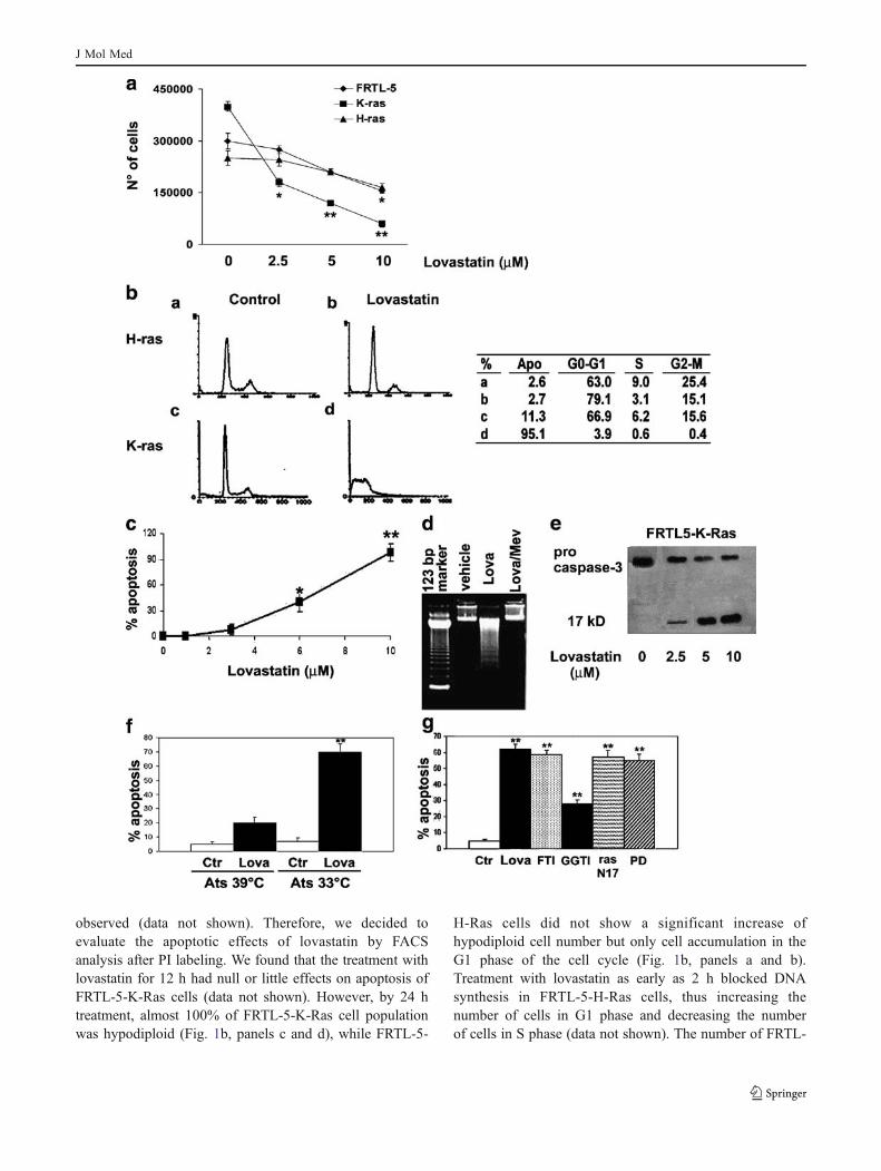
observed (data not shown). Therefore, we decided toevaluate the apoptotic effects of lovastatin by FACSanalysis after PI labeling. We found that the treatment withlovastatin for 12 h had null or little effects on apoptosis ofFRTL-5-K-Ras cells (data not shown). However, by 24 htreatment, almost 100% of FRTL-5-K-Ras cell populationwas hypodiploid (Fig. 1b, panels c and d), while FRTL-5-
H-Ras cells did not show a significant increase ofhypodiploid cell number but only cell accumulation in theG1 phase of the cell cycle (Fig. 1b, panels a and b).Treatment with lovastatin as early as 2 h blocked DNAsynthesis in FRTL-5-H-Ras cells, thus increasing thenumber of cells in G1 phase and decreasing the numberof cells in S phase (data not shown). The number of FRTL-
J Mol Med
5-K-Ras cells with hypodiploid DNA content, evaluatedafter 24 h of treatment by flow cytometry, increased in adose-dependent manner, attaining 100% at 10 µM lovastat-in concentration and 50% at a concentration of 7.5 µM(Fig. 1c). Loss of diploid DNA cell content started at 12 hfrom the beginning of lovastatin treatment and continued ina time-dependent manner (data not shown).
A further demonstration that the loss of DNA cellcontent induced by lovastatin was due to apoptosis wasobtained by DNA ladder analysis evaluated by agarose gelelectrophoresis that displayed the characteristic DNAfragmentation (Fig. 1d). A molecular assessment ofapoptosis induction was performed by evaluating, byWestern blot analysis, the levels and the cleavage into theactive form of pro-caspase 3. We observed a dose-dependent increase in the activation of caspase 3(Fig. 1e). Interestingly, the proapoptotic effect of lovastatinwas prevented by the addition of mevalonate (0.7 mM;Fig. 1d), whereas the same analysis performed on FRTL-5-H-Ras cells did not show any DNA degradation (data notshown). To further confirm that the induction of apoptosisby lovastatin was an effect specifically dependent on K-rastransformation of thyroid cells, we used a different FRTL-5-cell-derived line, Ats that is transformed with a Kirsten-murine sarcoma virus variant carrying a temperature-sensitive v-K-ras allele [21]. Lovastatin had no significanteffect on cells grown at 39°C, a nonpermissive temperaturefor p21 ras activity, whereas apoptosis was rapidly inducedby lovastatin at the permissive temperature of 33°C(Fig. 1f).
Role of farnesylation in lovastatin-induced apoptosisin K-ras-transformed cells
Lovastatin is an inhibitor of HMG-CoA reductase andaffects FTRL-5 cell proliferation mainly through an indirectinhibition of protein farnesylation [18]. In order to identifythe mechanisms responsible for the induction of apoptosisin cells expressing an active form of K-ras oncogene, wedetermined whether both farnesylation inhibition and K-raswere required for the observed effect and, thereafter,investigated the downstream mitogen-activated proteinkinase (MAPK) pathway activated by ras. To determinewhether inhibition of protein prenylation was responsiblefor apoptosis induced by lovastatin in FRTL-5-K-Ras cells,we used farnesyl- or geranylgeranyl-protein transferaseinhibitors (FTI-277 or GGTI-298, respectively; Fig. 1g).Treatment with 40 µM of GGTI-298 for 16 h inducedapoptosis only in a small percentage of cells, whereas FTI-277 completely reproduced the effect induced by lovastatin,thus suggesting a main role of farnesyl-protein transferaseinhibition (Fig. 1g). Furthermore, transfection of FRTL-5-K-Ras cells with a construct expressing a negative-
dominant mutant of ras protein produced effects compara-ble to those caused by lovastatin or FTI-277. These dataconfirmed the hypothesis that inhibition of HMG-CoAreductase by lovastatin occurred through the inactivation ofras protein due to the inhibition of its farnesylation(Fig. 1g).
Effects of lovastatin on ras-dependent survival signaltransduction pathways
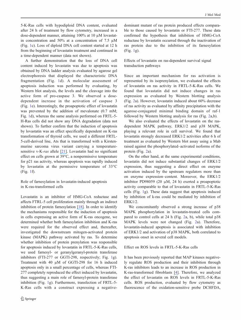
Since an important mechanism for ras activation isrepresented by its isoprenylation, we evaluated the effectsof lovastatin on ras activity in FRTL-5-K-Ras cells. Wefound that lovastatin did not induce changes in rasexpression as evaluated by Western blotting analysis(Fig. 2a). However, lovastatin induced about 60% decreaseof ras activity as evaluated by affinity precipitation with theagarose-conjugated minimal binding domain of raf-1followed by Western blotting analysis for ras (Fig. 2a,b).
We also evaluated the effects of lovastatin on the ras-dependent MAPK pathway, ERK1/2 and p38 MAPK,playing a relevant role in cell survival. We found thatlovastatin strongly decreased ERK1/2 activities after 6 h oftreatment as evaluated by Western blot assay using a Mabraised against the phosphorylated–activated isoforms of theprotein (Fig. 2a).
On the other hand, at the same experimental conditions,lovastatin did not induce substantial changes of ERK1/2expression, thus suggesting a direct effect on enzymeactivation induced by the upstream regulators more thanon enzyme expression–content. Moreover, the ERK1/2inhibitor PD98059 (20 μM, 24 h) exerted a proapoptoticactivity comparable to that of lovastatin in FRTL-5-K-Rascells (Fig. 1g). These data suggest that apoptosis inducedby inhibition of k-ras could be mediated by inhibition ofERK1/2.
We concomitantly observed a strong increase of p38MAPK phosphorylation in lovastatin-treated cells com-pared to control cells at 24 h (Fig. 2a, b), while total p38MAPK levels were not changed (Fig. 2a). Therefore,lovastatin-induced apoptosis is associated with inhibitionof ERK1/2 and activation of p38 MAPK, both correlated toapoptosis onset in several cell models.
Effect on ROS levels in FRTL-5-K-Ras cells
It has been previously reported that MAP kinases negative-ly regulate ROS production and their inhibition throughK-ras inhibitors leads to an increase in ROS production inK-ras-transformed fibroblasts [4]. Therefore, we analyzedthe effect of lovastatin on ROS levels in FRTL-5-K-Rascells. ROS production, evaluated by flow cytometry asfluorescence of the oxidation-sensitive probe DCHFDA,
J Mol Med
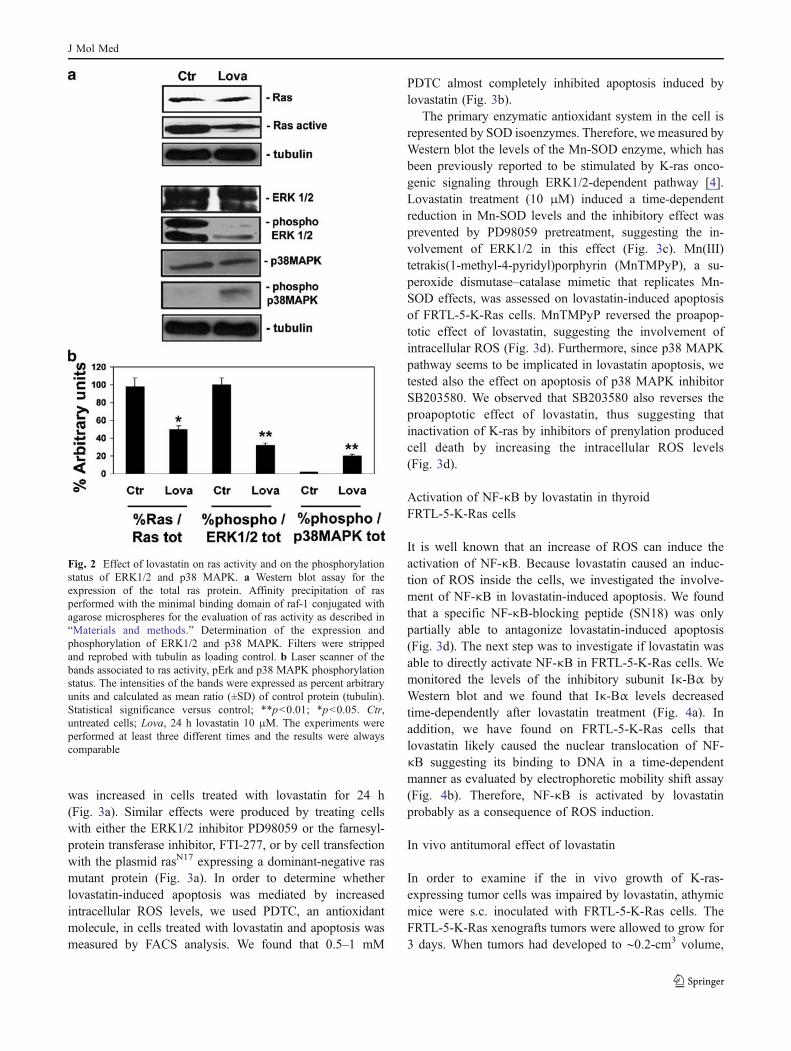
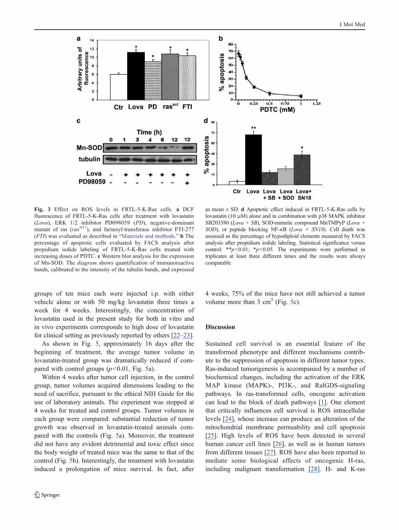
was increased in cells treated with lovastatin for 24 h(Fig. 3a). Similar effects were produced by treating cellswith either the ERK1/2 inhibitor PD98059 or the farnesyl-protein transferase inhibitor, FTI-277, or by cell transfectionwith the plasmid rasN17 expressing a dominant-negative rasmutant protein (Fig. 3a). In order to determine whetherlovastatin-induced apoptosis was mediated by increasedintracellular ROS levels, we used PDTC, an antioxidantmolecule, in cells treated with lovastatin and apoptosis wasmeasured by FACS analysis. We found that 0.5–1 mM
PDTC almost completely inhibited apoptosis induced bylovastatin (Fig. 3b).
The primary enzymatic antioxidant system in the cell isrepresented by SOD isoenzymes. Therefore, we measured byWestern blot the levels of the Mn-SOD enzyme, which hasbeen previously reported to be stimulated by K-ras onco-genic signaling through ERK1/2-dependent pathway [4].Lovastatin treatment (10 μM) induced a time-dependentreduction in Mn-SOD levels and the inhibitory effect wasprevented by PD98059 pretreatment, suggesting the in-volvement of ERK1/2 in this effect (Fig. 3c). Mn(III)tetrakis(1-methyl-4-pyridyl)porphyrin (MnTMPyP), a su-peroxide dismutase–catalase mimetic that replicates Mn-SOD effects, was assessed on lovastatin-induced apoptosisof FRTL-5-K-Ras cells. MnTMPyP reversed the proapop-totic effect of lovastatin, suggesting the involvement ofintracellular ROS (Fig. 3d). Furthermore, since p38 MAPKpathway seems to be implicated in lovastatin apoptosis, wetested also the effect on apoptosis of p38 MAPK inhibitorSB203580. We observed that SB203580 also reverses theproapoptotic effect of lovastatin, thus suggesting thatinactivation of K-ras by inhibitors of prenylation producedcell death by increasing the intracellular ROS levels(Fig. 3d).
Activation of NF-κB by lovastatin in thyroidFRTL-5-K-Ras cells
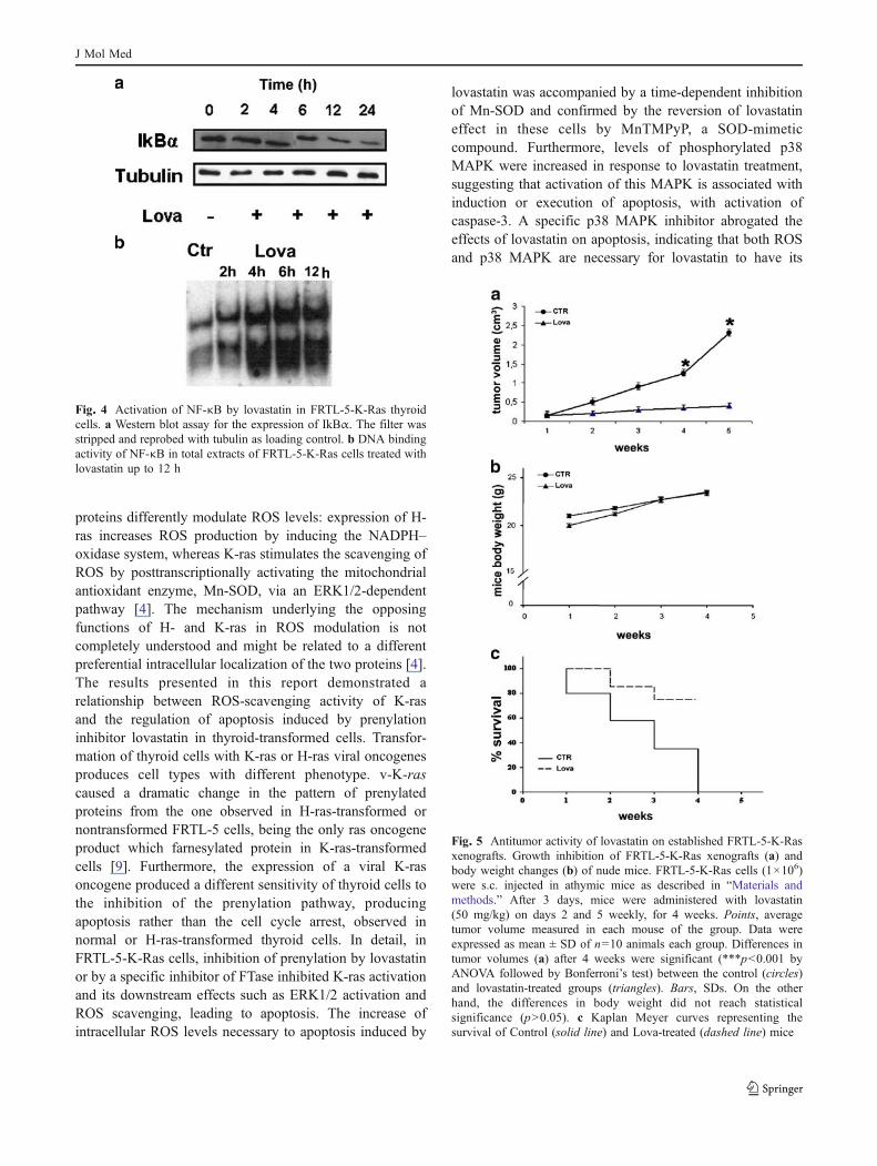
It is well known that an increase of ROS can induce theactivation of NF-κB. Because lovastatin caused an induc-tion of ROS inside the cells, we investigated the involve-ment of NF-κB in lovastatin-induced apoptosis. We foundthat a specific NF-κB-blocking peptide (SN18) was onlypartially able to antagonize lovastatin-induced apoptosis(Fig. 3d). The next step was to investigate if lovastatin wasable to directly activate NF-κB in FRTL-5-K-Ras cells. Wemonitored the levels of the inhibitory subunit Iκ-Bα byWestern blot and we found that Iκ-Bα levels decreasedtime-dependently after lovastatin treatment (Fig. 4a). Inaddition, we have found on FRTL-5-K-Ras cells thatlovastatin likely caused the nuclear translocation of NF-κB suggesting its binding to DNA in a time-dependentmanner as evaluated by electrophoretic mobility shift assay(Fig. 4b). Therefore, NF-κB is activated by lovastatinprobably as a consequence of ROS induction.
In vivo antitumoral effect of lovastatin
In order to examine if the in vivo growth of K-ras-expressing tumor cells was impaired by lovastatin, athymicmice were s.c. inoculated with FRTL-5-K-Ras cells. TheFRTL-5-K-Ras xenografts tumors were allowed to grow for3 days. When tumors had developed to ∼0.2-cm3 volume,
Fig. 2 Effect of lovastatin on ras activity and on the phosphorylationstatus of ERK1/2 and p38 MAPK. a Western blot assay for theexpression of the total ras protein. Affinity precipitation of rasperformed with the minimal binding domain of raf-1 conjugated withagarose microspheres for the evaluation of ras activity as described in“Materials and methods.” Determination of the expression andphosphorylation of ERK1/2 and p38 MAPK. Filters were strippedand reprobed with tubulin as loading control. b Laser scanner of thebands associated to ras activity, pErk and p38 MAPK phosphorylationstatus. The intensities of the bands were expressed as percent arbitraryunits and calculated as mean ratio (±SD) of control protein (tubulin).Statistical significance versus control; **p<0.01; *p<0.05. Ctr,untreated cells; Lova, 24 h lovastatin 10 μM. The experiments wereperformed at least three different times and the results were alwayscomparable
J Mol Med
groups of ten mice each were injected i.p. with eithervehicle alone or with 50 mg/kg lovastatin three times aweek for 4 weeks. Interestingly, the concentration oflovastatin used in the present study for both in vitro andin vivo experiments corresponds to high dose of lovastatinfor clinical setting as previously reported by others [22–23].
As shown in Fig. 5, approximately 16 days after thebeginning of treatment, the average tumor volume inlovastatin-treated group was dramatically reduced if com-pared with control groups (p<0.01, Fig. 5a).
Within 4 weeks after tumor cell injection, in the controlgroup, tumor volumes acquired dimensions leading to theneed of sacrifice, pursuant to the ethical NIH Guide for theuse of laboratory animals. The experiment was stopped at4 weeks for treated and control groups. Tumor volumes ineach group were compared: substantial reduction of tumorgrowth was observed in lovastatin-treated animals com-pared with the controls (Fig. 5a). Moreover, the treatmentdid not have any evident detrimental and toxic effect sincethe body weight of treated mice was the same to that of thecontrol (Fig. 5b). Interestingly, the treatment with lovastatininduced a prolongation of mice survival. In fact, after
4 weeks, 75% of the mice have not still achieved a tumorvolume more than 3 cm3 (Fig. 5c).
Discussion
Sustained cell survival is an essential feature of thetransformed phenotype and different mechanisms contrib-ute to the suppression of apoptosis in different tumor types.Ras-induced tumorigenesis is accompanied by a number ofbiochemical changes, including the activation of the ERKMAP kinase (MAPK)-, PI3K-, and RalGDS-signalingpathways. In ras-transformed cells, oncogene activationcan lead to the block of death pathways [1]. One elementthat critically influences cell survival is ROS intracellularlevels [24], whose increase can produce an alteration of themitochondrial membrane permeability and cell apoptosis[25]. High levels of ROS have been detected in severalhuman cancer cell lines [26], as well as in human tumorsfrom different tissues [27]. ROS have also been reported tomediate some biological effects of oncogenic H-ras,including malignant transformation [28]. H- and K-ras
Fig. 3 Effect on ROS levels in FRTL-5-K-Ras cells. a DCFfluorescence of FRTL-5-K-Ras cells after treatment with lovastatin(Lova), ERK 1/2 inhibitor PD098059 (PD), negative-dominantmutant of ras (rasN17), and farnesyl-transferase inhibitor FTI-277(FTI) was evaluated as described in “Materials and methods.” b Thepercentage of apoptotic cells evaluated by FACS analysis afterpropidium iodide labeling of FRTL-5-K-Ras cells treated withincreasing doses of PDTC. c Western blot analysis for the expressionof Mn-SOD. The diagram shows quantification of immunoreactivebands, calibrated to the intensity of the tubulin bands, and expressed
as mean ± SD. d Apoptotic effect induced in FRTL-5-K-Ras cells bylovastatin (10 μM) alone and in combination with p38 MAPK inhibitorSB203580 (Lova + SB), SOD-mimetic compound MnTMPyP (Lova +SOD), or peptide blocking NF-κB (Lova + SN18). Cell death wasassessed as the percentage of hypodiploid elements measured by FACSanalysis after propidium iodide labeling. Statistical significance versuscontrol: **p<0.01; *p<0.05. The experiments were performed intriplicates at least three different times and the results were alwayscomparable
J Mol Med
proteins differently modulate ROS levels: expression of H-ras increases ROS production by inducing the NADPH–oxidase system, whereas K-ras stimulates the scavenging ofROS by posttranscriptionally activating the mitochondrialantioxidant enzyme, Mn-SOD, via an ERK1/2-dependentpathway [4]. The mechanism underlying the opposingfunctions of H- and K-ras in ROS modulation is notcompletely understood and might be related to a differentpreferential intracellular localization of the two proteins [4].The results presented in this report demonstrated arelationship between ROS-scavenging activity of K-rasand the regulation of apoptosis induced by prenylationinhibitor lovastatin in thyroid-transformed cells. Transfor-mation of thyroid cells with K-ras or H-ras viral oncogenesproduces cell types with different phenotype. v-K-rascaused a dramatic change in the pattern of prenylatedproteins from the one observed in H-ras-transformed ornontransformed FRTL-5 cells, being the only ras oncogeneproduct which farnesylated protein in K-ras-transformedcells [9]. Furthermore, the expression of a viral K-rasoncogene produced a different sensitivity of thyroid cells tothe inhibition of the prenylation pathway, producingapoptosis rather than the cell cycle arrest, observed innormal or H-ras-transformed thyroid cells. In detail, inFRTL-5-K-Ras cells, inhibition of prenylation by lovastatinor by a specific inhibitor of FTase inhibited K-ras activationand its downstream effects such as ERK1/2 activation andROS scavenging, leading to apoptosis. The increase ofintracellular ROS levels necessary to apoptosis induced by
lovastatin was accompanied by a time-dependent inhibitionof Mn-SOD and confirmed by the reversion of lovastatineffect in these cells by MnTMPyP, a SOD-mimeticcompound. Furthermore, levels of phosphorylated p38MAPK were increased in response to lovastatin treatment,suggesting that activation of this MAPK is associated withinduction or execution of apoptosis, with activation ofcaspase-3. A specific p38 MAPK inhibitor abrogated theeffects of lovastatin on apoptosis, indicating that both ROSand p38 MAPK are necessary for lovastatin to have its
Fig. 5 Antitumor activity of lovastatin on established FRTL-5-K-Rasxenografts. Growth inhibition of FRTL-5-K-Ras xenografts (a) andbody weight changes (b) of nude mice. FRTL-5-K-Ras cells (1×10⁶)were s.c. injected in athymic mice as described in “Materials andmethods.” After 3 days, mice were administered with lovastatin(50 mg/kg) on days 2 and 5 weekly, for 4 weeks. Points, averagetumor volume measured in each mouse of the group. Data wereexpressed as mean ± SD of n=10 animals each group. Differences intumor volumes (a) after 4 weeks were significant (***p<0.001 byANOVA followed by Bonferroni’s test) between the control (circles)and lovastatin-treated groups (triangles). Bars, SDs. On the otherhand, the differences in body weight did not reach statisticalsignificance (p>0.05). c Kaplan Meyer curves representing thesurvival of Control (solid line) and Lova-treated (dashed line) mice
Fig. 4 Activation of NF-κB by lovastatin in FRTL-5-K-Ras thyroidcells. a Western blot assay for the expression of IkBα. The filter wasstripped and reprobed with tubulin as loading control. b DNA bindingactivity of NF-κB in total extracts of FRTL-5-K-Ras cells treated withlovastatin up to 12 h
J Mol Med
proapoptotic effects. p38 MAPK may also antagonizemalignant transformation induced by all oncogenic rasforms [29]. It has recently been reported that p38 MAPKspecifically modulates malignant transformation induced byoncogenes that produce ROS [30]. Interestingly, sincehighly tumorigenic cancer cells can override this p38function uncoupling its activation from oxidative stressproduction, its induction by a chemotherapy drug could bevery useful. In our model, p38 MAPK could be probablyactivated as a consequence of the high ROS levelsaccumulated in the cells after lovastatin inhibition of K-ras oncogene signaling pathways and of Mn-SOD, ratherthan a direct target of p38 MAPK signaling. In ourexperimental model, Mn-SOD expression was reduced byLOVA treatment even if a significant ROS elevation wasalso recorded. In fact, ROS activates the nuclear factor E2-related factor-2 (Nrf2) that is a member of CNC (cap ‘n’collar) family of b-Zip transcription factors and is anindispensable positive regulator of many antioxidant andphase II detoxifying enzymes [31]. On activation byoxidative or electrophilic stress, Nrf2 protein stabilizes,translocates to the nucleus, heterodimerizes with small Mafproteins, and binds to the so-called antioxidant responseelement, a common regulatory element found in the 5′-flanking regions of antioxidant and detoxification enzymesincluding SOD [32]. On the other hand, eukaryotic cellscan protect themselves from oxidative stress through theinduction of SOD expression via a ras-dependent pathway[33]. On the basis of these considerations, the decreasedMn-SOD expression found in our conditions and occurringtogether with ROS elevation could be, at least in part,explained by the block of ras activity induced by lovastatin.
ROS are emerging as key effectors in signal transductionand this role is especially evident in the pathways leading toprogrammed cell death. Another pathway that is underROS-mediated control in some systems is that leading toactivation of NF-κB, which is a central regulator of cellsurvival, being either as a proapoptotic or as an antiapop-totic protein depending on cell type and apoptotic inducers[34]. Our data suggest that lovastatin induced NF-κBtranscriptional activity in FRTL-5-K-Ras cells, probablyas a consequence of ROS induction, suggesting a possiblerole of this factor worth to be investigated. There is alsoevidence that p38 MAPK may be involved in NF-κBactivation [35]. Indeed, we found that a specific NF-κB-blocking peptide (SN18) was only partially able toantagonize lovastatin-induced apoptosis, probably becauseof the sensitization to apoptosis mediated per se by NF-κBblockade [36].
The antitumoral activity of lovastatin was assessed invivo on the growth of tumors generated by FRTL-5-K-Rass.c. cell injection in nude mice. The dramatic effectobserved in the treated group recommended further inves-
tigations on other benign or malignant thyroid proliferativediseases. It is important to note that lovastatin is able toexert apoptotic and differentiation effects against anaplasticthyroid carcinoma cell lines. We have recently shown thatlovastatin treatment significantly increased the effects of theoncolytic virus dl1520 against ATC cells [37]. In thiscellular model, the replication of dl1520 was enhanced bylovastatin treatment, and a significant increase of theexpression of the early gene E1A 13 S and the late genePenton was observed in lovastatin-treated cells. Finally,lovastatin treatment significantly enhanced the effects ofdl1520 against ATC tumor xenografts [37].
Both tumor and transformed cells are usually moreresistant to oxidative stress with respect to normal cells[38]. This peculiarity could interfere with the efficacy ofantitumor therapy based on free radical generation likeradiotherapy or chemotherapy with various drugs. There-fore, the finding of new therapeutic targets able to enhancefree-radical-mediated damage in tumor cells is of increasinginterest. Moreover, the present report is one of the firstdemonstrations that K-ras targeting could be a useful tool toincrease oxidative stress in thyroid cancer and the lattereffect induces modulation of multiple signals (ERK, NF-κB, p38 kinase) that could represent additional therapeutictargets in integrated anticancer strategies. In conclusion, thedifferential sensitivity of K-ras-transformed thyroid cells toinhibitors of prenylation with respect to induction ofapoptosis could be a solid ground to build therapeuticstrategies for those proliferative diseases sustained by aninappropriate K-ras expression.
Acknowledgments This work was supported by the Italian Associ-ation for Cancer Research (AIRC) and by the Associazione Educa-zione e Ricerca Medica Salernitana. Simona Pisanti was supported bya fellowship from FIRC. We thank Prof. Maria Caterina Turco,Department of Pharmaceutical Sciences, University of Salerno, Dr.Maria Rosaria Santillo and Dr. Rosalba Serù, Department ofNeuroscience, University of Naples Federico II, Italy, for usefuldiscussion and technical assistance.
References
1. Downward J (2003) Targeting RAS signalling pathways in cancertherapy. Nat Rev Cancer 3:11–22
2. Wittinghofer A, Nassar N (1996) How Ras-related proteins talk totheir effectors. Trends Biochem Sci 21:488–491
3. Rassool FV, Gaymes TJ, Omidvar N, Brady N, Beurlet S, Pla M,Reboul M, Lea N, Chomienne C, Thomas NS, Mufti GJ, PaduaRA (2007) Reactive oxygen species, DNA damage, and error-prone repair: a model for genomic instability with progression inmyeloid leukemia? Cancer Res 67(18):8762–8771
4. Santillo M, Mondola P, Serù R, Annella T, Cassano S, Ciullo I,Tecce MF, Iacomino G, Damiano S, Cuda G (2001) Opposingfunctions of Ki- and Ha-Ras genes in the regulation of redoxsignals. Current Biology 11:614–619
J Mol Med
5. Casey PJ, Solski PA, Der CJ, Buss JE (1989) p21ras is modifiedby a farnesyl isoprenoid. Proc Natl Acad Sci USA 86:8323–8327
6. Hancock JF, Magee AI, Childs JE, Marshall CJ (1989) All rasproteins are polyisoprenylated but only some are palmitoylated.Cell 57:1167–1177
7. Trahey M, McCormick F (1987) A cytoplasmic protein stimulatesnormal N-ras p21 GTPase, but does not affect oncogenic mutants.Science 238:542–545
8. Kondo T, Ezzat S, Asa SL (2006) Pathogenetic mechanisms inthyroid follicular-cell neoplasia. Nat Rev Cancer 6(4):292–306
9. Laezza C, Di Marzo V, Bifulco M (1998) v-K-ras leads topreferential farnesylation of p21(ras) in FRTL-5 cells: multipleinterference with the isoprenoid pathway. Proc Natl Acad SciUSA 95:13646–13651
10. Caraglia M, Budillon A, Tagliaferri P, Marra M, Abbruzzese A,Caponigro F (2005) Isoprenylation of intracellular proteins as anew target for the therapy of human neoplasms: preclinical andclinical implications. Curr Drug Targets 6:301–323
11. Caraglia M, Santini D, Marra M, Vincenti B, Tonini G, Budillon A(2006) Emerging anti-cancer molecular mechanisms of aminobi-sphosphonates. Endocr Rel Cancer 13:7–26
12. Agarwal B, Bhendwal S, Halmos B, Moss SF, Ramey WG,Holt PR (1999) Lovastatin augments apoptosis induced bychemotherapeutic agents in colon cancer cells. Clin Cancer Res5:2223–2229
13. Zhong WB, Liang YC, Wang CY, Chang TC, Lee WS (2005)Lovastatin suppresses invasiveness of anaplastic thyroid cancercells by inhibiting Rho geranylgeranylation and RhoA/ROCKsignaling. Endocr Relat Cancer 12:615–629
14. Yao CJ, Lai GM, Chan CF, Cheng AL, Yang YY, Chuang SE(2006) Dramatic synergistic anticancer effect of clinically achiev-able doses of lovastatin and troglitazone. Int J Cancer 118:773–779
15. Wong WW, Clendening JW, Martirosyan A, Boutros PC, Bros C,Khosravi F, Jurisica I, Stewart AK, Bergsagel PL, Penn LZ (2007)Determinants of sensitivity to lovastatin-induced apoptosis inmultiple myeloma. Mol Cancer Ther 6:1886–1897
16. Vitale M, Di Matola T, Rossi G, Laezza C, Fenzi G, Bifulco M(1999) Prenyltransferase inhibitors induce apoptosis in proliferat-ing thyroid cells through a p53-independent CrmA-sensitive, andcaspase-3-like protease-dependent mechanism. Endocrinology140:698–704
17. D’Acquisto F, de Cristofaro F, Maiuri MC, Tajana G, Carnuccio R(2001) Protective role of nuclear factor kappa B against nitricoxide-induced apoptosis in J774 macrophages. Cell Death Differ8:144–151
18. Bifulco M, Laezza C, Aloj SM (1999) Inhibition of farnesylationblocks growth but not differentiation in FRTL-5 thyroid cells.Biochimie 81:287–290
19. Fusco A, Pinto A, Ambesi-Impiombato FS, Vecchio G, Tsuchida N(1981) Transformation of rat thyroid epithelial cells by Kirstenmurine sarcoma virus. Int J Cancer 28:655–662
20. Fusco A, Portella G, Di Fiore PP, Berlingieri MT, Di Lauro R,Schneider AB, Vecchio G (1985) A mos oncogene-containingretrovirus, myeloproliferative sarcoma virus, transforms rat thy-roid epithelial cells and irreversibly blocks their differentiationpattern. J Virol 56:284–292
21. Colletta G, Pinto A, Di Fiore PP, Fusco A, Ferrentino M,Avvedimento VE, Tsuchida N, Vecchio G (1983) Dissociationbetween transformed and differentiated phenotype in rat thyroidepithelial cells after transformation with a temperature-sensitive
mutant of the Kirsten murine sarcoma virus. Mol Cell Biol3:2099–2109
22. McAnally JA, Gupta J, Sodhani S, Bravo L, Mo H (2007)Tocotrienols potentiate lovastatin-mediated growth suppression invitro and in vivo. Exp Biol Med 232:523–531
23. Shibata MA, Kavanaugh C, Shibata E, Abe H, Nguyen P, Otsuki Y,Trepel JB, Green JE (2003) Comparative effects of lovastatin onmammary and prostate oncogenesis in transgenic mouse models.Carcinogenesis 24:453–459
24. Nomura K, Imai H, Koumura T, Arai M, Nakagawa Y (1999)Mitochondrial phospholipid hydroperoxide glutathione peroxidasesuppresses apoptosis mediated by a mitochondrial death pathway.J Biol Chem 274:29294–29302
25. Takahashi A, Masuda A, Sun M, Centonze VE, Herman B (2004)Oxidative stress-induced apoptosis is associated with alterations inmitochondrial caspase activity and Bcl-2-dependent alterations inmitochondrial pH (pHm). Brain Res Bull 62:497–504
26. Szatrowski TP, Nathan CF (1991) Production of large amounts ofhydrogen peroxide by human tumor cells. Cancer Res 51:794–798
27. Toyokuni S, Okamoto K, Yodoi J, Hiai H (1995) Persistentoxidative stress in cancer. FEBS Lett 358:1–3
28. Mitsushita J, Lambeth JD, Kamata T (2004) The superoxidegen-erating oxidase Nox1 is functionally required for Ras oncogenetransformation. Cancer Res 64:3580–3585
29. Qi X, Tang J, Pramanik R, Schultz RM, Shirasawa S, Sasazuki T,Han J, Chen G (2004) p38 MAPK activation selectively inducescell death in K-ras-mutated human colon cancer cells throughregulation of vitamin D receptor. J Biol Chem 279:22138–22144
30. Dolado I, Swat A, Ajenjo N, De Vita G, Cuadrado A, Nebreda AR(2007) p38alpha MAP kinase as a sensor of reactive oxygenspecies in tumorigenesis. Cancer Cell 11:191–205
31. Motohashi H, Yamamoto M (2004) Nrf2-Keap1 defines aphysiologically important stress response mechanism. TrendsMol Med 10:549–557
32. Zhu H, Itoh K, Yamamoto M, Zweier JL, Li Y (2005) Role ofNrf2 signaling in regulation of antioxidants and phase 2 enzymesin cardiac fibroblasts: protection against reactive oxygen andnitrogen species-induced cell injury. FEBS Lett 579:3029–3036
33. Scorziello A, SantilloM, Adornetto A, Dell’aversano C, Sirabella R,Damiano S, Canzoniero LM, Renzo GF, Annunziato L (2007) NO-induced neuroprotection in ischemic preconditioning stimulatesmitochondrial Mn-SOD activity and expression via Ras/ERK1/2pathway. J Neurochem 103:1472–1480
34. Barkett M, Gilmore TD (1999) Control of apoptosis by Rel/NfkBtranscription factors. Oncogene 18:6910
35. Vanden Berghe W, Plaisance S, Boone E, De Bosscher K,Schmitz ML, Fiers W, Haegeman G (1998) p38 and extracellularsignal-regulated kinase mitogen-activated protein kinase pathwaysare required for nuclear factor-kB p65 transactivation mediated bytumor necrosis factor. J Biol Chem 273:3285–3290
36. Wang X, Chen W, Lin Y (2007) Sensitization of TNF-inducedcytotoxicity in lung cancer cells by concurrent suppression of theNF-kappa B and Akt pathways. Biochem Biophys Res Commun355:807–812
37. Libertini S, Iacuzzo I, Ferraro A, Vitale M, Bifulco M, Fusco A,Portella G (2007) Lovastatin enhances the replication of theoncolytic adenovirus dl1520 and its antineoplastic activityagainst anaplastic thyroid carcinoma cells. Endocrinology 148:5186–5194
38. Martindale JL, Holbrook NJ (2002) Cellular response to oxidativestress: signaling for suicide and survival. J Cell Physiol 192:1–15
J Mol Med
Related Documents