ORIGINAL ARTICLE Lost in translation: translational interference from a recurrent mutation in exon 1 of MECP2 A Saxena, D de Lagarde, H Leonard, S L Williamson, V Vasudevan, J Christodoulou, E Thompson, P MacLeod, D Ravine ............................................................................................................................... See end of article for authors’ affiliations ....................... Correspondence to: Professor David Ravine, Western Australian Institute for Medical Research, Centre for Medical Research and School of Medicine and Pharmacology, University of Western Australia, Level 2, North Block, Royal Perth Hospital, Perth 6000, WA, Australia; ravine@waimr. uwa.edu.au Revised version received 15 August 2005 Accepted for publication 30 August 2005 Published Online First 12 September 2005 ....................... J Med Genet 2006;43:470–477. doi: 10.1136/jmg.2005.036244 Background: Rett syndrome (RTT) is an X linked neuro-developmental disorder affecting mostly girls. Mutations in the coding region of MECP2 are found in 80% of classic RTT patients. Until recently, the region encoding MECP2 was believed to comprise exons 2, 3, and 4 with the ATG start site located at the end of exon 2 (MeCP2_e2). Methods: Recent reports of another mRNA transcript transcribed from exon 1 (MeCP2_e1) prompted us to screen exon 1 among RNA samples from 20 females with classic or atypical RTT. Results: A previously reported 11 base pair deletion in exon 1 was detected in one subject with a milder phenotype. Although RNA expression for both protein isoforms was detected from the mutant allele, evaluation of MeCP2 protein in uncultured patient lymphocytes by immunocytochemistry revealed that MeCP2 protein production was restricted to only 74–76% of lymphocytes. X chromosome inactivation studies of genomic DNA revealed similar XCI ratios at the HUMARA locus (73:27 with HpaII and 74:26 with McrBC). We have demonstrated that translation but not transcription of the MeCP2_e2 isoform is ablated by the 11 nucleotide deletion, 103 nucleotides upstream of the e2 translation start site. Conclusions: These findings reveal that nucleotides within the deleted sequence in the 59-UTR of the MeCP2_e2 transcript, while not required for transcription, are essential for translation. R TT syndrome (RTT) is a rare neuro-developmental disorder affecting 1 in 10 000 girls and is mostly caused by mutations in the X linked methyl CpG binding protein 2 gene (MECP2), which is subject to X inactivation. 1–3 Until recently, its protein coding region was believed to be restricted to sequence within exons 2, 3, and 4 with the translational start site positioned near the 39 end of exon 2. Reports in 2004 of an alternate mRNA transcript arising from exon 1, which is alternately spliced to exons 3 and 4, reveal a higher level of complexity than was previously appreciated. The protein isoform arising from exon 1 has been designated MeCP2_e1 (e1), while the originally defined protein isoform translated from exon 2 is now designated MeCP2_e2 (e2). Further, expression levels of the MeCP2_e1 mRNA transcript and protein isoform are much higher in brain than the expression levels of MeCP2_e2 mRNA transcript and its corresponding protein isoform. 45 Mutations in exons 3 and 4 are detected in 80% of patients with clinically diagnosed classic RTT and in a lower proportion of atypical RTT cases. 67 To date mutations have not been detected in exon 2, although a limited number of RTT causing mutations in exon 1 have now been reported. 589 Using primers in exons 1 and 3, we screened RNA prepared from fresh lymphocytes from 20 RTT cases that had been classified as either classic or atypical RTT within the Australian Rett Syndrome Database 10 and in whom earlier mutation screening had yielded wild type results. In one subject, we identified an 11 base pair deletion in exon 1 that has been previously reported. 89 The deletion lies in the AGG repeat region of exon 1 and was confirmed by direct sequence analysis of genomic DNA. Subsequent assessment of MeCP2 protein production in uncultured fresh lymphocytes by immunocytochemical analysis revealed absence of detectable immunostaining in 24–26% of cells. Since the deletion lies in the 59-UTR region of e2, we hypothesised that expression of the e2 RNA transcript may be disrupted. However, fragment length analysis of amplified cDNA revealed all four tran- scripts, two from the wild type allele and two from the mutant allele. We validated our immunocytochemistry results by quantitative assessment of the X inactivation ratio at the HUMARA locus in DNA prepared from the same uncultured patient cells and observed similar ratios of 73:27 with HpaII, and 74:26 with McrBC. Our observations suggest that the nucleotides within the deleted sequence in the 59- UTR of the e2 RNA transcript are essential for MeCP2_e2 protein translation. METHODS Sample collection Molecular and immunocytochemical studies were conducted on blood samples collected in tubes with ACD anti-coagulant from 20 female patients with classic or atypical RTT identified from the Australian Rett Syndrome Database and in whom earlier sequencing and MLPA screening of exons 2, 3, and 4 had not revealed a mutation. Prior approval for the study had been provided by the ethics committee of Princess Margaret Hospital for Children, Perth. Clinical details of the second case were provided from InterRett. 11 DNA analysis DNA was extracted from 1200 ml of blood using the Puregene DNA extraction kit (Gentra Systems, Minneapolis, MN) according to the manufacturer’s instructions. PCR analysis was performed on 50 ng of DNA as described previously. 12 mRNA analysis Total RNA was extracted from 1200 ml of blood using the Purescript RNA extraction kit (Gentra Systems) as per the 470 www.jmedgenet.com

Welcome message from author
This document is posted to help you gain knowledge. Please leave a comment to let me know what you think about it! Share it to your friends and learn new things together.
Transcript

ORIGINAL ARTICLE
Lost in translation: translational interference from arecurrent mutation in exon 1 of MECP2A Saxena, D de Lagarde, H Leonard, S L Williamson, V Vasudevan, J Christodoulou,E Thompson, P MacLeod, D Ravine. . . . . . . . . . . . . . . . . . . . . . . . . . . . . . . . . . . . . . . . . . . . . . . . . . . . . . . . . . . . . . . . . . . . . . . . . . . . . . . . . . . . . . . . . . . . . . . . . . . . . . . . . . . . . . . . . . . . . . . . . . . . . . .
See end of article forauthors’ affiliations. . . . . . . . . . . . . . . . . . . . . . .
Correspondence to:Professor David Ravine,Western AustralianInstitute for MedicalResearch, Centre forMedical Research andSchool of Medicine andPharmacology, Universityof Western Australia, Level2, North Block, Royal PerthHospital, Perth 6000, WA,Australia; [email protected]
Revised version received15 August 2005Accepted for publication30 August 2005Published Online First12 September 2005. . . . . . . . . . . . . . . . . . . . . . .
J Med Genet 2006;43:470–477. doi: 10.1136/jmg.2005.036244
Background: Rett syndrome (RTT) is an X linked neuro-developmental disorder affecting mostly girls.Mutations in the coding region of MECP2 are found in 80% of classic RTT patients. Until recently, theregion encoding MECP2 was believed to comprise exons 2, 3, and 4 with the ATG start site located at theend of exon 2 (MeCP2_e2).Methods: Recent reports of another mRNA transcript transcribed from exon 1 (MeCP2_e1) prompted us toscreen exon 1 among RNA samples from 20 females with classic or atypical RTT.Results: A previously reported 11 base pair deletion in exon 1 was detected in one subject with a milderphenotype. Although RNA expression for both protein isoforms was detected from the mutant allele,evaluation of MeCP2 protein in uncultured patient lymphocytes by immunocytochemistry revealed thatMeCP2 protein production was restricted to only 74–76% of lymphocytes. X chromosome inactivationstudies of genomic DNA revealed similar XCI ratios at the HUMARA locus (73:27 with HpaII and 74:26with McrBC). We have demonstrated that translation but not transcription of the MeCP2_e2 isoform isablated by the 11 nucleotide deletion, 103 nucleotides upstream of the e2 translation start site.Conclusions: These findings reveal that nucleotides within the deleted sequence in the 59-UTR of theMeCP2_e2 transcript, while not required for transcription, are essential for translation.
RTT syndrome (RTT) is a rare neuro-developmentaldisorder affecting 1 in 10 000 girls and is mostly causedby mutations in the X linked methyl CpG binding
protein 2 gene (MECP2), which is subject to X inactivation.1–3
Until recently, its protein coding region was believed to berestricted to sequence within exons 2, 3, and 4 with thetranslational start site positioned near the 39 end of exon 2.Reports in 2004 of an alternate mRNA transcript arising fromexon 1, which is alternately spliced to exons 3 and 4, reveal ahigher level of complexity than was previously appreciated.The protein isoform arising from exon 1 has been designatedMeCP2_e1 (e1), while the originally defined protein isoformtranslated from exon 2 is now designated MeCP2_e2 (e2).Further, expression levels of the MeCP2_e1 mRNA transcriptand protein isoform are much higher in brain than theexpression levels of MeCP2_e2 mRNA transcript and itscorresponding protein isoform.4 5
Mutations in exons 3 and 4 are detected in 80% of patientswith clinically diagnosed classic RTT and in a lowerproportion of atypical RTT cases.6 7 To date mutations havenot been detected in exon 2, although a limited number ofRTT causing mutations in exon 1 have now been reported.5 8 9
Using primers in exons 1 and 3, we screened RNA preparedfrom fresh lymphocytes from 20 RTT cases that had beenclassified as either classic or atypical RTT within theAustralian Rett Syndrome Database10 and in whom earliermutation screening had yielded wild type results. In onesubject, we identified an 11 base pair deletion in exon 1 thathas been previously reported.8 9 The deletion lies in the AGGrepeat region of exon 1 and was confirmed by direct sequenceanalysis of genomic DNA. Subsequent assessment of MeCP2protein production in uncultured fresh lymphocytes byimmunocytochemical analysis revealed absence of detectableimmunostaining in 24–26% of cells. Since the deletion lies in
the 59-UTR region of e2, we hypothesised that expression ofthe e2 RNA transcript may be disrupted. However, fragmentlength analysis of amplified cDNA revealed all four tran-scripts, two from the wild type allele and two from themutant allele. We validated our immunocytochemistryresults by quantitative assessment of the X inactivation ratioat the HUMARA locus in DNA prepared from the sameuncultured patient cells and observed similar ratios of 73:27with HpaII, and 74:26 with McrBC. Our observations suggestthat the nucleotides within the deleted sequence in the 59-UTR of the e2 RNA transcript are essential for MeCP2_e2protein translation.
METHODSSample collectionMolecular and immunocytochemical studies were conductedon blood samples collected in tubes with ACD anti-coagulantfrom 20 female patients with classic or atypical RTT identifiedfrom the Australian Rett Syndrome Database and in whomearlier sequencing and MLPA screening of exons 2, 3, and 4had not revealed a mutation. Prior approval for the study hadbeen provided by the ethics committee of Princess MargaretHospital for Children, Perth. Clinical details of the secondcase were provided from InterRett.11
DNA analysisDNA was extracted from 1200 ml of blood using the PuregeneDNA extraction kit (Gentra Systems, Minneapolis, MN)according to the manufacturer’s instructions. PCR analysiswas performed on 50 ng of DNA as described previously.12
mRNA analysisTotal RNA was extracted from 1200 ml of blood using thePurescript RNA extraction kit (Gentra Systems) as per the
470
www.jmedgenet.com
manufacturer’s instructions. RT-PCR was performed on RNAusing the Titan one step RT-PCR kit (Roche Applied Science,Castle Hill, NSW, Australia). In brief, 50 ng of RNA wasadded to the RT-PCR mix containing dNTPs (10 mM), DTT(125 mM), 56RT-PCR buffer, 50 ng each of forward (Ex1F2)and reverse (Ex4R2) primers in exons 1 and 4, respectively,and 1 U of enzyme (refer to table 1 for primer sequences).After reverse transcription at 48 C̊ (30 min), the sampleswere PCR amplified at an annealing temperature of 61 C̊ for25 cycles. A 1 ml aliquot of the product from the primary PCRwas further amplified in a semi-nested PCR reaction usingprimers in exon 1 (Ex1F2) and exon 3 (Ex3R2) and Tthpolymerase enzyme at an annealing temperature of 61 C̊ for25 cycles. PCR products were assessed visually by electro-phoresis on a 2% agarose gel.
Purification and sequencing of PCR productsPCR products were purified using the Qiaquick PCRpurification kit or gel extracted using the Qiaex II Gelpurification kit (Qiagen, Doncaster, Vic, Australia). Thepurified products were sequenced using BigDye terminatorchemistry version 3.1 on an ABI 3730 (Applied Biosystems,Foster City, CA, USA). Sequence chromatograms wereanalysed using Chromas software (Technelysium, Tewantin,Qld, Australia).
Immunocytochemical analysisBuffy coat was separated from blood samples within 24 h ofvenesection and frozen in DMSO with liquid nitrogen.Samples from healthy individuals were used as normal(positive staining) controls for immunostaining in eachassay. For every analysis, autofluorescence controls (contain-ing no primary or secondary antibody) and mock controls(containing no primary antibody but stained with secondaryantibody) were set up as negative controls from each sample.When required for immunochemical studies, frozen cellsfrom a patient and at least one healthy individual werethawed simultaneously and resuspended in RBC lysis bufferfor 10 min. The residual white cell pellet was collected bycentrifugation and fixed with 5% formaldehyde in PBS. Cellswere permeabilised for 10 min in PBS containing 0.1% TritonX-100 and blocked in PBS containing 1% BSA and 2% FCSbefore incubation with primary antibody (N-terminal anti-body generated using a peptide of amino acids 9–27 ofhuman MeCP2 and C-terminal antibody to amino acids 329–348 of human MeCP2, amino acid positions with reference tothe e2 protein isoform) in a dilution of 1:100 in PBScontaining 1% BSA, 2% FCS, and 0.1% Triton X for 45 minat room temperature. After three washes in PBS containing1% BSA and 2% FCS, cells were incubated with a secondarygoat anti-rabbit antibody conjugated with FITC (JacksonImmunochemicals, West Grove, PA, USA) in a dilution of1:100 in permeabilisation buffer containing 1% BSA and 2%FCS for 30 min at room temperature. The cells were thenwashed three times and cytospun onto slides. Slides weremounted in DAPI and cells were counted and analysed undera fluorescent microscope (Olympus, Melville, NY, USA).
Fragment length analysisFragment length analysis was performed on the products ofRT-PCR with FAM labelled forward primer 1F2 and reverseprimer 3R2 (table 1). Products were analysed using GeneScansoftware (Applied Biosystems).
X chromosome inactivation studiesX chromosome inactivation analysis was conducted on 2 mgof DNA sample with two different methylation sensitiverestriction enzymes. In one tube DNA was digested withHpaII, which digests unmethylated DNA, and in the otherwith McrBC, which digests methylated DNA. The digestedDNA was amplified using previously published labelledprimers for the androgen receptor gene and analysed usingGeneScan software according to previously published proto-cols.13 14 For quantification, the peak area underneath thesmaller allele was divided by the sum of the peak areas forboth alleles after correction.
Analysis of the MeCP2_e2 59-UTRWe analysed the secondary structure of the wild type andmutant sequence of MeCP2_e2 59-UTR using the Mfoldprogram available from the Burnet Institute, Melbourne athttp://mfold.burnet.edu.au/. This program predicts optimaland suboptimal secondary structures of RNA based on freeenergy minimisation.15 The 170 nucleotide 59-UTR sequence(GenBank accession number AY523575) up to the AUG startcodon and a sequence of 159 nucleotides (170 del 11)depicting the deletion of 11 nucleotides in the 59-UTR werefolded using the default parameters. Planar tree graphs wereconstructed for comparison of secondary structures.16
RESULTSProbandThe proband was a female born by spontaneous vertexdelivery after a pregnancy complicated only by hypertensionrequiring induction at term. The proband’s mother hadreported reduced fetal movements earlier in the pregnancy.Birth weight was 3317 g (10th–50th percentiles) and headcircumference was 35 cm (50th percentile). The proband wasinitially dusky with an Apgar score of 7 at 1 min. She wasbreast fed but later experienced difficulty sucking a bottle.
She sat at age 5.5 months. Concerns about her develop-ment emerged between 6 and 10 months. At 10 months shewas placid and bottom shuffling. Crawling began at12 months. By 13 months she was pulling herself up onfurniture and eventually walked at 21 months.
The family were initially worried, both because of hermotor delay as well as her overall slowing down ofdevelopment. Around 18 months she appeared to be sociallydisengaged and had autistic traits. She spoke her first wordsat 18 months but at about 30 months (at which time she hadbeen speaking single words) her speech began to deteriorate.Gradual loss of hand use and development of handstereotypies then emerged at 36 months. At the age of13 years, she remains able to pick up a grape and finger feed.
Seizures, which started at around 3 years, have respondedpartially to carbamazepine. Currently, she does not havescoliosis but suffers from constipation, grinds her teeth, andhas periodic rapid breathing. At the age of 13 years sheremains independently mobile and is well nourished with nogrowth or other major health problems. Her clinical featuresare summarised in table 2 alongside the clinical features ofthe first reported case5 notified to InterRett.17
Detection of an 11 nucleotide deletionWe determined the sequence of mRNA extracted from 20 RTTpatients (10 atypical, 10 classic) in whom mutation screeningof exons 2, 3, and 4 by sequencing and MLPA had yielded
Table 1 Sequences of primers used in reversetranscription and PCR analysis
Name Sequence Location
1F2 59 -TAAAAGCCGTCCGGAAAAT Exon 13R2 59 -CTCAGCAGAGTGGTGGGCTGA Exon 34R2 59 -AGTTTGAAAGGCATCTTGAC Exon 4
Lost in translation 471
www.jmedgenet.com

wild type results. In all patients, amplification with primersin exon 1 and exon 3 yielded two bands, which wereconfirmed by sequence analysis using forward and reverseprimers to be wild type e1 and e2 mRNA transcripts. In asample from one atypical RTT patient (212), a smaller bandwas also observed, which upon sequencing revealed apreviously reported 11 nucleotide deletion in exon 1(c.47_57del GCGAGGAGGAG) (fig 1A,B). Genomic DNAobtained from the patient was assessed by sequencing andthe deletion was again detected in exon 1 (fig 1C,D).
Absence of MeCP2 in the peripheral lymphocytesTo investigate the effects of the 11 nucleotide deletion onprotein translation, peripheral lymphocytes from the patientwere analysed immunocytochemically with anti-MeCP2antibodies directed against the N- and C-terminals whichdetect both MeCP2 protein isoforms. Peripheral lymphocytesfrom a normal subject were used as a positive control for theassay (fig 2A–D, panels i and ii). FITC staining was notdetectable in cells from autofluorescence and mock controlsfrom the normal individual (fig 2A, i and ii and fig 2B, i andii) and patient 212 (fig 2A, iii and iv and fig 2B, iii and iv). Wenoticed that cells positively stained with MeCP2 antibodiesshow variable intensity of staining (fig 2C,D, i–iv). Wecounted 250 cells from positive control slides stained with theN-terminal antibody and found 99.2% of cells (lower end of95% CI 97 to 99) staining positively (fig 2C, i and ii).Similarly, from the positive control slides stained with the C-terminal antibody (fig 2D, i and ii), we counted 289 cells andfound positive staining in 98.6% of cells (lower end of 95% CI96 to 99). In cells from patient 212, a mixture of fluorescing
(positive, indicated by arrowheads) and non-fluorescing(negative, indicated by arrows) cells were observed(fig 2C,D, iii and iv), indicating, in our interpretation, lossof both the MeCP2_e1 and MeCP2_e2 protein isoforms. Wecounted 321 cells from slides stained with the antibodyagainst the N-terminal epitope (fig 2C, iii and iv) and foundpositive staining in 76% of cells (95% CI 70 to 80). Similarly,on slides stained with the C-terminal antibody (fig 2D, iii andiv), 74% (95% CI 68 to 78) of 285 cells counted, stainedpositively, with the remainder showing absence of staining.
cDNA fragment length analysisFragment length was analysed by GeneScan on the RT-PCRproducts to confirm the presence of deletions in both the e1and e2 transcripts (fig 1E). Four transcripts, two wild typefragments (indicated by arrows in fig 1E) and twocorresponding mutant fragments that were 11 base pairsshorter than the wild type lengths (indicated by asterisks infig 1E), were revealed and this confirmed the presence of twoRNA transcripts from the mutant allele.
X chromosome inactivation studiesQuantitative XCI results from the HpaII digest revealed aratio of 73:27. An independent parallel assay with McrBCyielded a confirmatory ratio of 74:26.
Analysis of 59-UTR sequenceSince the 11 nucleotide deletion is upstream of the AUG startcodon for the e2 protein isoform, we examined its 59-UTRsequence for features that could explain the influence of thedeletion on the initiation of translation. The Mfold program
Table 2 Comparative table of phenotypical features from two reported cases with exon 1 mutation
Patient 212 (Australian case)Patient 052 (InterRett case published inMnatzakanian et al5)
PregnancyMaternal health Maternal hypertension requiring No problems
induction at termPregnancy duration 40 weeks 40 weeks
Birth and first week of lifeBirth weight 3317 g 3430 gBirth head circumference 35 cm Not availableAPGAR at 1 min 7 9First week of life No problems No problems
Early developmentParental comments about period up to 6 months Breast fed satisfactorily. Would not suck Vomited frequently
on a bottleParental comments about period from 6 Placid and rather slow. Wouldn’t crawl Unable to sit up unaided until 11 months.to 10 months Continued to vomit often in the morning.Age at independent walking 21 months 18 monthsAge at use of words with meaning 18 months 18 months
Parental concernsFirst concerns Reduced social engagement at 18 months Speech delayFirst diagnosis Autism AutismAge and year of clinical diagnosis of Rett syndrome 3 years (1994) 9 years (1985)
Features of regressionLevel of speech prior to deterioration Single words 3 word sentencesAge at loss of speech 30 months 36 monthsNature of deterioration Gradual GradualLoss of hand use Yes YesAge at deterioration of hand use 36 months 48 monthsAge at development of hand stereotypies 36 months 48 months
Current statusAge in 2005 13 years 28 yearsSpeech at present No NoAbility to walk Walks independently Walks independentlyAbility to finger feed Yes Able to fill and use a spoon with helpBest level of hand use today Can pick up a grape and finger feed Can finger feed, picks up a grape, holds a glass
to drink, and points to sign/objects with palmOther features
Presence of epilepsy Yes Yes mild, fully suppressed with TegretolAge at diagnosis of seizures 3 years 11 yearsPresence of scoliosis No Yes
472 Saxena, de Lagarde, Leonard, et al
www.jmedgenet.com

predicted six secondary structures for the wild type 59-UTRRNA sequence and 16 for the deletion mutant RNA. The fivemost stable secondary structures for wild type sequence andthe mutant sequence are shown in fig 3A,B. The Gibbs freeenergy of formation, which is approximated as a sum ofnegative contributions from helices plus positive contribu-tions from the loss of entropy when loops and bulges form,ranged from 252.3 kcal/mol to 249.8 kcal/mol for the wildtype sequence and from 242.6 kcal/mol to 240.5 kcal/mol
for the deletion mutant. Structural analysis of the twotranscripts revealed that the mutant sequence formed a morecomplicated secondary structure than the wild type sequence.Tree graphs of the secondary structures revealed a consistentY shaped structure in the wild type sequence, whereas onlytwo out of the 16 predicted structures of the mutant sequence(structure numbers 4 and 14, fig 3B, iv and v) had a Y shapedtree graph similar to the wild type sequence. Inspectionrevealed that the main stem of the Y shaped structure is
Figure 1 Sequence chromatograms and fragment length analysis. Numbers in the chromatograms correspond to c.DNA sequence for MeCP2_e1with the A of ATG start site numbered as 1. Arrows marked with an asterisk indicate the exon/exon junction in RNA sequences shown in panels A andB and exon/intron junction in DNA sequences shown in panels C and D. Nucleotides underlined in red are upstream of the deletion, while those inblack are downstream. The 11 nucleotide deletion is shown in the box in sequences from normal individuals. (A, B) Sequence chromatograms from theRNA of a normal individual (A) and patient 212 (B). The nucleotides in the box in panel A were found to be deleted in the chromatogram obtained fromthe RNA of patient 212 shown in panel B. (C, D) Sequence chromatograms from the DNA of a normal individual (C) and patient 212 (D). The deletionis seen as a mixed chromatogram beneath the underlined sequence in the patient DNA in panel D. (E) Fragment length analysis of the mRNAtranscripts with GeneScan software. All four RNA transcripts, two arising from the normal allele (indicated by arrows) and two from the mutant allele(indicated by asterisks), are clearly visible. The mutant RNA transcripts differ from the wild type RNA transcripts by 11 nucleotides.
Lost in translation 473
www.jmedgenet.com

made of 10 uninterrupted stacked pairs with three GUwobble bases in five out of six predicted structures for thewild type sequences. In contrast, the main stem of the Yshaped tree graph in structures 4 and 14 of mutant sequenceis made of four uninterrupted stacked pairs with no GUwobble bases. In the wild type sequence, the double strandedstem is interrupted by a multi-loop at one end consisting of11–15 single stranded nucleotides and a bulge of five to ninesingle stranded bases at the other end, but in the mutantstructures 4 and 14, the stem is interrupted by multi-loops atboth ends consisting of a maximum number of eight singlestranded nucleotides at one end and two at the other. Themulti-loop and bulge interrupting the stem in the wild typestructures have a GU wobble pair as the external closing pairwhereas no such closing pair was observed in the multi-loopsinterrupting the stem in the mutant sequence.
DISCUSSIONWe have identified a case with an exon 1 mutation, whichhas been previously reported, by screening samples from 20mutation negative classic and atypical RTT patients identifiedfrom the Australian Rett Syndrome Database.10 The mutationis an 11 base pair deletion 46 bases downstream from thestart codon in exon 1 (c.47_57del GCGAGGAGGAG). Thedeletion results in a frame shift and generates a missense
protein sequence after amino acid position 15, which stopsafter amino acid position 36 in the MeCP2_e1 proteinisoform. In contrast, the reading frame of the MeCP2_e2protein isoform is unaffected, as the 11 nucleotide deletion ispositioned within the 59-UTR starting 103 nucleotidesupstream of the e2 protein translation initiation site.Although the e1 protein isoform would be absent from aproportion of cells in this patient, we expected positiveimmunostaining in all lymphocytes due to the presence ofthe intact e2 protein isoform in all cells, which inlymphocytes is presumably much more abundantly producedthan the e1 protein isoform, in line with the observed RNAlevels.5 However, evaluation of MeCP2 protein in unculturedperipheral lymphocytes from the patient with anti-MeCP2antibodies directed against both the N- and C-terminalepitopes, demonstrated no discernible protein in 24% (95% CI19 to 29) and 26% (95% CI 21 to 31) of cells, respectively,which is significantly elevated compared with 0.8–1.4% (95%CI 0.2 to 3) of cells in the positive control slides that failed tostain.
To investigate further the reason for the absence of proteinin cells, we looked for evidence of a shorter MeCP2_e2 mRNAspecies, which, if present, would indicate transcription fromthe mutant allele. Demonstration of the mutant RNAtranscripts for both isoforms (indicated by asterisks in
Figure 2 Immunofluorescence analysis of peripheral blood lymphocytes. Panels i and ii show cells from a normal individual immunostained with DAPI(blue) and FITC (green), respectively. Panels iii and iv show cells from patient 212 stained with DAPI in blue and FITC in green, respectively. Panel Ashows autofluorescent controls where no primary or secondary antibody was used during staining. Panel B shows FITC only staining controls, wherecells were stained with the secondary antibody (mouse anti-rabbit FITC) alone. Note green staining of cells is not detectable in the negative controlsfrom a normal subject (i and ii) and patient 212 (iii and iv) shown in panels A and B. Cells stained with the antibody against the N-terminal epitope areshown in panel C and cells stained with the antibody against the C-terminal epitope are shown in panel D. All cells from a normal individual showpositive green staining with antibodies against the N-terminal and C-terminal shown in panels i and ii. Note the mixture of green staining positives(indicated by arrowheads) and non-staining negatives (indicated by arrows) in cells from the patient shown in panels iii and iv. In patient cells, 76% ofcells stained positive with the antibody against the N-terminal of MeCP2 and 74% stained positive with the antibody against the C-terminal of MeCP2.
474 Saxena, de Lagarde, Leonard, et al
www.jmedgenet.com
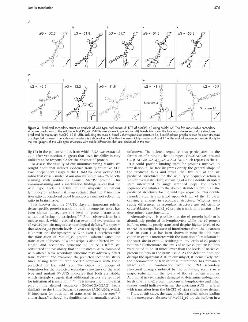
fig 1E) in the patient sample, from which RNA was extracted24 h after venesection, suggests that RNA instability is veryunlikely to be responsible for the absence of protein.
To assess the validity of our immunostaining results, wesought additional indirect evidence from quantitative XCI.Two independent assays at the HUMARA locus yielded XCIratios that closely matched our observation of 74–76% of cellsstaining with antibodies against MeCP2 protein. Ourimmunostaining and X inactivation findings reveal that thewild type allele is active in the majority of patientlymphocytes, although it is appreciated that the X inactiva-tion ratio in peripheral blood lymphocytes may not reflect theratio in brain tissue.
It is known that the 59-UTR plays an important role intissue specific protein translation and in some instances hasbeen shown to regulate the level of protein translationwithout affecting transcription.18–21 From observations in amouse model, which revealed that even mild overproductionof MeCP2 protein may cause neurological deficits,22 it appearsthat MeCP2_e2 protein levels in vivo are tightly regulated. Itis known that the upstream AUG in exon 1 interferes withthe translation of MeCP2_e2 protein isoform.4 Since thetranslation efficiency of a transcript is also affected by thelength and secondary structure of its 59-UTR,23 24 weconsidered the possibility that the upstream AUG combinedwith altered RNA secondary structure may adversely affecttranslation24 25 and examined the predicted secondary struc-tures arising from mutant 59-UTR compared with thosepredicted for the wild type. The Gibbs free energy offormation for the predicted secondary structures of the wildtype and mutant 59-UTRs indicates that both are stable,which strongly suggests that additional factors are requiredfor initiation of translation.24 26 27 It is interesting to note thatpart of the deleted sequence (GCGAGGAGGAG) bearssimilarity to the Shine-Dalgarno sequence (AGGAGG), whichis important for initiation of translation in prokaryotes28 29
and archaea,30 although its significance in mammalian cells is
unknown. The deleted sequence also participates in theformation of a nine nucleotide repeat GAGGAGGAG aroundGC (GAGGAGGAGGCGAGGAGGAG). Such repeats in the 59-UTR could provide binding sites for proteins involved intranslation.24 The tree diagrams clarify the general shape ofthe predicted folds and reveal that five out of the sixpredicted structures for the wild type sequence retain asimilar overall structure, consisting of a long double strandedstem interrupted by single stranded loops. The deletedsequence contributes to the double stranded stem in all thepredicted structures for the wild type sequence. This doublestranded stem is shortened upon deletion of the 11 basescausing a change in secondary structure. Whether suchsubtle differences in secondary structure are sufficient tocause ablation of MeCP2_e2 protein translation remains to bedetermined experimentally.
Alternatively, it is possible that the e1 protein isoform isabundantly produced in lymphocytes, while the e2 proteinisoform remains poorly translated despite higher levels of e2mRNA transcript, because of interference from the upstreamAUG in exon 1. It has been shown in vitro that the startcodon in exon 1 interferes with the initiation of translation atthe start site in exon 2, resulting in low levels of e2 proteinisoform.4 Furthermore, the levels of native e2 protein isoformwere noted to be 10 times lower than the levels of native e1protein isoform in the brain tissue. As the deletion does notdisrupt the upstream AUG in our subject, it seems likely thatthe phenomenon of translational interference has remainedintact and, in combination with the RNA secondarystructural changes induced by the mutation, results in amajor reduction in the levels of the e2 protein isoform.Additional in vivo studies designed to determine endogenouslevels of e1 and e2 protein isoforms in lymphocytes and othertissues would indicate whether the upstream AUG interfereswith translation from the MeCP2_e2 start site in these tissues.
Thus, at this stage, the exact molecular mechanism leadingto the unexpected absence of MeCP2_e2 protein isoform in a
i ii iii iv v
viviiiiii
∆ G = –52.3 ∆ G = –51.8 ∆ G = –51.7 ∆ G = –51.3 ∆ G = –50.8
Structure 4
Structure 14
∆ G = –40.7∆ G = –41.4∆ G = –41.9∆ G = –42.3∆ G = –42.6
A
B
Figure 3 Predicted secondary structure analysis of wild type and mutant 59-UTR of MeCP2_e2 using Mfold. (A) The five most stable secondarystructure predictions of the wild type MeCP2_e2 59-UTRs are shown in panels i–v. (B) Panels i–iv show the four most stable secondary structurespredicted for the mutant MeCP2_e2 59-UTR, including structure 4. Panel v shows predicted structure 14. Simplified tree graphs drawn for each structureare depicted as insets. The Y shaped structure is indicated in bold within the insets. Only structures 4 and 14 of the mutant sequence show similarity tothe tree graphs of the wild type structures with subtle differences that are discussed in the text.
Lost in translation 475
www.jmedgenet.com

proportion of cells in this patient remains undefined. Itcannot be explained on the basis of protein instability, as themutation is upstream of the coding region of the e2 RNAtranscript. Nonsense mediated decay is ruled out by ourobservation of the retention of the mutant e1 RNA transcriptcontaining the premature termination codon, as well as themutant e2 RNA transcript with the normal coding sequence.Combining the results of our analysis of the secondarystructures of mutant and wild type 59-UTR and a previousstudy on upstream AUG in exon 14 suggests that proteintranslation from the e2 mRNA transcript is regulated in partby the upstream AUG and factors binding within or tosequences overlapping the 11 deleted nucleotides in its 59-UTR, or to secondary structures induced by the deletedsequence. Further experiments designed to elucidate thesebinding factors should yield some insights into the exactmechanism of regulation. Nevertheless, our protein and RNAlevel analysis of this non-coding mutation extends knowl-edge of the biological consequences of this particularmutation. The splice site mutation reported by Amir et al9
also has the potential to disrupt both protein isoforms,although an aberrantly spliced RNA transcript was notobserved in that study. Importantly, our findings do notsupport a gathering view that the mutation we have studiedablates only the MeCP2_e1 protein isoform and that an RTT-like phenotype may occur in the presence of a normalMeCP2_e2 protein isoform.8
Comparison of the clinical features of the Australian andthe Canadian case, which was originally reported byMnatzakanian et al5 and subsequently notified to theinternational InterRett database,17 reveals some interestingsimilarities. Although the two subjects have different exon 1mutations at the genomic DNA level, because of tri-nucleotide repeats in the region of deletions, the 11nucleotide deletion reported in Mnatzakanian et al resultsin the generation of the same mutant DNA and RNAsequence as in our subject. Consequently, both the mutationsgenerate identical missense proteins that then terminate afteramino acid 36. Both subjects have a phenotype that is slightlymilder than usual. Regression in both subjects occurred laterthan normally expected and motor function (including handfunction) was better than average for subjects with classicRett syndrome. The Australian patient is 15 years youngerthan the Canadian subject, who, at 28 years, has a slightlylower level of mobility. Quantitative X inactivation studies onthe samples from the Canadian case revealed a ratio of 64:46(personal communication, Dr Patrick MacLeod). We notethat the atypical case reported by Amir et al,9 who has thesame 11 base pair deletion as our case, had random Xinactivation although no additional clinical details wereprovided. Until more clinical data are published on theseexon 1 cases, it is not possible to comment further on themost likely biological basis of the observed phenotypes, inparticular whether they reflect patterns of X inactivation,bearing in mind the immunostaining finding in theAustralian case that reveals the wild type allele is active inthe majority of cells, a finding that is consistent with theobserved milder clinical features in this child. Alternatively,this particular mutation may be characteristically milder inits clinical consequences, as has been found for mutationssuch as R133C and R306C.31 32
In conclusion, as well as revealing the biological impor-tance of the deleted non-coding region of RNA for proteintranslation, our findings confirm the emerging consensusthat mutations in exon 1 are responsible for only a minorityof RTT cases, which is not surprising given the smallproportion of nucleotides contributed by exon 1 to the gene’stotal coding sequence. Pooling the findings among our 20cases with the earlier published findings from exon 1
screening reveals that a total of seven exon 1 mutationshave been detected among 209 mutation negative RTTpatients (2/19 patients in Mnatzakanian et al,3 2/63 in Amiret al,9 0/97 in Evans et al, 2/10 in Ravn et al,8 and 1/20 in thisstudy). At a practical level, this permits an estimate to bemade of the prevalence of exon 1 mutations among mutationnegative RTT cases of 3.3% (95% CI 1.6 to 6.7). As mutationnegative RTT patients represent approximately one in five ofall cases, the overall contribution of exon 1 mutations to theoccurrence of RTT is less than 1%.
ACKNOWLEDGEMENTSWe would like to thank Dr Andrew Barker from the Laboratory forCancer Medicine, WAIMR, UWA for guidance and critical reading ofthis manuscript, Mrs Carol Philippe for her work in facilitatingsample collection, and Dr Wendy Robinson from the Department ofMedical Genetics, University of British Columbia, Canada, for Xinactivation analysis on the Canadian subject. We would also like toexpress our gratitude to all the families who have participated in thestudy, the Australian Paediatric Surveillance Unit, and the RettSyndrome Association of Australia who continue to facilitate caseascertainment of Rett syndrome in Australia.
ELECTRONIC-DATABASE INFORMATION
The web site of the Burnet Institute, Melbourne can befound at http://mfold.burnet.edu.au/
Authors’ affiliations. . . . . . . . . . . . . . . . . . . . .
A Saxena, D de Lagarde, Western Australian Institute for MedicalResearch, Centre for Medical Research, University of Western Australia,Perth, AustraliaH Leonard, Telethon Institute of Child Health Research, Centre for ChildHealth Research, The University of Western Australia, Perth, AustraliaS L Williamson, V Vasudevan, J Christodoulou, Western SydneyGenetics Program, Children’s Hospital at Westmead, and Discipline ofPaediatrics and Child Health, University of Sydney, Sydney, AustraliaE Thompson, Genetics Services of South Australia, Women’s andChildren’s Hospital, Adelaide, AustraliaP MacLeod, Department of Biology, Centre for Biomedical Research,University of Victoria, Victoria, BC, CanadaD Ravine, Western Australian Institute for Medical Research, Centre forMedical Research and School of Medicine and Pharmacology,University of Western Australia, Perth, Australia
The authors would like to acknowledge the Raine Foundation and RettSyndrome Australian Research Fund for funding of this project as well asthe National Institute of Child Health and Human Development for itscurrent funding of the Australian Rett Syndrome Database under NIHgrant number 1 R01 HD43100-01A1 PI. InterRett is funded by theInternational Rett Syndrome Association. DR is supported by a grantfrom the University of Western Australia, HL is funded by NHMRCprogram grant 353514, and JC is funded by NHMRC project grants185202 and 346603.
Competing interests: none declared
Patient details are published with consent
REFERENCES1 Renieri A, Meloni I, Longo I, Ariani F, Mari F, Pescucci C, Cambi F. Rett syndrome:
the complex nature of a monogenic disease. J Mol Med 2003;81(6):346–54.2 Colvin L, Leonard H, de Klerk N, Davis M, Weaving L, Williamson S,
Christodoulou J. Refining the phenotype of common mutations in Rettsyndrome. J Med Genet 2004;41(1):25–30.
3 Quaderi NA, Meehan RR, Tate PH, Cross SH, Bird AP, Chatterjee A,Herman GE, Brown SD. Genetic and physical mapping of a gene encoding amethyl CpG binding protein, Mecp2, to the mouse X chromosome. Genomics1994;22(3):648–51.
4 Kriaucionis S, Bird A. The major form of MeCP2 has a novel N-terminusgenerated by alternative splicing. Nucleic Acids Res 2004;32(5):1818–23.
5 Mnatzakanian GN, Lohi H, Munteanu I, Alfred SE, Yamada T, MacLeod PJ,Jones JR, Scherer SW, Schanen NC, Friez MJ, Vincent JB, Minassian BA. Apreviously unidentified MECP2 open reading frame defines a new proteinisoform relevant to Rett syndrome. Nat Genet 2004;36(4):339–41.
476 Saxena, de Lagarde, Leonard, et al
www.jmedgenet.com

6 Hagberg B. Clinical manifestations and stages of Rett syndrome. Ment RetardDev Disabil Res Rev 2002;8(2):61–5.
7 Amir RE, Van den Veyver IB, Wan M, Tran CQ, Francke U, Zoghbi HY. Rettsyndrome is caused by mutations in X-linked MECP2, encoding methyl-CpG-binding protein 2. Nat Genet 1999;23(2):185–8.
8 Ravn K, Nielson J, Schwartz M. Mutations found within exon 1 of MECP2 inDanish patients with Rett syndrome. Clin Genet 2005;67(6):532–3.
9 Amir RE, Fang P, Yu Z, Glaze DG, Percy AK, Zoghbi HY, Roa BB, Van denVeyver IB. Mutations in exon 1 of MECP2 are a rare cause of Rett syndrome.J Med Genet 2005;42(2):e15.
10 Colvin L, Fyfe S, Leonard S, Schiavello T, Ellaway C, De Klerk N,Christodoulou J, Msall M, Leonard H. Describing the phenotype in Rettsyndrome using a population database. Arch Dis Child 2003;88(1):38–43.
11 Fyfe S, Cream A, de Klerk N, Christodoulou J, Leonard H. InterRett andRettBASE: International Rett Syndrome Association databases for Rettsyndrome. J Child Neurol 2003;18(10):709–13.
12 Evans JC, Archer HL, Whatley SD, Kerr A, Clarke A, Butler R. Variation inexon 1 coding region and promoter of MECP2 in Rett syndrome and controls.Eur J Hum Genet 2005;13(1):124–6.
13 Pegoraro E, Schimke RN, Arahata K, Hayashi Y, Stern H, Marks H,Glasberg MR, Carroll JE, Taber JW, Wessel HB, et al. Detection of newpaternal dystrophin gene mutations in isolated cases of dystrophinopathy infemales. Am J Hum Genet 1994;54(6):989–1003.
14 Karasawa M, Tsukamoto N, Yamane A, Okamoto K, Maehara T,Yokohama A, Nojima Y, Omine M. Analysis of the distribution of CAGrepeats and X-chromosome inactivation status of HUMARA gene in healthyfemale subjects using improved fluorescence-based assay. Int J Hematol2001;74(3):281–6.
15 Zuker M. Prediction of RNA secondary structure by energy minimization.Methods Mol Biol 1994;25:267–94.
16 Gan HH, Pasquali S, Schlick T. Exploring the repertoire of RNA secondarymotifs using graph theory; implications for RNA design. Nucleic Acids Res2003;31(11):2926–43.
17 Moore H, Leonard H, Fyfe S, de Klerk NNL. InterRett - the application ofbioinformatics to international Rett syndrome research. Ann Hum Biol2005;32(2):228–236.
18 Kozak M. An analysis of vertebrate mRNA sequences: intimations oftranslational control. J Cell Biol 1991;115(4):887–903.
19 Meijer HA, Thomas AA. Control of eukaryotic protein synthesis by upstreamopen reading frames in the 59-untranslated region of an mRNA. Biochem J2002;367(Pt 1):1–11.
20 Wilkie GS, Dickson KS, Gray NK. Regulation of mRNA translation by 59- and39-UTR-binding factors. Trends Biochem Sci 2003;28(4):182–8.
21 Lammich S, Schobel S, Zimmer AK, Lichtenthaler SF, Haass C. Expression ofthe Alzheimer protease BACE1 is suppressed via its 59-untranslated region.EMBO Rep 2004;5(6):620–5.
22 Collins AL, Levenson JM, Vilaythong AP, Richman R, Armstrong DL,Noebels JL, David Sweatt J, Zoghbi HY. Mild overexpression of MeCP2 causesa progressive neurological disorder in mice. Hum Mol Genet2004;13(21):2679–89.
23 Gray NK, Wickens M. Control of translation initiation in animals. Annu RevCell Dev Biol 1998;14:399–458.
24 Mignone F, Gissi C, Liuni S, Pesole G. Untranslated regions of mRNAs.Genome Biol 2002;3(3):reviews0004.
25 Gray NK, Hentze MW. Regulation of protein synthesis by mRNA structure.Mol Biol Rep 1994;19(3):195–200.
26 Koromilas AE, Lazaris-Karatzas A, Sonenberg N. mRNAs containingextensive secondary structure in their 59 non-coding region translate efficientlyin cells overexpressing initiation factor eIF-4E. EMBO J 1992;11(11):4153–8.
27 van der Velden AW, Thomas AA. The role of the 59 untranslated region of anmRNA in translation regulation during development. Int J Biochem Cell Biol1999;31(1):87–106.
28 Shine J, Dalgarno L. Terminal-sequence analysis of bacterial ribosomal RNA.Correlation between the 39-terminal-polypyrimidine sequence of 16-S RNAand translational specificity of the ribosome. Eur J Biochem1975;57(1):221–30.
29 Shinedling S, Gayle M, Pribnow D, Gold L. Mutations affecting translation ofthe bacteriophage T4 rIIB gene cloned in Escherichia coli. Mol Gen Genet1987;207(2–3):224–32.
30 Karlin S, Brocchieri L, Campbell A, Cyert M, Mrazek J. Genomic andproteomic comparisons between bacterial and archaeal genomes and relatedcomparisons with the yeast and fly genomes. Proc Natl Acad Sci U S A2005;102(20):7309–14.
31 Leonard H, Colvin L, Christodoulou J, Schiavello T, Williamson S, Davis M,Ravine D, Fyfe S, de Klerk N, Matsuishi T, Kondo I, Clarke A, Hackwell S,Yamashita Y. Patients with the R133C mutation: is their phenotype differentfrom patients with Rett syndrome with other mutations? J Med Genet2003;40(5):e52.
32 Schanen C, Houwink EJ, Dorrani N, Lane J, Everett R, Feng A, Cantor RM,Percy A. Phenotypic manifestations of MECP2 mutations in classical andatypical Rett syndrome. Am J Med Genet 2004;126A(2):129–40.
Lost in translation 477
www.jmedgenet.com
Related Documents


![CRISPR/Cas9-mediated genome editing induces exon skipping ... · HeLa cells can cause skipping of exon 3, exon 4, or exons 3, 4, and 5 [18]. We also detected infrequent exon skipping](https://static.cupdf.com/doc/110x72/60db8f117fb86d112c69c947/crisprcas9-mediated-genome-editing-induces-exon-skipping-hela-cells-can-cause.jpg)