Current Biology 22, 1–10, December 18, 2012 ª2012 Elsevier Ltd All rights reserved http://dx.doi.org/10.1016/j.cub.2012.10.036 Article Loss of Nucleosomal DNA Condensation Coincides with Appearance of a Novel Nuclear Protein in Dinoflagellates Sebastian G. Gornik, 1 Kristina L. Ford, 2 Terrence D. Mulhern, 3 Antony Bacic, 2,3 Geoffrey I. McFadden, 1 and Ross F. Waller 1, * 1 School of Botany 2 ARC Centre of Excellence in Plant Cell Walls, School of Botany 3 Bio21 Molecular Science and Biotechnology Institute University of Melbourne, Victoria, 3010, Australia Summary Background: The packaging, expression, and maintenance of nuclear genomes using histone proteins is a ubiquitous and fundamental feature of eukaryotic cells, yet the phylum Dino- flagellata has apparently abandoned this model of nuclear organization. Their nuclei contain permanently condensed, liquid crystalline chromosomes that seemingly lack histone proteins, and contain remarkably large genomes. The molec- ular basis for this reorganization is poorly understood, as is the sequence of evolutionary events that led to such radical change. We have investigated nuclear organization in the closest relative to dinoflagellates, Perkinsus marinus, and an early-branching dinoflagellate, Hematodinium sp., to identify early changes that occurred during dinoflagellate nuclear evolution. Results: We show that P. marinus has a typical nuclear orga- nization that is based on the four core histones. By the early divergence of Hematodinium sp., however, dinoflagellate genome size is dramatically enlarged, chromosomes are permanently condensed, and histones are scarcely detect- able. In place of histones, we identify a novel, dominant family of nuclear proteins that is only found in dinoflagellates and, surprisingly, in a family of large algal viruses, the Phycodnavir- idae. These new proteins, which we call DVNPs (dinoflagellate/ viral nucleoproteins), are highly basic, bind DNA with similar affinity to histones, and occur in multiple posttranslationally modified forms. We find these proteins throughout all dinofla- gellates, including early- and late-branching taxa, but not in P. marinus. Conclusions: Gain of a major novel family of nucleoproteins, apparently from an algal virus, occurred early in dinoflagellate evolution and coincides with rapid and dramatic reorganiza- tion of the dinoflagellate nucleus. Introduction The dinoflagellate nucleus is so unusual that it was once believed to represent an intermediate stage between prokary- otes and eukaryotes—a so-called ‘‘mesokaryon’’ [1]. Dinofla- gellates are a successful and prolific lineage of eukaryotes fulfilling diverse environmental roles, including major marine primary producers and essential symbionts of reef-building corals [2]. About half of dinoflagellates have lost photosyn- thesis and are heterotrophic micropredators, saprophytes, or parasites of marine animals and other organisms. The notion of dinoflagellates as evolutionary intermediates was aban- doned when the sister relationship of dinoflagellates with apicomplexan parasites (e.g., the malaria causative agent Plasmodium) was recognized, because apicomplexans contain nuclei typical of most eukaryotes. The dinoflagellate nucleus, or ‘‘dinokaryon,’’ is therefore clearly a derived state. While the term dinokaryon lacks a strict definition, it is generally associated with the following characteristics: (1) chromosomes display a persistent condensed state and are conspicuous by light microscopy throughout the cell cycle [3]; (2) histone proteins are absent or below detectable limits [4]; (3) chromosomes have a cholesteric liquid crystalline orga- nization, evident by birefringence under polarized light and an arched fibrillar or banded appearance in electron micrographs [5–7]; and (4) nuclear DNA content is extremely large (up to 200 pg DNA, compared to 3 pg for humans, per haploid nucleus) [8, 9]. Dinoflagellate nuclei are also unusual in that up to 70% of thymine is replaced by the rare base 5-hydroxymethyluracil, they contain high levels of transition metals (Fe, Ni, Cu, and Zn), and chromosome number can be very high (>100) [4, 9]. The loss of histones as dominant nuclear proteins is perhaps the most striking of dinokaryotic characters because these proteins are central to the packing and regulation of eukaryotic nuclear DNA [10–12]. Histone octamers create nucleosome particles around which DNA is wrapped twice and organized into higher-order chromatin fibers. Dynamic modifications to nucleosomal histones or incorporation of specialized variant histone types regulate genomic functions such as gene expression, DNA repair, chromosome segregation, and meiotic recombination. The presence of a highly conserved set of histones throughout eukaryotes is testimony to their importance and suggests a seminal role in the evolution of eukaryotic nuclei. The radical change in dinoflagellates to a di- nokaryon therefore presents a clear challenge to the dogma of the eukaryotic nucleus [13]. Most dinoflagellates, so-called ‘‘typical’’ dinoflagellates or ‘‘dinokaryotes,’’ are thought to possess all of the derived nuclear characters (although thorough characterization has only been made for a handful of taxa). Lower-branching members, however, often display either variant or limited dino- karyotic characteristics. For example, some possess alternate cell stages that lack permanently condensed chromosomes (e.g., Amoebophrya, Noctiluca, and Blastodinium), and the condensed chromosomes in some taxa lack conspicuous fibrillar banding in electron micrographs (e.g., Hematodinium and Oxyrrhis)[14–17]. Further, chromosome number can be relatively small (e.g., four to ten in syndinians) [18, 19], and the presence or absence of histones is poorly characterized. Thus, it has been unclear what sequence of nuclear character changes led to the dinokaryotic state. To determine the early events in the evolution of the dino- karyon, we have investigated the nuclei of two early-branching members of the dinoflagellate lineage. The earliest member is Perkinsus marinus, a parasite of marine mollusks. P. marinus lacks the conspicuous features of the dinokaryon morphology, and although it is not considered to be a member of the dinoflagellates proper, it represents the closest known lineage *Correspondence: [email protected] Please cite this article in press as: Gornik et al., Loss of Nucleosomal DNA Condensation Coincides with Appearance of a Novel Nuclear Protein in Dinoflagellates, Current Biology (2012), http://dx.doi.org/10.1016/j.cub.2012.10.036

Welcome message from author
This document is posted to help you gain knowledge. Please leave a comment to let me know what you think about it! Share it to your friends and learn new things together.
Transcript
Please cite this article in press as: Gornik et al., Loss of Nucleosomal DNA Condensation Coincides with Appearance of a NovelNuclear Protein in Dinoflagellates, Current Biology (2012), http://dx.doi.org/10.1016/j.cub.2012.10.036
Loss of Nucleosomal DNA C
Current Biology 22, 1–10, December 18, 2012 ª2012 Elsevier Ltd All rights reserved http://dx.doi.org/10.1016/j.cub.2012.10.036
Articleondensation
Coincides with Appearance of a NovelNuclear Protein in Dinoflagellates
Sebastian G. Gornik,1 Kristina L. Ford,2
Terrence D. Mulhern,3 Antony Bacic,2,3
Geoffrey I. McFadden,1 and Ross F. Waller1,*1School of Botany2ARC Centre of Excellence in Plant Cell Walls, School ofBotany3Bio21 Molecular Science and Biotechnology InstituteUniversity of Melbourne, Victoria, 3010, Australia
Summary
Background: The packaging, expression, and maintenance ofnuclear genomes using histone proteins is a ubiquitous andfundamental feature of eukaryotic cells, yet the phylum Dino-flagellata has apparently abandoned this model of nuclearorganization. Their nuclei contain permanently condensed,liquid crystalline chromosomes that seemingly lack histoneproteins, and contain remarkably large genomes. The molec-ular basis for this reorganization is poorly understood, as isthe sequence of evolutionary events that led to such radicalchange. We have investigated nuclear organization in theclosest relative to dinoflagellates, Perkinsus marinus, and anearly-branching dinoflagellate, Hematodinium sp., to identifyearly changes that occurred during dinoflagellate nuclearevolution.Results: We show that P. marinus has a typical nuclear orga-nization that is based on the four core histones. By the earlydivergence of Hematodinium sp., however, dinoflagellategenome size is dramatically enlarged, chromosomes arepermanently condensed, and histones are scarcely detect-able. In place of histones, we identify a novel, dominant familyof nuclear proteins that is only found in dinoflagellates and,surprisingly, in a family of large algal viruses, the Phycodnavir-idae. These newproteins, which we call DVNPs (dinoflagellate/viral nucleoproteins), are highly basic, bind DNA with similaraffinity to histones, and occur in multiple posttranslationallymodified forms. We find these proteins throughout all dinofla-gellates, including early- and late-branching taxa, but not inP. marinus.Conclusions: Gain of a major novel family of nucleoproteins,apparently from an algal virus, occurred early in dinoflagellateevolution and coincides with rapid and dramatic reorganiza-tion of the dinoflagellate nucleus.
Introduction
The dinoflagellate nucleus is so unusual that it was oncebelieved to represent an intermediate stage between prokary-otes and eukaryotes—a so-called ‘‘mesokaryon’’ [1]. Dinofla-gellates are a successful and prolific lineage of eukaryotesfulfilling diverse environmental roles, including major marineprimary producers and essential symbionts of reef-buildingcorals [2]. About half of dinoflagellates have lost photosyn-thesis and are heterotrophic micropredators, saprophytes, or
*Correspondence: [email protected]
parasites of marine animals and other organisms. The notionof dinoflagellates as evolutionary intermediates was aban-doned when the sister relationship of dinoflagellates withapicomplexan parasites (e.g., the malaria causative agentPlasmodium) was recognized, because apicomplexanscontain nuclei typical of most eukaryotes. The dinoflagellatenucleus, or ‘‘dinokaryon,’’ is therefore clearly a derived state.While the term dinokaryon lacks a strict definition, it is
generally associated with the following characteristics: (1)chromosomes display a persistent condensed state and areconspicuous by light microscopy throughout the cell cycle[3]; (2) histone proteins are absent or below detectable limits[4]; (3) chromosomes have a cholesteric liquid crystalline orga-nization, evident by birefringence under polarized light and anarched fibrillar or banded appearance in electron micrographs[5–7]; and (4) nuclear DNA content is extremely large (up to 200pg DNA, compared to 3 pg for humans, per haploid nucleus)[8, 9]. Dinoflagellate nuclei are also unusual in that up to 70%of thymine is replaced by the rare base 5-hydroxymethyluracil,they contain high levels of transition metals (Fe, Ni, Cu, andZn), and chromosome number can be very high (>100) [4, 9].The loss of histones as dominant nuclear proteins is perhapsthe most striking of dinokaryotic characters because theseproteins are central to the packing and regulation of eukaryoticnuclear DNA [10–12]. Histone octamers create nucleosomeparticles around which DNA is wrapped twice and organizedinto higher-order chromatin fibers. Dynamic modifications tonucleosomal histones or incorporation of specialized varianthistone types regulate genomic functions such as geneexpression, DNA repair, chromosome segregation, andmeiotic recombination. The presence of a highly conservedset of histones throughout eukaryotes is testimony to theirimportance and suggests a seminal role in the evolution ofeukaryotic nuclei. The radical change in dinoflagellates to a di-nokaryon therefore presents a clear challenge to the dogma ofthe eukaryotic nucleus [13].Most dinoflagellates, so-called ‘‘typical’’ dinoflagellates or
‘‘dinokaryotes,’’ are thought to possess all of the derivednuclear characters (although thorough characterization hasonly been made for a handful of taxa). Lower-branchingmembers, however, often display either variant or limited dino-karyotic characteristics. For example, some possess alternatecell stages that lack permanently condensed chromosomes(e.g., Amoebophrya, Noctiluca, and Blastodinium), and thecondensed chromosomes in some taxa lack conspicuousfibrillar banding in electron micrographs (e.g., Hematodiniumand Oxyrrhis) [14–17]. Further, chromosome number can berelatively small (e.g., four to ten in syndinians) [18, 19], andthe presence or absence of histones is poorly characterized.Thus, it has been unclear what sequence of nuclear characterchanges led to the dinokaryotic state.To determine the early events in the evolution of the dino-
karyon, we have investigated the nuclei of two early-branchingmembers of the dinoflagellate lineage. The earliest member isPerkinsus marinus, a parasite of marine mollusks. P. marinuslacks the conspicuous features of the dinokaryonmorphology,and although it is not considered to be a member of thedinoflagellates proper, it represents the closest known lineage
180
360
1080900720540
100
500
1000
2000
pbpb
Perkinsus marinus Hematodinium sp.MNase digestion MNase digestion
6
15
1926
37
64
82
116
kDa
H3H1
H2BH2A
H4
0 0.2 0.4 0.8 1.6 0 0.2 0.4 0.8 1.6
6
15
1926
37
64
82
116
αH2B αH3K4me3 αH4 αH4K12ac kDa
H.P. H.P. H.P. H.P. H.P.
Acid ExtractedProteins
CBA Immuno-detection of Histones
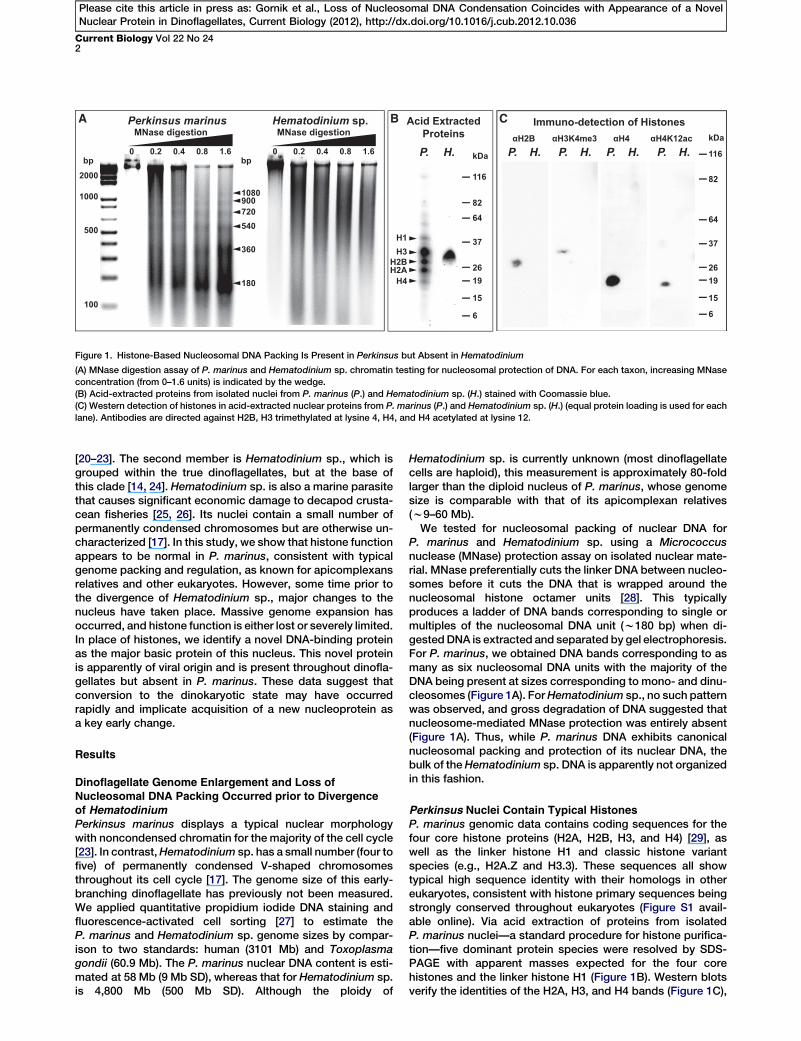
Figure 1. Histone-Based Nucleosomal DNA Packing Is Present in Perkinsus but Absent in Hematodinium
(A) MNase digestion assay of P. marinus and Hematodinium sp. chromatin testing for nucleosomal protection of DNA. For each taxon, increasing MNase
concentration (from 0–1.6 units) is indicated by the wedge.
(B) Acid-extracted proteins from isolated nuclei from P. marinus (P.) and Hematodinium sp. (H.) stained with Coomassie blue.
(C) Western detection of histones in acid-extracted nuclear proteins from P. marinus (P.) and Hematodinium sp. (H.) (equal protein loading is used for each
lane). Antibodies are directed against H2B, H3 trimethylated at lysine 4, H4, and H4 acetylated at lysine 12.
Current Biology Vol 22 No 242
Please cite this article in press as: Gornik et al., Loss of Nucleosomal DNA Condensation Coincides with Appearance of a NovelNuclear Protein in Dinoflagellates, Current Biology (2012), http://dx.doi.org/10.1016/j.cub.2012.10.036
[20–23]. The second member is Hematodinium sp., which isgrouped within the true dinoflagellates, but at the base ofthis clade [14, 24]. Hematodinium sp. is also a marine parasitethat causes significant economic damage to decapod crusta-cean fisheries [25, 26]. Its nuclei contain a small number ofpermanently condensed chromosomes but are otherwise un-characterized [17]. In this study, we show that histone functionappears to be normal in P. marinus, consistent with typicalgenome packing and regulation, as known for apicomplexansrelatives and other eukaryotes. However, some time prior tothe divergence of Hematodinium sp., major changes to thenucleus have taken place. Massive genome expansion hasoccurred, and histone function is either lost or severely limited.In place of histones, we identify a novel DNA-binding proteinas the major basic protein of this nucleus. This novel proteinis apparently of viral origin and is present throughout dinofla-gellates but absent in P. marinus. These data suggest thatconversion to the dinokaryotic state may have occurredrapidly and implicate acquisition of a new nucleoprotein asa key early change.
Results
Dinoflagellate Genome Enlargement and Loss ofNucleosomal DNA Packing Occurred prior to Divergence
of HematodiniumPerkinsus marinus displays a typical nuclear morphologywith noncondensed chromatin for the majority of the cell cycle[23]. In contrast,Hematodinium sp. has a small number (four tofive) of permanently condensed V-shaped chromosomesthroughout its cell cycle [17]. The genome size of this early-branching dinoflagellate has previously not been measured.We applied quantitative propidium iodide DNA staining andfluorescence-activated cell sorting [27] to estimate theP. marinus and Hematodinium sp. genome sizes by compar-ison to two standards: human (3101 Mb) and Toxoplasmagondii (60.9 Mb). The P. marinus nuclear DNA content is esti-mated at 58 Mb (9 Mb SD), whereas that for Hematodinium sp.is 4,800 Mb (500 Mb SD). Although the ploidy of
Hematodinium sp. is currently unknown (most dinoflagellatecells are haploid), this measurement is approximately 80-foldlarger than the diploid nucleus of P. marinus, whose genomesize is comparable with that of its apicomplexan relatives(w9–60 Mb).We tested for nucleosomal packing of nuclear DNA for
P. marinus and Hematodinium sp. using a Micrococcusnuclease (MNase) protection assay on isolated nuclear mate-rial. MNase preferentially cuts the linker DNA between nucleo-somes before it cuts the DNA that is wrapped around thenucleosomal histone octamer units [28]. This typicallyproduces a ladder of DNA bands corresponding to single ormultiples of the nucleosomal DNA unit (w180 bp) when di-gestedDNA is extracted and separated by gel electrophoresis.For P. marinus, we obtained DNA bands corresponding to asmany as six nucleosomal DNA units with the majority of theDNA being present at sizes corresponding to mono- and dinu-cleosomes (Figure 1A). ForHematodinium sp., no such patternwas observed, and gross degradation of DNA suggested thatnucleosome-mediated MNase protection was entirely absent(Figure 1A). Thus, while P. marinus DNA exhibits canonicalnucleosomal packing and protection of its nuclear DNA, thebulk of theHematodinium sp. DNA is apparently not organizedin this fashion.
Perkinsus Nuclei Contain Typical HistonesP. marinus genomic data contains coding sequences for thefour core histone proteins (H2A, H2B, H3, and H4) [29], aswell as the linker histone H1 and classic histone variantspecies (e.g., H2A.Z and H3.3). These sequences all showtypical high sequence identity with their homologs in othereukaryotes, consistent with histone primary sequences beingstrongly conserved throughout eukaryotes (Figure S1 avail-able online). Via acid extraction of proteins from isolatedP. marinus nuclei—a standard procedure for histone purifica-tion—five dominant protein species were resolved by SDS-PAGE with apparent masses expected for the four corehistones and the linker histone H1 (Figure 1B). Western blotsverify the identities of the H2A, H3, and H4 bands (Figure 1C),
Homo sapiens 1 137+++++++
Histone Fold
+ ++ + + ++ +
1 217Hematodinium sp.Histone Fold
+ + + + ++++++ +++ + + ++ + + ++++ ++ +++ +++ +
H2A
Hematodinium sp.
1 126Homo sapiensHistone Fold
+ ++++ ++++++ + ++ + + + + + +
Histone Fold
1 171+++ ++ +++++++ ++++++ +++++++ + ++ + ++ + +
H2B
+ + + + + ++1 134
Histone FoldHomo sapiens
142
Histone Fold
1+++++++++++++
Hematodinium sp.
+ +H3
103++ + + + + + + ++ +
1
Histone FoldHomo sapiens
Histone Fold
1761Hematodinium sp.+ + ++ + ++ + ++ + +++ +++ + + + ++ + + ++
++H4
Alpha-helixPhosphorylation site Acetylation
MethylationUbiqutination
+ Lysine residue
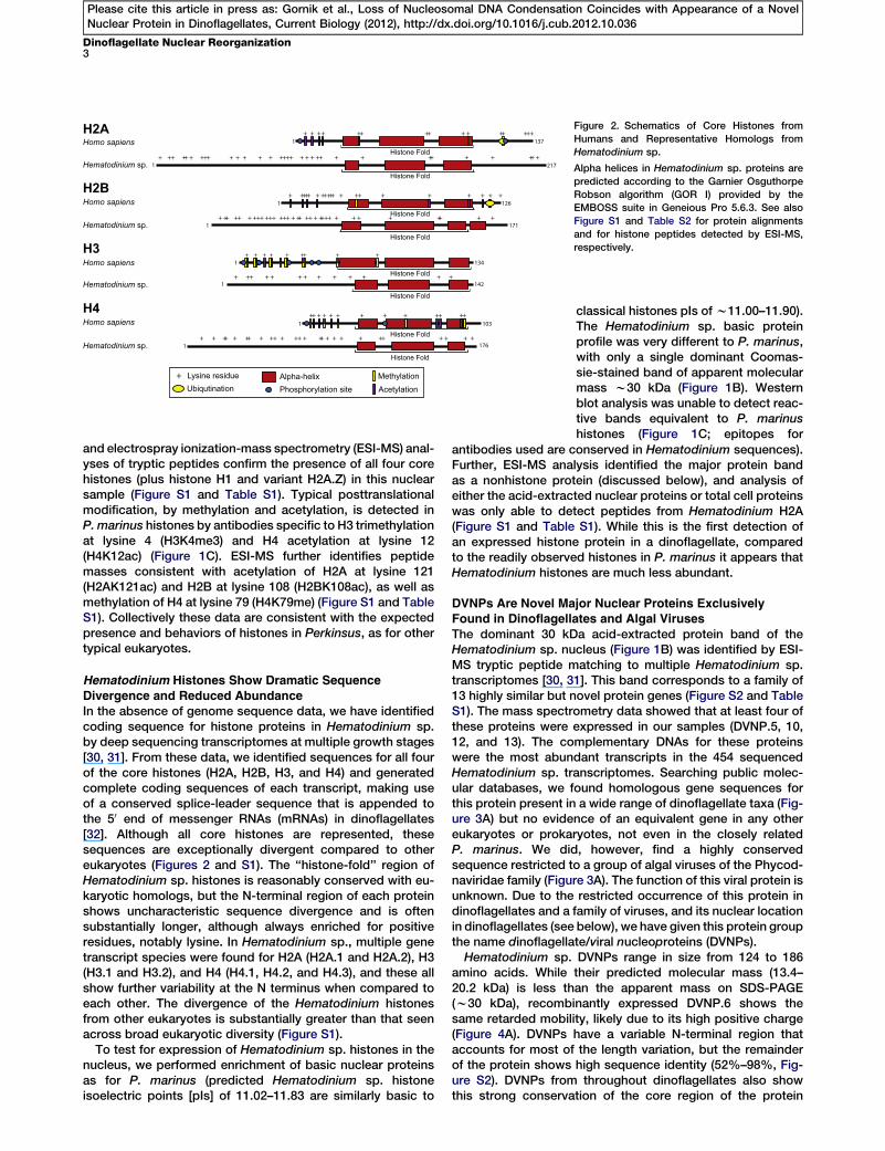
Figure 2. Schematics of Core Histones from
Humans and Representative Homologs from
Hematodinium sp.
Alpha helices in Hematodinium sp. proteins are
predicted according to the Garnier Osguthorpe
Robson algorithm (GOR I) provided by the
EMBOSS suite in Geneious Pro 5.6.3. See also
Figure S1 and Table S2 for protein alignments
and for histone peptides detected by ESI-MS,
respectively.
Dinoflagellate Nuclear Reorganization3
Please cite this article in press as: Gornik et al., Loss of Nucleosomal DNA Condensation Coincides with Appearance of a NovelNuclear Protein in Dinoflagellates, Current Biology (2012), http://dx.doi.org/10.1016/j.cub.2012.10.036
and electrospray ionization-mass spectrometry (ESI-MS) anal-yses of tryptic peptides confirm the presence of all four corehistones (plus histone H1 and variant H2A.Z) in this nuclearsample (Figure S1 and Table S1). Typical posttranslationalmodification, by methylation and acetylation, is detected inP. marinus histones by antibodies specific to H3 trimethylationat lysine 4 (H3K4me3) and H4 acetylation at lysine 12(H4K12ac) (Figure 1C). ESI-MS further identifies peptidemasses consistent with acetylation of H2A at lysine 121(H2AK121ac) and H2B at lysine 108 (H2BK108ac), as well asmethylation of H4 at lysine 79 (H4K79me) (Figure S1 and TableS1). Collectively these data are consistent with the expectedpresence and behaviors of histones in Perkinsus, as for othertypical eukaryotes.
Hematodinium Histones Show Dramatic Sequence
Divergence and Reduced AbundanceIn the absence of genome sequence data, we have identifiedcoding sequence for histone proteins in Hematodinium sp.by deep sequencing transcriptomes at multiple growth stages[30, 31]. From these data, we identified sequences for all fourof the core histones (H2A, H2B, H3, and H4) and generatedcomplete coding sequences of each transcript, making useof a conserved splice-leader sequence that is appended tothe 50 end of messenger RNAs (mRNAs) in dinoflagellates[32]. Although all core histones are represented, thesesequences are exceptionally divergent compared to othereukaryotes (Figures 2 and S1). The ‘‘histone-fold’’ region ofHematodinium sp. histones is reasonably conserved with eu-karyotic homologs, but the N-terminal region of each proteinshows uncharacteristic sequence divergence and is oftensubstantially longer, although always enriched for positiveresidues, notably lysine. In Hematodinium sp., multiple genetranscript species were found for H2A (H2A.1 and H2A.2), H3(H3.1 and H3.2), and H4 (H4.1, H4.2, and H4.3), and these allshow further variability at the N terminus when compared toeach other. The divergence of the Hematodinium histonesfrom other eukaryotes is substantially greater than that seenacross broad eukaryotic diversity (Figure S1).
To test for expression of Hematodinium sp. histones in thenucleus, we performed enrichment of basic nuclear proteinsas for P. marinus (predicted Hematodinium sp. histoneisoelectric points [pIs] of 11.02–11.83 are similarly basic to
classical histones pIs of w11.00–11.90).The Hematodinium sp. basic proteinprofile was very different to P. marinus,with only a single dominant Coomas-sie-stained band of apparent molecularmass w30 kDa (Figure 1B). Westernblot analysis was unable to detect reac-tive bands equivalent to P. marinushistones (Figure 1C; epitopes for
antibodies used are conserved in Hematodinium sequences).Further, ESI-MS analysis identified the major protein bandas a nonhistone protein (discussed below), and analysis ofeither the acid-extracted nuclear proteins or total cell proteinswas only able to detect peptides from Hematodinium H2A(Figure S1 and Table S1). While this is the first detection ofan expressed histone protein in a dinoflagellate, comparedto the readily observed histones in P. marinus it appears thatHematodinium histones are much less abundant.
DVNPs Are Novel Major Nuclear Proteins ExclusivelyFound in Dinoflagellates and Algal Viruses
The dominant 30 kDa acid-extracted protein band of theHematodinium sp. nucleus (Figure 1B) was identified by ESI-MS tryptic peptide matching to multiple Hematodinium sp.transcriptomes [30, 31]. This band corresponds to a family of13 highly similar but novel protein genes (Figure S2 and TableS1). The mass spectrometry data showed that at least four ofthese proteins were expressed in our samples (DVNP.5, 10,12, and 13). The complementary DNAs for these proteinswere the most abundant transcripts in the 454 sequencedHematodinium sp. transcriptomes. Searching public molec-ular databases, we found homologous gene sequences forthis protein present in a wide range of dinoflagellate taxa (Fig-ure 3A) but no evidence of an equivalent gene in any othereukaryotes or prokaryotes, not even in the closely relatedP. marinus. We did, however, find a highly conservedsequence restricted to a group of algal viruses of the Phycod-naviridae family (Figure 3A). The function of this viral protein isunknown. Due to the restricted occurrence of this protein indinoflagellates and a family of viruses, and its nuclear locationin dinoflagellates (see below), we have given this protein groupthe name dinoflagellate/viral nucleoproteins (DVNPs).Hematodinium sp. DVNPs range in size from 124 to 186
amino acids. While their predicted molecular mass (13.4–20.2 kDa) is less than the apparent mass on SDS-PAGE(w30 kDa), recombinantly expressed DVNP.6 shows thesame retarded mobility, likely due to its high positive charge(Figure 4A). DVNPs have a variable N-terminal region thataccounts for most of the length variation, but the remainderof the protein shows high sequence identity (52%–98%, Fig-ure S2). DVNPs from throughout dinoflagellates also showthis strong conservation of the core region of the protein
Esv (virus)FeldSpV (virus)
Alexandrium
SymbiodiniumKarlodinium
OxyrrhisHematodinium
Lingulodinium
Heterocapsa
Esv (virus)FeldSpV (virus)
Alexandrium
SymbiodiniumKarlodinium
OxyrrhisHematodinium
Lingulodinium
Heterocapsa
Esv (virus)FeldSpV (virus)
Alexandrium
SymbiodiniumKarlodinium
OxyrrhisHematodinium
Lingulodinium
Heterocapsa
1 132DVNP.5Helix-Turn-Helix
Alpha-helix
Predicted phosphorylation siteNuclear Localisation Signal
+ Lysine residue
++ +++ ++ ++ ++ ++++ + + + + + ++ ++ ++ ++ + + + + + ++ + ++B
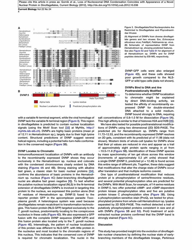
A Figure 3. Dinoflagellate/Viral Nucleoproteins Are
Restricted to Dinoflagellates and Phycodnaviri-
dae Viruses
(A) Alignment of DVNPs from diverse dinoflagel-
late genera and two viruses: Esv, Ectocarpus
siliculosus virus; FeldSpV, Feldmannia sp. virus.
(B) Schematic of representative DVNP from
Hematodinium sp. showing predicted features.
See also Figure S2 and Table S1 for alignment of
all Hematodinium sp. DVNPs and for DVNP
peptides detected by ESI-MS, respectively.
Current Biology Vol 22 No 244
Please cite this article in press as: Gornik et al., Loss of Nucleosomal DNA Condensation Coincides with Appearance of a NovelNuclear Protein in Dinoflagellates, Current Biology (2012), http://dx.doi.org/10.1016/j.cub.2012.10.036
with a variable N-terminal segment, while the viral homologs ofDVNP lack the variable N-terminal region (Figure 3). This regionin dinoflagellates is predicted to contain nuclear localizationsignals (using the Motif Scan tool [33] at MyHits, http://myhits.isb-sib.ch). DVNPs are highly basic proteins (mean pIof 12.11 in Hematodinium sp.), largely due to their high lysinecontent. Structural predictions of DVNP suggest severalhelical regions, including a potential helix-turn-helix conforma-tion in the conserved region (Figure 3B).
DVNP Locates to ChromatinImmumnofluorescent localization of DVNPs with an antibodyto the recombinantly expressed DVNP shows they occurexclusively in the Hematodinium sp. nucleus and colocatewith the condensed chromosomes clearly evident by DNAstaining (Figures 4A and 4B). Strong staining with alkalinefast green, a classic stain for basic nuclear proteins [34],confirms the abundance of basic proteins in the Hematodi-nium sp. nucleus (Figure 4C) (later-branching dinoflagellatenuclei are alkaline fast green negative [9]). To test whetherthe putative nucleus localization signal (NLS) in the N-terminalextension of dinoflagellate DVNPs is involved in targeting thisprotein to the nucleus, we expressed this portion alone (first47 residues of Hematodinium sp. DVNP.5) fused to thereporter protein GFP in the apicomplexan parasite Toxo-plasma gondii. A heterologous system was used becausedinoflagellates remain recalcitrant to transformation technolo-gies. This fusion protein (NLS-GFP) targeted exclusively to theT. gondii nucleus, predominantly locating to the conspicuousnucleolus in these cells (Figure 4D). We also expressed a GFPfusion with the complete DVNP sequence (DVNP-GFP) andthis fusion protein also located only to the T. gondii nucleus(Figure 4E). Interestingly, the pattern of nuclear localizationof this protein was different to NLS-GFP, with little protein inthe nucleolus and most located to the chromatin regions ofthis nucleus. This indicates that the conserved core of DVNPis required for chromatin localization. The nuclei in the
DVNP-GFP cells were also enlarged(Figure 4D), and these cells showedpoor growth compared to the NLS-GFP or wild-type cells (data not shown).
DVNPs Bind to DNA and ArePosttranslationally Modified
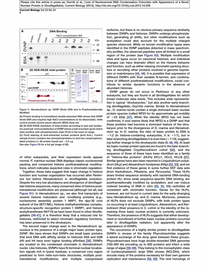
To determinewhether DVNP localizationto chromatin might be explainedby direct DNA-binding activity, wetested the affinity of recombinantly ex-pressed DVNP for double-strandedDNA attached to a solid medium.DVNP bound strongly to DNA, requiring
salt concentrations of 0.8–1.0 M for dissociation (Figure 5A).This high affinity is similar to that of histones H2A andH2B [35].We have also tested for possible posttranslational modifica-
tions of DVNPs using two dimensional (2D) SDS-PAGE. Thepredicted pIs for Hematodinium sp. DVNPs range from11.72–12.53, and the recombinantly expressed DVNP resolveson 2D gels, consistent with its predicted pI of 11.98 (data notshown). Western blots of whole-cell lysates for DVNPs showthat their pI values are reduced in vivo and appear as a trailof approximately eight protein spots ranging in pI fromw10.5–11.8 (Figure 5B). Identification of these DVNP speciesby mass spectrometry of ten gel slices across this window(increments of approximately 0.2 pH units) showed thata single DVNP (DVNP.5, predicted pI = 12.46) is found acrossthis entire range of different pIs. Together, these data suggestthat modifications that alter the charge state of DVNPs occurafter translation and that multiple isoforms coexist.One type of posttranslational modification that reduces
protein pI is phosphorylation of serine/threonine residues,and several potential phosphorylation sites are predicted forthe DVNPs.WithMotif Scan, for example, of 14 serine residuesin DVNP.5, two offer potential cAMP- and cGMP-dependentprotein kinase phosphorylation sites and five are putativeprotein kinase C phosphorylation sites (Figure 3B). Usinga phosphorylation-sensitive stain (ProQ), we tested for phos-phorylated proteins fromwhole-cell Hematodinium sp. lysatesseparated by 2D SDS-PAGE. This method detected a trail ofputatively phosphorylated proteins consistent in size and pIwith DVNPs (Figures 5B and S3). ProQ treatment of acid-extracted nuclear proteins confirmed that the DVNP band isstrongly stained (Figure 5C).
Discussion
This study has provided insight into the evolution of dinoflagel-late nuclear characters by defining the nuclear state of early-branching members of the dinoflagellate lineage, Perkinsus
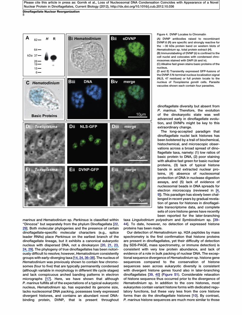
Figure 4. DVNP Locates to Chromatin
(A) DVNP antibodies raised to recombinant
DVNP.6 (R) are specific and strongly reactive for
the w30 kDa protein band on western blots of
Hematodinium sp. total protein extract (H).
(B) Immunolabeling of DVNP (ii) is confined to the
cell nuclei and colocates with condensed chro-
mosomes stained with DAPI (iii and iv).
(C) Alkaline fast green stains basic proteins of the
nucleus.
(D and E) Transiently expressed GFP-fusions of
the DVNP.5 N-terminal nucleus localization signal
(NLS, 47 residues) or full protein locate to the
nucleus of Toxoplasma gondii cells. Parasite
vacuoles shown each contain four parasites.
Dinoflagellate Nuclear Reorganization5
Please cite this article in press as: Gornik et al., Loss of Nucleosomal DNA Condensation Coincides with Appearance of a NovelNuclear Protein in Dinoflagellates, Current Biology (2012), http://dx.doi.org/10.1016/j.cub.2012.10.036
marinus and Hematodinium sp. Perkinsus is classified within‘‘Dinozoa’’ but separately from the phylum Dinoflagellata [22,29]. Both molecular phylogenies and the presence of certaindinoflagellate-specific molecular characters (e.g., spliceleader RNAs) place Perkinsus on the earliest branch of thedinoflagellate lineage, but it exhibits a canonical eukaryoticnucleus with dispersed DNA, not a dinokaryon [20, 21, 23,24, 29]. The phylogeny of true dinoflagellates has been notori-ously difficult to resolve; however, Hematodinium consistentlygroupswith early-diverging taxa [14, 24, 36–38]. The nucleus ofHematodinium was previously shown to contain few chromo-somes (four to five) that are typically permanently condensed(although variable in morphology in different life cycle stages)and lack conspicuous arched banding patterns in electronmicrographs [17]. Here, we have shown that althoughP. marinus fulfills all of the expectations of a typical eukaryoticnucleus, Hematodinium sp. has expanded its genome size,lacks nucleosomal DNA packing, possesses lowly expresseddivergent histones, and contains an abundant novel DNA-binding protein, DVNP, that is present throughout
,
dinoflagellate diversity but absent fromP. marinus. Therefore, the evolutionof the dinokaryotic state was welladvanced early in dinoflagellate evolu-tion, and DVNPs might be key to thisextraordinary change.The long-accepted paradigm that
dinoflagellate nuclei lack histones hasbeen bolstered by a trail of biochemical,histochemical, and microscopic obser-vations across a broad spread of dino-flagellate taxa, namely: (1) low ratios ofbasic protein to DNA, (2) poor stainingwith alkaline fast green for basic nuclearproteins, (3) lack of typical histonebands in acid extracted nuclear pro-teins, (4) absence of nucleosomalprotection of DNA in nuclease digestionassays, and (5) lack of evidence ofnucleosomal beads in DNA spreads forelectron microscopy (reviewed in [4,9]). This paradigm has slowly been chal-lenged in recent years by gradual revela-tion of genes for histones in dinoflagel-late transcriptome data, and completesets of core histone genes have recentlybeen reported for the later-branching
taxa Lingulodinium polyedrum and Symbiodinium sp. [39–44]. To date, however, no detection of expressed histoneproteins has been made.
Our detection of Hematodinium sp. H2A peptides by massspectrometry is the first confirmation that histone proteinsare present in dinoflagellates, yet their difficulty of detection(by SDS-PAGE, mass spectrometry, or immune detection) isconsistent with very low protein abundance, and lack ofevidence of a role in bulk packing of nuclear DNA. The excep-tional sequence divergence ofHematodinium sp. histone genesequences compared to the conservation of histonesequences seen across eukaryotic diversity is consistentwith divergent histone genes found also in later-branchingdinoflagellates [39, 40] (Figure S1). Considerable relaxationof histone sequence thus occurred prior to the divergence ofHematodinium sp. In addition to the core histones, mosteukaryotes contain variant histone forms with dedicated regu-latory functions, but these vary less from the core histoneforms than do the dinoflagellate histones [10]. By contrastP. marinus histone sequences are much more similar to those
kDa
37
26
5.0
unbo
und
was
h
0.2
0.4
0.6
0.8
1.0
NaCl concentration [M]
82BS
A
37
2619
37
2619
kDa10 12pH
anti-DVNP
Phosphoproteins (ProQ)
DV
NP
AcidExtr.
Cont. AcidExtr.
Cont.(+/-)(+/-)
+
+
--
+
+
--
ProQCoomassie
2D SDS-PAGE total protein
A
CB
37
2619
15
82
64
49
kDa
DNA Bindingpr
otei
n
Figure 5. Hematodinium sp. DVNP Binds DNA and Is Posttranslationally
Modified
(A) Protein binding to immobilized double-stranded DNA shows that DVNP
binds DNA and requires high NaCl concentrations to be dissociated, while
control protein bovine serum albumin (BSA) does not.
(B) 2D SDS-PAGE resolution of total protein according to size and isoelec-
tric point (pI). Immunodetection of DVNP shows a trail of protein spots which
stain positive with phosphoprotein stain ProQ in the lower pI range.
(C) ProQ staining of acid-extracted nuclear proteins (Acid Extr.). Control
sample (Cont.) contains two phosophoproteins (+) and two nonphosphory-
lated proteins (–). All protein bands are w0.5 mg.
See also Figure S3 for a full gel image of (B).
Current Biology Vol 22 No 246
Please cite this article in press as: Gornik et al., Loss of Nucleosomal DNA Condensation Coincides with Appearance of a NovelNuclear Protein in Dinoflagellates, Current Biology (2012), http://dx.doi.org/10.1016/j.cub.2012.10.036
of other eukaryotes, and their expression levels appearnormal. P. marinus nuclear DNA displays classic nucleosomalpacking and conserved histone posttranslational modifica-tions, which indicates expected roles in chromatin regulation.
Together, these data suggest that major change in histonefunction and nuclear organization has occurred after Perkin-sus but before Hematodinium in dinoflagellate evolution.Despite the very low abundance and divergence of dinoflagel-late histone sequences, many conserved sites of histone post-translational modification are preserved (although not all; seeFigure S1). In Hematodinium sp., we also find transcripts ofseveral histone maturation and -modifying proteins (e.g.,nucleosome assembly protein 1 NAP1, the dpy-30 coresubunit of the SET1/MLL histone methyltransferase complex,structure-specific recognition protein 1 SSRP1, and histoneacetyltransferase NAT10) as have been found in other dinofla-gellates [39–41]. It is therefore likely that a reduced role forhistones, restricted to select chromatin regulatory functions,has been preserved in the dinokaryon.
The second major change seen in the Hematodinium sp.nucleus is the presence of a single major basic protein type,DVNP. We have shown that DVNPs are small basic proteinsthat bind DNA with affinity similar to histones H2A and H2B(H3 and H4 have even higher binding affinities) [35]. DVNPsare located to the condensed chromatin in Hematodiniumnuclei. Like histones, DVNPs have N-terminal nuclear localiza-tion signals rich in lysine residues, a conserved core regionpredicted to form helix-turn-helix structures, multiple post-translational modifications, and multiple coexpressed
isoforms, but there is no obvious primary sequence similaritybetween DVNPs and histones. DVNPs undergo phosphoryla-tion, generating pI shifts, but other modifications such asacetylation could also account for the multiple chargedspecies observed. While no known modification types wereidentified in the DVNP peptides detected in mass spectrom-etry profiles, the observed peptides were all limited to a smallregion of the protein (see Figure S2). Multiple modificationsites and types occur on canonical histones, and individualchanges can have dramatic effect on the histone behaviorand function, such as either relaxing chromatin packing struc-ture or recruiting other proteins involved in genome expres-sion or maintenance [45, 46]. It is possible that expression ofdifferent DVNPs with their variable N-termini, and combina-tions of different posttranslational modifications, could con-tribute to similar dynamic functions in the absence ofabundant histones.DVNP genes do not occur in Perkinsus or any other
eukaryote, but they are found in all dinoflagellates for whichbroad molecular data exists. This includes wide representa-tion in typical ‘‘dinokaryotes,’’ but also another early-branch-ing dinoflagellate, Oxyrrhis marina. Similar to Hematodiniumsp., O. marina nuclei contain a single dominant basic nuclearprotein species (called NP23 for its approximate gel mobilityof w23 kDa) [47]. While the identity NP23 has not beenconfirmed, it now seems likely that NP23 is a DVNP and thatthese proteins had become a dominant feature of the dino-karyon prior to the divergences of O. marina and Hematodi-nium sp. In O. marina, the ratio of basic protein to DNA isw1:2 (in histone-containing eukaryotes, it is w1:1), but inlater-branching dinoflagellates it is even less (w1:10), suggest-ing further change to the dinokaryotic state [9, 48, 49]. At leastsix basic nuclear protein species are found in the later-branch-ing dinoflagellate Crypthecodinium cohnii [50], and thesequence of three of these have been identified as a familyof ‘‘histone-like proteins’’ (HLPs) (HCc1, HCc2, HCc3) [51].Similar genes have also been reported in Lingulodinium polye-drum (HLp) and Alexandrium fundyense (HAp) [52, 53], and wefind evidence of these genes in some further EST data sets(from Karlodinium, Pfiesteria, and Pyrocystis). These HLPsshare limited sequence similarity with bacterial DNA-bindingprotein HU, show weak sequence-specific DNA binding, areposttranslationally modified by acetylation, and can induceordered bending of DNA in vitro [52, 54, 55]—activities allconsistent with chromatin function. Genes for the HLPs,however, are not found in current data for the early-divergingtaxa Hematodinium sp. and O. marina. Moreover, presenceof HLPs does not exclude DVNPs, with both protein typesco-occurring in at least Lingulodinium, Alexandrium, and Kar-lodinium (their presence in C. cohnii or the identity of the re-maining three basic nuclear proteins is currently unknown).Therefore, the presence of HLPs suggests that either develop-ment or recruitment of further basic nuclear proteins occurredlater in dinoflagellate radiation, but apparently after theappearance of DVNPs.The occurrence of a highly similar protein to dinoflagellate
DVNPs in viruses of the family Phycodnaviridae suggestsa lateral exchange of the dvnp gene between these groups.Phycodnaviruses have huge double-stranded DNA genomes(160–560 kb) encoding up to 600 proteins and infect a widerange of algae [56–58]. They belong to the monophyletic classof nucleocytoplasmic large DNA viruses (NCLDVs), whichencode many of the proteins necessary for their own genomereplication and maintenance [59, 60]. The viral homologs of
Dinoflagellate Nuclear Reorganization7
Please cite this article in press as: Gornik et al., Loss of Nucleosomal DNA Condensation Coincides with Appearance of a NovelNuclear Protein in Dinoflagellates, Current Biology (2012), http://dx.doi.org/10.1016/j.cub.2012.10.036
DVNPs lack the N-terminal extension (implicated in nuclearlocalization) but are otherwise highly conserved with dinofla-gellate homologs (Figure 3A). Most Phycodnaviridae haveDVNP homologs. Given that this viral family is thought to beas old as eukaryotes themselves (2.0–2.7 billion years old)[56, 57], the viral homolog of DVNP likely predates the emer-gence of dinoflagellates some 800–900 million years ago[61]. This suggests that dinoflagellates gained the gene forDVNPs from the virus rather than vice versa, and acquisitionof the gene by dinoflagellates as a consequence of viralinfection is a possible scenario. It is highly unlikely that dinofla-gellate DVNPs are the product of a contemporary viralinfection because (1) dvnp mRNAs possess a dinoflagellate-specific splice leader; (2) DVNPs possess a nuclear localiza-tion signal, whereas the viral homologs do not; (3) multiple(over ten) different dvnp paralogues are found in dinoflagel-lates, but only a single gene copy in phycodnavirsuses; and(4) no further phycodnavirus genes could be found in theHem-atodinium sp. transcriptome data, despite searches with over140 viral genes. Given the occurrence of this common DNA-binding protein in both dinoflagellates and viruses, it isintriguing that both are possessed of far greater amounts ofDNA than their close relatives. The role of the DVNP homologsin phycodnaviruses remains to be determined, and it will beinteresting to explore whether it has a function in DNA packingfor these viruses with extraordinarily large genomes.
Regardless of whether DVNPs first evolved in viruses ordinoflagellates, their appearance as major nucleoproteins indinoflagellates coincides with the dramatic changes associ-ated with the dinokaryotic state. The nuclear genome size ofHematodinium sp. (4.8 Gb) is at least 80-fold that ofP. marinus, and O. marina has also greatly expanded itsgenome at 55 Gb [62]. This massive expansion of DNA contentcoincides with the loss of canonical, histone-based nucleo-somal packing and the appearance of DVNPs and persistentlycondensed chromosomes. Although the arched bandingpattern seen in chromosome electron micrographs of late-branching dinoflagellates is not evident in Hematodinium sp.,this is also the case in O. marina and vegetative cells of theNoctilucales and Blastodiniales. But the chromosomes ofthese cells do display limited birefringent properties, suggest-ing that cholesteric liquid crystal organization of chromo-somes does occur in these taxa [16]. The nuclei of early-branching taxa are also distinct in containing a greater basicprotein content compared to later-branching dinoflagellates,evident both biochemically and by positive alkaline fast greenstaining of nuclei seen only in the basal groups (e.g., Hemato-dinium, Oxyrrhis, Syndinium, Amoebophyra, Noctiluca, andBlastodinium) [9, 14, 49]. Therefore, there is evidence of furtherdevelopment of the dinokaryotic state during dinoflagellateradiation, although it appears that the most substantialbiochemical changes occurred very early. No dinoflagellateswith intermediate nuclear forms have been reported (i.e.,DVNPs combined with nucleosomal DNA packing, or abun-dant histones with liquid crystal chromosomes). Therefore, atpresent it is not possible to identify which change to the dino-flagellate nucleus came first andwhether subsequent changeswere dependent on early ones. However, it seems plausiblethat reduction in the role of histone proteins required an alter-native protein to be present, and DVNPs offer reasonablecandidates for this exchange. Whether DVNPs also dictatedan alternative chromosome packing structure and expandedgenome sizes or whether other factors contributed to theseradical changes is currently unknown.
Experimental Procedures
Cell Culture
Hematodinium sp. (ex Nephrops norvegicus) was cultured in the dark at
10�C in Nephrops saline media, supplemented with 10% fetal calf
serum, penicillin, and streptomycin [17]. Perkinsus marinus strain TXsc
(ATCC50983; American Type Culture Collection, VA) was cultured in the
dark at 26�C in ATCC1886mediumwith penicillin and streptomycin. The api-
complexan parasite Toxoplasma gondii and human-derived Jurkat cells
were cultured at 37�C as previously described [63, 64].
Genome Size Estimation
Propidium iodine (PI) FACS estimation of the cellular DNA content was
adapted from standard protocols [27] with stained permeablized cells.
Cultured cells (1–2 3 106) were washed in phosphate-buffered saline
(PBS) and fixed in 1 ml cold 70% ethanol for 1 hr at 4�C, washed twice in
PBS, and incubated at 4�C over night in 500 ml, 50 mg ml21 PI in PBS with
50 mg ml21 RNase A (Invitrogen). Approximately 10,000 cells per sample
were analyzed in a BD LSRII flow cytometer (Becton Dickinson & Company,
North Ryde, NSW, Australia) at a flow rate of w50 cells per second and at
constant instrument settings between samples. Nuclear DNA content was
calculated according to relative PI peak staining area with the standards
(Jurkat cells, T. gondii).
Micrococcus Nuclease Protection Assay
MNase assays were performed according to established protocols [65]. In
brief, 5 3 106 Hematodinium sp. or P. marinus cells were washed in Neph-
rops saline or artificial seawater, respectively. Cells were lysed by 30 min
incubation in hypotonic cell lysis buffer (10 mM Tris-Cl [pH 8.0], 1 mM
KCl, 1.5 mMMgCl2, and 1 mM DTT in 13 complete protease inhibitor cock-
tail [Roche]), and nuclei were enriched by centrifugation at 10,000 g for
10 min at 4�C. Pelleted nuclei were then resuspended in 1.2 ml chromatin
digestion buffer (20 mM Tris [pH 7.5], 15 mM NaCl, 60 mM KCl, 1 mM
CaCl2, 5 mM MgCl2, 300 mM sucrose, and 0.4% NP40) aliquoted to 6 3
200 ml and warmed to 37�C for 1 min. To each aliquot, 0, 0.2, 0.4, 0.8, or
1.6 units MNase (New England Biolabs) was added, incubated at 37�C for
3 min, and then stopped with 0.23 volumes 100 mM EDTA and 4% SDS.
Samples were proteinase K digested and DNA phenol-chloroform was ex-
tracted before agarose gel electrophoresis and ethidium bromide staining.
Acid Extraction, Electrophoresis, and Staining of Nuclear Proteins
Acid-soluble proteins were extracted from nuclei-enriched fractions of
Hematodinium sp. and P. marinus cells as for MNase assay (above). Nuclei
were resuspended in 400 ml 0.4 N H2SO4 and incubated with slow rotation
overnight at 4�C. Insoluble material was pelleted at 16,000 g for 10 min at
4�C, and soluble proteins in the supernatants were trichloroacetic acid
(TCA) precipitated, washed twice in ice-cold acetone, air dried, and then
dissolved in protease-free water. SDS-PAGE was performed in either 1D
(on 4%–12% gradient gels) or 2D with a linear pH range of 9–12 in the first
dimension. Western blots were performed with commercial rabbit poly-
clonal antibodies for histones (Abcam) H2B (ab1790), H3K4me3 (ab8580),
H4 (ab10158), and H4K12ac (ab 46983) or rabbit polyclonal antiserum to re-
combinantly expressed DVNP (see below) according to standard protocols.
Staining of phosphorylated proteins was performed with the ProQ Diamond
stain according to manufacturer’s protocols (Molecular Probes).
Mass Spectrometry
In-gel and in-solution digests with trypsin were performed as described in
[66]. RP-HPLC separation followed by SDS-PAGE was performed as
described in [67]. For the thermolysin digest, proteins were resuspended
in 8 M guanidine and diluted to 1 M guanidine with 100 mM tris (pH 8.0)
and 0.5 mM CaCl2, enzyme was added at 1:100, and digest was performed
at 70�C for 3 hr and prepared for MS as described in [66]. Peptides from
digests were loaded onto a reversed-phase precolumn (300 mm3 5mmZor-
bax sc300SB-C18; Agilent Technologies, Palo Alto, CA) attached to a Shi-
madzu Prominence nano LC system (Shimadzu Corporation, Kyoto, Japan).
The precolumn was washed with 0.1% formic acid and 5% acetonitrile at
a flow rate of 40 ml minute21 for 15 min before placement in line with
a 75 mm i.d. 3 150 mm Zorbax 300SB-C18 (Agilent Technologies)
reversed-phase column. Peptides were eluted with a gradient of 5% to
65% (v/v) acetonitrile in 0.1% formic acid over 60 min at a flow rate of
0.25 ml minute21. Peptides were analyzed via ESI on a QSTAR Elite hybrid
quadrupole time-of-flight mass spectrometer (AB Sciex, Foster City, CA).
Current Biology Vol 22 No 248
Please cite this article in press as: Gornik et al., Loss of Nucleosomal DNA Condensation Coincides with Appearance of a NovelNuclear Protein in Dinoflagellates, Current Biology (2012), http://dx.doi.org/10.1016/j.cub.2012.10.036
The mass spectrometer was operated in the positive-ion mode, ion
source voltage of 2,200 V, with 10 mm uncoated SilicaTips (New Objective,
Woburn, MA). Analyst QS 2.0 software (AB Sciex) was used to collect data
in a data-dependent acquisition mode for the three most intense ions fulfill-
ing the following criteria: m/z between 450 and 2,000, ion intensity 40
counts, and charge state between +2 and +5. After tandemmass spectrom-
etry (MS/MS) analysis, these ions were dynamically excluded for 18 s, using
amass tolerance of 50mDa. MS scans were accumulated for 0.5 s, andMS/
MS scanswere collected in automatic accumulationmode for amaximumof
2 s. Mass and charge state-dependent rolling collision energy was used,
and the mass spectrometer instrument was calibrated daily with [Glu]-fibri-
nopeptide B (Sigma-Aldrich, St. Louis, MO).
Peak lists from the MS/MS spectra were made with ProteinPilot software
version 2.0.1 (AB Sciex). The peak lists were searched against the UniProt
version 2012_7 database for P. marinus and in-house transcriptomes for
Hematodinium sp. [30, 31] with MASCOT 2.4 [68]. The MASCOT parame-
ters for trypsin digest samples were as follows: enzyme, trypsin; fixed
modifications, carbamidomethyl (C); variable modifications, methyl (K),
dimethyl (K), acetylation (K); MS peptide tolerance, 0.15 Da; MS/MS toler-
ance, 0.1 Da; and number of missed cleavages, up to two. The MASCOT
parameters for the thermolysin digest were as follows: enzyme, none; vari-
able modifications, methyl (K), dimethyl (K), acetylation (K); MS peptide
tolerance, 0.15 Da; and MS/MS tolerance, 0.1 Da. Only proteins with two
or more peptides with a p < 0.05 were reported, after satisfying manual
inspection.
DVNP Expression in Bacteria and Toxoplasma gondii
The coding sequence of DVNP.6 (GenBank accession number JX839699)
was cloned as an N-terminal 6 3 His fusion in expression vector TOPO
pET100/D and overexpressed in DE3 E. coli cells according to the manufac-
turer’s protocol (Invitrogen). Protein expressed into insoluble inclusion
bodies was isolated with BugBuster (Novagen) and purified on a Nickel-
agarose column under denaturing conditions according to the manufac-
turer’s protocol (QIAGEN). Protein was refolded in refolding buffer
(50 mM NaH2PO4 [pH 8.0]), concentrated with Amicon Ultra 3k Centrifugal
filters (Millipore), and stored at 220�C in 20% glycerol or lyophilized.
DVNP antisera were generated in rabbits (Walter and Eliza Hall Institute,
Bundoora, Australia). DVNP-GFP proteins were made either with the full
DVNP.5 (GenBank accession number JX839700) coding sequence (DVNP-
GFP) or the coding sequence for the first 47 residues (NLS-GFP) cloned
in frame with GFP in the T. gondii expression vector pCTG [69]. Fusion
proteins vectors were transformed and expressed in T. gondii according
to standard protocols [63], and expression was observed w48 hr after
transfection.
Microscopy
For immunofluorescence assays, Hematodinium sp. was fixed for 20 min in
cold acetone, washed in PBS, and then blocked in PBS + 1% bovine serum
albumin (BSA) at room temperature. Anti-DVNP antisera was applied at 1/
500 diluted in PBS + 1% BSA for 1 hr, washed, then labeled with anti-rabbit
Alexa-Flour 488 (Invitrogen). Cells were counter stained with DAPI. Alkaline
fast green staining was performed by acid hydrolysis (in 5% trichloroacetic
acid) of 4% paraformaldehyde-fixed Hematodinium cells followed by
30 min incubation in 0.1% Fast Green FCF (Sigma F7252) at pH 8.1 [34].
Cells were then washed with water, transferred to 96% ethanol, and
observed on an Olympus CKX41 microscope. All other fixed Hematodinium
sp. cells, and live T. gondii expressing GFP fusion proteins, were observed
and recorded on a Leica TCS2 confocal laser-scanning microscope (Wet-
zlar, Germany).
DNA Binding Assay
DNA binding of bacterially expressed DVNP.6 was assayed based on
previous methods [54]. A 10 mg matrix of cellulose beads bound to
w88 mg double-stranded calf thymusDNA (Sigma-Aldrich D8515) was equil-
ibrated with 1 ml DNA-binding buffer (10 mM Tris-HCl [pH 8.0], 5 mM b-mer-
captoethanol, 50 mM NaCl). In fresh buffer, 40 mg recombinant DVNP was
added to DNA matrix and incubated with rotation for 5 min at 4�C. Slurrywas transferred to a gravity column, unbound protein was collected, and
then the column was washed with 500 ml DNA-binding buffer (fraction
collected). Bound protein was then eluted by a series of increasing NaCl
concentrations (0.2, 0.4, 0.6, 0.8, 1.0, and 5.0 M). All fractions were TCA
precipitated and protein separated by SDS-PAGE stained with Coomassie
blue. BSA was used as a control.
Accession Numbers
The Hematodinium sp. histone and DVNP sequences are deposited at Gen-
Bank with accession numbers JX839685–JX839705.
Supplemental Information
Supplemental Information includes three figures and one table and can be
found with this article online at http://dx.doi.org/10.1016/j.cub.2012.10.036.
Acknowledgments
This work was supported by an Australian Research Council grant
(DP1093395). Giel van Dooren provided the pCTG vector and assistance
with T. gondii transformation. Michaela Petter provided advice for the
MNase assay. Douglas M. Neil provided Hematodinium cultures. K.L.F.
and A.B. acknowledge the support provided by the ARC Centre of Excel-
lence in Plant Cell Walls.
Received: September 19, 2012
Revised: October 18, 2012
Accepted: October 19, 2012
Published: November 15, 2012
References
1. Dodge, J.D. (1965). Chromosome structure in the dinoflagellates and
the problem of the mesokaryotic cell. Expcerpta Med. Int. Congr. Ser.
91, 339–345.
2. Trench, R.K. (1987). Dinoflagellates in non-parasitic symbioses. In The
Biology of Dinoflagellates, F. Taylor, ed. (Oxford: Blackwell Scientific
Publications), pp. 530–570.
3. Dodge, J.D., and Greuet, C. (1987). Dinoflagellate ultrastructure and
complex organelles. In The Biology of Dinoflagellates, F. Taylor, ed.
(Oxford: Blackwell Scientific Pulbications), pp. 93–142.
4. Rizzo, P.J. (1991). The enigma of the dinoflagellate chromosome.
J. Eukaryot. Microbiol. 38, 246–252.
5. Rill, R.L., Livolant, F., Aldrich, H.C., and Davidson, M.W. (1989). Electron
microscopy of liquid crystalline DNA: direct evidence for cholesteric-
like organization of DNA in dinoflagellate chromosomes. Chromosoma
98, 280–286.
6. Chow, M.H., Yan, K.T.H., Bennett, M.J., and Wong, J.T.Y. (2010).
Birefringence and DNA condensation of liquid crystalline chromo-
somes. Eukaryot. Cell 9, 1577–1587.
7. Livolant, F., and Bouligand, Y. (1980). Double helical arrangement of
spread dinoflagellate chromosomes. Chromosoma 80, 97–118.
8. LaJeunesse, T.C., Lambert, G., Andersen, R.A., Coffroth, M.A., and
Galbraith, D.W. (2005). Symbiodinium (Pyrrhophyta) genome sizes
(DNA conent) are smallest among dinoflagellates. J. Phycol. 41,
880–886.
9. Rizzo, P.J. (1987). Biochemistry of the dinoflagellate nucleus. In The
Biology of Dinoflagellates, F. Taylor, ed. (Oxford: Blackwell Scientific
Pulbications), pp. 143–173.
10. Talbert, P.B., and Henikoff, S. (2010). Histone variants—ancient wrap
artists of the epigenome. Nat. Rev. Mol. Cell Biol. 11, 264–275.
11. Luijsterburg, M.S., White, M.F., van Driel, R., and Dame, R.T. (2008). The
major architects of chromatin: architectural proteins in bacteria,
archaea and eukaryotes. Crit. Rev. Biochem. Mol. Biol. 43, 393–418.
12. Kornberg, R.D., and Lorch, Y. (1999). Twenty-five years of the nucleo-
some, fundamental particle of the eukaryote chromosome. Cell 98,
285–294.
13. Rizzo, P.J. (2003). Those amazing dinoflagellate chromosomes. Cell
Res. 13, 215–217.
14. Saldarriaga, J.F., Taylor, F.J.R., Cavalier-Smith, T., Menden-Deuer, S.,
and Keeling, P.J. (2004). Molecular data and the evolutionary history
of dinoflagellates. Eur. J. Protistol. 40, 85–111.
15. Miller, J.J., Delwiche, C.F., and Coats, D.W. (2012). Ultrastructure of
Amoebophrya sp. and its changes during the course of infection.
Protist 163, 720–745.
16. Cachon, J., Sato, H., Cachon, M., and Sato, Y. (1989). Analysis by polar-
izing microscopy of chromosomal structure among dinoflagellates and
its phylogenetic involvement. Biol. Cell 65, 51–60.
17. Appleton, P.L., and Vickerman, K. (1998). In vitro cultivation and devel-
opmental cycle in culture of a parasitic dinoflagellate (Hematodinium
Dinoflagellate Nuclear Reorganization9
Please cite this article in press as: Gornik et al., Loss of Nucleosomal DNA Condensation Coincides with Appearance of a NovelNuclear Protein in Dinoflagellates, Current Biology (2012), http://dx.doi.org/10.1016/j.cub.2012.10.036
sp.) associated with mortality of the Norway lobster (Nephrops norvegi-
cus) in British waters. Parasitology 116, 115–130.
18. Holland, A. (1974). E tude compare e de la mitose syndinienne, de celle
des peridiniens libres et des hypermastigines; infrastructure et cycle
evolutif des syndinides parasites de radiolaires. Protistologica (Paris)
10, 413–451.
19. Cachon, J. (1964). Contribution ‘a l’etude des peridiniens parasites: cy-
tologie, cycles evolutifs. Ann. Sci. Nat. 12, 1–158.
20. Bachvaroff, T.R., Handy, S.M., Place, A.R., and Delwiche, C.F. (2011).
Alveolate phylogeny inferred using concatenated ribosomal proteins.
J. Eukaryot. Microbiol. 58, 223–233.
21. Saldarriaga, J.F., McEwan, M.L., Fast, N.M., Taylor, F.J.R., and Keeling,
P.J. (2003). Multiple protein phylogenies show that Oxyrrhis marina and
Perkinsusmarinus are early branches of the dinoflagellate lineage. Int. J.
Syst. Evol. Microbiol. 53, 355–365.
22. Adl, S.M., Simpson, A.G.B., Farmer, M.A., Andersen, R.A., Anderson,
O.R., Barta, J.R., Bowser, S.S., Brugerolle, G., Fensome, R.A.,
Fredericq, S., et al. (2005). The new higher level classification of eukary-
otes with emphasis on the taxonomy of protists. J. Eukaryot. Microbiol.
52, 399–451.
23. Perkins, F. (1994). The structure of Perkinsus marinus (Mackin, Owen
and Collier, 1950) Levine, 1978 with comments on taxonomy and
phylogeny of Perkinsus spp. J. Shellfish Res. 15, 67–87.
24. Okamoto, N., Horak, A., and Keeling, P.J. (2012). Description of two
species of early branching dinoflagellates, Psammosa pacifica n. g.,
n. sp. and P. atlantica n. sp. PLoS ONE 7, e34900.
25. Stentiford, G.D., and Shields, J.D. (2005). A review of the parasitic dino-
flagellates Hematodinium species and Hematodinium-like infections in
marine crustaceans. Dis. Aquat. Organ. 66, 47–70.
26. Albalat, A., Gornik, S.G., Beevers, N., Atkinson, R.J.A., Miskin, D., and
Neil, D.M. (2012). Hematodinium sp. infection in Norway lobster
Nephrops norvegicus and its effects on meat quality. Dis. Aquat.
Organ. 100, 105–112.
27. Dolezel, J., Greilhuber, J., and Suda, J. (2007). Estimation of nuclear
DNA content in plants using flow cytometry. Nat. Protoc. 2, 2233–2244.
28. Clark, D.J. (2010). Nucleosome positioning, nucleosome spacing and
the nucleosome code. J. Biomol. Struct. Dyn. 27, 781–793.
29. Zhang, H., Campbell, D.A., Sturm, N.R., Dungan, C.F., and Lin, S. (2011).
Spliced leader RNAs, mitochondrial gene frameshifts and multi-protein
phylogeny expand support for the genus Perkinsus as a unique group of
alveolates. PLoS ONE 6, e19933.
30. Jackson, C.J., Gornik, S.G., and Waller, R.F. (2012). The mitochondrial
genome and transcriptome of the basal dinoflagellate Hematodinium
sp.: character evolution within the highly derived mitochondrial
genomes of dinoflagellates. Genome Biol. Evol. 4, 59–72.
31. Danne, J.C., Gornik, S.G., Macrae, J.I., McConville, M.J., and Waller,
R.F. (2012). Alveolatemitochondrial metabolic evolution: dinoflagellates
force reassessment of the role of parasitism as a driver of change in api-
complexans. Mol. Biol. Evol. Published online September 23, 2012.
10.1093/molbev/mss205.
32. Zhang, H., Hou, Y., Miranda, L., Campbell, D.A., Sturm, N.R.,
Gaasterland, T., and Lin, S. (2007). Spliced leader RNA trans-splicing
in dinoflagellates. Proc. Natl. Acad. Sci. USA 104, 4618–4623.
33. Pagni, M., Ioannidis, V., Cerutti, L., Zahn-Zabal, M., Jongeneel, C.V.,
Hau, J., Martin, O., Kuznetsov, D., and Falquet, L. (2007). MyHits:
improvements to an interactive resource for analyzing protein
sequences. Nucleic Acids Res. 35(Web Server issue), W433–W437.
34. Prento, P., and Lyon, H. (1973). Nucleoprotein staining. An analysis of
a standardized trichloroacetic acid—Fast Green FCF procedure.
Histochem. J. 5, 493–501.
35. Burton, D.R., Butler, M.J., Hyde, J.E., Phillips, D., Skidmore, C.J., and
Walker, I.O. (1978). The interaction of core histones with DNA: equilib-
rium binding studies. Nucleic Acids Res. 5, 3643–3663.
36. Groisillier, A., Massana, R., Valentin, K., Vaulot, D., andGuillou, L. (2006).
Genetic diversity and habitats of two enigmatic marine alveolate line-
ages. Aquat. Microb. Ecol. 42, 277–291.
37. Skovgaard, A., Massana, R., Balague, V., and Saiz, E. (2005).
Phylogenetic position of the copepod-infesting parasite Syndinium
turbo (Dinoflagellata, Syndinea). Protist 156, 413–423.
38. Hoppenrath, M., and Leander, B.S. (2010). Dinoflagellate phylogeny as
inferred from heat shock protein 90 and ribosomal gene sequences.
PLoS ONE 5, e13220.
39. Bayer, T., Aranda, M., Sunagawa, S., Yum, L.K., Desalvo, M.K.,
Lindquist, E., Coffroth, M.A., Voolstra, C.R., and Medina, M. (2012).
Symbiodinium transcriptomes: genome insights into the dinoflagellate
symbionts of reef-building corals. PLoS ONE 7, e35269.
40. Roy, S., and Morse, D. (2012). A full suite of histone and histone modi-
fying genes are transcribed in the dinoflagellate Lingulodinium. PLoS
ONE 7, e34340.
41. Lin, S. (2011). Genomic understanding of dinoflagellates. Res.
Microbiol. 162, 551–569.
42. Hackett, J.D., Scheetz, T.E., Yoon, H.S., Soares, M.B., Bonaldo, M.F.,
Casavant, T.L., and Bhattacharya, D. (2005). Insights into a dinoflagel-
late genome through expressed sequence tag analysis. BMC
Genomics 6, 80.
43. Okamoto, O.K., and Hastings, J.W. (2003). Genome-wide analysis of
redox-regulated genes in a dinoflagellate. Gene 321, 73–81.
44. Lin, S., Zhang, H., Zhuang, Y., Tran, B., andGill, J. (2010). Spliced leader-
based metatranscriptomic analyses lead to recognition of hidden
genomic features in dinoflagellates. Proc. Natl. Acad. Sci. USA 107,
20033–20038.
45. Kouzarides, T. (2007). Chromatin modifications and their function. Cell
128, 693–705.
46. Strahl, B.D., and Allis, C.D. (2000). The language of covalent histone
modifications. Nature 403, 41–45.
47. Kato, K.H., Moriyama, A., Huitorel, P., Cosson, J., Cachon, M., and Sato,
H. (1997). Isolation of the major basic nuclear protein and its localization
on chromosomes of the dinoflagellate, Oxyrrhis marina. Biol. Cell 89,
43–52.
48. Rizzo, P.J., and Nooden, L.D. (1974). Isolation and partial characteriza-
tion of dinoflagellate chromatin. Biochim. Biophys. Acta 349, 402–414.
49. Sun, Y.L. (1978). Characterization of acid-soluble proteins from dinofla-
gellate Oxyrrhis marina. Acta. Biol. Exp. Sinica 11, 297–302.
50. Vernet, G., Sala-Rovira, M., Maeder, M., Jacques, F., and Herzog, M.
(1990). Basic nuclear proteins of the histone-less eukaryote
Crypthecodinium cohnii (Pyrrhophyta): two-dimensional electropho-
resis and DNA-binding properties. Biochim. Biophys. Acta 1048,
281–289.
51. Sala-Rovira, M., Geraud, M.L., Caput, D., Jacques, F., Soyer-Gobillard,
M.O., Vernet, G., and Herzog, M. (1991). Molecular cloning and immuno-
localization of two variants of themajor basic nuclear protein (HCc) from
the histone-less eukaryote Crypthecodinium cohnii (Pyrrhophyta).
Chromosoma 100, 510–518.
52. Chudnovsky, Y., Li, J.F., Rizzo, P.J., Hastings, J., and Fagan, T.F. (2002).
Cloning, expression, and characterization of a histone-like protein from
the marine dinoflagellate Lingulodinium polyedrum (Dinophyceae).
J. Phycol. 38, 543–550.
53. Taroncher-Oldenburg, G., and Anderson, D.M. (2000). Identification and
characterization of three differentially expressed genes, encoding S-ad-
enosylhomocysteine hydrolase, methionine aminopeptidase, and
a histone-like protein, in the toxic dinoflagellate Alexandrium fun-
dyense. Appl. Environ. Microbiol. 66, 2105–2112.
54. Chan, Y.-H., andWong, J.T.Y. (2007). Concentration-dependent organi-
zation of DNA by the dinoflagellate histone-like protein HCc3. Nucleic
Acids Res. 35, 2573–2583.
55. Wong, J.T.Y., New, D.C., Wong, J.C.W., and Hung, V.K.L. (2003).
Histone-like proteins of the dinoflagellate Crypthecodinium cohnii
have homologies to bacterial DNA-binding proteins. Eukaryot. Cell 2,
646–650.
56. Van Etten, J.L., Graves, M.V., Muller, D.G., Boland, W., and Delaroque,
N. (2002). Phycodnaviridae—large DNA algal viruses. Arch. Virol. 147,
1479–1516.
57. Dunigan, D.D., Fitzgerald, L.A., and Van Etten, J.L. (2006).
Phycodnaviruses: a peek at genetic diversity. Virus Res. 117, 119–132.
58. Van Etten, J.L., and Dunigan, D.D. (2012). Chloroviruses: not your
everyday plant virus. Trends Plant Sci. 17, 1–8.
59. Koonin, E.V., and Yutin, N. (2010). Origin and evolution of eukaryotic
large nucleo-cytoplasmic DNA viruses. Intervirology 53, 284–292.
60. Yutin, N., Wolf, Y.I., Raoult, D., and Koonin, E.V. (2009). Eukaryotic large
nucleo-cytoplasmic DNA viruses: clusters of orthologous genes and
reconstruction of viral genome evolution. Virol. J. 6, 223.
61. Hackett, J.D., Yoon, H.S., Butterfield, N.J., Sanderson, M.J., and
Bhattacharya, D. (2007). Plastid endosymbiosis: sources and timing of
the major events. In Evolution of Primary Producers in the Sea, P.G.
Falkowski and A.H. Knoll, eds. (San Diego: Academic Press),
pp. 109–132.
Current Biology Vol 22 No 2410
Please cite this article in press as: Gornik et al., Loss of Nucleosomal DNA Condensation Coincides with Appearance of a NovelNuclear Protein in Dinoflagellates, Current Biology (2012), http://dx.doi.org/10.1016/j.cub.2012.10.036
62. Sano, J., and Kato, K.H. (2009). Localization and copy number of the
protein-coding genes actin, a-tubulin, and HSP90 in the nucleus of
a primitive dinoflagellate, Oxyrrhis marina. Zoolog. Sci. 26, 745–753.
63. Striepen, B., and Soldati, D. (2007). Genetic manipulation of
Toxoplasma gondii. In Toxoplasma gondii: The Model Apicomplexan -
Perspective and Methods, L. Weiss and K. Kim, eds. (San Diego:
Academic Press), pp. 391–415.
64. Tozeren, A., Sung, K.L., Sung, L.A., Dustin, M.L., Chan, P.Y., Springer,
T.A., and Chien, S. (1992). Micromanipulation of adhesion of a Jurkat
cell to a planar bilayer membrane containing lymphocyte function-asso-
ciated antigen 3 molecules. J. Cell Biol. 116, 997–1006.
65. Voss, T.S., Tonkin, C.J., Marty, A.J., Thompson, J.K., Healer, J., Crabb,
B.S., and Cowman, A.F. (2007). Alterations in local chromatin environ-
ment are involved in silencing and activation of subtelomeric var genes
in Plasmodium falciparum. Mol. Microbiol. 66, 139–150.
66. Natera, S.H.A., Ford, K.L., Cassin, A.M., Patterson, J.H., Newbigin, E.J.,
and Bacic, A. (2008). Analysis of the Oryza sativa plasma membrane
proteome using combined protein and peptide fractionation
approaches in conjunction with mass spectrometry. J. Proteome Res.
7, 1159–1187.
67. Shechter, D., Dormann, H.L., Allis, C.D., and Hake, S.B. (2007).
Extraction, purification and analysis of histones. Nat. Protoc. 2, 1445–
1457.
68. Perkins, D.N., Pappin, D.J., Creasy, D.M., and Cottrell, J.S. (1999).
Probability-based protein identification by searching sequence data-
bases using mass spectrometry data. Electrophoresis 20, 3551–3567.
69. van Dooren, G.G., Tomova, C., Agrawal, S., Humbel, B.M., and Striepen,
B. (2008). Toxoplasma gondii Tic20 is essential for apicoplast protein
import. Proc. Natl. Acad. Sci. USA 105, 13574–13579.
Related Documents