Regular Article MYELOID NEOPLASIA Loss of imprinting at the 14q32 domain is associated with microRNA overexpression in acute promyelocytic leukemia Floriana Manodoro, 1 Jacek Marzec, 1,2 Tracy Chaplin, 1 Farideh Miraki-Moud, 1 Eva Moravcsik, 3 Jelena V. Jovanovic, 3 Jun Wang, 1,2 Sameena Iqbal, 1 David Taussig, 1 David Grimwade, 3 John G. Gribben, 1 Bryan D. Young, 1 and Silvana Debernardi 1 1 Centre for Haemato-Oncology and 2 Molecular-Oncology, Barts Cancer Institute, Queen Mary University of London, London, United Kingdom; and 3 Department of Medical and Molecular Genetics, King’s College London School of Medicine, London, United Kingdom Key Points • Loss of imprinting occurs at the 14q32 domain in APL. • DNA methylation at the CTCF binding sites correlates with the overexpression of 14q32 miRNAs. Distinct patterns of DNA methylation characterize the epigenetic landscape of pro- myelocytic leukemia/retinoic acid receptor-a (PML-RARa)–associated acute promyelo- cytic leukemia (APL). We previously reported that the microRNAs (miRNAs) clustered on chromosome 14q32 are overexpressed only in APL. Here, using high-throughput bisulfite sequencing, we identified an APL-associated hypermethylation at the upstream differ- entially methylated region (DMR), which also included the site motifs for the enhancer blocking protein CCCTC-binding factor (CTCF). Comparing the profiles of diagnostic/ remission paired patient samples, we show that hypermethylation was acquired in APL in a monoallelic manner. The cytosine guanine dinucleotide status of the DMR correlated with expression of the miRNAs following a characteristic position-dependent pattern. Moreover, a signature of hypermethylation was also detected in leukemic cells from an established transgenic PML-RARA APL mouse model at the orthologous region on chromosome 12, including the CTCF binding site located upstream from the mouse miRNA cluster. These results, together with the demonstration that the region does not show DNA methylation changes during myeloid differentiation, provide evidence that 14q32 hypermethylation is implicated in the pathogenesis of APL. We propose a model in which loss of imprinting at the 14q32 domain leads to overexpression of the miRNAs in APL. (Blood. 2014;123(13):2066-2074) Introduction Acute promyelocytic leukemia (APL) is a subclass of acute myeloid leukemia (AML) characterized by the balanced reciprocal trans- location t(15;17)(q22;q11-12) resulting in the fusion between the promyelocytic leukemia gene (PML) and the retinoic acid receptor-a (RARA) gene. 1 The chimeric protein PML-RARa leads to a block of myeloid cell differentiation through constitutive repression of retinoic acid responsive genes. 2 This is consistent with the typical accumulation of abnormal hematopoietic progenitor cells blocked at the promyelocyte stage. Accordingly, PML-RARa has been shown to induce APL in transgenic mice. 3 However, deregulation of the retinoic acid pathway is insufficient to initiate APL, 4 and several studies have shown additional genetic and epigenetic processes that accompany the expression of the PML-RARa protein, 5,6 also involving master transcription regulators 7 and modulators of chroma- tin structure. 8,9 MicroRNAs (miRNAs) are single-stranded small noncoding RNAs (sncRNAs) that negatively regulate the expression of target genes. 10 They have been extensively associated with cancer as regulators of cell proliferation, differentiation, and apoptosis. 11 We have previously reported a signature of overexpressed miRNAs in APL. 12 These miRNAs are clustered in the DLK1-DIO3 imprinted domain on chromosome 14q32, 13,14 and their specific upregulation in primary APL cells was also confirmed by other groups in 3 independent studies. 15-17 Almost a hundred sncRNAs (53 miRNAs and 41 small nucleolar RNAs [snoRNAs]) are embedded in the DLK1-DIO3 domain arranged in 2 clusters and spanning more than 200 kb. There is a growing interest in the 14q32 miRNAs because they are deregulated in human diseases and cancers, 18-20 they possess oncogenic and tumor suppressor properties, 21,22 and they are likely to be involved in the imprinting regulation at 14q32. 23,24 In the DLK1-DIO3 domain, all noncoding genes are expressed only from the maternal allele. In particular, miRNAs are thought to be generated from polycistronic RNAs and coordinately regulated with the noncoding gene MEG3, located upstream. 13 In contrast, the protein-coding genes are paternally expressed. 25,26 This pattern of gene expression is under the control of 3 differentially methylated region (DMRs). 25,27 The primary imprinting regulation is exerted by the intergenic DMR (IG-DMR) that lies between the 2 reciprocally imprinted genes DLK1 and MEG3 and functions as the imprinting control region. 27,28 Two secondary DMRs, overlapping DLK1 (DLK1-DMR) and the promoter and beginning of MEG3 (MEG3-DMR) contribute to the regulation of the imprinted genes. The MEG3-DMR includes 7 putative binding sites for CCCTC-binding factor (CTCF), 25,29 an enhancer blocking protein implicated in transcriptional activation/repression and imprinting. Submitted December 11, 2012; accepted December 31, 2013. Prepublished online as Blood First Edition paper, February 3, 2014; DOI 10.1182/blood- 2012-12-469833. The online version of this article contains a data supplement. The publication costs of this article were defrayed in part by page charge payment. Therefore, and solely to indicate this fact, this article is hereby marked “advertisement” in accordance with 18 USC section 1734. © 2014 by The American Society of Hematology 2066 BLOOD, 27 MARCH 2014 x VOLUME 123, NUMBER 13 For personal use only. on February 9, 2016. by guest www.bloodjournal.org From

Welcome message from author
This document is posted to help you gain knowledge. Please leave a comment to let me know what you think about it! Share it to your friends and learn new things together.
Transcript
Regular Article
MYELOID NEOPLASIA
Loss of imprinting at the 14q32 domain is associated with microRNAoverexpression in acute promyelocytic leukemiaFloriana Manodoro,1 Jacek Marzec,1,2 Tracy Chaplin,1 Farideh Miraki-Moud,1 Eva Moravcsik,3 Jelena V. Jovanovic,3
Jun Wang,1,2 Sameena Iqbal,1 David Taussig,1 David Grimwade,3 John G. Gribben,1 Bryan D. Young,1
and Silvana Debernardi1
1Centre for Haemato-Oncology and 2Molecular-Oncology, Barts Cancer Institute, Queen Mary University of London, London, United Kingdom;
and 3Department of Medical and Molecular Genetics, King’s College London School of Medicine, London, United Kingdom
Key Points
• Loss of imprinting occurs atthe 14q32 domain in APL.
• DNA methylation at the CTCFbinding sites correlates withthe overexpression of 14q32miRNAs.
Distinct patterns of DNA methylation characterize the epigenetic landscape of pro-
myelocytic leukemia/retinoic acid receptor-a (PML-RARa)–associated acute promyelo-
cytic leukemia (APL). We previously reported that themicroRNAs (miRNAs) clustered on
chromosome14q32areoverexpressedonly inAPL.Here, usinghigh-throughput bisulfite
sequencing, we identified an APL-associated hypermethylation at the upstream differ-
entially methylated region (DMR), which also included the site motifs for the enhancer
blocking protein CCCTC-binding factor (CTCF). Comparing the profiles of diagnostic/
remissionpaired patient samples,we show that hypermethylationwas acquired in APL in
a monoallelic manner. The cytosine guanine dinucleotide status of the DMR correlated
with expressionof themiRNAs followinga characteristic position-dependent pattern.Moreover, a signature of hypermethylationwas
also detected in leukemic cells from an established transgenic PML-RARA APL mouse model at the orthologous region on
chromosome 12, including the CTCF binding site located upstream from the mouse miRNA cluster. These results, together with the
demonstration that the region does not show DNAmethylation changes during myeloid differentiation, provide evidence that 14q32
hypermethylation is implicated in the pathogenesis of APL.Weproposeamodel inwhich lossof imprintingat the 14q32domain leads
to overexpression of the miRNAs in APL. (Blood. 2014;123(13):2066-2074)
Introduction
Acute promyelocytic leukemia (APL) is a subclass of acute myeloidleukemia (AML) characterized by the balanced reciprocal trans-location t(15;17)(q22;q11-12) resulting in the fusion between thepromyelocytic leukemia gene (PML) and the retinoic acid receptor-a(RARA) gene.1 The chimeric protein PML-RARa leads to a blockof myeloid cell differentiation through constitutive repression ofretinoic acid responsive genes.2 This is consistent with the typicalaccumulation of abnormal hematopoietic progenitor cells blocked atthe promyelocyte stage. Accordingly, PML-RARa has been shownto induce APL in transgenic mice.3 However, deregulation of theretinoic acid pathway is insufficient to initiate APL,4 and severalstudies have shown additional genetic and epigenetic processesthat accompany the expression of the PML-RARa protein,5,6 alsoinvolving master transcription regulators7 and modulators of chroma-tin structure.8,9
MicroRNAs (miRNAs) are single-stranded small noncodingRNAs (sncRNAs) that negatively regulate the expression of targetgenes.10 They have been extensively associated with cancer asregulators of cell proliferation, differentiation, and apoptosis.11 Wehave previously reported a signature of overexpressed miRNAs inAPL.12 These miRNAs are clustered in the DLK1-DIO3 imprinteddomain on chromosome 14q32,13,14 and their specific upregulation
in primary APL cells was also confirmed by other groups in 3independent studies.15-17 Almost a hundred sncRNAs (53 miRNAsand 41 small nucleolar RNAs [snoRNAs]) are embedded in theDLK1-DIO3 domain arranged in 2 clusters and spanning more than200 kb. There is a growing interest in the 14q32 miRNAs becausethey are deregulated in humandiseases and cancers,18-20 they possessoncogenic and tumor suppressor properties,21,22 and they are likelyto be involved in the imprinting regulation at 14q32.23,24 In theDLK1-DIO3 domain, all noncoding genes are expressed only from thematernal allele. In particular, miRNAs are thought to be generated frompolycistronicRNAsandcoordinately regulatedwith thenoncodinggeneMEG3, located upstream.13 In contrast, the protein-coding genes arepaternally expressed.25,26 This pattern of gene expression is under thecontrol of 3 differentially methylated region (DMRs).25,27 The primaryimprinting regulation is exerted by the intergenic DMR (IG-DMR) thatlies between the 2 reciprocally imprinted genes DLK1 and MEG3and functions as the imprinting control region.27,28 Two secondaryDMRs, overlapping DLK1 (DLK1-DMR) and the promoter andbeginning ofMEG3 (MEG3-DMR) contribute to the regulation of theimprinted genes. The MEG3-DMR includes 7 putative binding sitesfor CCCTC-binding factor (CTCF),25,29 an enhancer blocking proteinimplicated in transcriptional activation/repression and imprinting.
Submitted December 11, 2012; accepted December 31, 2013. Prepublished
online as Blood First Edition paper, February 3, 2014; DOI 10.1182/blood-
2012-12-469833.
The online version of this article contains a data supplement.
The publication costs of this article were defrayed in part by page charge
payment. Therefore, and solely to indicate this fact, this article is hereby
marked “advertisement” in accordance with 18 USC section 1734.
© 2014 by The American Society of Hematology
2066 BLOOD, 27 MARCH 2014 x VOLUME 123, NUMBER 13
For personal use only.on February 9, 2016. by guest www.bloodjournal.orgFrom

CTCF exerts its regulatory function by binding to unmethylatedDNA in an allele-specific manner, thus preventing the expression oftarget genes.30,31
In the present study, we report the DNA methylation profiling ofthe IG-DMR and the MEG3-DMR at a single-nucleotide resolutionin a cohort of primary AML samples and normal hematopoietic cellsby next-generation sequencing. The method used, giving outputsequence reads with an average read length of 400 bp, enabled theallele-specific analysis of the cytosine guanine dinucleotide (CpG)sites. We detected loss of imprinting (LOI) in primary APL cells atthe MEG3-DMR. Hypermethylation of the CTCF binding sites wasconsistent with the previously reported overexpression of miRNAs.A similar DNA methylation pattern was also detected in APL blastsin an established transgenic mouse model, further implicating LOIat 14q32 in APL pathogenesis.
Methods
Patients and samples
Bone marrow (BM) and peripheral blood (PB) samples were obtained fromSt. Bartholomew’s Hospital tissue bank. Specimens were collected forresearch purposes after written informed consent. This study was conductedin accordance with the Declaration of Helsinki. Six BM/PB samples fromhealthy donors were obtained from the transplantation department, whereasthe remaining 2 were purchased fromLonzaGroup Ltd. (Basel, Switzerland).Ethical approval to access the storedmaterial and perform the study describedhere was obtained from the East London and The City Health AuthorityResearch Ethics Committee (ref. 10/H0704/65). Of a total of 51 specimens(supplemental Table 1; see the BloodWeb site), 27 were diagnostic samplesfrom 15 patients with APL with t(15;17)/PML-RARA, 10 were diagnosticsamples (normal karyotype [NK], n5 4; inv(16), n5 3; t(8;21), n5 3), and 2patients with non-Hodgkin lymphomawith noninfiltrated BMwere included.Diagnosis was based on the World Health Organization criteria.32 Ascontrols, we also included in the study the remission paired samples for 12patients with APL and for the 4 patients with AMLNK and 8BM/PB samplesfrom healthy donors.
Isolation of human myeloid progenitors
Samples of granulocyte colony-stimulating factor–mobilized PB cells from4 healthy donors were collected following informed consent. Mononuclearcells were obtained by density centrifugation using Ficoll-Paque (GEHealthcare Life Sciences) and stained with PerCP anti-CD34 (clone 8G12),PECY7 anti-CD38 (clone HIT2), APC anti-CD45RA (clone H100), PE anti-CD123 (clone 7G3), and fluorescein isothiocyanate–conjugated antibodiesspecific for lineage (Lin)mixture 1, all fromBDBiosciences, for 30minutesat 4°C. Cells were then washed in 2% fetal calf serum with phosphate-buffered saline and resuspended in 0.2 ug/mL of 49,6-diamidino-2-phenylindole dihydrochloride. Sorting was performed on a BD Aria.Gates were set up to exclude debris and nonviable cells and sorted inthe following fractions: Lin–CD341CD38– (hematopoietic stem cells,HSCs), Lin–CD341CD381CD45RA–CD1231 (common myeloid progen-itors), Lin–CD341CD381CD45RA1CD1231 (granulocyte-monocyteprogenitors), and Lin–CD341CD381CD45RA–CD123– (megakaryocyte-erythrocyte progenitors). Purity checks were performed to ensure sortquality. Genomic DNAwas extracted and successfully used for sequencinganalysis from all the sorted cell fractions, with the exception of the HSCfraction, for which only 2 samples were recovered.
Isolation of transgenic PML-RARA APL cells
To generate transgenic APL cells for epigenetic profiling, serial trans-plantation of blasts was undertaken using an established hMRP8-PML-RARAtransgenic model.3 Bulk APL leukemic cells (5-8 3 105) were injected
intravenously into 3 recipient mice (FVB/N) after sublethal irradiation(4.5 Gy). Mice developed overt APL in 4 weeks posttransplantation. BMcells were collected by flushing buffered saline through mouse long bones.Cells were pelleted by low-speed centrifugation and were resuspended inTrizol. As controls, 3 healthy mice and 1 irradiated mouse were alsoincluded in the study. Experiments were conducted following ethicalapproval (ref. PPL 70/6766).
Bisulfite sequencing
Details of the 14q32domainwere obtained from theGenomeEnsemble browser(http://www.ensembl.org/index.html) and previous publications.25,29,33
Genomic DNA was bisulfite converted using the EZ DNA Methylation kitaccording to the manufacturer’s instructions (Zymo Research). The primersused (supplemental Table 2) were designed using the MethPrimer program(http://www.urogene.org/methprimer/index1.html) and included the Roche454 adapter sequence and 2 alternative 4-nucleotide sequence tags (bar-code).The list of the primer pairs used for DNA amplification is presented insupplemental Table 2. All primerswere tested for their ability to yield specificproducts. Amplicons were generated using FastStart High Fidelity PCRSystem (RocheAppliedScience). Polymerase chain reaction (PCR) ampliconswere subjected to quality checks using the Agilent Bioanalyzer, purified withAMPure beads (Agencourt, Beverly, MA), and quantified using the Quant-iTPicogreen dsDNA assay kit. To minimize the number of experimentsperformed on the Roche 454 platform, we used pools of the human remissionsamples, whereas all the diagnostic, healthy donor, and murine specimenswere treated separately. However, 6 remission samples from 5 APL and 1AMLNKwere sequenced separately to obtain data for 6 diagnostic/remissionsample pairs. The purified PCR products were sequenced using the Roche 454Life Sciences Genome Sequencer GS FLX Titanium according to themanufacturer’s protocols (454 Life Sciences, Branford, CT).
Sequence data analysis
After trimming off the bar-code and adapter sequence, the CpG methylationstatus was determined using the character-based user interface version of theQUMA tool (quantification tool for methylation analysis; http://quma.cdb.riken.jp)34 referring to the human and mouse genome sequence (GRCh37/hg19; GRCm38/mm10). The percent identity score was normalized to theread length. Sequence reads were filtered depending on the identity score(sequences with identity score ,90% or .10 mismatches were discarded)and the conversion efficiency (sequenceswith conversion efficiency,95%or.5 unconverted cytosines in CpH [non-CpG] sites were discarded). The Rproject (R version 2.12.1; http://www.R-project.org) was used for statisticaland cluster analysis. Unsupervised clustering analysis of samples and CpGsites was performed using Manhattan and Pearson distance metrics, respec-tively. Pairwise Student t tests with Benjamini and Hochberg correction formultiple testingwere applied across the sample groups. For the comparison ofmethylation levels between diagnostic/remission sample pairs, a threshold of3s from bootstrap mean estimation of methylation differences was used. Thesignificance of overlap between differentially methylated CpG sets wasverified by means of the hypergeometric test.35 Kruskall-Wallis statisticswere used to compare the methylation profiles determined for murinesamples. A search of the single-nucleotide polymorphism (SNP) databaseavailable at the University of California, Santa Cruz genome annotationdatabase identified 18 SNPs in total (http://hgdownload.cse.ucsc.edu/goldenPath/hg19/database; dbSNP build 131). C/T SNPs were excludedbecause they were not suitable for bisulfite-treated DNA, and 12 SNPs(supplemental Table 2) were used to interrogate the sequence reads. Allele-specific methylation changes between diagnosis/remission paired sampleswere identified using a x2 test.
Quantitative real-time PCR
Total RNAwas extracted using Trizol, as per the manufacturer’s instructions(Invitrogen, Life Technologies, Carlsbad, CA) from a cohort of samplesincluding the diagnostic samples from 9 patients with APL, 3 AML NK,3 AML t(8;21), 3 AML inv(16), and 5 complete remission samples. Con-centration and quality were assessed using the Agilent Bioanalyzer. The
BLOOD, 27 MARCH 2014 x VOLUME 123, NUMBER 13 DNA METHYLATION PROFILING BY DEEP SEQUENCING 2067
For personal use only.on February 9, 2016. by guest www.bloodjournal.orgFrom

relative expression of each miRNA was determined using TaqMan miRNAassays (Applied Biosystems, Foster City, CA). Experiments were performedin triplicate on an ABI Prism 7700 Sequence Detection System (AppliedBiosystems). Analysis of relative gene expression was performed using theΔCt method.36 Normalization was performed against RNAU6.
Correlation between DNA methylation and gene expression
DNA methylation data of each CpG site were correlated with miRNAexpression values (2ΔCt) across the samples computing the Pearson cor-relation coefficients.AP-value threshold of .05was applied to selectmiRNAspositively/negatively correlated with the methylation status at each CpG site.
Results
DNA methylation analysis by high-throughput sequencing
For theDNAmethylation analysis of the 51 samples (see “Methods”),we generated amplicon libraries from 9 distinct regions in the human14q32 domain, overlapping the IG-DMR, the MEG3-DMR, 4 CpGislands, and including 7 CTCF putative binding sites enclosed in theMEG3-DMR (supplemental Figure 1). The identification of CTCFbinding sites (CTCF A-B-C-D-E-F-G) (supplemental Table 2) wasbased on previously published sequences experimentally confirmedby electrophoretic mobility shift assay and chromatin immunopre-cipitation.29 The domain is highly conserved inmouse spanning 1Mbat the distal region of chromosome 12.33 Further amplicon librarieswere obtained from the mouse genome at the CpG island locatedupstream from theMEG3 ortholog gene (mouseGtl2) and the CTCFbinding site corresponding to the human CTCFG. In total, 641 singleamplicon libraries ranging from 332 bp to 496 bp (supplementalTable 2) were produced from human and murine samples. Fora detailed overview of samples’ characteristics, see “Methods.”
A total of 1 318 267 read sequences were generated using high-throughput amplicon bisulfite sequencing (454 GS FLX Titanium;Roche) (Table 1). After quality control check, 1 058 189 sequenceswith a bisulfite conversion rate of 99.01% and a sequencing accuracyof 99.62% were used to determine the methylation status of 248CpGs. On average, 1802 sequence reads were analyzed per ampliconper sample.
Overall methylation profiling at human 14q32
Data from the human sampleswere classified into 4 sample groups asdescribed in supplemental Table 1. For each group, we determinedthe average methylation profile (Figure 1) and showed that APL washypermethylated at the promoter (regions 2-3-4-5) and gene body(region 7) of MEG3 as compared with the remaining groups. Theunsupervised hierarchical cluster analysis based on the methylationlevels of the 202 human CpGs identified 2 clusters (Figure 2) andseparated APL samples from controls. APL showed prominent
hypermethylation, whereas remission samples, with the exceptionof 1 case only, segregated with controls in the second and largelyhypomethylated cluster. Notably, CpGs belonging to the sameamplicon clustered together, indicating that this domain is character-ized by a well-defined site-specific CpG methylation patterningas expected for a region regulated by DNA methylation. Becausestatistical analysis confirmed that controls and remission samplesshared amatching profile (supplemental Table 3), we nextmerged thedata into 1 group. The pairwise Student t test performed againstthe APL group showed that 110 CpG sites, accounting for 54.5% ofthe total CpGs analyzed, were hypermethylated in APL (supplemen-tal Table 4). Hypermethylation, encompassing bothMEG3 promoterand gene body, included all 7 CTCF binding sites. Conversely, noevidence of statistically significant differences resulted for the CpGsembedded in the IG-DMR (supplemental Table 4).
To validate the reliability and reproducibility of the method used,we compared the methylation level of independently sequencedoverlapping CpGs (supplemental Figure 2).
LOI in APL at human 14q32
Because the regions selected for the study lie within an imprinteddomain, differential methylation between alleles was expected at theCpG sites. Unsupervised hierarchical cluster analysis applied tosingle amplicon CpG methylation pattern identified regions display-ing a clear partition into 2 classes of methylation profile, equallydistributed among the reads and presumably representing the 2 alleles(Figure 3Ai). When heterozygous SNPs were observed, clusteranalysis was applied to determine the allelic methylation pattern(Figure 3Aii).
Our interest was to characterize the APL-associated hyper-methylation at the allelic level. We compared the allelic methylationprofile of the 6 diagnostic/remissionpaired samples and demonstrated
Table 1. Run summary
Attribute analyzed Value
CpGs analyzed 248
Amplicons generated 641
Mean amplicon length 437
Mean of CpGs per amplicon 21
Total sequence reads from 454 sequencer 1 318 267
Average read length 375.59
Median read length 440.86
454 sequencing accuracy 99.62
CpH conversion efficiency 99.01
Total number of sequences used for methylation
analysis
1 058 189
Total number of sequences used for methylation
analysis from forward read
507 451
Total number of sequences used for methylation
analysis from reverse read
550 738
Figure 1. Bar plots of the average methylation level
for each CpG site per sample group. The average
DNA methylation level determined for the APL, remis-
sion (R), control (C), and AML sample groups is
represented by blue columns. A black horizontal line
indicates the 50% level of methylation. Amplicon names
are indicated underneath the bar plots. Sample groups
are labeled on the left of each panel. CpG islands and
CTCF binding sites are indicated with green and red
bars, respectively.
2068 MANODORO et al BLOOD, 27 MARCH 2014 x VOLUME 123, NUMBER 13
For personal use only.on February 9, 2016. by guest www.bloodjournal.orgFrom

that APL is characterized by monoallelic hypermethylation at theMEG3-DMR (supplemental Table 5). Figure 3 shows the unsuper-vised cluster analysis of the allelic CpG patterning at regions 2(Figure 3B) and 7 (Figure 3C) obtained for the diagnosis (i) andremission (ii) stages of patients P9 and P31 with APL. Notably, when
the disease is established, the leukemic cells carry both alleles fullymethylated. In contrast, at the remission stage 1 of the 2 allelesexhibits significantly lower methylation resulting in a profile thatmirrors the healthy donor (iii). Taken together, these data show thatLOI occurs at the MEG3-DMR in APL cells.
Figure 2. Unsupervised hierarchical cluster analysis.
The heat map represents the unsupervised hierarchical
cluster analysis of the 4 sample groups based on the
DNA methylation at the 202 CpG sites. Each row
represents a CpG site, and each column a sample. The
percentage of CpG methylation is depicted using color
scales of red (CpG methylation.50%) and green (CpG
methylation ,50%). Sample group labels are also
indicated at the top of the heat map.
BLOOD, 27 MARCH 2014 x VOLUME 123, NUMBER 13 DNA METHYLATION PROFILING BY DEEP SEQUENCING 2069
For personal use only.on February 9, 2016. by guest www.bloodjournal.orgFrom

Correlation of DNA methylation with gene expression profiles
To investigate the epigenetic regulation of the 14q32 miRNAs,we quantified the expression of 6 miRNAs included in the cluster(miR-127, miR-136, miR-154, miR-337, miR-379, and miR-485) ina collection of 23 samples representative of the total cohort ofspecimens (see “Methods”) and correlated the miRNA expressionprofile with theDNAmethylation data at bothDMRs (Figure 4). Theregion showed a distinctive trend of correlation with a bivalentpattern at the MEG3-DMR that differentiates the promoter (regions2-3-4-5) from the gene body (regions 7-8-9). We selected the CpGs(P , .05) whose methylation status was significantly negativelycorrelated with expression of the miRNAs (supplemental Table 6):these CpGs were included in the IG-DMR and in the gene body(region 8) and showed a good consistency across the miRNAsanalyzed, with the exception of miR-136, which exhibited lowerlevels of correlation for these regions. Conversely, for the CpGslocated in the promoter of MEG3 (regions 3 and 4), the correlationwas consistently positive. In particular, all miRNAs were positivelycorrelated with the methylation status of at least 1 CpG included orlocalized in the bordering area of a CTCF binding site (miR-485).
Hypermethylation at 14q32 in APL does not reflect a stage of
myeloid cell differentiation
APL cells resemble normal promyelocytes, a stage of the myeloiddifferentiation pathway. It was therefore important to determine thathypermethylation at 14q32 was leukemia related. Accordingly,granulocyte colony-stimulating factor–mobilized PB cell samplesfrom 4 healthy donors were flow sorted into the HSC, commonmyeloid progenitor, megakaryocyte-erythrocyte progenitor, and
granulocyte-monocyte progenitor populations, and high-throughputbisulfite sequencingwas used to determine themethylation profiles at14q32. For this analysis, we generated amplicon libraries from theregions that displayed APL-associated hypermethylation (regions2-3-4-5-7). The IG-DMRwas also included to investigate for possibleDNA methylation changes at the imprinting control region duringmyeloid differentiation. The analysis of variance test did not detectany significant difference across the cell types (supplementalTable 7),and the DNA methylation pattern obtained for each fraction wasdistinct from the prominently hypermethylated profile observed in theleukemic samples from patients with APL (supplemental Figure 4).These results indicated that the observed 14q32 epigenetic alterationsare associatedwithAPLpathogenesis and do not occur during normalmyeloid differentiation.
The hypermethylation signature is conserved in murine
PML-RARA transgenic APL cells
TheDLK1-DIO3 domain is highly conserved inmammals and sharessimilar imprinting regulation of coding and noncoding genes.33
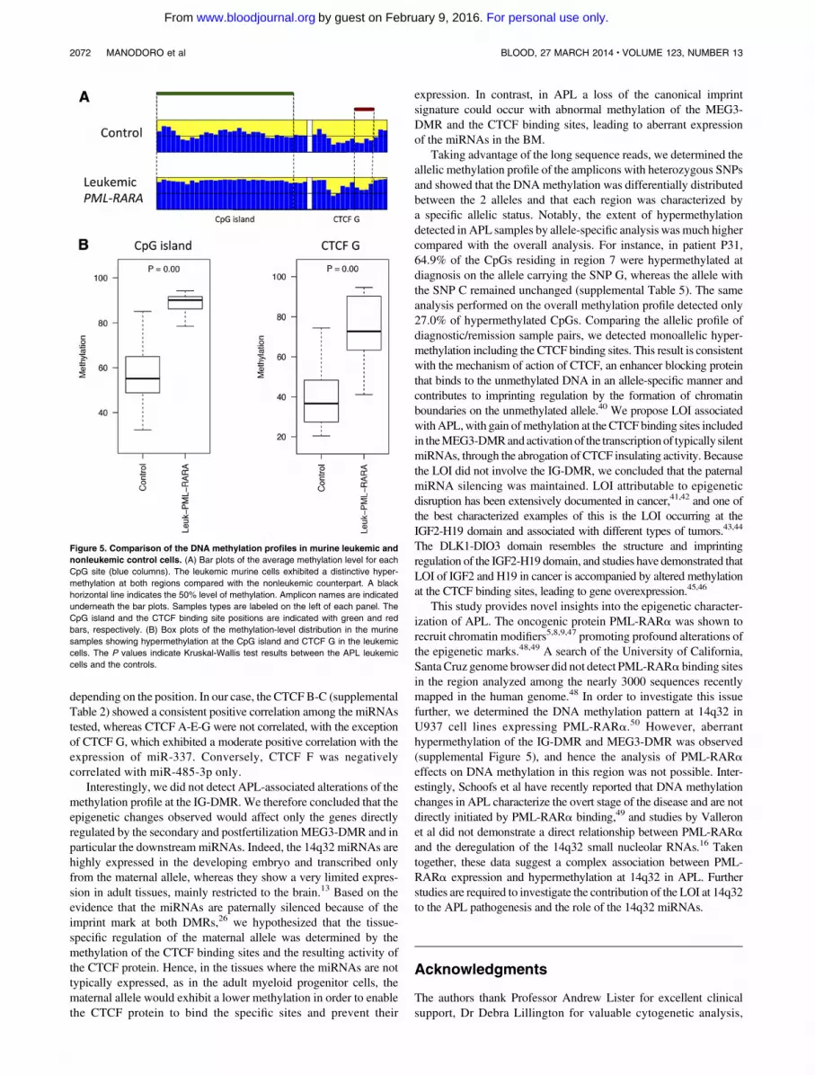
Therefore, we investigated whether the region is similarly dereg-ulated in murine APL, utilizing the established hMRP8-PML-RARatransgenic model.3Murine transgenic APL cells were collected fromthe BM of 3 leukemic mice and 4 controls (see “Methods”). High-throughput bisulfite sequencing was performed to determine themethylation profile of the CpG island and the CTCF binding sitelocated upstream from the mouse GTL2 gene. Of the 7 samples, 5were successfully sequenced (4 controls and 1 leukemic mouse).Leukemic APL cells exhibited hypermethylation at both the CpGisland and the CTCF binding site as compared with the controls
Figure 3. Allele-specific DNA methylation profiling. Unsupervised cluster analysis was performed on the CpG methylation pattern obtained for each sample and amplicon.
When a heterozygous SNP was observed, sequence reads were separated accordingly to the SNP genotype, and the CpG methylation pattern of each allele was analyzed by
cluster analysis. (A) Differential methylation between alleles at region 9 in the healthy donor C55. (i) Overall cluster analysis identified 2 clusters according to the methylation
pattern; (ii) DNA methylation pattern for each allele. (B) Allele-specific DNA methylation differences at region 2 between diagnosis (i) and complete remission (ii) stages of
patient P9 with APL; (iii) allele-specific DNA methylation at region 2 in the healthy donor C55. (C) Allele-specific DNA methylation differences at region 7 between diagnosis (i)
and complete remission (ii) stages of patient P31 with APL; (iii) allele-specific DNA methylation at region 7 in the healthy donor C52. Heat maps show clustering results. Each
column represents a CpG site, and each row the methylation pattern of a single sequence read. The color indicates the methylation status of each CpG: blue, methylated;
yellow, not methylated. The bar plot at the top of each heat map shows the overall methylation level of each CpG site.
2070 MANODORO et al BLOOD, 27 MARCH 2014 x VOLUME 123, NUMBER 13
For personal use only.on February 9, 2016. by guest www.bloodjournal.orgFrom

(Figure 5). These results validate the hypothesis that LOI at 14q32 isassociated with the pathogenesis of PML-RARa–induced leukemia.
Discussion
The present study shows that a LOI occurs at the DLK1-DIO3imprinted domain inAPL cells in associationwith the overexpressionof the downstream clustered miRNAs. Epigenetic changes occurcommonly in cancer, and because they are reversible, their iden-tification has had profound effects in the development of targetedtherapies. DNA methylation inhibitors such as azacitidine anddecitabine are currently included in clinical trials for the treatmentof hematologic malignancies.37,38 Recently, a large-scale study dem-onstrated that DNA methylation patterns segregate AML subtypesaccording to their karyotype, suggesting that the specific fusiononcoproteins can drive epigenetic changes and contribute to themalignant transformation by the deregulation of sets of genes.39
The DLK1-DIO3 miRNAs are epigenetically regulated andsilenced in most adult tissues but overexpressed in APL cells, asshown by ourselves and others,12,15,17 suggesting that they may con-tribute to APL development in cooperation with the PML-RARaprotein. Here, we show that the MEG3-DMR that is involved inthe imprinting regulation of the DLK1-DIO3 domain27 is hyper-methylated in APL. The MEG3-DMR hypermethylation was strictlydisease-associated, among the cases analyzed, which includedAMLswith NK, t(8;21), and inv(16), being detected only in the APL cases
and not present in remission BM or normal controls. Furthermore,myeloid progenitor cells showed a distinctive hypomethylated profilein this region indicating that the observed epigenetic changes did notreflect a stage of cell differentiation. Finally, the leukemic BM cellscollected from the murine APL model showed marked hypermeth-ylation at the orthologous region on chromosome 12, also includingthe unique CTCF binding site located upstream from the mousemiRNA cluster. Taken together, these data demonstrate that APL ischaracterized by hypermethylation at the 14q32 domain.
We also showed that the DNAmethylation profile at the 2 DMRscorrelatedwith expression of the downstreammiRNAs. This result isconsistent with the hypothesis that the expression of the sncRNAsclustered in the 14q32 domain is under the control of the IG-DMRand MEG3-DMR because they are organized in repeated arrays andthey might be processed from a long transcript starting from thepromoter of MEG3,13 although the mechanisms have not yet beenelucidated. In our experiments, the miRNAs tested were negativelycorrelated with the IG-DMR, whereas the MEG3-DMR displayeda bivalent pattern: methylation at the promoter of MEG3 positivelycorrelated with expression of the miRNAs, whereas methylation atthe MEG3 gene body showed a less consistent and predominantlynegative correlation. This atypical patterning (hypermethylationassociatedwith higher expression)was attributable to the presence ofthe binding sites for the insulator CTCF. Indeed, we showed that themiRNAs tested were positively correlated with the methylation of atleast 1 CpG included in a CTCF binding site. Furthermore, thisresult indicates that CTCF binding sites display different properties
Figure 4. Correlation between DNA methylation levels and gene expression profiles. The expression of 6 miRNAs included in the 14q32 cluster (miR-127, miR-136,
miR-154, miR-337, miR-379, and miR-485) was correlated with DNA methylation data at the DMRs. The position of the CpG island, the CTCF binding sites, and the amplicon
is labeled with green, red, and blue horizontal bars, respectively. The correlation is represented with red and green vertical bars indicating positive and negative values,
respectively. Each column indicates a CpG site. The distance of each gene from the IG-DMR is indicated on the right.
BLOOD, 27 MARCH 2014 x VOLUME 123, NUMBER 13 DNA METHYLATION PROFILING BY DEEP SEQUENCING 2071
For personal use only.on February 9, 2016. by guest www.bloodjournal.orgFrom
depending on the position. In our case, the CTCFB-C (supplementalTable 2) showed a consistent positive correlation among the miRNAstested, whereas CTCF A-E-G were not correlated, with the exceptionof CTCF G, which exhibited a moderate positive correlation with theexpression of miR-337. Conversely, CTCF F was negativelycorrelated with miR-485-3p only.
Interestingly, we did not detect APL-associated alterations of themethylation profile at the IG-DMR.We therefore concluded that theepigenetic changes observed would affect only the genes directlyregulated by the secondary and postfertilization MEG3-DMR and inparticular the downstream miRNAs. Indeed, the 14q32 miRNAs arehighly expressed in the developing embryo and transcribed onlyfrom the maternal allele, whereas they show a very limited expres-sion in adult tissues, mainly restricted to the brain.13 Based on theevidence that the miRNAs are paternally silenced because of theimprint mark at both DMRs,26 we hypothesized that the tissue-specific regulation of the maternal allele was determined by themethylation of the CTCF binding sites and the resulting activity ofthe CTCF protein. Hence, in the tissues where the miRNAs are nottypically expressed, as in the adult myeloid progenitor cells, thematernal allele would exhibit a lower methylation in order to enablethe CTCF protein to bind the specific sites and prevent their
expression. In contrast, in APL a loss of the canonical imprintsignature could occur with abnormal methylation of the MEG3-DMR and the CTCF binding sites, leading to aberrant expressionof the miRNAs in the BM.
Taking advantage of the long sequence reads, we determined theallelic methylation profile of the amplicons with heterozygous SNPsand showed that the DNAmethylation was differentially distributedbetween the 2 alleles and that each region was characterized bya specific allelic status. Notably, the extent of hypermethylationdetected in APL samples by allele-specific analysis wasmuch highercompared with the overall analysis. For instance, in patient P31,64.9% of the CpGs residing in region 7 were hypermethylated atdiagnosis on the allele carrying the SNP G, whereas the allele withthe SNP C remained unchanged (supplemental Table 5). The sameanalysis performed on the overall methylation profile detected only27.0% of hypermethylated CpGs. Comparing the allelic profile ofdiagnostic/remission sample pairs, we detected monoallelic hyper-methylation including the CTCF binding sites. This result is consistentwith the mechanism of action of CTCF, an enhancer blocking proteinthat binds to the unmethylated DNA in an allele-specific manner andcontributes to imprinting regulation by the formation of chromatinboundaries on the unmethylated allele.40 We propose LOI associatedwithAPL,with gain ofmethylation at the CTCF binding sites includedin theMEG3-DMRandactivationof the transcriptionof typically silentmiRNAs, through the abrogation of CTCF insulating activity. Becausethe LOI did not involve the IG-DMR, we concluded that the paternalmiRNA silencing was maintained. LOI attributable to epigeneticdisruption has been extensively documented in cancer,41,42 and one ofthe best characterized examples of this is the LOI occurring at theIGF2-H19 domain and associated with different types of tumors.43,44
The DLK1-DIO3 domain resembles the structure and imprintingregulation of the IGF2-H19 domain, and studies have demonstrated thatLOI of IGF2 and H19 in cancer is accompanied by altered methylationat the CTCF binding sites, leading to gene overexpression.45,46
This study provides novel insights into the epigenetic character-ization of APL. The oncogenic protein PML-RARa was shown torecruit chromatin modifiers5,8,9,47 promoting profound alterations ofthe epigenetic marks.48,49 A search of the University of California,Santa Cruz genome browser did not detect PML-RARa binding sitesin the region analyzed among the nearly 3000 sequences recentlymapped in the human genome.48 In order to investigate this issuefurther, we determined the DNA methylation pattern at 14q32 inU937 cell lines expressing PML-RARa.50 However, aberranthypermethylation of the IG-DMR and MEG3-DMR was observed(supplemental Figure 5), and hence the analysis of PML-RARaeffects on DNA methylation in this region was not possible. Inter-estingly, Schoofs et al have recently reported that DNA methylationchanges in APL characterize the overt stage of the disease and are notdirectly initiated by PML-RARa binding,49 and studies by Valleronet al did not demonstrate a direct relationship between PML-RARaand the deregulation of the 14q32 small nucleolar RNAs.16 Takentogether, these data suggest a complex association between PML-RARa expression and hypermethylation at 14q32 in APL. Furtherstudies are required to investigate the contribution of the LOI at 14q32to the APL pathogenesis and the role of the 14q32 miRNAs.
Acknowledgments
The authors thank Professor Andrew Lister for excellent clinicalsupport, Dr Debra Lillington for valuable cytogenetic analysis,
Figure 5. Comparison of the DNA methylation profiles in murine leukemic and
nonleukemic control cells. (A) Bar plots of the average methylation level for each
CpG site (blue columns). The leukemic murine cells exhibited a distinctive hyper-
methylation at both regions compared with the nonleukemic counterpart. A black
horizontal line indicates the 50% level of methylation. Amplicon names are indicated
underneath the bar plots. Samples types are labeled on the left of each panel. The
CpG island and the CTCF binding site positions are indicated with green and red
bars, respectively. (B) Box plots of the methylation-level distribution in the murine
samples showing hypermethylation at the CpG island and CTCF G in the leukemic
cells. The P values indicate Kruskal-Wallis test results between the APL leukemic
cells and the controls.
2072 MANODORO et al BLOOD, 27 MARCH 2014 x VOLUME 123, NUMBER 13
For personal use only.on February 9, 2016. by guest www.bloodjournal.orgFrom

Professor Scott Kogan for provision of the MRP8-PML-RARAtransgenic APL blasts, and Professor Pier Giuseppe Pelicci forprovision of the inducible U937 cell lines.
Thisworkwas supported by a project grant fromCancer ResearchUnited Kingdom (C6277/A6789) (B.D.Y. and S.D.) and a Specialistprogram award from Leukaemia & Lymphoma Research (10023)(D.G. and E.M.).
Authorship
Contribution: F.M., B.D.Y., and S.D. designed the research andanalyzed data; F.M., T.C., F.M.-M., E.M., and J.V.J. performed theexperiments; J.M. performed bioinformatics analysis, analyzed data,
andmade the figures; J.W. assisted with bioinformatics analysis; S.I.collected patient samples and performed DNA extraction; D.G. andD.T. contributed to research design; J.M., D.G., J.G.G., B.D.Y.,and S.D. critically reviewed the manuscript; and F.M. wrote themanuscript.
Conflict-of-interest disclosure: The authors declare no competingfinancial interests.
Correspondence: Floriana Manodoro, Centre for Haemato-Oncology,Barts Cancer Institute, QueenMaryUniversity of London,John Vane Science Centre, Charterhouse Square, London EC1M6BQ, United Kingdom; e-mail: [email protected]; andSilvana Debernardi, Centre for Haemato-Oncology, Barts CancerInstitute, Queen Mary University of London, John Vane ScienceCentre, Charterhouse Square, LondonEC1M6BQ,UnitedKingdom;e-mail: [email protected].
References
1. de The H, Chomienne C, Lanotte M, Degos L,Dejean A. The t(15;17) translocation of acutepromyelocytic leukaemia fuses the retinoic acidreceptor alpha gene to a novel transcribed locus.Nature. 1990;347(6293):558-561.
2. Melnick A, Licht JD. Deconstructing a disease:RARalpha, its fusion partners, and their rolesin the pathogenesis of acute promyelocyticleukemia. Blood. 1999;93(10):3167-3215.
3. Brown D, Kogan S, Lagasse E, et al. APMLRARalpha transgene initiates murine acutepromyelocytic leukemia. Proc Natl Acad Sci U S A.1997;94(6):2551-2556.
4. Kogan SC, Hong SH, Shultz DB, Privalsky ML,Bishop JM. Leukemia initiated by PMLRARalpha:the PML domain plays a critical role while retinoicacid-mediated transactivation is dispensable.Blood. 2000;95(5):1541-1550.
5. Carbone R, Botrugno OA, Ronzoni S, et al.Recruitment of the histone methyltransferaseSUV39H1 and its role in the oncogenic propertiesof the leukemia-associated PML-retinoic acidreceptor fusion protein. Mol Cell Biol. 2006;26(4):1288-1296.
6. Hoemme C, Peerzada A, Behre G,et al. Chromatin modifications induced byPML-RARalpha repress critical targets inleukemogenesis as analyzed by ChIP-Chip.Blood. 2008;111(5):2887-2895.
7. Wang K, Wang P, Shi J, et al. PML/RARalphatargets promoter regions containing PU.1consensus and RARE half sites in acutepromyelocytic leukemia. Cancer Cell. 2010;17(2):186-197.
8. Di Croce L, Raker VA, Corsaro M, et al.Methyltransferase recruitment and DNAhypermethylation of target promoters by anoncogenic transcription factor. Science. 2002;295(5557):1079-1082.
9. Villa R, Pasini D, Gutierrez A, et al. Role ofthe polycomb repressive complex 2 in acutepromyelocytic leukemia. Cancer Cell. 2007;11(6):513-525.
10. Bartel DP. MicroRNAs: genomics, biogenesis,mechanism, and function. Cell. 2004;116(2):281-297.
11. Ambros V. The functions of animal microRNAs.Nature. 2004;431(7006):350-355.
12. Dixon-McIver A, East P, Mein CA, et al. Distinctivepatterns of microRNA expression associated withkaryotype in acute myeloid leukaemia. PLoSONE. 2008;3(5):e2141.
13. Seitz H, Royo H, Bortolin ML, Lin SP, Ferguson-Smith AC, Cavaille J. A large imprinted microRNAgene cluster at the mouse Dlk1-Gtl2 domain.Genome Res. 2004;14(9):1741-1748.
14. Kircher M, Bock C, Paulsen M. Structuralconservation versus functional divergence ofmaternally expressed microRNAs in the Dlk1/Gtl2imprinting region. BMC Genomics. 2008;9:346.
15. Jongen-Lavrencic M, Sun SM, Dijkstra MK, ValkPJ, Lowenberg B. MicroRNA expression profilingin relation to the genetic heterogeneity of acutemyeloid leukemia. Blood. 2008;111(10):5078-5085.
16. Valleron W, Laprevotte E, Gautier EF, et al.Specific small nucleolar RNA expression profilesin acute leukemia. Leukemia. 2012;26(9):2052-2060.
17. Cancer Genome Atlas Research Network.Genomic and epigenomic landscapes of adultde novo acute myeloid leukemia. N Engl J Med.2013;368(22):2059-2074.
18. Glazov EA, McWilliam S, Barris WC, DalrympleBP. Origin, evolution, and biological role of miRNAcluster in DLK-DIO3 genomic region in placentalmammals. Mol Biol Evol. 2008;25(5):939-948.
19. Benetatos L, Voulgaris E, Vartholomatos G.
DLK1-MEG3 imprinted domain microRNAs incancer biology. Crit Rev Eukaryot Gene Expr.2012;22(1):1-15.
20. Benetatos L, Hatzimichael E, Londin E, et al.The microRNAs within the DLK1-DIO3 genomicregion: involvement in disease pathogenesis. CellMol Life Sci. 2013;70(5):795-814.
21. Swarbrick A, Woods SL, Shaw A, et al. miR-380-5prepresses p53 to control cellular survival and isassociated with poor outcome in MYCN-amplifiedneuroblastoma. Nat Med. 2010;16(10):1134-1140.
22. Benetatos L, Vartholomatos G, Hatzimichael E.MEG3 imprinted gene contribution intumorigenesis. Int J Cancer. 2011;129(4):773-779.
23. Seitz H, Youngson N, Lin SP, et al. ImprintedmicroRNA genes transcribed antisense toa reciprocally imprinted retrotransposon-likegene. Nat Genet. 2003;34(3):261-262.
24. Liu L, Luo GZ, Yang W, et al. Activation ofthe imprinted Dlk1-Dio3 region correlates withpluripotency levels of mouse stem cells. J BiolChem. 2010;285(25):19483-19490.
25. Wylie AA, Murphy SK, Orton TC, Jirtle RL.Novel imprinted DLK1/GTL2 domain on humanchromosome 14 contains motifs that mimic thoseimplicated in IGF2/H19 regulation. Genome Res.2000;10(11):1711-1718.
26. da Rocha ST, Edwards CA, Ito M, Ogata T,Ferguson-Smith AC. Genomic imprinting at themammalian Dlk1-Dio3 domain. Trends Genet.2008;24(6):306-316.
27. Kagami M, O’Sullivan MJ, Green AJ, et al.The IG-DMR and the MEG3-DMR at humanchromosome 14q32.2: hierarchical interactionand distinct functional properties as imprintingcontrol centers. PLoS Genet. 2010;6(6):e1000992.
28. Lin SP, Youngson N, Takada S, et al. Asymmetricregulation of imprinting on the maternal andpaternal chromosomes at the Dlk1-Gtl2 imprintedcluster on mouse chromosome 12. Nat Genet.2003;35(1):97-102.
29. Rosa AL, Wu YQ, Kwabi-Addo B, Coveler KJ,Reid Sutton V, Shaffer LG. Allele-specificmethylation of a functional CTCF binding siteupstream of MEG3 in the human imprinteddomain of 14q32. Chromosome Res. 2005;13(8):809-818.
30. Bell AC, West AG, Felsenfeld G. The proteinCTCF is required for the enhancer blockingactivity of vertebrate insulators. Cell. 1999;98(3):387-396.
31. Szabo P, Tang SH, Rentsendorj A, Pfeifer GP,Mann JR. Maternal-specific footprints at putativeCTCF sites in the H19 imprinting control regiongive evidence for insulator function. Curr Biol.2000;10(10):607-610.
32. Vardiman JW, Thiele J, Arber DA, et al. The 2008revision of the World Health Organization (WHO)classification of myeloid neoplasms and acuteleukemia: rationale and important changes.Blood. 2009;114(5):937-951.
33. Paulsen M, Takada S, Youngson NA, et al.Comparative sequence analysis of the imprintedDlk1-Gtl2 locus in three mammalian speciesreveals highly conserved genomic elements andrefines comparison with the Igf2-H19 region.Genome Res. 2001;11(12):2085-2094.
34. Kumaki Y, Oda M, Okano M. QUMA:quantification tool for methylation analysis.Nucleic Acids Res. 2008;36(suppl 2):W170-W175.
35. Fury W, Batliwalla F, Gregersen PK, Li W.Overlapping probabilities of top ranking gene lists,hypergeometric distribution, and stringency ofgene selection criterion. Conf Proc IEEE Eng MedBiol Soc. 2006;1:5531-5534.
36. Livak KJ, Schmittgen TD. Analysis of relativegene expression data using real-time quantitativePCR and the 2(-Delta Delta C(T)) method.Methods. 2001;25(4):402-408.
37. Scandura JM, Roboz GJ, Moh M, et al. Phase 1study of epigenetic priming with decitabine prior tostandard induction chemotherapy for patients withAML. Blood. 2011;118(6):1472-1480.
38. Kantarjian HM, Thomas XG, Dmoszynska A, et al.Multicenter, randomized, open-label, phase III trial
BLOOD, 27 MARCH 2014 x VOLUME 123, NUMBER 13 DNA METHYLATION PROFILING BY DEEP SEQUENCING 2073
For personal use only.on February 9, 2016. by guest www.bloodjournal.orgFrom
of decitabine versus patient choice, with physicianadvice, of either supportive care or low-dosecytarabine for the treatment of older patients withnewly diagnosed acute myeloid leukemia. J ClinOncol. 2012;30(21):2670-2677.
39. Figueroa ME, Lugthart S, Li Y, et al. DNAmethylation signatures identify biologically distinctsubtypes in acute myeloid leukemia. Cancer Cell.2010;17(1):13-27.
40. Bell AC, Felsenfeld G. Methylation of aCTCF-dependent boundary controls imprintedexpression of the Igf2 gene. Nature. 2000;405(6785):482-485.
41. Feinberg AP, Ohlsson R, Henikoff S. Theepigenetic progenitor origin of human cancer. NatRev Genet. 2006;7(1):21-33.
42. Jelinic P, Shaw P. Loss of imprinting and cancer.J Pathol. 2007;211(3):261-268.
43. Cui H, Cruz-Correa M, Giardiello FM, et al. Lossof IGF2 imprinting: a potential marker of colorectalcancer risk. Science. 2003;299(5613):1753-1755.
44. Honda S, Arai Y, Haruta M, et al. Lossof imprinting of IGF2 correlates withhypermethylation of the H19 differentiallymethylated region in hepatoblastoma. Br JCancer. 2008;99(11):1891-1899.
45. Ulaner GA, Vu TH, Li T, et al. Loss of imprinting ofIGF2 and H19 in osteosarcoma is accompaniedby reciprocal methylation changes of a CTCF-binding site. Hum Mol Genet. 2003;12(5):535-549.
46. Pant V, Kurukuti S, Pugacheva E, et al. Mutationof a single CTCF target site within the H19imprinting control region leads to loss of Igf2imprinting and complex patterns of de novomethylation upon maternal inheritance. Mol CellBiol. 2004;24(8):3497-3504.
47. Grignani F, De Matteis S, Nervi C, et al. Fusionproteins of the retinoic acid receptor-alpha recruithistone deacetylase in promyelocytic leukaemia.Nature. 1998;391(6669):815-818.
48. Martens JH, Brinkman AB, Simmer F, et al. PML-RARalpha/RXR alters the epigenetic landscape inacute promyelocytic leukemia. Cancer Cell. 2010;17(2):173-185.
49. Schoofs T, Rohde C, Hebestreit K, et al. DNAmethylation changes are a late event in acutepromyelocytic leukemia and coincide with loss oftranscription factor binding. Blood. 2013;121(1):178-187.
50. Grignani F, Ferrucci PF, Testa U, et al. The acutepromyelocytic leukemia-specific PML-RAR alphafusion protein inhibits differentiation and promotessurvival of myeloid precursor cells. Cell. 1993;74(3):423-431.
2074 MANODORO et al BLOOD, 27 MARCH 2014 x VOLUME 123, NUMBER 13
For personal use only.on February 9, 2016. by guest www.bloodjournal.orgFrom
online February 3, 2014 originally publisheddoi:10.1182/blood-2012-12-469833
2014 123: 2066-2074
Young and Silvana DebernardiJovanovic, Jun Wang, Sameena Iqbal, David Taussig, David Grimwade, John G. Gribben, Bryan D. Floriana Manodoro, Jacek Marzec, Tracy Chaplin, Farideh Miraki-Moud, Eva Moravcsik, Jelena V. overexpression in acute promyelocytic leukemiaLoss of imprinting at the 14q32 domain is associated with microRNA
http://www.bloodjournal.org/content/123/13/2066.full.htmlUpdated information and services can be found at:
(1450 articles)Myeloid Neoplasia Articles on similar topics can be found in the following Blood collections
http://www.bloodjournal.org/site/misc/rights.xhtml#repub_requestsInformation about reproducing this article in parts or in its entirety may be found online at:
http://www.bloodjournal.org/site/misc/rights.xhtml#reprintsInformation about ordering reprints may be found online at:
http://www.bloodjournal.org/site/subscriptions/index.xhtmlInformation about subscriptions and ASH membership may be found online at:
Copyright 2011 by The American Society of Hematology; all rights reserved.of Hematology, 2021 L St, NW, Suite 900, Washington DC 20036.Blood (print ISSN 0006-4971, online ISSN 1528-0020), is published weekly by the American Society
For personal use only.on February 9, 2016. by guest www.bloodjournal.orgFrom
Related Documents