Molecular Biology of the Cell Vol. 16, 665– 675, February 2005 Loss of Function of KRE5 Suppresses Temperature Sensitivity of Mutants Lacking Mitochondrial Anionic Lipids Quan Zhong,* † Jelena Gvozdenovic-Jeremic,* † Paul Webster, ‡ Jingming Zhou,* and Miriam L. Greenberg* § *Department of Biological Sciences, Wayne State University, Detroit, MI 48202; and ‡ House Ear Institute, Los Angeles, CA 90057 Submitted September 15, 2004; Revised November 9, 2004; Accepted November 14, 2004 Monitoring Editor: Howard Riezman Disruption of PGS1, which encodes the enzyme that catalyzes the committed step of cardiolipin (CL) synthesis, results in loss of the mitochondrial anionic phospholipids phosphatidylglycerol (PG) and CL. The pgs1 mutant exhibits severe growth defects at 37°C. To understand the essential functions of mitochondrial anionic lipids at elevated temperatures, we isolated suppressors of pgs1 that grew at 37°C. One of the suppressors has a loss of function mutation in KRE5, which is involved in cell wall biogenesis. The cell wall of pgs1 contained markedly reduced -1,3-glucan, which was restored in the suppressor. Stabilization of the cell wall with osmotic support alleviated the cell wall defects of pgs1 and suppressed the temperature sensitivity of all CL-deficient mutants. Evidence is presented suggesting that the previously reported inability of pgs1 to grow in the presence of ethidium bromide was due to defective cell wall integrity, not from “petite lethality.” These findings demonstrated that mitochondrial anionic lipids are required for cellular functions that are essential in cell wall biogenesis, the maintenance of cell integrity, and survival at elevated temperature. INTRODUCTION Cardiolipin (CL), a unique anionic phospholipid with dimeric structure, is ubiquitous in eukaryotes and primarily found in the mitochondrial inner membrane (Schlame et al., 2000). CL plays a key role in mitochondrial bioenergetics (Jiang et al., 2000; Koshkin and Greenberg, 2000, 2002; Schlame et al., 2000; Pfeiffer et al., 2003) and is also involved in mitochondrial biogenesis (Kawasaki et al., 1999; Jiang et al., 2000). Defective remodeling of CL is associated with Barth syndrome, a severe genetic disorder characterized by cardiomyopathy, neutropenia, skeletal myopathy, and re- spiratory chain defects (Vreken et al., 2000). The phenotype of Barth syndrome is dependent upon multiple factors that are not well understood (Barth et al., 1983, 1996). Elucidation of the functions of CL will help to clarify the abnormalities associated with this disorder. The biosynthesis of CL is conserved in eukaryotic organisms. It occurs via three enzymatic reactions (Schlame et al., 2000), including formation of phosphatidylglycerolphosphate (PGP) from CDP-DAG and glycerol-3-P, dephosphorylation of PGP to phosphatidylglycerol (PG), and condensation of CDP-DAG and PG to form CL. Disruption of PGS1, the structural gene encoding PGP synthase, results in the complete loss of both PG and CL (Janitor et al., 1996; Chang et al., 1998a). The crd1 mutant, which lacks CL synthase, has no detectable CL but accumulates PG (Jiang et al., 1997; Chang et al., 1998b; Tuller et al., 1998; Jiang et al., 2000; Pfeiffer et al., 2003; Zhong et al., 2004). The human taffazin gene (TAZ1), which is associated with Barth syndrome, encodes a transacylase that may be involved in the remodeling of CL (Xu et al., 2003). Deletion of the yeast homolog of this gene, TAZ1, leads to decreased CL, aberrant CL acyl species, and accumulation of monolysocardiolipin (Gu et al., 2004). Mutants deficient in CL biosynthesis exhibit growth defects at elevated temperatures. The taz1 mutant is temperature sensitive for growth on ethanol but grows well on other carbon sources at elevated temperature (Gu et al., 2004). The crd1 mutant loses viability on both fermentable and non- fermentable carbon sources at elevated temperature, and it does not form colonies from single cells seeded on YPD plates (Jiang et al., 1999, 2000; Zhong et al., 2004). The pgs1 mutant exhibits the most severe growth defects and cannot grow at all at 37°C, even on glucose (Chang et al., 1998a; Dzugasova et al., 1998). The temperature-sensitive growth defects observed in CL-deficient mutants suggest that CL plays an essential role in maintaining cell viability at elevated temperature. The greater degree of temperature sensitivity of the pgs1 mutant com- pared with the crd1 mutant indicates that PG can substitute for some essential functions of CL. Mitochondria from crd1 (Koshkin and Greenberg, 2000, 2002) and taz1 (Ma et al., 2004) exhibit defective energetic coupling at elevated temperatures. Although thermal sensitivity of the bioenergetic functions may explain temperature sensitivity of these mutants in nonfer- mentable medium, the reason for loss of viability on glucose is not known. In addition to the temperature-sensitive growth defects, CL-deficient mutants exhibit decreased mitochondrial ge- nome stability. Mutant cells of crd1 grown in the presence Article published online ahead of print in MBC in Press on Novem- ber 24, 2004 (http://www.molbiolcell.org/cgi/doi/10.1091/mbc. E04-09-0808). † These authors contributed equally to this study. § Corresponding author. E-mail address: [email protected]. wayne.edu. Abbreviations used: CFW, calcoflour white; CL, cardiolipin; CSIII, chitin synthase III; GS, glucan synthase; mtDNA, mitochondrial DNA; PGP, phophatidylglycerolphosphate; PG, phosphatidylglyc- erol; Ura , synthetic drop out medium without uracil; YPD, yeast extract, peptone, and dextrose; YPGE, yeast extract, glycerol and ethanol; YPDS, YPD supplemented with 1 M sorbitol. © 2005 by The American Society for Cell Biology 665

Welcome message from author
This document is posted to help you gain knowledge. Please leave a comment to let me know what you think about it! Share it to your friends and learn new things together.
Transcript

Molecular Biology of the CellVol. 16, 665–675, February 2005
Loss of Function of KRE5 Suppresses Temperature Sensitivityof Mutants Lacking Mitochondrial Anionic LipidsQuan Zhong,*† Jelena Gvozdenovic-Jeremic,*† Paul Webster,‡ Jingming Zhou,*and Miriam L. Greenberg*§
*Department of Biological Sciences, Wayne State University, Detroit, MI 48202; and ‡House Ear Institute, LosAngeles, CA 90057
Submitted September 15, 2004; Revised November 9, 2004; Accepted November 14, 2004Monitoring Editor: Howard Riezman
Disruption of PGS1, which encodes the enzyme that catalyzes the committed step of cardiolipin (CL) synthesis, results inloss of the mitochondrial anionic phospholipids phosphatidylglycerol (PG) and CL. The pgs1� mutant exhibits severegrowth defects at 37°C. To understand the essential functions of mitochondrial anionic lipids at elevated temperatures, weisolated suppressors of pgs1� that grew at 37°C. One of the suppressors has a loss of function mutation in KRE5, whichis involved in cell wall biogenesis. The cell wall of pgs1� contained markedly reduced �-1,3-glucan, which was restoredin the suppressor. Stabilization of the cell wall with osmotic support alleviated the cell wall defects of pgs1� andsuppressed the temperature sensitivity of all CL-deficient mutants. Evidence is presented suggesting that the previouslyreported inability of pgs1� to grow in the presence of ethidium bromide was due to defective cell wall integrity, not from“petite lethality.” These findings demonstrated that mitochondrial anionic lipids are required for cellular functions thatare essential in cell wall biogenesis, the maintenance of cell integrity, and survival at elevated temperature.
INTRODUCTION
Cardiolipin (CL), a unique anionic phospholipid withdimeric structure, is ubiquitous in eukaryotes and primarilyfound in the mitochondrial inner membrane (Schlame et al.,2000). CL plays a key role in mitochondrial bioenergetics(Jiang et al., 2000; Koshkin and Greenberg, 2000, 2002;Schlame et al., 2000; Pfeiffer et al., 2003) and is also involvedin mitochondrial biogenesis (Kawasaki et al., 1999; Jiang etal., 2000). Defective remodeling of CL is associated withBarth syndrome, a severe genetic disorder characterized bycardiomyopathy, neutropenia, skeletal myopathy, and re-spiratory chain defects (Vreken et al., 2000). The phenotypeof Barth syndrome is dependent upon multiple factors thatare not well understood (Barth et al., 1983, 1996). Elucidationof the functions of CL will help to clarify the abnormalitiesassociated with this disorder.
The biosynthesis of CL is conserved in eukaryotic organisms.It occurs via three enzymatic reactions (Schlame et al., 2000),including formation of phosphatidylglycerolphosphate (PGP)from CDP-DAG and glycerol-3-P, dephosphorylation of PGPto phosphatidylglycerol (PG), and condensation of CDP-DAG
and PG to form CL. Disruption of PGS1, the structural geneencoding PGP synthase, results in the complete loss of both PGand CL (Janitor et al., 1996; Chang et al., 1998a). The crd1�mutant, which lacks CL synthase, has no detectable CL butaccumulates PG (Jiang et al., 1997; Chang et al., 1998b; Tuller etal., 1998; Jiang et al., 2000; Pfeiffer et al., 2003; Zhong et al., 2004).The human taffazin gene (TAZ1), which is associated withBarth syndrome, encodes a transacylase that may be involvedin the remodeling of CL (Xu et al., 2003). Deletion of the yeasthomolog of this gene, TAZ1, leads to decreased CL, aberrantCL acyl species, and accumulation of monolysocardiolipin (Guet al., 2004). Mutants deficient in CL biosynthesis exhibitgrowth defects at elevated temperatures. The taz1� mutant istemperature sensitive for growth on ethanol but grows well onother carbon sources at elevated temperature (Gu et al., 2004).The crd1� mutant loses viability on both fermentable and non-fermentable carbon sources at elevated temperature, and itdoes not form colonies from single cells seeded on YPD plates(Jiang et al., 1999, 2000; Zhong et al., 2004). The pgs1� mutantexhibits the most severe growth defects and cannot grow at allat 37°C, even on glucose (Chang et al., 1998a; Dzugasova et al.,1998). The temperature-sensitive growth defects observed inCL-deficient mutants suggest that CL plays an essential role inmaintaining cell viability at elevated temperature. The greaterdegree of temperature sensitivity of the pgs1� mutant com-pared with the crd1� mutant indicates that PG can substitutefor some essential functions of CL. Mitochondria from crd1�(Koshkin and Greenberg, 2000, 2002) and taz1� (Ma et al., 2004)exhibit defective energetic coupling at elevated temperatures.Although thermal sensitivity of the bioenergetic functions mayexplain temperature sensitivity of these mutants in nonfer-mentable medium, the reason for loss of viability on glucose isnot known.
In addition to the temperature-sensitive growth defects,CL-deficient mutants exhibit decreased mitochondrial ge-nome stability. Mutant cells of crd1� grown in the presence
Article published online ahead of print in MBC in Press on Novem-ber 24, 2004 (http://www.molbiolcell.org/cgi/doi/10.1091/mbc.E04-09-0808).† These authors contributed equally to this study.§ Corresponding author. E-mail address: [email protected].
Abbreviations used: CFW, calcoflour white; CL, cardiolipin; CSIII,chitin synthase III; GS, glucan synthase; mtDNA, mitochondrialDNA; PGP, phophatidylglycerolphosphate; PG, phosphatidylglyc-erol; Ura�, synthetic drop out medium without uracil; YPD, yeastextract, peptone, and dextrose; YPGE, yeast extract, glycerol andethanol; YPDS, YPD supplemented with 1 M sorbitol.
© 2005 by The American Society for Cell Biology 665

of fermentable or nonfermentable carbon sources segregatelarge numbers of petites (respiratory incompetent cells) afterprolonged culture at elevated temperature (Jiang et al., 2000;Zhong et al., 2004). The pgs1� mutant was initially deter-mined to be “petite lethal” because the mutant cells did notsurvive ethidium bromide mutagenesis, which induces pe-tite formation (Janitor and Subik, 1993; Dzugasova et al.,1998). However, 4�,6-diamidino-2-phenylindole (DAPI) stain-ing of pgs1� revealed only the presence of nuclear DNA(Chang et al., 1998b). The absence of mitochondrial DNA(mtDNA) staining was attributed to a lack of elongated mito-chondrial structure, but loss of mtDNA was not ruled out.
In a large-scale screen to identify genes involved in cellwall biogenesis, Lussier et al. (1997) reported that disruptionof the PGS1 promoter results in several cell wall defects,including decreased glucosamine levels and hypersensitiv-ity to cell wall-perturbing agents such as zymolyase, cal-cofluor white (CFW), papulacandin, and caffeine. The yeastcell wall is an essential organelle that determines the shapeand preserves the osmotic integrity of the cell by counter-acting the internal turgor pressure. Mutants with weakenedcell walls tend to form swollen cells with more sphericalappearance and larger cell size than the wild-type (Popolo etal., 1993; de Nobel et al., 2000). The yeast cell wall has atwo-layered structure. The highly glycosylated mannopro-teins form the outer layer. The internal fibrillar layer con-tains glucans and chitin (Klis et al., 2002). �-1,3-Glucan formsa hollow helical structure that resembles a flexible wirespring. Together with chitin, it confers mechanical strengthto the cell wall. �-1,6-Glucan in its mature form is a highlybranched water-soluble polymer that interconnects all othercell wall components in a lattice (Klis et al., 2002). Chitin isenriched in the chitin ring in and around bud scars, and aminor portion is also uniformly dispersed in the lateral wall(Molano et al., 1980; Shaw et al., 1991; Cabib et al., 2001).Chitin deposition, however, is increased upon weakening ofthe cell wall (Popolo et al., 2001; Klis et al., 2002).
The yeast cell wall is a highly dynamic structure. It un-dergoes a number of modifications during different stages ofthe cell cycle and in response to environmental changes(Cabib et al., 2001; Smits et al., 2001). Elaborate control mech-anisms are strictly coordinated in the regulation of cell wall
biogenesis. Stress due to heat shock or ethanol induces theproduction of trehalose (Attfield, 1987; Hottiger et al., 1987;Neves and Francois, 1992; Hottiger et al., 1994; Singer andLindquist, 1998) and glycerol (Alonso-Monge et al., 2001),which increase turgor pressure. Defects in the assembly ofthe cell wall compromise the response to stress and severelythreaten cell survival. Supplementation with sorbitol, anosmotic stabilizer, supports the growth of cell wall mutants,presumably by balancing the turgor pressure on the plasmamembrane and stabilizing cell wall structure (Popolo et al.,2001; Klis et al., 2002).
To understand the essential functions of CL at elevatedtemperature, we took the genetic approach of isolating spon-taneous suppressor mutants of pgs1� that grow at elevatedtemperatures, one of which was identified to have a loss offunction allele of KRE5, which is involved in cell wall bio-genesis. In this report, we demonstrated that the absence ofmitochondrial anionic phospholipids PG and CL results indefective cell wall assembly. Disruption of KRE5 induces�-1,3-glucan synthesis, strengthening the cell wall structurein pgs1� and enabling it to survive at elevated temperature.These data suggest that mitochondrial anionic phospholip-ids are required for processes that are essential in cell wallbiogenesis and the maintenance of cell integrity.
MATERIALS AND METHODS
MaterialsAll chemicals used were reagent grade or better. The polymerase chainreaction (PCR) was performed using the native pfu enzyme kit from Invitro-gen (Carlsbad, CA). The Zymoprep yeast plasmid mini prep kit was fromZymoResearch (Orange, CA). The Wizard Plus Miniprep DNA purificationsystem was from Promega (Madison, WI). All other buffers and enzymes werepurchased from Sigma-Aldrich (St. Louis, MO). Glucose, yeast extract, andpeptone were purchased from Difco (Detroit, MI).
Yeast Strains and Growth MediaThe Saccharomyces cerevisiae strains used in this work are listed in Table 1.Synthetic complete medium (SCD) contained amino acids adenine (20.25mg/l), arginine (20 mg/l), histidine (20 mg/l), leucine (60 mg/l), lysine (200mg/l), methionine (20 mg/l), threonine (300 mg/l), tryptophan (20 mg/l),and uracil (20 mg/l), vitamins, salts (essentially components of Difco VitaminFree Yeast Base without amino acids), inositol (75 �M), and glucose (2%).Synthetic drop out medium (Ura�) contained all ingredients, except uracil for
Table 1. Plasmids and yeast strains used in this study
Plasmid or strain Characteristics or genotype Source or reference
pYES2/CT 2 �m, URA3 InvitrogenpRS415-PGS1 derivative of pYES2/CT, expresses PGS1 from Gal1 promoter He and Greenberg (2004)Ycp50 Centromere, URA3 Rose et al. (1987)Ycp50-KRE5 derivative of Ycp50, expresses KRE5 from its own promoter This studyGAD74D3A MAT �, ade8, ura 3, trp1, his3, leu2 Dzugasova et al. (1998)GAD74D3C MAT �, ade8, ura 3, trp1, his3, leu2, pgs1::HIS3 Dzugasova et al. (1998)FGY3 MAT �, ura 3-52, lys2-801, ade2-101, trp1�1, his3�200, leu2�1 Jiang et al. (1997)FGY3 (�0) rho0 mutant derived from FGY3 This studyQZY24B (�0) MAT a, ura 3-52, lys2-801, trp1�1, his3�200, leu2�1, pgs1�::TRP1 This studyQZY11A (�0) MAT a, ura 3-52, lys2-801, trp1�1, his3�200, leu2�1, pgs1�::TRP1, kre5W1166X This studyFGY2 MAT �, ura 3-52, lys2-801, ade2-101, trp1�1, his3�200, leu2�1, crd1�::URA3 Jiang et al. (1997)100 MAT a, ade1, oxi2 (�) C. Dieckmann101 MAT a, ade1, oxi1 (�) C. Dieckmann102 MAT a, ade1, oxi3 (�) C. Dieckmann103 MAT a, ade1, cob (�) C. Dieckmann104 MAT �, met6, oxi2 (�) C. Dieckmann105 MAT �, met6, oxi1 (�) C. Dieckmann106 MAT �, met6, oxi3 (�) C. Dieckmann107 MAT �, met6, cob (�) C. DieckmannT158c/S14a Diploid prototroph S. cerevisiae ATCC (46427)
Q. Zhong et al.
Molecular Biology of the Cell666
selection. Sporulation medium contained potassium acetate (1%), glucose(0.05%), and the essential amino acids. Complex media contained yeast ex-tract (1%), peptone (2%), and glucose (2%) (YPD) or glycerol (3%) and ethanol(1%) (YPGE). Complex YPDS medium was YPD supplemented with 1 Msorbitol. Solid medium contained agar (2%) in addition to the above-men-tioned ingredients.
DAPI StainYeast cells were grown to early stationary phase, fixed in 70% ethanol at roomtemperature for 30 min, and stained with 1 �g/ml DAPI for 5 min. Cells wereviewed with an Olympus BX41 epifluorescence microscope, WU filter, and a100� oil immersion objective. Images were captured with a Q-color3 cameraand represent at least 200 observed cells.
Isolation of Extragenic Suppressors of pgs1�Disruption of the PGS1 gene was performed as described previously (Zhongand Greenberg, 2003). Haploid pgs1� mutants of opposite mating types wereobtained. YPD medium was inoculated from single colonies of pgs1� cells andgrown for 24 h. About 108 cells from each independent culture were plated ona fresh YPD plate and incubated at 39°C. Single colonies from each plate werereexamined for growth at 39°C. Cells that grew at 39°C were analyzed further.To test the dominant and recessive character of the suppressor mutation,suppressor mutants were crossed to the parent strain, and growth of thediploid cells at 39°C was examined. Genetic complementation analysis wascarried out with recessive mutants.
Plasmid ComplementationA yeast genomic DNA library in plasmid YCp50 (Rose et al., 1987) was usedto clone the suppressor genes by complementation. Suppressor mutant cellswere transformed with library DNA, plated on Ura� plates, and incubated at25°C until colonies formed. Transformants were replicated onto YPD plates,and growth at 39°C was examined. Plasmid DNA was extracted from trans-formants that lost the capability to grow at 39°C, amplified in Escherichia coliDH5�, and retransformed into the suppressor mutant to confirm complemen-tation of the suppressor phenotype. The DNA inserts of the positive cloneswere sequenced using primer YCp50 forward (5�-TTGGAGCCATATCGAC-TACG-3�) and YCp50 reverse (5�-ATGCGTCCGGCGTAGAGGATC-3�).
Identification of the Suppressor MutationGenomic DNA of pgs1� (QZY24B) and the suppressor QZY11A was ex-tracted. DNA of the KRE5 region was amplified and sequenced using thefollowing primers F1 (5�-TGTATTGGTTCATACCGGCA-3�); F2 (5�-ATAT-AGGGTTCTGAATTG-3�); R2 (5�-ATTGGAAGTTAGCGCCACAA-3�); F3(5�-ACGATATGGCATACCCGAAT-3�); F4 (5�-TTATGGAAGCAATGAATG-3�);R4 (5�-AGAACCCTGGAATTGTGTGGA-3�); F5 (5�-TCCGTACAATTTGCT-TACTGC-3�); F6 (5�-CGCCCGTTTAGAAGATAG-3�); R6 (5�-CACCAA-CAAAGGAAGTATGCA-3�); F7 (5�-GCGTAAGGGACTTATTGCATT-3�); F8(5�-AAAGGTAAAAAGTCACAC-3�); R8 (5�-GAATCGACAAGTGCTAGGC-AT-3�); F9 (5�-TGCCGACACTGGAATTAAACA-3�); F10 (5�-CGGATAAAA-AAATTGCTC-3�); R10 (5�-ACCAGCATCTAACTCCCGAAA-3�); F11 (5�-TC-AAACGTGCACCTCTAGGA-3�); and R12 (5�-CAGCCCATACCTACTTTC-CAT-3�).
K1 Killer Toxin AssaySensitivity to K1 killer toxin was evaluated by a seeded plate assay using amodified YPD medium supplemented with 50 mM sodium citrate buffer (pH3.7–3.8) and 0.003% methylene blue as described previously (Boone et al., 1990).
Sensitivity to Nikkomycin ZLog phase cells were harvested and resuspended in SCD liquid medium at1 � 105 cells/ml. Nikkomycin Z was added to a final concentration of 0.1 or1 mM and incubated at 30°C for 48 h. A550 was measured, and sensitivity wasdetermined by comparing A550 in treated versus untreated cells.
Transmission Electron MicroscopyCells were grown in YPD media at 30°C to an A550 of 0.5–1 and fixed in 2.5%glutaraldehyde in 100 mM sodium cacodylate buffer (pH 7.2) by addition of anequal amount of double-strength fixative to cells in suspension. Cells werecentrifuged into a pellet, left to fix for 24 h, and then washed successively in 100mM sodium cacodylate and in water, and resuspended in 10% gelatin at 37°C.The cells were pelleted while the gelatin was still warm, and the pellets were leftto cool on ice. The gelatin-embedded pellets were cut out of their tubes and slicedinto thin strips, which were fixed in 2.5% buffered glutaraldehyde for 1 h.Postfixation, dehydration, and resin infiltration were carried out in a microwaveprocessor using a protocol modified from Giberson and Demaree (1999). A PelcoBioWave Microwave processor (Ted Pella, Redding, CA) was used for all irra-diation steps. A flat chamber through which cold water was circulated wasplaced on the floor of the processor, and specimens were placed on top. Thegelatin-embedded yeast cells were placed into glass vials containing ice-coldaqueous 2% osmium tetroxide and irradiated at full power for 40 s at a maximum
temperature of 30°C, left at room temperature for 5 min, cooled on ice, andirradiated for an additional 40 s at full power. The osmium tetroxide wasremoved and replaced with cold water. Acetone dehydration was performedusing the following steps: 1 � 50%, 1 � 70%, 1 � 90%, 2 � 100% acetone. Eachstep was performed in the microwave processor with 100% power for 40 s at atemperature maximum of 37°C. Infiltration with uncatalyzed epoxy resin con-sisted of full-power irradiation for 15 min at 45°C and full power in 1:1 acetone:resin followed by a similar irradiation in 100% resin. The specimens were thenremoved from the microwave processor and placed on a rotating table wherethey were infiltrated for 3 d in epoxy resin, changing the resin each day. Finally,the specimens were embedded in epoxy resin containing a catalyst and left topolymerize overnight at 60°C. Sections were prepared using an Ultracut S ultra-microtome (Leica Microsystems, Deerfield, IL) equipped with a diamond knife(Diatome US, Hatfield, PA). Sections, on metal grids were contrasted with uranylacetate and lead citrate, and imaged in a CM120 BioTwin transmission electronmicroscope (FEI, Hillsboro, OR) operating at 80 kV.
Alkali-Insoluble �-Glucan QuantificationYeast cells were grown in 50–100 ml of YPD or synthetic Ura� medium toearly stationary phase. Cells were harvested and washed once with distilledwater. Half the cells were used to determine the dry weight, and the other halfwere prepared for alkali extraction following the protocol described previ-ously (Boone et al., 1990). The insoluble pellet that remained after zymolasedigestion was removed with centrifugation and dialyzed against distilledwater using Slide-a-Lyze 7000-Da molecular weight cut-off cassettes (PierceChemical, Rockford, IL). Total alkaline insoluble �-1,3 and �-1,6-glucan wasdetermined by analysis of the carbohydrate content of the supernatant beforedialysis by using the phenol-sulfuric acid method (Dubois et al., 1956). Anal-ysis of the carbohydrate content of the retained fraction after dialysis deter-mined the proportion of �-1,6 glucan.
Alkali-Soluble �-1,3-Glucan QuantificationAlkali-soluble �-1,3-glucan immunodetection was performed as describedpreviously (Lussier et al., 1998). Briefly, cells were grown to early stationaryphase at 30°C in YPD, harvested, and washed once with 5 ml of water. Cellpellets were resuspended in 100 �l of water with 100 �l of glass beads andsubjected to five cycles of vortexing for 30 s, interspersed with 30-s incuba-tions on ice. Total cellular protein was determined with the Bradford assaybefore alkali extraction (1.5 N NaOH, 1 h, 75°C). A set of 1:2 serial dilutionsof the alkali-soluble fractions was spotted on nitrocellulose membrane. Theimmunoblotting was performed in Tris-buffered saline/Tween 20 containing5% nonfat dried milk powder by using a 1000-fold dilution of anti-�-1,3-glucan primary antibody (Biosupplies Australia, Victoria, Australia), and a5000-fold dilution of alkaline phosphatase conjugated goat anti-mouse sec-ondary antibody (Promega). The membranes were developed with an APdetection kit (Promega). Dot blots were scanned with a ScanMaker 6800scanner, and signals were quantitated with Adobe Photoshop software, byusing the histogram function.
Chitin QuantificationYeast cells were grown in 50- to 100-ml cultures to early stationary phase. Chitinlevels were determined as described previously (Reissig et al., 1955). Briefly,�600–800 mg of cells was harvested. Half the cells were used to determine thecell dry weight, and the other half were transferred to 13 � 100 borosilicate tubes,resuspended in 4 ml of 6% KOH, and incubated at 80°C for 90 min to remove themannan layer of the cell wall. After alkali treatment, 0.4 ml of glacial acetic acidwas added. Cells were centrifuged at 4000 � g for 4 min and washed twice withcold water. Chitinase from Serratia marcescens (0.4 U) was resuspended in 2 ml of50 mM sodium phosphate buffer (pH 6.3) and added to samples. Digestion wascarried out at 30°C overnight, and 400 �l of supernatant was incubated for 1 h at37°C with cytohelicase (Sigma-Aldrich). A 100-�l portion of each sample, blankor standard, was mixed to 100 �l of 0.27 M potassium-tetraborate pH 9.0, boiledfor 3 min, and then cooled on ice. Color was developed by addition of 3 ml offreshly diluted DMAB reagent (Ehrlich’s reagent, consisting of 10 g of p-dimeth-ylaminobenzaldehyde in 12.5 ml of concentrated HCl and 87.5 ml of glacial aceticacid, diluted 1:10 with glacial acetic acid). Absorbance at A490 and A585 wasmeasured using a Bio Spec-1601 Shimadzu spectrophotometer after 20-min in-cubation at room temperature.
Chitin DistributionYeast cells grown to early stationary phase were harvested by centrifugationat 2000 � g. Chitin stain was performed using Oregon Green 488 fromMolecular Probes following the procedures from the manufacturer and ob-served using Olympus BX41 NIB filter. Images captured represent at least 200observed cells.
RESULTS
Disruption of PGS1 Leads to Loss of Mitochondrial DNAThe pgs1� mutant in the FGY3 strain background was gen-erated as described previously (Zhong and Greenberg,
PGS1 Is Essential for Cell Integrity
Vol. 16, February 2005 667

2003). Consistent with a previous report (Chang et al.,1998a), the haploid pgs1� mutant was viable but did notgrow on a nonfermentable carbon source. To determinewhether pgs1� cells contained a mitochondrial genome, cellswere stained with DAPI (Figure 1A). In contrast to isogenicwild-type cells, only nuclear DNA was visible in pgs1� cells,whereas mtDNA was not evident. To exclude the possibilitythat pgs1� cells contained incomplete mtDNA, 28 indepen-dent haploid pgs1� mutant cells derived from sporulation ofheterozygous diploids were tested by complementationwith �� tester strains for growth on nonfermentable me-dium (YPGE). Representative crosses are shown in Figure1B. None of the 28 pgs1� strains was complemented by anyof the �� tester strains. As a control, diploid cells that carriedcomplementary �� mtDNA mutations grew on YPGE. Thisdemonstrates that pgs1� cells grown on glucose mediumexhibited loss of mtDNA, even at the optimal growth tem-perature.
Disruption of KRE5 Suppresses Temperature Sensitivity ofpgs1�
To gain insight into the role of anionic phospholipids atelevated temperature, we used the genetic approach of iso-lating spontaneous suppressors of pgs1� temperature sensi-tivity. Eighteen recessive suppressor mutants that grew atnonpermissive temperatures were isolated, and these iden-tified three complementation groups. One of the suppres-sors, QZY11A, was characterized further. In addition tocomplementation of growth at 37°C (Figure 2A), the sup-pressor complemented the enlarged cell size phenotype ofthe pgs1� mutant (Figure 2B). To clone the gene identified bythe suppressor mutation, the suppressor was transformedwith a genomic library and transformants were screened forinability to grow at nonpermissive temperatures. A plasmidbearing 5.5-kb DNA containing the KRE5 locus comple-mented the suppressor phenotype (Figure 2A). No otheropen reading frame was present on the plasmid. Subsequentsequencing analysis revealed a single G-to-A mutation re-sulting in a nonsense codon at the KRE5 locus of the sup-pressor mutant, leading to a deduced 201-amino acid dele-tion from the C terminus of the protein (Figure 3A). Wedesignated this mutation kre5W1166X.
KRE5 encodes an N-glycoprotein of �200 kDa that local-izes to the endoplasmic reticulum (Levinson et al., 2002).
Biochemical activity of Kre5p has not been determined.However, analysis of truncated versions of Kre5p indicatedthat all major regions of the protein are required for function(Levinson et al., 2002). Disruption of KRE5 results in de-creased �-1,6-glucan and resistance to K1 killer toxin, the
Figure 1. Disruption of PGS1 results in loss of mtDNA. (A) Iso-genic wild-type (FGY3), �0, and pgs1� (QZY24B) cells were grown inYPD to early stationary phase. DNA was visualized by staining withDAPI as described in Materials and Methods. (B) Haploid pgs1� and�� tester strains (100–107) were crossed on a YPD plate. Diploidcells were selected on synthetic minimal medium. Mitochondrialfunction was determined by assessing growth on nonfermentablemedium (YPGE).
Figure 2. KRE5 complements the suppressor phenotype. (A) Cellsfrom wild-type (FGY3), �0, pgs1� (QZY24B), the suppressor(QZY11A), and the suppressor transformed with empty vectorYCp50 (�vec), or with YCp50 containing KRE5 (�KRE5) were se-rially diluted, spotted on YPD plates, and incubated at the indicatedtemperatures. (B) Wild-type (FGY3), �0, pgs1� (QZY24B), and sup-pressor (QZY11A) cells were grown to mid-log phase in YPD andexamined microscopically.
Figure 3. The suppressor mutant carries a loss of function allele ofKRE5. (A) DNA of the KRE5 locus from pgs1� (QZY24B) and thesuppressor (QZY11A) was sequenced as described in Materials andMethods. The single nonsense mutation in the coding sequence ofKRE5 causes deletion of 201 amino acids from the C terminus of theprotein. (B) K1 killer toxin producing cells (T158c/S14a) were spot-ted on plates preseeded with wild-type (FGY3), �0, pgs1� (QZY24B),and suppressor (QZY11A) cells and incubated at 30°C for 2 d.Sensitivity to K1 killer toxin is indicated by the presence of a killingzone surrounding cells. (C) The suppressor mutant (QZY11A) wascrossed to the wild-type strain (FGY3). Diploid cells were sporu-lated, and meiotic tetrad analysis was performed. Genotypes of thehaploid spores from six tetrads are shown.
Q. Zhong et al.
Molecular Biology of the Cell668
binding of which requires �-1,6-glucan (Meaden et al., 1990).The kre5W1166X mutant was assayed for K1 killer toxin sen-sitivity. The absence of a killer zone in the suppressor mu-tant (Figure 3B) suggests that kre5W1166X is a loss of functionallele. To confirm that the suppressor phenotype resultedfrom the kre5W1166X allele, diploid cells heterozygous forpgs1� and kre5W1166X were sporulated, and suppression oftemperature sensitivity of the pgs1� mutant was analyzed in18 tetrads. Suppression of temperature sensitivity cosegre-gated with K1 killer toxin resistance (Figure 3C). Loss ofkre5p often leads to extremely slow growth (Meaden et al.,1990) or inviability in some strain backgrounds (Shahinian etal., 1998). Interestingly, the single mutant kre5W1166X in theFGY3 strain background exhibited only slightly compro-mised growth phenotypes. The above-mentioned geneticanalysis demonstrated that the loss of function allelekre5W1166X is an extragenic suppressor of pgs1�, which en-ables it to grow at elevated temperature.
Disruption of PGS1 Leads to a Defective Cell WallIdentification of a mutant in cell wall synthesis as a suppres-sor of pgs1� temperature sensitivity suggests that pgs1�temperature sensitivity results from perturbation of cell wallbiosynthesis and/or structure. We therefore examined thecell wall properties of pgs1� and the suppressor mutant.
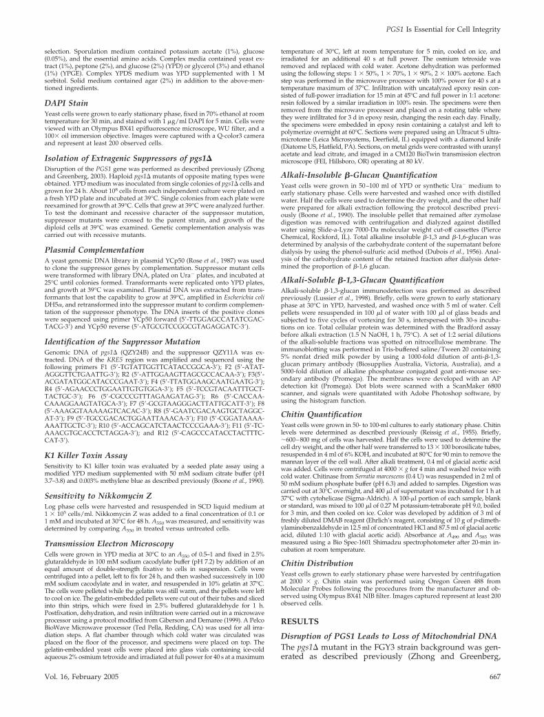
Mutants with defective cell wall structure exhibit sensitiv-ity to cell wall-perturbing agents. Consistent with a previousreport (Lussier et al., 1997), pgs1� was hypersensitive tocaffeine and CFW. In the presence of 1 M sorbitol, an osmoticstabilizer, sensitivity to CFW and caffeine in pgs1� wassignificantly reduced (Figure 4A). Furthermore, the suppres-sor pgs1� kre5W1166X was no longer sensitive to CFW, al-though sensitivity to caffeine in pgs1� was not suppressed.Thus, the suppressor mutation conferred a beneficial effecton the cell wall of pgs1� cells (Figure 4A).
Deletion of KRE5 leads to a number of changes in the cellwall, such as diffuse cell wall surface, a complete loss of theelectron-dense mannoprotein layer (Simons et al., 1998), andenlarged cell wall structure (Levinson et al., 2002). Electronmicroscopic analysis was performed to compare the ultra-structure of the cell wall of pgs1� and the pgs1� kre5W1166X
suppressor strain. As seen in Figure 4B, the wild-type cellwall has a finely delineated dark-staining mannoproteinlayer. In contrast, the cell wall of the suppressor was twice asthick and exhibited a rough appearance, similar to thatreported for the kre5 mutant (Simons et al., 1998; Levinson etal., 2002). However, aberrations in cell wall morphologywere not observed in pgs1� cells.
To gain further insight into how kre5W1166X suppressedpgs1� temperature sensitivity, we characterized the cell wallcomposition of pgs1� and the suppressor strain. Levels of themajor cell wall components were measured, including �-1,3and �-1,6-glucan and chitin (Table 2). Because pgs1� cells losemtDNA and are all �0 cells (Figure 1), we compared cell wallcomposition in pgs1� and isogenic wild-type �0 cells. Surpris-ingly, the loss of mtDNA from wild-type cells resulted indecreased alkaline-soluble and -insoluble �-1,3-glucan (50 and12% decrease, respectively) and a decrease (37%) in �-1,6-glucan. Disruption of PGS1 led to an even more pronounceddecrease in �-1,3-glucan. Alkaline-soluble and -insoluble �-1,3-glucan was reduced to 76 and 68% of the isogenic �0 wild-typelevels. In contrast, �-1,3 glucan was dramatically increased inthe suppressor mutant to levels greater than those observed inwild-type cells. Alkaline-insoluble �-1,6-glucan was reducedapproximately twofold in the pgs1� mutant compared with theisogenic �0 wild-type and to an even greater extent in thesuppressor mutant.
In contrast to �-glucans, chitin levels were not affected byloss of mtDNA (Table 2). In both pgs1� and the suppressormutant, chitin levels were �3 times higher than in wild-typecells (Table 2). Examination of the distribution of chitin byusing specific fluorescent probes revealed the presence ofchitin predominantly in the bud scars of wild-type cells(Figure 5A). In pgs1� and suppressor mutants, however,chitin staining was uniformly distributed to the lateral cellwall, and the bud scar was hardly distinguishable from therest of the cell wall. Hyperaccumulation of chitin in cell wallmutants is mediated by chitin synthase III (CSIII) (Osmondet al., 1999; Valdivieso et al., 2000). When grown in thepresence of 0.1 mM Nikkomycin Z, a known inhibitor ofCSIII (Gaughran et al., 1994), viability of the pgs1� mutantwas reduced by 40% (our unpublished data). In contrast, the
Figure 4. Cell wall properties of pgs1� andthe suppressor pgs1� kre5W1166X. (A) Cellsfrom wild-type (FGY3), �0, pgs1� (QZY24B),and the suppressor (QZY11A) mutant wereserially diluted and spotted on YPD or YPDSplates supplemented with 5 �g/ml CFW or 5mM caffeine. Cells were incubated at 30°C. (B)Transmission electron microscopy of the cellwall in wild-type (FGY3), �0, pgs1� (QZY24B),and the suppressor (QZY11A) was performedas described in Materials and Methods. The hor-izontal bar represents 500 nm.
PGS1 Is Essential for Cell Integrity
Vol. 16, February 2005 669

wild-type and suppressor mutant tolerated 1 mM Nikkomy-cin Z with no obvious loss of viability. Hypersensitivity toNikkomycin Z suggested that increased chitin is essential forthe survival of pgs1�. We wished to determine whether afurther increase in chitin synthesis led to increased viabilityof pgs1� cells at 37°C. Glucosamine was recently shown toinduce chitin synthesis via CSIII in wild-type cells as well asin cell wall mutants (Bulik et al., 2003). As shown in Figure5B, in the presence of 10 mM glucosamine, growth of pgs1�was slightly improved at 30°C. Chitin levels in pgs1� weredoubled at 37°C compared with 30°C in the presence of 10mM glucosamine (our unpublished data). However, at 37°C,pgs1� cells exhibited only limited growth and lost viabilityafter 20 h (Figure 5B). Thus, 10 mM glucosamine did notrestore growth to levels observed in the presence of thesuppressor mutation. Supplementation with 5–20 mM glu-cosamine led to a shortened lag in growth and increasedsaturation optical density of the pgs1� mutant at 30°C, butwas not sufficient to support growth at elevated temperature(our unpublished data).
When transformed with a plasmid-borne copy of thePGS1 gene under the control of the PGAL1 promoter, �-1,3-glucan levels in the pgs1� mutant significantly increased andchitin decreased (Table 2). This was observed even duringgrowth in glucose, in which low levels of expression of PGS1from this plasmid are sufficient to restore growth of pgs1� atelevated temperature and synthesis of PG and CL (He andGreenberg, 2004). Expression of the plasmid-borne copy ofKRE5 in the suppressor mutant restored �-1,6-glucan anddecreased �-1,3-glucan levels (Table 2). Together, these ex-periments indicate that disruption of PGS1 leads to multiplecell wall defects resulting in defective growth at elevatedtemperature. Increasing cell wall synthesis, particularly�-1,3-glucan and chitin, restored growth at 37°C.
Osmotic Stabilization of the Cell Wall Restores Growthof pgs1� at Elevated TemperatureSuppression of pgs1� temperature sensitivity by increased�-1,3-glucan synthesis suggested that osmotic stabilizationof the cell wall might alleviate cell wall stress and supportgrowth of pgs1� at elevated temperature. To address this
possibility, we examined the effect of sorbitol on cell wallcomposition and growth of pgs1� at 37°C. Mutant cells ofpgs1� grown in the presence of 1 M sorbitol contained 1.8and 2.4-fold increased alkaline soluble �-1,3-glucan and al-kaline insoluble �-1,6-glucan levels, respectively, comparedwith levels observed in pgs1� cells grown in YPD lackingsorbitol, whereas chitin levels were significantly reduced(Table 2). Consistent with this finding, the majority of pgs1�cells displayed the wild-type pattern of chitin staining ofbud scars and exhibited normal cell size in the presence ofsorbitol (data not shown). Supplementation with sorbitolrestored growth of pgs1� in two strain backgrounds, FGY3and GA74D (Figure 6A). Sorbitol also supported colonyformation of crd1� on YPD (Figure 6B), as well as growth oftaz1� on ethanol (data not shown) at 37°C.
As this report has shown, pgs1� cells in the FGY3 back-ground grown on YPD are all �0 cells. However, the pgs1�mutant in the GA74D strain background, GA74D3C, waspreviously thought to be “petite lethal”, because the mutantcells did not survive ethidium bromide mutagenesis, whichinduces loss of mtDNA (Janitor and Subik, 1993; Dzugasovaet al., 1998). A likely explanation for the inability of pgs1�cells to grow in the presence of ethidium bromide is thatpgs1� �0 cells in the GA74D strain background fail to survivedue to loss of cell wall integrity. To resolve this discrepancy,we examined the effects of ethidium bromide on pgs1� cellsin the presence or absence of osmotic support. Consistentwith the previous report (Janitor and Subik, 1993; Dzu-gasova et al., 1998), pgs1� (GA74D3C) cells failed to grow onplates containing 25 �g/ml ethidium bromide. However,when supplemented with 1 M sorbitol, the cells grew in thepresence of ethidium bromide (Figure 7). Failure to comple-ment �� tester strains for growth on YPGE (our unpublisheddata) confirmed the loss of mtDNA. The pgs1� �0 mutant inthis genetic background was viable on YPD but exhibitedslower growth than the isogenic �� strain. Consistent withthese observations, a greater decrease in alkaline-solubleand -insoluble �-1,3-glucan was observed in pgs1� �0 cellsthan in pgs1� �� cells (our unpublished data). These datasuggest that pgs1� cells can survive ethidium bromide treat-ment in the presence of increased osmotic support.
Table 2. Cell wall composition in pgs1� and suppressor mutants
Strain Medium
Alkaline-insoluble glucan Alkaline-soluble
Chitin�-1,6 �-1,6 � �-1,3 �-1,3 �-1,3-glucan
WT YPD 37.8 � 0.9 141.4 � 4.4 103.6 100% 4.65 � 0.39WT �0 YPD 23.7 � 1.4 114.7 � 8.1 91.0 50 � 3% 4.54 � 0.26pgs1� �0 YPD 12.1 � 1.9 74.7 � 1.4 62.6 38 � 2% 12.36 � 1.67pgs1� kre5W1166X �0 YPD 7.7 � 2.5 155.8 � 1.4 148.1 174 � 15% 12.35 � 0.39pgs1� �0 YPDS 21.0 � 1.6 98.8 � 18.8 77.8 90 � 25% 8.09 � 1.52
pgs1� �0 � PGS1 Ura��� 25.8 � 5.3 131.6 � 33.5 105.8 100% 7.76 � 0.32pgs1� �
0� vec Ura��� 21.4 � 2.1 69.6 � 22.7 48.2 41 � 6% 12.04 � 0.80
pgs1� kre5W1166X �0 � vec Ura��� 7.4 � 1.8 118.4 � 17.3 111.0 151 � 21% 12.09 � 3.14pgs1� kre5W1166X �0 � KRE5 Ura��� 27.1 � 0.9 99.3 � 9.6 72.2 52 � 5% 10.52 � 0.37
Glucan and chitin levels were measured as described in Materials and Methods in wild-type (FGY3), �0, pgs1� (QZY24B), and suppressor
mutant pgs1� kre5W1166X (QZY11A) cells grown in YPD or YPDS; pgs1� (QZY24B) cells transformed with empty vector pYES2/CT (�vec)or pYES2/CT-PGS1 (�PGS1); and pgs1� kre5W1166X suppressor mutant (QZY11A) cells transformed with empty vector YCp50 (�vec) or thegenomic clone of KRE5 (�KRE5) grown in synthetic ura� medium. Alkaline insoluble glucan and chitin are expressed as micrograms permilligram of cell dry weight. Alkaline soluble �-1,3-glucan in cells grown in complex medium (top) was expressed as a percentage of thatof wild-type (FGY3) cells. Alkaline soluble �-1,3-glucan in cells grown in synthetic ura� medium (bottom) was expressed relative to pgs1�(QZY24B) cells transformed with pYES2/CT-PGS1. Data represent three independent experiments.
Q. Zhong et al.
Molecular Biology of the Cell670

In summary, disruption of PGS1 leads to a defective cellwall and inability to grow at elevated temperature. Stabili-zation of the cell wall, either by increased synthesis of �-1,3-glucan or increased osmotic support, restores growth at37°C. These findings show that mitochondrial anionic phos-pholipids are essential in cellular functions required for cellwall biogenesis and maintenance of cell integrity.
DISCUSSION
The pgs1� mutant, which lacks mitochondrial anionic phos-pholipids PG and CL, exhibits severe temperature sensitiv-ity for growth even on fermentable carbon sources (Chang et
al., 1998a; Dzugasova et al., 1998), suggesting that anionicphospholipids are required for essential cellular functions.This report shows that mutants lacking these lipids aredefective in cell wall biogenesis. Reorganization of the cellwall in the pgs1� kre5W1166X suppressor or osmotic stabili-zation with sorbitol restores growth at elevated temperature.The pgs1� mutant exhibited a marked decrease in �-1,3-glucan (Table 2), the lack of which greatly impairs the me-chanical strength of the cell wall and severely threatensviability of cells at elevated temperature (Klis et al., 2002). Asa result, pgs1� mutant cells become enlarged and rounded(Figure 2), resembling another cell wall mutant, gas1�,which is also defective in �-glucan synthesis (Popolo et al.,
Figure 5. Increased chitin deposition inpgs1� and the suppressor mutant. (A) Cellsfrom wild-type (FGY3), �0, pgs1� (QZY24B),and the suppressor (QZY11A) were grown inYPD to early stationary phase. Chitin was vi-sualized by staining with Oregon Green 488as described in Materials and Methods. Chitindistribution was visualized by focusing ontwo planes. (B) Cells from pgs1� (QZY24B)and the suppressor mutant (QZY11A) weregrown in YPD in the presence or absence of 10mM glucosamine at the indicated tempera-tures. Viable cells were determined by serialdilution and plating.
PGS1 Is Essential for Cell Integrity
Vol. 16, February 2005 671

1993; Ram et al., 1998). Likely as a result of dramaticallyincreased �-1,3-glucan in the pgs1� kre5W1166X suppressorstrain (Table 2), the cell wall structure was thicker (Figure4B), and cell size and growth at elevated temperature wererestored (Figure 2). Consistent with these findings, osmoticstabilization with sorbitol suppressed temperature sensitiv-ity of all CL deficient mutants at elevated temperature (Fig-ure 6), suggesting that deficiency in mitochondrial anionicphospholipids results in cell wall defects that lead to atemperature-sensitive growth phenotype.
Cell wall biogenesis is a highly regulated process. �-1,3-Glucan is synthesized by �-1,3-glucan synthase (GS) local-ized on the plasma membrane (Qadota et al., 1996). GS iscomposed of a catalytic subunit encoded by the two homol-ogous genes FKS1 and FKS2 (Inoue et al., 1995; Mazur et al.,1995), and a regulatory subunit, the small GTPase, Rho1p
(Drgonova et al., 1996; Qadota et al., 1996). The Rho-typeGTPase is generally regulated by switching between a GDP-bound inactive state and a GTP-bound active state (Wei etal., 1997; Ihara et al., 1998). Various factors are involved inthe regulation of �-1,3-glucan synthesis by Rho1p in yeastcells. The putative cell surface sensor protein Wsc1p plays acritical role in stimulating nucleotide exchange of Rho1pthrough the GDP/GTP exchange factor, Rom2p (Philip andLevin, 2001). Exchange of GDP for GTP stimulates Rho1p,leading to activation of GS activity. Lrg1p, a GTPase-activat-ing protein, promotes formation of GDP-bound Rho1p, thusnegatively regulating �-1,3-glucan synthesis (Watanabe etal., 2001). Thus, overexpression of ROM2 or WSC1 (Sekiya-Kawasaki et al., 2002), or loss of function of LRG1 (Watanabeet al., 2001) restores the impaired �-1,3-glucan synthesisobserved in GS mutants. In addition, posttranslational mod-ification of Rho1p by the geranylgeranyl group is requiredfor binding of Rho1p to GS and activation of GS activity(Inoue et al., 1999). Other factors affect �-1,3-glucan synthesisby regulation of the catalytic subunit of GS. Movement ofFks1p driven by actin is required for the construction of auniform and solid cell wall (Utsugi et al., 2002). Transcrip-tion of FKS2 is up-regulated in response to cell wall stressinduced by heat, cell wall mutations, and cell wall-perturb-ing agents (Zhao et al., 1998; de Nobel et al., 2000; Lagorce etal., 2003; Garcia et al., 2004). Deletion of KRE5 leads to a114-fold up-regulation of FKS2 in response to an impairedcell wall (Kapteyn et al., 1999). Restored �-1,3-glucan levelsin pgs1� mutant cells in the presence of the kre5W1166X sup-pressor mutation (Table 2) could be mediated by up-regu-lation of FKS2 expression. In fact, increased �-1,3-glucan is ageneral characteristic shared by several kre mutants, alongwith defective �-1,6-glucan synthesis (Roemer et al., 1994;Dijkgraaf et al., 1996; Shahinian et al., 1998; Shahinian andBussey, 2000).
Decreased �-glucan levels in �0 cells and in mutants lack-ing mitochondrial anionic lipids suggest the existence of aregulatory link between mitochondrial biogenesis and cellwall synthesis. It has been suggested that cytoplasmic petitemutants isolated after ethidium bromide mutagenesis havealtered cell wall assembly (Wauters et al., 2001). In thisstudy, we have shown for the first time that loss of mtDNAalone led to a significant decrease in �-glucan. These defectswere exacerbated in the pgs1� mutant (Table 2). Our find-ings that greater cell wall defects were observed in pgs1�than in the wild-type �0 cells suggests that, along withoxidative phosphorylation, other mitochondrial functionsrequiring PG and/or CL may be required for cell wall bio-genesis. A link between mitochondrial functions and cellwall biogenesis has been implicated in several previousstudies as well. In addition to pgs1, Lussier et al. (1997)reported that mutations in four other genes with mitochon-drial associated functions, IMP2�, IFM1, SMP2, and COX11have cell wall defects. Three of these genes (IFM1, SMP2,and COX11) are required for mtDNA stability (Vambutas etal., 1991; Irie et al., 1993; Tzagoloff et al., 1993). A genome-wide screen for deletion mutants that exhibit increased re-sistance to K1 killer toxin, which indicates alterations in thecell surface, identified 17 deletion mutants affecting genesfor respiration and ATP metabolism (Page et al., 2003). All ofthe mutants are respiratory deficient, and four are involvedin mitochondrial genome maintenance.
The identification of cell wall defects in mutants withmitochondrial dysfunction suggests that mitochondria mayplay a general role in the regulation of cell wall biogenesis.Several enzymes involved in �-1,3-glucan synthesis werefound to have dual localization in the plasma membrane and
Figure 6. Supplementation with sorbitol supports growth of CL-deficient mutants at 37°C. (A) Isogenic wild-type and pgs1� mutantcells from two genetic backgrounds (FGY3 �0 and GA74D ��) wereserially diluted and spotted on YPD or YPDS plates and incubatedat the indicated temperature. (B) Wild-type (FGY3) and crd1�(FGY2) cells were grown overnight in YPD. Single cells were seededon YPD or YPDS plates and incubated at the indicated temperature.
Figure 7. Pgs1� survives ethidium bromide mutagenesis in thepresence of osmotic stabilizer. Wild-type GA74D3A and isogenicpgs1� mutant GA74D3c cells were streaked on synthetic minimalplates with ethidium bromide or sorbitol as indicated and incubatedat 30°C.
Q. Zhong et al.
Molecular Biology of the Cell672

mitochondria. Both the catalytic and the regulatory subunitof GS are localized on the plasma membrane at the site ofcell wall synthesis (Qadota et al., 1996). Interestingly, bothFks1p and Rho1p are also present in mitochondria (Sick-mann et al., 2003). Gas1p, a putative �-1,3-glucan–remodel-ing enzyme (Popolo and Vai, 1999; Mouyna et al., 2000), theloss of which also results in reduced �-1,3-glucan in the cellwall (Popolo et al., 1993; Ram et al., 1998), is attached to theplasma membrane via a glycosyl-phosphatidylinositol an-chor (Conzelmann et al., 1988; Nuoffer et al., 1991). Gas1palso is found in mitochondria (Grandier-Vazeille et al., 2001;Sickmann et al., 2003). It is not known whether dual plasmamembrane/mitochondria localization of these enzymes hasany physiological relevance. It is tempting, however, to as-sume that mitochondria are required for the maturation ormodification of those enzymes, in which case mitochondrialdysfunction would result in decreased enzyme activity andcell wall defects. Alternatively, mitochondrial dysfunctionmay trigger signals that prevent proper mobilization ofthose enzymes to the site of cell wall biosynthesis.
Our finding that pgs1� in the FGY3 strain backgroundexhibited loss of mtDNA even at optimal growth tempera-ture suggests that PG and CL are required for maintainingmtDNA. Mutants lacking only CL exhibit a strain-depen-dent decrease in mtDNA stability at elevated temperature(Jiang et al., 2000; Zhong et al., 2004). We have noticed thatcrd1� mutants from different strain backgrounds differgreatly with respect to the temperature at which growth isdefective and mtDNA becomes unstable (Zhong et al., 2004).In addition, crd1� was less thermotolerant on synthetic me-dium than on rich medium (Zhong et al., 2004). Althoughpgs1� in the GA74D strain background does not losemtDNA on YPD at 30°C, it cannot grow on synthetic me-dium with glycerol and ethanol as carbon source (Dzu-gasova et al., 1998), suggesting that it may lose mtDNAunder this condition. Furthermore, the results presentedhere show that pgs1� is not “petite lethal,” and the previ-ously reported inability of pgs1� to survive ethidium bro-mide (Janitor and Subik, 1993; Dzugasova et al., 1998) wasdue to defective cell wall integrity. Pgs1� �0 cells in theGA74D strain background were obtained after ethidiumbromide treatment in the presence of osmotic support andwere viable on YPD (Figure 7B). Those petite cells exhibitedslower growth than the isogenic �� strain, which presum-ably resulted from the exacerbated cell wall defects causedby loss of mtDNA. The further compromised growth ob-served in pgs1� �0 cells suggests a “synthetic sick” interac-tion between pgs1� and the �0 mutation. This interactionpredicts that the number of pgs1� cells surviving the loss ofmtDNA would be low. This seems paradoxical in light ofour finding that lack of PG and CL in the pgs1� mutantstrain resulted in 100% petite formation on YPD. Interest-ingly, mutations in ATP15 and ATP16, two structural genesencoding � and � subunit of F1-ATPase, lead to similarphenotypes. On one hand, atp15 and atp16 exhibited anextremely high frequency of petite formation. However, thepetite mutants have severe growth defects. Like PGS1,ATP15 and ATP16 also were thought to be essential in apetite background (Giraud and Velours, 1997; Lai-Zhang etal., 1999; Contamine and Picard, 2000).
In summary, we have isolated and identified an extra-genic suppressor of pgs1�, the loss of function allele ofKRE5, kre5W1166X. Characterization of pgs1� and the sup-pressor strain strongly suggests that temperature sensitivityof CL-deficient mutants and the previously reported “petitelethal” phenotype of pgs1� mutant cells were primarily dueto defective cell wall integrity. This work is the first demon-
stration of defective cell wall biosynthesis in mutants lackingmitochondrial anionic phospholipids PG and CL. Our find-ings thus provide new insights into the essential functions ofthese lipids and point to a regulatory role of mitochondria incell wall biogenesis.
ACKNOWLEDGMENTS
We thank Professor Julius Subık for providing the pgs1� strain in the GA74Dstrain background. We also thank Shuliang Chen and Guiling Li for helpinitiating this study and Dr. Deirdre Vaden for advice. This work was sup-ported by grant HL-62263 from the National Institutes of Health.
REFERENCES
Alonso-Monge, R., Real, E., Wojda, I., Bebelman, J. P., Mager, W. H., andSiderius, M. (2001). Hyperosmotic stress response and regulation of cell wallintegrity in Saccharomyces cerevisiae share common functional aspects. Mol.Microbiol. 41, 717–730.
Attfield, P. V. (1987). Trehalose accumulates in Saccharomyces cerevisiae duringexposure to agents that induce heat shock response. FEBS Lett. 225, 259–263.
Barth, P. G., Scholte, H. R., Berden, J. A., Van der Klei-Van Moorsel, J. M.,Luyt-Houwen, I. E., Van ’t Veer-Korthof, E. T., Van der Harten, J. J., andSobotka-Plojhar, M. A. (1983). An X-linked mitochondrial disease affectingcardiac muscle, skeletal muscle and neutrophil leucocytes. J. Neurol. Sci. 62,327–355.
Barth, P. G., Van den Bogert, C., Bolhuis, P. A., Scholte, H. R., van Gennip,A. H., Schutgens, R. B., and Ketel, A. G. (1996). X-linked cardioskeletalmyopathy and neutropenia (Barth syndrome): respiratory-chain abnormali-ties in cultured fibroblasts. J. Inherit. Metab. Dis. 19, 157–160.
Boone, C., Sommer, S. S., Hensel, A., and Bussey, H. (1990). Yeast KRE genesprovide evidence for a pathway of cell wall beta-glucan assembly. J. Cell Biol.110, 1833–1843.
Bulik, D. A., Olczak, M., Lucero, H. A., Osmond, B. C., Robbins, P. W., andSpecht, C. A. (2003). Chitin synthesis in Saccharomyces cerevisiae in response tosupplementation of growth medium with glucosamine and cell wall stress.Eukaryot. Cell 2, 886–900.
Cabib, E., Roh, D. H., Schmidt, M., Crotti, L. B., and Varma, A. (2001). Theyeast cell wall and septum as paradigms of cell growth and morphogenesis.J. Biol. Chem. 276, 19679–19682.
Chang, S. C., Heacock, P. N., Clancey, C. J., and Dowhan, W. (1998a). ThePEL1 gene (renamed PGS1) encodes the phosphatidylglycero-phosphate syn-thase of Saccharomyces cerevisiae. J. Biol. Chem. 273, 9829–9836.
Chang, S. C., Heacock, P. N., Mileykovskaya, E., Voelker, D. R., and Dowhan,W. (1998b). Isolation and characterization of the gene (CLS1) encoding cardi-olipin synthase in Saccharomyces cerevisiae. J. Biol. Chem. 273, 14933–14941.
Contamine, V., and Picard, M. (2000). Maintenance and integrity of themitochondrial genome: a plethora of nuclear genes in the budding yeast.Microbiol. Mol. Biol. Rev. 64, 281–315.
Conzelmann, A., Riezman, H., Desponds, C., and Bron, C. (1988). A major125-kd membrane glycoprotein of Saccharomyces cerevisiae is attached to thelipid bilayer through an inositol-containing phospholipid. EMBO J. 7, 2233–2240.
de Nobel, H., Ruiz, C., Martin, H., Morris, W., Brul, S., Molina, M., and Klis,F. M. (2000). Cell wall perturbation in yeast results in dual phosphorylation ofthe Slt2/Mpk1 MAP kinase and in an Slt2-mediated increase in FKS2-lacZexpression, glucanase resistance and thermotolerance. Microbiology 146,2121–2132.
Dijkgraaf, G. J., Brown, J. L., and Bussey, H. (1996). The KNH1 gene ofSaccharomyces cerevisiae is a functional homolog of KRE9. Yeast 12, 683–692.
Drgonova, J., Drgon, T., Tanaka, K., Kollar, R., Chen, G. C., Ford, R. A., Chan,C. S., Takai, Y., and Cabib, E. (1996). Rho1p, a yeast protein at the interfacebetween cell polarization and morphogenesis. Science 272, 277–279.
Dubois, M., Gilles, K. A., Hamilton, J. K., Rebers, P. A., and Smith, F. (1956).Colorimetric method for determination of sugars and related substances.Anal. Chem. 28, 350–356.
Dzugasova, V., Obernauerova, M., Horvathova, K., Vachova, M., Zakova, M.,and Subik, J. (1998). Phosphatidylglycerolphosphate synthase encoded by thePEL1/PGS1 gene in Saccharomyces cerevisiae is localized in mitochondria andits expression is regulated by phospholipid precursors. Curr. Genet. 34,297–302.
PGS1 Is Essential for Cell Integrity
Vol. 16, February 2005 673

Garcia, R., Bermejo, C., Grau, C., Perez, R., Rodriguez-Pena, J. M., Francois, J.,Nombela, C., and Arroyo, J. (2004). The global transcriptional response totransient cell wall damage in Saccharomyces cerevisiae and its regulation by thecell integrity signaling pathway. J. Biol. Chem. 279, 15183–15195.
Gaughran, J. P., Lai, M. H., Kirsch, D. R., and Silverman, S. J. (1994). Ni-kkomycin Z is a specific inhibitor of Saccharomyces cerevisiae chitin synthaseisozyme Chs3 in vitro and in vivo. J. Bacteriol. 176, 5857–5860.
Giberson, R. T., and Demaree, R.S.J. (1999). Microwave processing tech-niques for electron microscopy: a four-hour protocol. Methods Mol. Biol.117, 145–158.
Giraud, M. F., and Velours, J. (1997). The absence of the mitochondrial ATPsynthase delta subunit promotes a slow growth phenotype of rho� yeast cellsby a lack of assembly of the catalytic sector F1. Eur. J. Biochem. 245, 813–818.
Grandier-Vazeille, X., Bathany, K., Chaignepain, S., Camougrand, N., Manon,S., and Schmitter, J. M. (2001). Yeast mitochondrial dehydrogenases are asso-ciated in a supramolecular complex. Biochemistry 40, 9758–9769.
Gu, Z., Valianpour, F., Chen, S., Vaz, F. M., Hakkaart, G. A., Wanders, R. J.,and Greenberg, M. L. (2004). Aberrant cardiolipin metabolism in the yeasttaz1 mutant: a model for Barth syndrome. Mol. Microbiol. 51, 149–158.
He, Q., and Greenberg, M. L. (2004). Post-translational regulation of phos-phatidylglycerolphosphate synthase in response to inositol. Mol. Microbiol.53, 1243–1249.
Hottiger, T., Boller, T., and Wiemken, A. (1987). Rapid changes of heat anddesiccation tolerance correlated with changes of trehalose content in Saccha-romyces cerevisiae cells subjected to temperature shifts. FEBS Lett. 220, 113–115.
Hottiger, T., De Virgilio, C., Hall, M. N., Boller, T., and Wiemken, A. (1994).The role of trehalose synthesis for the acquisition of thermotolerance in yeast.II. Physiological concentrations of trehalose increase the thermal stability ofproteins in vitro. Eur. J. Biochem. 219, 187–193.
Ihara, K., Muraguchi, S., Kato, M., Shimizu, T., Shirakawa, M., Kuroda, S.,Kaibuchi, K., and Hakoshima, T. (1998). Crystal structure of human RhoA ina dominantly active form complexed with a GTP analogue. J. Biol. Chem. 273,9656–9666.
Inoue, S. B., Qadota, H., Arisawa, M., Watanabe, T., and Ohya, Y. (1999).Prenylation of Rho1p is required for activation of yeast 1,3-beta-glucan syn-thase. J. Biol. Chem. 274, 38119–38124.
Inoue, S. B., Takewaki, N., Takasuka, T., Mio, T., Adachi, M., Fujii, Y.,Miyamoto, C., Arisawa, M., Furuichi, Y., and Watanabe, T. (1995). Character-ization and gene cloning of 1,3-beta-D-glucan synthase from Saccharomycescerevisiae. Eur. J. Biochem. 231, 845–854.
Irie, K., Takase, M., Araki, H., and Oshima, Y. (1993). A gene, SMP2, involvedin plasmid maintenance and respiration in Saccharomyces cerevisiae encodes ahighly charged protein. Mol. Gen. Genet. 236, 283–288.
Janitor, M., Obernauerova, M., Kohlwein, S. D., and Subik, J. (1996). The pel1mutant of Saccharomyces cerevisiae is deficient in cardiolipin and does notsurvive the disruption of the CHO1 gene encoding phosphatidylserine syn-thase. FEMS Microbiol. Lett. 140, 43–47.
Janitor, M., and Subik, J. (1993). Molecular cloning of the PEL1 gene ofSaccharomyces cerevisiae that is essential for the viability of petite mutants.Curr. Genet. 24, 307–312.
Jiang, F., Gu, Z., Granger, J. M., and Greenberg, M. L. (1999). Cardiolipinsynthase expression is essential for growth at elevated temperature and isregulated by factors affecting mitochondrial development. Mol. Microbiol. 31,373–379.
Jiang, F., Rizavi, H. S., and Greenberg, M. L. (1997). Cardiolipin is not essentialfor the growth of Saccharomyces cerevisiae on fermentable or non-fermentablecarbon sources. Mol. Microbiol. 26, 481–491.
Jiang, F., Ryan, M. T., Schlame, M., Zhao, M., Gu, Z., Klingenberg, M.,Pfanner, N., and Greenberg, M. L. (2000). Absence of cardiolipin in the crd1null mutant results in decreased mitochondrial membrane potential andreduced mitochondrial function. J. Biol. Chem. 275, 22387–22394.
Kapteyn, J. C., Van Egmond, P., Sievi, E., Van Den Ende, H., Makarow, M.,and Klis, F. M. (1999). The contribution of the O-glycosylated protein Pir2p/Hsp150 to the construction of the yeast cell wall in wild-type cells and beta1,6-glucan-deficient mutants. Mol. Microbiol. 31, 1835–1844.
Kawasaki, K., Kuge, O., Chang, S. C., Heacock, P. N., Rho, M., Suzuki, K.,Nishijima, M., and Dowhan, W. (1999). Isolation of a Chinese hamster ovary(CHO) cDNA encoding phosphatidylglycerophosphate (PGP) synthase, ex-pression of which corrects the mitochondrial abnormalities of a PGP syn-thase-defective mutant of CHO-K1 cells. J. Biol. Chem. 274, 1828–1834.
Klis, F. M., Mol, P., Hellingwerf, K., and Brul, S. (2002). Dynamics of cell wallstructure in Saccharomyces cerevisiae. FEMS Microbiol. Rev. 26, 239–256.
Koshkin, V., and Greenberg, M. L. (2000). Oxidative phosphorylation incardiolipin-lacking yeast mitochondria. Biochem. J. 347, 687–691.
Koshkin, V., and Greenberg, M. L. (2002). Cardiolipin prevents rate-depen-dent uncoupling and provides osmotic stability in yeast mitochondria. Bio-chem. J. 364, 317–322.
Lagorce, A., Hauser, N. C., Labourdette, D., Rodriguez, C., Martin-Yken, H.,Arroyo, J., Hoheisel, J. D., and Francois, J. (2003). Genome-wide analysis of theresponse to cell wall mutations in the yeast Saccharomyces cerevisiae. J. Biol.Chem. 278, 20345–20357.
Lai-Zhang, J., Xiao, Y., and Mueller, D. M. (1999). Epistatic interactions ofdeletion mutants in the genes encoding the F1-ATPase in yeast Saccharomycescerevisiae. EMBO J. 18, 58–64.
Levinson, J. N., Shahinian, S., Sdicu, A. M., Tessier, D. C., and Bussey, H.(2002). Functional, comparative and cell biological analysis of Saccharomycescerevisiae Kre5p. Yeast 19, 1243–1259.
Lussier, M., Sdicu, A. M., Shahinian, S., and Bussey, H. (1998). The Candidaalbicans KRE9 gene is required for cell wall beta-1, 6-glucan synthesis and isessential for growth on glucose. Proc. Natl. Acad. Sci. USA 95, 9825–9830.
Lussier, M., et al. (1997). Large scale identification of genes involved in cellsurface biosynthesis and architecture in Saccharomyces cerevisiae. Genetics 147,435–450.
Ma, L., Vaz, F. M., Gu, Z., Wanders, R. J., and Greenberg, M. L. (2004). Thehuman TAZ gene complements mitochondrial dysfunction in the yeasttaz1delta mutant. Implications for Barth syndrome. J. Biol. Chem. 279, 44394–44399.
Mazur, P., Morin, N., Baginsky, W., el-Sherbeini, M., Clemas, J. A., Nielsen,J. B., and Foor, F. (1995). Differential expression and function of two homol-ogous subunits of yeast 1,3-beta-D-glucan synthase. Mol. Cell. Biol. 15, 5671–5681.
Meaden, P., Hill, K., Wagner, J., Slipetz, D., Sommer, S. S., and Bussey, H.(1990). The yeast KRE5 gene encodes a probable endoplasmic reticulumprotein required for (1–6)-beta-D-glucan synthesis and normal cell growth.Mol. Cell. Biol. 10, 3013–3019.
Molano, J., Bowers, B., and Cabib, E. (1980). Distribution of chitin in the yeastcell wall. An ultrastructural and chemical study. J. Cell Biol. 85, 199–212.
Mouyna, I., Fontaine, T., Vai, M., Monod, M., Fonzi, W. A., Diaquin, M.,Popolo, L., Hartland, R. P., and Latge, J. P. (2000). Glycosylphosphatidylino-sitol-anchored glucanosyltransferases play an active role in the biosynthesisof the fungal cell wall. J. Biol. Chem. 275, 14882–14889.
Neves, M. J., and Francois, J. (1992). On the mechanism by which a heat shockinduces trehalose accumulation in Saccharomyces cerevisiae. Biochem. J. 288,859–864.
Nuoffer, C., Jeno, P., Conzelmann, A., and Riezman, H. (1991). Determinantsfor glycophospholipid anchoring of the Saccharomyces cerevisiae GAS1 proteinto the plasma membrane. Mol. Cell. Biol. 11, 27–37.
Osmond, B. C., Specht, C. A., and Robbins, P. W. (1999). Chitin synthase III:synthetic lethal mutants and “stress related” chitin synthesis that bypasses theCSD3/CHS6 localization pathway. Proc. Natl. Acad. Sci. USA 96, 11206–11210.
Page, N., et al. (2003). A Saccharomyces cerevisiae genome-wide mutant screenfor altered sensitivity to K1 killer toxin. Genetics 163, 875–894.
Pfeiffer, K., Gohil, V., Stuart, R. A., Hunte, C., Brandt, U., Greenberg, M. L.,and Schagger, H. (2003). Cardiolipin stabilizes respiratory chain supercom-plexes. J. Biol. Chem. 278, 52873–52880.
Philip, B., and Levin, D. E. (2001). Wsc1 and Mid2 are cell surface sensors forcell wall integrity signaling that act through Rom2, a guanine nucleotideexchange factor for Rho1. Mol. Cell. Biol. 21, 271–280.
Popolo, L., Gualtieri, T., and Ragni, E. (2001). The yeast cell-wall salvagepathway. Med. Mycol. 39 (suppl) 1, 111–121.
Popolo, L., and Vai, M. (1999). The Gas1 glycoprotein, a putative wall polymercross-linker. Biochim. Biophys. Acta 1426, 385–400.
Popolo, L., Vai, M., Gatti, E., Porello, S., Bonfante, P., Balestrini, R., andAlberghina, L. (1993). Physiological analysis of mutants indicates involve-ment of the Saccharomyces cerevisiae GPI-anchored protein gp115 in morpho-genesis and cell separation. J. Bacteriol. 175, 1879–1885.
Qadota, H., Python, C. P., Inoue, S. B., Arisawa, M., Anraku, Y., Zheng, Y.,Watanabe, T., Levin, D. E., and Ohya, Y. (1996). Identification of yeast Rho1pGTPase as a regulatory subunit of 1,3-beta-glucan synthase. Science 272,279–281.
Ram, A. F., Kapteyn, J. C., Montijn, R. C., Caro, L. H., Douwes, J. E., Baginsky,W., Mazur, P., van den Ende, H., and Klis, F. M. (1998). Loss of the plasmamembrane-bound protein Gas1p in Saccharomyces cerevisiae results in the
Q. Zhong et al.
Molecular Biology of the Cell674

release of beta1,3-glucan into the medium and induces a compensation mech-anism to ensure cell wall integrity. J. Bacteriol. 180, 1418–1424.
Reissig, J. L., Storminger, J. L., and Leloir, L. F. (1955). A modified color-imetric method for the estimation of N-acetylamino sugars. J. Biol. Chem.217, 959 –966.
Roemer, T., Paravicini, G., Payton, M. A., and Bussey, H. (1994). Character-ization of the yeast (1-6)-beta-glucan biosynthetic components, Kre6p andSkn1p, and genetic interactions between the PKC1 pathway and extracellularmatrix assembly. J. Cell Biol. 127, 567–579.
Rose, M. D., Novick, P., Thomas, J. H., Botstein, D., and Fink, G. R. (1987). ASaccharomyces cerevisiae genomic plasmid bank based on a centromere-con-taining shuttle vector. Gene 60, 237–243.
Schlame, M., Rua, D., and Greenberg, M. L. (2000). The biosynthesis andfunctional role of cardiolipin. Prog. Lipid Res. 39, 257–288.
Sekiya-Kawasaki, M., Abe, M., Saka, A., Watanabe, D., Kono, K., Minemura-Asakawa, M., Ishihara, S., Watanabe, T., and Ohya, Y. (2002). Dissection ofupstream regulatory components of the Rho1p effector, 1,3-beta-glucan syn-thase, in Saccharomyces cerevisiae. Genetics 162, 663–676.
Shahinian, S., and Bussey, H. (2000). beta-1,6-Glucan synthesis in Saccharomy-ces cerevisiae. Mol. Microbiol. 35, 477–489.
Shahinian, S., Dijkgraaf, G. J., Sdicu, A. M., Thomas, D. Y., Jakob, C. A., Aebi,M., and Bussey, H. (1998). Involvement of protein N-glycosyl chain glucosy-lation and processing in the biosynthesis of cell wall beta-1,6-glucan ofSaccharomyces cerevisiae. Genetics 149, 843–856.
Shaw, J. A., Mol, P. C., Bowers, B., Silverman, S. J., Valdivieso, M. H., Duran,A., and Cabib, E. (1991). The function of chitin synthases 2 and 3 in theSaccharomyces cerevisiae cell cycle. J. Cell Biol. 114, 111–123.
Sickmann, A., et al. (2003). The proteome of Saccharomyces cerevisiae mitochon-dria. Proc. Natl. Acad. Sci. USA 100, 13207–13212.
Simons, J. F., Ebersold, M., and Helenius, A. (1998). Cell wall 1,6-beta-glucansynthesis in Saccharomyces cerevisiae depends on ER glucosidases I and II, andthe molecular chaperone BiP/Kar2p. EMBO J. 17, 396–405.
Singer, M. A., and Lindquist, S. (1998). Thermotolerance in Saccharomycescerevisiae: the Yin and Yang of trehalose. Trends Biotechnol. 16, 460–468.
Smits, G. J., van den Ende, H., and Klis, F. M. (2001). Differential regulation ofcell wall biogenesis during growth and development in yeast. Microbiology147, 781–794.
Tuller, G., Hrastnik, C., Achleitner, G., Schiefthaler, U., Klein, F., and Daum,G. (1998). YDL142c encodes cardiolipin synthase (Cls1p) and is non-essentialfor aerobic growth of Saccharomyces cerevisiae. FEBS Lett. 421, 15–18.
Tzagoloff, A., Nobrega, M., Gorman, N., and Sinclair, P. (1993). On thefunctions of the yeast COX10 and COX11 gene products. Biochem. Mol. Biol.Int. 31, 593–598.
Utsugi, T., Minemura, M., Hirata, A., Abe, M., Watanabe, D., and Ohya, Y.(2002). Movement of yeast 1,3-beta-glucan synthase is essential for uniformcell wall synthesis. Genes Cells 7, 1–9.
Valdivieso, M. H., Ferrario, L., Vai, M., Duran, A., and Popolo, L. (2000).Chitin synthesis in a gas1 mutant of Saccharomyces cerevisiae. J. Bacteriol. 182,4752–4757.
Vambutas, A., Ackerman, S. H., and Tzagoloff, A. (1991). Mitochondrialtranslational-initiation and elongation factors in Saccharomyces cerevisiae. Eur.J. Biochem. 201, 643–652.
Vreken, P., Valianpour, F., Nijtmans, L. G., Grivell, L. A., Plecko, B., Wanders,R. J., and Barth, P. G. (2000). Defective remodeling of cardiolipin and phos-phatidylglycerol in Barth syndrome. Biochem. Biophys. Res. Commun. 279,378–382.
Watanabe, D., Abe, M., and Ohya, Y. (2001). Yeast Lrg1p acts as a specializedRhoGAP regulating 1,3-beta-glucan synthesis. Yeast 18, 943–951.
Wauters, T., Iserentant, D., and Verachtert, H. (2001). Impact of mitochondrialactivity on the cell wall composition and on the resistance to tannic acid inSaccharomyces cerevisiae. J. Gen. Appl. Microbiol. 47, 21–26.
Wei, Y., Zhang, Y., Derewenda, U., Liu, X., Minor, W., Nakamoto, R. K.,Somlyo, A. V., Somlyo, A. P., and Derewenda, Z. S. (1997). Crystal structureof RhoA-GDP and its functional implications. Nat. Struct. Biol. 4, 699–703.
Xu, Y., Kelley, R. I., Blanck, T. J., and Schlame, M. (2003). Remodeling ofcardiolipin by phospholipid transacylation. J. Biol. Chem. 278, 51380–51385.
Zhao, C., Jung, U. S., Garrett-Engele, P., Roe, T., Cyert, M. S., and Levin, D. E.(1998). Temperature-induced expression of yeast FKS2 is under the dualcontrol of protein kinase C and calcineurin. Mol. Cell. Biol. 18, 1013–1022.
Zhong, Q., Gohil, V. M., Ma, L., and Greenberg, M. L. (2004). Absence ofcardiolipin results in temperature sensitivity, respiratory defects, and mito-chondrial DNA instability independent of pet56. J. Biol. Chem. 279, 32294–32300.
Zhong, Q., and Greenberg, M. L. (2003). Regulation of phosphatidylglycero-phosphate synthase by inositol in Saccharomyces cerevisiae is not at the level ofPGS1 mRNA abundance. J. Biol. Chem. 278, 33978–33984.
PGS1 Is Essential for Cell Integrity
Vol. 16, February 2005 675
Related Documents


