Loss of Anti-Viral Immunity by Infection with a Virus Encoding a Cross-Reactive Pathogenic Epitope Alex T. Chen 1¤a , Markus Cornberg 1¤b , Stephanie Gras 2 , Carole Guillonneau 3¤c , Jamie Rossjohn 2 , Andrew Trees 4 , Sebastien Emonet 4 , Juan C. de la Torre 4 , Raymond M. Welsh 1 *, Liisa K. Selin 1 1 Department of Pathology, University of Massachusetts Medical School, Worcester, Massachusetts, United States of America, 2 Department of Biochemistry and Molecular Biology, School of Biomedical Sciences, Monash University, Clayton, Victoria, Australia, 3 Department of Microbiology and Immunology, University of Melbourne, Parkville, Victoria, Australia, 4 Department of Immunology and Microbial Science, The Scripps Research Institute, La Jolla, California, United States of America Abstract T cell cross-reactivity between different strains of the same virus, between different members of the same virus group, and even between unrelated viruses is a common occurrence. We questioned here how an intervening infection with a virus containing a sub-dominant cross-reactive T cell epitope would affect protective immunity to a previously encountered virus. Pichinde virus (PV) and lymphocytic choriomeningitis virus (LCMV) encode subdominant cross-reactive NP 205–212 CD8 T cell epitopes sharing 6 of 8 amino acids, differing only in the MHC anchoring regions. These pMHC epitopes induce cross- reactive but non-identical T cell receptor (TCR) repertoires, and structural studies showed that the differing anchoring amino acids altered the conformation of the MHC landscape presented to the TCR. PV-immune mice receiving an intervening infection with wild type but not NP205-mutant LCMV developed severe immunopathology in the form of acute fatty necrosis on re-challenge with PV, and this pathology could be predicted by the ratio of NP205-specific to the normally immunodominant PV NP 38–45 -specific T cells. Thus, cross-reactive epitopes can exert pathogenic properties that compromise protective immunity by impairing more protective T cell responses. Citation: Chen AT, Cornberg M, Gras S, Guillonneau C, Rossjohn J, et al. (2012) Loss of Anti-Viral Immunity by Infection with a Virus Encoding a Cross-Reactive Pathogenic Epitope. PLoS Pathog 8(4): e1002633. doi:10.1371/journal.ppat.1002633 Editor: Christopher M. Walker, Nationwide Children’s Hospital, United States of America Received October 4, 2011; Accepted February 23, 2012; Published April 19, 2012 Copyright: ß 2012 Chen et al. This is an open-access article distributed under the terms of the Creative Commons Attribution License, which permits unrestricted use, distribution, and reproduction in any medium, provided the original author and source are credited. Funding: This work was supported by United States National Institutes of Health grants AI047140 (JCT), AI077719 (JCT), AI079665 (JCCT), AI017672 (RMW), AI081675 (RMW), AI046578 (LKS), a German Research Foundation fellowship CO310-2/1 (MC), an institutional Diabetes Endocrinology Research Center DK52530, an Australian Research Council Federation Fellowship (JR). The funders had no role in study design, data collection and analysis, decision to publish, or preparation of the manuscript. Competing Interests: The authors have declared that no competing interests exist. * E-mail: [email protected] ¤a Current address: Infectious and Inflammatory disease Center, Sanford-Burnham Medical Research Institute, La Jolla, California, United States of America ¤b Current address: Department of Gastroenterology, Hepatology and Endocrinology, Hannover Medical School, Hannover, Germany ¤c Current address: CR2-CNRS, INSERM U643-ITERT, CHU Hotel-Dieu, Nantes, France Introduction The desired consequence of vaccination or viral infection is long lasting immunity that protects the host from re-infection or else quickly restricts viral replication to prevent disease and immune pathology. In many cases neutralizing antibody produced by stable plasma cell populations restricts re-infection for the lifetime of the host. In other cases effective neutralizing antibody responses may wane with time or not develop, and resistance relies more on a rapid response by memory T cells [1]. CD8 T cell memory is stable in a pristine environment, but it can be compromised by subsequent viral or bacterial infections [2,3]. This compromise may be in the form of type 1 interferon (IFN)-induced attrition, resulting in a Bim- dependent apoptosis and loss of memory T cells [4]. Alternatively, this compromise may be in the form of skewing the memory T cell repertoire as a consequence of CD8 T cell cross-reactivity between heterologous agents. Such cross-reactivity is commonplace and is seen in humans between influenza A virus (IAV) and hepatitis C virus (HCV), between IAV and Epstein-Barr virus, and within members of the flavi-, hanta-, and orthomyxo-virus groups [3]. It could therefore be expected that protective immunity could be altered by an intervening viral infection, especially against an agent poorly controlled by neutralizing antibodies and reliant on T cell-dependent immunity, as exemplified by the New World arenavirus Pichinde virus (PV) [5,6]. PV is distantly related to LCMV, an Old World arenavirus, and these two viruses encode cross-reactive epitopes at nucleoprotein (NP) positions 205–212. Heterologous challenge of LCMV-immune mice with PV results in about a 10-fold reduction in PV titer by day 4 post-infection (PI) when compared to naı ¨ve controls, and PV-immune mice synthesize about 2–5 times less LCMV on LCMV challenge [6,7]. Alterations in the T cell epitope immunodominance hierarchy of the previously immunized animals occurs following heterologous challenge in the LCMV and PV system in either direction [6]. T cell responses to the NP205 epitopes are normally subdominant during infections with either virus alone, even after re-challenge with homologous virus, but in mice sequentially infected with heterologous virus, they become dominant, with narrowly focused oligoclonal repertoires [8]. The beneficial effects of CD8 T cell-mediated clearance of viral infections are sometimes offset by immunopathology, and in experimental models of autoimmunity specific so-called ‘‘patho- genic epitopes’’ may elicit immunopathology due to their cross- reactivity with self-antigens [9]. Herpes simplex virus-1-induced PLoS Pathogens | www.plospathogens.org 1 April 2012 | Volume 8 | Issue 4 | e1002633

Welcome message from author
This document is posted to help you gain knowledge. Please leave a comment to let me know what you think about it! Share it to your friends and learn new things together.
Transcript
Loss of Anti-Viral Immunity by Infection with a VirusEncoding a Cross-Reactive Pathogenic EpitopeAlex T. Chen1¤a, Markus Cornberg1¤b, Stephanie Gras2, Carole Guillonneau3¤c, Jamie Rossjohn2,
Andrew Trees4, Sebastien Emonet4, Juan C. de la Torre4, Raymond M. Welsh1*, Liisa K. Selin1
1 Department of Pathology, University of Massachusetts Medical School, Worcester, Massachusetts, United States of America, 2 Department of Biochemistry and
Molecular Biology, School of Biomedical Sciences, Monash University, Clayton, Victoria, Australia, 3 Department of Microbiology and Immunology, University of
Melbourne, Parkville, Victoria, Australia, 4 Department of Immunology and Microbial Science, The Scripps Research Institute, La Jolla, California, United States of America
Abstract
T cell cross-reactivity between different strains of the same virus, between different members of the same virus group, andeven between unrelated viruses is a common occurrence. We questioned here how an intervening infection with a viruscontaining a sub-dominant cross-reactive T cell epitope would affect protective immunity to a previously encountered virus.Pichinde virus (PV) and lymphocytic choriomeningitis virus (LCMV) encode subdominant cross-reactive NP205–212 CD8 T cellepitopes sharing 6 of 8 amino acids, differing only in the MHC anchoring regions. These pMHC epitopes induce cross-reactive but non-identical T cell receptor (TCR) repertoires, and structural studies showed that the differing anchoring aminoacids altered the conformation of the MHC landscape presented to the TCR. PV-immune mice receiving an interveninginfection with wild type but not NP205-mutant LCMV developed severe immunopathology in the form of acute fattynecrosis on re-challenge with PV, and this pathology could be predicted by the ratio of NP205-specific to the normallyimmunodominant PV NP38–45 -specific T cells. Thus, cross-reactive epitopes can exert pathogenic properties thatcompromise protective immunity by impairing more protective T cell responses.
Citation: Chen AT, Cornberg M, Gras S, Guillonneau C, Rossjohn J, et al. (2012) Loss of Anti-Viral Immunity by Infection with a Virus Encoding a Cross-ReactivePathogenic Epitope. PLoS Pathog 8(4): e1002633. doi:10.1371/journal.ppat.1002633
Editor: Christopher M. Walker, Nationwide Children’s Hospital, United States of America
Received October 4, 2011; Accepted February 23, 2012; Published April 19, 2012
Copyright: � 2012 Chen et al. This is an open-access article distributed under the terms of the Creative Commons Attribution License, which permitsunrestricted use, distribution, and reproduction in any medium, provided the original author and source are credited.
Funding: This work was supported by United States National Institutes of Health grants AI047140 (JCT), AI077719 (JCT), AI079665 (JCCT), AI017672 (RMW),AI081675 (RMW), AI046578 (LKS), a German Research Foundation fellowship CO310-2/1 (MC), an institutional Diabetes Endocrinology Research Center DK52530,an Australian Research Council Federation Fellowship (JR). The funders had no role in study design, data collection and analysis, decision to publish, orpreparation of the manuscript.
Competing Interests: The authors have declared that no competing interests exist.
* E-mail: [email protected]
¤a Current address: Infectious and Inflammatory disease Center, Sanford-Burnham Medical Research Institute, La Jolla, California, United States of America¤b Current address: Department of Gastroenterology, Hepatology and Endocrinology, Hannover Medical School, Hannover, Germany¤c Current address: CR2-CNRS, INSERM U643-ITERT, CHU Hotel-Dieu, Nantes, France
Introduction
The desired consequence of vaccination or viral infection is long
lasting immunity that protects the host from re-infection or else
quickly restricts viral replication to prevent disease and immune
pathology. In many cases neutralizing antibody produced by stable
plasma cell populations restricts re-infection for the lifetime of the
host. In other cases effective neutralizing antibody responses may
wane with time or not develop, and resistance relies more on a rapid
response by memory T cells [1]. CD8 T cell memory is stable in a
pristine environment, but it can be compromised by subsequent
viral or bacterial infections [2,3]. This compromise may be in the
form of type 1 interferon (IFN)-induced attrition, resulting in a Bim-
dependent apoptosis and loss of memory T cells [4]. Alternatively,
this compromise may be in the form of skewing the memory T cell
repertoire as a consequence of CD8 T cell cross-reactivity between
heterologous agents. Such cross-reactivity is commonplace and is
seen in humans between influenza A virus (IAV) and hepatitis C
virus (HCV), between IAV and Epstein-Barr virus, and within
members of the flavi-, hanta-, and orthomyxo-virus groups [3].
It could therefore be expected that protective immunity could
be altered by an intervening viral infection, especially against an
agent poorly controlled by neutralizing antibodies and reliant on T
cell-dependent immunity, as exemplified by the New World
arenavirus Pichinde virus (PV) [5,6]. PV is distantly related to
LCMV, an Old World arenavirus, and these two viruses encode
cross-reactive epitopes at nucleoprotein (NP) positions 205–212.
Heterologous challenge of LCMV-immune mice with PV results
in about a 10-fold reduction in PV titer by day 4 post-infection (PI)
when compared to naıve controls, and PV-immune mice
synthesize about 2–5 times less LCMV on LCMV challenge
[6,7]. Alterations in the T cell epitope immunodominance
hierarchy of the previously immunized animals occurs following
heterologous challenge in the LCMV and PV system in either
direction [6]. T cell responses to the NP205 epitopes are normally
subdominant during infections with either virus alone, even after
re-challenge with homologous virus, but in mice sequentially
infected with heterologous virus, they become dominant, with
narrowly focused oligoclonal repertoires [8].
The beneficial effects of CD8 T cell-mediated clearance of viral
infections are sometimes offset by immunopathology, and in
experimental models of autoimmunity specific so-called ‘‘patho-
genic epitopes’’ may elicit immunopathology due to their cross-
reactivity with self-antigens [9]. Herpes simplex virus-1-induced
PLoS Pathogens | www.plospathogens.org 1 April 2012 | Volume 8 | Issue 4 | e1002633
conjunctivitis and Theiler’s virus-induced encephalitis are cases
where viral epitopes induce cross-reactive T cells that target
proteins of the eye and brain, respectively [10,11]. We questioned
here whether select epitopes cross-reactive between two viruses
may at times act as pathogenic epitopes and cause immune
pathology even in the absence of autoimmunity and show here
how an LCMV infection disrupts protective immunity to PV due
to the presence of a cross-reactive ‘‘pathogenic’’ epitope.
Results
Generation and analysis of NP205 variantsTo analyze the role of cross-reactive epitopes in the elicitation
or disruption of protective immunity and immune pathology, we
first characterized the molecular properties of wild type and
mutant epitopes cross-reactive between LCMV and PV. This
study uses both the Armstrong strain of LCMV and its highly
disseminating Clone 13 derivative; these viruses differ by only
three amino acids and have identical T cell epitopes [12]. LCMV
NP205–212 (YTVKYPNL) and PV NP205–212 (YTVKFPNM) are
class I MHC H2Kb-restricted epitopes that share 6 of 8 amino
acids (Figure 1). To evaluate the conformational differences
between the LCMV and PV epitopes, we solved the crystal
structures of WT H2Kb –NP205–212 from LCMV and PV to
2.50 A resolution (Table S1). The structures show that positions 2,
5 and 8 are the anchor residues, whereas positions 2 and 6 are
partially exposed, and positions 1, 4 and 7 are solvent-exposed and
thus represent potential TCR contact points.
Overall the conformation of the WT LCMV and PV peptides
bound to H2Kb is similar, with a root mean square deviation
(rmsd) of ,0.24 A (Figure 1A). The WT NP205 peptides from
LCMV and PV differ by only two residues at position 5 and 8,
which are MHC anchor residues, and are thus inaccessible for
direct TCR contact. The respective H2Kb binding clefts adopt
similar conformations (rmsd of ,0.3 A) (Figure 1B), with the
largest difference in a specific region of the a2-helix (rmsd .0.5 to
0.9 A) (Figure 1C). The presence of the P5-Tyrosine hydroxyl
group of the LCMV peptide, instead of the P5-Phenylalanine
found in the PV peptide, accounts for this perturbation of the a2-
helix. Namely, P5-Tyrosine alters the conformation of Serine-99,
which is located within the b-strand at the floor of the cleft
(Figure 1C), the effect of which is transmitted through the cleft by
a rearrangement of side chains of the Glutamine-114, Leucine-
Author Summary
The purpose of vaccination against viruses is to inducestrong neutralizing antibody responses that inactivateviruses on contact and strong T cell responses that attackand kill virus-infected cells. Some viruses, however, like HIVand hepatitis C virus, are only weakly controlled byneutralizing antibody, so T cell immunity is very importantfor control of these infections. T cells recognize small virus-encoded peptides, called epitopes, presented on thesurface of infected cells, and some of these epitopesinduce strongly protective and others weakly protective Tcell responses. However, the same T cells can sometimesdemonstrate cross-reactivity and recognize similar epi-topes encoded by two different viruses. We questionedhere what infection with a virus encoding a weak cross-reactive epitope would do to immunity to a previously-encountered virus. Here we report that such an infectioncan compromise protective immunity by enhancing thenormally weak response and suppressing the normallystrong response. Under these conditions such epitopesfunction as ‘‘pathogenic’’ epitopes, and we suggest thatthe potential for inducing responses to pathogenicepitopes should be an important consideration in thedesign of T cell vaccines.
Figure 1. Analysis and comparisons of NP205-Kb structures. (A), (B) and (C): superposition of LCMV (pink) with PV (blue) structures, with thepeptide in stick representation and the MHC H2Kb in grey cartoon. The tip of the a2-helix is colored accordingly to the peptides bound by the H2Kb
molecules, representing the section from residue 150 to 156 of the a2-helix (B & C). (C) shows, with a different orientation, the residues that changeconformation between the peptide-MHC complexes, namely Serine-99, Glutamine-114, Leucine-156, Glutamate-152 as well as Glycine-151, for whichthe Ca atom is represented by a sphere. (D) superposition of the LCMV (pink) with LCMV-V207A (green) structures, with peptide in stickrepresentation and MHC in grey cartoon. (E) and (F): comparison of LCMV (pink) and LCMV-V207A (green) mutant peptide, both bound to the H2Kb
molecule (grey cartoon) in the same orientation. The P3 residues are colored in yellow. Arginine-155, Glutamate-152 and Alanine-151 of the H2Kb
molecule are represented as grey stick to show the different interaction of their side chains between both structures. The red dashed lines representthe hydrogen bond made between the residues.doi:10.1371/journal.ppat.1002633.g001
Loss of Immunity Due to a Cross-Reactive Epitope
PLoS Pathogens | www.plospathogens.org 2 April 2012 | Volume 8 | Issue 4 | e1002633
156, Glutamate-152 and Glycine-151, for which a maximum
displacement of 0.9 A is observed. This altered positioning of the
a2-helix could affect the interaction with the TCR, as differences
in this region of the MHC has been shown to impact on TCR
ligations in many other systems [13,14]. This indicates that,
although the pMHC complexes are similar, they are not identical
epitopes from the perspective of the T cell, and this is reflected by
differences in the LCMV-specific vs. PV-specific NP205 reper-
toires of TCR generated by infection in vivo [8].
In our previous study we isolated a T cell escape variant of
LCMV Clone 13, where the Valine in the third position of the
LCMV NP205 epitope was converted into an Alanine (NP
V207A). This mutant epitope stabilized the expression of H2Kb on
RMA/S cells, indicating that it could be presented by the MHC
[8]. The PV-NP205, WT LCMV-NP205, and LCMV V207A
mutant peptides had very similar effects at stabilizing H2Kb in that
the pMHC complexes had an average Tm of 47uC.
To understand the impact of the V207A mutation, the crystal
structure of the H2Kb-NP V207A epitope was determined to
2.30 A resolution (Table S1). The structure shows that the
mutation at P3-Valine of the LCMV peptide into Alanine (NP
V207A) did not affect the overall conformation of the H2Kb
binding cleft (rmsd .0.3 A) (Figure 1D). The difference between
the LCMV WT and LCMV-V207A structures is limited to a
change in the Arginine-155 conformation between the two pMHC
complexes. Namely, within the H2Kb-NP205 complex, Arginine-
155 hydrogen bonds to the main chain of the P4-Lysine residue of
the peptide (Figure 1E). In the H2Kb-NP V207A epitope, on
account of subtle movement of the peptide, the conformation of
Arginine-155 is shifted such that it now points towards the tip of
the a2-helix and hydrogen bonds with the Alanine-151 (Figure 1F).
Arginine-155, a position previously termed the gatekeeper residue,
has been shown to be involved in interacting with the TCR in
most of the structures of TCR-pMHC solved to date and often
changes conformation upon TCR ligation [15,16]. The change of
conformation observed for the Arginine-155 due to the Valine to
Alanine mutation at position 3 between the LCMV WT and
V207A structures explains the effect on the TCR recognition and
on T cell activity that is associated with epitope escape.
Since the naturally selected V207A mutant was generated
during LCMV Clone 13 infection and may have had additional
mutations, we used reverse genetics approaches to generate
rLCMV (rV207A) with the specific mutation V207A within the
NP205–212 epitope of the Armstrong strain. As a control we also
used reverse genetics to rescue WT Armstrong virus (rWT),
thereby giving us highly defined viruses differing in a single
nucleotide. Because the LCMV-V207A peptides could stabilize
H2Kb and induce a weak but detectable T cell response, we tested
Alanine substitutions in different residues of the NP205 epitope to
find a variant that would not stabilize H2Kb. This was done by
converting a Leucine into an Alanine in the eighth (and anchoring)
position of the peptide, thereby eliminating MHC stabilization
(Figure 2). These results led us to design and generate by reverse
genetics the LCMV Armstrong anchoring mutant rL212A. Armed
with this assembly of mutant viruses and the knowledge of their
structures and biochemical properties we could now address the
biological aspects of heterologous immunity between LCMV and
PV.
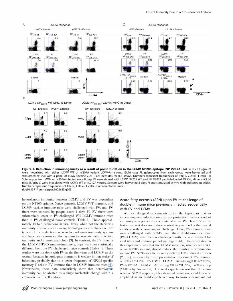
Reduction in immunogenicity due to point mutations inthe LCMV NP205 epitope
The newly engineered rV207A variant of LCMV-Armstrong
was similar to the natural Clone 13 variant in that it induced
normal responses to all tested epitopes except for NP205
(Figures 3A and S1) [8]. Infection with the rV207A LCMV-Arm
variant resulted at day 8 PI in greatly diminished responses against
either the WT LCMV NP205 or the PV NP205 epitopes (e.g.
LCMV NP205 response induced by rWT = 1.860.24% vs.
rV207A = 0.160.05%, n = 3/group, p = 0.0002) (Figures 3A and
S1). H2Kb-MHC-Ig dimers were also employed to ensure that the
diminished NP205-specific CD8 T cell response in variant-
infected mice was due to a loss in specific T cell number and
not just due to an alteration in T cell function detected by ICS
assays. In the host infected with the rWT virus, similar frequencies
of antigen-specific CD8 T cell populations were detected using
either LCMV WT NP205-loaded MHC-Ig dimers or LCMV NP
V207A-loaded MHC-Ig dimers (2.060.1% vs. 1.860.25%,
respectively, n = 2) (Figures 3B and S1). On the other hand,
MHC-Ig dimers loaded with either peptide could detect only a
very small percentage of CD8 T cells in mice infected with the
rV207A variant virus (Figure 3B). The LCMV Armstrong rL212A
anchoring variant, whose NP205 peptide does not stabilize H2Kb,
induced T cell responses well against the LCMV GP33 and
NP396 epitopes but failed to induce NP205 responses at all above
background (Figures 3C and S1). Note that the L212A peptide did
sensitize targets to killing, but that effect was very sensitive to
dilution, much as we previously showed with the V207A peptide,
in comparison to wild type NP205–212 (data not shown) [8].
These mutants made it possible to assess the role of the NP205
epitope in heterologous immunity.
Association of cross-reactive NP205-specific CD8 T cellswith heterologous immunity
We used the three LCMV mutants that poorly induced NP205-
specific CD8 T cell responses to test the hypothesis that
Figure 2. Lack of MHC stabilization by LCMV NP L212A. MHCstabilization assays for LCMV WT (NP205) and mutant (L212A) peptides.RMA-S cells were incubated with different concentrations of peptidesand stained against H2Kb to detect its stabilization on the cell surface.doi:10.1371/journal.ppat.1002633.g002
Loss of Immunity Due to a Cross-Reactive Epitope
PLoS Pathogens | www.plospathogens.org 3 April 2012 | Volume 8 | Issue 4 | e1002633
heterologous immunity between LCMV and PV was dependent
on the NP205 epitope. Naıve controls, LCMV WT immune, and
LCMV variant-immune mice were challenged with PV, and PV
titers were assessed by plaque assay 4 days PI. PV titers were
substantially lower in PV-challenged WT-LCMV-immune mice
than in PV-challenged naıve controls (Table 1). These approxi-
mately 10-fold reductions in viral titers, while not the sterilizing
immunity normally seen during homologous virus challenge, are
typical of the reductions seen in heterologous immunity systems
and have been shown in other systems to correlate with protective
immunity and immunopathology [3]. In contrast, the PV titers in
the LCMV NP205 mutant-immune groups were not statistically
different from the PV-challenged naıve controls (Table 1). These
studies were not done with PV as the first virus and LCMV as the
second, because heterologous immunity is weaker in that order of
infections, probably due to a lower frequency of NP205-specific
memory T cells in PV-immune than in LCMV-immune mice [6].
Nevertheless, these data conclusively show that heterologous
immunity can be ablated by a single nucleotide change within a
cross-reactive T cell epitope.
Acute fatty necrosis (AFN) upon PV re-challenge ofdouble immune mice previously infected sequentiallywith PV and LCMV
We next designed experiments to test the hypothesis that an
intervening viral infection may disrupt protective T cell-dependent
immunity to a previously encountered virus. We chose PV as the
first virus, as it does not induce neutralizing antibodies that would
interfere with a homologous challenge. Here, PV-immune mice
were challenged with LCMV, and these double-immune mice
(PV+LCMV) were then re-challenged with PV and assessed for
viral titers and immune pathology (Figure 4A). The expectation in
this experiment was that the LCMV infection, whether with WT
or an NP205 mutant, should reduce the number of immunodo-
minant PV NP38-specific memory cells by IFN-induced attrition
[2,6,17], as shown by this representative experiment: PV immune
only = 5.464.2%; PV+rWT LCMV Armstrong = 0.9660.2%;
PV+rV207A LCMV Armstrong = 0.6760.09%, n = 5/group
(p,0.05 by Anova test). The next expectation was that the cross-
reactive NP205 response, after its initial reduction, should then be
amplified in an LCMV-preferred way to form a dominant but
Figure 3. Reduction in immunogenicity as a result of point mutation in the LCMV NP205 epitope (NP V207A). (A) B6 mice (3/group)were inoculated with either rLCMV WT or rV207A variant LCMV-Armstrong. Eight days PI, splenocytes from each group were harvested andstimulated ex vivo with a panel of LCMV-specific CD8 T cell peptides for ICS assays. Numbers represent frequencies of IFNc+, CD8a+ T cells. (B)Splenocytes from rWT- or rV207A-infected mice 8 days PI were stained with LCMV NP205 WT and NP V207A peptide-loaded MHC-Ig dimers. (C) B6mice (3/group) were inoculated with rLCMV WT or rL212A viruses. Spleens were harvested 8 days PI and stimulated ex vivo with indicated peptides.Numbers represent frequencies of IFNc+, CD8a+ T cells in representative mice.doi:10.1371/journal.ppat.1002633.g003
Loss of Immunity Due to a Cross-Reactive Epitope
PLoS Pathogens | www.plospathogens.org 4 April 2012 | Volume 8 | Issue 4 | e1002633
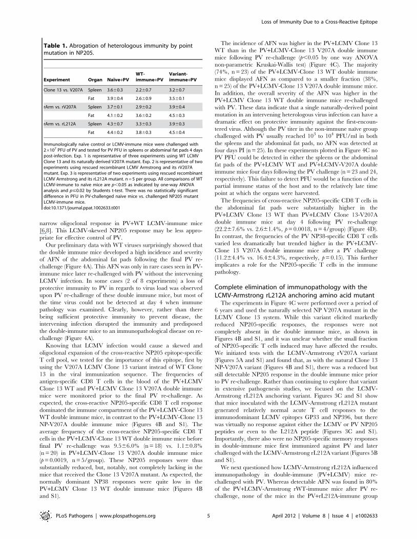
narrow oligoclonal response in PV+WT LCMV-immune mice
[6,8]. This LCMV-skewed NP205 response may be less appro-
priate for effective control of PV.
Our preliminary data with WT viruses surprisingly showed that
the double immune mice developed a high incidence and severity
of AFN of the abdominal fat pads following the final PV re-
challenge (Figure 4A). This AFN was only in rare cases seen in PV-
immune mice later re-challenged with PV without the intervening
LCMV infection. In some cases (2 of 8 experiments) a loss of
protective immunity to PV in regards to virus load was observed
upon PV re-challenge of these double immune mice, but most of
the time virus could not be detected at day 4 when immune
pathology was examined. Clearly, however, rather than there
being sufficient protective immunity to prevent disease, the
intervening infection disrupted the immunity and predisposed
the double-immune mice to an immunopathological disease on re-
challenge (Figure 4A).
Knowing that LCMV infection would cause a skewed and
oligoclonal expansion of the cross-reactive NP205 epitope-specific
T cell pool, we tested for the importance of this epitope, first by
using the V207A LCMV Clone 13 variant instead of WT Clone
13 in the viral immunization sequence. The frequencies of
antigen-specific CD8 T cells in the blood of the PV+LCMV
Clone 13 WT and PV+LCMV Clone 13 V207A double immune
mice were monitored prior to the final PV re-challenge. As
expected, the cross-reactive NP205-specific CD8 T cell response
dominated the immune compartment of the PV+LCMV-Clone 13
WT double immune mice, in contrast to the PV+LCMV-Clone 13
NP-V207A double immune mice (Figures 4B and S1). The
average frequency of the cross-reactive NP205-specific CD8 T
cells in the PV+LCMV-Clone 13 WT double immune mice before
final PV re-challenge was 9.566.0% (n = 18) vs. 1.160.8%
(n = 20) in PV+LCMV-Clone 13 V207A double immune mice
(p = 0.0019, n = 5/group). These NP205 responses were thus
substantially reduced, but, notably, not completely lacking in the
mice that received the Clone 13 V207A mutant. As expected, the
normally dominant NP38 responses were quite low in the
PV+LCMV Clone 13 WT double immune mice (Figures 4B
and S1).
The incidence of AFN was higher in the PV+LCMV Clone 13
WT than in the PV+LCMV-Clone 13 V207A double immune
mice following PV re-challenge (p,0.05 by one way ANOVA
non-parametric Kruskai-Wallis test) (Figure 4C). The majority
(74%, n = 23) of the PV+LCMV-Clone 13 WT double immune
mice displayed AFN as compared to a smaller fraction (38%,
n = 25) of the PV+LCMV-Clone 13 V207A double immune mice.
In addition, the overall severity of the AFN was higher in the
PV+LCMV Clone 13 WT double immune mice re-challenged
with PV. These data indicate that a single naturally-derived point
mutation in an intervening heterologous virus infection can have a
dramatic effect on protective immunity against the first-encoun-
tered virus. Although the PV titer in the non-immune naıve group
challenged with PV usually reached 103 to 104 PFU/ml in both
the spleens and the abdominal fat pads, no AFN was detected at
four days PI (n = 25). In these experiments plotted in Figure 4C no
PV PFU could be detected in either the spleens or the abdominal
fat pads of the PV+LCMV WT and PV+LCMV-V207A double
immune mice four days following the PV challenge (n = 23 and 24,
respectively). This failure to detect PFU would be a function of the
partial immune status of the host and to the relatively late time
point at which the organs were harvested.
The frequencies of cross-reactive NP205-specific CD8 T cells in
the abdominal fat pads were substantially higher in the
PV+LCMV Clone 13 WT than PV+LCMV Clone 13-V207A
double immune mice at day 4 following PV re-challenge
(22.267.6% vs. 2.661.4%, p = 0.0018, n = 4/group) (Figure 4D).
In contrast, the frequencies of the PV NP38-specific CD8 T cells
varied less dramatically but trended higher in the PV+LCMV-
Clone 13 V207A double immune mice after a PV challenge
(11.264.4% vs. 16.464.3%, respectively, p = 0.15). This further
implicates a role for the NP205-specific T cells in the immune
pathology.
Complete elimination of immunopathology with theLCMV-Armstrong rL212A anchoring amino acid mutant
The experiments in Figure 4C were performed over a period of
6 years and used the naturally selected NP V207A mutant in the
LCMV Clone 13 system. While this variant elicited markedly
reduced NP205-specific responses, the responses were not
completely absent in the double immune mice, as shown in
Figures 4B and S1, and it was unclear whether the small fraction
of NP205-specific T cells induced may have affected the results.
We initiated tests with the LCMV-Armstrong rV207A variant
(Figures 5A and S1) and found that, as with the natural Clone 13
NP-V207A variant (Figures 4B and S1), there was a reduced but
still detectable NP205 response in the double immune mice prior
to PV re-challenge. Rather than continuing to explore that variant
in extensive pathogenesis studies, we focused on the LCMV-
Armstrong rL212A anchoring variant. Figures 3C and S1 show
that mice inoculated with the LCMV-Armstrong rL212A mutant
generated relatively normal acute T cell responses to the
immunodominant LCMV epitopes GP33 and NP396, but there
was virtually no response against either the LCMV or PV NP205
peptides or even to the L212A peptide (Figures 3C and S1).
Importantly, there also were no NP205-specific memory responses
in double-immune mice first immunized against PV and later
challenged with the LCMV-Armstrong rL212A variant (Figures 5B
and S1).
We next questioned how LCMV-Armstrong rL212A influenced
immunopathology in double-immune (PV+LCMV) mice re-
challenged with PV. Whereas detectable AFN was found in 80%
of the PV+LCMV-Armstrong rWT-immune mice after PV re-
challenge, none of the mice in the PV+rL212A-immune group
Table 1. Abrogation of heterologous immunity by pointmutation in NP205.
Experiment Organ Naıve+PVWT-immune+PV
Variant-immune+PV
Clone 13 vs. V207A Spleen 3.660.3 2.260.7 3.260.7
Fat 3.960.4 2.660.9 3.560.1
rArm vs. rV207A Spleen 3.760.1 2.960.2 3.960.4
Fat 4.160.2 3.660.2 4.560.3
rArm vs. rL212A Spleen 4.360.7 3.360.3 3.960.3
Fat 4.460.2 3.860.3 4.560.4
Immunologically naıve control or LCMV-immune mice were challenged with26107 PFU of PV and tested for PV PFU in spleens or abdominal fat pads 4 dayspost-infection. Exp. 1 is representative of three experiments using WT LCMVClone 13 and its naturally derived V207A mutant. Exp. 2 is representative of twoexperiments using rescued recombinant LCMV Armstrong and its rV207Amutant. Exp. 3 is representative of two experiments using rescued recombinantLCMV Armstrong and its rL212A mutant. n = 5 per group. All comparisons of WTLCMV-immune to naıve mice are p,0.05 as indicated by one-way ANOVAanalysis and p#0.02 by Students t-test. There was no statistically significantdifference in PFU in PV-challenged naıve mice vs. challenged NP205 mutantLCMV-immune mice.doi:10.1371/journal.ppat.1002633.t001
Loss of Immunity Due to a Cross-Reactive Epitope
PLoS Pathogens | www.plospathogens.org 5 April 2012 | Volume 8 | Issue 4 | e1002633
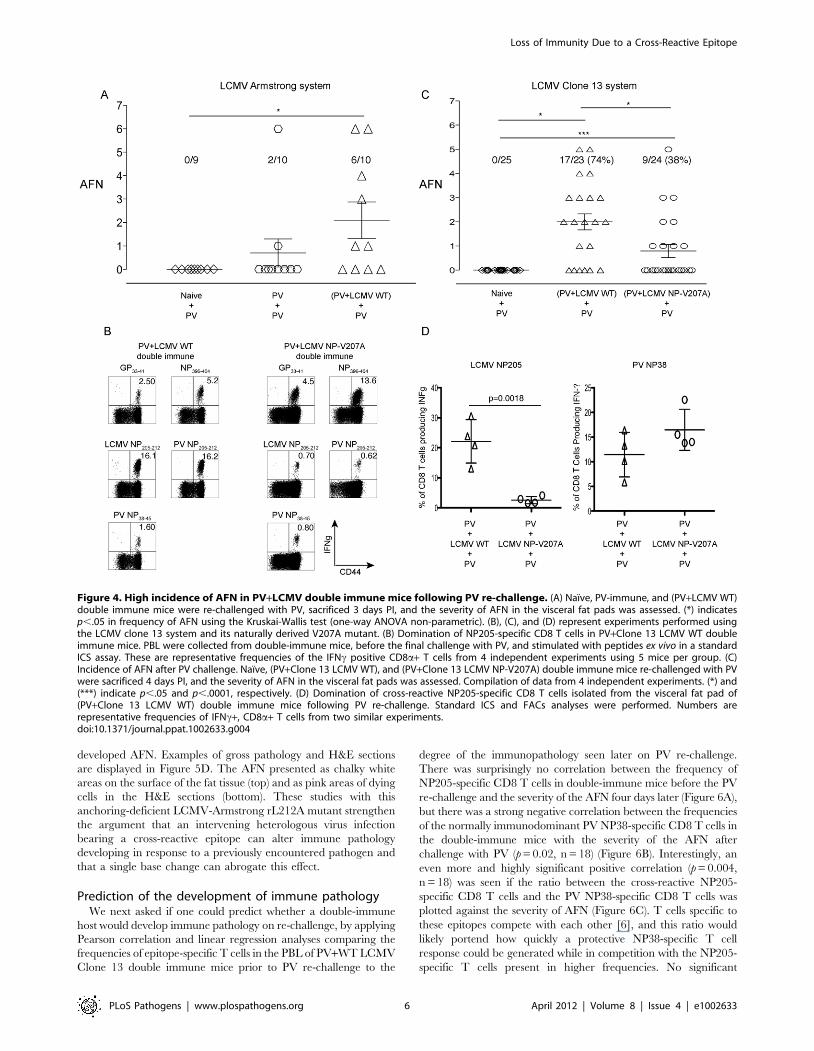
developed AFN. Examples of gross pathology and H&E sections
are displayed in Figure 5D. The AFN presented as chalky white
areas on the surface of the fat tissue (top) and as pink areas of dying
cells in the H&E sections (bottom). These studies with this
anchoring-deficient LCMV-Armstrong rL212A mutant strengthen
the argument that an intervening heterologous virus infection
bearing a cross-reactive epitope can alter immune pathology
developing in response to a previously encountered pathogen and
that a single base change can abrogate this effect.
Prediction of the development of immune pathologyWe next asked if one could predict whether a double-immune
host would develop immune pathology on re-challenge, by applying
Pearson correlation and linear regression analyses comparing the
frequencies of epitope-specific T cells in the PBL of PV+WT LCMV
Clone 13 double immune mice prior to PV re-challenge to the
degree of the immunopathology seen later on PV re-challenge.
There was surprisingly no correlation between the frequency of
NP205-specific CD8 T cells in double-immune mice before the PV
re-challenge and the severity of the AFN four days later (Figure 6A),
but there was a strong negative correlation between the frequencies
of the normally immunodominant PV NP38-specific CD8 T cells in
the double-immune mice with the severity of the AFN after
challenge with PV (p = 0.02, n = 18) (Figure 6B). Interestingly, an
even more and highly significant positive correlation (p = 0.004,
n = 18) was seen if the ratio between the cross-reactive NP205-
specific CD8 T cells and the PV NP38-specific CD8 T cells was
plotted against the severity of AFN (Figure 6C). T cells specific to
these epitopes compete with each other [6], and this ratio would
likely portend how quickly a protective NP38-specific T cell
response could be generated while in competition with the NP205-
specific T cells present in higher frequencies. No significant
Figure 4. High incidence of AFN in PV+LCMV double immune mice following PV re-challenge. (A) Naıve, PV-immune, and (PV+LCMV WT)double immune mice were re-challenged with PV, sacrificed 3 days PI, and the severity of AFN in the visceral fat pads was assessed. (*) indicatesp,.05 in frequency of AFN using the Kruskai-Wallis test (one-way ANOVA non-parametric). (B), (C), and (D) represent experiments performed usingthe LCMV clone 13 system and its naturally derived V207A mutant. (B) Domination of NP205-specific CD8 T cells in PV+Clone 13 LCMV WT doubleimmune mice. PBL were collected from double-immune mice, before the final challenge with PV, and stimulated with peptides ex vivo in a standardICS assay. These are representative frequencies of the IFNc positive CD8a+ T cells from 4 independent experiments using 5 mice per group. (C)Incidence of AFN after PV challenge. Naıve, (PV+Clone 13 LCMV WT), and (PV+Clone 13 LCMV NP-V207A) double immune mice re-challenged with PVwere sacrificed 4 days PI, and the severity of AFN in the visceral fat pads was assessed. Compilation of data from 4 independent experiments. (*) and(***) indicate p,.05 and p,.0001, respectively. (D) Domination of cross-reactive NP205-specific CD8 T cells isolated from the visceral fat pad of(PV+Clone 13 LCMV WT) double immune mice following PV re-challenge. Standard ICS and FACs analyses were performed. Numbers arerepresentative frequencies of IFNc+, CD8a+ T cells from two similar experiments.doi:10.1371/journal.ppat.1002633.g004
Loss of Immunity Due to a Cross-Reactive Epitope
PLoS Pathogens | www.plospathogens.org 6 April 2012 | Volume 8 | Issue 4 | e1002633
correlation was found between the frequencies of the LCMV GP33-
specific CD8 T cells or the LCMV GP33/PV NP38 ratio and the
level of AFN (Figure 6D). On a smaller scale with double-immune
mice using the rWT Armstrong virus, two experiments that had
strong AFN on re-challenge with PV showed positive correlations
with the frequencies both of NP205-specific T cells (R2 = 0.48;
p = 0.027) and with the ratio of NP205- to NP38-specific T cells
(R2 = 0.41; p = 0.046) with the severity of AFN (n = 10). Thus, there
was predictive value in knowing the frequencies of the cross-reactive
and immunodominant PV-specific epitopes.
Discussion
This report shows that protective immunity to a virus can be
disrupted by an otherwise well-tolerated and controlled infection
with a second and different virus. Further, it shows that a single
cross-reactive CD8 T cell epitope on that second virus can dictate
the degree of immune pathology on re-challenge with the first
virus. The NP205 epitopes encoded by LCMV and PV are highly
cross-reactive because they differ only in their MHC-anchoring
amino acids, and our studies presented in Table 1 with NP205
mutants clearly implicate this cross-reactive epitope in protective
heterologous immunity between these viruses. However, these
epitopes induce distinct TCR repertoires, and sequential infections
with these viruses result in very narrowly focused repertoires
skewed in favor of the second-encountered virus [8]. These
inappropriate T cell repertoires may interfere with strong
protective immunity to the first encountered virus.
The effects of buried MHC polymorphisms on TCR recogni-
tion have been previously evaluated [8,18,19], and we show here
Figure 5. Analysis of immune response and immunopathology with the LCMV-Armstrong rL212A anchoring amino acid mutant. (A)Diminished cross-reactive NP205 CD8 T cell responses in the (PV+rV207A) double immune mice. PV-immune mice were immunized with either rWT orrV207A variant Armstrong strain LCMV. After six weeks, PBL were collected and stimulated with LCMV-specific CD8 T cell peptides. The data representaverage frequencies of the IFNc-positive, CD8a+ T cells. This is representative of 3 experiments, with n = 5/group. (B) Complete elimination of cross-reactive NP205 CD8 T cell responses in (PV+rL212A) double immune mice. PV-immune mice were immunized with either rWT or rL212A LCMVArmstrong. After six weeks, PBL were collected and stimulated with LCMV-specific CD8 T cell peptides. Data represent average frequencies of IFNc-positive, CD8a+ T cells. This is representative of two experiments, with n = 5/group. (C) Prevention of AFN by the rL212A anchoring mutant. Naıve, PV-immune, (PV+rWT) and (PV+rL212A) double immune groups were re-challenged with PV. Four days later fat pads were harvested and AFN scoresevaluated. This is a compilation of two similar experiments. (***) indicates p,.0001. (D) Photographs of abdominal fat pads and tissue histologysections. Abdominal fat pads were harvested, photographed (top), and then fixed in 10% neutral buffered formaldehyde and embedded in paraffin atthe UMMS histology core facility. Thin tissue sections (5 mm) were stained with hemotoxylin and eosin (bottom). The digital photographs of thesections were taken using a Nikon Eclipse E300 microscope system.doi:10.1371/journal.ppat.1002633.g005
Loss of Immunity Due to a Cross-Reactive Epitope
PLoS Pathogens | www.plospathogens.org 7 April 2012 | Volume 8 | Issue 4 | e1002633
that epitope-anchoring amino acids buried within the MHC can
alter the conformations of determinants accessible to the TCR
[20], explaining why different TCR repertoires can react with
these LCMV and PV NP205 epitopes (Figure 1A,B,C). Further,
we show how a mutation in the third position of the LCMV
NP205 epitope will allow for epitope binding to the MHC yet alter
its interaction with T cells generated in response to the wild type
epitope (Figure 1D,E,F). Strikingly, single nucleotide changes
altering the cross-reactive epitope of the second intervening virus
removed its ability to interfere with the protection from disease
(Figures 4 and 5). In this case we suggest that the loss of T cells
specific to a protective and normally immunodominant epitope
(NP38) by a combination of IFN-induced attrition and competi-
tion with T cells responding to a normally subdominant cross-
reactive epitope (NP205) tips the balance from efficient protective
immunity to less efficient immunopathology.
Previous studies, as well as results presented here, have shown
that the immunodominant PV NP38 response is substantially
reduced in double (PV+LCMV)-immune mice in comparison to
PV only-immune mice [6,17]. Protective T cell-dependent
immunity to tumors can be lost after bacterial infections [21],
and a recent report shows that protective immunity to Plasmodium
is lost in mice subjected to a series of infections [22]. In our present
study, however, the reduction of the immunodominant NP38-
specific T cell response caused by the intervening LCMV infection
was partially compensated for by the cross-reactive NP205
response, which became dominant in double immune mice. The
price for the increased cross-reactive response, which was not ideal
for protection against PV, was enhanced disease associated with
immune pathology on PV rechallenge. If LCMV NP205 was
mutated in a way (V207A) that resulted in a reduced though still
detectable T cell response, the PV+LCMV-double immune hosts
responded to PV re-challenge with less pathology; if the
intervening LCMV was mutated in an MHC anchoring site
(L212A) to prevent any T cell response at all, the PV+LCMV
double-immune hosts responded to PV re-challenge with even less
pathology, which was undetectable.
The ratio of NP205-specific to NP38-specific T cells in double-
immune mice had strong predictive value for the production of
immune pathology on re-challenge with PV (Figure 6). The ratios
Figure 6. Correlation of frequencies of CD8 T cells in double-immune mice with pathology after PV re-challenge. Linear regressionanalyses comparing the frequencies of antigen-specific CD8 T cells in the (PV+WT LCMV) double immune mice with the severity of AFN following PVre-challenge. These represent data compiled from four independent experiments using LCMV Clone 13 virus. (A) LCMV NP205-specific CD8 T cellresponse. (B) PV NP38-specific CD8 T cell response. (C) Ratio of LCMV NP205 to PV NP38. (D) LCMV GP33 and the ratio of GP33/PV NP38.doi:10.1371/journal.ppat.1002633.g006
Loss of Immunity Due to a Cross-Reactive Epitope
PLoS Pathogens | www.plospathogens.org 8 April 2012 | Volume 8 | Issue 4 | e1002633
of these epitope-specific T cells in double immune mice might
predict their relative abilities to compete with each other in their
early response to the PV re-challenge. Analyses of T cells in
diseased tissue day 4 after PV re-challenge are complicated by the
severe necrosis and collateral cell damage in the adipose tissue, but
many NP205-specific T cells are found at that time (Figure 4D). It
is likely, however, that T cell responses occurring very early after
challenge may have controlled viral load and affected the
outcome.
Severe immune pathologies associated with cross-reactive T cell
responses in humans have been reported in fulminant HCV-
associated hepatitis, infectious mononucleosis, and dengue hem-
orrhagic fever and shock syndrome [23–26]. Aberrant pathology
associated with cross-reactive pathogenic epitopes is thus an issue
that should be considered in vaccine construction. For instance,
some strains of HCV encode an epitope that strongly cross-reacts
with an epitope of IAV [27], and HCV vaccines containing this
cross-reactive epitope are under evaluation [28]. One wonders
what a sequence of an IAV infection (or vaccine) and an HCV
vaccination, in either order, would have on a subsequent
encounter with either virus. We suggest that these concerns would
be less for viruses or viral vaccines that would induce high levels of
neutralizing antibody, which might prevent infection in the first
place. However, viruses like HCV, HIV, and CMV are relatively
poor at inducing effective neutralizing antibody responses, and
individuals infected with these viruses often become super-infected
with slightly different variants. Cross-reactive pathogenic epitopes
might also be an issue with influenza virus infections when
individuals with poor neutralizing antibodies but strong cross-
reactive T cells to new influenza virus strains become infected.
The panniculitis described in our current model may seem
unusual, but panniculitis is a pathology commonly found in
humans in the form of erythema nodosum, which involves
inflammation of subcutaneous fat tissue [29]. Erythema nodosum
sometimes occurs following infections or in association with
autoimmune diseases such as Crohn’s [30]. Of relevance to our
present work, panniculitis is sometimes found in humans after
vaccinations for smallpox, hepatitis B and papilloma viruses [31–
33]. In mice, panniculitis and AFN of visceral fat pads is a
common feature of virus infections by the intraperitoneal route,
but it is particularly noticeable in models of heterologous
immunity, where memory T cells induced by an earlier
heterologous viral infection rapidly respond to but inefficiently
clear an infection of the fat pads by a second virus [3,7,34]. The
Armstrong strain of LCMV replicates poorly in the fat pads and
does not directly elicit AFN, but a history of an LCMV infection
can prime a mouse for AFN after infection with certain
heterologous viruses that do grow in the fat. The mechanism of
AFN is best studied in LCMV-immune mice infected with vaccinia
virus, where cross-reactive T cells enter the fat pads and stimulate
necrosis through an IFNc, TNF, and Fas ligand-dependent
mechanism that reflects the private specificity of the T cell
populations in LCMV-immune mice, as shown in assays using
adoptive transfers of immune T cell [7,35,36]. PV, used in the
current study, does replicate in fat tissue, and the disruption of the
memory T cell response specific to PV by the LCMV infection has
apparently created the conditions that predispose to AFN rather
than pathology-free clearance of virus on re-challenge with PV.
The unique finding of our current study, however, is not simply
another demonstration of heterologous immunity. Rather, it is the
finding that the heterologous immunity associated with an
intervening infection with a virus containing a cross-reactive
epitope can have a profound impact on the homologous immunity
against a previously immunized pathogen. Hence, lasting
immunity to a previously encountered pathogen can be compro-
mised by subsequent infections with other pathogens bearing
cross-reactive pathogenic epitopes.
Materials and Methods
Ethics statementThis study was carried out in strict accordance with the
recommendations in the Guide for the Care and Use of
Laboratory Animals of the U. S. National Institutes of Health.
All animal work was reviewed and approved by the UMMS
institutional Animal Care Committee (Animal Welfare Assurance
# A3306-01), and all the efforts were made to minimize suffering
of mice.
MiceC57BL/6 (B6, H2Kb) male mice were purchased from the
Jackson Laboratory (Bar Harbor, ME) and maintained under
specific pathogen-free conditions at the University of Massachu-
setts Medical School (UMMS) Department of Animal Medicine.
All animal work was reviewed and approved by the UMMS
Institutional Animal Care Committee.
VirusesThe AN3739 strain of PV and several strains and variants of
LCMV were propagated in BHK-21 cells. These include LCMV,
strain Armstrong, and recombinant (r) Armstrong variants
harboring laboratory-directed mutations in the NP205–212 epitope:
rLCMV wild type (rWT), rV207A, and rL212A. The Clone 13
natural variant of LCMV, which has mutations in the glycoprotein
and polymerase that allow for greater replication and dissemina-
tion in vivo [37] was also used, as well as a naturally-derived CD8 T
cell escape variant in the NP205–212 epitope, LCMV-clone 13
V207A. Clone 13 can be used at high doses to establish persistent
infections, but the experiments described here use lower doses that
generate immune responses that clear infection similarly to that of
the Armstrong strain. To avoid immune responses generated to
bovine serum following sequential infections, PV was purified by
sucrose density gradient ultra-centrifugation and diluted in serum-
free HBSS before immunization [7]. LCMV stocks were
propagated to titers over 107 PFU/ml as assayed on vero cell
monolayers and diluted in serum-free HBSS prior to infection of
mice.
Experimental procedures used for the generation and rescue of
recombinant LCM viruses (rLCMV) were as described [38].
Briefly, BHK-21 cells were transfected with T7 RNA polymerase
(T7RNP)-based expression plasmids that directed intracellular
synthesis of full-length S and L genome RNA species of LCMV
Armstrong strain, together with pol II-based expression plasmids
expressing T7RNP and the minimal viral trans-acting factors (L
and NP) required for virus RNA replication and gene transcrip-
tion. At 60 h post-transfection, tissue culture supernatants were
collected (referred to as P0), clarified at low speed and used to
infect fresh monolayers of BHK-21 cells. At 48 h p.i., TCS were
collected (P1) and titrated by plaque assay.
All inoculations were by the intraperitonal route. For primary
infections with LCMV, male mice 6–8 weeks of age were
inoculated with 56104 to 56105 PFU of LCMV. For primary
infections with PV, mice were inoculated with 26107 PFU of
purified PV. Mice were considered immune 6 weeks after
immunization. For homologous challenge, PV-immune mice were
inoculated with 26107 PFU of purified PV. For heterologous
challenge, LCMV-immune mice were inoculated with 26107 PFU
of purified PV. For generation of double immune mice, PV-
Loss of Immunity Due to a Cross-Reactive Epitope
PLoS Pathogens | www.plospathogens.org 9 April 2012 | Volume 8 | Issue 4 | e1002633
immune animals were challenged with 56105 to 16106 PFU of
LCMV WT or LCMV variants. Re-challenge of (PV+LCMV)
double immune mice was done with 26107 PFU of purified PV.
Synthetic peptidesLCMV-encoded peptide epitopes used were GP33–41 (KAVYN-
FATC), NP396–404 (FQPQNGQFI), LCMV NP205–212 (YTV-
KYPNL), mutated NP205–212 V207A (YTAKYPNL) and L212A
(YTVKYPNA). PV-encoded epitopes were NP38–45 (SALDFHKV)
and PV NP205–212 (YTVKFPNM). Synthetic peptides were from
BioSource International or 21st Century Biochemicals at 90%
purity.
Peptide/MHC stabilization assayTAP-1 deficient RMA-S cells [39] were seeded into 96-well U-
bottom plates at 56105 cells per well. Following incubation in 5%
CO2 at 27uC for 4 hours, variants of LCMV NP205 peptides were
added at different concentrations and incubated overnight. The
cells were then stained with mAb to H2Kb (clone AF6 88.5)
conjugated with PE (BD Bioscience) and analyzed by fluorescence-
activated cell sorting (FACS).
Protein expression, purification, crystallization andstructure determination
H2Kb and b2-microglobulin molecules were expressed in
Escherichia coli as inclusion bodies, refolded with the LCMV-
NP205, PV-NP205 or LCMV-V3A (V207A) NP205 peptides and
purified as previously described [40]. The three plasmid (p) MHC
complexes were concentrated to 2–5 mg/ml, using the hanging-
drop vapor diffusion technique at 20uC. Crystals were grown with a
reservoir containing 16–24% polyethylene glycol (PEG) 3350,
0.1 M Na-Cacodylate, pH 6.5, and 0.2 M Na acetate. The crystals
belong to space group P21 and the unit cell dimensions were
consistent with two molecules per asymmetric units (Table S1).
The crystals were flash frozen to a temperature of 100 K before
data collection using an in-house X-ray generator with a RAXIS-
IV detector for the H2Kb-LCMV NP205 or at the Australian
Synchrotron on the BM1 beamline with a MarCCD or an ADSC
Q210r detector for the H2Kb-LCMV-V207A (V3A) and H2-Kb-
PV NP205 structures. The data were processed and scaled with
the XDS [41]. The crystal structure was solved using the
molecular replacement method in the program Phaser [42] from
the CCP4 suite of programs (1994). The search probe used to solve
the structure was the structure of mouse MHC class I H2Kb minus
the peptide (Protein Data Bank accession number 2ZSV) [43].
The progress of refinement was monitored by the Rfree value with
neither a sigma nor a low-resolution cut-off being applied to the
data. This protocol includes several cycles of refinement with the
PHENIX software [44] followed by manual model rebuilding with
Coot program [45]. Final refinement statistics are summarized in
Table S1. The coordinates of the three complexes have been
deposited with the Protein Data Bank under accession numbers
3P4M, 3P4N and 3P4O for the H2Kb-NP205-LCMV, H2Kb-
NP205-PV and H2Kb-LCMV-V3A (NP V207A), respectively.
Thermostability measurements of recombinant class Icomplexes using circular dichroism (CD)
Circular Dichroism Spectra were measured on a Jasco 815
spectropolarimeter using a thermostatically controlled cuvette. A
far-UV spectra was collected from 190 nm to 250 nm. The UV
minimum was determined as 219 nm for the three peptide-MHX
complexes. The measurements for the thermal melting experi-
ments were made at the minimum, at intervals of 0.1uC at a rate of
1uC/min from 20uC to 90uC. The Jasco Spectra Manager
software was used to view and smooth the traces, and then the
GraphPad Prism software was used to plot temperature versus %
unfolded. The midpoint of thermal denaturation (Tm) for each
protein was determined as the point at which 50% unfolding was
achieved. The measurements were done in duplicate at two
concentrations (5 mM and 10 mM) in a solution of 10 mM Tris
pH 8, 150 mM NaCl.
Acute fatty necrosis (AFN) scoresThe severity of AFN was scored based on the guidelines from a
previous publication: (1–2) very mild to mild disease with a few
white necrotic spots on one or both lower abdominal fat pads; (3–4)mildly moderate and moderate with larger patches of necrosis of the
lower abdominal fat pads and extension into the upper left quadrant
fat pad around the spleen; (5–6) moderately severe to severe with
very extensive large patches of necrosis on the lower abdominal fat
pads and spotty fatty necrosis throughout omental fat pads as well as
the splenic fat pad; (7) very severe disease with such severe fatty
necrosis that the organs are adherent to each other [7,35].
Tissue histologyAbdominal fat pads from different groups of mice were
harvested and fixed in 10% neutral buffered formaldehyde and
embedded in paraffin at the UMMS histology core facility. Thin
tissue sections (5 mm) were stained with hemotoxylin and eosin.
The digital photographs of the sections were taken using the Nikon
Eclipse E300 microscope system at the UMMS core facility.
Isolation of lymphocytes from adipose tissueThe infiltrating leukocytes in the fat pads were isolated by
mincing and digesting with collagenase B (200 mg/ml) in MEM
plus 4% BSA for 1 hour at 37uC, and then by separation over
Lympholyte-M from Cederlane Laboratory (Burlington, ON).
Intracellular cytokine staining (ICS)Leukocytes from spleens, blood and abdominal fat pads (16106
cells/well) were stimulated with 1 mM peptides in medium
containing 0.2 ml of GolgiPlug and human recombinant IL-2
(BD Pharmingen) at 37uC for 5 hours. Intracellular cytokine
staining (ICS) was performed using a cytofix/cytoperm kit from
BD Bioscience. Intracellular cytokine-producing cells were detect-
ed with allophycocyanin (APC)-conjugated anti-mouse IFNcmonoclonal antibodies (1:1000) (XMG1.2) and phycoerythrin-
Cy7 (PE-Cy7)-conjugated anti-mouse TNFa (1:200) (MP6-XT22).
Cell surface and MHC-Ig dimer staining by flowcytometry
All the surface antibodies were used at a 1:200 dilution per well.
Single-cell suspensions of splenocytes or blood lymphocytes were
first incubated with anti-mouse CD16/CD32 Fc-block antibody
(1 ml/well) for 15 minutes on ice. Subsequently a cell surface
staining procedure was performed using PerCP-Cy5.5 anti-mouse
CD8a (clone 53-6.7) and FITC anti-mouse CD44 (clone IM7).
The samples were incubated on ice for 20 minutes.
Soluble dimeric mouse H2Kb-Ig fusion proteins (MHC-Ig
dimer) were purchased from BD Bioscience (San Diego, CA). The
LCMV WT NP205 and mutant V207A peptides (1 mg/ml) were
incubated with the MHC-Ig dimer at an 800 to 1 molar ratio and
with recombinant human beta-2 micro-globulin (0.15 mg/mg of
dimer) from BD Bioscience at 4uC for 4 days for passive loading of
the peptide onto the MHC. The final products were used for
surface staining assays as above and previously reported [2].
Loss of Immunity Due to a Cross-Reactive Epitope
PLoS Pathogens | www.plospathogens.org 10 April 2012 | Volume 8 | Issue 4 | e1002633
Statistical analysisStatistical analysis was performed using GraphPad Prism software
(5.0b). Comparisons between two groups were performed using the
unpaired Student’s t test (2-tailed). Comparisons between more than
two groups were performed using one way Anova analysis (2-tailed).
Pearson’s correlation and linear regression tests were used to
measure the correlation between two independent variables. P values
less than 0.05 were considered statistically significant.
Supporting Information
Figure S1 Graphic analysis of dot plots from Figures 3,4, and 5. This figure graphs the magnitude and variance of
epitope-specific T cell responses from replicas associated with the
representative data presented in Fig. 3A (n = 3/group), 3B (n = 2/
group), 3C (n = 3/group), 4B (n = 5/group), 5A (n = 5/group), and
5B (n = 5/group). All show means 6 standard deviations
(p,0.05*).
(TIF)
Table S1 Data collection and refinement statistics.
(DOC)
Acknowledgments
We thank Drs. Stephen Turner and Michael Brehm for advice, and Robb
Wesselingh, Brian Sheridan, and the staff at the Australian synchrotron for
assistance.
Author Contributions
Conceived and designed the experiments: ATC, MC, RMW, LKS, SG,
CG, JR, AT, SE, and JCT all participated in conceiving and designing the
experiments. Performed the experiments: ATC, MC, RMW, and LKS did
the infections, T cell analyses, immune pathology, and wrote the paper.
SG, CG, and JR performed the peptide-MHC structural studies. AT, SE,
and JCT generated by reverse genetics the recombinant viruses. Analyzed
the data: ATC MC RMW LKS SG CG JR AT SE JCT. Wrote the paper:
ATC RMW. JCT designed and contributed recombinant viruses or this
work.
References
1. Amanna IJ, Slifka MK (2011) Contributions of humoral and cellular immunityto vaccine-induced protection in humans. Virology 411: 206–215.
2. Selin LK, Lin MY, Kraemer KA, Schneck JP, Pardoll D, et al. (1999) Attrition
of T cell memory:selective loss of lymphocytic choriomeningitis virus (LCMV)
epitope-specific memory CD8 T cells following infections with heterologousviruses. Immunity 11: 733–742.
3. Welsh RM, Che JW, Brehm MA, Selin LK (2010) Heterologous immunity
between viruses. Immunol Rev 235: 244–266.
4. Bahl K, Huebner A, Davis RJ, Welsh RM (2010) Analysis of apoptosis of
memory T cells and dendritic cells during the early stages of viral infection orexposure to toll-like receptor agonists. J Virol 84: 4866–4877.
5. Chanas AC, Young PR, Ellis DS, Mann G, Stamford S, et al. (1980) Evaluationof plaque size reduction as a method for the detection of Pichinde virus antibody.
Arch Virol 65: 157–167.
6. Brehm MA, Pinto AK, Daniels KA, Schneck JP, Welsh RM, et al. (2002) T cell
immunodominance and maintenance of memory regulated by unexpectedlycross-reactive pathogens. Nat Immunol 3: 627–634.
7. Selin LK, Varga SM, Wong IC, Welsh RM (1998) Protective heterologous
antiviral immunity and enhanced immunopathogenesis mediated by memory T
cell populations. J Exp Med 188: 1705–1715.
8. Cornberg M, Chen AT, Wilkinson LA, Brehm MA, Kim SK, et al. (2006)Narrowed TCR repertoire and viral escape as a consequence of heterologous
immunity. J Clin Invest 116: 1443–1456.
9. Welsh RM, Fujinami RS (2007) Pathogenic epitopes, heterologous immunity
and vaccine design. Nat Rev Microbiol 5: 555–563.
10. Zhao Z-S, Granucci F, Yeh L, Schaffer PA, Cantor H (1998) Molecular mimicry
by herpes simplex vrus-type 1: autoimmune disease after viral infection. Science279: 13441347.
11. Tsunoda I, Kuang LQ, Kobayashi-Warren M, Fujinami RS (2005) Central
nervous system pathology caused by autoreactive CD8+ T-cell clones following
virus infection. J Virol 79: 14640–14646.
12. Sullivan BM, Emonet SF, Welch MJ, Lee AM, Campbell KP, et al. (2011) Pointmutation in the glycoprotein of lymphocytic choriomeningitis virus is necessary
for receptor binding, dendritic cell infection, and long-term persistence. Proc
Natl Acad Sci U S A 108: 2969–2974.
13. Tynan FE, Reid HH, Kjer-Nielsen L, Miles JJ, Wilce MC, et al. (2007) A T cellreceptor flattens a bulged antigenic peptide presented by a major histocompat-
ibility complex class I molecule. Nat Immunol 8: 268–276.
14. Godfrey DI, Rossjohn J, McCluskey J (2008) The fidelity, occasional
promiscuity, and versatility of T cell receptor recognition. Immunity 28:304–314.
15. Tynan FE, Burrows SR, Buckle AM, Clements CS, Borg NA, et al. (2005) T cellreceptor recognition of a ‘super-bulged’ major histocompatibility complex class
I-bound peptide. Nat Immunol 6: 1114–1122.
16. Burrows SR, Chen Z, Archbold JK, Tynan FE, Beddoe T, et al. (2010) Hard
wiring of T cell receptor specificity for the major histocompatibility complex isunderpinned by TCR adaptability. Proc Natl Acad Sci U S A 107:
10608–10613.
17. Kim SK, Welsh RM (2004) Comprehensive early and lasting loss of memory
CD8 T cells and functional memory during acute and persistent viral infections.J Immunol 172: 3139–3150.
18. Macdonald WA, Chen Z, Gras S, Archbold JK, Tynan FE, et al. (2009) T cell
allorecognition via molecular mimicry. Immunity 31: 897–908.
19. Archbold JK, Macdonald WA, Gras S, Ely LK, Miles JJ, et al. (2009) Natural
micropolymorphism in human leukocyte antigens provides a basis for geneticcontrol of antigen recognition. J Exp Med 206: 209–219.
20. Theodossis A, Guillonneau C, Welland A, Ely LK, Clements CS, et al. (2010)
Constraints within major histocompatibility complex class I restricted peptides:
presentation and consequences for T-cell recognition. Proc Natl Acad Sci U S A107: 5534–5539.
21. Smith DK, Dudani R, Pedras-Vasconcelos JA, Chapdelaine Y, van Faassen H,
et al. (2002) Cross-reactive antigen is required to prevent erosion of established T
cell memory and tumor immunity: a heterologous bacterial model of attrition.J Immunol 169: 1197–1206.
22. Schmidt NW, Harty JT (2011) Cutting edge: attrition of Plasmodium-specific
memory CD8 T cells results in decreased protection that is rescued by boosterimmunization. J Immunol 186: 3836–3840.
23. Urbani S, Amadei B, Fisicaro P, Pilli M, Missale G, et al. (2005) Heterologous T
cell immunity in severe hepatitis C virus infection. J Exp Med 201: 675–680.
24. Clute SC, Watkin LB, Cornberg M, Naumov YN, Sullivan JL, et al. (2005) Cross-
reactive influenza virus-specific CD8+ T cells contribute to lymphoproliferation inEpstein-Barr virus-associated infectious mononucleosis. J Clin Invest 115: 3602–3612.
25. Spaulding AC, Kurane I, Ennis FA, Rothman AL (1999) Analysis of murine
CD8(+) T-cell clones specific for the Dengue virus NS3 protein: flavivirus cross-
reactivity and influence of infecting serotype. J Virol 73: 398–403.
26. Mongkolsapaya J, Dejnirattisai W, Xu XN, Vasanawathana S,Tangthawornchaikul N, et al. (2003) Original antigenic sin and apoptosis in
the pathogenesis of dengue hemorrhagic fever. Nat Med 9: 921–927.
27. Wedemeyer H, Mizukoshi E, Davis AR, Bennink JR, Rehermann B (2001)
Cross-reactivity between hepatitis C virus and influenza A virus determinant-specific cytotoxic T cells. J Virol 75: 11392–11400.
28. Schlaphoff V, Klade CS, Jilma B, Jelovcan SB, Cornberg M, et al. (2007)
Functional and phenotypic characterization of peptide-vaccine-induced HCV-specific CD8+ T cells in healthy individuals and chronic hepatitis C patients.
Vaccine 25: 6793–6806.
29. Requena L, Requena C (2002) Erythema nodosum. Dermatol Online J 8: 4.
30. Smoller BR, Weishar M, Gray MH (1990) An unusual cutaneous manifestation
in Crohn’s disease. Arch Pathol Lab Med 6: 609–610.
31. Di Giusto CA, Bernhard JD (1986) Erythema nodosum provoked by hepatitis B
vaccine. Lancet 2: 1042.
32. Ojaimi S, Buttery JP, Korman TM (2009) Quadrivalent Human Papillomavirusrecombinant vaccine associated lipoatrophy. Vaccine 27: 4876–4878.
33. Gaertner EM, Groo S, Kim J (2004) Papular spongiotic dermatitis of smallpox
vaccination: report of 2 cases with review of the literature. Arch Pathol Lab Med128: 1173–1175.
34. Yang H, Joris I, Majno G, Welsh RM (1985) Necrosis of adipose tissue inducedby sequential infections with unrelated viruses. Am J Pathol 120: 173–177.
35. Nie S, Lin SJ, Kim SK, Welsh RM, Selin LK (2010) Pathological features of
heterologous immunity are regulated by the private specificities of the immunerepertoire. Am J Pathol 176: 2107–2112.
36. Selin LK, Wlodarczyk MF, Kraft AR, Nie S, Kenney LL, et al. (2011)Heterologous immunity: immunopathology, autoimmunity and protection
during viral infections. Autoimmunity 44: 328–347.
37. Matloubian M, Kolhekar SR, Somasundaram T, Ahmed R (1993) Moleculardeterminants of macrophage tropism and viral persistence:importance of single
amino acid changes in the polymerase and glycoprotein of lymphocytic
choriomeningitis virus. J Virol 67: 7340–7349.
38. Sanchez AB, de la Torre JC (2006) Rescue of the prototypic Arenavirus LCMVentirely from plasmid. Virology 350: 370–380.
39. Townsend A, Ohlen C, Bastin J, Ljunggren H, Foster L, et al. (1989) Association
of class I major histocompatibility heavy and light chains induced by viral
peptides. Nature 340: 443–448.
Loss of Immunity Due to a Cross-Reactive Epitope
PLoS Pathogens | www.plospathogens.org 11 April 2012 | Volume 8 | Issue 4 | e1002633
40. Clements CS, Kjer-Nielsen L, Macdonald WA, Brooks AG, Purcell AW, et al.
(2002) The production, purification and crystallization of a soluble heterodi-meric form of a highly selected T-cell receptor in its unliganded and liganded
state. Acta Crystallogr D Biol Crystallogr 58: 2131–2134.
41. Kabsch W (2010) Integration, scaling, space-group assignment and post-refinement. Acta Crystallogr D Biol Crystallogr 66: 133–144.
42. Read RJ (2001) Pushing the boundaries of molecular replacement withmaximum likelihood. Acta Crystallogr D Biol Crystallogr 57: 1373–1382.
43. Butler NS, Theodossis A, Webb AI, Nastovska R, Ramarathinam SH, et al.
(2008) Prevention of cytotoxic T cell escape using a heteroclitic subdominantviral T cell determinant. PLoS Pathog 4: e1000186.
44. Adams PD, Afonine PV, Bunkoczi G, Chen VB, Davis IW, et al. (2010)
PHENIX: a comprehensive Python-based system for macromolecular structuresolution. Acta Crystallogr D Biol Crystallogr 66: 213–221.
45. Emsley P, Lohkamp B, Scott WG, Cowtan K (2010) Features and developmentof Coot. Acta Crystallogr D Biol Crystallogr 66: 486–501.
Loss of Immunity Due to a Cross-Reactive Epitope
PLoS Pathogens | www.plospathogens.org 12 April 2012 | Volume 8 | Issue 4 | e1002633
Related Documents