ORIGINAL RESEARCH ARTICLE published: 09 May 2013 doi: 10.3389/fncel.2013.00066 Long-term fluoxetine treatment induces input-specific LTP and LTD impairment and structural plasticity in the CA1 hippocampal subfield Francisco J. Rubio 1† , Estíbaliz Ampuero 1† , Rodrigo Sandoval 2 , Jorge Toledo 1 , Floria Pancetti 2 and Ursula Wyneken 1 * 1 Laboratorio de Neurociencias, Centro de Investigaciones Biológicas, Universidad de los Andes, Santiago, Chile 2 Laboratorio de Neurotoxicologia Ambiental, Universidad Católica del Norte, Coquimbo, Chile Edited by: Rena Li, Roskamp Institute, USA Reviewed by: Maarten H. P. Kole, Netherlands Institute for Neuroscience, Netherlands Rif S. El-Mallakh, University of Louisville School of Medicine, USA *Correspondence: Ursula Wyneken, Laboratorio de Neurociencias, Centro de Investigaciones Biológicas, Universidad de los Andes, San Carlos de Apoquindo, 2200 Las Condes 762001, Santiago, Chile. e-mail: [email protected] † These authors have contributed equally to this work. Antidepressant drugs are usually administered for several weeks for the treatment of major depressive disorder. However, they are also prescribed in several additional psychiatric conditions as well as during long-term maintenance treatments. Antidepressants induce adaptive changes in several forebrain structures which include modifications at glutamatergic synapses. We recently found that repetitive administration of the selective serotonin reuptake inhibitor (SSRI) fluoxetine to naïve adult male rats induced an increase of mature, mushroom-type dendritic spines in several forebrain regions. This was associated with an increase of GluA2-containing α-amino-3-hydroxy-5-methylisoxazole-4-propionate receptors (AMPA-Rs) in telencephalic postsynaptic densities. To unravel the functional significance of such a synaptic re-arrangement, we focused on glutamate neurotransmission in the hippocampus. We evaluated the effect of four weeks of 0.7mg/kg fluoxetine on long-term potentiation (LTP) and long-term depression (LTD) in the CA1 hippocampal subfield. Recordings in hippocampal slices revealed profound deficits in LTP and LTD at Schaffer collateral-CA1 synapses associated to increased spine density and enhanced presence of mushroom- type spines, as revealed by Golgi staining. However, the same treatment had neither an effect on spine morphology, nor on LTP and LTD at perforant path-CA1 synapses. Cobalt staining and immunohistochemical experiments revealed decreased AMPA-R Ca 2+ permeability in the stratum radiatum (s.r.) together with increased GluA2-containing Ca 2+ impermeable AMPA-Rs. Therefore, 4 weeks of fluoxetine treatment promoted structural and functional adaptations in CA1 neurons in a pathway-specific manner that were selectively associated with impairment of activity-dependent plasticity at Schaffer collateral-CA1 synapses. Keywords: antidepressants, LTP, LTD, dendritic spines, glutamate receptors INTRODUCTION Fluoxetine is a selective serotonin reuptake inhibitor (SSRI) that is widely used to treat anxiety- and mood-related disorders, but in addition, its use has been expanded to other psychiatric conditions and is often continued after remission of symptoms (Schatzberg, 2000; Blier et al., 2007). The therapeutic effect of antidepressant drugs is mediated by cellular events which include adult hippocampal neurogenesis, maturation of nascent neu- rons and changes in gene expression. It has been shown that the activation of gene transcription following fluoxetine treat- ment is elicited by epigenetic remodeling of chromatin structure leading to increased plasticity and dendritic spine remodeling (Maya Vetencourt et al., 2011; Wang et al., 2011). This has been proposed to underlie plastic changes in glutamate neuro- transmission (Pittenger and Duman, 2008; Sanacora et al., 2008; Maya-Vetencourt et al., 2012). It had been previously described by us that 28 days of 0.7 mg/kg fluoxetine administration to adult naïve rats induced growth of dendritic spines and changes in glutamate receptor subunit composition in cerebrocortical synapses (Ampuero et al., 2010). In addition, such changes led to impairment of remote, but not recent, hippocampus-dependent memory (Ampuero et al., 2013). To study the effect of fluoxetine on cellular plasticity, we focused on CA1 pyramidal neurons which receive spatially seg- regated direct and indirect excitatory inputs from the entorhinal cortex (EC) via the perforant path and the Schaffer collaterals originating in CA3, respectively. While Schaffer collaterals termi- nate in the CA1 stratum radiatum (s.r.) on proximal dendrites, the direct perforant path makes synapses on the distal dendrites of CA1 neurons that are localized in the stratum lacunosum moleculare (s.l.m.) (Steward, 1976). The induction of long-term potentiation (LTP) and long-term depression (LTD) of excitatory synaptic transmission at CA1 requires both N-methyl-D-aspartate receptor (NMDA-R) acti- vation and trafficking of α-amino-3-hydroxy-5-methylisoxazole- 4-propionate receptors (AMPA-Rs) (Citri and Malenka, 2008). Frontiers in Cellular Neuroscience www.frontiersin.org May2013 | Volume 7 | Article 66 | 1 CELLULAR NEUROSCIENCE

Welcome message from author
This document is posted to help you gain knowledge. Please leave a comment to let me know what you think about it! Share it to your friends and learn new things together.
Transcript
ORIGINAL RESEARCH ARTICLEpublished: 09 May 2013
doi: 10.3389/fncel.2013.00066
Long-term fluoxetine treatment induces input-specific LTPand LTD impairment and structural plasticity in the CA1hippocampal subfieldFrancisco J. Rubio1†, Estíbaliz Ampuero 1†, Rodrigo Sandoval2, Jorge Toledo1, Floria Pancetti 2 andUrsula Wyneken 1*
1 Laboratorio de Neurociencias, Centro de Investigaciones Biológicas, Universidad de los Andes, Santiago, Chile2 Laboratorio de Neurotoxicologia Ambiental, Universidad Católica del Norte, Coquimbo, Chile
Edited by:
Rena Li, Roskamp Institute, USA
Reviewed by:
Maarten H. P. Kole, NetherlandsInstitute for Neuroscience,NetherlandsRif S. El-Mallakh, University ofLouisville School of Medicine, USA
*Correspondence:
Ursula Wyneken, Laboratorio deNeurociencias, Centro deInvestigaciones Biológicas,Universidad de los Andes, SanCarlos de Apoquindo, 2200Las Condes 762001, Santiago, Chile.e-mail: [email protected]†These authors have contributedequally to this work.
Antidepressant drugs are usually administered for several weeks for the treatmentof major depressive disorder. However, they are also prescribed in severaladditional psychiatric conditions as well as during long-term maintenance treatments.Antidepressants induce adaptive changes in several forebrain structures whichinclude modifications at glutamatergic synapses. We recently found that repetitiveadministration of the selective serotonin reuptake inhibitor (SSRI) fluoxetine to naïveadult male rats induced an increase of mature, mushroom-type dendritic spines inseveral forebrain regions. This was associated with an increase of GluA2-containingα-amino-3-hydroxy-5-methylisoxazole-4-propionate receptors (AMPA-Rs) in telencephalicpostsynaptic densities. To unravel the functional significance of such a synapticre-arrangement, we focused on glutamate neurotransmission in the hippocampus. Weevaluated the effect of four weeks of 0.7 mg/kg fluoxetine on long-term potentiation(LTP) and long-term depression (LTD) in the CA1 hippocampal subfield. Recordings inhippocampal slices revealed profound deficits in LTP and LTD at Schaffer collateral-CA1synapses associated to increased spine density and enhanced presence of mushroom-type spines, as revealed by Golgi staining. However, the same treatment had neitheran effect on spine morphology, nor on LTP and LTD at perforant path-CA1 synapses.Cobalt staining and immunohistochemical experiments revealed decreased AMPA-R Ca2+permeability in the stratum radiatum (s.r.) together with increased GluA2-containingCa2+ impermeable AMPA-Rs. Therefore, 4 weeks of fluoxetine treatment promotedstructural and functional adaptations in CA1 neurons in a pathway-specific manner thatwere selectively associated with impairment of activity-dependent plasticity at Schaffercollateral-CA1 synapses.
Keywords: antidepressants, LTP, LTD, dendritic spines, glutamate receptors
INTRODUCTIONFluoxetine is a selective serotonin reuptake inhibitor (SSRI) thatis widely used to treat anxiety- and mood-related disorders,but in addition, its use has been expanded to other psychiatricconditions and is often continued after remission of symptoms(Schatzberg, 2000; Blier et al., 2007). The therapeutic effect ofantidepressant drugs is mediated by cellular events which includeadult hippocampal neurogenesis, maturation of nascent neu-rons and changes in gene expression. It has been shown thatthe activation of gene transcription following fluoxetine treat-ment is elicited by epigenetic remodeling of chromatin structureleading to increased plasticity and dendritic spine remodeling(Maya Vetencourt et al., 2011; Wang et al., 2011). This hasbeen proposed to underlie plastic changes in glutamate neuro-transmission (Pittenger and Duman, 2008; Sanacora et al., 2008;Maya-Vetencourt et al., 2012).
It had been previously described by us that 28 days of0.7 mg/kg fluoxetine administration to adult naïve rats induced
growth of dendritic spines and changes in glutamate receptorsubunit composition in cerebrocortical synapses (Ampuero et al.,2010). In addition, such changes led to impairment of remote, butnot recent, hippocampus-dependent memory (Ampuero et al.,2013). To study the effect of fluoxetine on cellular plasticity, wefocused on CA1 pyramidal neurons which receive spatially seg-regated direct and indirect excitatory inputs from the entorhinalcortex (EC) via the perforant path and the Schaffer collateralsoriginating in CA3, respectively. While Schaffer collaterals termi-nate in the CA1 stratum radiatum (s.r.) on proximal dendrites,the direct perforant path makes synapses on the distal dendritesof CA1 neurons that are localized in the stratum lacunosummoleculare (s.l.m.) (Steward, 1976).
The induction of long-term potentiation (LTP) and long-termdepression (LTD) of excitatory synaptic transmission at CA1requires both N-methyl-D-aspartate receptor (NMDA-R) acti-vation and trafficking of α-amino-3-hydroxy-5-methylisoxazole-4-propionate receptors (AMPA-Rs) (Citri and Malenka, 2008).
Frontiers in Cellular Neuroscience www.frontiersin.org May 2013 | Volume 7 | Article 66 | 1
CELLULAR NEUROSCIENCE
Rubio et al. Chronic fluoxetine alters synaptic plasticity
NMDA-Rs are heterotetramers composed mandatorily of twoGluN1 and two GluN2 subunit types (A to D), whereas AMPA-Rs are comprised of four unique subunits, GluA1-GluA4. Inthe adult hippocampus, AMPA-Rs are heterotetramers assem-bled preferentially by GluA1 and GluA2/GluA3 subunits. Subunitcomposition is a major determinant of biophysical channel prop-erties, downstream signaling, receptor trafficking and synapticplasticity. In such a way, GluA2-containing receptors are Ca2+impermeable, in contrast to GluA2-lacking channels that are per-meable to the divalent ion (Traynelis et al., 2010). High frequencysynaptic stimulation induces LTP via NMDA-R-dependent Ca2+influx, activation of several protein kinases and recruitmentof GluA1-containing AMPA-Rs to the synapse. In contrast,LTD is induced by low frequency synaptic stimulation, pro-longed increases in calcium levels leading to protein phosphataseactivation and GluA1- and GluA2-containing AMPA-R inter-nalization (Derkach et al., 2007; Isaac et al., 2007; Citri andMalenka, 2008). Although the contribution of specific AMPA-R subunits is not firmly established for LTD, GluA1 subunitsat least play an important role (Meng et al., 2003; He et al.,2011). Additionally, LTP and LTD are accompanied by oppos-ing changes in spine density and morphology, i.e., spine length,head volume, and neck diameter. As such, LTP correlates withgrowth of new spines and enlargement of preexisting spineswhereas LTD associates with shrinkage and loss of spines (Segal,2005).
Based on the fluoxetine-induced enrichment of GluN2Aover GluN2B-containing NMDA-Rs and GluA2 over GluA1-containing AMPA-Rs (Ampuero et al., 2010) associated toincreased mushroom-type spines, we hypothesized that suchchanges might negatively affect further potentiation. Therefore,we examined the consequences of repetitive fluoxetine treatmenton spine morphology as well as LTP and LTD in the proximaland distal dendrites within the CA1 s.r. and s.l.m., respectively.Our data indicated that fluoxetine did not induce structural norfunctional changes at perforant path-CA1 synapses. However,consistent with our hypothesis, increased mushroom-type den-dritic spines on proximal dendrites, where Schaffer collateral toCA1 synapses occur in the s.r. were associated with a signifi-cant reduction of LTP. Interestingly, LTD was also impaired atthese synapses. In addition, AMPA-R-dependent Ca2+ perme-ability was decreased specifically in the s.r. along with increasedGluA2 content in AMPA-Rs.
MATERIALS AND METHODSANIMALSAdult male Sprague–Dawley rats weighting 280–300 g (∼12weeks old) at the beginning of the fluoxetine treatment wereused for all experiments. All procedures involving animals wereapproved by the Universidad de los Andes Bioethical Committeeand were performed in accordance with the National Institute ofHealth Guide for the Care and Use of Laboratory Animals (NIHPublications No. 80-23). Either saline (0.9% NaCl, control group)or 0.7 mg/kg fluoxetine (Ely-Lilly Co., Indianapolis, USA) dis-solved in saline was administered daily via i.p. injection between9:00 and 10:00 AM for 28 days (flx 4 weeks). A second controlgroup of rats received a single fluoxetine injection (flx 24 h).
In total, 85 rats were sacrificed 24 h following the lastfluoxetine or saline injection to perform electrophysiologicalrecordings (n = 39), Golgi staining (n = 17), immunohisto-chemistry (n = 12), and cobalt staining (n = 17). For immuno-histochemistry or for Golgi staining, rats were sacrificed underketamine (50 mg/kg) and xylazine (10 mg/kg) anesthesia and thenperfused intracardially with saline followed by 300 ml of 4%paraformaldehyde in PBS.
EXTRACELLULAR FIELD RECORDINGS IN THE CA1 HIPPOCAMPALSUBFIELDRecordings were performed in hippocampal slices, as previouslyreported (Olmos et al., 2009). In each experimental group, 2–3slices per rat were recorded. The animals were anesthetized withhalothane gas and then intracardially perfused with artificial cere-brospinal fluid (aCSF, in mM; 124 NaCl; 5 KCl; 1.25 NaH2PO4;1.0 MgCl2; 2.0 CaCl2; 26 NaHCO3; 10 glucose; pH 7.4). After per-fusion, the animals were decapitated and brains rapidly removed,immersed in ice-cold dissection buffer (in mM; 212.7 sucrose;5 KCl; 1.25 NaH2PO4; 3 MgSO4; 1 CaCl2; 26 NaHCO3; 10glucose; pH 7.4) and then the hippocampi dissected to obtaintransverse slices (400 μm thickness) with a vibratome (CampdenInstruments, Leicester, UK). Slices were transferred to an inter-face chamber containing aCSF saturated with 95% O2/5% CO2
at 36◦C, left at these conditions for 45 min, and then main-tained at 24◦C for 1 h. Single slices were transferred to a recordingchamber containing aCSF and continually perfused at a flow of2 ml/min.
Recording electrodes were glass micropipettes (1–3 M�) filledwith aCSF. For CA3-CA1 experiments, the concentric bipo-lar stimulating electrode (FHC Corporate and Manufacturing,Bowdoin, ME, USA) was placed on the Schaffer collateral fibers,and the recording electrode into the s.r. of the CA1 region. Forinput-output (I/O) experiments, increasing current steps from 0to 500 μA were applied. Averaged fiber volley amplitude vs. fEPSPslope was plotted. For LTP experiments, stimuli able to elicit 50%of the maximum fEPSP response were used for baseline record-ings and theta burst stimulation (TBS). LTP was elicited after20 min of a stable baseline using electrical stimulation (constantcurrent, 200 μs stimuli) delivered every 15 s. The TBS protocolconsisted of five stimuli trains with an inter-train interval of 10 s.Each train consisted of 10 bursts at 5 Hz, each burst having fourpulses at 100 Hz. After TBS, potentiation was recorded for 1 h.For LTD experiments, the stimulus intensity was adjusted to elicit80% of the maximum response for baseline recordings and lowfrequency stimulation (LFS). LTD was elicited after 20 min ofa stable baseline using electrical stimulation (constant current,200 μs stimuli) delivered every 30 s. The LFS protocol to induceLTD consisted of 1200 pulses at 2 Hz and thereafter, data acquisi-tion lasted 60 min. To test the plasticity of the direct pathway fromthe EC to CA1 synapses, the stimulation electrode was placedon the temporoammonic pathway while recordings were per-formed in the s.l.m. of the CA1 hippocampal area using the samestimulation protocol as in Schaffer collateral-CA1 s.r. record-ings (Remondes and Schuman, 2002), but in this case the aCSFcontained 4.0 mM CaCl2. Hippocampal slices were mechanicallyde-afferented from Schaffer collaterals.
Frontiers in Cellular Neuroscience www.frontiersin.org May 2013 | Volume 7 | Article 66 | 2
Rubio et al. Chronic fluoxetine alters synaptic plasticity
Voltage recordings were acquired using an extracellularamplifier (Dagan Corporation, Minneapolis, MI, USA) and adata acquisition board (National Instruments, USA) controlledthrough Igor Pro software (Wavemetrics Inc., USA). LTP and LTDplots were obtained by measuring the fEPSP slopes, consideringthe baseline as 100%.
SPINE DENSITY AND MORPHOLOGYBrains were processed using the FD Rapid GolgiStainTM kit (FDNeuro Technologies, Baltimore, USA) and analyzed as alreadydescribed (Ampuero et al., 2007, 2010). Briefly, 11 selected pyra-midal neurons of hippocampal layer CA1 were examined perexperimental condition. Proximal secondary dendrites in the s.r.that emerged from the primary dendrite at <50 μm away fromthe soma were examined. Distal dendrites were at >150 μm awayfrom the soma and corresponded to s.l.m. dendrites. It is knownthat histological techniques lead to shrinkage due to fixation, pro-cessing, dehydration, and mounting of samples. In our case, thedentate gyrus was clearly visible at about 380 μm away from thepyramidal cell layer, indicating that at stretches between 200 and350 μm we are positioned in the s.l.m. In two cases, based on asecond criterion used to select s.l.m. dendrites: i.e., that s.r. pyra-midal cell dendrites run perpendicular to the hippocampal fissurewhile this perpendicular orientation is absent in s.l.m. (Pyapaliet al., 1998; Megias et al., 2001), selected dendrite stretches beganat ∼160 μm away from the cell body. Spines were quantifiedalong segments of 50 μm. Spine density was calculated as num-ber of spines per μm and spine shape classification was performedunder the microscope at different focal planes into three groups:(1) filopodia/thin, (2) stubby and (3) mushroom/branched. Ina few cases (less than 10%) in which spine category was notclear to the observer, spine, and neck diameters were measuredat the focal plane in which the image appeared at its maximal sizeand classified according to measures reported by Harris (Harriset al., 1992). Photomicrographs for representative images werecaptured with a ZeissAxioplan 2 microscope (100× objective,numerical aperture 1.3) attached to a Nikon COOLPIX995 digitalcamera (final magnification of 4700×).
IMMUNOSTAININGAnti-GluA1 (1:50) and anti-GluA2 (1:400) from Millipore(Temecula, CA, USA) for immunofluorescence and immuno-histochemical staining were used. For immunofluorescence, thesecondary antibody Alexa Fluor® 488 conjugated to goat anti-rabbit IgG (1:400) from Invitrogen (Eugene, OR, USA) was used.Fluorescent images were obtained on a confocal microscope (LSM510 Meta, Zeiss, USA; 20× magnification) using LSM 5 Pascalsoftware. For immunohistochemistry, primary antibodies weredetected with biotinylated anti-rabbit secondary antibody (1:200,Jackson ImmunoResearch Laboratories, Inc., West Grove, PA,USA) and 3,3-diaminobenzidine tetrahydrochloride (DAB) usinga nickel-intensified reaction according to the published protocol(Ampuero et al., 2010). For quantification of immunochemicalstaining, brain slices were visualized under a light microscope(AxiosKop, Zeiss, Germany; 10× magnification; numeric aper-ture 0.3), and images of serial sections were captured with a digitalcamera (Nikon, CoolPIX 995) with a final magnification of 37×.
Digitized images were analyzed with the NIH ImageJ software.Optical density of hippocampal s.r., s.l.m., and stratum pyrami-dale (s.p.) layers in CA1 was analyzed from 3 to 5 sections peranimal restricted to bregma −3.14 mm and bregma −4.52 mmand five consecutive areas of 47.3 ± 1.6 μm2 were quantified. Inthe case of the s.l.m., these areas were selected at 30–80 μm awayfrom the hippocampal fissure, perpendicular toward the s.p. celllayer.
COBALT STAININGThe Ca2+ permeability of GluA1-containing AMPARs was eval-uated in fresh hippocampal sections using a modified cobaltstaining technique, adapted from Osswald et al. (2007). Briefly,three to five slices per animal (n = 9 animals per condition)were incubated with 5 mM CoCl2 in assay buffer for 10 min inpresence of a NMDA-R antagonist (40 μM aminophosphono-valerate, APV), a voltage-dependent Na+ channel blocker (1 μMtetrodotoxin), and a Ca2+ channel blocker (2 μM Nimodipine).The AMPA-R antagonist, 6-cyano-7-nitroquinoxaline-2,3-dione(20 μM CNQX), was added to negative controls. Sections wereincubated with the AMPA-R agonist quiscualic acid (100 μM)for 20 min. Intracellular Co2+ was precipitated with 0.24%ammonium sulfide, and slices were fixed in 0.8% glutaralde-hyde. Silver enhancement of the Co2+ sulfide precipitate wasobtained with AgNO3. Photomicrographs of hippocampal sec-tions were captured with a stereoscopic microscope attached toa Nikon COOLPIX995 digital camera at a final magnification of3.74×. For each hippocampal digital image, the dorsal-most CA1subfield was selected and the mean optical density of 114.0 ×28.5 μm (3250 μm2) rectangles was calculated for each layer, i.e.,s.p., s.l.m., and s.r., and using NIH ImageJ software.
STATISTICAL ANALYSISThe GraphPad PRISM 4.0 software was used to analyze data. Dataare presented as mean ± SEM. For morphological and opticaldensity analysis, the Mann-Whitney U-test was applied. In theelectrophysiological experiments n-value represents the mean of2–3 slices per rat per condition. For input/output curves, datafrom each experimental group was fit to a linear regression andcompared using covariate analysis. LTP and LTD data was sub-jected to one-way analysis of variance (ANOVA), followed by aBonferroni post-hoc test.
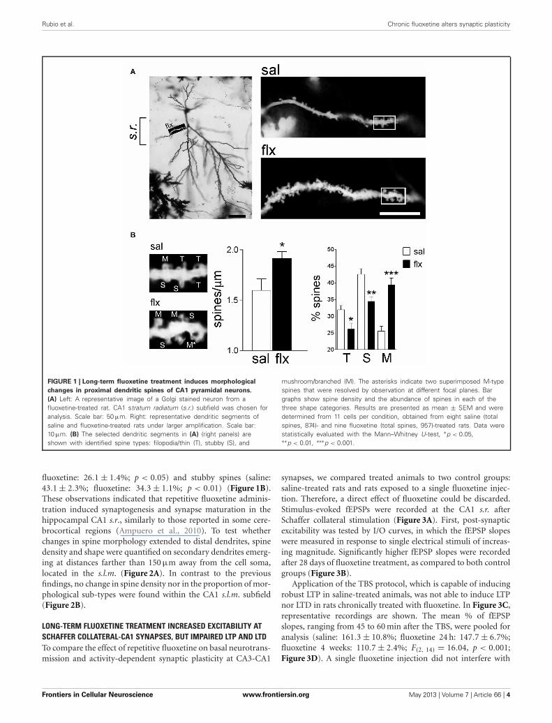
RESULTSFLUOXETINE INDUCED CHANGES IN SPINE DENSITY ANDMUSHROOM-TYPE MORPHOLOGY IN THE CA1 s.r., BUT NOTIN THE s.l.m.In order to examine the morphology of the post-synaptic com-partment in CA1, i.e., dendritic spines, the Golgi staining methodwas used. Spine morphology and spine density of secondary den-drites was analyzed at proximal segments emerging <50 μm awayfrom the soma of hippocampal CA1 neurons (Figure 1A). Spinedensity (spines/μm) increased significantly from 1.59 ± 0.11 to1.92 ± 0.07 after 4 weeks of fluoxetine treatment (p < 0.05). Thisincrease was accompanied by a higher proportion of mushroom-type spines (saline: 26.2 ± 1.7%; fluoxetine: 39.6 ± 1.8%, p <
0.001) and a concomitant decrease of thin (saline: 30.7 ± 1.4%;
Frontiers in Cellular Neuroscience www.frontiersin.org May 2013 | Volume 7 | Article 66 | 3
Rubio et al. Chronic fluoxetine alters synaptic plasticity
FIGURE 1 | Long-term fluoxetine treatment induces morphological
changes in proximal dendritic spines of CA1 pyramidal neurons.
(A) Left: A representative image of a Golgi stained neuron from afluoxetine-treated rat. CA1 stratum radiatum (s.r.) subfield was chosen foranalysis. Scale bar: 50 μm. Right: representative dendritic segments ofsaline and fluoxetine-treated rats under larger amplification. Scale bar:10 μm. (B) The selected dendritic segments in (A) (right panels) areshown with identified spine types: filopodia/thin (T), stubby (S), and
mushroom/branched (M). The asterisks indicate two superimposed M-typespines that were resolved by observation at different focal planes. Bargraphs show spine density and the abundance of spines in each of thethree shape categories. Results are presented as mean ± SEM and weredetermined from 11 cells per condition, obtained from eight saline (totalspines, 874)- and nine fluoxetine (total spines, 957)-treated rats. Data werestatistically evaluated with the Mann–Whitney U-test, ∗p < 0.05,∗∗p < 0.01, ∗∗∗p < 0.001.
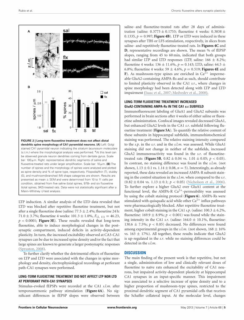
fluoxetine: 26.1 ± 1.4%; p < 0.05) and stubby spines (saline:43.1 ± 2.3%; fluoxetine: 34.3 ± 1.1%; p < 0.01) (Figure 1B).These observations indicated that repetitive fluoxetine adminis-tration induced synaptogenesis and synapse maturation in thehippocampal CA1 s.r., similarly to those reported in some cere-brocortical regions (Ampuero et al., 2010). To test whetherchanges in spine morphology extended to distal dendrites, spinedensity and shape were quantified on secondary dendrites emerg-ing at distances farther than 150 μm away from the cell soma,located in the s.l.m. (Figure 2A). In contrast to the previousfindings, no change in spine density nor in the proportion of mor-phological sub-types were found within the CA1 s.l.m. subfield(Figure 2B).
LONG-TERM FLUOXETINE TREATMENT INCREASED EXCITABILITY ATSCHAFFER COLLATERAL-CA1 SYNAPSES, BUT IMPAIRED LTP AND LTDTo compare the effect of repetitive fluoxetine on basal neurotrans-mission and activity-dependent synaptic plasticity at CA3-CA1
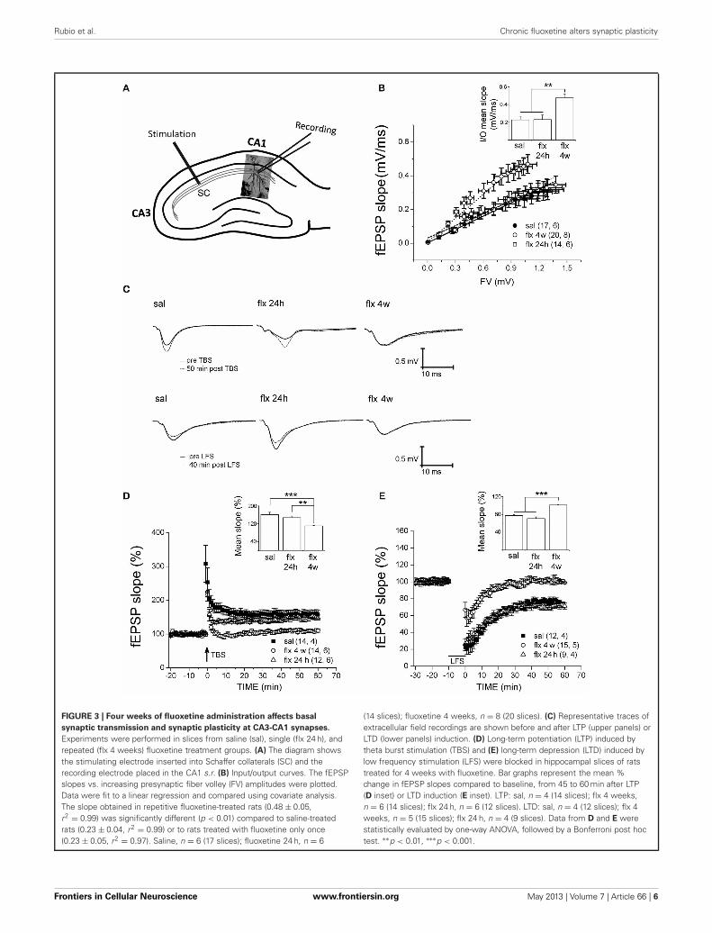
synapses, we compared treated animals to two control groups:saline-treated rats and rats exposed to a single fluoxetine injec-tion. Therefore, a direct effect of fluoxetine could be discarded.Stimulus-evoked fEPSPs were recorded at the CA1 s.r. afterSchaffer collateral stimulation (Figure 3A). First, post-synapticexcitability was tested by I/O curves, in which the fEPSP slopeswere measured in response to single electrical stimuli of increas-ing magnitude. Significantly higher fEPSP slopes were recordedafter 28 days of fluoxetine treatment, as compared to both controlgroups (Figure 3B).
Application of the TBS protocol, which is capable of inducingrobust LTP in saline-treated animals, was not able to induce LTPnor LTD in rats chronically treated with fluoxetine. In Figure 3C,representative recordings are shown. The mean % of fEPSPslopes, ranging from 45 to 60 min after the TBS, were pooled foranalysis (saline: 161.3 ± 10.8%; fluoxetine 24 h: 147.7 ± 6.7%;fluoxetine 4 weeks: 110.7 ± 2.4%; F(2, 14) = 16.04, p < 0.001;Figure 3D). A single fluoxetine injection did not interfere with
Frontiers in Cellular Neuroscience www.frontiersin.org May 2013 | Volume 7 | Article 66 | 4
Rubio et al. Chronic fluoxetine alters synaptic plasticity
FIGURE 2 | Long-term fluoxetine treatment does not affect distal
dendritic spine morphology of CA1 pyramidal neurons. (A) Left: Golgistained CA1 pyramidal neuron indicating the stratum lacunosum moleculare(s.l.m.) where the morphological analysis was performed. #At this level canbe observed granule neuron dendrites coming from dentate gyrus. Scalebar: 100 μm. Right: representative dendritic segments of saline andfluoxetine-treated rats under larger amplification. Scale bar: 10 μm. (B) Thenumber of spines and the morphology of spines were analyzed and plottedas spine density and % of spine type, respectively. Filopodia/thin (T), stubby(S), and mushroom/branched (M) shape categories are shown. Results arepresented as mean ± SEM and were determined from 10 to 11 cells percondition, obtained from five saline (total spines, 979)- and six fluoxetine(total spines, 942)-treated rats. Data were not statistically significant after aMann–Whitney U-test analysis.
LTP induction. A similar analysis of the LTD data revealed thatLTD was blocked after repetitive fluoxetine treatment, but notafter a single fluoxetine dose (saline: 77.5 ± 2.4%; fluoxetine 24 h:71.0 ± 3.7%; fluoxetine 4 weeks: 101.3 ± 1.0%, F(2, 11) = 46.23,p < 0.0001; Figure 3E). These results revealed that long-termfluoxetine, able to induce morphological changes in the post-synaptic compartment, induced deficits in activity-dependentplasticity. In turn, the increased excitability observed at CA3-CA1synapses can be due to increased spine density and/or the fact thatlarge spines are known to generate a larger postsynaptic responses(Spruston, 2008).
To further clarify whether the detrimental effects of fluoxetineon LTP and LTD were associated with the changes in spine mor-phology and density, electrophysiological recordings at perforantpath-CA1 synapses were performed.
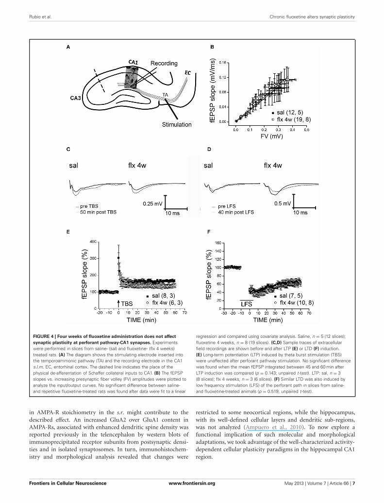
LONG-TERM FLUOXETINE TREATMENT DID NOT AFFECT LTP NOR LTDAT PERFORANT PATH-CA1 SYNAPSESStimulus-evoked fEPSPs were recorded at the CA1 s.l.m. aftertemporoammonic pathway stimulation (Figure 4A). No sig-nificant differences in fEPSP slopes were observed between
saline- and fluoxetine-treated rats after 28 days of adminis-tration (saline: 0.3773 ± 0.1753; fluoxetine 4 weeks: 0.3838 ±0.1335, p = 0.997; Figure 4B). LTP or LTD were induced in thesesynapses after TBS or LFS stimulation, respectively, in slices fromsaline- and repetitively fluoxetine-treated rats. In Figures 4C andD, representative recordings are shown. The mean % of fEPSPslopes, ranging from 45 to 60 min, indicated that both groupshad similar LTP and LTD responses (LTP, saline: 166 ± 8.2%;fluoxetine 4 weeks: 136 ± 11.6%, p = 0.143; LTD, saline: 64.5 ±6.8%; fluoxetine 4 weeks: 59 ± 4.6%, p = 0.519; Figures 4E andF). As mushroom-type spines are enriched in Ca2+ imperme-able GluA2-containing AMPA-Rs and as such, should contributeto limited plasticity observed in the CA1 s.r., where changes inspine morphology had been detected along with LTP and LTDimpairment (Isaac et al., 2007; Medvedev et al., 2008).
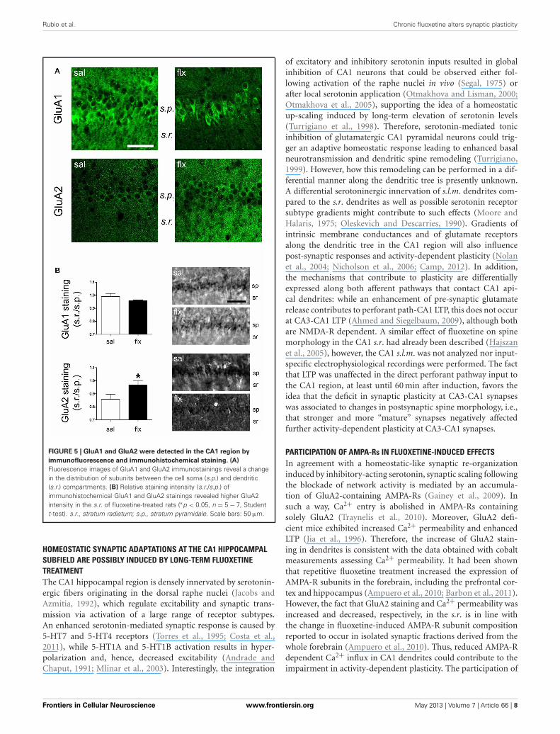
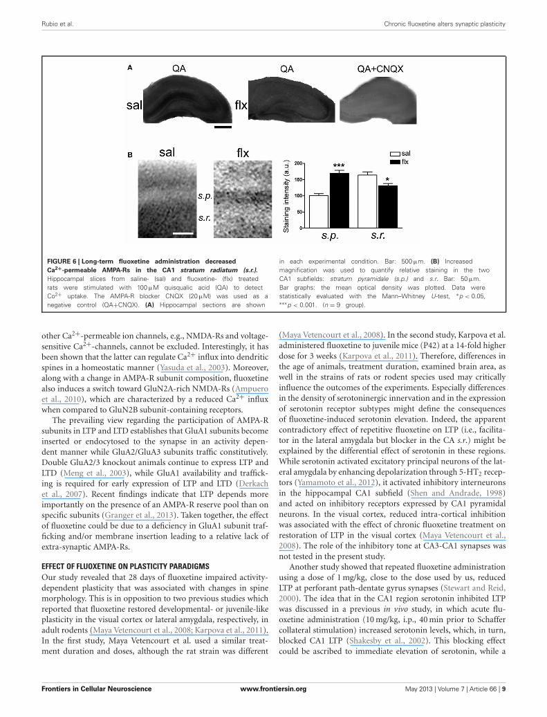
LONG-TERM FLUOXETINE TREATMENT INCREASEDGluA2-CONTAINING AMPA-Rs IN THE CA1 s.r. SUBFIELDImmunofluorescent labeling of GluA1 and GluA2 subunits wasperformed in brain sections after 4 weeks of either saline or fluox-etine administration. Confocal images revealed decreased GluA1,but enhanced GluA2 levels in the CA1 s.r. subfield following flu-oxetine treatment (Figure 5A). To quantify the relative content ofthese subunits in hippocampal subfields, immunohistochemicalstaining was performed. The relative staining intensity comparedto the s.p. in the s.r. and in the s.l.m. was assessed. While GluA1staining did not change in neither of the subfields, increasedGluA2 immunoreactivity was found in the s.r. of fluoxetine-treated rats (Figure 5B, 0.82 ± 0.04 vs. 1.01 ± 0.05; p < 0.05).In contrast, no staining difference was found in the s.l.m. (notshown, 1.13 ± 0.1 vs. 1.14 ± 0.08, n = 5). Interestingly, as alreadyreported, these data revealed an increased AMPA-R subunit stain-ing in the control situation in the s.l.m. when compared to the s.r.(0.82 ± 0.04 vs. 1.13 ± 0.1; p < 0.05) (Nicholson et al., 2006).To further explore a higher GluA2 over GluA1 content at thefunctional level, the AMPA-R Ca2+-permeability was assessedby using the cobalt staining protocol (Figure 6). AMPA-Rs werestimulated with quisqualic acid while other Ca2+ influx pathwayswere pharmacologically blocked. After repetitive fluoxetine treat-ment, higher cobalt staining in the CA1 s.p. (saline: 100.3 ± 6.2%,fluoxetine: 169.9 ± 8.9%; p < 0.001) was found while the stain-ing intensity in the CA1 s.r. (saline: 164.0 ± 10.1%, fluoxetine:130.6 ± 7.5%; p < 0.05) decreased. No differences were foundamong experimental groups in the s.l.m. (not shown, 168 ± 16%vs. 163 ± 17%). All together, these results indicate that GluA2is up-regulated in the s.r. while no staining differences could bedetected in the s.l.m.
DISCUSSIONThe main finding of the present work is that repetitive, but nota single, administration of low and clinically relevant doses offluoxetine to naïve rats enhanced the excitability of CA1 neu-rons, but impaired activity-dependent plasticity at hippocampalCA1 synapses in an input-specific manner. This impairmentwas associated to a selective increase of spine density and to ahigher proportion of mushroom-type spines, restricted to theproximal dendritic segment of CA1 pyramidal cells that receivesthe Schaffer collateral input. At the molecular level, changes
Frontiers in Cellular Neuroscience www.frontiersin.org May 2013 | Volume 7 | Article 66 | 5
Rubio et al. Chronic fluoxetine alters synaptic plasticity
FIGURE 3 | Four weeks of fluoxetine administration affects basal
synaptic transmission and synaptic plasticity at CA3-CA1 synapses.
Experiments were performed in slices from saline (sal), single (flx 24 h), andrepeated (flx 4 weeks) fluoxetine treatment groups. (A) The diagram showsthe stimulating electrode inserted into Schaffer collaterals (SC) and therecording electrode placed in the CA1 s.r. (B) Input/output curves. The fEPSPslopes vs. increasing presynaptic fiber volley (FV) amplitudes were plotted.Data were fit to a linear regression and compared using covariate analysis.The slope obtained in repetitive fluoxetine-treated rats (0.48 ± 0.05,r2 = 0.99) was significantly different (p < 0.01) compared to saline-treatedrats (0.23 ± 0.04, r2 = 0.99) or to rats treated with fluoxetine only once(0.23 ± 0.05, r2 = 0.97). Saline, n = 6 (17 slices); fluoxetine 24 h, n = 6
(14 slices); fluoxetine 4 weeks, n = 8 (20 slices). (C) Representative traces ofextracellular field recordings are shown before and after LTP (upper panels) orLTD (lower panels) induction. (D) Long-term potentiation (LTP) induced bytheta burst stimulation (TBS) and (E) long-term depression (LTD) induced bylow frequency stimulation (LFS) were blocked in hippocampal slices of ratstreated for 4 weeks with fluoxetine. Bar graphs represent the mean %change in fEPSP slopes compared to baseline, from 45 to 60 min after LTP(D inset) or LTD induction (E inset). LTP: sal, n = 4 (14 slices); flx 4 weeks,n = 6 (14 slices); flx 24 h, n = 6 (12 slices). LTD: sal, n = 4 (12 slices); flx 4weeks, n = 5 (15 slices); flx 24 h, n = 4 (9 slices). Data from D and E werestatistically evaluated by one-way ANOVA, followed by a Bonferroni post hoctest. ∗∗p < 0.01, ∗∗∗p < 0.001.
Frontiers in Cellular Neuroscience www.frontiersin.org May 2013 | Volume 7 | Article 66 | 6
Rubio et al. Chronic fluoxetine alters synaptic plasticity
FIGURE 4 | Four weeks of fluoxetine administration does not affect
synaptic plasticity at perforant pathway-CA1 synapses. Experimentswere performed in slices from saline- (sal) and fluoxetine- (flx 4 weeks)treated rats. (A) The diagram shows the stimulating electrode inserted intothe temporoammonic pathway (TA) and the recording electrode in the CA1s.l.m. EC, entorhinal cortex. The dashed line indicates the place of thephysical de-afferentation of Schaffer collateral inputs to CA1. (B) The fEPSPslopes vs. increasing presynaptic fiber volley (FV) amplitudes were plotted toanalyze the input/output curves. No significant difference between saline-and repetitive fluoxetine-treated rats was found after data were fit to a linear
regression and compared using covariate analysis. Saline, n = 5 (12 slices);fluoxetine 4 weeks, n = 8 (19 slices). (C,D) Sample traces of extracellularfield recordings are shown before and after LTP (E) or LTD (F) induction.(E) Long-term potentiation (LTP) induced by theta burst stimulation (TBS)were unaffected after perforant pathway stimulation. No significant differencewas found when the mean fEPSP integrated between 45 and 60 min afterLTP induction was compared (p = 0.143; unpaired t-test). LTP: sal, n = 3(8 slices); flx 4 weeks, n = 3 (6 slices). (F) Similar LTD was also induced bylow frequency stimulation (LFS) of the perforant path in slices from saline-and fluoxetine-treated animals (p = 0.519; unpaired t-test).
in AMPA-R stoichiometry in the s.r. might contribute to thedescribed effect. An increased GluA2 over GluA1 content inAMPA-Rs, associated with enhanced dendritic spine density wasreported previously in the telencephalon by western blots ofimmunoprecipitated receptor subunits from postsynaptic densi-ties and in isolated synaptosomes. In turn, immunohistochem-istry and morphological analysis revealed that changes were
restricted to some neocortical regions, while the hippocampus,with its well-defined cellular layers and dendritic sub-regions,was not analyzed (Ampuero et al., 2010). To now explore afunctional implication of such molecular and morphologicaladaptations, we took advantage of the well-characterized activity-dependent cellular plasticity paradigms in the hippocampal CA1region.
Frontiers in Cellular Neuroscience www.frontiersin.org May 2013 | Volume 7 | Article 66 | 7
Rubio et al. Chronic fluoxetine alters synaptic plasticity
FIGURE 5 | GluA1 and GluA2 were detected in the CA1 region by
immunofluorescence and immunohistochemical staining. (A)
Fluorescence images of GluA1 and GluA2 immunostainings reveal a changein the distribution of subunits between the cell soma (s.p.) and dendritic(s.r.) compartments. (B) Relative staining intensity (s.r./s.p.) ofimmunohistochemical GluA1 and GluA2 stainings revealed higher GluA2intensity in the s.r. of fluoxetine-treated rats (∗p < 0.05, n = 5 − 7, Studentt-test). s.r., stratum radiatum; s.p., stratum pyramidale. Scale bars: 50 μm.
HOMEOSTATIC SYNAPTIC ADAPTATIONS AT THE CA1 HIPPOCAMPALSUBFIELD ARE POSSIBLY INDUCED BY LONG-TERM FLUOXETINETREATMENTThe CA1 hippocampal region is densely innervated by serotonin-ergic fibers originating in the dorsal raphe nuclei (Jacobs andAzmitia, 1992), which regulate excitability and synaptic trans-mission via activation of a large range of receptor subtypes.An enhanced serotonin-mediated synaptic response is caused by5-HT7 and 5-HT4 receptors (Torres et al., 1995; Costa et al.,2011), while 5-HT1A and 5-HT1B activation results in hyper-polarization and, hence, decreased excitability (Andrade andChaput, 1991; Mlinar et al., 2003). Interestingly, the integration
of excitatory and inhibitory serotonin inputs resulted in globalinhibition of CA1 neurons that could be observed either fol-lowing activation of the raphe nuclei in vivo (Segal, 1975) orafter local serotonin application (Otmakhova and Lisman, 2000;Otmakhova et al., 2005), supporting the idea of a homeostaticup-scaling induced by long-term elevation of serotonin levels(Turrigiano et al., 1998). Therefore, serotonin-mediated tonicinhibition of glutamatergic CA1 pyramidal neurons could trig-ger an adaptive homeostatic response leading to enhanced basalneurotransmission and dendritic spine remodeling (Turrigiano,1999). However, how this remodeling can be performed in a dif-ferential manner along the dendritic tree is presently unknown.A differential serotoninergic innervation of s.l.m. dendrites com-pared to the s.r. dendrites as well as possible serotonin receptorsubtype gradients might contribute to such effects (Moore andHalaris, 1975; Oleskevich and Descarries, 1990). Gradients ofintrinsic membrane conductances and of glutamate receptorsalong the dendritic tree in the CA1 region will also influencepost-synaptic responses and activity-dependent plasticity (Nolanet al., 2004; Nicholson et al., 2006; Camp, 2012). In addition,the mechanisms that contribute to plasticity are differentiallyexpressed along both afferent pathways that contact CA1 api-cal dendrites: while an enhancement of pre-synaptic glutamaterelease contributes to perforant path-CA1 LTP, this does not occurat CA3-CA1 LTP (Ahmed and Siegelbaum, 2009), although bothare NMDA-R dependent. A similar effect of fluoxetine on spinemorphology in the CA1 s.r. had already been described (Hajszanet al., 2005), however, the CA1 s.l.m. was not analyzed nor input-specific electrophysiological recordings were performed. The factthat LTP was unaffected in the direct perforant pathway input tothe CA1 region, at least until 60 min after induction, favors theidea that the deficit in synaptic plasticity at CA3-CA1 synapseswas associated to changes in postsynaptic spine morphology, i.e.,that stronger and more “mature” synapses negatively affectedfurther activity-dependent plasticity at CA3-CA1 synapses.
PARTICIPATION OF AMPA-Rs IN FLUOXETINE-INDUCED EFFECTSIn agreement with a homeostatic-like synaptic re-organizationinduced by inhibitory-acting serotonin, synaptic scaling followingthe blockade of network activity is mediated by an accumula-tion of GluA2-containing AMPA-Rs (Gainey et al., 2009). Insuch a way, Ca2+ entry is abolished in AMPA-Rs containingsolely GluA2 (Traynelis et al., 2010). Moreover, GluA2 defi-cient mice exhibited increased Ca2+ permeability and enhancedLTP (Jia et al., 1996). Therefore, the increase of GluA2 stain-ing in dendrites is consistent with the data obtained with cobaltmeasurements assessing Ca2+ permeability. It had been shownthat repetitive fluoxetine treatment increased the expression ofAMPA-R subunits in the forebrain, including the prefrontal cor-tex and hippocampus (Ampuero et al., 2010; Barbon et al., 2011).However, the fact that GluA2 staining and Ca2+ permeability wasincreased and decreased, respectively, in the s.r. is in line withthe change in fluoxetine-induced AMPA-R subunit compositionreported to occur in isolated synaptic fractions derived from thewhole forebrain (Ampuero et al., 2010). Thus, reduced AMPA-Rdependent Ca2+ influx in CA1 dendrites could contribute to theimpairment in activity-dependent plasticity. The participation of
Frontiers in Cellular Neuroscience www.frontiersin.org May 2013 | Volume 7 | Article 66 | 8
Rubio et al. Chronic fluoxetine alters synaptic plasticity
FIGURE 6 | Long-term fluoxetine administration decreased
Ca2+-permeable AMPA-Rs in the CA1 stratum radiatum (s.r.).
Hippocampal slices from saline- (sal) and fluoxetine- (flx) treatedrats were stimulated with 100 μM quisqualic acid (QA) to detectCo2+ uptake. The AMPA-R blocker CNQX (20 μM) was used as anegative control (QA+CNQX). (A) Hippocampal sections are shown
in each experimental condition. Bar: 500 μm. (B) Increasedmagnification was used to quantify relative staining in the twoCA1 subfields: stratum pyramidale (s.p.) and s.r. Bar: 50 μm.Bar graphs: the mean optical density was plotted. Data werestatistically evaluated with the Mann–Whitney U-test, ∗p < 0.05,∗∗∗p < 0.001. (n = 9 group).
other Ca2+-permeable ion channels, e.g., NMDA-Rs and voltage-sensitive Ca2+-channels, cannot be excluded. Interestingly, it hasbeen shown that the latter can regulate Ca2+ influx into dendriticspines in a homeostatic manner (Yasuda et al., 2003). Moreover,along with a change in AMPA-R subunit composition, fluoxetinealso induces a switch toward GluN2A-rich NMDA-Rs (Ampueroet al., 2010), which are characterized by a reduced Ca2+ influxwhen compared to GluN2B subunit-containing receptors.
The prevailing view regarding the participation of AMPA-Rsubunits in LTP and LTD establishes that GluA1 subunits becomeinserted or endocytosed to the synapse in an activity depen-dent manner while GluA2/GluA3 subunits traffic constitutively.Double GluA2/3 knockout animals continue to express LTP andLTD (Meng et al., 2003), while GluA1 availability and traffick-ing is required for early expression of LTP and LTD (Derkachet al., 2007). Recent findings indicate that LTP depends moreimportantly on the presence of an AMPA-R reserve pool than onspecific subunits (Granger et al., 2013). Taken together, the effectof fluoxetine could be due to a deficiency in GluA1 subunit traf-ficking and/or membrane insertion leading to a relative lack ofextra-synaptic AMPA-Rs.
EFFECT OF FLUOXETINE ON PLASTICITY PARADIGMSOur study revealed that 28 days of fluoxetine impaired activity-dependent plasticity that was associated with changes in spinemorphology. This is in opposition to two previous studies whichreported that fluoxetine restored developmental- or juvenile-likeplasticity in the visual cortex or lateral amygdala, respectively, inadult rodents (Maya Vetencourt et al., 2008; Karpova et al., 2011).In the first study, Maya Vetencourt et al. used a similar treat-ment duration and doses, although the rat strain was different
(Maya Vetencourt et al., 2008). In the second study, Karpova et al.administered fluoxetine to juvenile mice (P42) at a 14-fold higherdose for 3 weeks (Karpova et al., 2011). Therefore, differences inthe age of animals, treatment duration, examined brain area, aswell in the strains of rats or rodent species used may criticallyinfluence the outcomes of the experiments. Especially differencesin the density of serotoninergic innervation and in the expressionof serotonin receptor subtypes might define the consequencesof fluoxetine-induced serotonin elevation. Indeed, the apparentcontradictory effect of repetitive fluoxetine on LTP (i.e., facilita-tor in the lateral amygdala but blocker in the CA s.r.) might beexplained by the differential effect of serotonin in these regions.While serotonin activated excitatory principal neurons of the lat-eral amygdala by enhancing depolarization through 5-HT2 recep-tors (Yamamoto et al., 2012), it activated inhibitory interneuronsin the hippocampal CA1 subfield (Shen and Andrade, 1998)and acted on inhibitory receptors expressed by CA1 pyramidalneurons. In the visual cortex, reduced intra-cortical inhibitionwas associated with the effect of chronic fluoxetine treatment onrestoration of LTP in the visual cortex (Maya Vetencourt et al.,2008). The role of the inhibitory tone at CA3-CA1 synapses wasnot tested in the present study.
Another study showed that repeated fluoxetine administrationusing a dose of 1 mg/kg, close to the dose used by us, reducedLTP at perforant path-dentate gyrus synapses (Stewart and Reid,2000). The idea that in the CA1 region serotonin inhibited LTPwas discussed in a previous in vivo study, in which acute flu-oxetine administration (10 mg/kg, i.p., 40 min prior to Schaffercollateral stimulation) increased serotonin levels, which, in turn,blocked CA1 LTP (Shakesby et al., 2002). This blocking effectcould be ascribed to immediate elevation of serotonin, while a
Frontiers in Cellular Neuroscience www.frontiersin.org May 2013 | Volume 7 | Article 66 | 9
Rubio et al. Chronic fluoxetine alters synaptic plasticity
single fluoxetine administration, even at higher doses, should notlead to maintained high serotonin levels 24 h after the injection(Caccia et al., 1990). This suggested that the effect we observedafter 4 weeks of administration, but measured 24 after the lastdose, was a consequence of adaptive plastic changes that requiredrepeated fluoxetine administration, and not to acute changes inserotonin. Consistent with our data, it was shown that repeatedadministration of methamphetamine, a drug that acts on centralmonoamine neurotransmission, including the serotonin system,enhanced basal neurotransmission and had a detrimental effecton CA3-CA1 synaptic plasticity (Swant et al., 2010).
The increased fEPSPs revealed in I/O curves in fluoxetine-treated animals could be due to direct effects of fluoxetine onglutamate receptors. However, fluoxetine has inhibitory effects onAMPA and NMDA receptors (Szasz et al., 2007; Kim et al., 2013)and therefore, decreased fEPSPs should be expected. In conse-quence, the most straightforward explanation is that larger spinesare able to elicit larger post-synaptic responses. This could bemediated by several plasticity-related signals that are expressedfollowing fluoxetine treatment, such as BDNF (Castren, 2005;Bath et al., 2012) or IGF-1 (Corvin et al., 2012), which in turnare able to stimulate glutamate receptor expression and recep-tor phosphorylation (Slack et al., 2004; First et al., 2011). Achanged inhibition due to serotonin-induced re-arrangements ofinterneurons can also contribute to this phenomenon (Mendezet al., 2012).
The concept of meta-plasticity posits that synapses that havepreviously been potentiated are more likely to express LTD andless likely to express LTP, whereas the opposite applies if LTD hasbeen previously induced (Bear, 2003). It is therefore difficult toreconcile that both plasticity paradigms are impaired under the
used experimental conditions. If LTP is occluded due to a synap-tic up-scaling at CA3-CA1 synapses, LTD should be enhanced, butnot decreased, at these synapses. We therefore propose that Ca2+-influx pathways and/or Ca2+ changes in dendritic spines causedby intracellular release or changes in buffering capacity, might bealtered thereby affecting both plasticity paradigms. Further exper-iments, using different stimulation protocols, increasing extra-cellular Ca2+ concentration or blocking inhibitory interneuronsneed to be performed to get insight into underlying mechanisms.However, it is clear that maintaining the stimulation conditionsof the control group in fluoxetine-treated animals, both LTP andLTD are impaired. Similarly, a specific stimulation protocol andlearning procedures can lead to occlusion of both LTP and LTD(Liang et al., 2002; Delvendahl et al., 2010), as we found influoxetine-treated animals.
In agreement with our findings, chronic escitalopram treat-ment inhibited CA3-CA1 LTP in healthy rats in a stress-resistantstrain (Flinders Resistant Line rats, Karolinska Institutet). Thisphenomenon was attenuated after exposure to stress (Ryan et al.,2009), thereby confirming a distinct result in naïve subjects.Although the effect of fluoxetine described by us may be restrictedto non-stressed or non-depressed subjects, we report that repeti-tive treatment impacts significantly glutamate neurotransmissionand plasticity in the hippocampus.
ACKNOWLEDGMENTSFunding for this study was provided by grants to Ursula Wynekenfrom Conicyt (Anillo ACT09_2006) and by Universidad de losAndes (MED-001-09) to Francisco J. Rubio. The authors thankfor the critical review by Dr. Bruce Hope (NIDA, NIH). We alsothank Ximena Orellana for her technical assistance.
REFERENCESAhmed, M. S., and Siegelbaum, S. A.
(2009). Recruitment of N-TypeCa(2+) channels during LTPenhances low release efficacy ofhippocampal CA1 perforant pathsynapses. Neuron 63, 372–385.
Ampuero, E., Dagnino-Subiabre, A.,Sandoval, R., Zepeda-Carreno, R.,Sandoval, S., Viedma, A., et al.(2007). Status epilepticus inducesregion-specific changes in dendriticspines, dendritic length and TrkBprotein content of rat brain cortex.Brain Res. 1150, 225–238.
Ampuero, E., Rubio, F. J., Falcon,R., Sandoval, M., Diaz-Veliz, G.,Gonzalez, R. E., et al. (2010).Chronic fluoxetine treatmentinduces structural plasticity andselective changes in glutamatereceptor subunits in the rat cerebralcortex. Neuroscience 169, 98–108.
Ampuero, E., Stehberg, J., Gonzalez, D.,Besser, N., Ferrero, M., Diaz-Veliz,G., et al. (2013). Repetitive fluox-etine treatment affects long-termmemories but not learning. Behav.Brain Res. 247, 92–100.
Andrade, R., and Chaput, Y. (1991).5-Hydroxytryptamine4-like recep-tors mediate the slow excitatoryresponse to serotonin in the rat hip-pocampus. J. Pharmacol. Exp. Ther.257, 930–937.
Barbon, A., Caracciolo, L., Orlandi,C., Musazzi, L., Mallei, A., LaVia, L., et al. (2011). Chronicantidepressant treatments inducea time-dependent up-regulation ofAMPA receptor subunit protein lev-els. Neurochem. Int. 59, 896–905.
Bath, K. G., Jing, D. Q., Dincheva,I., Neeb, C. C., Pattwell, S. S.,Chao, M. V., et al. (2012). BDNFVal66Met impairs fluoxetine-induced enhancement ofadult hippocampus plasticity.Neuropsychopharmacology 37,1297–1304.
Bear, M. F. (2003). Bidirectional synap-tic plasticity: from theory to reality.Philos. Trans. R. Soc. Lond. B Biol.Sci. 358, 649–655.
Blier, P., Keller, M. B., Pollack, M. H.,Thase, M. E., Zajecka, J. M., andDunner, D. L. (2007). Preventingrecurrent depression: long-term
treatment for major depressivedisorder. J. Clin. Psychiatry 68, e06.
Caccia, S., Cappi, M., Fracasso, C., andGarattini, S. (1990). Influence ofdose and route of administrationon the kinetics of fluoxetine andits metabolite norfluoxetine in therat. Psychopharmacology (Berl.) 100,509–514.
Camp, A. J. (2012). Intrinsic neuronalexcitability: a role in homeostasisand disease. Front. Neurol. 3:50. doi:10.3389/fneur.2012.00050
Castren, E. (2005). Is mood chemistry?Nat. Rev. Neurosci. 6, 241–246.
Citri, A., and Malenka, R. C. (2008).Synaptic plasticity: multiple forms,functions, and mechanisms.Neuropsychopharmacology 33,18–41.
Corvin, A. P., Molinos, I., Little, G.,Donohoe, G., Gill, M., Morris,D. W., et al. (2012). Insulin-likegrowth factor 1 (IGF1) and itsactive peptide (1-3)IGF1 enhancethe expression of synaptic markersin neuronal circuits through differ-ent cellular mechanisms. Neurosci.Lett. 520, 51–56.
Costa, L., Trovato, C., Musumeci, S.A., Catania, M. V., and Ciranna,L. (2011). 5-HT(1A) and 5-HT(7)receptors differently modulateAMPA receptor-mediated hip-pocampal synaptic transmission.Hippocampus 22, 790–801.
Delvendahl, I., Jung, N. H., Mainberger,F., Kuhnke, N. G., Cronjaeger, M.,and Mall, V. (2010). Occlusion ofbidirectional plasticity by preced-ing low-frequency stimulation inthe human motor cortex. Clin.Neurophysiol. 121, 594–602.
Derkach, V. A., Oh, M. C., Guire,E. S., and Soderling, T. R. (2007).Regulatory mechanisms of AMPAreceptors in synaptic plasticity. Nat.Rev. Neurosci. 8, 101–113.
First, M., Gil-Ad, I., Taler, M.,Tarasenko, I., Novak, N., andWeizman, A. (2011). The effectsof fluoxetine treatment in achronic mild stress rat model ondepression-related behavior, brainneurotrophins and ERK expression.J. Mol. Neurosci. 45, 246–255.
Gainey, M. A., Hurvitz-Wolff, J. R.,Lambo, M. E., and Turrigiano, G. G.
Frontiers in Cellular Neuroscience www.frontiersin.org May 2013 | Volume 7 | Article 66 | 10
Rubio et al. Chronic fluoxetine alters synaptic plasticity
(2009). Synaptic scaling requires theGluR2 subunit of the AMPA recep-tor. J. Neurosci. 29, 6479–6489.
Granger, A. J., Shi, Y., Lu, W., Cerpas,M., and Nicoll, R. A. (2013). LTPrequires a reserve pool of glutamatereceptors independent of subunittype. Nature 493, 495–500.
Hajszan, T., Maclusky, N. J., andLeranth, C. (2005). Short-termtreatment with the antidepressantfluoxetine triggers pyramidal den-dritic spine synapse formation inrat hippocampus. Eur. J. Neurosci.21, 1299–1303.
Harris, K. M., Jensen, F. E., andTsao, B. (1992). Three-dimensionalstructure of dendritic spines andsynapses in rat hippocampus (CA1)at postnatal day 15 and adult ages:implications for the maturationof synaptic physiology and long-term potentiation. J. Neurosci. 12,2685–2705.
He, K., Lee, A., Song, L., Kanold, P. O.,and Lee, H. K. (2011). AMPA recep-tor subunit GluR1 (GluA1) serine-845 site is involved in synapticdepression but not in spine shrink-age associated with chemical long-term depression. J. Neurophysiol.105, 1897–1907.
Isaac, J. T., Ashby, M. C., and McBain,C. J. (2007). The role of the GluR2subunit in AMPA receptor functionand synaptic plasticity. Neuron 54,859–871.
Jacobs, B. L., and Azmitia, E. C. (1992).Structure and function of the brainserotonin system. Physiol. Rev. 72,165–229.
Jia, Z., Agopyan, N., Miu, P., Xiong,Z., Henderson, J., Gerlai, R., et al.(1996). Enhanced LTP in mice defi-cient in the AMPA receptor GluR2.Neuron 17, 945–956.
Karpova, N. N., Pickenhagen, A.,Lindholm, J., Tiraboschi, E.,Kulesskaya, N., Agustsdottir, A.,et al. (2011). Fear erasure in micerequires synergy between antide-pressant drugs and extinctiontraining. Science 334, 1731–1734.
Kim, H. J., Kim, T. H., Choi, S. J.,Hong, Y. J., Yang, J. S., Sung, K. W.,et al. (2013). Fluoxetine suppressessynaptically induced [Ca(2)(+)]ispikes and excitotoxicity in culturedrat hippocampal neurons. BrainRes. 1490, 23–34.
Liang, P. I., Yang, H. W., Lin, Y. W., Yen,C. D., and Min, M. Y. (2002). Theeffect of prior prolonged low fre-quency stimulation on the furthersynaptic plasticity at hippocampalCA1 synapses. Chin. J. Physiol. 45,63–67.
Maya Vetencourt, J. F., Sale, A., Viegi,A., Baroncelli, L., De Pasquale, R.,
O’Leary, O. F., et al. (2008). Theantidepressant fluoxetine restoresplasticity in the adult visual cortex.Science 320, 385–388.
Maya-Vetencourt, J. F., Tiraboschi,E., Greco, D., Restani, L., Cerri,C., Auvinen, P., et al. (2012).Experience-dependent expressionof NPAS4 regulates plasticity inadult visual cortex. J. Physiol. 590,4777–4787.
Maya Vetencourt, J. F., Tiraboschi,E., Spolidoro, M., Castren, E., andMaffei, L. (2011). Serotonin triggersa transient epigenetic mechanismthat reinstates adult visual cortexplasticity in rats. Eur. J. Neurosci. 33,49–57.
Medvedev, N. I., Rodríguez-Arellano,J. J., Popov, V. I., Davies, H. A.,Tigaret, C. M., Schoepfer, R., et al.(2008). The glutamate receptor 2subunit controls post-synaptic den-sity complexity and spine shape inthe dentate gyrus. Eur. J. Neurosci.27, 315–325.
Megias, M., Emri, Z., Freund, T. F., andGulyas, A. I. (2001). Total numberand distribution of inhibitory andexcitatory synapses on hippocampalCA1 pyramidal cells. Neuroscience102, 527–540.
Mendez, P., Pazienti, A., Szabo, G.,and Bacci, A. (2012). Directalteration of a specific inhibitorycircuit of the hippocampus byantidepressants. J. Neurosci. 32,16616–16628.
Meng, Y., Zhang, Y., and Jia, Z. (2003).Synaptic transmission and plasticityin the absence of AMPA glutamatereceptor GluR2 and GluR3. Neuron39, 163–176.
Mlinar, B., Falsini, C., and Corradetti,R. (2003). Pharmacologicalcharacterization of 5-HT(1B)receptor-mediated inhibition oflocal excitatory synaptic trans-mission in the CA1 region of rathippocampus. Br. J. Pharmacol. 138,71–80.
Moore, R. Y., and Halaris, A. E. (1975).Hippocampal innervation by sero-tonin neurons of the midbrainraphe in the rat. J. Comp. Neurol.164, 171–183.
Nicholson, D. A., Trana, R., Katz, Y.,Kath, W. L., Spruston, N., andGeinisman, Y. (2006). Distance-dependent differences in synapsenumber and AMPA receptor expres-sion in hippocampal CA1 pyramidalneurons. Neuron 50, 431–442.
Nolan, M. F., Malleret, G., Dudman, J.T., Buhl, D. L., Santoro, B., Gibbs,E., et al. (2004). A behavioralrole for dendritic integration:HCN1 channels constrain spa-tial memory and plasticity at
inputs to distal dendrites of CA1pyramidal neurons. Cell 119,719–732.
Oleskevich, S., and Descarries, L.(1990). Quantified distribution ofthe serotonin innervation in adultrat hippocampus. Neuroscience 34,19–33.
Olmos, C., Sandoval, R., Rozas, C.,Navarro, S., Wyneken, U., Zeise, M.,et al. (2009). Effect of short-termexposure to dichlorvos on synap-tic plasticity of rat hippocampalslices: involvement of acylpeptidehydrolase and alpha(7) nicotinicreceptors. Toxicol. Appl. Pharmacol.238, 37–46.
Osswald, I. K., Galan, A., and Bowie,D. (2007). Light triggers expressionof philanthotoxin-insensitive Ca2+-permeable AMPA receptors in thedeveloping rat retina. J. Physiol. 582,95–111.
Otmakhova, N. A., Lewey, J., Asrican,B., and Lisman, J. E. (2005).Inhibition of perforant path inputto the CA1 region by serotonin andnoradrenaline. J. Neurophysiol. 94,1413–1422.
Otmakhova, N. A., and Lisman, J. E.(2000). Dopamine, serotonin, andnoradrenaline strongly inhibit thedirect perforant path-CA1 synapticinput, but have little effect on theSchaffer collateral input. Ann. N.Y.Acad. Sci. 911, 462–464.
Pittenger, C., and Duman, R. S.(2008). Stress, depression,and neuroplasticity: a con-vergence of mechanisms.Neuropsychopharmacology 33,88–109.
Pyapali, G. K., Sik, A., Penttonen,M., Buzsaki, G., and Turner, D.A. (1998). Dendritic properties ofhippocampal CA1 pyramidal neu-rons in the rat: intracellular stainingin vivo and in vitro. J. Comp. Neurol.391, 335–352.
Remondes, M., and Schuman, E. M.(2002). Direct cortical input mod-ulates plasticity and spiking in CA1pyramidal neurons. Nature 416,736–740.
Ryan, B., Musazzi, L., Mallei, A.,Tardito, D., Gruber, S. H.,El Khoury, A., et al. (2009).Remodelling by early-life stress ofNMDA receptor-dependent synap-tic plasticity in a gene-environmentrat model of depression. Int.J. Neuropsychopharmacol. 12,553–559.
Sanacora, G., Zarate, C. A., Krystal,J. H., and Manji, H. K. (2008).Targeting the glutamatergic systemto develop novel, improved ther-apeutics for mood disorders. Nat.Rev. Drug Discov. 7, 426–437.
Schatzberg, A. F. (2000). New indi-cations for antidepressants. J. Clin.Psychiatry 61(Suppl. 11), 9–17.
Segal, M. (1975). Physiological andpharmacological evidence for aserotonergic projection to thehippocampus. Brain Res. 94,115–131.
Segal, M. (2005). Dendritic spinesand long-term plasticity. Nat. Rev.Neurosci. 6, 277–284.
Shakesby, A. C., Anwyl, R., and Rowan,M. J. (2002). Overcoming the effectsof stress on synaptic plasticity inthe intact hippocampus: rapidactions of serotonergic and antide-pressant agents. J. Neurosci. 22,3638–3644.
Shen, R. Y., and Andrade, R. (1998).5-Hydroxytryptamine2 receptorfacilitates GABAergic neurotrans-mission in rat hippocampus.J. Pharmacol. Exp. Ther. 285,805–812.
Slack, S. E., Pezet, S., McMahon, S. B.,Thompson, S. W., and Malcangio,M. (2004). Brain-derived neu-rotrophic factor induces NMDAreceptor subunit one phosphory-lation via ERK and PKC in the ratspinal cord. Eur. J. Neurosci. 20,1769–1778.
Spruston, N. (2008). Pyramidal neu-rons: dendritic structure and synap-tic integration. Nat. Rev. Neurosci. 9,206–221.
Steward, O. (1976). Topographic orga-nization of the projections from theentorhinal area to the hippocam-pal formation of the rat. J. Comp.Neurol. 167, 285–314.
Stewart, C. A., and Reid, I. C. (2000).Repeated ECS and fluoxetineadministration have equivalenteffects on hippocampal synapticplasticity. Psychopharmacology(Berl.) 148, 217–223.
Swant, J., Chirwa, S., Stanwood,G., and Khoshbouei, H. (2010).Methamphetamine reduces LTP andincreases baseline synaptic trans-mission in the CA1 region of mousehippocampus. PLoS ONE 5:e11382.doi: 10.1371/journal.pone.0011382
Szasz, B. K., Mike, A., Karoly, R.,Gerevich, Z., Illes, P., Vizi, E.S., et al. (2007). Direct inhibitoryeffect of fluoxetine on N-methyl-D-aspartate receptors in the centralnervous system. Biol. Psychiatry 62,1303–1309.
Torres, G. E., Chaput, Y., and Andrade,R. (1995). Cyclic AMP andprotein kinase A mediate 5-hydroxytryptamine type 4 receptorregulation of calcium-activatedpotassium current in adult hip-pocampal neurons. Mol. Pharmacol.47, 191–197.
Frontiers in Cellular Neuroscience www.frontiersin.org May 2013 | Volume 7 | Article 66 | 11
Rubio et al. Chronic fluoxetine alters synaptic plasticity
Traynelis, S. F., Wollmuth, L. P.,McBain, C. J., Menniti, F. S., Vance,K. M., Ogden, K. K., et al. (2010).Glutamate receptor ion channels:structure, regulation, and function.Pharmacol. Rev. 62, 405–496.
Turrigiano, G. G. (1999). Homeostaticplasticity in neuronal networks: themore things change, the more theystay the same. Trends Neurosci. 22,221–227.
Turrigiano, G. G., Leslie, K. R., Desai,N. S., Rutherford, L. C., and Nelson,S. B. (1998). Activity-dependentscaling of quantal amplitude inneocortical neurons. Nature 391,892–896.
Wang, Y., Neumann, M., Hansen,K., Hong, S. M., Kim, S., Noble-Haeusslein, L. J., et al. (2011).Fluoxetine increases hippocampalneurogenesis and induces epigeneticfactors but does not improvefunctional recovery after traumaticbrain injury. J. Neurotrauma 28,259–268.
Yamamoto, R., Ueta, Y., Sugai, T.,and Kato, N. (2012). A serotoner-gic discrimination favoring synapticinputs that accompany robust spikefiring in lateral amygdala neurons.Neuroscience 220, 119–130.
Yasuda, R., Sabatini, B. L., andSvoboda, K. (2003). Plasticity
of calcium channels in den-dritic spines. Nat. Neurosci. 6,948–955.
Conflict of Interest Statement: Theauthors declare that the researchwas conducted in the absence of anycommercial or financial relationshipsthat could be construed as a potentialconflict of interest.
Received: 07 February 2013; accepted: 20April 2013; published online: 09 May2013.Citation: Rubio FJ, Ampuero E, SandovalR, Toledo J, Pancetti F and Wyneken U
(2013) Long-term fluoxetine treatmentinduces input-specific LTP and LTDimpairment and structural plasticity inthe CA1 hippocampal subfield. Front.Cell. Neurosci. 7:66. doi: 10.3389/fncel.2013.00066Copyright © 2013 Rubio, Ampuero,Sandoval, Toledo, Pancetti andWyneken. This is an open-accessarticle distributed under the terms of theCreative Commons Attribution License,which permits use, distribution andreproduction in other forums, providedthe original authors and source arecredited and subject to any copyrightnotices concerning any third-partygraphics etc.
Frontiers in Cellular Neuroscience www.frontiersin.org May 2013 | Volume 7 | Article 66 | 12
Related Documents


