REPORT Long-range signaling by phosphoprotein waves arising from bistability in protein kinase cascades Nick I Markevich 1 , Mikhail A Tsyganov 1 , Jan B Hoek and Boris N Kholodenko* Department of Pathology, Anatomy and Cell Biology, Thomas Jefferson University, Locust St, Philadelphia, PA, USA * Corresponding author. Department of Pathology, Anatomy and Cell Biology, Thomas Jefferson University, 1020 Locust St, Philadelphia, PA 19107, USA. Tel.: þ 1 215 503 1614; Fax: þ 1 215 923 2218; E-mail: [email protected] 1 These authors contributed equally to this work Received 10.7.06; accepted 2.10.06 A hallmark of protein kinase/phosphatase cascades, including mitogen-activated protein kinase (MAPK) pathways, is the spatial separation of their components within cells. The top-level kinase, MAP3K, is phosphorylated at the cell membrane, and cytoplasmic kinases at sequential downstream levels (MAP2K and MAPK) spread the signal to distant targets. Given measured protein diffusivity and phosphatase activities, signal propagation by diffusion would result in a steep decline of MAP2K activity and low bisphosphorylated MAPK (ppMAPK) levels near the nucleus, especially in large cells, such as oocytes. Here, we show that bistability in a two-site MAPK (de)phosphorylation cycle generates a novel type of phosphoprotein wave that propagates from the surface deep into the cell interior. Positive feedback from ppMAPK to cytoplasmic MAP2K enhances the propagation span of the ppMAPK wave, making it possible to convey phosphorylation signals over exceedingly long distances. The finding of phosphorylation waves traveling with constant amplitude and high velocity may solve a long-standing enigma of survival signaling in developing neurons. Molecular Systems Biology 14 November 2006; doi:10.1038/msb4100108 Subject Categories: simulation and data analysis; signal transduction Keywords: bistable switches; long-range signaling; MAPK cascades; phosphorylation waves; retrograde signaling in developing neurons Introduction Cascades of coupled phosphorylation/dephosphorylation cycles, such as mitogen-activated protein kinase (MAPK) pathways, integrate external stimuli and propagate signals from the plasma membrane (PM) to the nucleus. A typical, three-tier cascade comprises MAPK, MAPK kinase (MAP2K) and MAP2K kinase (MAP3K). MAP2K is activated by MAP3K at the cell membrane and then drifts into the cell interior, where it phosphorylates MAPK on two conserved threonine and tyrosine residues. Various phosphatases reverse these phosphorylations. The biological outcome of stimulation depends on the temporal profile of active, bisphosphorylated MAPK (ppMAPK) in the perinuclear area (Murphy et al, 2002). Owing to dephosphorylation during diffusion through the cytoplasm, the phosphorylation level of downstream kinases may become low near the nucleus. In fact, for a protein phosphorylated by a membrane-bound kinase and dephos- phorylated by a cytosolic phosphatase, Brown and Kholoden- ko (1999) showed that there can be a precipitous spatial gradient of the phosphorylated form, high close to the membrane and low within the cell. The existence of a phosphoprotein gradient of this kind has been confirmed theoretically (Lipkow et al, 2005) and experimentally (Kalab et al, 2002; Niethammer et al, 2004). Additional tiers in a kinase cascade can make shallower the phosphoprotein gradients (Kholodenko, 2006); yet signaling across a distance of more than about 5–10 mm requires more than diffusion. This includes (i) trafficking of phosphorylated kinases within endocytic vesicles or non-vesicular signaling complexes by molecular motors (Kholodenko, 2002; Howe and Mobley, 2004; Miaczynska et al, 2004; Perlson et al, 2005) and (ii) phosphoprotein waves propagating from the membrane over long distances, especially in large cells, such as Xenopus oocytes (Kholodenko, 2003; Slepchenko and Terasaki, 2003). Phosphoprotein waves may also be a key to the long-standing enigma of retrograde signaling in developmental neurons, where tyrosine phosphorylation in neuron bodies starts as early as a few minutes after nerve growth factor (NGF) stimulation of distal axons located centimeters away (MacIn- nis et al, 2003). Such a rapid propagation of survival signals over centimeters of axon length cannot be accounted for by the retrograde transport of endosomes or translocation of phos- phorylated kinases by molecular motors moving with rates of a few mm/s (Hill et al, 2004). An alternative mechanism for the initial, rapid transfer of phosphorylation signals might be & 2006 EMBO and Nature Publishing Group Molecular Systems Biology 2006 1 Molecular Systems Biology (2006) doi:10.1038/msb4100108 & 2006 EMBO and Nature Publishing Group All rights reserved 1744-4292/06 www.molecularsystemsbiology.com Article number: 61

Welcome message from author
This document is posted to help you gain knowledge. Please leave a comment to let me know what you think about it! Share it to your friends and learn new things together.
Transcript

REPORT
Long-range signaling by phosphoprotein wavesarising from bistability in protein kinase cascades
Nick I Markevich1, Mikhail A Tsyganov1, Jan B Hoek and Boris N Kholodenko*
Department of Pathology, Anatomy and Cell Biology, Thomas Jefferson University, Locust St, Philadelphia, PA, USA* Corresponding author. Department of Pathology, Anatomy and Cell Biology, Thomas Jefferson University, 1020 Locust St, Philadelphia, PA 19107, USA.Tel.: þ 1 215 503 1614; Fax: þ 1 215 923 2218; E-mail: [email protected] These authors contributed equally to this work
Received 10.7.06; accepted 2.10.06
A hallmark of protein kinase/phosphatase cascades, including mitogen-activated protein kinase(MAPK) pathways, is the spatial separation of their components within cells. The top-level kinase,MAP3K, is phosphorylated at the cell membrane, and cytoplasmic kinases at sequentialdownstream levels (MAP2K and MAPK) spread the signal to distant targets. Given measuredprotein diffusivity and phosphatase activities, signal propagation by diffusion would result in asteep decline of MAP2K activity and low bisphosphorylated MAPK (ppMAPK) levels near thenucleus, especially in large cells, such as oocytes. Here, we show that bistability in a two-site MAPK(de)phosphorylation cycle generates a novel type of phosphoprotein wave that propagates from thesurface deep into the cell interior. Positive feedback from ppMAPK to cytoplasmic MAP2K enhancesthe propagation span of the ppMAPK wave, making it possible to convey phosphorylation signalsover exceedingly long distances. The finding of phosphorylation waves traveling with constantamplitude and high velocity may solve a long-standing enigma of survival signaling in developingneurons.Molecular Systems Biology 14 November 2006; doi:10.1038/msb4100108Subject Categories: simulation and data analysis; signal transductionKeywords: bistable switches; long-range signaling; MAPK cascades; phosphorylation waves; retrogradesignaling in developing neurons
Introduction
Cascades of coupled phosphorylation/dephosphorylationcycles, such as mitogen-activated protein kinase (MAPK)pathways, integrate external stimuli and propagate signalsfrom the plasma membrane (PM) to the nucleus. A typical,three-tier cascade comprises MAPK, MAPK kinase (MAP2K)and MAP2K kinase (MAP3K). MAP2K is activated by MAP3Kat the cell membrane and then drifts into the cell interior,where it phosphorylates MAPK on two conserved threonineand tyrosine residues. Various phosphatases reverse thesephosphorylations. The biological outcome of stimulationdepends on the temporal profile of active, bisphosphorylatedMAPK (ppMAPK) in the perinuclear area (Murphy et al, 2002).
Owing to dephosphorylation during diffusion through thecytoplasm, the phosphorylation level of downstream kinasesmay become low near the nucleus. In fact, for a proteinphosphorylated by a membrane-bound kinase and dephos-phorylated by a cytosolic phosphatase, Brown and Kholoden-ko (1999) showed that there can be a precipitous spatialgradient of the phosphorylated form, high close to themembrane and low within the cell. The existence of aphosphoprotein gradient of this kind has been confirmed
theoretically (Lipkow et al, 2005) and experimentally (Kalabet al, 2002; Niethammer et al, 2004). Additional tiers in akinase cascade can make shallower the phosphoproteingradients (Kholodenko, 2006); yet signaling across a distanceof more than about 5–10 mm requires more than diffusion. Thisincludes (i) trafficking of phosphorylated kinases withinendocytic vesicles or non-vesicular signaling complexes bymolecular motors (Kholodenko, 2002; Howe and Mobley,2004; Miaczynska et al, 2004; Perlson et al, 2005) and (ii)phosphoprotein waves propagating from the membrane overlong distances, especially in large cells, such as Xenopusoocytes (Kholodenko, 2003; Slepchenko and Terasaki, 2003).Phosphoprotein waves may also be a key to the long-standingenigma of retrograde signaling in developmental neurons,where tyrosine phosphorylation in neuron bodies starts asearly as a few minutes after nerve growth factor (NGF)stimulation of distal axons located centimeters away (MacIn-nis et al, 2003). Such a rapid propagation of survival signalsover centimeters of axon length cannot be accounted for by theretrograde transport of endosomes or translocation of phos-phorylated kinases by molecular motors moving with rates ofa few mm/s (Hill et al, 2004). An alternative mechanism forthe initial, rapid transfer of phosphorylation signals might be
& 2006 EMBO and Nature Publishing Group Molecular Systems Biology 2006 1
Molecular Systems Biology (2006) doi:10.1038/msb4100108& 2006 EMBO and Nature Publishing Group All rights reserved 1744-4292/06www.molecularsystemsbiology.comArticle number: 61

phosphoprotein waves that travel from the distal axon to theneuron body (Kholodenko, 2003).
A phosphoprotein wave amplifies the phosphorylation levelat the wave front following the arrival of active molecules bydiffusion. This local amplification can arise from a positivefeedback enabling reaction cascades to display switch-like,bistable behavior. In fact, traveling waves in bistable systemsare known in physics, chemistry and biology (Zhabotinskyand Zaikin, 1973; Keener and Sneyd, 1998; Reynolds et al,2003). For MAPK cascades, bistability is thought to arisefrom activation of kinases at the top of the cascade (MAP3K)by downstream kinase (MAPK) (Bhalla et al, 2002; Xiongand Ferrell, 2003). However, these long positive feedbackloops will increase the phosphorylation levels close to thePM where activated MAP3K is localized, but not atdistant cytoplasmic areas. To bring about phosphoproteinwaves, kinases in the cytoplasm should exhibit bistability,as the intrinsically bistable MAPK cycle does (Markevichet al, 2004). Importantly, a theoretical prediction thata two-site MAPK phosphorylation circuit can generatebistability was recently confirmed experimentally for theextracellular signal-regulated kinase (ERK) cycle in BHK cells(Harding et al, 2005) and for the Fus3/MAPK cycle in yeast(Wang et al, 2006).
In the present paper, we demonstrate a novel type ofphosphoprotein wave that emerges from the bistable behaviorof kinase and phosphatase circuits in the cytoplasm and relaysextracellular signals from the PM to distant targets. We unveilhow the transfer of phosphorylation signals depends on theinput–output relationships of sequential cascade levels. Thefindings show that bistability intrinsic to the MAPK activationcycle can give rise to a wave of active MAPK that propagates abinary phosphorylation signal. Positive feedback from MAPKto cytoplasmic MAP2K enables this wave to propagate withhigh velocity over exceedingly long distances. These travelingwaves of protein phosphorylation present a novel mechanismof long-range signaling within cells, when phosphorylationsignals cannot be transferred by diffusion. We demonstratehow waves of phosphorylated kinases may be involved inretrograde signaling in neurons.
Results and discussion
Computational analysis of the spatio-temporaldynamics
Our MAPK cascade models explicitly incorporate heteroge-neous spatial distribution of activated kinases. For illustrative
Table I Equations and boundary conditions describing the dynamics of MAP cascade kinases
Equations Boundary conditions
1 d[pMAP3 K]/dt¼v1m–v2
m
2q½pMAP2K�
qt¼ D
L2
� �� 1
x2
qqx
x2 q½pMAP2K�qx
� �þ vc
5 � vc6
q½pMAP2K�qx
����x¼1
¼ L2
3Dðvm
3 � vm4 þ vm
5 � vm6 Þ
q½pMAP2K�qx
����x¼Q=L
¼ 0
3q½ppMAP2K�
qt¼ D
L2
� �1
x2
qqx
x2 q½ppMAP2K�qx
� �� vc
5
q½ppMAP2K�qx
����x¼1
¼ L2
3Dðvm
4 � vm5 Þ
q½ppMAP2K�qx
����x¼Q=L
¼ 0
4q½pMAPK�
qt¼ D
L2
� �1
x2
qqx
x2 q½pMAPK�qx
� �þ vc
7 � vc8 þ vc
9 � vc10
q½pMAPK�qx
����x¼1
¼ 0
q½pMAPK�qx
����x¼Q=L
¼ 0
5q½ppMAPK�
qt¼ D
L2
� �1
x2
qqx
x2 q½ppMAPK�qx
� �þ vc
8 � vc9
q½ppMAPK�qx
����x¼1
¼ 0
q½ppMAPK�qx
����x¼Q=L
¼ 0
L is the cell radius, Q is the nuclear radius, D is the diffusivity of proteins in the cytosol (for illustrative purposes, assumed to be equal to 2 mm2/s for both thephosphorylated and unphosphorylated forms) and x is a dimensionless coordinate, equal to the distance from the cell center divided by the cell radius, L. The distance dfrom the cell membrane is expressed in terms of x as, d¼(1�x)L. The rate expressions are given in Tables II and III.
Long-range signaling by phosphoprotein wavesNI Markevich et al
2 Molecular Systems Biology 2006 & 2006 EMBO and Nature Publishing Group
purposes, we initially consider a spherical cell of radiusL¼15 mm with a nucleus of radius Q¼6 mm (SupplementaryFigure S1). Much larger distances up to centimeters and one-dimensional geometry are considered for long-range signalingin developing neurons. The spatio-temporal dynamics of theMAPK cascade is described by partial differential equations(known as reaction–diffusion equations; Table I).
Cell size (L) and protein diffusivity (D) influence the spatio-temporal dynamics as a single parameter combination L2/D,known as the ‘diffusion-time’ (Table I). The protein diffusivityin the cytoplasm was reported to be in a range of 1–10 mm2/s(reviewed by Brown and Kholodenko, 1999). For illustrativepurposes, we assume D¼2 mm2/s under ‘crowded’ cytoplas-mic condition (Khan et al, 2004). The assumption of higherlimit of D-values of 10mm2/s will not change our findings,if traveling distances (L) increase approximately two-fold.Importantly, variations in the reported diffusivity values do
not change our conclusion of inability of diffusion to propagatephosphorylation signals over distances of tens of micrometersand our analysis of long-range signaling in neurons.
Stimulus–response relationships and the spatialpropagation of the signals
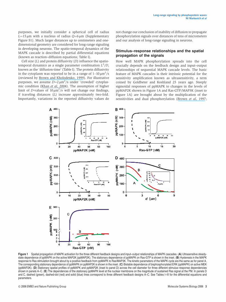
How well MAPK phosphorylation spreads into the cellcrucially depends on the feedback design and input–outputrelationships of sequential MAPK cascade levels. The basicfeature of MAPK cascades is their intrinsic potential for thesensitivity amplification known as ultrasensitivity, a termcoined by Goldbeter and Koshland 25 years ago. Steeplysigmoidal responses of ppMAPK to changes in the levels ofppMAP2K shown in Figure 1A and Ras-GTP/MAP3K (inset toFigure 1A) are brought about by the multiplication of thesensitivities and dual phosphorylation (Brown et al, 1997;
Figure 1 Spatial propagation of MAPK activation for the three different feedback designs and input–output relationships of MAPK cascades. (A) Ultrasensitive steady-state dependence of ppMAPK on the active MAP2K (ppMAP2K). The stationary dependence of ppMAPK on Ras-GTP is shown in the inset. (B) Hysteresis in the MAPKresponse to Ras stimulation brought about by a positive feedback from ppMAPK to Ras/MAP3K. The kinetic parameters of the MAPK cycle are the same as for panel A.The corresponding stationary dependence of ppMAPK on ppMAP2K is shown in the inset. (C) Bistable dependence of bisphosphorylated ERK (ppMAPK) on active MEK(ppMAP2K). (D) Stationary spatial profiles of ppMAPK and ppMAP2K (inset to panel D) across the cell diameter for three different stimulus–response dependenciesshown in panels A–C. (E) The dependences of the stationary ppMAPK level at the nuclear membrane on the magnitude of sustained Ras signal at the PM. In panels Dand C, dashed (green), dashed-dot (red) and solid (blue) lines correspond to three different feedback designs A–C. See Tables I–III for the differential equations andparameters.
Long-range signaling by phosphoprotein wavesNI Markevich et al
& 2006 EMBO and Nature Publishing Group Molecular Systems Biology 2006 3
Ferrell, 1997). A long positive feedback further increases thesteepness of the input–output relationship, making it similarto a step-function and allowing MAPK cascades to operate asbistable switches (Bhalla et al, 2002). For an MAPK cascadeimbedded in a long positive feedback, Figure 1B illustratesbistable responses of ppMAPK to external stimuli (Ras-GTP)and ultrasensitive responses to ppMAP2K (inset to Figure 1B).Importantly, the two-site phosphorylation and dephosphor-ylation MAPK cycle by itself can already exhibit bistability(Figure 1C). In this cycle, the same ppMAP2K level can switchMAPK into either an active or inactive state, depending onwhether the initial MAPK concentration was higher or lowerthan the value corresponding to an unstable state (dashedline).
When MAP2K is phosphorylated at the cell surface anddephosphorylated while drifting in the cytoplasm, spatialgradients of ppMAP2K are generated both before and at steadystate. A strong decline in ppMAP2K with the distance from themembrane causes the ppMAPK fraction to decrease, reaching anadir in the perinuclear area. For the three stimulus–responserelationships shown in Figure 1A–C (one ultrasensitive andtwo mechanistically distinct, bistable responses of MAPK),Figure 1D compares steady-state spatial profiles of ppMAPK(inset to Figure 1D shows ppMAP2K profiles) activated bya sustained elevation of Ras-GTP/MAP3K levels (snapshotsof ppMAPK and ppMAP2K spatial profiles at different timesfollowing the onset of activation are shown in SupplementaryFigure S2). When the response is ultrasensitive (line A) or ifbistability arises from a long feedback (line B), MAPK activityin the perinuclear area appears to be surprisingly low. Onlywhen hysteresis occurs in the cytoplasm (line C) are ppMAPKconcentrations near the PM and the nucleus similar, despite aprecipitous decline in ppMAP2K. In fact, although the MAPK-induced feedback stimulation of cell-surface MAP3K increasesMAP2K activity close to the membrane, this feedback is unableto substantially increase ppMAPK concentration deep in thecytoplasm (inset to Figure 1D). As a result, for all three distinctkinetic scenarios of signaling from MAP3K to MAPK, the levelof ppMAP2K in the perinuclear area is roughly the same.Bistability within the MAPK cycle allows for self-perpetuationof MAPK activation all the way down to the nucleus, whereMAP2K activity is low.
We may ask whether a significant increase in sustained Ras/MAP3K activity enhances MAPK signaling in the perinucleararea. Figure 1E demonstrates that although for all threefeasible mechanisms of MAPK responses shown in Figure 1A–C,the stationary ppMAPK level near the nucleus increases withthe magnitude of the sustained Ras-GTP activity, the maximalppMAPK signal appears to be spectacularly higher when itspropagation is supported by hysteresis in the MAPK cycle (lineC). Positive feedback from MAPK to membrane-confinedMAP3K may slightly decrease the amount of Ras-GTP neededfor half-maximal MAPK activation (line B), but has practicallyno effect on the maximal achievable perinuclear level ofppMAPK, which is similar to that obtained for a merelysigmoidal response (line A). We conclude that MAPKbistability in the cytoplasm aids in the relay of phosphoryla-tion cues.
Activation of cell-surface receptors is usually transient andresults in a transient rather than sustained activation of Ras/
MAP3K at the cell membrane. The analysis of a transientstimulation of the MAPK cascade confirms the results shownfor a sustained activation: the peak ppMAPK level near thenucleus is similar to the level near the cell surface only whenthe relay of the phosphorylated signal is enhanced bybistability in the MAPK cycle in the cytoplasm (SupplementaryFigure S3). Supplementary Figure S4 further confirms thisconclusion and illustrates how MAPK activation spreads in thespace and eventually vanishes following a transient stimula-tion of Ras at the PM. Importantly, calculations also suggestthat even a marked decrease in the phosphorylated MAPKfraction seen in the perinuclear area may not be readilydetectable when ppMAPK is averaged over the whole cell(Supplementary Figure S3B), as it occurs for usual Westernblot protocols.
Regulation of phosphatase activities plays acrucial role in signal propagation
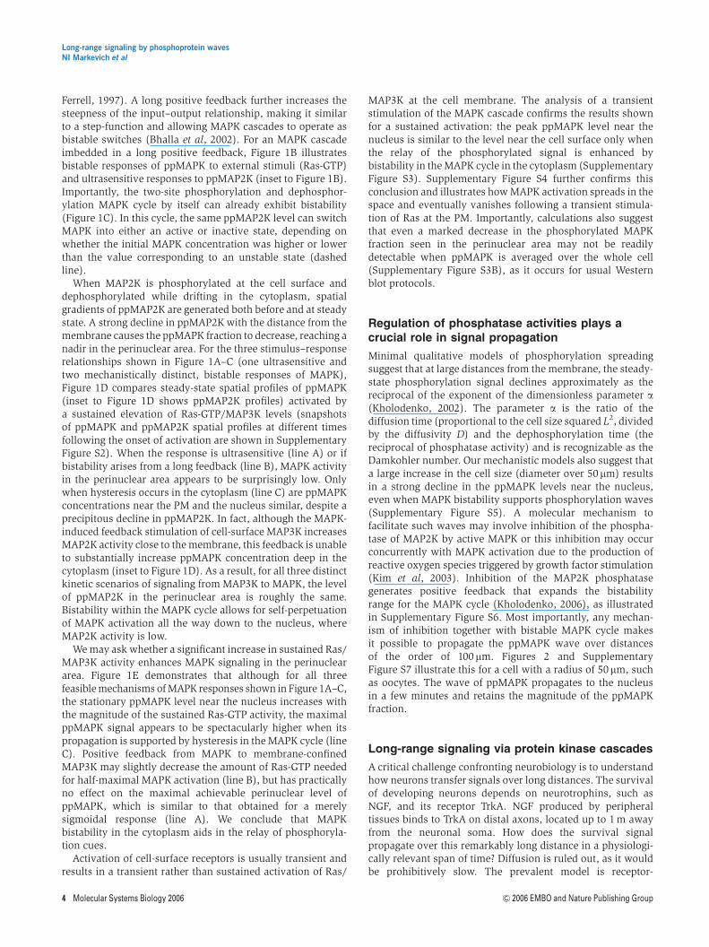
Minimal qualitative models of phosphorylation spreadingsuggest that at large distances from the membrane, the steady-state phosphorylation signal declines approximately as thereciprocal of the exponent of the dimensionless parameter a(Kholodenko, 2002). The parameter a is the ratio of thediffusion time (proportional to the cell size squared L2, dividedby the diffusivity D) and the dephosphorylation time (thereciprocal of phosphatase activity) and is recognizable as theDamkohler number. Our mechanistic models also suggest thata large increase in the cell size (diameter over 50mm) resultsin a strong decline in the ppMAPK levels near the nucleus,even when MAPK bistability supports phosphorylation waves(Supplementary Figure S5). A molecular mechanism tofacilitate such waves may involve inhibition of the phospha-tase of MAP2K by active MAPK or this inhibition may occurconcurrently with MAPK activation due to the production ofreactive oxygen species triggered by growth factor stimulation(Kim et al, 2003). Inhibition of the MAP2K phosphatasegenerates positive feedback that expands the bistabilityrange for the MAPK cycle (Kholodenko, 2006), as illustratedin Supplementary Figure S6. Most importantly, any mechan-ism of inhibition together with bistable MAPK cycle makesit possible to propagate the ppMAPK wave over distancesof the order of 100 mm. Figures 2 and SupplementaryFigure S7 illustrate this for a cell with a radius of 50mm, suchas oocytes. The wave of ppMAPK propagates to the nucleusin a few minutes and retains the magnitude of the ppMAPKfraction.
Long-range signaling via protein kinase cascades
A critical challenge confronting neurobiology is to understandhow neurons transfer signals over long distances. The survivalof developing neurons depends on neurotrophins, such asNGF, and its receptor TrkA. NGF produced by peripheraltissues binds to TrkA on distal axons, located up to 1 m awayfrom the neuronal soma. How does the survival signalpropagate over this remarkably long distance in a physiologi-cally relevant span of time? Diffusion is ruled out, as it wouldbe prohibitively slow. The prevalent model is receptor-
Long-range signaling by phosphoprotein wavesNI Markevich et al
4 Molecular Systems Biology 2006 & 2006 EMBO and Nature Publishing Group
mediated endocytosis, followed by the retrograde transport ofsignaling endosomes containing the NGF–pTrkA complexes tothe cell body (Ginty and Segal, 2002). However, data indicatethat although the endosomes may carry survival signals, theremust be additional mechanisms independent of the NGFtransport, as tyrosine phosphorylation is observed in cell
bodies at least 30 min before the first detection of radio-iodinated NGFarriving from the locally stimulated distal axons(Senger and Campenot, 1997). Lateral propagation of TrkAphosphorylation along the axon membrane is excluded, asnearly complete suppression of TrkA signaling in the cellbodies/proximal axons did not affect neuronal survival,
Figure 2 Phosphorylation waves emerging from the feedback inhibition of MAP2K phosphatases and bistability in the MAPK cycle. (A) Radial propagation profiles ofthe ppMAPK wave from the cell membrane (solid blue lines). Profiles of ppMAP2K are shown by dashed red lines. (B) Concentration profile of the ppMAPK wave for atwo-dimensional cross-section taken through the center of a cell. Cell radius L¼50 mm, nuclear radius Q¼20 mm. The rate equations are given in Tables II and III andSupplementary Table I.
Long-range signaling by phosphoprotein wavesNI Markevich et al
& 2006 EMBO and Nature Publishing Group Molecular Systems Biology 2006 5
whereas TrkA inhibition at distal axons induced apoptosis(MacInnis et al, 2003).
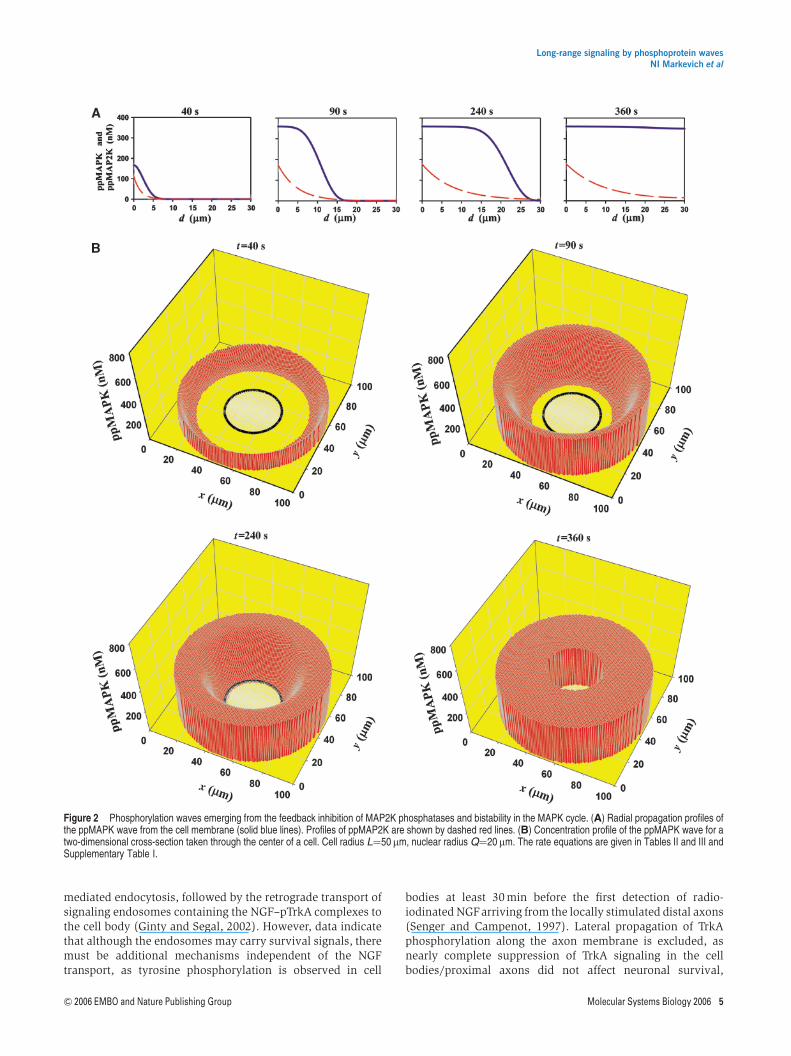
Our analysis suggests a novel mechanism where signalstraveling along the axon are not accompanied by NGF and donot require activated TrkA in the cell body. These fast survivalsignals can be transmitted by a wave of protein phosphoryla-tion in a kinase cascade, such as MAPK or PI3K/Akt cascades.If a downstream kinase stimulates the phosphorylation of anupstream kinase in the cytoplasm (directly or via a regulatorycircuit), a resulting bistable switch generates a travelingcytoplasmic wave that propagates over increasingly longdistances with constant amplitude and velocity (Figure 3).After reaching the target, such as the nucleus, this wavegradually turns off owing to prompt dephosphorylation ofkinases in the nucleus. Thus, protein kinase/phosphatasecascades possessing positive feedback circuits in the cyto-plasm can generate biological switches in space and time thatare capable of propagating phosphorylation signals overexceedingly long distances.
It was proposed that bistability would make proteinphosphorylation cascades capable of ‘all-or-none’ switchingand allow cells to ‘remember’ a transient differentiationstimulus long after the stimulus was removed (Xiong andFerrell, 2003). Here, we show yet another role of bistableprotein kinase cascades: to support waves of phosphoproteinsthat propagate signals to remote cellular locations. In contrastwith traveling waves previously described (Zhabotinsky andZaikin, 1973; Slepchenko and Terasaki, 2003), phosphoproteinwaves arising from bistability in MAPK cycle feature a spatiallyinhomogeneous activation threshold, because the ppMAP2Kconcentration decreases as the distance from the membrane
increases (inset to Figure 1D). Interestingly, the fact thatsignals spread from the cell periphery to the interior where thesurface of the wave front decreases facilitates the signalpropagation. Supplementary Figures S8 and S9 illustrate thisspace-condensing effect; at the same values of kineticparameters, a one-dimensional wave vanishes at distanceswhere the wave that travels inside the cell still delivers thesignal.
The analysis above suggests experiments that can testwhether the rapid propagation of phosphorylation signalsarises from bistability in kinase cascades. Although counter-intuitive at first glance, a marked decrease in the activity ofERK phosphatases (and/or in the ERK abundance) leads to abifurcation where the bistable ERK response becomes merelyultrasensitive (Markevich et al, 2004), and the input–outputrelationship in Figure 1C changes to the one shown inFigure 1A. Thus, the propagation of a phosphorylation wavethat delivers the survival or proliferation signal may beabolished by inhibition of the phosphatase of the kinase inthe bistable cascade cycle.
Signaling via phosphoprotein waves has key advantagesover solely diffusive signal propagation. Owing to phosphataseactivity, the phosphorylation state of a protein that diffuses inthe cytoplasm will decrease much strongly than the amplitudeof a phosphoprotein wave that essentially propagates a binarysignal. Positive feedback emerging from cytoplasmic interac-tions enables protein kinase/phosphatase cascades to transmitphosphorylation signals over much longer distances than100 mm. The velocity of the wave significantly increases withthe strength of this feedback, readily reaching hundreds of mm/s(Supplementary Figure S10). The finding that phosphorylation
Figure 3 Traveling wave of active MAPK carrying a binary phosphorylation signal over exceedingly long distances. The ppMAPK wave propagation (one-dimensionalgeometry) is supported by the feedback activation of MAP2K. This wave maintains the signal magnitude and velocity and propagates over 1 cm with a velocity of 25mm/s.Profiles of ppMAPK and ppMAP2K are shown by solid (blue) line and dashed (red) lines, respectively. The wave fronts at 20 s are shown at a higher scale resolution. Seethe rate equations in Tables I–III and Supplementary Table III.
Long-range signaling by phosphoprotein wavesNI Markevich et al
6 Molecular Systems Biology 2006 & 2006 EMBO and Nature Publishing Group
signals can propagate as traveling waves with a constantamplitude and high velocity over distances exceeding hun-dreds, or even thousands of millimeters, may solve a long-standing enigma of survival signaling in neuronal biology.When short positive regulatory circuits are in place, thesewaves provide a robust mechanism of signal transfer along theaxon. Waves of protein phosphorylation may be a widespreadmechanism of intracellular signal transfer.
Materials and methods
Description of the spatio-temporal dynamicsby reaction–diffusion equations
At the cell membrane, the dynamics of the MAP3K and pMAP3Kconcentrations is described by the ordinary differential equation (seethe first row in Table I); the total concentration, [MAP3K]tot, of theseforms remains constant. Whereas the concentration of MAP3K and
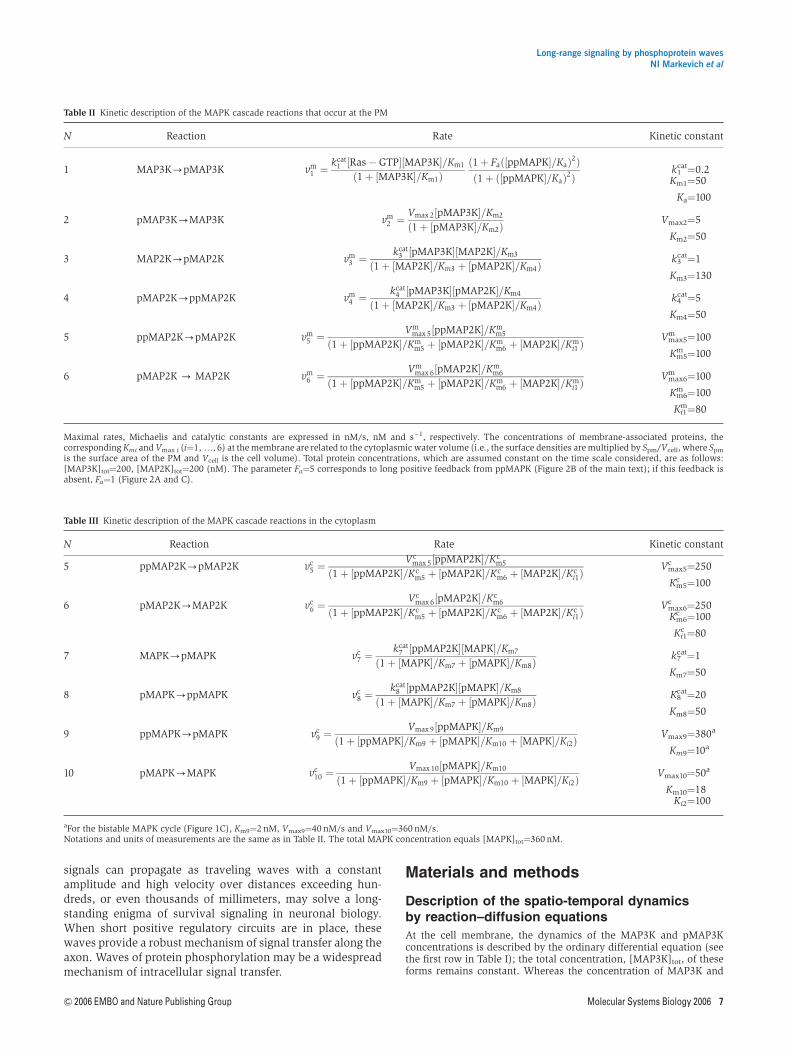
Table II Kinetic description of the MAPK cascade reactions that occur at the PM
N Reaction Rate Kinetic constant
1 MAP3K-pMAP3K vm1 ¼ kcat
1 ½Ras � GTP�½MAP3K�=Km1
ð1 þ ½MAP3K�=Km1Þð1 þ Fað½ppMAPK�=KaÞ2Þð1 þ ð½ppMAPK�=KaÞ2Þ
k1cat¼0.2
Km1¼50
Ka¼100
2 pMAP3K-MAP3K vm2 ¼ Vmax 2½pMAP3K�=Km2
ð1 þ ½pMAP3K�=Km2ÞVmax2¼5
Km2¼50
3 MAP2K-pMAP2K vm3 ¼ kcat
3 ½pMAP3K�½MAP2K�=Km3
ð1 þ ½MAP2K�=Km3 þ ½pMAP2K�=Km4Þk3
cat¼1
Km3¼130
4 pMAP2K-ppMAP2K vm4 ¼ kcat
4 ½pMAP3K�½pMAP2K�=Km4
ð1 þ ½MAP2K�=Km3 þ ½pMAP2K�=Km4Þk4
cat¼5
Km4¼50
5 ppMAP2K-pMAP2K vm5 ¼ Vm
max 5½ppMAP2K�=Kmm5
ð1 þ ½ppMAP2K�=Kmm5 þ ½pMAP2K�=Km
m6 þ ½MAP2K�=Kmi1 Þ
Vmax5m ¼100
Km5m ¼100
6 pMAP2K - MAP2K vm6 ¼ Vm
max 6½pMAP2K�=Kmm6
ð1 þ ½ppMAP2K�=Kmm5 þ ½pMAP2K�=Km
m6 þ ½MAP2K�=Kmi1 Þ
Vmax6m ¼100
Km6m ¼100
Ki1m¼80
Maximal rates, Michaelis and catalytic constants are expressed in nM/s, nM and s�1, respectively. The concentrations of membrane-associated proteins, thecorresponding Kmi and Vmax i (i¼1,y, 6) at the membrane are related to the cytoplasmic water volume (i.e., the surface densities are multiplied by Spm/Vcell, where Spm
is the surface area of the PM and Vcell is the cell volume). Total protein concentrations, which are assumed constant on the time scale considered, are as follows:[MAP3K]tot¼200, [MAP2K]tot¼200 (nM). The parameter Fa¼5 corresponds to long positive feedback from ppMAPK (Figure 2B of the main text); if this feedback isabsent, Fa¼1 (Figure 2A and C).
Table III Kinetic description of the MAPK cascade reactions in the cytoplasm
N Reaction Rate Kinetic constant
5 ppMAP2K-pMAP2K vc5 ¼ Vc
max 5½ppMAP2K�=Kcm5
ð1 þ ½ppMAP2K�=Kcm5 þ ½pMAP2K�=Kc
m6 þ ½MAP2K�=Kci1Þ
Vmax5c ¼250
Km5c ¼100
6 pMAP2K-MAP2K vc6 ¼ Vc
max 6½pMAP2K�=Kcm6
ð1 þ ½ppMAP2K�=Kcm5 þ ½pMAP2K�=Kc
m6 þ ½MAP2K�=Kci1Þ
Vmax6c ¼250Km6
c ¼100
Ki1c ¼80
7 MAPK-pMAPK vc7 ¼ kcat
7 ½ppMAP2K�½MAPK�=Km7
ð1 þ ½MAPK�=Km7 þ ½pMAPK�=Km8Þk7
cat¼1
Km7¼50
8 pMAPK-ppMAPK vc8 ¼ kcat
8 ½ppMAP2K�½pMAPK�=Km8
ð1 þ ½MAPK�=Km7 þ ½pMAPK�=Km8ÞK8
cat¼20
Km8¼50
9 ppMAPK-pMAPK vc9 ¼ Vmax 9½ppMAPK�=Km9
ð1 þ ½ppMAPK�=Km9 þ ½pMAPK�=Km10 þ ½MAPK�=Ki2ÞVmax9¼380a
Km9¼10a
10 pMAPK-MAPK vc10 ¼ Vmax 10½pMAPK�=Km10
ð1 þ ½ppMAPK�=Km9 þ ½pMAPK�=Km10 þ ½MAPK�=Ki2ÞVmax10¼50a
Km10¼18Ki2¼100
aFor the bistable MAPK cycle (Figure 1C), Km9¼2 nM, Vmax9¼40 nM/s and Vmax10¼360 nM/s.Notations and units of measurements are the same as in Table II. The total MAPK concentration equals [MAPK]tot¼360 nM.
Long-range signaling by phosphoprotein wavesNI Markevich et al
& 2006 EMBO and Nature Publishing Group Molecular Systems Biology 2006 7
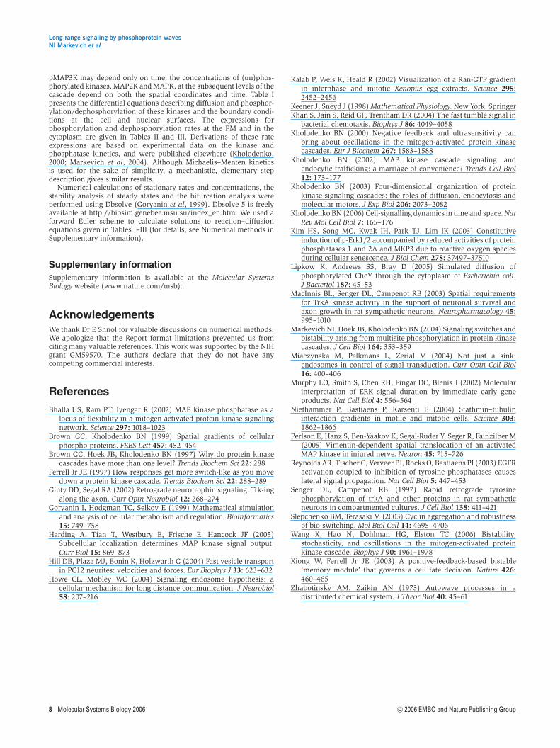
pMAP3K may depend only on time, the concentrations of (un)phos-phorylated kinases, MAP2K and MAPK, at the subsequent levels of thecascade depend on both the spatial coordinates and time. Table Ipresents the differential equations describing diffusion and phosphor-ylation/dephosphorylation of these kinases and the boundary condi-tions at the cell and nuclear surfaces. The expressions forphosphorylation and dephosphorylation rates at the PM and in thecytoplasm are given in Tables II and III. Derivations of these rateexpressions are based on experimental data on the kinase andphosphatase kinetics, and were published elsewhere (Kholodenko,2000; Markevich et al, 2004). Although Michaelis–Menten kineticsis used for the sake of simplicity, a mechanistic, elementary stepdescription gives similar results.
Numerical calculations of stationary rates and concentrations, thestability analysis of steady states and the bifurcation analysis wereperformed using Dbsolve (Goryanin et al, 1999). Dbsolve 5 is freelyavailable at http://biosim.genebee.msu.su/index_en.htm. We used aforward Euler scheme to calculate solutions to reaction–diffusionequations given in Tables I–III (for details, see Numerical methods inSupplementary information).
Supplementary information
Supplementary information is available at the Molecular SystemsBiology website (www.nature.com/msb).
AcknowledgementsWe thank Dr E Shnol for valuable discussions on numerical methods.We apologize that the Report format limitations prevented us fromciting many valuable references. This work was supported by the NIHgrant GM59570. The authors declare that they do not have anycompeting commercial interests.
References
Bhalla US, Ram PT, Iyengar R (2002) MAP kinase phosphatase as alocus of flexibility in a mitogen-activated protein kinase signalingnetwork. Science 297: 1018–1023
Brown GC, Kholodenko BN (1999) Spatial gradients of cellularphospho-proteins. FEBS Lett 457: 452–454
Brown GC, Hoek JB, Kholodenko BN (1997) Why do protein kinasecascades have more than one level? Trends Biochem Sci 22: 288
Ferrell Jr JE (1997) How responses get more switch-like as you movedown a protein kinase cascade. Trends Biochem Sci 22: 288–289
Ginty DD, Segal RA (2002) Retrograde neurotrophin signaling: Trk-ingalong the axon. Curr Opin Neurobiol 12: 268–274
Goryanin I, Hodgman TC, Selkov E (1999) Mathematical simulationand analysis of cellular metabolism and regulation. Bioinformatics15: 749–758
Harding A, Tian T, Westbury E, Frische E, Hancock JF (2005)Subcellular localization determines MAP kinase signal output.Curr Biol 15: 869–873
Hill DB, Plaza MJ, Bonin K, Holzwarth G (2004) Fast vesicle transportin PC12 neurites: velocities and forces. Eur Biophys J 33: 623–632
Howe CL, Mobley WC (2004) Signaling endosome hypothesis: acellular mechanism for long distance communication. J Neurobiol58: 207–216
Kalab P, Weis K, Heald R (2002) Visualization of a Ran-GTP gradientin interphase and mitotic Xenopus egg extracts. Science 295:2452–2456
Keener J, Sneyd J (1998) Mathematical Physiology. New York: SpringerKhan S, Jain S, Reid GP, Trentham DR (2004) The fast tumble signal in
bacterial chemotaxis. Biophys J 86: 4049–4058Kholodenko BN (2000) Negative feedback and ultrasensitivity can
bring about oscillations in the mitogen-activated protein kinasecascades. Eur J Biochem 267: 1583–1588
Kholodenko BN (2002) MAP kinase cascade signaling andendocytic trafficking: a marriage of convenience? Trends Cell Biol12: 173–177
Kholodenko BN (2003) Four-dimensional organization of proteinkinase signaling cascades: the roles of diffusion, endocytosis andmolecular motors. J Exp Biol 206: 2073–2082
Kholodenko BN (2006) Cell-signalling dynamics in time and space. NatRev Mol Cell Biol 7: 165–176
Kim HS, Song MC, Kwak IH, Park TJ, Lim IK (2003) Constitutiveinduction of p-Erk1/2 accompanied by reduced activities of proteinphosphatases 1 and 2A and MKP3 due to reactive oxygen speciesduring cellular senescence. J Biol Chem 278: 37497–37510
Lipkow K, Andrews SS, Bray D (2005) Simulated diffusion ofphosphorylated CheY through the cytoplasm of Escherichia coli.J Bacteriol 187: 45–53
MacInnis BL, Senger DL, Campenot RB (2003) Spatial requirementsfor TrkA kinase activity in the support of neuronal survival andaxon growth in rat sympathetic neurons. Neuropharmacology 45:995–1010
Markevich NI, Hoek JB, Kholodenko BN (2004) Signaling switches andbistability arising from multisite phosphorylation in protein kinasecascades. J Cell Biol 164: 353–359
Miaczynska M, Pelkmans L, Zerial M (2004) Not just a sink:endosomes in control of signal transduction. Curr Opin Cell Biol16: 400–406
Murphy LO, Smith S, Chen RH, Fingar DC, Blenis J (2002) Molecularinterpretation of ERK signal duration by immediate early geneproducts. Nat Cell Biol 4: 556–564
Niethammer P, Bastiaens P, Karsenti E (2004) Stathmin–tubulininteraction gradients in motile and mitotic cells. Science 303:1862–1866
Perlson E, Hanz S, Ben-Yaakov K, Segal-Ruder Y, Seger R, Fainzilber M(2005) Vimentin-dependent spatial translocation of an activatedMAP kinase in injured nerve. Neuron 45: 715–726
Reynolds AR, Tischer C, Verveer PJ, Rocks O, Bastiaens PI (2003) EGFRactivation coupled to inhibition of tyrosine phosphatases causeslateral signal propagation. Nat Cell Biol 5: 447–453
Senger DL, Campenot RB (1997) Rapid retrograde tyrosinephosphorylation of trkA and other proteins in rat sympatheticneurons in compartmented cultures. J Cell Biol 138: 411–421
Slepchenko BM, Terasaki M (2003) Cyclin aggregation and robustnessof bio-switching. Mol Biol Cell 14: 4695–4706
Wang X, Hao N, Dohlman HG, Elston TC (2006) Bistability,stochasticity, and oscillations in the mitogen-activated proteinkinase cascade. Biophys J 90: 1961–1978
Xiong W, Ferrell Jr JE (2003) A positive-feedback-based bistable‘memory module’ that governs a cell fate decision. Nature 426:460–465
Zhabotinsky AM, Zaikin AN (1973) Autowave processes in adistributed chemical system. J Theor Biol 40: 45–61
Long-range signaling by phosphoprotein wavesNI Markevich et al
8 Molecular Systems Biology 2006 & 2006 EMBO and Nature Publishing Group
Related Documents