Medical Laser Application 20 (2005) 255–266 Long-pulsed Nd:YAG 1064 nm in the treatment of leg veins: Check up of results at 6 months in 100 patients Mario A. Trelles a, , Ine´s Allones a , Xavier A ´ lvarez a , Mariano Ve´lez a , Carmen Buil a , Ricardo Luna a , Oswaldo Trelles b a Instituto Me´dico Vilafortuny/Fundacio´n Antoni De Gimbernat, Av. Vilafortuny 31, E-43850 Cambrils, Tarragona, Spain b Computer Architecture Department, University of Ma´laga, Ma´laga, Spain Received 2 September 2005; accepted 5 September 2005 Abstract This study assessed at 6 months, subjectively and objectively, the efficacy of a long-pulsed Nd:YAG laser system in clearing leg veins. A hundred female patients (25–60 y.o., skin types II–IV) with leg vein varicosities were treated with single pulse shots by a long-pulsed 1064 nm Nd:YAG laser, with 3, 5, 7 and 10 mm spot size diameters with related energies at 130, 120, 110 and 100 J/cm 2 , and pulse lengths of 20, 30, 40 and 50 ms, respectively. One or two treatments were given at 2-month intervals, with post-treatment assessments at 6 months. Patients subjectively assessed the treatment and their results were used to identify a satisfaction index (SI). Objective assessment was based on clinical photography and computer-generated data from a vein clearance detection program. The overall patient satisfaction rate was 57% and objective assessments based on the clinical photography and computer assessment were 64% and 71%, respectively. The Nd:YAG 1064 nm long-pulsed laser offered efficient treatment of leg veins irrespective of skin phototypes and results were better on blue and thick vessels. Side effects were minimal and transient. r 2005 Elsevier GmbH. All rights reserved. Keywords: Leg veins; Long-pulsed Nd:YAG; Vascular lasers Introduction Leg veins can represent an aesthetic problem for many women, without the existence, necessarily, of a phenomenon of hemodynamic relevance [1]. Today we face a growing demand for the treatment of the small veins of the legs with lasers since they are considered as being an important technological advance which is capable of providing less invasive treatment [2]. Sclerotherapy, when suitably chosen for the treatment of small leg veins and practiced with a refined technique offers excellent results [3,4]. Laser is an alternative to such treatment, particularly when there is a resistance to sclerotherapy and in those cases in which the vein caliber is smaller and does not allow the insertion of the needle for the sclerosant injection or on superficial veins. Laser’s coagulation effect inside the vessel rarely leads to extravasation, and, as a side effect, matting is less frequent. Laser is also greatly effective at treating superficial blue-colored veins and there is no need to inject foreign substances into the body. ARTICLE IN PRESS www.elsevier.de/mla 1615-1615/$ - see front matter r 2005 Elsevier GmbH. All rights reserved. doi:10.1016/j.mla.2005.09.001 Corresponding author. Tel.: +34 977 361320; fax: +34 977 791024. E-mail address: [email protected] (M.A. Trelles).

Welcome message from author
This document is posted to help you gain knowledge. Please leave a comment to let me know what you think about it! Share it to your friends and learn new things together.
Transcript
ARTICLE IN PRESS
1615-1615/$ - se
doi:10.1016/j.m
�CorrespondE-mail addr
Medical Laser Application 20 (2005) 255–266www.elsevier.de/mla
Long-pulsed Nd:YAG 1064 nm in the treatment of leg veins: Check up of
results at 6 months in 100 patients
Mario A. Trellesa,�, Ines Allonesa, Xavier Alvareza, Mariano Veleza, Carmen Buila,Ricardo Lunaa, Oswaldo Trellesb
aInstituto Medico Vilafortuny/Fundacion Antoni De Gimbernat, Av. Vilafortuny 31, E-43850 Cambrils, Tarragona, SpainbComputer Architecture Department, University of Malaga, Malaga, Spain
Received 2 September 2005; accepted 5 September 2005
Abstract
This study assessed at 6 months, subjectively and objectively, the efficacy of a long-pulsed Nd:YAG laser system inclearing leg veins.
A hundred female patients (25–60 y.o., skin types II–IV) with leg vein varicosities were treated with single pulseshots by a long-pulsed 1064 nm Nd:YAG laser, with 3, 5, 7 and 10mm spot size diameters with related energies at 130,120, 110 and 100 J/cm2, and pulse lengths of 20, 30, 40 and 50ms, respectively.
One or two treatments were given at 2-month intervals, with post-treatment assessments at 6 months. Patientssubjectively assessed the treatment and their results were used to identify a satisfaction index (SI). Objective assessmentwas based on clinical photography and computer-generated data from a vein clearance detection program.
The overall patient satisfaction rate was 57% and objective assessments based on the clinical photography andcomputer assessment were 64% and 71%, respectively.
The Nd:YAG 1064 nm long-pulsed laser offered efficient treatment of leg veins irrespective of skin phototypes andresults were better on blue and thick vessels. Side effects were minimal and transient.r 2005 Elsevier GmbH. All rights reserved.
Keywords: Leg veins; Long-pulsed Nd:YAG; Vascular lasers
Introduction
Leg veins can represent an aesthetic problem formany women, without the existence, necessarily, of aphenomenon of hemodynamic relevance [1]. Today weface a growing demand for the treatment of the smallveins of the legs with lasers since they are considered asbeing an important technological advance which iscapable of providing less invasive treatment [2].
e front matter r 2005 Elsevier GmbH. All rights reserved.
la.2005.09.001
ing author. Tel.: +34977 361320; fax: +34977 791024.
ess: [email protected] (M.A. Trelles).
Sclerotherapy, when suitably chosen for the treatmentof small leg veins and practiced with a refined techniqueoffers excellent results [3,4]. Laser is an alternative tosuch treatment, particularly when there is a resistance tosclerotherapy and in those cases in which the vein caliberis smaller and does not allow the insertion of the needlefor the sclerosant injection or on superficial veins.Laser’s coagulation effect inside the vessel rarely leads toextravasation, and, as a side effect, matting is lessfrequent. Laser is also greatly effective at treatingsuperficial blue-colored veins and there is no need toinject foreign substances into the body.
ARTICLE IN PRESSM.A. Trelles et al. / Medical Laser Application 20 (2005) 255–266256
The thermal effect generated at the point of absorp-tion in the treatment of leg veins by the laser is broughtabout by the absorption of near-infrared light between800 and 1100 nm by oxyhemoglobin. The 1064 nm laserbeam compared to other wavelengths has greater skinpenetration and allows adaptations to cooling devices soas to adequately control the damage done to theepidermis. On the other hand, absorption by melaninlocated in the epidermis is relatively low and does notinterfere with the penetration of a substantial amount ofenergy to coagulate the vessels located in the dermis.
The long-pulsed 1064 nm Nd:YAG laser has offeredbetter results in leg veins up to 4mm in diameter, inconjunction with epidermal cooling [5,6]. Furtherdevelopments of long-pulsed Nd:YAG technology haveallowed the delivery of a macropulse that could varydepending on the size and depth of leg veins [7].
The current study assesses subjectively and objec-tively, at 6 months after the final treatment, the efficacyof the 1064 nm long-pulsed Nd:YAG developed by theCompany Cutera Altus, namely the Vantage.
Materials and methods
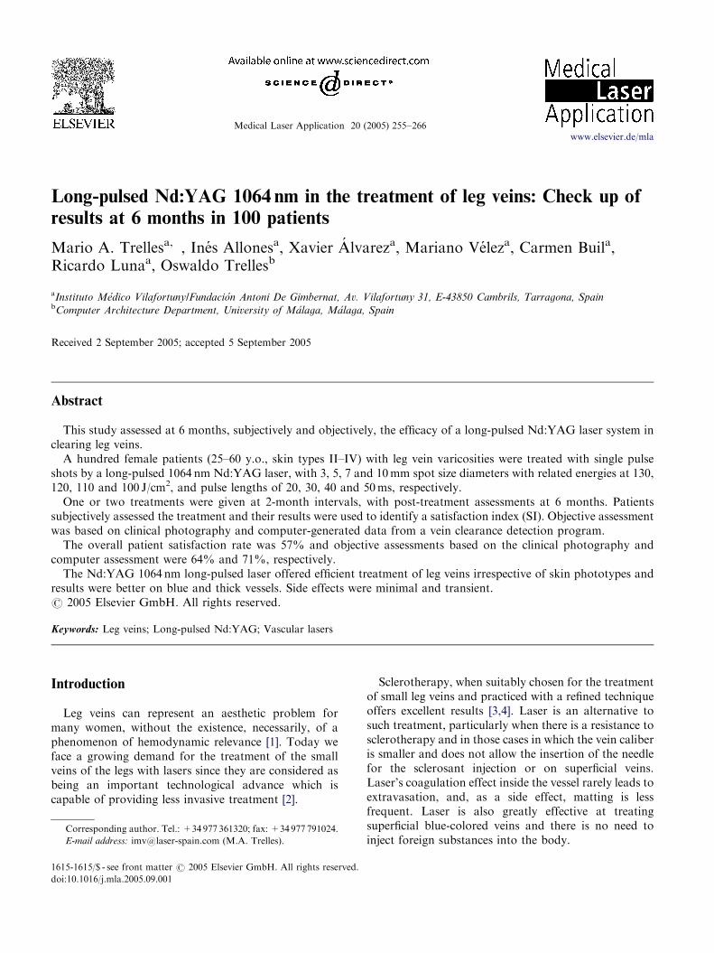
This retrospective study was based on the screening of250 medical files at the Instituto Medico Vilafortuny,Cambrils (Spain) of women treated with the Nd:YAGlaser for leg veins for a cosmetic reason. A hundred files,corresponding to female patients were randomly se-lected for this study. The check up of all related filescontaining information was approved by the EthicsCommittee of the ANTONI DE GIMBERNATFOUNDATION. The clinical characteristics of pa-
Table 1A. Clinical characteristics of treated vessel in relation to p
tients, vessel diameters and colors are listed inTables 1A and B. Phototypes of all women treated wereof Fitzpatrick’s skin types II–IV, whose ages rangedfrom 25 to 60 (mean age 44). No patient treated waspregnant and none had a history of scarring or hadpreviously undergone laser and/or sclerotherapy treat-ment. All patients had signed a document of informedconsent for both their treatment and for the clinicalphotography. In addition, all of them had beeninformed as to the nature of the study and instructedconcerning the characteristics of the questionnaires theyhad completed at the time of the treatment. Prior to thetreatment, all patients had been examined by ecographyto confirm that the saphenous vein and its collateralswere competent, without reticular or perforating vessels.All patients had undergone a maximum of twotreatments at 2-month intervals and results evaluatedat 6 months after the last treatment. The secondtreatment 2 months after the first, was only carried outif the patient had not been satisfied with the resultsobtained with the first treatment and if the physicianconsidered that the results could be improved upon ifthe treatment session was repeated. To this end, allpatients were instructed prior to treatment as to thecharacteristics of the questionnaire and how improve-ment should be estimated, discussing the details withthem of treatment, expectations and limitations [8].They had been made to understand that improvementwould require time (based upon relevant literature [9]and our own experience). Consequently, patients wereinstructed only to return for the second treatment if theyconsidered that the results had not been satisfactory inaccordance with the instructions received.
The laser used was a long-pulsed Nd:YAG at 1064 nmwavelength ‘‘Altus Vantage’’ (Cutera, Burlingame,
atients’ age and skin phototype
ARTICLE IN PRESS
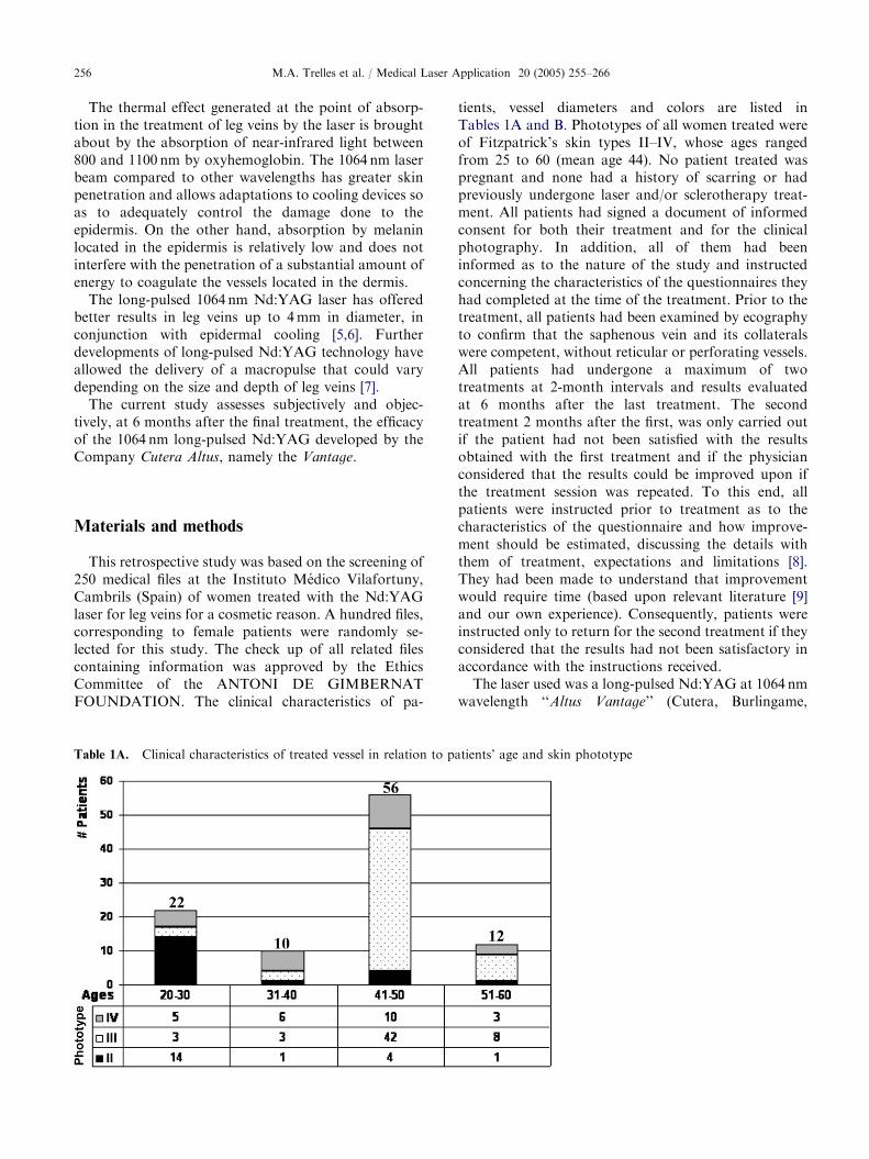
Table 1B. Number of treated vessels in relation to their color and diameter
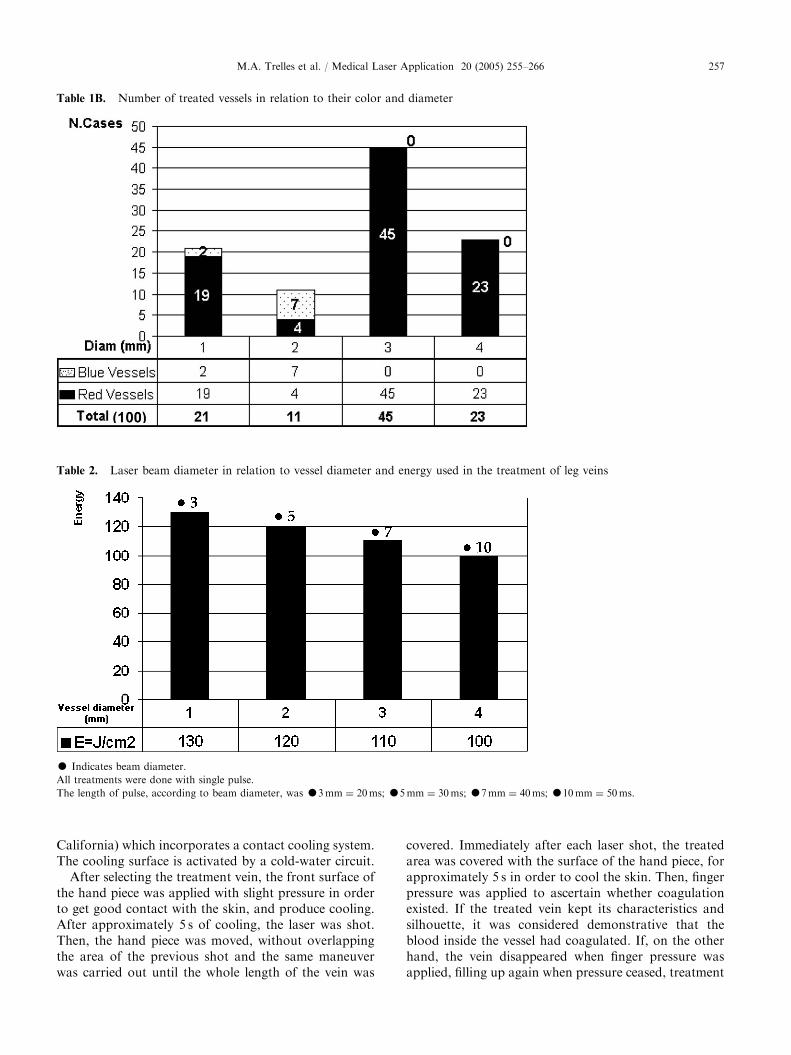
Table 2. Laser beam diameter in relation to vessel diameter and energy used in the treatment of leg veins
K Indicates beam diameter.
All treatments were done with single pulse.
The length of pulse, according to beam diameter, was K3mm ¼ 20ms; K5mm ¼ 30ms; K7mm ¼ 40ms; K10mm ¼ 50ms.
M.A. Trelles et al. / Medical Laser Application 20 (2005) 255–266 257
California) which incorporates a contact cooling system.The cooling surface is activated by a cold-water circuit.
After selecting the treatment vein, the front surface ofthe hand piece was applied with slight pressure in orderto get good contact with the skin, and produce cooling.After approximately 5 s of cooling, the laser was shot.Then, the hand piece was moved, without overlappingthe area of the previous shot and the same maneuverwas carried out until the whole length of the vein was
covered. Immediately after each laser shot, the treatedarea was covered with the surface of the hand piece, forapproximately 5 s in order to cool the skin. Then, fingerpressure was applied to ascertain whether coagulationexisted. If the treated vein kept its characteristics andsilhouette, it was considered demonstrative that theblood inside the vessel had coagulated. If, on the otherhand, the vein disappeared when finger pressure wasapplied, filling up again when pressure ceased, treatment
ARTICLE IN PRESSM.A. Trelles et al. / Medical Laser Application 20 (2005) 255–266258
was considered not to have been sufficient. Then, havingfinished treatment on the whole vein, cooling was doneas previously described, but this time cooling contactwas for a 10 s period, and the same treatment sequenceas previously outlined was repeated with the same lasersettings.
The parameters used to treat the veins varieddepending on their diameter. If the two treatments weredone, the second was carried out at 2 months. Thesettings were the same for all patients who presented thesame vein diameter and for both treatments. Nodifferentiation was made due to vein color or itsapparent depth. To select the treatment program, only thevein diameter was taken into account: pulse length of20ms and 130 J/cm2 was used for the 1mm veins; 30msand 120 J/cm2 for the 2mm veins; 40ms and 110 J/cm2
for the 3mm veins; and 50ms and 100 J/cm2 for the4mm veins (Table 2).
No topical anesthesia was administered to any patientand neither were compression bandages applied aftertreatment. Patients were recommended the use of atopical ointment of Prednicarbato (Peitel
TM
, Lab. NovagS.A, Barcelona, Spain), 3 times per day for 5 days, andto avoid exposure to sunlight over the next 2 weeks aftertreatment.
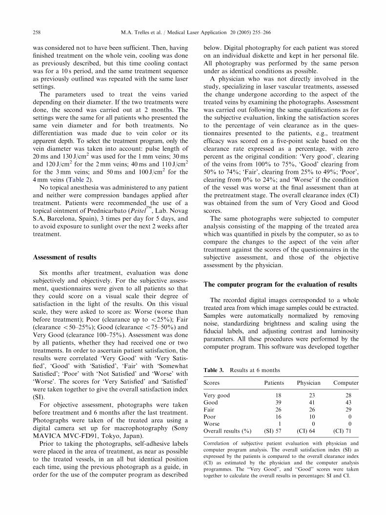
Table 3. Results at 6 months
Scores Patients Physician Computer
Very good 18 23 28
Good 39 41 43
Fair 26 26 29
Poor 16 10 0
Worse 1 0 0
Overall results (%) (SI) 57 (CI) 64 (CI) 71
Correlation of subjective patient evaluation with physician and
computer program analysis. The overall satisfaction index (SI) as
expressed by the patients is compared to the overall clearance index
(CI) as estimated by the physician and the computer analysis
programmes. The ‘‘Very Good’’, and ‘‘Good’’ scores were taken
together to calculate the overall results in percentages: SI and CI.
Assessment of results
Six months after treatment, evaluation was donesubjectively and objectively. For the subjective assess-ment, questionnaires were given to all patients so thatthey could score on a visual scale their degree ofsatisfaction in the light of the results. On this visualscale, they were asked to score as: Worse (worse thanbefore treatment); Poor (clearance up to o25%); Fair(clearance o50–25%); Good (clearance o75–50%) andVery Good (clearance 100–75%). Assessment was doneby all patients, whether they had received one or twotreatments. In order to ascertain patient satisfaction, theresults were correlated ‘Very Good’ with ‘Very Satis-fied’, ‘Good’ with ‘Satisfied’, ‘Fair’ with ‘SomewhatSatisfied’; ‘Poor’ with ‘Not Satisfied’ and ‘Worse’ with‘Worse’. The scores for ‘Very Satisfied’ and ‘Satisfied’were taken together to give the overall satisfaction index(SI).
For objective assessment, photographs were takenbefore treatment and 6 months after the last treatment.Photographs were taken of the treated area using adigital camera set up for macrophotography (SonyMAVICA MVC-FD91, Tokyo, Japan).
Prior to taking the photographs, self-adhesive labelswere placed in the area of treatment, as near as possibleto the treated vessels, in an all but identical positioneach time, using the previous photograph as a guide, inorder for the use of the computer program as described
below. Digital photography for each patient was storedon an individual diskette and kept in her personal file.All photography was performed by the same personunder as identical conditions as possible.
A physician who was not directly involved in thestudy, specializing in laser vascular treatments, assessedthe change undergone according to the aspect of thetreated veins by examining the photographs. Assessmentwas carried out following the same qualifications as forthe subjective evaluation, linking the satisfaction scoresto the percentage of vein clearance as in the ques-tionnaires presented to the patients, e.g., treatmentefficacy was scored on a five-point scale based on theclearance rate expressed as a percentage, with zeropercent as the original condition: ‘Very good’, clearingof the veins from 100% to 75%, ‘Good’ clearing from50% to 74%; ‘Fair’, clearing from 25% to 49%; ‘Poor’,clearing from 0% to 24%; and ‘Worse’ if the conditionof the vessel was worse at the final assessment than atthe pretreatment stage. The overall clearance index (CI)was obtained from the sum of Very Good and Goodscores.
The same photographs were subjected to computeranalysis consisting of the mapping of the treated areawhich was quantified in pixels by the computer, so as tocompare the changes to the aspect of the vein aftertreatment against the scores of the questionnaires in thesubjective assessment, and those of the objectiveassessment by the physician.
The computer program for the evaluation of results
The recorded digital images corresponded to a wholetreated area from which image samples could be extracted.Samples were automatically normalized by removingnoise, standardizing brightness and scaling using thefiducial labels, and adjusting contrast and luminosityparameters. All these procedures were performed by thecomputer program. This software was developed together
ARTICLE IN PRESSM.A. Trelles et al. / Medical Laser Application 20 (2005) 255–266 259
with the Department of Computer Architecture, theUniversity of Malaga (Spain)[10].
A Canny operator was next used as an optimal edgedetector, working in a multistage process and whichresulted in an image made up of one-pixel-thickconnected segments that closely followed the faintmargins of the feathering of the veins. In this way,the pre- and post-treatment areas of the affectedzone in each image were computed as a percentage.This could thus be used as a sensitive and objectivecomparative measurement not only for diagnosticreports on the pretreatment condition of veins, but
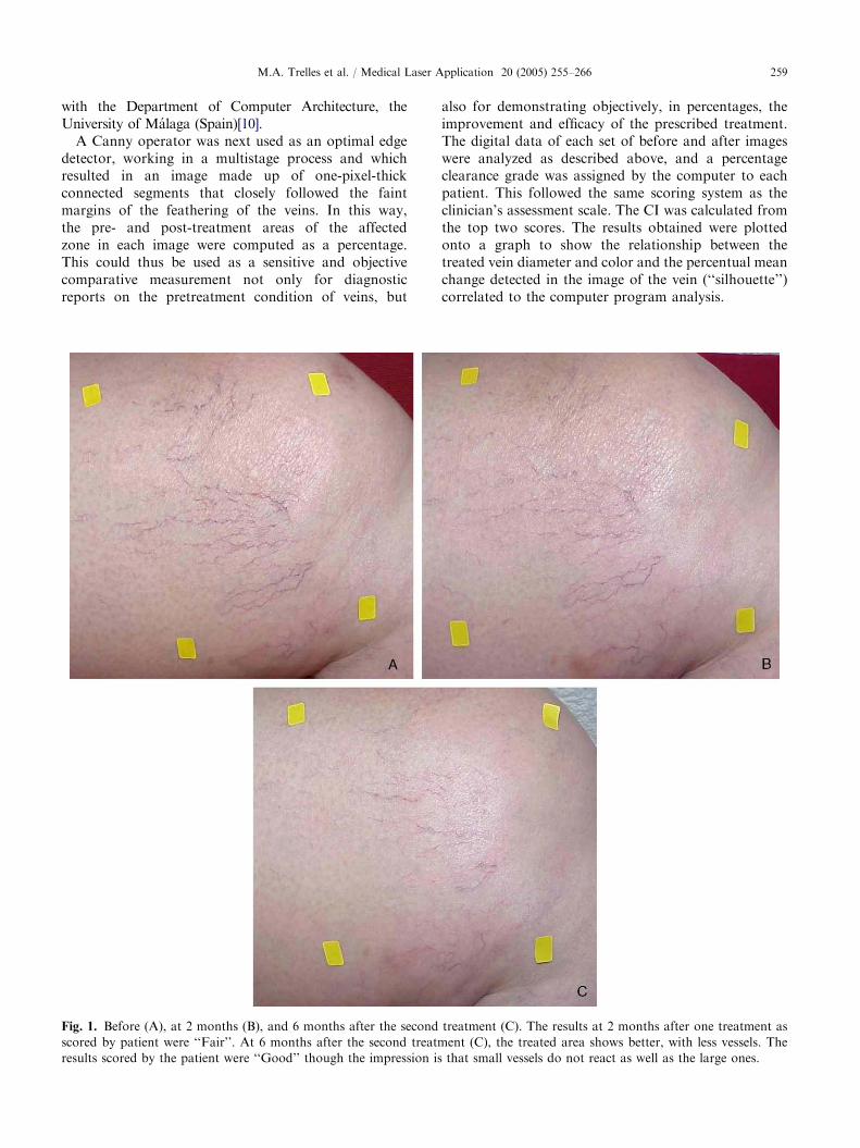
Fig. 1. Before (A), at 2 months (B), and 6 months after the second
scored by patient were ‘‘Fair’’. At 6 months after the second treatm
results scored by the patient were ‘‘Good’’ though the impression is
also for demonstrating objectively, in percentages, theimprovement and efficacy of the prescribed treatment.The digital data of each set of before and after imageswere analyzed as described above, and a percentageclearance grade was assigned by the computer to eachpatient. This followed the same scoring system as theclinician’s assessment scale. The CI was calculated fromthe top two scores. The results obtained were plottedonto a graph to show the relationship between thetreated vein diameter and color and the percentual meanchange detected in the image of the vein (‘‘silhouette’’)correlated to the computer program analysis.
treatment (C). The results at 2 months after one treatment as
ent (C), the treated area shows better, with less vessels. The
that small vessels do not react as well as the large ones.
ARTICLE IN PRESSM.A. Trelles et al. / Medical Laser Application 20 (2005) 255–266260
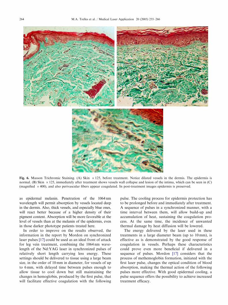
Histology
From the selected files treated, 10 patients werechosen at random from the 100 cases studied and onwhom histologies had been performed. These histologiesit was assumed, represented a cross sample of the casestreated. Samples were taken with a sufficiently broaddiameter punch, before and immediately after treat-ment. The sample taken after treatment was done abouta centimeter away from the biopsy taken prior totreatment, once the vein had been treated along itswhole length.
All specimens were routinely processed and stainedwith the Masson Trichromic technique.
Results
Two months after treatment, 55 patients returned forconsultation. The remaining 45 were telephoned andthey stated that they were satisfied with the results andthat they preferred to wait until the 6-month check up.Of these 55 patients, 38 qualified their results as being‘‘Fair’’, 16 ‘‘Poor’’, and 1 ‘‘Worse’’. This patient hadsuffered small burns, that had already been detected,which she said had formed blisters a few hours aftertreatment and she had treated with the aforementionedPrednicarbato ointment. At 2 months after the firsttreatment, the now repaired lesion presented pigmentarychanges, and so it was decided to wait and not performany further laser treatment on her. This patient wasespecially recommended to avoid sunlight. All those 55
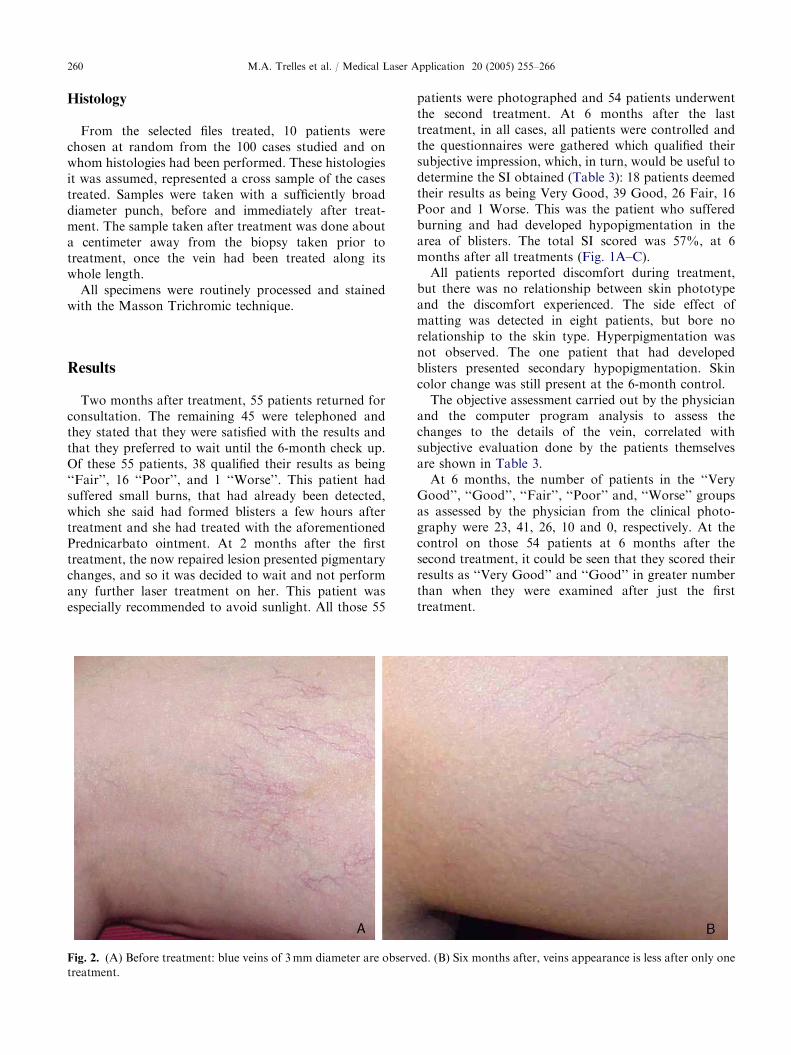
Fig. 2. (A) Before treatment: blue veins of 3mm diameter are observ
treatment.
patients were photographed and 54 patients underwentthe second treatment. At 6 months after the lasttreatment, in all cases, all patients were controlled andthe questionnaires were gathered which qualified theirsubjective impression, which, in turn, would be useful todetermine the SI obtained (Table 3): 18 patients deemedtheir results as being Very Good, 39 Good, 26 Fair, 16Poor and 1 Worse. This was the patient who sufferedburning and had developed hypopigmentation in thearea of blisters. The total SI scored was 57%, at 6months after all treatments (Fig. 1A–C).
All patients reported discomfort during treatment,but there was no relationship between skin phototypeand the discomfort experienced. The side effect ofmatting was detected in eight patients, but bore norelationship to the skin type. Hyperpigmentation wasnot observed. The one patient that had developedblisters presented secondary hypopigmentation. Skincolor change was still present at the 6-month control.
The objective assessment carried out by the physicianand the computer program analysis to assess thechanges to the details of the vein, correlated withsubjective evaluation done by the patients themselvesare shown in Table 3.
At 6 months, the number of patients in the ‘‘VeryGood’’, ‘‘Good’’, ‘‘Fair’’, ‘‘Poor’’ and, ‘‘Worse’’ groupsas assessed by the physician from the clinical photo-graphy were 23, 41, 26, 10 and 0, respectively. At thecontrol on those 54 patients at 6 months after thesecond treatment, it could be seen that they scored theirresults as ‘‘Very Good’’ and ‘‘Good’’ in greater numberthan when they were examined after just the firsttreatment.
ed. (B) Six months after, veins appearance is less after only one
ARTICLE IN PRESSM.A. Trelles et al. / Medical Laser Application 20 (2005) 255–266 261
In total, the subjective SI value for all 100 patients,taking the ‘‘Very Good’’ and ‘‘Good’’ results, was 57(57%) although 45 patients had received only onetreatment, as they were already satisfied (Fig. 2A,B).The clinician and the computer analysis placed theobjective assessments higher, at 64% and 71%, respec-tively (Table 3). This had been noticed in previousstudies due to patients’ expectations of results beinghigher than the realistic possibilities offered by treat-ment [8].
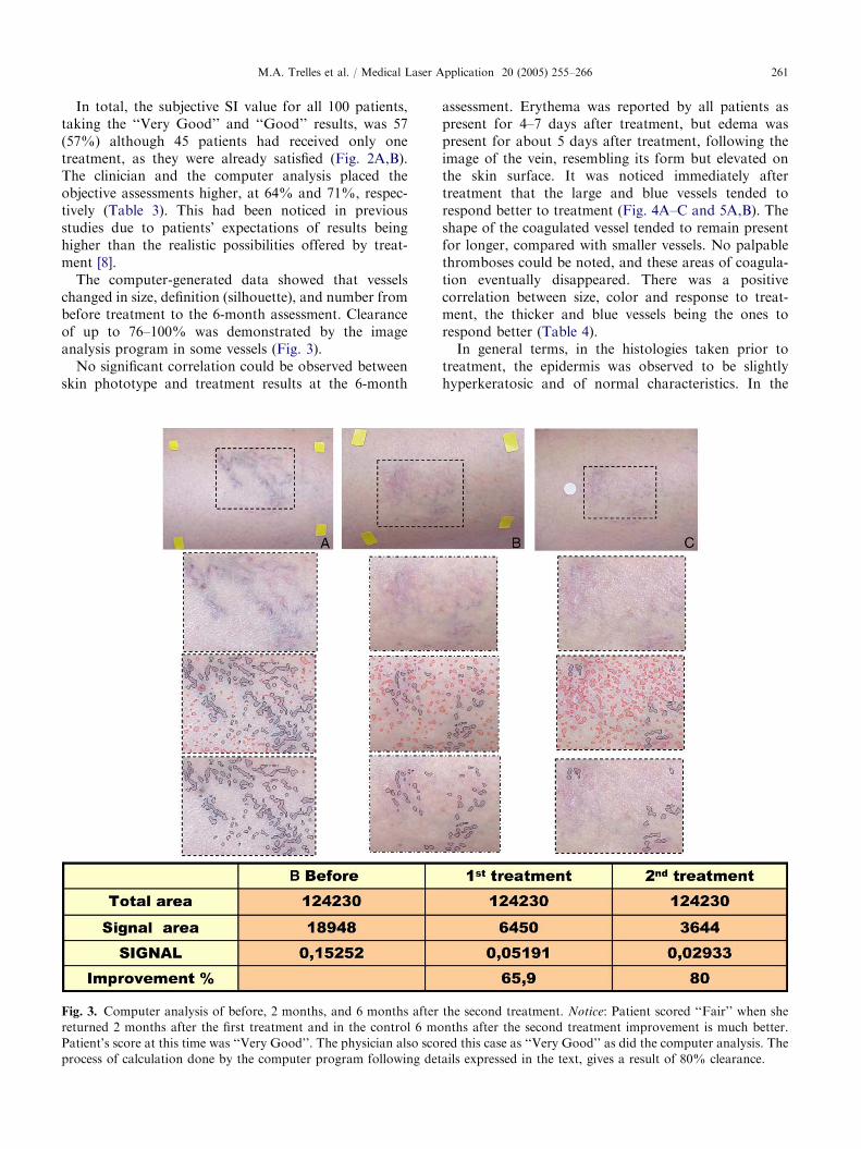
The computer-generated data showed that vesselschanged in size, definition (silhouette), and number frombefore treatment to the 6-month assessment. Clearanceof up to 76–100% was demonstrated by the imageanalysis program in some vessels (Fig. 3).
No significant correlation could be observed betweenskin phototype and treatment results at the 6-month
Fig. 3. Computer analysis of before, 2 months, and 6 months after
returned 2 months after the first treatment and in the control 6 mo
Patient’s score at this time was ‘‘Very Good’’. The physician also sco
process of calculation done by the computer program following det
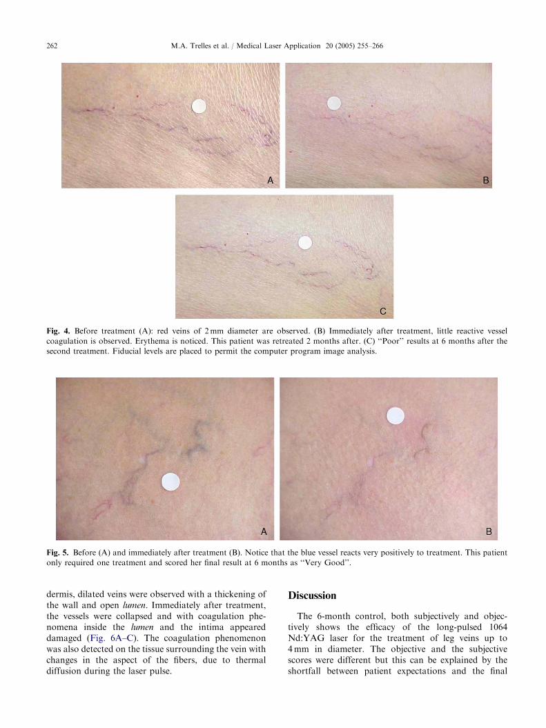
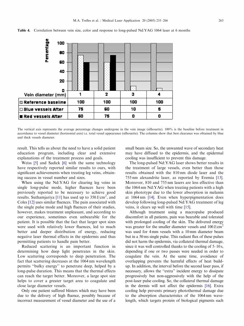
assessment. Erythema was reported by all patients aspresent for 4–7 days after treatment, but edema waspresent for about 5 days after treatment, following theimage of the vein, resembling its form but elevated onthe skin surface. It was noticed immediately aftertreatment that the large and blue vessels tended torespond better to treatment (Fig. 4A–C and 5A,B). Theshape of the coagulated vessel tended to remain presentfor longer, compared with smaller vessels. No palpablethromboses could be noted, and these areas of coagula-tion eventually disappeared. There was a positivecorrelation between size, color and response to treat-ment, the thicker and blue vessels being the ones torespond better (Table 4).
In general terms, in the histologies taken prior totreatment, the epidermis was observed to be slightlyhyperkeratosic and of normal characteristics. In the
the second treatment. Notice: Patient scored ‘‘Fair’’ when she
nths after the second treatment improvement is much better.
red this case as ‘‘Very Good’’ as did the computer analysis. The
ails expressed in the text, gives a result of 80% clearance.
ARTICLE IN PRESS
Fig. 4. Before treatment (A): red veins of 2mm diameter are observed. (B) Immediately after treatment, little reactive vessel
coagulation is observed. Erythema is noticed. This patient was retreated 2 months after. (C) ‘‘Poor’’ results at 6 months after the
second treatment. Fiducial levels are placed to permit the computer program image analysis.
Fig. 5. Before (A) and immediately after treatment (B). Notice that the blue vessel reacts very positively to treatment. This patient
only required one treatment and scored her final result at 6 months as ‘‘Very Good’’.
M.A. Trelles et al. / Medical Laser Application 20 (2005) 255–266262
dermis, dilated veins were observed with a thickening ofthe wall and open lumen. Immediately after treatment,the vessels were collapsed and with coagulation phe-nomena inside the lumen and the intima appeareddamaged (Fig. 6A–C). The coagulation phenomenonwas also detected on the tissue surrounding the vein withchanges in the aspect of the fibers, due to thermaldiffusion during the laser pulse.
Discussion
The 6-month control, both subjectively and objec-tively shows the efficacy of the long-pulsed 1064Nd:YAG laser for the treatment of leg veins up to4mm in diameter. The objective and the subjectivescores were different but this can be explained by theshortfall between patient expectations and the final
ARTICLE IN PRESS
Table 4. Correlation between vein size, color and response to long-pulsed Nd:YAG 1064 laser at 6 months
The vertical axis represents the average percentage changes undergone in the vein image (silhouette). 100% is the baseline before treatment in
accordance to vessel diameter (horizontal axis) i.e. total vessel appearance (silhouette). The columns show that best clearance was obtained by blue
and thick vessels diameter.
M.A. Trelles et al. / Medical Laser Application 20 (2005) 255–266 263
result. This tells us about the need to have a solid patienteducation program, including clear and extensiveexplanations of the treatment process and goals.
Weiss [5] and Sadick [6] with the same technologyhave respectively reported similar results to ours, withsignificant achievements when treating leg veins, obtain-ing success in vessel number and sizes.
When using the Nd:YAG for clearing leg veins insingle long-pulse mode, higher fluences have beenpreviously reported to be necessary to achieve goodresults. Suthamjariya [11] has used up to 350 J/cm2, andColes [12] uses similar fluences. The pain associated withthe single pulse mode and high fluences of their studies,however, makes treatment unpleasant, and according toour experience, sometimes even unbearable for thepatient. It is possible that the fact that larger spot sizeswere used with relatively lower fluences, led to muchbetter and deeper distribution of energy, reducingnegative laser thermal effects in the epidermis and thuspermitting patients to handle pain better.
Reduced scattering is an important function indetermining how deep light penetrates in the skin.Low scattering corresponds to deep penetration. Thefact that scattering decreases at the 1064 nm wavelengthpermits ‘‘bulky energy’’ to penetrate deep, helped by along-pulse duration. This means that the thermal effectscan reach the target better. Moreover, a large spot sizehelps to cover a greater target area to coagulate andclose large diameter vessels.
Only one patient suffered blisters which may have beendue to the delivery of high fluence, possibly because ofincorrect measurement of vessel diameter and the use of a
small beam size. So, the unwanted wave of secondary heatmay have diffused to the epidermis, and the epidermalcooling was insufficient to prevent this damage.
The long-pulsed Nd:YAG laser shows better results inthe treatment of large vessels, even better than thoseresults obtained with the 810 nm diode laser and the755 nm alexandrite laser, as reported by Eremia [13].Moreover, 810 and 755 nm lasers are less effective thanthe 1064 nm Nd:YAG when treating patients with a highskin phototype due to the lower absorption in melaninat 1064 nm [14]. Even when hyperpigmentation doesdevelop following long-pulsed Nd:YAG treatment of legveins, it clears up well with time [15].
Although treatment using a macropulse produceddiscomfort in all patients, pain was bearable and toleratedwith prolonged cooling of the skin. The delivered energywas greater for the smaller diameter vessels and 100 J/cm2
was used for 4mm vessels with a 10mm diameter beambut in a 50ms single pulse. This radiant flux of these pulsesdid not harm the epidermis, via collateral thermal damage,since it was well controlled thanks to the cooling of 5–10 s,depending if one or two passes were needed in order tocoagulate the vein. At the same time, avoidance ofoverlapping prevents the harmful effects of heat build-up. In addition, the interval before the second laser pass, ifnecessary, allows the ‘‘extra’’ incident energy to dissipateprogressively but non-aggressively with the help of thepost-laser pulse cooling. So, the collateral thermal damagein the dermis will not affect the epidermis [16]. Extracooling help prevents primary photothermal damage dueto the absorption characteristics of the 1064nm wave-length, which targets protein of biological pigments such
ARTICLE IN PRESS
Fig. 6. Masson Trichromic Staining. (A) Skin � 125, before treatment. Notice dilated vessels in the dermis. The epidermis is
normal. (B) Skin � 125, immediately after treatment shows vessels wall collapse and lesion of the intima, which can be seen in (C)
(magnified � 400), and also perivascular fibers appear coagulated. In post-treatment images epidermis is preserved.
M.A. Trelles et al. / Medical Laser Application 20 (2005) 255–266264
as epidermal melanin. Penetration of the 1064nmwavelength will permit absorption by vessels located deepin the dermis. Also, thick vessels, and especially blue ones,will react better because of a higher density of theirpigment content. Absorption will be more favorable at thelevel of vessels than at the melanin of the epidermis, evenin those darker phototype patients treated here.
In order to improve on the results observed, theinformation in the report by Mordon on synchronizedlaser pulses [17] could be used as an ideal front of attackfor leg vein treatment, combining the 1064 nm wave-length of the Nd:YAG laser in synchronized pulses ofrelatively short length carrying less energy. Thesesettings should be delivered to tissue using a large beamsize, in the order of 10mm in diameter, for vessels of upto 6mm, with delayed time between pulses enough toallow tissue to cool down but still maintaining thechanges in hemoglobin, produced by the first pulse, thatwill facilitate effective coagulation with the following
pulse. The cooling process for epidermis protection hasto be prolonged before and immediately after treatment.A sequence of pulses in a synchronized manner, with atime interval between them, will allow build-up andaccumulation of heat, sustaining the coagulation pro-cess. At the same time, the incidence of unwantedthermal damage by heat diffusion will be lowered.
The energy delivered by the laser used in thesetreatments in a large diameter beam (up to 10mm), iseffective as is demonstrated by the good response ofcoagulation in vessels. Perhaps these characteristicscould prove even more beneficial if delivered in asequence of pulses. Mordon [17] considers that theprocess of methemoglobin formation, initiated with thefirst laser pulse, changes the optical condition of bloodabsorption, making the thermal action of the followingpulses more effective. With good epidermal cooling, apulse sequence offers the possibility to achieve increasedtreatment efficacy.
ARTICLE IN PRESSM.A. Trelles et al. / Medical Laser Application 20 (2005) 255–266 265
Long-term assessment allows confirmation of thecomplete disappearance of treated vessels [18]. Surely,the computer-generated image extraction program pre-sents the improvement much more clearly than thepatients’ subjective evaluation and the clinical photogra-phy. The program calculates the area of venous lesionsbefore and 6 months after treatment with a pixel-basedgoniometric program which permits being well presentedto patients to possibly help to bring the SI scores more inline with the physician’s appreciation of results.
Hyperpigmentation at high fluences with theNd:YAG can be a side effect as reported by Omura[19]. This drawback has not been seen even in photo-type IV patients. This must be due to relatively lowerenergy, delivered in a large spot size, though aprolonged pulse length in comparison to that ofOmura, but only for blue vessels. Small red vesselsusually required more than one treatment. Kauvar [20]has noticed that high clearance rates can be obtainedin vessels over 1.5mm in diameter and that theyrespond often in only one treatment when using the1064 nm Nd:YAG laser. Our observations coincidewith this.
Conclusions
The long-pulsed 1064 nm Nd:YAG laser as applied inthe presented cases offered a Very Good solution in thetreatment of leg veins up to 4mm in diameter. Theobjective assessments of the clinical results were VeryGood, accompanied by a high SI from the group of 100patients. Result obtained by a single treatment couldenhance with a second one. Final results were achievedwith very minimal complications.
Acknowledgements
The clinical and laboratory subject matter of thispaper is registered in the activities of the FUNDACIONANTONI DE GIMBERNAT (2004/5) whose granthelped support these investigations. The authors thankMrs. Maribel Andreu for her help in the preparation ofthe final version of this paper.
Zusammenfassung
Der Langpuls-Nd:YAG-Laser (1064 nm) zur Behandlung
von Besenreiser-Varizen: Ergebnisse nach 6 Monaten bei
100 Patienten
In dieser Studie wird die Wirksamkeit eines Langpuls-Nd:YAG-Lasers zur Entfernung von Besenreiser-Vari-zen 6 Monate nach Behandlung mittels objektiver undsubjektiver Methoden untersucht.
100 Patientinnen (25–60 Jahre, Hauttypen II-IV) mitBesenreiser-Varizen wurden mit einem LangpulsNd:YAG-Laser mit 3, 5, 7 und 10mm Fokusdurchmes-ser, Pulslangen von 20, 30, 40 und 50ms und denentsprechenden Energiedichten von 130, 120, 110und 100 J/cm2 (mit jeweils Einzelpulsen pro Flache)behandelt. Es wurden ein oder zwei Behandlungeninnerhalb von zwei Monaten durchgefuhrt, die Nach-beobachtungszeit betrug 6 Monate. Die Patientenbeurteilten die Ergebnisse selbst, aus diesen Beurteilun-gen wurde ein
’’satisfaction index ’’ (SI) gebildet. Die
objektiven Ergebnisse wurden anhand von Fotos undmit einem speziellen Bildverabeitungsprogramm, mitdem die entfernten Besenreiser detektiert wurden,ermittelt.
Die mittlere Zufriedenheitsrate lag bei 57%, dieobjektive Beurteilung anhand Fotos und des Compu-terprogamms ergab 64% bzw. 71% Behandlungserfolg.
Der Langpuls-Nd:YAG-Laser (1064 nm) ermoglichteine effektive Behandlung von Besenreiser-Varizenunabhangig vom Hauttyp, die Ergebnisse waren besserbei dickeren und blau erscheinenden Gefaßen. Uner-wunschte Nebenwirkungen waren minimal und voru-bergehend.r 2005 Elsevier GmbH. All rights reserved.
Schlusselworter: Besenreiser, Langpuls-Nd:YAG-Laser, Gefaß-
laser
References
[1] Weiss RA, Feied CF, Weiss MA. Vein diagnosis and
treatment: a comprehensive approach. New York:
McGraw-Hill; 2001.
[2] Dover JS, Sadick NS, Goldman MP. The role of lasers
and light sources in the treatment of leg veins. Dermatol
Surg 1999;24:328–36.
[3] Cabrera Garrido JR, Cabrera Garcia-Olmedo JR, Gar-
cia-Olmedo Dominguez MA. Elargissement des limites de
la schlerotherapie: noveaux produits. Sclerosants Phlebo-
logie 1997;50:181–8.
[4] Mingo-Garcia J. Esclerosis venosa con espuma: foam
medical system. Rev Esp Med Cir Cosmet 1999;7:29–31.
[5] Weiss RA, Weiss MA. Early clinical results with a
multiple synchronized pulse 1064 nm laser for leg
telangiectasias and reticular veins. Dermatol Surg 1999;
25:399–402.
[6] Sadick NS. Long-term results with a multiple synchro-
nized-pulse 1064 nm Nd:YAG laser for the treatment of
leg venulectasias and reticular veins. Dermatol Surg
2001;27:365–9.
[7] Trelles MA, Allones I, Martın-Vazquez MJ, Trelles O,
Velez M, Mordon S. Long pulsed Nd:YAG laser for
treatment of leg veins in 40 patients with assessments at 6
and 12 Months. Lasers Surg Med 2004;35:68–76.
[8] Trelles MA, Pardo L, Ayliffe P, Trelles K, Velez M,
Garcıa-Solana L. Patients’ answers to a postoperative
ARTICLE IN PRESSM.A. Trelles et al. / Medical Laser Application 20 (2005) 255–266266
questionnaire related to laser resurfacing. Facial Plast
Surg 2001;17:187–92.
[9] Sadick NS. Vasculight and other 1064 nm wavelength
lasers for treatment of lower extremity veins. Scope Phleb
Lymphol 2000;29:175–8.
[10] Trelles MA, Martın-Vazquez MJ, Trelles O. Objec-
tive follow-up by computational evaluation of tissue
changes alter light treatment. Lasers Med Sci 2003;
18(Suppl. 1).
[11] Suthamjariya K, Farinelli WA, Anderson RR. Treatment
of telangiectasias and reticular veins with a long-pulsed
Nd:YAG laser. Lasers Surg Med 2002;14(Suppl.):41.
[12] Coles CM, Werner RS, Zelickson BD. Comparative pilot
study evaluating the treatment of leg veins with a long
pulse Nd:YAG laser and sclerotherapy. Lasers Surg Med
2002;30(2):154–9.
[13] Eremia S, Li C, Umar SH. A side-by-side comparative
study of 1064 nm Nd:YAG, 810 nm diode, and 755 nm
alexandrite lasers for treatment of 0.3–3 nm leg veins.
Dermatol Surg 2002;28(3):224–30.
[14] Trelles MA, Allones I, Trelles O. An 810 nm diode laser in
the treatment of small (o1.0mm) leg veins: a preliminary
assessment. Laser Med Sci 2004;19(1):21–6.
[15] Sadick NS, Weiss RA, Goldman MP. Advances in laser
surgery for leg veins: bimodal wavelength approach to
lower extremity vessels, new cooling techniques, and
longer pulse durations. Dermatol Surg 2002;28:16–20.
[16] Bowes LE, Goldman MP. Treatment of leg telangiectasias
with a 1064 nm long pulse Nd:YAG laser using
dynamic versus contact cooling. Lasers Surg Med 2002;
14(Suppl.):40.
[17] Mordon S, Brisot D, Fournier N. Using a ‘‘Non Uniform
Pulse Sequence’’ can improve selective coagulation with a
Nd:YAG laser (1.06 mm) thanks to met-hemoglobin
absorption: a clinical study on blue leg veins. Lasers Surg
Med 2003;32:160–70.
[18] Rogachefsky AS, Silapunt S, Goldberg DJ. Nd:YAG
laser (1064 nm) irradiation for lower extremity telangiec-
tasias and small reticular veins. Efficacy as measured by
vessel color and size. Dermatol Surg 2002;28(3):220–3.
[19] Omura NE, Dover JS, Arndt KA, Kauvar AN. Treat-
ment of reticular leg veins with a 1064 nm long pulse
Nd:YAG laser. Jam Acad Dermatol 2003;48(1):76–81.
[20] Kauvar AN. Optimizing treatment of small (0.1–1.0mm)
and large (1.0–2.0mm) leg telangiectasia with long pulsed
Nd:YAG laser. Lasers Surg Med 2001;13(Suppl.):24.
Related Documents