ARTICLE Received 18 Mar 2014 | Accepted 28 Aug 2014 | Published 6 Oct 2014 Long-distance endosome trafficking drives fungal effector production during plant infection Ewa Bielska 1, *, Yujiro Higuchi 1, * ,w , Martin Schuster 1, *, Natascha Steinberg 1,w , Sreedhar Kilaru 1 , Nicholas J. Talbot 1 & Gero Steinberg 1 To cause plant disease, pathogenic fungi can secrete effector proteins into plant cells to suppress plant immunity and facilitate fungal infection. Most fungal pathogens infect plants using very long strand-like cells, called hyphae, that secrete effectors from their tips into host tissue. How fungi undergo long-distance cell signalling to regulate effector production during infection is not known. Here we show that long-distance retrograde motility of early endo- somes (EEs) is necessary to trigger transcription of effector-encoding genes during plant infection by the pathogenic fungus Ustilago maydis. We demonstrate that motor-dependent retrograde EE motility is necessary for regulation of effector production and secretion during host cell invasion. We further show that retrograde signalling involves the mitogen-activated kinase Crk1 that travels on EEs and participates in control of effector production. Fungal pathogens therefore undergo long-range signalling to orchestrate host invasion. DOI: 10.1038/ncomms6097 OPEN 1 School of Biosciences, College of Life and Environmental Sciences, University of Exeter, Stocker Road, Exeter EX4 4QD, UK. *These authors contributed equally to this work. w Present addresses: Department of Bioscience and Biotechnology, Facultyof Agriculture, Kyushu University, 6-10-1 Hakozaki, Fukuoka 812-8581, Japan (Y.H.); School of Geography, College of Life and Environmental Sciences, University of Exeter, Rennes Drive, Exeter EX4 4RJ, UK (N.S.). Correspondence and requests for materials should be addressed to G.S. (email: [email protected]). NATURE COMMUNICATIONS | 5:5097 | DOI: 10.1038/ncomms6097 | www.nature.com/naturecommunications 1 & 2014 Macmillan Publishers Limited. All rights reserved.

Welcome message from author
This document is posted to help you gain knowledge. Please leave a comment to let me know what you think about it! Share it to your friends and learn new things together.
Transcript

ARTICLE
Received 18 Mar 2014 | Accepted 28 Aug 2014 | Published 6 Oct 2014
Long-distance endosome trafficking drives fungaleffector production during plant infectionEwa Bielska1,*, Yujiro Higuchi1,*,w, Martin Schuster1,*, Natascha Steinberg1,w, Sreedhar Kilaru1,
Nicholas J. Talbot1 & Gero Steinberg1
To cause plant disease, pathogenic fungi can secrete effector proteins into plant cells to
suppress plant immunity and facilitate fungal infection. Most fungal pathogens infect plants
using very long strand-like cells, called hyphae, that secrete effectors from their tips into host
tissue. How fungi undergo long-distance cell signalling to regulate effector production during
infection is not known. Here we show that long-distance retrograde motility of early endo-
somes (EEs) is necessary to trigger transcription of effector-encoding genes during plant
infection by the pathogenic fungus Ustilago maydis. We demonstrate that motor-dependent
retrograde EE motility is necessary for regulation of effector production and secretion during
host cell invasion. We further show that retrograde signalling involves the mitogen-activated
kinase Crk1 that travels on EEs and participates in control of effector production. Fungal
pathogens therefore undergo long-range signalling to orchestrate host invasion.
DOI: 10.1038/ncomms6097 OPEN
1 School of Biosciences, College of Life and Environmental Sciences, University of Exeter, Stocker Road, Exeter EX4 4QD, UK. * These authors contributedequally to this work. w Present addresses: Department of Bioscience and Biotechnology, Faculty of Agriculture, Kyushu University, 6-10-1 Hakozaki, Fukuoka812-8581, Japan (Y.H.); School of Geography, College of Life and Environmental Sciences, University of Exeter, Rennes Drive, Exeter EX4 4RJ, UK (N.S.).Correspondence and requests for materials should be addressed to G.S. (email: [email protected]).
NATURE COMMUNICATIONS | 5:5097 | DOI: 10.1038/ncomms6097 | www.nature.com/naturecommunications 1
& 2014 Macmillan Publishers Limited. All rights reserved.

Plant pathogenic fungi invade their host plants by hyphal tipgrowth, which is an important determinant of fungalvirulence. In response to infection, plant innate defences
recognize pathogens and trigger a complex set of physiologicalresponses1,2. Fungal pathogens overcome host immunity bysecretion of a battery of effector proteins3–5. These effectorproteins suppress plant immunity, thereby allowing rapid fungalcolonization of plant tissue. Such interplay between pathogen andhost forms the basis of the biotrophic interaction of the corn smutfungus Ustilago maydis and maize6. In this interaction, the fungusexplores the plant surface by tip growth, but upon recognition ofhost surface cues initiates penetration of plant epidermal cells6,7.During this initial phase of development, a number of plantdefence genes are induced, suggesting that the host recognizes thefungal intruder8. However, the host defence reaction issuppressed rapidly by secreted fungal effectors. A subset ofthese have been studied in depth, such as Pep1, required forpenetration and for inhibition of apoplastic plant peroxidases9,10,Pit2, which inhibits apoplastic plant proteases11,12 and Cmu1, achorismate mutase, which attenuates plant salicylic acid levels13
and thus suppresses plant defence responses.As plant defence responses occur within minutes of perception
of a pathogen14–16, fungal effector proteins must be produced andsecreted equally rapidly. Effector production must therefore beinduced as soon as the hyphal tip begins invasion. However, thearchitecture of an invading hyphal cell poses a challenge, becausethe nucleus is located at a considerable distance from the invadingtip17. It must therefore perceive a long-range signal from theplasma membrane to induce effector transcription. In animal
neurons, such retrograde signalling from the synapse to thenucleus is mediated by early endosomes (EEs). Theydeliver signalling components, including mitogen-activatedprotein kinases (MAPKs; ref. 18) along microtubules to thenucleus19–21. Microtubule-dependent EE motility was describedin U. maydis22,23 and other fungi24,25. Individual organelles cantravel up to 90 mm, and therefore potentially traverse the distancebetween the hyphal tip and nucleus26,27. However, a role forfungal EEs in cell signalling has not been reported.
Here, we focus on the mechanism of long-range signallingduring early infection of maize by U. maydis. We presentevidence that effectors are secreted at the invading hyphal tip. Wehave developed a method to immobilize EEs very specifically, andwe show that EE motility is crucial for transcription andsubsequent secretion of the Cmu1, Pep1 and Pit2 effectors. EEstherefore transmit signals to the nucleus to regulate plantinfection. Moreover, we provide evidence that the MAPK Crk1localizes to moving EEs and that effector production andsecretion is deregulated when crk1 is deleted. Collectively, ourresults are consistent with a role for motor-driven EE motility inretrograde signalling during infection. This long-range signallingmechanism is therefore of pivotal importance to fungalpathogenicity.
ResultsFungal EEs move retrogradely during plant infection. The cornsmut fungus, U. maydis colonizes plant tissues using invasivehyphae (Fig. 1a) that grow at their tips. During infection, the
FungusPlant
FungusNucleus
Distance
20
n=16719 cells
NucleusMean
95% Cl
15
10
5
0
0–4
<32
28–3
2
24–2
8
20–2
4
16–2
0
12–1
6
8–124–
8
Retrograde run-length (μm)
Tim
eR
elat
ive
num
ber
(%)
2 d.p.i.
1 d.p.i.
405 nm
1 d.p.i.
****** * *
NucleusEEs
Plant
Cmu1cytoplasmic GFP
EEs (paGFP-Rab5a)
Cell wall Nucleus (H4)
Figure 1 | Endosomes travel from the invading hyphal tip to the nucleus. (a) Lectin-stained hyphal cells (green) colonizing a leaf at 2 d.p.i. Plant cell
walls were stained using propidium iodide (red). Scale bars, 20mm (left panel) and 10mm (right panel). (b) Invasion of hyphal cells, expressing a
nuclear RFP (nucleus; appearing yellow) and cytoplasmic GFP (green), at 1 d.p.i. Dotted line indicates plant surface. Chloroplasts visualized by their
auto-fluorescence (asterisks, red). Arrow head points towards invading hyphal tip. Scale bar, 10mm. See Supplementary Movie 1. (c) Secretion of effector
protein Cmu1-mCherry (red) during early fungal invasion (fungus in green). Right panels reveal Cmu1-mCherry at the fungus–plant interface. Scale bars,
2 mm. See Supplementary Movie 2. (d) Image and kymograph show retrograde motility of photo-activated paGFP-Rab5a-labelled EEs (green) towards sub-
apical nucleus (red, labelled with histone4-mCherry) in an invading fungal cell. The fungal cell wall was stained with Calcofluor White (blue). Point of
photo-activation indicated by open arrowhead. Solid arrows in lower panel indicate long range motility. Scale bars, 10mm (upper panel), 3 s (vertical, lower
panel) and 5 mm (horizontal, lower panel). (e) Retrograde run-length of photo-activated EEs in invading hyphae (1 d.p.i.). Mean position of nucleus is
indicated in red; 95% confidence intervals (CIs) indicated by green dotted line. Sample size from 43 experiments is shown.
ARTICLE NATURE COMMUNICATIONS | DOI: 10.1038/ncomms6097
2 NATURE COMMUNICATIONS | 5:5097 | DOI: 10.1038/ncomms6097 | www.nature.com/naturecommunications
& 2014 Macmillan Publishers Limited. All rights reserved.

Table 1 | Strains and plasmids used in this study.
Strain or plasmid Genotype Reference
SG200 a1 mfa2 bW2 bE1, bleR Ref. 35
SG200G3_NLSR3 a1 mfa2 bW2 bE1, bleR/poG3/poNLS3RFP This studySG200Cmu1Ch_G3 a1 mfa2 bW2 bE1, Pcmu1-cmu1-mcherry, bleR, hygR/poG3 This studySG200Cmu1Ch_paGRab5a_H4Ch a1 mfa2 bW2 bE1, Pcmu1-cmu1-mcherry, bleR, hygR/pNopaGRab5a/pCoH4Ch This studyAB33GRab5a a2 Pnar-bW2 Pnar-bE1, bleR/poGRab5a Ref. 64
AB33GRab5a_cEAP a2 Pnar-bW2 Pnar-bE1, bleR/poGRab5a/pHPcEAP This studyAB33Kin3G_ChRab5a_cEAP a2 Pnar-bW2 Pnar-bE1, Pkin3-kin3-egfp, bleR, hygR/pomChRab5a/pCPcEAP This studySG200GRab5a a1 mfa2 bW2 bE1, bleR/poGRab5a This studySG200GRab5a_cEAP a1 mfa2 bW2 bE1, bleR/poGRab5a/pHPcEAP This studySG200GRab5a_mEAP a1 mfa2 bW2 bE1, bleR/poGRab5a/pCPmEAP This studySG200GRab5a_cEAP_Cmu1Ch a1 mfa2 bW2 bE1, Pcmu1-cmu1-mcherry, bleR, cbxR/poGRab5a/pHPcEAP This studySG200GRab5a_cEAP_Pep1Ch a1 mfa2 bW2 bE1, Ppep1-pep1-mcherry, bleR, cbxR/poGRab5a/pHPcEAP This studySG200GRab5a_cEAP_DPit2_nPit2Ch a1 mfa2 bW2 bE1, Dpit2, bleR, G418R/poGRab5a/pCnPit2Ch This studySG200DKin3_GRab5a a1 mfa2 bW2 bE1, Dkin3, bleR, natR/pCoGRab5a This studyAB33DKin3_ChRab5a_Kin3G a2 Pnar-bW2 Pnar-bE1, Dkin3, bleR, natR/pHomChRab5a/pKin3G Ref. 27
AB33G3Dyn2_ChRab5a a2 Pnar-bW2 Pnar-bE1, Pdyn2-3xegfp-dyn2, bleR, hygR/pomChRab5a Ref. 27
AB33G3Dyn2_ChRab5a_cEAP a2 Pnar-bW2 Pnar-bE1, Pdyn2-3xegfp-dyn2, bleR, hygR/pomChRab5a/pCPcEAP This studyAB33Mcs1G3 a2 Pnar-bW2 Pnar-bE1, Pmcs1-mcs1-3xegfp, bleR, hygR This studyAB33Mcs1G3_cEAP a2 Pnar-bW2 Pnar-bE1, Pmcs1-mcs1-3xegfp, bleR, hygR/pCPcEAP This studySG200crgG a1 mfa2 bW2 bE1, bleR/pcrg1GFP This studySG200migG a1 mfa2 bW2 bE1, bleR/pmig1GFP This studySG200Cmu1Ch_G3 a1 mfa2 bW2 bE1, Pcmu1-cmu1-mcherry, bleR, hygR/poG3 This studySG200GRab5a_Pep1Ch_G3 a1 mfa2 bW2 bE1, Ppep1-pep1-mcherry, bleR, hygR/poGRab5a/poG3 This studySG200G3_nPit2Ch a1 mfa2 bW2 bE1, bleR/poG3/pHnPit2Ch This studySG200Cmu1Ch a1 mfa2 bW2 bE1, Pcmu1-cmu1-mcherry, bleR, hygR This studyAB33DHok1 a2 Pnar-bW2 Pnar-bE1, Dhok1, bleR,, hygR Ref. 37
AB33GRab5a_DHok1 a2 Pnar-bW2 Pnar-bE1, Dhok1, bleR, hygR/ poGRab5a Ref. 37
SG200DHok1 a1 mfa2 bW2 bE1, Dhok1, bleR, hygR This studySG200nPit2Ch a1 mfa2 bW2 bE1, bleR/pHnPit2Ch This studySG200nPit2Ch_DHok1 a1 mfa2 bW2 bE1, Dhok1, bleR, natR/pHnPit2Ch This studySG200Cmu1Ch_DHok1 a1 mfa2 bW2 bE1, Pcmu1-cmu1-mcherry, Dhok1, bleR, hygR, natR This studySG200Pep1Ch_DHok1 a1 mfa2 bW2 bE1, Ppep1-pep1-mcherry, Dhok1, bleR, hygR, natR This studyAB33DRab5a a2 Pnar-bW2 Pnar-bE1, Drab5a, bleR, natR This studyAB33DRab5a_Yup1G a2 Pnar-bW2 Pnar-bE1, Drab5a, bleR, natR/pYup1SG2 This studySG200DRab5a a1 mfa2 bW2 bE1, Drab5a, bleR, natR This studySG200DRab5a_Cmu1Ch a1 mfa2 bW2 bE1, Drab5a, Pcmu1-cmu1-mcherry, bleR, natR, hygR This studySG200Pep1Ch a1 mfa2 bW2 bE1, Ppep1-pep1-mcherry, bleR, hygR This studySG200DRab5a_Pep1Ch a1 mfa2 bW2 bE1, Drab5a, Ppep1-pep1-mcherry, bleR, natR, hygR This studySG200nPit2Ch_DRab5a a1 mfa2 bW2 bE1, Drab5a, bleR, natR/pHnPit2Ch This studyAB33mChRab5a_Kpp2G a2 Pnar-bW2 Pnar-bE1, bleR/pomChRab5a/pKpp2G This studyAB33mChRab5a_Kpp4G a2 Pnar-bW2 Pnar-bE1, bleR/pomChRab5a/pKpp4G This studyAB33mChRab5a_Fuz7G a2 Pnar-bW2 Pnar-bE1, bleR/pomChRab5a/pFuz7G This studyAB33mChRab5a_Gsk3bG a2 Pnar-bW2 Pnar-bE1, bleR/pomChRab5a/pGsk3bG This studyAB33mChRab5a_Kpp6G a2 Pnar-bW2 Pnar-bE1, bleR/pomChRab5a/pKpp6G This studyAB33mChRab5a_Crk1G a2 Pnar-bW2 Pnar-bE1, bleR/pomChRab5a/pCrk1G This studyAB33mChRab5a_Crk1G3 a2 Pnar-bW2 Pnar-bE1, Pcrk1-crk1-3xegfp, bleR, hygR/pomChRab5a This studySG200Crk1G3 a1 mfa2 bW2 bE1, Pcrk1-crk1-3xegfp, bleR, hygR This studySG200cmu1G a1 mfa2 bW2 bE1, bleR/pcmu1GFP This studySG200Cmu1Ch_DCrk1 a1 mfa2 bW2 bE1, Pcmu1-cmu1-mcherry, Dcrk1, bleR, hygR, G418R This studySG200cmu1G_DCrk1 a1 mfa2 bW2 bE1, Dcrk1, bleR, G418R/pcmu1GFP This studySG200cmu1G_DHok1 a1 mfa2 bW2 bE1, Dhok1, bleR, hygR/pcmu1GFP This studypoG3 Potef-3xegfp, cbxR This studypoNLS3RFP Potef-gal4s-3xmrfp, natR Ref. 27
pNopaGRab5a Potef-pagfp-rab5a, natR This studypCoH4Ch Potef-his4-mcherry, cbxR This studypHCmu1Ch Pcmu1-cmu1-mcherry, hygR This studypCCmu1Ch Pcmu1-cmu1-mcherry, cbxR This studypHPep1Ch Ppep1-pep1-mcherry, hygR This studypCPep1Ch Ppep1-pep1-mcherry, cbxR This studypoGRab5a Potef-egfp-rab5a, natR Ref. 64
pCoGRab5a Potef-egfp-rab5a, cbxR Ref. 65
pomChRab5a Potef-mcherry-rab5a, natR Ref. 27
pHomChRab5a Potef-mcherry-rab5a, hygR Ref. 33
pKin3G Pkin3-kin3-egfp, cbxR Ref. 69
pHPcEAP Pcrg1-kin1G96E,1-739-PX, hygR This studypCPcEAP Pcrg1-kin1G96E,1-739-PX, cbxR This studypCPmEAP Pmig1-kin1G96E,1-739-PX, cbxR This studypDPit2 Dpit2, G418R This study
NATURE COMMUNICATIONS | DOI: 10.1038/ncomms6097 ARTICLE
NATURE COMMUNICATIONS | 5:5097 | DOI: 10.1038/ncomms6097 | www.nature.com/naturecommunications 3
& 2014 Macmillan Publishers Limited. All rights reserved.

fungus secretes a battery of effector proteins9,11–13,28,29. Theseeffector proteins allow the pathogen to remain masked from plantrecognition, thereby overcoming plant defence. We visualized thefungal nucleus, during the initial phase of plant infection, byexpressing triple red fluorescent protein (RFP) fused to a nuclearlocalization signal in cells that also expressed cytoplasmic greenfluorescent protein (GFP; for all genotypes see Table 1; and forusage of strains see Supplementary Table 1). We found thenucleus routinely located 22.00±2.01 mm behind the distalinvading tip (Fig. 1b and Supplementary Movie 1). Toinvestigate effector secretion, we visualized the metaboliceffector chorismate mutase Cmu1 (ref. 13). We tested whetherCmu1 is secreted upon host penetration by fusing mCherry to theendogenous cmu1 gene and expressing the translational fusion ina U. maydis strain that co-expressed cytoplasmic GFP. This didnot impair virulence, demonstrating that the fusion protein isfully functional (Supplementary Fig. 1a,b). Cmu1 surrounded theinvading hyphal tip (Fig. 1c), consistent with its role insuppressing plant defence13. Significantly, this occurred at along distance from the nucleus of the invading hypha thatremained on the leaf surface (Fig. 1b and Supplementary Movie1). We then tested whether EE motility might provide themechanism for long-range signalling between the invadinghyphal tip and nucleus. For this, we co-expressed a photo-activatable EE marker paGFP-Rab5a (ref. 26) and mCherry-histone-4 in invasive hyphae in U. maydis. Following laser-basedphoto-activation, EEs became visible and travelled rapidly fromthe hyphal tip towards the nucleus (Supplementary Movie 2 andFig. 1d, lower panel shows kymograph, in which motility isrepresented by diagonal lines). We found that 36% of signals(n¼ 167) reached or passed the nucleus (Fig. 1e), demonstratingthat retrograde EE motility could mediate communicationbetween the hyphal tip and nucleus during infection.
A synthetic protein blocks specifically EE motility. Bi-direc-tional EE motility in U. maydis is driven by the motor proteinkinesin-3 (Kin3, ref. 26). We found that a Dkin3 mutant was lessvirulent (Supplementary Fig. 2a,b), suggesting that EE motility is
important for infection. However, Kin3 has been implicated insecretion in U. maydis30 and it is possible, therefore, thatinhibition of an EE-independent kinesin-3 function attenuatesvirulence. To investigate this possibility, we designed a syntheticprotein to immobilize EEs more specifically, without blockingkinesin-3 motility. We used an EE anchoring protein (EAP)consisting of a non-motile kinesin-1 motor head31, fused to theEE-binding Phox domain of t-SNARE Yup1 (ref. 23; Fig. 2a). TheEAP construct was expressed under the inducible crg1 promoter32,which is glucose-repressible and strongly-inducible by arabinose,and transformed into strains expressing GFP-Rab5a-labelled EEs.In the absence of the synthetic protein, normal EE motility wasobserved (Fig. 2b,d; no EAP), but expression of the chimaericEAP strongly inhibited EE movement (Fig. 2b,d; þ EAP;Supplementary Movie 3). Expression of EAP did not inhibit EE-independent motility of fluorescent Kin3 in yeast or hyphal cells(Fig. 2c,e; Supplementary Movie 4) or fluorescent dynein (Fig.2f–h), nor did it inhibit apical localization of chitin synthase Mcs1(Fig. 2i,j), which is dependent on the Kin1 motor33. EAP did nottherefore affect dynein, kinesin-1 or kinesin-3-mediated transportalong microtubules. As these motors mediate the majority of theintracellular transport in U. maydis30, we conclude that EAPspecifically blocks EE motility, but without inhibition of othermotor-driven intracellular trafficking.
EE motility is essential for early plant infection. Havingestablished EAP as a specific tool to block EE motility, weinvestigated EE motility during early and late plant colonization.EAP was expressed under control of the crg1- and plant-inducedmig1 promoter, which is repressed during early plant invasion34.We confirmed the expression pattern of both promoters bymonitoring cytoplasmic GFP in planta. The crg1 promoterinduces GFP expression during early stages of infection and isrepressed after 2–3 days, whereas mig1-driven expression isobserved at 3 days post infection (d.p.i.) onwards (SupplementaryFig. 3).
To test the role of EE motility in plant infection, we generatedSG200 strains, expressing GFP-Rab5a and two mutants that
Table 1 (Continued)
Strain or plasmid Genotype Reference
pCnPit2Ch Ppit2-pit2-mcherry, cbxR This studypHnPit2Ch Ppit2-pit2-mcherry, hygR This studypYup1SG2 Potef-yup1-sgfp, cbxR Ref. 23
pMcs1G3 Pmcs1-mcs1-3xegfp, hygR This studypHDHok1 Dhok1, hygR, Ref. 37
pNDHok1 Dhok1, natR Ref. 37
pDRab5a Drab5a, natR This studypcrg1GFP Pcrg1-egfp, cbxR This studypmig1GFP Pmig1-egfp, cbxR This studypKpp2G Potef-kpp2-egfp, cbxR This studypKpp4G Potef-kpp4-egfp, cbxR This studypFuz7G Potef-fuz7-egfp, cbxR This studypGsk3bG Potef-gsk3b-egfp, cbxR This studypKpp6G Potef-kpp6-egfp, cbxR This studypCrk1G Potef-crk1-egfp, cbxR This studypCrk1G3 Pcrk1-crk1-3xegfp, hygR This studypDCrk1 Dcrk1, G418R This studypcmu1GFP Pcmu1-egfp, cbxR This study
D, deletion; ‘� ’, fusion; ‘/’, ectopically integrated; a, b, mating type loci; bleR, phleomycin resistance; cbxR, carboxin resistance; cmu1, chorismate mutase; crg1, conditional arabinose-induced promoter;dyn2, C-terminal half of the dynein heavy chain; EAP, synthetic early endosomes anchoring protein; E1, W2, genes of the b mating type loci; egfp, enhanced green fluorescent protein; G418R, geneticinresistance; his4, histone-4; hygR, hygromycin resistance; kin1, kinesin-1; kin3, kinesin-3; mcherry, monomeric cherry; mcs1, myosin-chitin synthase 1; mig1, maize-inducible promoter; mrfp, monomeric redfluorescent protein; natR, nourseothricin resistance; NLS, nuclear localization signal of the GAL-4 DNA-binding domain from pC-ACT1 (Clontech, Mountain View, CA, USA); otef, constitutive promoter; P,promoter; pagfp, photo-activatable monomeric green fluorescent protein; pep1, immunity suppressor; pit2, protease inhibitor; PX, Phox domain from Yup1 (aa 4-148); rab5a, small endosomal Rab5-likeGTPase; yup1, endosomal t-SNARE.
ARTICLE NATURE COMMUNICATIONS | DOI: 10.1038/ncomms6097
4 NATURE COMMUNICATIONS | 5:5097 | DOI: 10.1038/ncomms6097 | www.nature.com/naturecommunications
& 2014 Macmillan Publishers Limited. All rights reserved.

contained EAP under the crg1 promoter (crg1EAP) or mig1promoter (mig1EAP). SG200 strains are widely used in virulencestudies in U. maydis9,13,29, because they are able to auto-inducesa pheromone cascade, which initiates hyphal growth oncharcoal-containing agar medium and during plant infectionwithout the need for fusion of two compatible cells35. Using
these strains we observed EEs during plant tissue invasion(Fig. 3a and Supplementary Movie 5). In control experiments,EEs moved bidirectionally throughout infection (Fig. 3b,c andSupplementary Movie 5; control). When plants were infectedwith crg1EAP, pre-grown in arabinose-medium, EE motilitywas blocked for the first 2 days but re-established when
Yeast Hypha
100
80
*** ***
60
Mot
ile E
Es
(%)
40
20
0
No EAP
+EAP
No EAP
+EAPn=≥50
Yeast Hypha
15 ** ***
10
0.0
0.4
0.8
1.2P=0.403
n=20 n=113/114
0.0
0.5
1.0
1.5
2.0
Hypha
P=0.823
Vel
ocity
(μm
× s
–1)
Flu
x (c
ount
× s
–1)
Hypha
Kin
3 m
otili
ty (
%)
5
0
No EAP
+EAP
No EAP
+EAP
No EAP
+EAP
No EAP
+EAPn=41–57
Kin1 Yup1
PX885
Coiled coilMotor head
HyphaT
ime
DistanceDistance
Tim
e
No EAP +EAPEEs
EEsKin3
G96E1
No EAP
+ EAP
Tim
e
Distance
Dynein
0
No EAP
EAP
No EAP
EAPn=50/51/29/30
2.5
0
1
2
Inte
nsity
(a.
u. ×
102 )
3
4
7.5
10
Yeast
P=0.372
Hypha No EAP + EAP
Mcs1EdgeP=0.336
5
Figure 2 | A synthetic anchorage protein blocks fungal EE motility. (a) Organization of synthetic linker EAP; N-terminal part of an immobile kinesin-1rigour
protein (aa 1-739) fused to an EE-binding Phox domain (PX) of the putative t-SNARE Yup1 (ref. 23). (b) Contrast-inverted kymographs showing motility of
GFP-Rab5a-labelled EEs. Cells were pre-grown in glucose-containing (no EAP) or arabinose-containing medium (þ EAP). The linker blocks EE motility.
Scale bars, 3 s (vertical) and 3mm (horizontal). See Supplementary Movie 3. (c) Kymograph showing motility of kinesin-3-GFP (green, arrowheads),
unbound to stationary EEs (red) in a hyphal cell expressing EAP for 5 h. Scale bars, 2 s (vertical) and 2mm (horizontal). See Supplementary Movie 4.
(d) Motile EEs in yeast-like (yeast) and hyphal cells (hypha) in absence (no EAP) or presence of EAP expression (þ EAP). Mean±s.e. and sample size
from two experiments is shown. ***Significance at Po0.001, unpaired Student’s t-test. (e) EE-independent movement of GFP-labelled kinesin-3 motors
in the presence (þ EAP) or absence of EAP (no EAP) in yeast-like cells and hyphae. Mean±s.e. and sample size n from two to three experiments is
shown. **Significance at P¼0.0066, ***significance at P¼0.0004, unpaired Student’s t-test. (f) Flux of dynein in the presence (þ EAP) or absence of EAP
(no EAP). Mean±s.e. and sample size n from two experiments is shown. No significant difference at P¼0.4030, unpaired Student’s t-test. (g) Velocity of
retrograde dynein in the presence (þ EAP) or absence of EAP (no EAP). Mean±s.e. and sample size n from two experiments is shown. No significant
difference at P¼0.823, unpaired Student’s t-test. (h) Contrast-inverted kymographs showing retrograde motility of dynein in the absence of EAP
(no EAP) and after 15 h of expression of EAP (þ EAP). Scale bars, 1 s (vertical) and 1 mm (horizontal). (i) Apical Mcs1-GFP3 signal intensity at the growth
region of yeast-like and hyphal cells in the presence (þ EAP) or absence of EAP (no EAP). Mean±s.e. and sample size n from one experiment is
shown. The result was confirmed by a non-quantitative experiment. No significant difference at an error probability of P¼0.372 and 0.336, unpaired
Student’s t-test. (j) Apical Mcs1-GFP3 at the growth region of hyphal cells in the presence (þ EAP) or absence of EAP (no EAP). Scale bar, 2 mm.
NATURE COMMUNICATIONS | DOI: 10.1038/ncomms6097 ARTICLE
NATURE COMMUNICATIONS | 5:5097 | DOI: 10.1038/ncomms6097 | www.nature.com/naturecommunications 5
& 2014 Macmillan Publishers Limited. All rights reserved.
the crg1 promoter was glucose-repressed at later time points(Fig. 3b,c and Supplementary Movie 6). By contrast, EEmotility was initially normal in the mig1EAP strain, butblocked 3–4 days after infection (Fig. 3b,c and SupplementaryMovie 6). Thus, by placing EAP under control of differentpromoters, we established an infection-stage-specific means ofinhibiting EE motility.
We then investigated the role of EE motility during early andlate plant infection. Inoculation of maize plants with controlstrain or mig1EAP caused disease symptoms, including inhibitionof plant growth (Fig. 3d) and ‘tumour’ formation (Fig. 3e). Wetherefore conclude that EE motility is not essential at late stages ofinfection. However, infection with crg1EAP led to severeattenuation of disease symptoms (Fig. 3d,e). Blocking EE motilityduring early infection therefore strongly reduced virulence of thefungus. We reasoned that this might be owing to an inability tosuppress plant immunity. One early defence reaction of the plantis to release H2O2 during an oxidative burst, which can bevisualized by precipitation of diaminobenzidine36. We stainedinfected leaves with diaminobenzidine and found little evidenceof an oxidative burst in wild-type or mig1EAP infections at 4–8d.p.i. (Fig. 3f,g), which is consistent with suppression of earlyimmune responses during successful U. maydis infection9,12,13.By contrast, crg1EAP strains induced an oxidative burst
(Fig. 3f,g). We conclude that EE motility is required to suppresshost defences during early stages of infection.
Endosome motility is required for fungal effector production.Suppression of plant immunity responses suggested the EEmotility may be necessary for fungal effector production andsecretion. To test this idea, we expressed functional gene fusionsof mCherry and Cmu1 (ref. 13), the immunity suppressor Pep1(ref. 9) and the protease inhibitor Pit2 (ref. 12), and observedtheir secretion in planta. Fungi expressing Cmu1-mCherry, Pep1-mCherry caused strong disease symptoms (SupplementaryFig. 1a,b), indicating that the fusion protein is fully functional.In contrast, Pit2-mCherry producing strains showed attenuatedvirulence (Supplementary Fig. 1a,b). Each effector accumulated inthe apoplastic space (Supplementary Fig. 1c–e), as previouslyreported9,12,13. We then infected plants with crg1EAP strains co-expressing each fluorescent effector protein and the EE markerGFP-Rab5a. When cells were pre-grown in glucose-containingmedia, infecting fungi showed EE motility and normal secretionof fluorescent effectors (Fig. 4a, left panels; lower left kymographsshow EE motility by diagonal lines). When cells were grown ininducing (arabinose-containing) media, EAP was produced andEE motility was inhibited (Fig. 4a, right panels; lower right
2 d.p.i.
EEs2 d.p.i.
Distance
No Small Large
14 d.p.i. *
*
******
n=3 exp.54–58 plants
n=21–28
4 d.p.i. 8 d.p.i.R
elat
ive
num
ber
(%)
Rel
ativ
e nu
mbe
r (%
)
Rel
ativ
e nu
mbe
r (%
)
100
75
50
25
0
100
75
50
25
0
100
80
60
40
20
0
14 d.p.i.
Tim
e
Con
trol
crg1
EA
Pmig1E
AP
6 d.p.i.
EEs
mig1
EAP
crg1
EAP
mig1EAPcrg1EAPControl
mig1EAPcrg1EAPControl
Control
Contro
l
mig1
EAP
crg1
EAP
Contro
l
Days post infection
n=4–11 cells,48–248 EEs
0 2 3 4 5 6 7 8
mig1EAP
crg1EAP
4 d.p.i.
8 d.p.i.
4 d.p.i.
Figure 3 | EE motility is essential for early plant infection. (a) GFP-Rab5a-labelled EEs in invasive fungal cell (2 d.p.i.). Scale bar, 10mm. See
Supplementary Movie 5. (b) Contrast-inverted kymographs showing EE motility in invasive hyphae. Expression of EAP under crg1 promoter blocks EE
motility at 2 d.p.i., whereas mig1 promoter-driven expression inhibits motility at 6 d.p.i. Scale bars, 3 s (vertical) and 1 mm (horizontal). See Supplementary
Movie 6. (c) Graph showing EE motility during pathogenic development. Mean±s.e. and sample size n from one experiment is shown. The result was
confirmed by a non-quantitative experiment. (d) Maize infected with wild-type U. maydis (control) and mutants that block EE motility during early infection
(crg1EAP) and late infection (mig1EAP) at 14 d.p.i. (e) Disease symptoms post infection with control, crg1EAP and mig1EAP. Mean±s.e.; sample sizes
(¼ number of experiments (exp)) are shown. *Significantly different from control at P¼0.0468 (small tumours) and P¼0.0205 (large tumours); unpaired
Student’s t-test. No tumours: anthocyanin and chlorosis but no swellings; small tumours: swellings o3 mm; large tumours: swellings 43 mm.
(f) Diaminobenzidine precipitation indicates plant defence-associated H2O2 in leaves infected with wild type, crg1EAP and mig1EAP. Mean±s.e. and sample
sizes from one experiment is shown. The result was confirmed by a non-quantitative experiment. ***Significantly different from control at P¼0.0003 (4
d.p.i.) and P¼0.0009 (8 d.p.i.); unpaired Student’s t-test. (g) Diaminobenzidine precipitation in control, crg1EAP and mig1EAP mutants. Scale bar, 5 mm.
ARTICLE NATURE COMMUNICATIONS | DOI: 10.1038/ncomms6097
6 NATURE COMMUNICATIONS | 5:5097 | DOI: 10.1038/ncomms6097 | www.nature.com/naturecommunications
& 2014 Macmillan Publishers Limited. All rights reserved.

kymograph shows the absence of motility). Importantly, in thesecells, the peripheral signal of all three effector proteins wasdecreased markedly (Fig. 4a,b and Supplementary Fig. 1c–e),suggesting that a block in EE motility impairs effector secretion.This defect in effector secretion raised the possibility thatretrograde EE-based signalling is required to induce effectorgene transcription at the nucleus. We therefore measuredtranscript levels of cmu1, pep1 and pit2 using quantitative real-time PCR (qRT–PCR). None of the effectors was expressed inaxenic culture, whereas effector expression was strongly inducedin planta when EAP was not present (Fig. 4c; bars show inductioncompared with liquid culture). However, when EE motility wasblocked by expression of EAP under the crg1 promoter, weobserved significantly reduced effector transcription (Fig. 4d,þEAP; error probability Pcmu1: 0.0009; Ppep1: 0.0149 and Ppit2:0.0026; unpaired Student’s t-test). We conclude that effectortranscription and, consequently, effector secretion dependson EE motility.
Many signalling pathways are located to EEs, includingphosphatidylinositol 3-phosphate (PtdIns3P) signalling19. ThePhox domain of EAP is expected to bind to PtdIns3P, and thusmight have unrecognized side effects. We therefore set out to findEAP-independent ways of blocking EE motility and the putative
retrograde signalling mechanism. Recently, the orthologous hookproteins Hok1 and HookA, identified, respectively, in U. maydis37
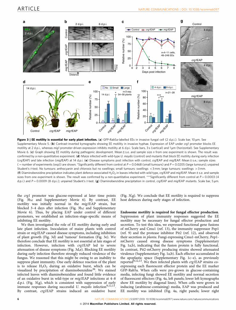
and Aspergillus nidulans38, were shown to act as adaptors thatlink microtubule-dependent motors to EEs. They are thus crucialfor fungal EE motility, and we set out to investigate effectortranscription and secretion in Dhok1 mutants. We found thathok1 null mutants were still able to generate hyphal cells(Supplementary Fig. 4), demonstrating that pheromone signallingpathways were not affected in this strain. Consistent withprevious reports, these hyphae remained short, and weobserved immobile EEs clustered at their cell ends (Fig. 5a,b)(ref. 37). Similar to EAP-expressing strains, hok1 null mutantswere impaired in their ability to cause disease (Fig. 5c,d). Moststrikingly, the transcription and secretion of all tested effectorswas reduced significantly in hok1 null mutants (Fig. 5e–g). Theseresults are therefore consistent with our conclusion that EEmotility is required for the induction of effector production andexocytosis. To test this hypothesis further, we deleted the fungalhomologue of the small GTPase Rab5 (ref. 39), which is of centralimportance for EE function in mammalian cells40. In the absenceof Rab5a, short hyphae formed (Fig. 6a), containing largelyimmobile EE accumulations at the ends of cells (Fig. 6b), whichwere labelled by the EE marker Yup1-GFP (ref. 23). Similar to
No EAP
Distance
Cmu1120
100
80
60
40
20
0n=20 n=4 n=4
No
EA
P
+E
AP
No
EA
P
+E
AP
No
EA
P
+E
AP
No
EA
P
cmu1
PeP
1
pit2
+E
AP
No
EA
P
+E
AP
No
EA
P
+E
AP
*** *** ***
*** ***
Pep1 Pit2 cmu1 pep1 pit2
Tim
eR
elat
ive
inte
nsity
(%
)
120
100
80
60
40
20
0
Rel
ativ
e in
tens
ity (
%)
120 4
3
2
1
0
100
80
60
40
20
0
Rel
ativ
e in
tens
ity (
%)
Fol
d-in
duct
ion
(×10
3 )
120
100
80
60
40
20
0
Rel
ativ
e ex
pres
sion
(%
) 120
100
80
60
40
20
0
Rel
ativ
e ex
pres
sion
(%
) 120
100
80
60
40
20
0
Rel
ativ
e ex
pres
sion
(%
)
Distance
Tim
e
Distance
Tim
e
+EAP No EAP +EAP No EAP +EAPCmu1 PeP1 Pit2
5
EEs EEs EEs EEs EEs EEs
Figure 4 | EE motility is required for effector secretion and transcription of effector-encoding genes in U. maydis (a) Impaired secretion of effectors
during infection. Cmu1-mCherry (Cmu1), Pep1-mCherry (Pep1) or Pit2-mCherry (Pit2) expressed in cells that contain EAP, which impaired EE motility
(contrast-inverted kymographs, stationary signals appear as vertical lines). Cells were pre-grown in glucose- (no EAP) and arabinose-containing medium
(þ EAP). Scale bars, 5 mm (upper panels), 3 s (vertical, lower panels) and 2 mm (horizontal, lower panels). (b) Secretion of fluorescent protein-effector
fusions in plants at 2 d.p.i. Mutants were pre-grown in glucose- (no EAP) and arabinose-containing medium (þ EAP). Mean±s.e. and sample size from
one experiment is shown. The result was confirmed by a non-quantitative experiment. ***Significance at error probability Pcmu1 and Ppit2¼0.0002,
Ppep1o0.0001; unpaired Student’s t-test. Signals were corrected by subtracting intracellular fluorescent background. (c) Induction of fungal effector
transcription during in plant infection. Expression in planta at 1 d.p.i. was compared with transcript levels in liquid culture. Mean±s.e. and sample sizes
(¼ number of experiments) are shown. (d) Relative transcript levels of cmu1, pep1 and pit2 genes in infectious fungal cells grown at 1–2 d.p.i. Cells
were pre-grown in glucose- (no EAP) and arabinose-containing medium (þ EAP). See Supplementary Methods section. Expression of effector genes in
control cells was set to 100%. Mean±s.e. and sample sizes (¼ number of experiments) are shown. *Different from control at Ppep1¼0.0149, **different
from control at Ppit2¼0.0026 and ***different from control at Pcmu1¼0.0009; unpaired Student’s t-test.
NATURE COMMUNICATIONS | DOI: 10.1038/ncomms6097 ARTICLE
NATURE COMMUNICATIONS | 5:5097 | DOI: 10.1038/ncomms6097 | www.nature.com/naturecommunications 7
& 2014 Macmillan Publishers Limited. All rights reserved.

Dhok1 mutants, rab5a null mutants caused only mildpathogenicity symptoms (Fig. 6c,d), and, again, transcriptionand secretion of all tested effectors were significantly reduced(Fig. 6e–g). We conclude that inhibition of EE motility, either byexpression of EAP, deletion of the motor adaptor Hok1 ordeletion of rab5a, reduces effector transcription and secretion,thereby preventing the fungus from successful colonization ofplant tissue.
An endosome-associated MAPK controls effector transcrip-tion. Our observations indicated that Rab5a-positive EEs mediateretrograde signaling that initiates transcription and subsequentsecretion of effectors. We therefore set out to identify potentialsignalling components in this pathway. In mammalian cells,purified Rab5-positive EEs contain numerous long-rangesignalling proteins. Among these are MAPKs of the extracellularsignal-regulated kinase (ERK) signalling pathway, includingB-Raf (B-rapidly accelerated fibrosarcoma), MEK1 (MAPK orERK kinase), ERK1/2 (ERKs 1 and 2) and kinase p38 (MAPKp38; overview in ref. 21). In addition, the GTPases Rap1
(Ras-related protein 1), the adaptor protein Gab2 (growthfactor receptor-bound protein 2; ref. 18) and the EE-associatedadaptor protein, phosphotyrosine interaction, PH domain andleucine zipper containing 1 (APPL1; ref. 41) are involved insignalling. Finally, components of the Akt signalling pathway,such as glycogen synthase kinase-3 beta (GSK3b; ref. 42) and thetransforming growth factor beta (TGF-b)/smad signallingpathway, including the signalling mediators SMAD2 andSMAD4 (ref. 43), the FYVE-domain containing endofin44 andthe SMAD anchor for receptor activation (SARA; ref. 45)transport signals from the plasma membrane to the nucleus(overview in refs 19,21). We used the sequences of these humanproteins (see Methods section for accession codes) in a reciprocalBLAST search and screened the published genome sequence of U.maydis for fungal orthologues of any of these signalling proteins.We did not find APPL-like proteins or components of theSMAD/SARA pathway nor did we identify homologues of Gab2or Rap1. However, we found a GSK3b homologue (64% identicalto human GSK3b; NCBI accession code: XP_756707.1) and theMAPK Kpp2/Ubc2 (refs 46,47; NCBI accession code:XP_759452.1; 49% identical to human ERK1 and 54% identical
Δhok1 Δhok1
Δhok1Δhok1
Δhok1
9 d.p.i.
Tim
e120
100
80
60
40
20
0
120
100
80
60
40
20
0
120
100
80
60
40
20
0
n=20
2 d.p.i.
Cmu1 Cmu1 Cmu1
Control 2 d.p.i. 2 d.p.i.
n=3/4/4
Rel
ativ
e in
tens
ity (
%)
Rel
ativ
e in
tens
ity (
%)
Rel
ativ
e in
tens
ity (
%)
Distance
****
***
Cmu1 Pep1 Pit2 cmu1 pep1 pit2
Rel
ativ
e nu
mbe
r (%
)
Control
No Small Large
n=4 exp.106/113 plants
**
***
75
50
25
0
Contro
l
Δhok1
Contro
l
Δhok1
Contro
l
40
120
100
80
60
20
0
120
Contro
l
100
80
60
40
20
0 0
20
40
60
80
100
120
Rel
ativ
e ex
pres
sion
(%
)
Rel
ativ
e ex
pres
sion
(%
)
Rel
ativ
e ex
pres
sion
(%
)
** ******
Δhok1
Contro
l
Δhok1
Contro
l
Δhok1
Contro
l
EEs
EEs
Δhok1 Δhok1
Figure 5 | Effector secretion and pathogenicity are impaired in hok1 null mutants. (a) Phenotype of hok1 deletion mutants. Scale bar, 20mm. (b) EE
localization and motility in hok1 deletion mutants. Upper panel shows EE clusters (GFP-Rab5a, green, arrowheads) at the poles of the short mutant cells.
Lower kymograph shows the absence of EE motility in Dhok1 mutants. Scale bars, 20mm (upper panel), 3 s (vertical, lower panel) and 1 mm (horizontal,
lower panel). (c) Plant disease symptoms at 9 days after infection of maize plants with control and Dhok1 strains. (d) Plant symptoms at 14 days after
infection with control and hok1 deletion mutants. Mean±s.e. and sample sizes n (¼ number of experiments (exp)) are shown. *Different from control at
error probability P¼0.019; **different from control at error probability P¼0.0047; unpaired Student’s t-test. (e) Secretion of fluorescent effectors,
indicated by the signal intensity at the periphery of the invasive hyphae in plants at 2 d.p.i. Mean±s.e. and sample sizes from one experiment are shown.
The result was confirmed by a non-quantitative experiment. *Different from control at Ppep1¼0.0184, ***different from control at Pcmu1¼0.0002 and
Ppit2o0.0001; unpaired Student’s t-test. (f) Relative transcript levels of cmu1, pep1 and pit2 in cells grown in infected plant tissue at 1–2 d.p.i. Effector
expression in infecting control strains was set as 100%. Mean±s.e. and sample sizes n (¼ number of experiments) are shown. **Significance at error
probability Pcmu1¼0.0021 and Ppep1¼0.0096; ***significance at error probability Ppit2o0.0001; unpaired Student’s t-test. (g) Secretion of Cmu1-mCherry
in control and Dhok1 mutants. Note that secretion of the effector in Dhok1 mutants is reduced compared with control strain. Scale bar, 5mm.
ARTICLE NATURE COMMUNICATIONS | DOI: 10.1038/ncomms6097
8 NATURE COMMUNICATIONS | 5:5097 | DOI: 10.1038/ncomms6097 | www.nature.com/naturecommunications
& 2014 Macmillan Publishers Limited. All rights reserved.
to human MAPK p38 and ERK2) and the MAPK kinase kinaseKpp4/Ubc4 (refs 48,49; 37% identical to human MEK1).U. maydis contains a single MAPK module49. This consists ofthe MAPKK kinase Kpp4/Ubc4 (refs 48,49), the downstreamMAPK Fuz7 (ref. 50) and the MAPKs Kpp2/Ubc2 (refs 46,47),Kpp6 (ref. 51) and Crk1 (refs 52,53 and Supplementary Fig. 5a).All MAPK module components are required for full virulence inU. maydis46,47,49–52 and thus could be involved in virulence-associated signalling. We therefore fused single GFP to allthese kinases and to the GSK3b homologue, and asked whetherany of these show long-range motility within hyphal cells.In these experiments, the MAPK Crk1 showed constant anddirected movements. The MAPKK kinase Kpp4 and GSK3bhomologue occasionally moved, whereas the MAPK kinase Fuz7and the MAPKs Kpp2 and Kpp6 were not motile (SupplementaryFig. 5b). Interestingly, all moving kinases were also concentratedin the fungal nucleus (Supplementary Fig. 5c), which supports thenotion that they serve roles in membrane-to-nucleus signalling.
In the initial localization experiments, very faint Crk1-GFPsignals moved over long distances. To test whether Crk1 istransported via retrograde moving EEs, we fused the endogenous
copy of crk1 to a triple-GFP tag and co-expressed the fusionprotein with endosomal mCherry-Rab5a in a strain that formshyphal cells in liquid culture. We observed that Crk1-GFP3
localized to rapidly moving Rab5a-positive EEs (Fig. 7a andSupplementary Movie 7). Retrograde Crk1-GFP3 signals travelledfor up to 30 mm (Fig. 7a), which is consistent with the notion thatthe MAPK could participate in retrograde signalling to thenucleus during plant infection. To test this idea further, weexpressed Crk1-GFP3 in the solo-pathogenic strain SG200 andinvestigated Crk1 motility in planta. We found that the MAPKalso travelled over long distances in invasive hyphae and localizedin the nucleus (Fig. 7b and Supplementary Fig. 6a). This resultsuggests that Crk1 could participate in transmitting signals fromthe hyphal tip to the nucleus, thereby controlling effectortranscription and secretion. We addressed this possibility bydeleting crk1 in a strain that expressed Cmu1-mCherry.Surprisingly, we found that effector secretion was significantlyincreased in the absence of Crk1 (Fig. 7c, both images areidentically processed; Fig. 7d), suggesting an inhibitory signallingfunction. We therefore set out to test whether expression of cmu1was induced by deletion of the MAPK crk1. The resultant crk1
Δrab5a Δrab5a
EEs
2 d.p.i.
Cmu1 Cmu1 Cmu1
Tim
e
DistanceΔrab5aControl
Control 2 d.p.i. �rab5a 2 d.p.i. �rab5a
9 d.p.i.
Δrab5a
Rel
ativ
e nu
mbe
r (%
) 75
50
25
0
Contro
l
No Small Large
n=3 exp.70 plants
Δrab5a
120
100
80
60
40
20
0
n=20
Rel
ativ
e in
tens
ity (
%)
***
Cmu1
Contro
l
Δrab5a
120
100
80
60
40
20
0
Rel
ativ
e in
tens
ity (
%)
***
Pep1 Pit2 cmu1 pep1 pit2
Contro
l
Δrab5a
120
100
80
60
40
20
0
Rel
ativ
e in
tens
ity (
%)
***
Contro
l
Δrab5a
120
100
80
60
40
20
0Rel
ativ
e ex
pres
sion
(%
)
*
Contro
ln=4
Δrab5a
120
100
80
60
40
20
0Rel
ativ
e ex
pres
sion
(%
)
**
Contro
l
Δrab5a
120
100
80
60
40
20
0Rel
ativ
e ex
pres
sion
(%
)
**
**
*
Contro
l
Figure 6 | Effector secretion and pathogenicity are impaired in rab5a null mutants. (a) Phenotype of rab5a deletion mutants. Scale bar, 20mm. (b) EE
localization and motility in rab5a deletion mutants. Upper panel shows EE clusters (Yup1-GFP, green, arrowheads) at the poles of the short mutant cells.
Lower kymograph shows the absence of EE motility in Drab5a mutants. Scale bars, 3mm (upper panel), 5 s (vertical, lower panel) and 2mm (horizontal,
lower panel). (c) Plant disease symptoms at 9 days after infection of maize plants with control and Drab5a strains. (d) Plant symptoms at 14 days
after infection with control and rab5a deletion mutants. Mean±s.e. and sample sizes n (¼ number of experiments (exp)) are shown. *Different from
control at error probability P¼0.019; **different from control at error probability P¼0.0047; unpaired Student’s t-test. (e) Secretion of fluorescent
effectors indicated by the signal intensity at the periphery of the invasive hyphae in plants at 2 d.p.i. Mean±s.e. and sample size n from one experiment is
shown. The result was confirmed by a non-quantitative experiment. ***Significance at error probability Po0.0001; unpaired Student’s t-test. (f) Relative
transcript levels of cmu1, pep1 and pit2 in cells grown in infected plant tissue at 1–2 d.p.i. Effector expression in infecting control strains was set
as 100%. Mean±s.e. and sample sizes n (¼ number of experiments) are shown. *Significance at error probability Pcmu1¼0.0257, **significance at error
probabilities Ppep1¼0.0083 and Ppit2¼0.007; unpaired Student’s t-test. (g) Secretion of Cmu1-mCherry in Control and Drab5a mutants. Note that
secretion in Drab5 is reduced, compared to the control strain. Scale bar, 5 mm.
NATURE COMMUNICATIONS | DOI: 10.1038/ncomms6097 ARTICLE
NATURE COMMUNICATIONS | 5:5097 | DOI: 10.1038/ncomms6097 | www.nature.com/naturecommunications 9
& 2014 Macmillan Publishers Limited. All rights reserved.

null mutants, however, showed a defect in morphology and weresignificantly impaired in penetration of the plant. Consequently,RT–PCR analysis of cmu1 could not be reliably carried outbecause of the small proportion of fungal hyphae that successfullypenetrated host tissue, which would normally express cmu1specifically. To circumvent this problem, we made a transcrip-tional fusion of cytoplasmic GFP under the control of the cmu1promoter (Pcmu1) and quantitatively analysed the GFP signalintensity in control hyphae at 1 d.p.i., when the fungus had notyet invaded the plant, and 2 d.p.i., when fungal cells were foundinside the plant tissue. We found only traces of cytoplasmic GFPat 1 d.p.i., but a strong fluorescent signal when the fungus hadpenetrated the plant (Supplementary Fig. 6b,c). This is consistentwith the induction of Pcmu1 during plant invasion. Significantly,less fluorescence was detected in Dhok1 mutants (SupplementaryFig. 6b,c), which is consistent with the role of EE motility ineffector transcription. We next generated a crk1 null mutant,expressing the Pcmu1-GFP construct. We infected plants and
visualized infection structures after 2 d.p.i. Interestingly, we founda significant increase in cytoplasmic GFP fluorescence, indicatingthat Pcmu1 is more strongly induced in Dcrk1 mutants (Fig. 7e,f).When considered together, these results provide evidence thatthe EE-associated MAPK, Crk1, acts as a repressor of effectortranscription.
DiscussionAn increasing body of evidence suggests that pathogenic fungiand oomycetes secrete a large repertoire of effector proteins intoplant cells to suppress plant immunity and facilitate biotrophicgrowth1,3–5,8–13,29. In this report, we provide evidence thatmotility of EEs is essential for effector production and subsequentsecretion by the filamentous fungal pathogen U. maydis. Thisconclusion is supported by several independent experimentalstrategies. First, we generated an artificial anchor protein EAPthat consists of a mutated kinesin-1 motor head that tightly binds
Crk1
Control
Cmu12 d.p.i.
Control
Cmu1***500
400
300
200
100
0n=20
Crk1
Nucleus
Tim
e
Distance
Tip
Merge
EEs (Rab5a)
Tim
e
Distance
Tip
Contro
l
Δcrk1
Rel
ativ
e in
tens
ity (
%)
Pcmu1-GFP
***250
200
150
100
50
0n=29/25
Contro
l
Δcrk1
Rel
ativ
e in
tens
ity (
%)
Δcrk1
Δcrk1
Pcmu1-GFP2 d.p.i.
Figure 7 | EE-associated localization of the MAPK Crk1 and its role in effector secretion and transcription. (a) Kymographs showing motility
of Crk1-GFP3 (Crk1) on EEs (Rab5a, arrowheads). Direction towards the hyphal tip is indicated (Tip). Note that the endogenous copy of crk1 was fused to a
triple-GFP tag. Motility was observed in a photo-bleached area. The upper two kymographs are contrast inverted. Scale bars, 5 s (vertical) and 5 mm
(horizontal). See Supplementary Movie 7. (b) Kymographs showing motility of Crk1-GFP3 in a fungal hypha during invasion of plant tissue. Note that
trajectories of Crk1-GFP3 signals are discontinuous, as signals move in and out of the confocal focus. Scale bars, 3mm (upper panel), 5 s (vertical, lower
panel) and 2 mm (horizontal, lower panel). See Supplementary Movie 8. (c) Secretion of fluorescent Cmu1 effector in control and crk1 null mutants (Dcrk1).
Both images were scaled identically to show different degree of secreted Cmu1-mCherry. Inset shows Cmu1-mCherry after scaling the control images. Scale
bar, 5 mm. (d) Secretion of fluorescent Cmu1 indicated by the signal intensity at the periphery of the invasive control and Dcrk1 mutant hyphae in plants
at 2 d.p.i. Mean±s.e. and sample size n from two experiments is shown. ***Significance at error probability P¼0.0005; unpaired Student’s t-test.
(e) Expression of cytoplasmic GFP under the cmu1 promoter in invasive control and Dcrk1 mutant hyphae in plants at 2 d.p.i. Both images were scaled
identically. Scale bar, 5 mm. (f) Expression of cytoplasmic GFP under the cmu1 promoter in invasive control and Dcrk1 mutant hyphae in plants at
2 d.p.i. Mean±s.e. and sample size n from two experiments is shown. ***Significance at error probability Po0.0001; unpaired Student’s t-test.
ARTICLE NATURE COMMUNICATIONS | DOI: 10.1038/ncomms6097
10 NATURE COMMUNICATIONS | 5:5097 | DOI: 10.1038/ncomms6097 | www.nature.com/naturecommunications
& 2014 Macmillan Publishers Limited. All rights reserved.
to microtubules31 and the C-terminal Phox domain of theSNARE Yup1 (ref. 23). Phox domains bind to the lipid PtdIns3P(ref. 54), which is, itself, enriched in the membrane of EEs55.Strong expression of the chimaeric EAP almost abolished EEmotility and significantly reduced the secretion of three testedeffector proteins into the apoplastic space between the fungus andthe plant cell. One could argue that the presence of EAP simplyblocks microtubule-dependent delivery of effector proteins. Weconsider this possibility unlikely, however, because microtubule-based membrane trafficking in U. maydis depends on kinesin-1,kinesin-3 and dynein30, and EAP expression did not impairmotility of fluorescent kinesin-3 or dynein nor did it affectdelivery of chitin synthase Mcs1 to the growing tip of the cell, aprocess depending on kinesin-1 (ref. 33). We conclude from theseresults that EAP specifically blocks EE movement but does notaffect other membrane trafficking pathways. Consequently, weconsider it very likely that impaired effector secretion is aconsequence of reduced EE motility. This notion is furthersupported by the observation that hok1 and rab5a null mutantsalso show almost identical defects in effector secretion. It wasrecently reported that hok1 null mutants have a defect in motor-to-cargo coupling, which results in a severe reduction in EEtransport37. A similar defect is seen in rab5a null mutants,although these cells are most likely impaired in various EEfunctions, because this GTPase is of central importance formotility, identity and function of these organelles40. However,when taken together, all three mutant lines (EAP-expressing,hok1 and rab5a null mutants) show very significant reductions insecretion of the three tested effectors. Motile EEs therefore appearto be essential during the interaction of the fungal pathogen andthe plant cell.
How then do moving EEs support effector secretion duringplant infection? We recognized that elongated fungal cellsface a considerable challenge during host infection in commu-nicating between the invading distal tip and the nucleus. This isclearly necessary in order to produce effector proteins specificallyduring the very early stages of infection. In U. maydis, thenucleus is, for instance, B22mm behind the invading hyphal tip.In the crowded cytoplasm, even a few micrometres pose aninsurmountable problem because passive diffusion is severelyhindered56. This suggests that communication between the hyphaltip and nucleus involves an active transport-dependent signallingmechanism. In mammalian neurons, such long-range signallinginvolves EEs, which travel along microtubules, deliveringsignalling components from the plasma membrane to thenucleus19–21. Long-range and microtubule-dependent motility ofEEs have been reported in U. maydis23,26 and other fungi24,25
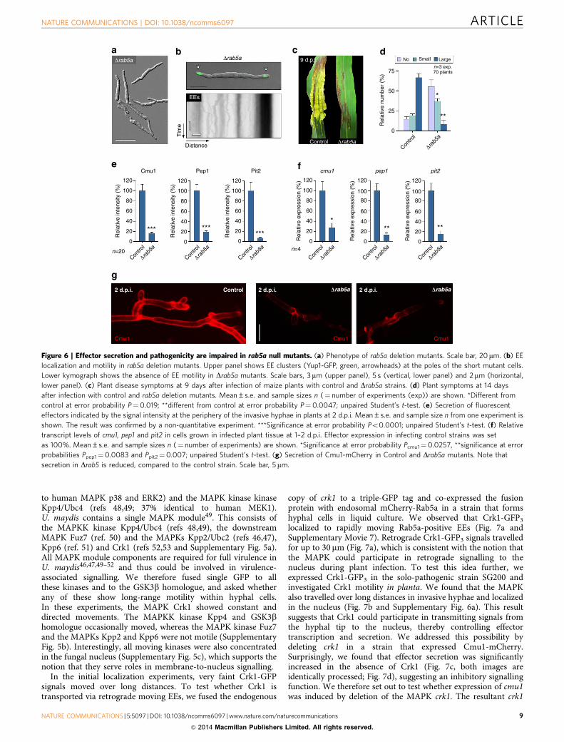
(overview in ref. 22), and we have previously speculated thatmoving fungal EEs might participate in retrograde signalling57.The evidence provided here is consistent with such a function. Theobserved defects in effector secretion, for example, are most likelyowing to impaired effector gene transcription as a consequence ofblocked EE-dependent retrograde signalling (Fig. 8). Thus, EEmotility acts at the level of transcription, which implies a novel,endosome-based signalling mechanism that mediates communi-cation between the fungal nucleus and invading hyphal tip, andwhich is essential for fungal pathogenicity.
A role for fungal EEs in long-range signalling has not hithertobeen described. Indeed, our understanding of the signallingpathways associated with fungal EEs is in its infancy. Inmammalian cells, several signalling pathways are located onEEs that include the MAPKs of the ERK signalling pathway19,21.Our search for homologues of retrograde signalling components,associated with EEs in humans, revealed known components of aMAPK module. This included the MAPK Kpp2/Ubc2, which isshown to be involved in pathogenic development46,47, and
therefore is a good candidate for EE-associated signalling.However, Kpp2 did not show directed movement, whereas theMAPK Crk1 was transported by EEs over long distances. Thisobservation, coupled with localization of Crk1 in the fungalnucleus, and de-repressed effector transcription and secretion,strongly suggests that Crk1 participates in long-range EE-basedsignalling. We also find the MAPK kinase Fuz7 and the MAPKKkinase Kpp4 in the nucleus, and at least Kpp4 showed occasionalmotility. Therefore, we consider it most likely that theMAPK module in U. maydis delivers signals from the plasmamembrane into the nucleus. Crk1 appears to be of keyimportance in this pathway, which adds to the known roles ofCrk1 in morphogenesis52 and mating53.
Surprisingly, crk1 null mutants showed increased expressionand secretion of effector protein. This suggests that Crk1 is anegative regulator that represses effector transcription. Wespeculate that it might therefore counteract an unknown EE-bound phosphatase to collectively fine-tune expression of effectorgenes during infection. Such balanced activities of phosphatasesand kinases mediate many essential cellular processes, includingcell-cycle progression58, axonal migration and transport59,60, andvarious signalling pathways61. Finally, it is worth mentioning thatGsk3b also showed directed movement in the cell. MammalianGsk3b is a direct target of the serine/threonine kinase Akt(ref. 62), and both dynamically interact with the Rab5-effectorAPPL1 on EEs42. U. maydis does not contain an APPLhomologue, and whether the Gsk3b homologue is associatedwith fungal EEs is not yet clear. However, the identification of anEE-associated and MAPK-based signalling pathway in effectorproduction raises the possibility that EEs have more widespreadroles in the plasma membrane-to-nucleus signalling than hashitherto been recognized in pathogenic fungi.
Tip growth
EE motility
Effector transcription
Defence
Invasion site selection
Endocytosis-dependent plant recognition
Effector production and secretion
Figure 8 | Model for retrograde EE motility in effector-mediated plant
invasion by pathogenic fungi. The invading hyphal tip perceives plant cues
during the initial stages of infection. Endocytic uptake relays this
information to motile EEs that transport the signal to the nucleus. This
retrograde signalling mechanism triggers effector gene expression.
Subsequently, effectors are secreted at the hyphal tip to suppress plant
immunity and facilitate fungal invasion.
NATURE COMMUNICATIONS | DOI: 10.1038/ncomms6097 ARTICLE
NATURE COMMUNICATIONS | 5:5097 | DOI: 10.1038/ncomms6097 | www.nature.com/naturecommunications 11
& 2014 Macmillan Publishers Limited. All rights reserved.

MethodsStrains and plasmids. All U. maydis strains were constructed in the geneticbackground of AB33 (ref. 63) or SG200 (ref. 35; Table 1). To observe EEs motilityduring plant infection, plasmid poGRab5a (ref. 64) was integrated ectopically intostrain SG200, resulting in SG200GRab5a. Strains AB33mChRab5a andAB33Kin3G_mChRab5a were generated by integrating the plasmid pomChRab5ainto AB33 and AB33Kin3G (ref. 26), respectively. SG200DKin3 and SG200DHok1were generated by deleting the endogenous kin3 or hok1 gene using pDKin3(ref. 27) or pHDHok1 (ref. 37), respectively. SG200Cmu1Ch_DHok1,SG200Pep1Ch_DHok1 and SG200nPit2Ch_DHok1 were generated by deleting theendogenous hok1 gene using pNDHok1 (ref. 37). Deletions were confirmed bySouthern blot. Ectopic integration of plasmid pCoGRab5a (ref. 65) resulted inSG200DKin3_GRab5a. All plasmids were generated by standard cloningprocedures or in vivo recombination in the yeast Saccharomyces cerevisiae66.Detailed cloning information is provided in the Supplementary Methods.
Growth conditions. All cultures of U. maydis strains were grown overnight at28 �C in either YEPSlight medium (1.0% yeast extract, 0.4% peptone and 0.4%sucrose) or complete medium (CM), containing 1% (w/v) glucose (CMglc) or1% (w/v) arabinose (CMara) at 200 r.p.m. Hyphal growth was induced bytransferring cells, grown in CMglc, into nitrate minimal (NM) medium supple-mented with 1% (w/v) glucose. Cells were grown under these conditions for 5–10 hat 200 r.p.m. (28 �C).
Microscopic techniques. Spinning disc confocal microscopy of infected plantleaves was performed using a VisiScope Confocal Cell Explorer (Visitron System,Munich, Germany) that consisted of an IX81 motorized inverted microscope(Olympus, Hamburg, Germany), a CSU-X1 Spinning Disc unit (Yokogawa, Tokyo,Japan), a PlanApo UPlanSApo � 63/1.35 oil objective (Olympus, Hamburg,Germany) and a Photometrics CoolSNAP HQ2 camera (Roper Scientific,Germany). The fluorescent tags and dyes were excited using a VS-LMS6 LaserMerge System with two solid-state lasers (488 nm/100 mW and 561 nm/100 mW,Visitron System) and a 405-nm/100-mW diode laser, which was 50/50 split. Forphoto-activation studies, the 405-nm/100-mW diode laser split and directed into a2D-VisiFRAP Realtime Scanner (Visitron System). Co-observation of GFP andRFP was done using an OptoSplit II LS Image Splitter (Cairn Research Limited,Faversham, UK). All parts of the system were under the control of the softwarepackage VisiView (Visitron System).
Laser-based epi-fluorescence microscopy of cultured cells was done aspreviously described27. In brief, cells were placed onto a 2% (w/v) agar cushion andimmediately observed using an IX81 motorized inverted microscope, equippedwith a PlanApo � 100/1.45 Oil TIRF (Olympus, Hamburg, Germany). A VS-LMS4Laser-Merge-System with solid-state lasers (488 nm/50 mW or 75 mW and561 nm/50 mW or 75 mW; Visitron System) was used to excite the fluorescentprotein tags. Photo-bleaching experiments were done using a 405-nm/60-mWdiode laser, which was dimmed by an ND 0.6 filter, resulting in 15 mW outputpower, coupled into the light path by an OSI-IX 71 adaptor (Visitron System). Thesystem was regulated by a UGA-40 controller (Rapp OptoElectronic GmbH,Hamburg, Germany) and a VisiFRAP 2D FRAP control software for MetaSeries7.5.1 (Visitron System). Dual imaging of GFP and RFP was carried out using aDual-View Micro imager (Photometrics, Tucson, USA) equipped with a dual-linebeam splitter (z491/561, Chroma, Rockingham, USA) with an emission beamsplitter (565 DCXR, Chroma, Rockingham, USA), an ET-Bandpass 525/50(Chroma, Rockingham, USA) and a BrightLine HC 617/73 (Semrock, Rochester,USA). A Photometrics CoolSNAP HQ2 camera was used to capture all images. Allparts of the system were under the control of the software package MetaMorph(MDS Analytical Technologies, Winnersh, UK).
Scanning electron microscopy was performed using yeast-like cells of strainSG200DHok1, which grown on CM agar supplemented with 1% glucose and 1%charcoal for 5 days at 28 �C. Images were acquired using Nikon D5000 camera thatwas connected to the Nikon stereomicroscope SMZ800 (Nikon, Kingston, Surrey,UK). The 10-mm diameter agarose discs containing yeast-like and hyphal cellswere attached to a cryostage and rapidly frozen in liquid nitrogen slush, followedby water sublimation at � 95 �C for 3 min in Jeol JSM-6390LV scanning electronmicroscope (JEOL, Ltd, Welwyn Garden City, UK). This was followed by goldsputter coating using an Alto 2100 chamber (Gatan Ltd., Oxfordshire, UK) andobservation in a Jeol JSM-6390LV scanning electron microscope (JEOL, Ltd).
Quantitative analysis of motility ex planta. EE motility in the presence orabsence of EAP was measured in cells grown in NMglu (no EAP) and NMara
(þ EAP, expression induced for 5 h). GFP-Rab5a signals that did not move for 10 swere considered as non-motile EEs. The effect of EAP on flux of fluorescent dyneinwas measured within photo-bleached hyphal cells, beginning 5 mm behind the tipand extending for 20mm in NMglu (15 h) and NMara (expression induced for 15 h).Motility was analysed in kymographs. Kinesin-3 motility was measured in photo-bleaching apical parts of yeast-like and hyphal cells, using a 405-nm/60-mWdiode laser, dimmed to 15 mW output using kymographs. Only signals that did notco-localize with mCherry-Rab5a were counted and compared with all kinesin-3-GFP signals, irrespective of co-localization with EEs. The degree of secretion of
Mcs1-GFP3 was estimated by measuring the average intensity of the GFP signal inthe apical parts of cells forming a medium-sized bud. The intensity values werecorrected by the cytoplasmic background.
Fungal plant infection. U. maydis strains were grown in either YEPSlight, CMglc orCMara to an OD600 of 0.8–1.0. Cells were harvested by centrifugation and re-suspended in water. A quantity of 0.2 or 0.5 ml of this cell suspension (OD600B1.0)were injected into 10-day-old maize plants (Golden Bantam; Chase Organics Ltd.,Hersham, Surrey, UK). Pathogenicity assays were performed as described pre-viously67. In brief, infected plants were grown in a GroBank (CSF Plant Climatics,Wertingen, Germany; 16 h light, 8 h darkness, 28 �C). Eight- to nine-day-old plantswere infected by injection of 0.5 ml U. maydis cells, pre-grown to OD600¼ 0.8 andwashed with water into the stem of the plant. Disease symptoms were scored at9–14 days after infection. The scoring criteria were as follows: ‘no tumours’(chlorosis and antocyanin formation), ‘small tumours’ (chlorosis, antocyaninformation and tumours up to 3 mm) and ‘large tumours’ (chlorosis, antocyaninformation and tumours over 3 mm). All infection assays were done in at least threeexperiments and compared by Student’s t-test by using the software Prism 4.03(GraphPad Software Inc., La Jolla, CA, USA).
Microscopy of infected plant tissue. Propidium iodide/wheat germ agglutinin-AF488 and diaminobenzidine (Sigma-Aldrich, UK) staining in infected plant leaveswas done as described previously67. In brief, infected leaf sections were incubatedin staining solution (1 mg ml� 1 diaminobenzidine, 0.05% (v/v) Tween 20 and10 mM Na2HPO4) under mild vacuum for 60 min, followed by 4 h shaking at80–100 r.p.m. at room temperature. Samples were incubated in ethanol:aceticacid:glycerol (3:1:1) and stored in fresh de-stain solution at 4 �C (at 90 �C for15 min). Before microscopy, samples were washed in PBS, pH 7.4, andmicroscopically analysed.
To visualize nucleus and effector secretion in early stages of infection, plantswere infected with the strain SG200G3_NLSR3 and SG200Cmu1Ch_G3 (forexperimental usage of strains, see Supplementary Table 2). An B1� 1-cm regionat B5 cm beneath the infection point was taken from the third inner leaf andplaced on Carolina observation Gel (Carolina Biological Supply Company,Burlington, USA). The invasion site was identified by bright-field microscopy,followed by acquiring Z-stacks capturing red and green fluorescence, using anOptoSplit II LS Image Splitter. Z-stacks covered 30 mm at 0.2 mm step size(exposure time of 200 ms, binning 1 and the 488-/561-nm laser at 20%/50%). Aftermerging stacks, maximum projections and three-dimensional reconstructions weredone using MetaMorph (Molecular Devices, Winnersh, UK).
EE motility inside invading fungal cells was measured after infection of maizeplants with strain SG200GRab5a. Leaf samples were collected at several timepoints post infection, and GFP-Rab5a motility was recorded in randomly selectedinvasive hyphal cells. The proportion of moving EEs was determined in movies,covering 8–10 s observation time.
To analyse EE motility in fungal cells at 1 d.p.i., infected leaf tissue was preparedfor microscopic analysis. The samples were incubated in 2 mg ml� 1 Calcofluorsolution (F3543-Fluorescent Brightener 28, Sigma, UK) for 30 s, water rinsed andobserved on Carolina Observation Gel, using a spinning disc confocal microscope.Invasion sites were identified and Z-stacks were generated as described above.Calcofluor was excited using a 405-nm laser at 15% output power. In photo-activation experiments, paGFP-Rab5a was excited at the invasion site using afocussed 405-nm laser at 15% output power, followed by immediate acquisitionof 150 planes using the OptoSplit II LS Image Splitter (Cairn Research Limited),and the 488- and 561-nm observation lasers at 30% and 50% output power,respectively. Motility of photo-activated EEs was analysed in kymographs by usingMetaMorph.
To test for crg1 or mig1 promoters activities in planta, maize plants wereinfected with 0.5 ml suspension of strain SG200crgG and SG200migG, and pre-grown in CMara or CMglc. Maize tissue samples were collected and observed usingan IX81 microscope and a VS-LMS4 Laser Merge System, the 488-nm observationlaser at 100% output power at an exposure time of 150 ms and image binning 1.
Quantitative analysis of fluorescent intensity of Cmu1-mCherry, Pep1-mCherryand Pit2-mCherry was done by measuring the average fluorescent intensityat the periphery of hyphal cells of strains SG200GRab5a_cEAP_Cmu1Ch,SG200GRab5a_cEAP_Pep1Ch and SG200GRab5a_cEAP_DPit2_nPit2Ch thatshowed reduced or no EE motility. Images were taken at 200 ms exposure time. Theaverage fluorescence intensity over length of B5 mm was measured and correctedfor the background intensity in the cytoplasm. All statistic testing was done usingthe software Prism 4.03 (GraphPad Software Inc.).
To analyse expression of cytoplasmic GFP expressed under the cmu1 promoter,images (200 ms exposure time, spinning disc microscope) were taken of strainsSG200Pcmu1-GFP, SG200DHok1_Pcmu1-GFP and SG200DCrk1_Pcmu1-GFP at1 and 2 d.p.i. The average fluorescent intensity was measured and corrected for thebackground adjacent to the cell. All statistic testing was done using the softwarePrism 4.03 (GraphPad Software Inc.).
Quantitative RT–PCR analysis. Quantitative analysis of effector expression wasperformed using qRT–PCR, according to published protocols9,11,13,68. In brief, leaf
ARTICLE NATURE COMMUNICATIONS | DOI: 10.1038/ncomms6097
12 NATURE COMMUNICATIONS | 5:5097 | DOI: 10.1038/ncomms6097 | www.nature.com/naturecommunications
& 2014 Macmillan Publishers Limited. All rights reserved.

samples were removed 2–4 cm beneath the injection point and harvested from fourindependent experiments, 30 plants each. The material was frozen in liquidnitrogen, ground by mortar and pestle, and total RNA was extracted by using anRNeasy Plant Mini Kit (Qiagen, Hilden, Germany) with DNase I treatment,according to the manufacturer’s instructions. cDNA was synthesized usingSuperScript III First-Strand Synthesis SuperMix (Invitrogen, Carlsbad, USA). qRT–PCR was performed with an Mx3005P thermocycler (Stratagene, La Jolla, USA) byusing Brilliant III Ultra-Fast SYBR Green QPCR Master Mix (Stratagene) andB250 or 500 ng cDNAs, estimated by NanoDrop ND-1000 (LabTech InternationalLtd, East Sussex, UK), from samples of liquid cultures or plants, respectively. qRT–PCR conditions were as follows: 3 min at 95 �C, followed by 40 cycles of 95 �C for20 s and 60 �C for 20 s. To normalize expression levels, the constitutively expressedpeptidyl–prolyl cis–trans isomerase gene (ppi) was used, as described previously9.
Bioinformatics. Putative components of EE-associated signalling were identifiedby reciprocal BLAST search (http://blast.ncbi.nlm.nih.gov/Blast.cgi). All proteinsequences used were fetched from the NCBI server (http://www.ncbi.nlm.nih.gov/pubmed). Accession codes of the protein sequences used in this approach were asfollows: human ERK1: P27361.4; human ERK2: P28482.3; human MEK1:NP_005912.1; human MAPK p38: NP_002736.3; human Gab2: BAA76737.1;human RAP1: ABA64473.1; human GSK3beta: NP_002084.2; mouse RAF-B:NP_647455.3; human APPL1: NP_036228.1; human APPL2: NP_001238833.1;human SMAD2: AAC39657.1; human SMAD4: Q13485.1; human endofin:AAL30772.1 and human SARA: NP_004790.2.
References1. Dodds, P. N. & Rathjen, J. P. Plant immunity: towards an integrated view of
plant-pathogen interactions. Nat. Rev. Genet. 11, 539–548 (2010).2. Jones, J. D. & Dangl, J. L. The plant immune system. Nature 444, 323–329
(2006).3. Kale, S. D. & Tyler, B. M. Entry of oomycete and fungal effectors into plant and
animal host cells. Cell. Microbiol. 13, 1839–1848 (2011).4. Valent, B. & Khang, C. H. Recent advances in rice blast effector research. Curr.
Opin. Plant Biol. 13, 434–441 (2010).5. Rafiqi, M., Ellis, J. G., Ludowici, V. A., Hardham, A. R. & Dodds, P. N.
Challenges and progress towards understanding the role of effectors in plant-fungal interactions. Curr. Opin. Plant Biol. 15, 477–482 (2012).
6. Brefort, T. et al. Ustilago maydis as a pathogen. Annu. Rev. Phytopathol. 47,423–445 (2009).
7. Mendoza-Mendoza, A. et al. Physical-chemical plant-derived signals inducedifferentiation in Ustilago maydis. Mol. Microbiol. 71, 895–911 (2009).
8. Doehlemann, G. et al. Reprogramming a maize plant: transcriptional andmetabolic changes induced by the fungal biotroph Ustilago maydis. Plant J. 56,181–195 (2008).
9. Doehlemann, G. et al. Pep1, a secreted effector protein of Ustilago maydis, isrequired for successful invasion of plant cells. PLoS Pathog. 5, e1000290 (2009).
10. Hemetsberger, C., Herrberger, C., Zechmann, B., Hillmer, M. & Doehlemann,G. The Ustilago maydis effector Pep1 suppresses plant immunity by inhibitionof host peroxidase activity. PLoS Pathog. 8, e1002684 (2012).
11. Doehlemann, G., Reissmann, S., Assmann, D., Fleckenstein, M. & Kahmann, R.Two linked genes encoding a secreted effector and a membrane protein areessential for Ustilago maydis-induced tumour formation. Mol. Microbiol. 81,751–766 (2011).
12. Mueller, A. N., Ziemann, S., Treitschke, S., Assmann, D. & Doehlemann, G.Compatibility in the Ustilago maydis-maize interaction requires inhibition ofhost cysteine proteases by the fungal effector Pit2. PLoS Pathog. 9, e1003177(2013).
13. Djamei, A. et al. Metabolic priming by a secreted fungal effector. Nature 478,395–398 (2011).
14. Grand, C., Sarni, F. & Lamb, C. J. Rapid induction by fungal elicitor of thesynthesis of cinnamyl-alcohol dehydrogenase, a specific enzyme of ligninsynthesis. Eur. J. Biochem. 169, 73–77 (1987).
15. Lamb, C. J., Lawton, M. A., Dron, M. & Dixon, R. A. Signals and transductionmechanisms for activation of plant defenses against microbial attack. Cell 56,215–224 (1989).
16. Apostol, I., Heinstein, P. F. & Low, P. S. Rapid Stimulation of an oxidative burstduring elicitation of cultured plant cells: role in defense and signal transduction.Plant Physiol. 90, 109–116 (1989).
17. Flor-Parra, I., Vranes, M., Kamper, J. & Perez-Martin, J. Biz1, a zinc fingerprotein required for plant invasion by Ustilago maydis, regulates the levels of amitotic cyclin. Plant Cell 18, 2369–2387.
18. Delcroix, J. D. et al. NGF signaling in sensory neurons: evidence that earlyendosomes carry NGF retrograde signals. Neuron 39, 69–84 (2003).
19. Scita, G. & Di Fiore, P. P. The endocytic matrix. Nature 463, 464–473 (2010).20. Gould, G. W. & Lippincott-Schwartz, J. New roles for endosomes: from
vesicular carriers to multi-purpose platforms. Nat. Rev. Mol. Cell Biol. 10,287–292 (2009).
21. Miaczynska, M., Pelkmans, L. & Zerial, M. Not just a sink: endosomes incontrol of signal transduction. Curr. Opin. Cell. Biol. 16, 400–406 (2004).
22. Steinberg, G. Endocytosis and early endosome motility in filamentous fungi.Curr. Opin. Microbiol. 20C, 10–18 (2014).
23. Wedlich-Soldner, R., Bolker, M., Kahmann, R. & Steinberg, G. A putativeendosomal t-SNARE links exo- and endocytosis in the phytopathogenic fungusUstilago maydis. EMBO J. 19, 1974–1986 (2000).
24. Abenza, J. F., Pantazopoulou, A., Rodriguez, J. M., Galindo, A. & Penalva, M. A.Long-distance movement of Aspergillus nidulans early endosomes onmicrotubule tracks. Traffic 10, 57–75 (2009).
25. Seidel, C., Moreno-Velasquez, S. D., Riquelme, M. & Fischer, R. Neurosporacrassa NKIN2, a kinesin-3 motor, transports early endosomes and is requiredfor polarized growth. Eukaryot. Cell 12, 1020–1032 (2013).
26. Schuster, M. et al. Kinesin-3 and dynein cooperate in long-range retrogradeendosome motility along a nonuniform microtubule array. Mol. Biol. Cell 22,3645–3657 (2011).
27. Schuster, M., Lipowsky, R., Assmann, M. A., Lenz, P. & Steinberg, G. Transientbinding of dynein controls bidirectional long-range motility of earlyendosomes. Proc. Natl Acad. Sci. USA 108, 3618–3623 (2011).
28. Kamper, J. et al. Insights from the genome of the biotrophic fungal plantpathogen Ustilago maydis. Nature 444, 97–101 (2006).
29. Tanaka, S. et al. A secreted Ustilago maydis effector promotes virulence bytargeting anthocyanin biosynthesis in maize. Elife 3, e01355 (2014).
30. Schuchardt, I., Assmann, D., Thines, E., Schuberth, C. & Steinberg, G.Myosin-V, Kinesin-1, and Kinesin-3 cooperate in hyphal growth of the fungusUstilago maydis. Mol. Biol. Cell 16, 5191–5201 (2005).
31. Straube, A., Hause, G., Fink, G. & Steinberg, G. Conventional kinesin mediatesmicrotubule-microtubule interactions in vivo. Mol. Biol. Cell 17, 907–916(2006).
32. Bottin, A., Kamper, J. & Kahmann, R. Isolation of a carbon source-regulatedgene from Ustilago maydis. Mol. Gen. Genet. 253, 342–352 (1996).
33. Schuster, M. et al. Myosin-5, kinesin-1 and myosin-17 cooperate in secretion offungal chitin synthase. EMBO J. 31, 214–227 (2012).
34. Basse, C. W., Stumpferl, S. & Kahmann, R. Characterization of a Ustilagomaydis gene specifically induced during the biotrophic phase: evidence fornegative as well as positive regulation. Mol. Cell. Biol. 20, 329–339 (2000).
35. Bolker, M., Genin, S., Lehmler, C. & Kahmann, R. Genetic regulation of matingand dimorphism in Ustilago maydis. Can. J. Bot. 73, 320–325 (1995).
36. Thordal-Christensen, H., Zhang, Z., Wei, Y. & Collinge, D. B. Subcellularlocalization of H2O2 in plants. H2O2 accumulation in papillae andhypersensitive response during the barley-powdery mildew interaction. Plant J.11, 1187–1194 (1997).
37. Bielska, E. et al. Hook is an adapter that coordinates kinesin-3 and dyneincargo-attachment on early endosomes. J. Cell. Biol. 204, 989–1007 (2014).
38. Zhang, J., Qiu, R., Arst, Jr H. N., Penalva, M. A. & Xiang, X. HookA is a noveldynein-early endosome linker critical for cargo movement in vivo. J. Cell. Biol.204, 1009–1026 (2014).
39. Fuchs, U., Hause, G., Schuchardt, I. & Steinberg, G. Endocytosis is essential forpathogenic development in the corn smut fungus Ustilago maydis. Plant Cell18, 2066–2081 (2006).
40. Zerial, M. & McBride, H. Rab proteins as membrane organizers. Nat. Rev. Mol.Cell Biol. 2, 107–117 (2001).
41. Miaczynska, M. et al. APPL proteins link Rab5 to nuclear signal transductionvia an endosomal compartment. Cell 116, 445–456 (2004).
42. Schenck, A. et al. The endosomal protein Appl1 mediates Akt substratespecificity and cell survival in vertebrate development. Cell 133, 486–497(2008).
43. Zhang, Y., Musci, T. & Derynck, R. The tumor suppressor Smad4/DPC 4as acentral mediator of Smad function. Curr. Biol. 7, 270–276 (1997).
44. Chen, Y. G., Wang, Z., Ma, J., Zhang, L. & Lu, Z. Endofin, a FYVE domainprotein, interacts with Smad4 and facilitates transforming growth factor-betasignaling. J. Biol. Chem. 282, 9688–9695 (2007).
45. Tsukazaki, T., Chiang, T. A., Davison, A. F., Attisano, L. & Wrana, J. L. SARA, aFYVE domain protein that recruits Smad2 to the TGFbeta receptor. Cell 95,779–791 (1998).
46. Muller, P., Aichinger, C., Feldbrugge, M. & Kahmann, R. The MAP kinase kpp2regulates mating and pathogenic development in Ustilago maydis. Mol.Microbiol. 34, 1007–1017 (1999).
47. Mayorga, M. E. & Gold, S. E. The ubc2 gene of Ustilago maydis encodes aputative novel adaptor protein required for filamentous growth, pheromoneresponse and virulence. Mol. Microbiol. 41, 1365–1379 (2001).
48. Andrews, D. L., Egan, J. D., Mayorga, M. E. & Gold, S. E. The Ustilago maydisubc4 and ubc5 genes encode members of a MAP kinase cascade required forfilamentous growth. Mol. Plant Microbe Interact. 13, 781–786 (2000).
49. Muller, P., Weinzierl, G., Brachmann, A., Feldbrugge, M. & Kahmann, R.Mating and pathogenic development of the smut fungus Ustilago maydis areregulated by one mitogen-activated protein kinase cascade. Eukaryot. Cell 2,1187–1199 (2003).
NATURE COMMUNICATIONS | DOI: 10.1038/ncomms6097 ARTICLE
NATURE COMMUNICATIONS | 5:5097 | DOI: 10.1038/ncomms6097 | www.nature.com/naturecommunications 13
& 2014 Macmillan Publishers Limited. All rights reserved.

50. Banuett, F. & Herskowitz, I. Identification of fuz7, a Ustilago maydisMEK/MAPKK homolog required for a-locus-dependent and -independentsteps in the fungal life cycle. Genes Dev. 15, 1367–1378 (1994).
51. Brachmann, A., Schirawski, J., Muller, P. & Kahmann, R. An unusual MAPkinase is required for efficient penetration of the plant surface by Ustilagomaydis. EMBO J. 22, 2199–2210 (2003).
52. Garrido, E. & Perez-Martin, J. The crk1 gene encodes an Ime2-related proteinthat is required for morphogenesis in the plant pathogen Ustilago maydis. Mol.Microbiol. 47, 729–743 (2003).
53. Garrido, E. et al. The induction of sexual development and virulence in thesmut fungus Ustilago maydis depends on Crk1, a novel MAPK protein. GenesDev. 18, 3117–3130 (2004).
54. Lemmon, M. A. Phosphoinositide recognition domains. Traffic. 4, 201–213 (2003).55. Gillooly, D. J. et al. Localization of phosphatidylinositol 3-phosphate in yeast
and mammalian cells. EMBO J. 19, 4577–4588 (2000).56. Luby-Phelps, K. Cytoarchitecture and physical properties of cytoplasm: volume,
viscosity, diffusion, intracellular surface area. Int. Rev. Cytol. 192, 189–221 (2000).57. Steinberg, G. On the move: endosomes in fungal growth and pathogenicity.
Nat. Rev. Microbiol. 5, 309–316 (2007).58. Kraft, C. Mitotic entry: tipping the balance. Curr. Biol. 13, R445–R446 (2003).59. Morfini, G., Pigino, G., Beffert, U., Busciglio, J. & Brady, S. T. Fast axonal
transport misregulation and Alzheimer’s disease. Neuromolecular Med. 2,89–99 (2002).
60. Schaar, B. T., Kinoshita, K. & McConnell, S. K. Doublecortin microtubuleaffinity is regulated by a balance of kinase and phosphatase activity at theleading edge of migrating neurons. Neuron 41, 203–213 (2004).
61. Bononi, A. et al. Protein kinases and phosphatases in the control of cell fate.Enzyme Res. 2011, 329098 (2011).
62. Manning, B. D. & Cantley, L. C. AKT/PKB signaling: navigating downstream.Cell 129, 1261–1274 (2007).
63. Brachmann, A., Weinzierl, G., Kamper, J. & Kahmann, R. Identification ofgenes in the bW/bE regulatory cascade in Ustilago maydis. Mol. Microbiol. 42,1047–1063 (2001).
64. Schuster, M. et al. Controlled and stochastic retention concentrates dyneinat microtubule ends to keep endosomes on track. EMBO J. 30, 652–664 (2011).
65. Higuchi, Y., Ashwin, P., Roger, Y. & Steinberg, G. Early endosome motilityspatially organizes polysome distribution. J. Cell. Biol. 204, 343–357 (2014).
66. Raymond, C. K., Pownder, T. A. & Sexson, S. L. General method for plasmidconstruction using homologous recombination. Biotechniques 26, 134–141 (1999).
67. Treitschke, S., Doehlemann, G., Schuster, M. & Steinberg, G. The myosin motordomain of fungal chitin synthase V is dispensable for vesicle motility butrequired for virulence of the maize pathogen Ustilago maydis. Plant Cell 22,2476–2494 (2010).
68. Wilson, R. A., Gibson, R. P., Quispe, C. F., Littlechild, J. A. & Talbot, N. J. AnNADPH-dependent genetic switch regulates plant infection by the rice blastfungus. Proc. Natl Acad. Sci. USA 107, 21902–21907 (2010).
69. Wedlich-Soldner, R., Straube, A., Friedrich, M. W. & Steinberg, G. A balance ofKIF1A-like kinesin and dynein organizes early endosomes in the fungusUstilago maydis. EMBO J. 21, 2946–2957 (2002).
AcknowledgementsWe would like to thank Drs T. Penn, M. Martin-Urdiroz, X. Yan, Mr P. Splatt and MrsL. Yeves-Gonzalez for their technical support. In particular, we wish to highlight theexcellent support provided by Mrs S. Das-Gupta. We thank Drs G. Doehlemann andS. Treitschke for their help with lectin staining and in planta microscopy. We are gratefulto receive strains and plasmids from Drs N. Clark, U. Fuchs, and G. Fink andMrs G. Dagdas. We wish to thank Professor S. Gurr for comments on the manuscript.This work was supported by the Biotechnology and Biological Sciences Research Council(BB/H019774/1; BB/J009903/1).
Author contributionsG.S. developed the research plan and the experimental strategy, directed the project,analysed the data, assembled all figures and movies, and wrote the manuscript(with the exception of Methods, which were mainly written by E.B., Y.H., M.S. and S.K.).E.B. generated 15 plasmids, 24 strains and designed the Drab5a experiments, analysedmotor motility, diaminobenzidine precipitation and morphology of Drab5a andDhok1 mutants. Y.H. generated 9 plasmids and 13 strains, partly performed and analysedthe experiments of pathogenicity, effector transcription and secretion. M.S. analysed EEmotility in planta, analysed dynein motility, Mcs1 secretion and effector secretion inDhok1, investigated co-motility of Crk1 and EEs, motility and nuclear localization ofother signalling components, motility of Crk1 in planta, established the analysis ofPcmu1-GFP reporter construct and analysed expression of this in control Dhok1 andDcrk1 mutants. S.K. generated four strains and one plasmid, participated in thescanning electron microscopy experiments and analysed expression of effectors inDhok1 mutants. N.S. established, performed and analysed plant infection assays anddiaminobenzidine staining of reactive oxygen. N.J.T. contributed to writing themanuscript and participated in the research plan design. All authors corrected themanuscript and discussed the data.
Additional informationSupplementary Information accompanies this paper at http://www.nature.com/naturecommunications
Competing financial interests: The authors declare no competing financial interests.
Reprints and permission information is available online at http://npg.nature.com/reprintsandpermissions/
How to cite this article: Bielska, E. et al. Long-distance endosome traffickingdrives fungal effector production during plant infection. Nat. Commun. 5:5097doi: 10.1038/ncomms6097 (2014).
This work is licensed under a Creative Commons Attribution 4.0International License. The images or other third party material in this
article are included in the article’s Creative Commons license, unless indicated otherwisein the credit line; if the material is not included under the Creative Commons license,users will need to obtain permission from the license holder to reproduce the material.To view a copy of this license, visit http://creativecommons.org/licenses/by/4.0/
ARTICLE NATURE COMMUNICATIONS | DOI: 10.1038/ncomms6097
14 NATURE COMMUNICATIONS | 5:5097 | DOI: 10.1038/ncomms6097 | www.nature.com/naturecommunications
& 2014 Macmillan Publishers Limited. All rights reserved.
Related Documents