Anatomy of long bone and classification of Joints Prepared by Dr Dipendra Maharjan 1 st yr Resident, NAMS

Welcome message from author
This document is posted to help you gain knowledge. Please leave a comment to let me know what you think about it! Share it to your friends and learn new things together.
Transcript
Anatomy of long bone and classification of Joints
Prepared byDr Dipendra Maharjan1st yr Resident, NAMS
Bone
• Calcified, living, connective tissue that forms the majority of skeletal system
• Intercellular calcified matrix which consist collagen fiber
• Functions as– Supportive structure– Protector– Reservoir– Act as a lever– Act as a container
Type of Bone
• Compact– Dense bone tissue composed of osteons, which
resist pressure and shocks and protect the spongy tissue
– forms especially the diaphysis of the long bones.• Spongy– Tissue made of bony compartments separated by
cavities filled with bone marrow, blood vessels and nerves
– gives bones their lightness.
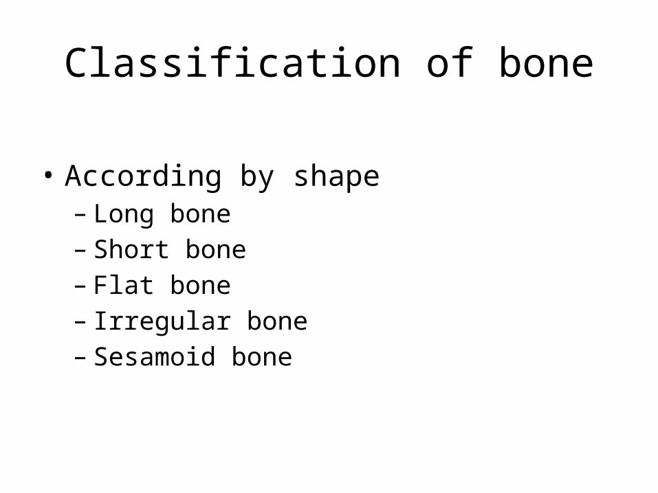
Classification of bone
• According by shape– Long bone– Short bone– Flat bone– Irregular bone– Sesamoid bone

Long bone
• Longer than they are wide. • Reflects the elongated shape rather than the
overall size. • Consist of a shaft plus two ends and are
constructed primarily of compact bone• may contain substantial amounts of spongy
bone. • All bones of the limbs, except the patella, wrist
and ankle bones, are long bones.
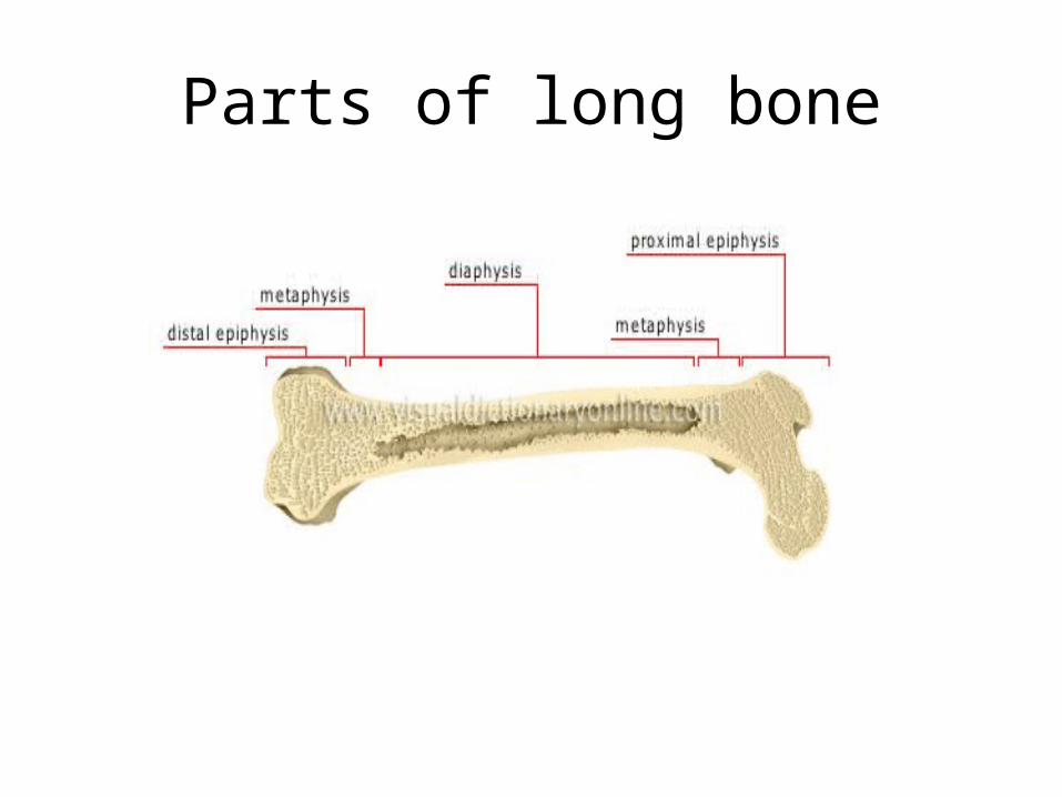
Parts of long bone

• Epiphysis– Are expanded articular ends – separated from the shaft by the epiphyseal plate
during bone growth– composed of a spongy bone surrounded by a thin
layer of compact bone.– Proximal epiphysis
• Enlarged terminal part of the bone, • nearest the center of the body,
– Distal epiphysis• Enlarged terminal part of the bone, • farthest from the center of the body,
• Metaphysis– Part of the bone between the epiphysis and the
diaphysis; – it contains the connecting cartilage enabling the
bone to grow– disappears at adulthood.
• Diaphysis– Elongated hollow central portion of the bone
located between the methaphyses; – made of compact tissue– encloses the medullary cavity.
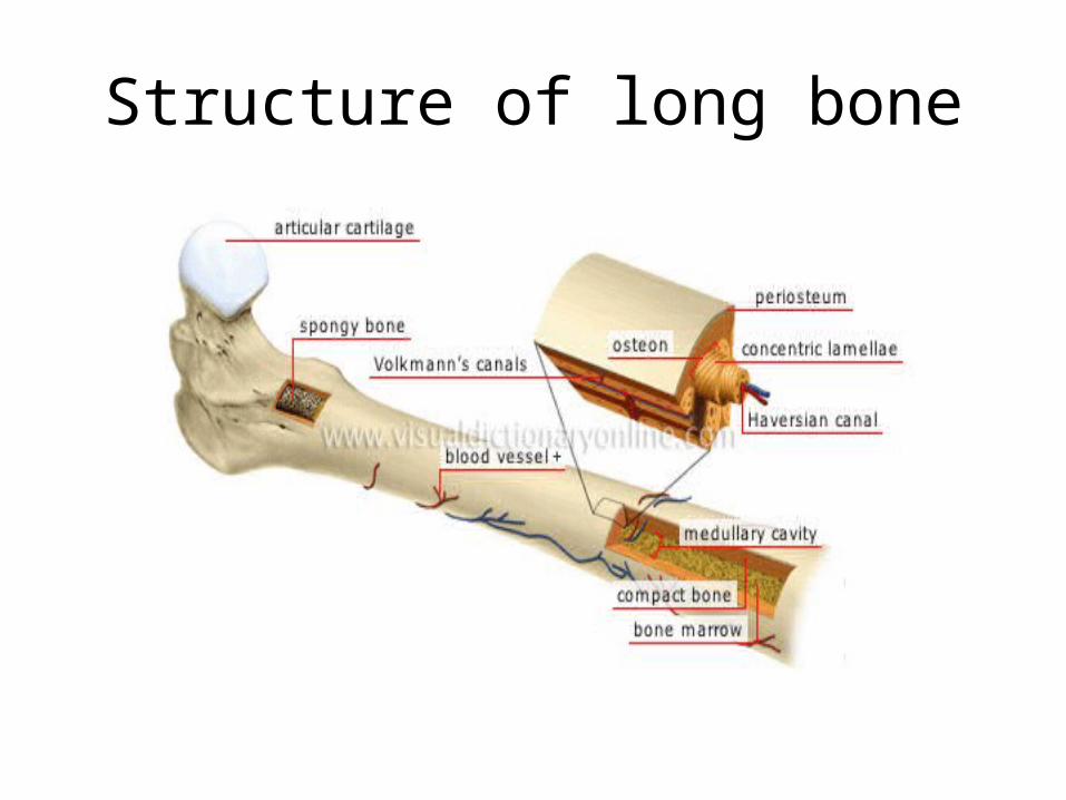
Structure of long bone

• Osteon – Elementary cylindrical structure of the compact
bone – Runs parallel to longest axis of bone– Surrounds and opens into Haversian canal.
• Haversian canal – Lengthwise central canal of the osteon – enclose blood vessels and nerves.

• Volkmann’s canals – Perforating canal– Transverse canals of the compact bone enclosing
blood vessels and nerves– they connect the Haversian canals and with the
medullary cavity and the periosteum.• Medullary cavity – Cylindrical central cavity of the bone containing
the bone marrow– encloses lipid-rich yellow bone marrow.
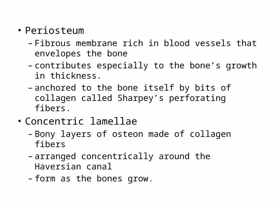
• Periosteum – Fibrous membrane rich in blood vessels that
envelopes the bone– contributes especially to the bone’s growth in
thickness.– anchored to the bone itself by bits of collagen
called Sharpey’s perforating fibers.• Concentric lamellae – Bony layers of osteon made of collagen fibers– arranged concentrically around the Haversian canal– form as the bones grow.
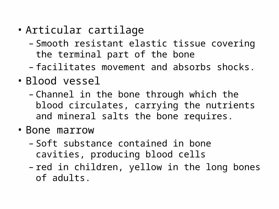
• Articular cartilage – Smooth resistant elastic tissue covering the terminal
part of the bone– facilitates movement and absorbs shocks.
• Blood vessel – Channel in the bone through which the blood
circulates, carrying the nutrients and mineral salts the bone requires.
• Bone marrow– Soft substance contained in bone cavities, producing
blood cells– red in children, yellow in the long bones of adults.

JOINTS
the site where two or more skeletal elements come together
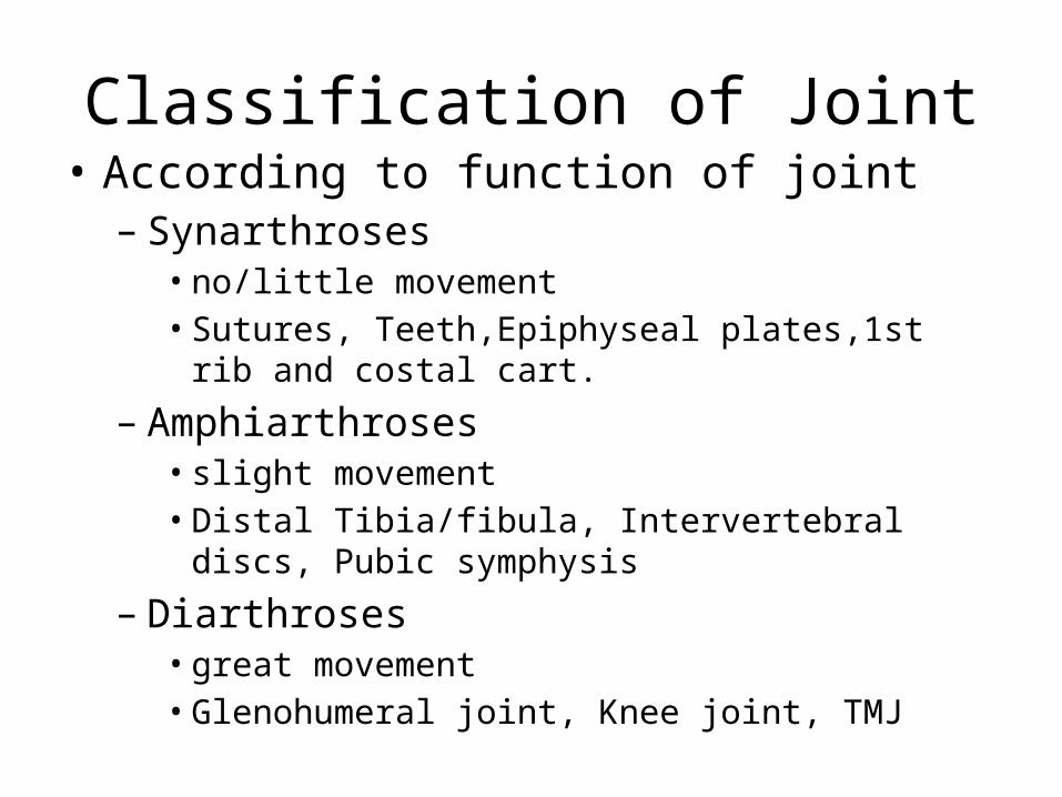
Classification of Joint• According to function of joint– Synarthroses• no/little movement• Sutures, Teeth,Epiphyseal plates,1st rib and costal cart.
– Amphiarthroses• slight movement• Distal Tibia/fibula, Intervertebral discs, Pubic symphysis
– Diarthroses• great movement• Glenohumeral joint, Knee joint, TMJ
• According to the structure of Joint– Cartilaginous– Fibrous – Synovial

• Cartilaginous Joint– are connected entirely by cartilage– allow more movement between bones than a
fibrous joint but less than the highly mobile synovial joint
– also forms the growth regions of immature long bones and the intervertebral discs of the spinal column.
– Types• Synchondrosis• Symphysis
Cartilaginous• Synchondrosis (synarthroses)
– Primary cartilaginous joints– Occur where two ossification centre in a developing bone
remain seperated by a layer of cartilage– Growth plate between head and shaft of developing long bone– Allow bone growth and eventually become completely ossified
• Symphysis (amphiarthroses)– Secondary cartileginous joints– Two separate bones are interconnected by cartilage– Mostly occur in midline– Pubis symphysis, intervertebral disc between adjacent
vertebrae
• Fibrous Joint– are connected by dense connective tissue,
consisting mainly of collagen– Types• Sutures• Syndesmoses• Gomphosis

Fibrous
• Suture– Only in skull where adjacent bones are linked by a thin layer
of connective tissue• Gomphoses
– Occur only between the teeth and adjacent bone– Short collagen tissue fibre in the periodontal ligament run
between the root of the tooth and the bony socket• Syndesmoses
– Joints in which two adjacent bones are linked by a ligament– Are moveable– Ligamentum flavum, interosseos membrane
Synovial Joint
• Are diarthrosis• the most common and most movable type• achieve movement at the point of contact of the
articulating bones.• The main structural differences between synovial
and fibrous joints are – the existence of capsules surrounding the articulating
surfaces of a synovial joint– the presence of lubricating synovial fluid within those
capsules.
Classification of synovial Joint
• Based upon movement– Uniaxial joint– Biaxial Joint– Multiaxial Joint

Classification of synovial Joint
• Based on the shape of their articular surface– Planar Joint– Hinge Joint– Pivot Joint– Bicondylar Joint– Condylar Joint– Saddle Joint– Ball and socket joint

• Plane Joint– Also called Gliding
Joints– One moves across the
surface of another– Allow sliding or gliding
movements– Acromoclavicular joint

• Hinge Joint– Also known as
ginglymus joint– Allow movement
around one axis that passes transversly through the joint
– Permit flexion and extension
– Humeroulnar joint
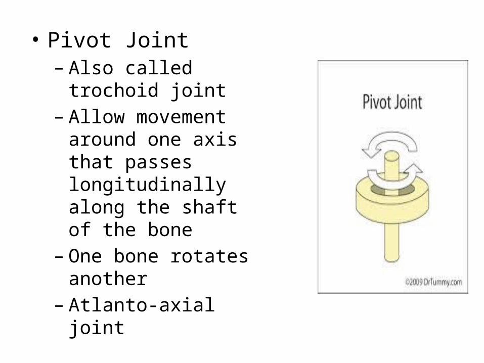
• Pivot Joint– Also called trochoid joint– Allow movement around
one axis that passes longitudinally along the shaft of the bone
– One bone rotates another
– Atlanto-axial joint

• Bicondylar Joint– Formed by two convex
condyles that articulate with concave or flat surface
– Allow movement mostly in one axis with limited rotation around a second axis
– Knee joint
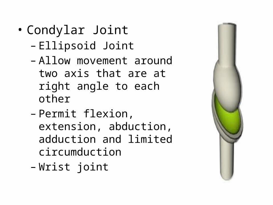
• Condylar Joint– Ellipsoid Joint– Allow movement around two
axis that are at right angle to each other
– Permit flexion, extension, abduction, adduction and limited circumduction
– Wrist joint
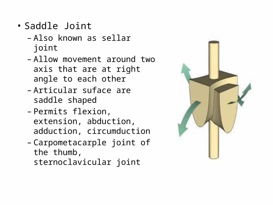
• Saddle Joint– Also known as sellar joint– Allow movement around
two axis that are at right angle to each other
– Articular suface are saddle shaped
– Permits flexion, extension, abduction, adduction, circumduction
– Carpometacarple joint of the thumb, sternoclavicular joint

• Ball and Socket Joint– Universal joint,
spheroidal joint– Allow movement
around multiple axis– Permits extension,
flexion, abduction, adduction, circumduction except gliding
– Hip joint, glenohumeral joint

Thank You!

References
• Gray’s anatomy for student by Drake, Vogi• Gray’s anatomy : the anatomical by susan
standring• Clinical anatomy by region – Richard snell• Netter’s Anatomy• Gross Anatomy BRSeries by Kyung• Clinically oriented Anatomy by Moore• Last’s Anatomy regional and applied
Related Documents