VASCULARBIOLOGY Localized TRPA1 channel Ca 2+ signals stimulated by reactive oxygen species promote cerebral artery dilation Michelle N. Sullivan, 1 Albert L. Gonzales, 2 Paulo W. Pires, 3 Allison Bruhl, 1 M. Dennis Leo, 4 Wencheng Li, 1 Agathe Oulidi, 3 Frederick A. Boop, 5 Yumei Feng, 1 Jonathan H. Jaggar, 4 Donald G. Welsh, 6 Scott Earley 3 * Reactive oxygen species (ROS) can have divergent effects in cerebral and peripheral circulations. We found that Ca 2+ -permeable transient receptor potential ankyrin 1 (TRPA1) channels were present and co- localized with NADPH (reduced form of nicotinamide adenine dinucleotide phosphate) oxidase 2 (NOX2), a major source of ROS, in the endothelium of cerebral arteries but not in other vascular beds. We recorded and characterized ROS-triggered Ca 2+ signals representing Ca 2+ influx through single TRPA1 channels, which we called “TRPA1 sparklets.” TRPA1 sparklet activity was low under basal conditions but was stim- ulated by NOX-generated ROS. Ca 2+ entry during a single TRPA1 sparklet was twice that of a TRPV4 spark- let and ~200 times that of an L-type Ca 2+ channel sparklet. TRPA1 sparklets representing the simultaneous opening of two TRPA1 channels were more common in endothelial cells than in human embryonic kidney (HEK) 293 cells expressing TRPA1. The NOX-induced TRPA1 sparklets activated intermediate-conductance, Ca 2+ -sensitive K + channels, resulting in smooth muscle hyperpolarization and vasodilation. NOX-induced activation of TRPA1 sparklets and vasodilation required generation of hydrogen peroxide and lipid- peroxidizing hydroxyl radicals as intermediates. 4-Hydroxy-nonenal, a metabolite of lipid peroxidation, also increased TRPA1 sparklet frequency and dilated cerebral arteries. These data suggest that in the ce- rebral circulation, lipid peroxidation metabolites generated by ROS activate Ca 2+ influx through TRPA1 channels in the endothelium of cerebral arteries to cause dilation. INTRODUCTION Regulation of the cerebral circulation differs from that of the rest of the body to meet the metabolic demands and specific anatomical constraints of the brain. One example of this disparity is the observation that reactive oxygen species (ROS) such as superoxide anions (O 2 − ) and hydrogen per- oxide (H 2 O 2 ) primarily cause vasodilation in the cerebral circulation and vasoconstriction in peripheral arteries (1). The NADPH (reduced form of nicotinamide adenine dinucleotide phosphate) oxidase (NOX) family, con- sisting of five isoforms (NOX1 to NOX5), is a major source of ROS in the vasculature (2, 3). Although the generation of ROS by NOX is much greater in healthy cerebral arteries than in other vascular beds (4), the mo- lecular mechanism responsible for translating these higher amounts of ROS into a qualitatively different vascular response is not known. Potential candidate mediators of the vascular actions of ROS include Ca 2+ -permeable members of the transient receptor potential (TRP) chan- nel family, several of which are present in the vasculature and can be regu- lated by ROS and ROS-derived products (5–8), including the ankyrin TRP (TRPA) channel TRPA1. Initially characterized as a detector of noxious electrophilic substances in nociceptive and sensory neurons (9, 10), TRPA1 has since been shown to be present in mast cells, enterochromaffin cells, epithelial cells, and other tissues, suggesting a broader biological role for this channel. TRPA1 is present in the endothelium and mediates vasodila- tion of cerebral arteries in response to allyl isothiocyanate (AITC) (11), a pungent compound found in mustard oil. Endogenous regulators of TRPA1 activity in the endothelium are currently unknown. In vagal and sensory nerves, hypoxic and hyperoxic conditions can increase TRPA1 activity (12). Additional evidence indicates that TRPA1 in neurons can be activated by oxidative modification of cysteines in its cytoplasmic N terminus by ROS, including H 2 O 2 and O 2 − (7, 13). TRPA1 is also activated by compounds produced by peroxidation of w6 polyunsaturated fatty acids in the plasma membrane, such as 4-hydroxy-nonenal (4-HNE), 4-oxo-2-nonenal (4-ONE), and 4-hydroxy-hexenal (7, 14). 4-HNE and related substances are produced by hydroxyl radicals (OH•) formed during degradation of H 2 O 2 (15), sug- gesting that the oxidant and redox signaling mechanisms acting on TRPA1 could be linked by the lipid peroxidation process. This proposed signaling cascade has not been studied in vascular endothelium, and its effects on TRPA1 activity, endothelial function, and vasomotor responses have not been characterized. Elementary Ca 2+ influx events through single TRPV4 channels have been optically recorded from endothelial cells using total internal reflection fluorescence (TIRF) and confocal microscopy ( 16, 17). These events, termed “TRPV4 sparklets, ” are fundamental signals underlying endothelium- dependent dilation of mesenteric arteries (16). In theory, all Ca 2+ -permeable TRP channels with sufficient conductance are capable of being detected optically as sparklets, with amplitude, frequency, and spatial spread re- flecting the unitary conductance, Ca 2+ permeability, and gating kinetics of the channel. Previous studies are consistent with the possibility that TRPA1 channels are critical sensors of cellular oxidant and redox status. However, little is currently known about the relationship between ROS generation and 1 Vascular Physiology Research Group, Department of Biomedical Sciences, Colorado State University, Fort Collins, CO 80523, USA. 2 Department of Phar- macology, University of Vermont School of Medicine, Burlington, VT 05405, USA. 3 Department of Pharmacology, University of Nevada School of Medicine, Reno, NV 89557–0318, USA. 4 Department of Physiology, University of Tennessee Health Science Center, Memphis, TN 38163, USA. 5 Department of Neurosurgery, University of Tennessee Health Science Center, Memphis, TN 38163, USA. 6 Depart- ment of Physiology and Pharmacology, University of Calgary, Calgary, Alberta T2N 4N1, Canada. *Corresponding author. E-mail: [email protected] RESEARCHARTICLE www.SCIENCESIGNALING.org 6 January 2015 Vol 8 Issue 358 ra2 1 on January 6, 2015 http://stke.sciencemag.org/ Downloaded from

Welcome message from author
This document is posted to help you gain knowledge. Please leave a comment to let me know what you think about it! Share it to your friends and learn new things together.
Transcript

R E S E A R C H A R T I C L E
V A S C U L A R B I O L O G Y
Localized TRPA1 channel Ca2+ signals stimulatedby reactive oxygen species promote cerebralartery dilationMichelle N. Sullivan,1 Albert L. Gonzales,2 Paulo W. Pires,3 Allison Bruhl,1 M. Dennis Leo,4
Wencheng Li,1 Agathe Oulidi,3 Frederick A. Boop,5 Yumei Feng,1 Jonathan H. Jaggar,4
Donald G. Welsh,6 Scott Earley3*
http://stke.sD
ownloaded from
Reactive oxygen species (ROS) can have divergent effects in cerebral and peripheral circulations. Wefound that Ca2+-permeable transient receptor potential ankyrin 1 (TRPA1) channels were present and co-localized with NADPH (reduced form of nicotinamide adenine dinucleotide phosphate) oxidase 2 (NOX2), amajor source of ROS, in the endothelium of cerebral arteries but not in other vascular beds. We recordedand characterized ROS-triggered Ca2+ signals representing Ca2+ influx through single TRPA1 channels,which we called “TRPA1 sparklets.” TRPA1 sparklet activity was low under basal conditions but was stim-ulated by NOX-generated ROS. Ca2+ entry during a single TRPA1 sparklet was twice that of a TRPV4 spark-let and ~200 times that of an L-type Ca2+ channel sparklet. TRPA1 sparklets representing the simultaneousopening of two TRPA1 channels were more common in endothelial cells than in human embryonic kidney(HEK) 293 cells expressing TRPA1. The NOX-induced TRPA1 sparklets activated intermediate-conductance,Ca2+-sensitive K+ channels, resulting in smooth muscle hyperpolarization and vasodilation. NOX-inducedactivation of TRPA1 sparklets and vasodilation required generation of hydrogen peroxide and lipid-peroxidizing hydroxyl radicals as intermediates. 4-Hydroxy-nonenal, a metabolite of lipid peroxidation,also increased TRPA1 sparklet frequency and dilated cerebral arteries. These data suggest that in the ce-rebral circulation, lipid peroxidation metabolites generated by ROS activate Ca2+ influx through TRPA1channels in the endothelium of cerebral arteries to cause dilation.
ci
on January 6, 2015encemag.org/
INTRODUCTION
Regulation of the cerebral circulation differs from that of the rest of thebody to meet the metabolic demands and specific anatomical constraintsof the brain. One example of this disparity is the observation that reactiveoxygen species (ROS) such as superoxide anions (O2
−) and hydrogen per-oxide (H2O2) primarily cause vasodilation in the cerebral circulation andvasoconstriction in peripheral arteries (1). The NADPH (reduced form ofnicotinamide adenine dinucleotide phosphate) oxidase (NOX) family, con-sisting of five isoforms (NOX1 to NOX5), is a major source of ROS in thevasculature (2, 3). Although the generation of ROS by NOX is muchgreater in healthy cerebral arteries than in other vascular beds (4), the mo-lecular mechanism responsible for translating these higher amounts ofROS into a qualitatively different vascular response is not known.
Potential candidate mediators of the vascular actions of ROS includeCa2+-permeable members of the transient receptor potential (TRP) chan-nel family, several of which are present in the vasculature and can be regu-lated by ROS and ROS-derived products (5–8), including the ankyrin TRP(TRPA) channel TRPA1. Initially characterized as a detector of noxiouselectrophilic substances in nociceptive and sensory neurons (9, 10), TRPA1has since been shown to be present in mast cells, enterochromaffin cells,
1Vascular Physiology Research Group, Department of Biomedical Sciences,Colorado State University, Fort Collins, CO 80523, USA. 2Department of Phar-macology, University of Vermont School of Medicine, Burlington, VT 05405,USA. 3Department of Pharmacology, University of Nevada School of Medicine,Reno, NV 89557–0318, USA. 4Department of Physiology, University of TennesseeHealth Science Center, Memphis, TN 38163, USA. 5Department of Neurosurgery,University of Tennessee Health Science Center, Memphis, TN 38163, USA. 6Depart-ment of Physiology and Pharmacology, University of Calgary, Calgary, AlbertaT2N 4N1, Canada.*Corresponding author. E-mail: [email protected]
w
epithelial cells, and other tissues, suggesting a broader biological role forthis channel. TRPA1 is present in the endothelium and mediates vasodila-tion of cerebral arteries in response to allyl isothiocyanate (AITC) (11), apungent compound found in mustard oil. Endogenous regulators of TRPA1activity in the endothelium are currently unknown. In vagal and sensorynerves, hypoxic and hyperoxic conditions can increase TRPA1 activity (12).Additional evidence indicates that TRPA1 in neurons can be activated byoxidative modification of cysteines in its cytoplasmic N terminus by ROS,including H2O2 and O2
− (7, 13). TRPA1 is also activated by compoundsproduced by peroxidation of w6 polyunsaturated fatty acids in the plasmamembrane, such as 4-hydroxy-nonenal (4-HNE), 4-oxo-2-nonenal (4-ONE),and 4-hydroxy-hexenal (7, 14). 4-HNE and related substances are producedby hydroxyl radicals (OH•) formed during degradation of H2O2 (15), sug-gesting that the oxidant and redox signaling mechanisms acting on TRPA1could be linked by the lipid peroxidation process. This proposed signalingcascade has not been studied in vascular endothelium, and its effects onTRPA1 activity, endothelial function, and vasomotor responses have notbeen characterized.
Elementary Ca2+ influx events through single TRPV4 channels havebeen optically recorded from endothelial cells using total internal reflectionfluorescence (TIRF) and confocal microscopy (16, 17). These events, termed“TRPV4 sparklets,” are fundamental signals underlying endothelium-dependent dilation of mesenteric arteries (16). In theory, all Ca2+-permeableTRP channels with sufficient conductance are capable of being detectedoptically as sparklets, with amplitude, frequency, and spatial spread re-flecting the unitary conductance, Ca2+ permeability, and gating kineticsof the channel.
Previous studies are consistent with the possibility that TRPA1 channelsare critical sensors of cellular oxidant and redox status. However, little iscurrently known about the relationship between ROS generation and
ww.SCIENCESIGNALING.org 6 January 2015 Vol 8 Issue 358 ra2 1

R E S E A R C H A R T I C L E
on January 6, 2015http://stke.sciencem
ag.org/D
ownloaded from
TRPA1 channel activity in the endothelium. Moreover, it is not clear howthe potential of these channels to serve as ROS sensors in the vasculaturecould account for differences in the effects of ROS between the peripheraland cerebral circulations. Here, we investigated how NOX-generatedROS could provoke vasodilation through activation of TRPA1 channelsand sought to determine how this response differed between cerebral andperipheral arteries. We showed that TRPA1 colocalizes with NOX isoform2 (NOX2) in the endothelium of cerebral arteries but not in other vascularbeds. We further developed a method for recording and characterizingTRPA1 sparklets, demonstrating the presence of large-amplitude TRPA1sparklets with unique properties in cerebral artery endothelial cells. Final-ly, we demonstrated that ROS generated by NOX increased TRPA1 spark-let frequency and dilated cerebral arteries through a process that requiresthe peroxidation of membrane lipids. Together, these data indicate that Ca2+
influx through TRPA1 channels elicits endothelium-dependent vasodilationin response to the generation of lipid peroxidation products, and this re-sponse is specific to the cerebral vasculature.
RESULTS
TRPA1 colocalizes with NOX2 in the endothelium ofcerebral arteries but not in other vascular bedsWe detected TRPA1 mRNA by reverse transcription polymerase chainreaction (RT-PCR) in rat cerebral arteries but not in renal, coronary, ormesenteric arteries (Fig. 1A). To confirm this unusual distribution pattern,we mounted arteries en face and immunolabeled the endothelium with an
w
antibody against TRPA1. TRPA1 protein was detected in rat cerebralendothelia, where it was abundant in perinuclear regions; it was also presentwithin fenestrations of the internal elastic lamina (IEL), sites of close contact
Fig. 1. TRPA1 colocalizes with NOX2 in the endothelium of cerebral arteries
but not in other vascular beds. (A) RT-PCR for TRPA1 (A1) and eNOS (+)mRNA in rat coronary (C), renal (R), mesenteric (M), and cerebral (CA) ar-teries. NTC, no complementary DNA (cDNA) template control (n = 2 rats).(B) Top: Immunolabeling for TRPA1 in coronary, renal, mesenteric, and ce-rebral arteries; scale bar, 10 mm. Autofluorescence of the IEL is green.TRPA1 protein (red) was only detected in the endothelium of cerebral ar-teries and was present within myoendothelial projections (arrows, inset;scale bar, 5 mm). Bottom: Immunolabeling was not detected when theprimary antibody was omitted. n = 3 rats. (C) RT-PCR for TRPA1 mRNAin whole human cerebral arteries (CA) and smooth muscle cells isolatedfrom human cerebral arteries (SMC) (n = 3 independent biological repli-cates). (D) Western blot for TRPA1 in human cerebral arteries (CA) (n =2 independent biological replicates). (E) RT-PCR detection of humanTRPA1 (A1) and eNOS (+) mRNA in primary coronary (C), renal (R), dermal(D), and cerebral artery (CA) endothelial cells. TRPA1mRNA was detectedonly in CA. Representative of three independent experiments. (F) Top: Ce-rebral arteries immunolabeled for NOX1 (left), NOX2 (middle), or NOX4(right) (red); scale bars, 10 mm. NOX2 and NOX4 are more abundant withinmyoendothelial projections compared with NOX1 (arrows, insets; bars, 5 mm).Endothelial cell nuclei are stained with 4′,6-diamidino-2-phenylindole (DAPI)(blue), and IEL autofluorescence is green. Bottom: No primary antibodycontrol. n = 3 rats per group. (G) PLA experiments for TRPA1:NOX4(top) and TRPA1:NOX2 (bottom) in cerebral artery endothelial cells. Punctacorresponding to positive PLA results are red, cellular autofluorescence isgreen, and DAPI-stained nuclei are blue. Puncta density is summarized atthe right (n = 4 cells per group, 3 rats); *P ≤ 0.05 versus TRPA1:NOX4. (H) PLAfor TRPA1:NOX4 (top) and TRPA1:NOX2 (bottom). TRPA1:NOX2 punctawithin IEL fenestrations are indicated by arrows. *, an example magnifiedand shown in cross section in the insets. Puncta density is summarized atthe right (n = 8 to 14 vessels per group, 3 rats); *P ≤ 0.05 compared withTRPA1:NOX4.ww.SCIENCESIGNALING.org 6 January 2015 Vol 8 Issue 358 ra2 2

R E S E A R C H A R T I C L E
on January 6, 2015http://stke.sciencem
ag.org/D
ownloaded from
between the endothelial and smooth muscle cell plasma membranes knownas myoendothelial projections (Fig. 1B, arrows, inset). TRPA1 immunola-beling was not detected in coronary, mesenteric, or renal arterial endothelium(Fig. 1B), suggesting that this channel was selectively present in the cerebralendothelia. Arteries from human subjects displayed the same TRPA1distribution pattern. TRPA1 mRNAwas detected by RT-PCR in whole hu-man cerebral arteries but not in isolated smooth muscle cells (Fig. 1C),and TRPA1 protein was detected by Western blotting in tissue homogenatesof human cerebral arteries (Fig. 1D). TRPA1 message was not detected byRT-PCR in RNA isolated from human coronary, renal glomerular, or neo-natal dermal artery endothelial cells, but was found in endothelial cellsfrom human cerebral arteries (Fig. 1E). These findings demonstrate thatthe presence of TRPA1 in the endothelium is a distinctive feature of thecerebral circulation.
To test our proposal that ROS and/or ROS-derived products are endog-enous agonists of TRPA1 in the endothelium, we first examined cerebralarteries for the presence of NOX isoforms. NOX5 is not present in rats ormice, and NOX3 is found only in the inner ear (18); however, the otherthree isoforms are present in the rodent vasculature (19, 20). We detectedthree NOX isoforms—NOX1, NOX2, and NOX4—in the endothelium ofcerebral arteries by immunolabeling (Fig. 1F and fig. S1). All three iso-forms were present in perinuclear regions and IEL fenestrations, butNOX2 and NOX4 were more abundant in myoendothelial projectionscompared with NOX1 (Fig. 1F, insets, and fig. S2).
We used in situ proximity ligation assays (PLAs) (21) to investigatewhether NOX2 and NOX4 colocalize with TRPA1 in the endothelium.PLAs for TRPA1:NOX2 produced abundant red puncta, indicating signif-icant colocalization (≤40 nm) in cerebral artery endothelial cells (Fig. 1G).PLAs for TRPA1:NOX4 revealed few red puncta (Fig. 1G). Few puncta(about one to two per cell) were present when primary antibodies wereomitted. We also used PLAs to assess TRPA1 and NOX colocalizationin intact cerebral arteries (Fig. 1H). These assays revealed the presenceof puncta throughout the endothelium, including in IEL fenestrations, inarteries probed with antibodies against NOX2 and TRPA1 (Fig. 1H andfig. S3). Our data indicate that TRPA1:NOX2 complexes are present incerebral artery endothelial cells within myoendothelial junctions and inother regions. Coimmunoprecipitation for NOX2 and TRPA1 failed to de-tect direct interaction between the two proteins (fig. S4A). These datademonstrate that NOX2 and TRPA1 selectively colocalize but do notphysically interact in the cerebral artery endothelium, suggesting that me-tabolites generated by NOX2 could regulate TRPA1 activity in this tissue.
ROS stimulate TRPA1 sparklet activity in cerebral arteryendothelial cellsWe studied changes in TRPA1 activity in cerebral artery endothelial cellsin response to ROS by using TIRF microscopy to record subcellular Ca2+
signals representing Ca2+ influx through single TRPA1 channels, which wecalled TRPA1 sparklets. To initially characterize TRPA1 sparklets, wetreated primary cerebral artery endothelial cells loaded with the Ca2+ in-dicator dye Fluo-4 AM with the TRPA1 agonist AITC. Very few eventswere seen under basal conditions, but AITC induced a concentration-dependent increase in sparklet frequency (Fig. 2, A and B, and movieS1). The half-maximal effective concentration (EC50) for sparklet activa-tion was 4.4 mM (Fig. 2B), similar to the previously reported EC50 forAITC-induced dilation of cerebral arteries (EC50, 16.4 mM) (11). The in-crease in sparklet frequency stimulated by AITC was not affected by de-pletion of intracellular stores with cyclopiazonic acid (fig. S5A) but wasabsent when cells were bathed in Ca2+-free solution (fig. S5B), indicatingthat these signals are generated by influx of Ca2+. The selective TRPA1antagonist HC-030031 (22) blocked AITC-induced increases in sparklet
w
frequency (Fig. 2C), confirming that the subcellular Ca2+ influx eventsactivated by AITC were bona fide TRPA1 sparklets. Single-site analysisindicated that the total number of active TRPA1 sparklet sites increasedfrom 1.4 ± 0.7 sites to 6.1 ± 1.8 sites after the addition of AITC (Fig. 2D),suggesting that AITC recruited previously inactive TRPA1 channels anddid not increase the frequency of basally active sites. Further, these datashowed that only about four to eight TRPA1 sparklet sites per cell wereactive during maximal stimulation.
We compared TRPA1 sparklets recorded from cerebral artery endothe-lial cells to those recorded from human embryonic kidney (HEK) 293 cellstransfected with a TRPA1-GFP (green fluorescent protein) fusion protein.AITC caused an increase in TRPA1 sparklet frequency in transfected HEKcells, which was inhibited by the TRPA1 blocker HC-030031 and absent inuntransfected HEK cells (fig. S6, A and B). Modal duration, attack time,decay time, and spatial spread of TRPA1 sparklets recorded from TRPA1-GFP–transfected HEK cells were essentially identical to those recordedfrom cerebral artery endothelial cells (table S1). Duration histograms in-dicated that the most frequently occurring TRPA1 sparklets lasted less than200 ms and fit single exponential functions in endothelial cells (t = 408 ms;Fig. 2E) and transfected HEK cells (t = 210 ms; fig. S6C). Spatial spreaddistribution was also similar between TRPA1 sparklets recorded from endo-thelial cells and HEK cells, with most events having an area of less than1 mm2 (Fig. 2E and fig. S6C).
A multiple Gaussian fit of a histogram of TRPA1 sparklet amplitudesrecorded from transfected HEK cells (fig. S6C) indicated three distinctpeaks of F/F0 = 1.13, 1.26, and 1.39. These data suggest that the unitaryTRPA1 sparklet amplitude is DF/F0 = 0.13, and the peaks represent theopening of one, two, or three individual TRPA1 channels, with the mostfrequently occurring TRPA1 sparklets (F/F0 = 1.13) signifying the open-ing of a single TRPA1 channel. Identical results were obtained in endo-thelial cells (F/F0 = 1.13, 1.26, 1.39), consistent with a unitary TRPA1sparklet amplitude of DF/F0 = 0.13 in these native cells (Fig. 2E). This isalso apparent from plots of fluorescence intensity over time for regions ofinterest (ROIs) with active TRPA1 sparklet sites, where three distinct am-plitudes are seen (Fig. 2F and fig. S6D). Unlike TRPA1 sparklets in HEKcells, the most commonly occurring TRPA1 sparklets recorded from en-dothelial cells had an amplitude of F/F0 = 1.26 (DF/F0 = 0.26), indicatingthat simultaneous opening of two TRPA1 channels at the same active siteoccurred much more frequently than expected. These data suggest thatnative TRPA1 channels exhibit binary coupled gating more frequentlyin cerebral artery endothelial cells compared with cloned TRPA1 channelsexpressed in HEK cells.
Colocalization of NOX2 and TRPA1 (Fig. 1H) in the cerebral arteryendothelium supports the concept that NOX-derived ROS could regulateTRPA1 channel activity in this tissue. To test this hypothesis, NOX activ-ity was stimulated by administration of NADPH (23). We found thatenhanced NOX activity increased cerebral artery TRPA1 sparklet frequencyin a concentration-dependent manner (Fig. 2, G and H) and that the TRPA1blocker HC-030031 abolished this response (Fig. 2I). These findings in-dicate that NOX-derived ROS stimulate TRPA1 sparklet activity in cere-bral artery endothelial cells, suggesting that TRPA1 is a ROS sensor inthis tissue.
ROS generated by NOX dilate cerebral arteries byactivating TRPA1The effects of ROS-induced increases in TRPA1 sparklet frequency onvessel diameter were studied using intact cerebral arteries that were pres-surized to physiological values (80 mmHg) to allow spontaneous myogenictone to develop. Our data showed that the addition of NADPH to increasegeneration of ROS by NOX caused concentration-dependent vasodilation
ww.SCIENCESIGNALING.org 6 January 2015 Vol 8 Issue 358 ra2 3

R E S E A R C H A R T I C L E
on January 6, 2015http://stke.sciencem
ag.org/D
ownloaded from
(Fig. 3, A and B). This response was abolished by the TRPA1 blocker HC-030031 (Fig. 3, A and C), indicating that NOX-derived ROS bring aboutdilation of cerebral arteries by increasing the frequency of TRPA1 sparklets.
One mechanism by which the endothelium can promote arterial dilationis by releasing diffusible substances such as nitric oxide (NO) or prostacyclin(PGI2). We found that NADPH-induced dilation of cerebral arteries wasnot altered by blocking NO and PGI2 synthesis with L-NG-nitroarginine(L-NNA) and indomethacin, respectively (Fig. 3, D and E), indicating thatthese pathways are not involved in the response. The endothelium can alsocause dilation by direct electrotonic spread of endothelial cell membrane hy-perpolarization through myoendothelial gap junctions to underlying vascu-lar smooth muscle cells. Small-conductance and intermediate-conductanceCa2+-activated K+ (IK) channels are implicated in this form of endothelium-dependent vasodilation, and functional IK channels are present in cerebralartery endothelial cells at myoendothelial junctions (11, 24). Our datashowed that intraluminal administration of the selective IK channel blocker
w
TRAM34 nearly abolished NADPH-induced vasodilation (Fig. 3, F and G),suggesting that TRPA1 sparklets act through IK channels in the endothe-lium to dilate cerebral arteries.
To determine if increases in TRPA1 sparklet activity were associatedwith smooth muscle cell hyperpolarization, we recorded the membranepotential of arterial myocytes from intact, pressurized (80 mmHg) cerebralarteries using intracellular microelectrodes. We found that administrationof NADPH to stimulate ROS generation hyperpolarized the membranes ofsmooth muscle cells by about −8 mV (Fig. 3, H and I). Blocking TRPA1channels with HC-030031 abolished NADPH-induced membrane poten-tial hyperpolarization (Fig. 3, H and I), demonstrating that NOX-derivedROS hyperpolarize smooth muscle cells in pressurized cerebral arteries byactivating TRPA1. TRPA1 channels are not present in smooth muscle cellsin cerebral arteries (Fig. 1C) (11, 25), suggesting that this response is mediatedby the endothelium. Together, our findings suggest that Ca2+ influx throughTRPA1 channels in the endothelium activates nearby IK channels to initiate
Fig. 2. ROS stimulate TRPA1 sparklets in cerebral artery endothelial cells.(A) Time-lapse image of an AITC-induced TRPA1 sparklet recorded froma cerebral artery endothelial cell; scale bar, 8 mm. (B) AITC induces aconcentration-dependent increase in TRPA1 sparklet frequency in cerebralartery endothelial cells (n = 5 to 52 cells per concentration, 4 independentcell isolations). (C) Summary data showing that HC-030031 inhibits AITC-induced increases in TRPA1 sparklet frequency in cerebral artery endo-thelial cells (n = 10 to 24 cells, 3 independent cell isolations); *P ≤ 0.05compared to control at baseline. (D) Active TRPA1 sparklet sites per cellbefore and after administration of AITC (n = 10 cells, 5 rats). (E) Amplitude(left), duration (middle), and spatial spread (right) histograms for TRPA1
sparklets (n = 762 total events, 43 independent experiments). (F) Repre-sentative recordings of change in fluorescence (F/F0) within an ROI onprimary cerebral artery endothelial cells stimulated by AITC. Dotted linesindicate the opening of one, two, or three TRPA1 channels. (G) Time-lapse image of a TRPA1 sparklet stimulated by NADPH; scale bar, 8 mm.(H) The NOX substrate NADPH induced a concentration-dependent in-crease in TRPA1 sparklet frequency (n = 9 to 22 cells per concentration,4 independent cell isolations). (I) Summary data indicating that HC-030031inhibits NADPH-induced increases in TRPA1 sparklet frequency (n = 12 to28 cells per group, 3 independent cell isolations); *P ≤ 0.05 compared withbaseline, control.
ww.SCIENCESIGNALING.org 6 January 2015 Vol 8 Issue 358 ra2 4

R E S E A R C H A R T I C L E
w
on January 6, 2015http://stke.sciencem
ag.org/D
ownloaded from
K+ efflux. The ensuing hyperpolarization of the endothelial cell plasmamembrane is conducted to underlying smooth muscle to hyperpolarizeand relax that tissue, resulting in arterial dilation.
ROS-derived lipid peroxidation metabolites stimulateTRPA1 sparklets and dilate cerebral arteriesNOX-derived ROS could stimulate endothelial cell TRPA1 activity direct-ly or through generation of lipid peroxidation products (Fig. 4A). Wefound that the NOX inhibitor apocynin attenuated NADPH-induced in-creases in TRPA1 sparklet frequency (Fig. 4B) and inhibited vasodilationin response to NADPH (Fig. 4C). These findings were supported by ex-periments showing that NADPH-induced increases in TRPA1 sparkletfrequency and NADPH-induced dilation were attenuated by the NOX2inhibitory peptide gp91ds-tat (26) but not by a scrambled control peptide(scr. gp91ds-tat) (Fig. 4, B and D). These data confirm that NADPH in-creases the generation of ROS by a NOX isoform (probably NOX2), re-sulting in increased TRPA1 sparklet frequency and vasodilation.
When present on the plasma membrane, NOX produces O2− in the ex-
tracellular space, which is rapidly dismutated to H2O2 by spontaneous orsuperoxide dismutase–catalyzed reactions (Fig. 4A). We investigated whetherH2O2 downstream of NOX was involved in the activation of TRPA1 byusing catalase, a membrane-impermeable enzyme that rapidly degradesH2O2. Catalase prevented NADPH-induced increases in TRPA1 sparklet fre-quency (Fig. 4B), indicating that extracellular generation of H2O2 is requiredfor this response. Administration of NADPH in the presence of catalaseconstricted intact cerebral arteries (Fig. 4E), indicating that H2O2 is requiredfor the vasodilatory response. Our data indicate that increases in endothelialcell TRPA1 sparklet activity and cerebral artery dilation in response to ROSgenerated by NOX require the generation of extracellular H2O2.
In the presence of iron (Fe2+), H2O2 is degraded to OH• through theFenton reaction. OH• are highly unstable and rapidly react with poly-unsaturated fatty acids in the plasma membrane to generate lipid peroxi-dation products such as 4-HNE. To distinguish between the direct effectsof H2O2 on TRPA1 activity and by-products generated by OH•, we che-lated iron with deferoxamine to inhibit the Fenton reaction and diminishthe formation of OH• and lipid peroxidation (15). Deferoxamine blockedNADPH-induced increases in TRPA1 sparklet frequency (Fig. 4B) andblunted NADPH-induced dilation of intact cerebral arteries (Fig. 4F), sug-gesting that generation of OH• and lipid peroxidation are necessary forNOX-induced increases in TRPA1 sparklet activity and cerebral arterydilation.
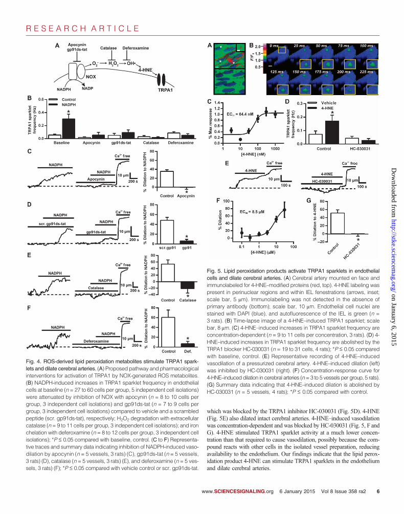
These findings are consistent with the possibility that compounds gen-erated by ROS-dependent peroxidation of polyunsaturated fatty acids suchas 4-HNE and related compounds could serve as endogenous agonists ofTRPA1 channels in cerebral arteries. These substances react with cysteine,histidine, and lysine residues to form stable protein adducts that are recog-nized by specific antibodies (27). To determine if lipid peroxidation me-tabolites are present, we probed intact cerebral arteries, mounted en face,with an antibody that binds to 4-HNE–modified proteins. These experimentsrevealed that 4-HNE–modified proteins were abundant in the cerebral arteryendothelium (Fig. 5A), with immunolabeling present in perinuclear regionsand within IEL fenestrations (Fig. 5A, arrows, inset) where NOX2, NOX4,and TRPA1 channels are also present. Coimmunoprecipitation experimentsindicated association of TRPA1 and 4-HNE in cerebral arteries (fig. S4B).
To test our hypothesis that lipid peroxide metabolites activate TRPA1channels in the endothelium, we examined the effects of endogenous ad-ministration of these substances on TRPA1 sparklet activity and cerebralartery diameter. Our findings show that exogenous administration of 4-HNEinduced a concentration-dependent increase in TRPA1 sparklet frequencyin cerebral artery endothelial cells (EC50, 64.8 ± 50.4 nM) (Fig. 5C),
Fig. 3. ROS generated by NOX dilate cerebral arteries by activating TRPA1.(A) Representative recordings of the intraluminal diameter of an intact, pres-
surized cerebral artery over time. Introduction of NADPH to the bathing solutioninduced vasodilation, which was nearly abolished by the TRPA1 blocker HC-030031. (B) NADPH-induced vasodilation is concentration-dependent (n = 3vessels per concentration, 3 rats). (C) Summary data indicating that NADPH-induced dilation is attenuated by HC-030031 (n = 5 vessels, 3 rats); *P ≤ 0.05compared to control. (D and E) Representative recordings (D) and summarydata (E) indicating that the NO synthase inhibitor L-NNA and the cyclooxy-genase inhibitor indomethacin do not affect NADPH-induced vasodilation(n = 3 vessels, 3 rats). (F and G) Representative recordings (F) and summarydata (G)showing thatNADPH-inducedvasodilation (left) is inhibitedwhen the IKchannel blocker TRAM34 ispresent in the lumen (right) (n=5vessels, 3 rats);*P ≤ 0.05 compared with control. (H) Representative recordings of smoothmuscle cell membrane potential (Em) in a pressurized cerebral artery. SmoothmusclecellswerehyperpolarizedbyNADPH.NADPH-inducedhyperpolarizationwas blocked by HC-030031. (I) Summary data (n = 4 vessels, 4 rats); *P ≤ 0.05compared with vehicle, control; #P ≤ 0.05 compared with vehicle, NADPH.ww.SCIENCESIGNALING.org 6 January 2015 Vol 8 Issue 358 ra2 5
R E S E A R C H A R T I C L E
w
on January 6, 2015http://stke.sciencem
ag.org/D
ownloaded from
which was blocked by the TRPA1 inhibitor HC-030031 (Fig. 5D). 4-HNE(Fig. 5E) also dilated intact cerebral arteries. 4-HNE–induced vasodilationwas concentration-dependent and was blocked by HC-030031 (Fig. 5, F andG). 4-HNE stimulated TRPA1 sparklet activity at a much lower concen-tration than that required to cause vasodilation, possibly because the com-pound reacts with other cells in the isolated vessel preparation, reducingavailability to the endothelium. Our findings indicate that the lipid perox-idation product 4-HNE can stimulate TRPA1 sparklets in the endotheliumand dilate cerebral arteries.
Fig. 5. Lipid peroxidation products activate TRPA1 sparklets in endothelialcells and dilate cerebral arteries. (A) Cerebral artery mounted en face andimmunolabeled for 4-HNE–modified proteins (red, top). 4-HNE labeling waspresent in perinuclear regions and within IEL fenestrations (arrows, inset;scale bar, 5 mm). Immunolabeling was not detected in the absence ofprimary antibody (bottom); scale bar, 10 mm. Endothelial cell nuclei arestained with DAPI (blue), and autofluorescence of the IEL is green (n =3 rats). (B) Time-lapse image of a 4-HNE–induced TRPA1 sparklet; scalebar, 8 mm. (C) 4-HNE–induced increases in TRPA1 sparklet frequency areconcentration-dependent (n = 9 to 11 cells per concentration, 3 rats). (D) 4-HNE–induced increases in TRPA1 sparklet frequency are abolished by theTRPA1 blocker HC-030031 (n = 19 to 31 cells, 4 rats); *P ≤ 0.05 comparedwith baseline, control. (E) Representative recording of 4-HNE–inducedvasodilation of a pressurized cerebral artery. 4-HNE–induced dilation (left)was inhibited by HC-030031 (right). (F) Concentration-response curve for4-HNE–induced dilation in cerebral arteries (n=3 to 5 vessels per group, 5 rats).(G) Summary data indicating that 4-HNE–induced dilation is abolished byHC-030031 (n = 5 vessels, 4 rats); *P ≤ 0.05 compared with control.
Fig. 4. ROS-derived lipid peroxidation metabolites stimulate TRPA1 spark-
lets and dilate cerebral arteries. (A) Proposed pathway and pharmacologicalinterventions for activation of TRPA1 by NOX-generated ROS metabolites.(B) NADPH-induced increases in TRPA1 sparklet frequency in endothelialcells at baseline (n = 27 to 60 cells per group, 5 independent cell isolations)were attenuated by inhibition of NOX with apocynin (n = 8 to 10 cells pergroup, 3 independent cell isolations) and gp91ds-tat (n = 7 to 9 cells pergroup, 3 independent cell isolations) compared to vehicle and a scrambledpeptide (scr. gp91ds-tat), respectively; H2O2 degradation with extracellularcatalase (n = 9 to 11 cells per group, 3 independent cell isolations); and ironchelation with deferoxamine (n = 8 to 12 cells per group, 3 independent cellisolations); *P ≤ 0.05 compared with baseline, control. (C to F) Representa-tive traces and summary data indicating inhibition of NADPH-induced vaso-dilation by apocynin (n = 5 vessels, 3 rats) (C), gp91ds-tat (n = 5 vessels,3 rats) (D), catalase (n = 5 vessels, 3 rats) (E), and deferoxamine (n = 5 ves-sels, 3 rats) (F); *P ≤ 0.05 compared with vehicle control or scr. gp91ds-tat.ww.SCIENCESIGNALING.org 6 January 2015 Vol 8 Issue 358 ra2 6

R E S E A R C H A R T I C L E
ROS-derived lipid peroxidation products fail todilate cerebral arteries from endothelial cell–specificTRPA1-knockout miceWe used endothelial cell–specific TRPA1 knockout (eTRPA1−/−) mice tofurther investigate the involvement of TRPA1 channels in ROS-induceddilation in the cerebral circulation. TRPA1 immunolabeling was detectedin the endothelium of cerebral arteries from mice but not in the coronary,renal, or mesenteric arterial beds (fig. S7). TRPA1 was not detected in West-ern blots of whole cerebral arteries from eTRPA1−/− mice (Fig. 6A), andimmunolabeling studies demonstrated that TRPA1 was present in dorsalroot ganglion (DRG) neurons but not in the endothelium of cerebral arteriesof eTRPA1−/− mice (Fig. 6B). AITC failed to increase the frequency ofTRPA1 sparklets in cerebral artery endothelial cells from eTRPA1−/− mice(Fig. 6C). Passive diameter, myogenic tone, and vasoconstriction in re-sponse to KCl did not differ between cerebral arteries from control or
w
eTRPA1−/− mice (table S2). However, administration of 4-HNE or NADPHfailed to dilate intact pressurized cerebral arteries from eTRPA1−/− mice(Fig. 6, D and E), demonstrating the critical involvement of TRPA1 chan-nels in this response.
DISCUSSION
The brain requires continuous perfusion to provide a constant supply ofoxygen and nutrients, but the enclosing skull tightly limits vascular disten-sion, presenting unique challenges to the cerebral circulation. Among oth-er factors, endothelial control of arterial diameter is critical for preciseregulation of global blood flow to the brain and for matching regional flowto metabolic demand. The ability of the endothelium to rapidly detectchanges in local oxidant and redox status and effect appropriate vasomotorresponses is centrally important for this function but is poorly understood.
ww.SCIENCESIGNALING.org
on January 6, 2015http://stke.sciencem
ag.org/D
ownloaded from
The results of the current study provide strongevidence that TRPA1 channels in the endo-thelium sense ROS generated by NOX.The resulting increase in Ca2+ influx throughTRPA1 channels causes vasodilation. Thispathway appears to be present only in cere-bral arteries, identifying a fundamental dis-tinction in function between blood vesselsin the brain and those in the periphery.
Our results show that elementary Ca2+
signals arising from the influx of Ca2+ throughsingle TRPA1 channels (TRPA1 sparklets)cause endothelium-dependent dilation of ce-rebral vessels in response to ROS genera-tion. Our data indicate that TRPA1 sparkletsare very large Ca2+ influx events, with a uni-tary amplitude about twice that of a TRPV4sparklet reported in our previous publication(17) (DF/F0 = 0.13 versus 0.06), consistentwith the larger single-channel conductanceand greater Ca2+ permeability of TRPA1(10, 28–30). The signal mass of a TRPV4sparklet was estimated to be ~100 timesthat of an L-type Ca2+ channel sparklet (31),suggesting that TRPA1 sparklets are at least~200 times greater than L-type Ca2+ chan-nel sparklets. We also showed that the mostfrequently occurring TRPA1 sparklets recordedfrom endothelial cells have amplitudes con-sistent with the simultaneous opening oftwo TRPA1 channels, doubling the amountof Ca2+ entering during a typical event. Thiscoupled gating arrangement supports theconcept that TRPA1 channels are present ina tight binary structure in the endothelial cellplasma membrane and that the opening ofone of the channels in this pair triggers theadjacent channel, perhaps through bindingof incoming Ca2+ to a Ca2+-sensing EF-handdomain on the N terminus (9). Coupled gatingof TRPA1 has not been previously described,and we showed that it occurred less frequent-ly in HEK cells expressing TRPA1, suggest-ing that formation of binary structures is notan inherent property of the channel but
Fig. 6. ROS-derived lipid peroxidation products fail to dilatecerebral arteries from endothelial cell–specific TRPA1-knockout mice. (A) Western blot for TRPA1 protein (138 kD)in cerebral arteries from control and eTRPA1−/− mice (n =3 mice per group). (B) TRPA1 immunolabeling in the ce-rebral endothelium (top) and DRG neurons (bottom) fromcontrol and eTRPA1−/− mice (n = 3 mice per group). (C)Summary data indicating the effect of AITC on the fre-quency of sparklets recorded from endothelial cells iso-lated from control compared with eTRPA1−/− mice (n =20 cells per group, 3 independent cell isolations pergroup). (D and E) 4-HNE (D) and NADPH (E) dilate cere-
bral arteries from control but not eTRPA1−/− mice (n = 5 vessels per group, 3 mice per group); *P ≤ 0.05compared with control. (F) Proposed signaling pathway: In endothelial cells (EC), NOX generates O2
−,which is rapidly dismutated to H2O2. In the presence of iron, H2O2 undergoes the Fenton reaction toyield OH•. Oxidation of membrane lipids by OH• generates lipid peroxidation products (LPP), which ac-tivate TRPA1 sparklets through binary-coupled TRPA1 channels. Ca2+ domains created by TRPA1 spark-lets stimulate outward K+ currents through IK channels to hyperpolarize the EC plasma membrane (Em).Electrotonic spread of EC hyperpolarization through myoendothelial gap junctions (MEGJ) causes smoothmuscle cell (SMC) hyperpolarization and vasodilation.
6 January 2015 Vol 8 Issue 358 ra2 7

R E S E A R C H A R T I C L E
on January 6, 2015http://stke.sciencem
ag.org/D
ownloaded from
may be mediated by adaptor or scaffolding proteins selectively expressedby the endothelium. It is possible that TRPA1 channels could also couplewith Ca2+-sensitive channels, such as TRPV4, but our TRPA1 sparkletdata provide no evidence for such an arrangement. Our data also showedthat only a few (about four to eight) TRPA1 sparklet sites are active percell under conditions sufficient to induce maximal dilation of cerebral ar-teries. These findings support a scheme in which a large amount of Ca2+
entering the cell during individual TRPA1 sparklet events is sufficient toallow these few active sites to generate very high local Ca2+ concentrationin subcellular domains, particularly within the confined space of myoen-dothelial projections where TRPA1 and IK channels are present (11). Wepropose that local increases in Ca2+ created by TRPA1 sparklets activatenearby IK channels in myoendothelial projections, either directly or in-directly through Ca2+-induced Ca2+ release from inositol trisphosphate re-ceptors (32). The resulting efflux of K+ hyperpolarizes the endothelial cellmembrane, and the electrotonic spread of this influence through myoen-dothelial gap junctions subsequently hyperpolarizes the underlying smoothmuscle to cause vasodilation (Fig. 6F) (33, 34).
ROS are involved in the control of cerebral endothelial cell function,vascular reactivity, and blood flow. NOX is a major ROS generator in thecerebral circulation (35, 36), and NOX abundance and basal O2
− productionare up to 120-fold higher in cerebral arteries than in the aorta and carotid,mesenteric, and renal arteries (4). Our data demonstrated that ROS gener-ated by NOX stimulates TRPA1 activity in the cerebral endothelium. Wealso showed that NOX2 colocalizes with TRPA1 and that NOX-inducedTRPA1 activation and vasodilation are blocked by the gp91ds-tat peptide,which is thought to be a specific inhibitor of NOX2 (26). These data pro-vide evidence that generation of ROS by NOX2 increases TRPA1 activity inthe cerebral artery endothelium. Our findings also indicate that NOX-derived O2
− does not directly activate TRPA1 in the endothelium but requiresthe generation of H2O2 and OH• intermediates. Immunolabeling studiespresented here indicated that NOX2-, TRPA1-, and 4-HNE–modified pro-teins are abundant within IEL fenestrations. This arrangement suggests thatlocalized generation of lipid peroxidation products within myoendothelialprojections activates TRPA1 sparklets to elicit vasodilation, a concept sup-ported by our data showing that 4-HNE increased TRPA1 sparklet activityin endothelial cells and dilated cerebral arteries. Together, these findingsprovide support for a signaling cascade in which O2
− generated by NOX2is converted to H2O2 and then to OH•, leading to peroxidation of membranelipids. The resulting metabolites activate TRPA1 channels in the endothelium.Ca2+ influx through TRPA1 channels activates IK channels to hyperpolarizethe endothelial and smooth muscle cell plasma membrane, resulting in arterialdilation (Fig. 6F).
Collectively, our data demonstrate that TRPA1 channels are central to aROS-sensing signaling pathway unique to the cerebral circulation thatcauses endothelium-dependent vasodilation. The effects of NOX-derivedROS were investigated here, but it is also possible that other sources ofROS, such as mitochondrial respiration, could influence TRPA1 activity orthat NOX-derived ROS could influence other ROS-sensitive channels,such as TRPM2 or TRPM7. NOX activity and ROS production areincreased in cerebral arteries during hypertension and other pathologicalconditions (37), suggesting that TRPA1 channels and the pathway we de-scribed here may provide some protection against cerebrovascular disease.
MATERIALS AND METHODS
AnimalsMale Sprague-Dawley rats (300 to 400 g; Harlan) were deeply anesthetizedwith pentobarbital sodium (50 mg, intraperitoneal) and euthanized by
w
exsanguination. Male and female mice (10 to 12 weeks old) were deeplyanesthetized by isoflurane inhalation (3%) and euthanized by cervical dis-location followed by decapitation. Brains were isolated and placed in ice-cold Mops-buffered saline. Cerebral and cerebellar arteries were isolatedfrom the brain, cleaned of connective tissue, and stored in Mops-bufferedsaline. The University of Nevada School of Medicine and Colorado StateUniversity Institutional Animal Care and Use Committees approved allanimal procedures.
Generation of eTRPA1−/− miceeTRPA1−/− mice were created by crossing mice homozygous for loxP-flankedTRPA1 S5/S6 transmembrane domain construct (“floxed TRPA1”; TheJackson Laboratory, 008650 B.129-TRPA1tm2KyKw>/J) and mice hemizygousfor Cre recombinase under the control of the receptor tyrosine kinase Tek(Tie2) promoter/enhancer [The Jackson Laboratory, B6.Cg-Tg(Tek-cre)1Ywa/J].Tek imparts consistent Cre expression exclusively in the endothelium dur-ing development and adulthood. F1 progeny positive for Cre were matedwith homozygous floxed TRPA1 mice. F2 progeny were genotyped byPCR with genomic DNA obtained from ear or tail biopsies, and thosepositive for Cre and homozygous for floxed TRPA1 were crossed withhomozygous floxed TRPA1 mice. eTRPA1−/− mice are viable and fertile.Control mice were homozygous for floxed TRPA1 but negative for Cre.
RNA isolation and RT-PCRFor rat samples, total RNA was extracted and purified from the left mainand septal coronary, renal interlobar, cerebral, cerebellar, and first- to fourth-order mesenteric arteries. For human samples, total RNAwas isolated fromhuman cerebral arteries or isolated smooth muscle cells, as well as primaryhuman microvascular endothelial cells from neonate dermis and humanbrain microvascular endothelial cells; total RNA from human renal glo-merular endothelial cells (4005) and human cardiac microvascular endo-thelial cells (6005) was obtained from ScienCell Research Laboratories.First-strand cDNAwas synthesized, and PCR was performed using primersets specific for rat TRPA1, rat eNOS, human TRPA1, or human eNOS.PCRs always included a template-free negative control. Approval to usethe human tissues and cells was granted by the University of Calgary In-stitutional Review Board.
ImmunohistochemistryImmunohistochemistry was performed on arteries in the en face prepara-tion as previously described (11). Briefly, arteries were isolated, cleaned ofconnective tissue, and cut open longitudinally. Tissue was fixed with 4%formaldehyde, then permeabilized and blocked with a phosphate-bufferedsaline (PBS) solution containing 1% Triton X-100 and 2% bovine serumalbumin (BSA). Arteries were incubated with primary antibody overnightat 4°C, then washed and incubated with a Texas red–conjugated secondaryantibody for 2 hours at room temperature. Tissue was washed andmounted on a slide with UltraCruz Mounting Medium (Santa Cruz Bio-technology, sc-24941), which contains DAPI nuclear stain. Fluorescenceimages were obtained using a FluoView 1000 laser-scanning confocal mi-croscope (Olympus).
Western blotting for human TRPA1Human brain samples were obtained in accordance with the guidelines ofthe Declaration of Helsinki after obtaining approval from the University ofTennessee Health and Science Center review board and receiving writteninformed consent. A temporal lobe sample was obtained from an adolescentmale who underwent a lobectomy and had no history of hypertension orstroke. The cerebral tissue was immediately placed in chilled Dulbecco’smodified Eagle’s medium for transportation. Human cerebral arteries were
ww.SCIENCESIGNALING.org 6 January 2015 Vol 8 Issue 358 ra2 8

R E S E A R C H A R T I C L E
on January 6, 2015http://stke.sciencem
ag.org/D
ownloaded from
dissected from the sample within 1 to 2 hours of surgery and placed in ice-cold physiologic saline solution (PSS). Arteries were then transferred tochilled radioimmunoprecipitation assay (RIPA) buffer for lysis, after whichSDS was added and samples were heated in a boiling water bath for 3 min.After protein estimation, samples were run on an SDS-polyacrylamide gel,transferred onto a nitrocellulose membrane, blocked with 5% milk, and in-cubated overnight with anti-TRPA1 antibody. The lower part of the sameblot was cut and probed independently for actin. Membranes were washedwith tris-buffered saline–Tween 20 buffer and incubated with secondaryantibody for 1 hour. Proteins were visualized using SuperSignal West PicoChemiluminescent Substrate (Thermo Fisher Scientific).
Western blotting for mouse TRPA1Cerebral and cerebellar arteries from each mouse were isolated and snap-frozen in liquid nitrogen. Fifty microliters of RIPA lysis buffer (Pierce)containing a protease inhibitor cocktail (Calbiochem) was added to eachartery sample and homogenized by sonication (20 × 2–s pulses) andmechanical disruption by a Fisher Scientific Tissuemiser (10 s) on ice.Samples were then centrifuged at 13,000 rpm for 10 min, and the super-natant was transferred to a new tube. Protein concentration for each sam-ple was determined using a bicinchoninic acid (BCA) protein assay (Pierce).Ten nanograms of each protein sample was added to SDS sample bufferand heated at 70°C for 10 min. Immediately after denaturation, proteinswere separated by SDS–polyacrylamide gel electrophoresis (SDS-PAGE)and then transferred onto a nitrocellulose membrane. Membranes were blockedwith 5% milk, 1% BSA in PBS containing 0.1% Tween and 0.02% sodiumazide (PBS-TA) for 30 min at room temperature on a rocker and then ex-posed to a rabbit anti-TRPA1 antibody (1:500, Alomone Labs) in 5% milk,1% BSA (PBS-TA) overnight at room temperature on a rocker. The mem-brane was then washed with PBS-T 3 × 5 min and exposed to a goat anti-rabbit horseradish peroxidase–conjugated secondary antibody (1:10,000;Invitrogen) in 5% milk, 1% BSA (PBS-T) for 2 hours at room temperatureon a rocker. The membrane was then washed 5 × 5 min with PBS-T, in-cubated in SuperSignal ECL substrate (Pierce) for 1 to 3 min, and imaged.Protein amount was quantified using ImageJ software.
CoimmunoprecipitationCerebral and cerebellar arteries from each mouse were snap-frozen in liquidnitrogen. Fifty microliters of IP Lysis Buffer (Pierce) containing a proteaseinhibitor cocktail (Calbiochem) was added to each artery sample andhomogenized using a Fisher Scientific Tissuemiser (10 s) on ice. Sampleswere then centrifuged at 13,000 rpm for 10 min, and the supernatant wastransferred to a new tube. Protein concentration for each sample wasdetermined using a BCA protein assay (Pierce). Coimmunoprecipitationwas performed using a Dynabeads Protein G Immunoprecipitation Kit(Invitrogen) according to the manufacturer’s protocol. Fifty microlitersof Dynabeads was added to a tube and placed on the DynaMag-2 magnetto separate the beads from the solution. The solution was removed, and200 ml of antibody binding and washing buffer containing 2 mg of rabbitanti-TRPA1 antibody (Alomone Labs) or goat anti-NOX2 antibody (SantaCruz Biotechnology) was added to the beads. Tubes were incubated atroom temperature on a rocker to allow antibodies to bind the beads for10 min. The tube was then placed on the magnet to separate the bead-antibody complexes from the solution and unbound antibodies. Bead-antibodycomplexes were then washed with 200 ml of antibody binding and wash-ing buffer and again separated from the supernatant. Ten micrograms ofprotein was diluted in 100 ml of immunoprecipitation buffer and added tothe tube containing the bead-antibody complexes and incubated at roomtemperature for 10 min on a rocker. The tube was then placed on the mag-net, and the supernatant was transferred to a clean tube for later use. The
w
bead-antibody-antigen complexes were then washed three times with 200 mlof washing buffer, resuspended in 100 ml of washing buffer, transferred toa new tube, and placed on the magnet to remove the supernatant. Thebead-antibody-antigen complexes were resuspended in 20 ml of elutionbuffer, 7.5 ml of 4× SDS sample buffer, and 2.5 ml of double-distilled water(ddH2O). The supernatant and 10 mg of input protein samples were dilutedin 7.5 ml of 4× SDS sample buffer and ddH2O to a total volume of 30 ml.All three samples (pull-down, supernatant, and input) were heated at 70°Cfor 10 min. Samples were immediately used for SDS-PAGE and Westernblotting, using a similar method as described above with an anti-TRPA1antibody (1:500, Alomone Labs) or an anti–4-HNE antibody (1:1000, Abcam).
Cerebral artery endothelial cell isolationBasilar arteries were cut into three segments. Each segment was thenpinned onto a Silgard block, cut open longitudinally, and placed intimaside down onto 35-mm microwell MatTek dishes coated with Matrigelcontaining 1 drop of supplemented medium. Tissue was incubated at 37°C,6% CO2 for 4 to 5 hours to allow adherence, and then additional medium wasadded. The medium was changed every 2 to 3 days. After 1 week, tissue wasremoved from culture, and migrated endothelial cells were allowed to pro-liferate. Endothelial cells were identified by their typical cobblestone-likeappearance. RT-PCR and immunocytochemistry were used to confirm thepresence of eNOS and TRPA1 mRNA in the isolated cells.
TRPA1 sparklet recording and analysisTIRF microscopy was performed essentially as previously described (17).Briefly, TIRFM recordings (3-ms exposure time) were acquired using athrough-the-lens TIRF system built around an inverted Olympus IX-70 mi-croscope equipped with an Olympus PlanApo 60× oil-immersion lens(numeral aperture, 1.45) and an Andor iXon charge-coupled device cam-era. Cells in a physiological Hepes-buffered solution were loaded withFluo-4 AM for 20 min at 37°C, 6% CO2 in the dark and imaged. All ex-periments were performed at room temperature (22° to 25°C). Endothelialcells were superfused with Hepes-buffered solution containing AITC (30 mM),NADPH (10 mM), HC-030031 (10 mM), apocynin (30 mM), gp91ds-tat(1 mM), scr. gp91ds-tat (1 mM), catalase (500 U/ml), deferoxamine (100 mM),or 4-HNE (300 nM). Each recording was 1500 frames and ~30 to 60 sin length.
TIRF image data were processed using a custom algorithm implementedas a plugin (LC_Pro) for ImageJ software essentially as previously described(17). Location (x, y), amplitude, duration, attack time, decay time, and spa-tial spread were then calculated for each event. Fluorescence was calcu-lated as DF, the difference between peak fluorescence (F), and the localminimum fluorescence within each ROI. Duration was defined as the timeinterval at 50% maximum peak fluorescence. Attack and decay time weredetermined by the time interval from 50% peak-to-peak fluorescence (attack)or from peak fluorescence to 50% peak (decay). Spatial spread was cal-culated as the area of the maximum best-fit ellipse at 95% of the peakfluorescence of an event.
Isolated vessel experimentsIsolated vessel experiments were performed as previously described (11).Briefly, after being transferred to a vessel chamber (Living Systems), ar-terial segments were cannulated on a glass micropipette, secured withmonofilament thread, pressurized to 20 mmHg with PSS, and superfusedwith warmed (37°C), aerated PSS to equilibrate for 15 min. Inner diameterwas continuously monitored using video microscopy and edge-detectionsoftware (IonOptix). Viability of the tissue was assessed by exposing pres-surized arteries (20 mmHg) to isotonic PSS containing 60 mMKCl. Arterieswere allowed to equilibrate for an additional 15 min, then pressurized to
ww.SCIENCESIGNALING.org 6 January 2015 Vol 8 Issue 358 ra2 9

R E S E A R C H A R T I C L E
on January 6, 2015http://stke.sciencem
ag.org/D
ownloaded from
80 mmHg (rat) or 60 mmHg (mouse) and allowed to develop stable my-ogenic tone. A change in diameter in response to NADPH (10 mM), HC-030031 (10 mM), LNNA (300 mM), indomethacin (10 mM), TRAM-34(1 mM), apocynin (30 mM), gp91ds-tat (1 mM), scr. gp91ds-tat (1 mM),catalase (750 U/ml), deferoxamine (100 mM), or 4-HNE (10 mM) wasrecorded. Passive diameter was determined by superfusing vessels withCa2+-free PSS (no added Ca2+, 3 mM EGTA). Percent myogenic tone wascalculated as the difference in active and passive diameter at 80 mmHgdivided by the passive diameter and multiplied by 100. Percent dilationwas calculated as the change in myogenic tone between baseline andtreatment.
Proximity ligation assayColocalization of TRPA1 with either NOX2 or NOX4 in primary rat ce-rebral artery endothelial cells or intact, en face rat cerebral arteries wasstudied using an in situ PLA detection kit (Duolink, Olink BiosciencesInc.) (21), essentially as previously described (38). Cells and vessels werefixed with 4% formaldehyde for 10 or 20 min, respectively, at room tem-perature followed by a 2-hour fixation at 4°C. After being washed withPBS, cells or vessels were permeabilized with cold methanol (−80°C) or1% Triton X-100, respectively, and incubated overnight in a 2% BSAblocking solution containing primary antibodies. After incubation withprimary antibodies, cells or vessels were washed in blocking solution fol-lowed by three 10-min washes with 5 ml of Duolink In Situ Wash BufferA. Cells and vessels were incubated in a humidified chamber at 37°C for1 hour with secondary anti-rabbit PLUS and anti-goat MINUS PLAprobes and then washed three times (5 min each) in 5 ml of Wash BufferA at room temperature. Samples were incubated in ligation-ligase solutionfor 30 min at 37°C in a humidified chamber and then washed three times(2 min each) in 5 ml of Wash Buffer A at room temperature. Last, sampleswere incubated in Amplification-Polymerase solution for 100 min at 37°Cin a humidified chamber and then washed twice (2 min each) in 5 ml ofDuolink In Situ Wash Buffer B. Cells were further washed in 1% WashBuffer B for 1 min and mounted using Duolink In Situ Mounting Mediumcontaining DAPI nuclear stain. Fluorescence images were obtained usinga spinning disc confocal microscope (Andor) and a 100× oil-immersionobjective. Positive signals (bright red puncta) were only generated whenthe two PLA probes were in close proximity (<40 nm). Excitation of flu-orescent puncta was achieved at 543 nm, and autofluorescence of thecytosol was illuminated at 488 nm. Images were analyzed with Volocityimaging software (v6.0, Perkin-Elmer Inc.). Negative control experimentswere performed by omitting primary antibodies or PLA probes; no posi-tive signals were detected under these conditions. The density of positivepuncta per cell was determined using an automated object-finding pro-tocol in Volocity.
Smooth muscle cell membrane potentialSmooth muscle cell membrane potential recordings were performed inisolated, pressurized cerebral arteries as previously described (11). Briefly,cerebral arteries were isolated and pressurized to 80 mmHg. Smoothmuscle cells were impaled through the adventitia with glass intracellularmicroelectrodes (tip resistance, 100 to 200 megohms). A WPI Intra 767amplifier was used for recording membrane potential (Em). Analog outputfrom the amplifier was recorded using IonWizard software (sample fre-quency, 20 Hz). Criteria for acceptance of Em recordings were (i) an abruptnegative deflection of potential as the microelectrode was advanced into acell, (ii) stable membrane potential for at least 1 min, and (iii) an abruptchange in potential to ∼0 mV after the electrode was retracted from thecell. Changes in smooth muscle membrane potential in response to NADPH(10 mM) and HC-030031 (10 mM) were assessed.
ww
Data analysis and statisticsAll data are means ± SE. Statistical analyses were performed, and graphswere constructed using SigmaPlot v11.0. Unpaired or paired t tests wereused to compare two groups. Multiple groups were compared using one-way or two-way analysis of variance followed by a Student-Newman-Keulspost hoc test to ascertain statistical differences. A value of P ≤ 0.05 wasconsidered statistically significant for all experiments. Histograms wereconstructed and fit to multiple Gaussian functions using OriginPro v8.5,and SigmaPlot was used to create the figures. Concentration-responsecurves were made by fitting data to a four-parameter logistic equationusing SigmaPlot.
SUPPLEMENTARY MATERIALSwww.sciencesignaling.org/cgi/content/full/8/358/ra2/DC1Fig. S1. NOX antibody specificity.Fig. S2. NADPH oxidase proteins are present in myoendothelial projections.Fig. S3. TRPA1 and NOX2 colocalize near IEL fenestrations.Fig. S4. TRPA1 does not physically interact with NOX2 but is modified by 4-HNE.Fig. S5. AITC stimulates Ca2+ influx in endothelial cells.Fig. S6. TRPA1 sparklets in TRPA1-GFP–transfected HEK 293 cells.Fig. S7. TRPA1 protein in mouse arteries.Table S1. TRPA1 sparklet properties.Table S2. Vascular reactivity of cerebral arteries from control and eTRPA1−/− mice.Movie S1. TRPA1 sparklets in endothelial cells.
REFERENCES AND NOTES1. T. M. Paravicini, C. G. Sobey, Cerebral vascular effects of reactive oxygen species:
Recent evidence for a role of NADPH-oxidase. Clin. Exp. Pharmacol. Physiol. 30,855–859 (2003).
2. B. M. Babior, R. S. Kipnes, J. T. Curnutte, Biological defense mechanisms. The pro-duction by leukocytes of superoxide, a potential bactericidal agent. J. Clin. Invest. 52,741–744 (1973).
3. A. A. Miller, G. R. Drummond, C. G. Sobey, Reactive oxygen species in the cerebralcirculation: Are they all bad? Antioxid. Redox Signal. 8, 1113–1120 (2006).
4. A. A. Miller, G. R. Drummond, H. H. Schmidt, C. G. Sobey, NADPH oxidase activityand function are profoundly greater in cerebral versus systemic arteries. Circ. Res.97, 1055–1062 (2005).
5. N. Weissmann, A. Sydykov, H. Kalwa, U. Storch, B. Fuchs, M. Mederos y Schnitzler,R. P. Brandes, F. Grimminger, M. Meissner, M. Freichel, S. Offermanns, F. Veit, O. Pak,K. H. Krause, R. T. Schermuly, A. C. Brewer, H. H. Schmidt, W. Seeger, A. M. Shah,T. Gudermann, H. A. Ghofrani, A. Dietrich, Activation of TRPC6 channels is essen-tial for lung ischaemia–reperfusion induced oedema in mice. Nat. Commun. 3, 649(2012).
6. L. Zhang, P. Papadopoulos, E. Hamel, Endothelial TRPV4 channels mediate dilationof cerebral arteries: Impairment and recovery in cerebrovascular pathologies relatedto Alzheimer’s disease. Br. J. Pharmacol. 170, 661–670 (2013).
7. D. A. Andersson, C. Gentry, S. Moss, S. Bevan, Transient receptor potential A1 is asensory receptor for multiple products of oxidative stress. J. Neurosci. 28, 2485–2494(2008).
8. N. Takahashi, D. Kozai, R. Kobayashi, M. Ebert, Y. Mori, Roles of TRPM2 in oxidativestress. Cell Calcium 50, 279–287 (2011).
9. S. E. Jordt, D. M. Bautista, H. H. Chuang, D. D. McKemy, P. M. Zygmunt, E. D. Högestätt,I. D. Meng, D. Julius, Mustard oils and cannabinoids excite sensory nerve fibres throughthe TRP channel ANKTM1. Nature 427, 260–265 (2004).
10. G. M. Story, A. M. Peier, A. J. Reeve, S. R. Eid, J. Mosbacher, T. R. Hricik, T. J. Earley,A. C. Hergarden, D. A. Andersson, S. W. Hwang, P. McIntyre, T. Jegla, S. Bevan,A. Patapoutian, ANKTM1, a TRP-like channel expressed in nociceptive neurons, isactivated by cold temperatures. Cell 112, 819–829 (2003).
11. S. Earley, A. L. Gonzales, R. Crnich, Endothelium-dependent cerebral artery dilationmediated by TRPA1 and Ca2+-activated K+ channels. Circ. Res. 104, 987–994 (2009).
12. N. Takahashi, T. Kuwaki, S. Kiyonaka, T. Numata, D. Kozai, Y. Mizuno, S. Yamamoto,S. Naito, E. Knevels, P. Carmeliet, T. Oga, S. Kaneko, S. Suga, T. Nokami, J. Yoshida,Y. Mori, TRPA1 underlies a sensing mechanism for O2. Nat. Chem. Biol. 7, 701–711(2011).
13. Y. Sawada, H. Hosokawa, K. Matsumura, S. Kobayashi, Activation of transient receptorpotential ankyrin 1 by hydrogen peroxide. Eur. J. Neurosci. 27, 1131–1142 (2008).
14. K. Uchida, 4-Hydroxy-2-nonenal: A product and mediator of oxidative stress. Prog. LipidRes. 42, 318–343 (2003).
w.SCIENCESIGNALING.org 6 January 2015 Vol 8 Issue 358 ra2 10

R E S E A R C H A R T I C L E
on Januahttp://stke.sciencem
ag.org/D
ownloaded from
15. J. M. Gutteridge, R. Richmond, B. Halliwell, Inhibition of the iron-catalysed formationof hydroxyl radicals from superoxide and of lipid peroxidation by desferrioxamine. Biochem.J. 184, 469–472 (1979).
16. S. K. Sonkusare, A. D. Bonev, J. Ledoux, W. Liedtke, M. I. Kotlikoff, T. J. Heppner,D. C. Hill-Eubanks, M. T. Nelson, Elementary Ca2+ signals through endothelialTRPV4 channels regulate vascular function. Science 336, 597–601 (2012).
17. M. N. Sullivan, M. Francis, N. L. Pitts, M. S. Taylor, S. Earley, Optical recording re-veals novel properties of GSK1016790A-induced vanilloid transient receptor potentialchannel TRPV4 activity in primary human endothelial cells.Mol. Pharmacol. 82, 464–472(2012).
18. D. Mukherjea, S. Jajoo, K. Sheehan, T. Kaur, S. Sheth, J. Bunch, C. Perro, L. P. Rybak,V. Ramkumar, NOX3 NADPH oxidase couples transient receptor potential vanilloid 1 tosignal transducer and activator of transcription 1-mediated inflammation and hearing loss.Antioxid. Redox Signal. 14, 999–1010 (2011).
19. A. Görlach, R. P. Brandes, K. Nguyen, M. Amidi, F. Dehghani, R. Busse, Agp91phox containing NADPH oxidase selectively expressed in endothelial cellsis a major source of oxygen radical generation in the arterial wall. Circ. Res. 87,26–32 (2000).
20. T. Ago, T. Kitazono, J. Kuroda, Y. Kumai, M. Kamouchi, H.Ooboshi, M.Wakisaka, T. Kawahara,K. Rokutan, S. Ibayashi, M. Iida, NAD(P)H oxidases in rat basilar arterial endothelial cells.Stroke 36, 1040–1046 (2005).
21. S. Fredriksson, M. Gullberg, J. Jarvius, C. Olsson, K. Pietras, S. M. Gústafsdóttir, A. Ostman,U. Landegren, Protein detection using proximity-dependent DNA ligation assays.Nat. Biotechnol. 20, 473–477 (2002).
22. C. R. McNamara, J. Mandel-Brehm, D. M. Bautista, J. Siemens, K. L. Deranian, M. Zhao,N. J. Hayward, J. A. Chong, D. Julius, M. M. Moran, C. M. Fanger, TRPA1 mediatesformalin-induced pain. Proc. Natl. Acad. Sci. U.S.A. 104, 13525–13530 (2007).
23. S. P. Didion, F. M. Faraci, Effects of NADH and NADPH on superoxide levels and ce-rebral vascular tone. Am. J. Physiol. Heart Circ. Physiol. 282, H688–H695 (2002).
24. R. M. Hannah, K. M. Dunn, A. D. Bonev, M. T. Nelson, Endothelial SKCa and IKCa
channels regulate brain parenchymal arteriolar diameter and cortical cerebral bloodflow. J. Cereb. Blood Flow Metab. 31, 1175–1186 (2011).
25. S. Earley, A. L. Gonzales, Z. I. Garcia, A dietary agonist of transient receptor potentialcation channel V3 elicits endothelium-dependent vasodilation. Mol. Pharmacol. 77, 612–620 (2010).
26. F. E. Rey, M. E. Cifuentes, A. Kiarash, M. T. Quinn, P. J. Pagano, Novel competitiveinhibitor of NAD(P)H oxidase assembly attenuates vascular O2
− and systolic bloodpressure in mice. Circ. Res. 89, 408–414 (2001).
27. K. Uchida, L. I. Szweda, H. Z. Chae, E. R. Stadtman, Immunochemical detection of 4-hydroxynonenal protein adducts in oxidized hepatocytes. Proc. Natl. Acad. Sci. U.S.A.90, 8742–8746 (1993).
28. K. Nagata, A. Duggan, G. Kumar, J. García-Añoveros, Nociceptor and hair cell trans-ducer properties of TRPA1, a channel for pain and hearing. J. Neurosci. 25, 4052–4061(2005).
29. H. Watanabe, J. Vriens, S. H. Suh, C. D. Benham, G. Droogmans, B. Nilius, Heat-evokedactivation of TRPV4 channels in a HEK293 cell expression system and in native mouseaorta endothelial cells. J. Biol. Chem. 277, 47044–47051 (2002).
ww
30. R. Strotmann, G. Schultz, T. D. Plant, Ca2+-dependent potentiation of the non-selective cation channel TRPV4 is mediated by a C-terminal calmodulin binding site.J. Biol. Chem. 278, 26541–26549 (2003).
31. J. Mercado, R. Baylie, M. F. Navedo, C. Yuan, J. D. Scott, M. T. Nelson, J. E. Brayden,L. F. Santana, Local control of TRPV4 channels by AKAP150-targeted PKC in arterialsmooth muscle. J. Gen. Physiol. 143, 559–575 (2014).
32. X. Qian, M. Francis, V. Solodushko, S. Earley, M. S. Taylor, Recruitment of dynamicendothelial Ca2+ signals by the TRPA1 channel activator AITC in rat cerebral arteries.Microcirculation 20, 138–148 (2013).
33. H. A. Coleman, M. Tare, H. C. Parkington, EDHF is not K+ but may be due to spreadof current from the endothelium in guinea pig arterioles. Am. J. Physiol. Heart Circ.Physiol. 280, H2478–H2483 (2001).
34. H. A. Coleman, M. Tare, H. C. Parkington, K+ currents underlying the action of endothelium-derived hyperpolarizing factor in guinea-pig, rat and human blood vessels. J. Physiol. 531,359–373 (2001).
35. R. M. Touyz, A. M. Briones, Reactive oxygen species and vascular biology: Implica-tions in human hypertension. Hypertens. Res. 34, 5–14 (2011).
36. K. Bedard, K. H. Krause, The NOX family of ROS-generating NADPH oxidases: Phys-iology and pathophysiology. Physiol. Rev. 87, 245–313 (2007).
37. T. M. Paravicini, S. Chrissobolis, G. R. Drummond, C. G. Sobey, Increased NADPH-oxidase activity and Nox4 expression during chronic hypertension is associated withenhanced cerebral vasodilatation to NADPH in vivo. Stroke 35, 584–589 (2004).
38. A. L. Gonzales, Y. Yang, M. N. Sullivan, L. Sanders, F. Dabertrand, D. C. Hill-Eubanks,M. T. Nelson, S. Earley, A PLCg1-dependent, force-sensitive signaling network in themyogenic constriction of cerebral arteries. Sci. Signal. 7, ra49 (2014).
Acknowledgments: We thank D. Hill-Eubanks for critical comments on the manuscriptand editorial assistance. Funding: This work was supported by the NIH [grants HL091905(to S.E.), F31HL094145 (to A.L.G.), and HL067061, HL110347, and HL094378 (to J.H.J.)], anAmerican Heart Association Postdoctoral Fellowship (to M.D.L.), an American Heart Associ-ation Scientist Development Grant (11SDG7360050 to Y.F.), an operating grant from theCanadian Institute of Health Research (to D.G.W.), and a Monfort Excellence Award fromthe Monfort Family Foundation (to S.E.). Author contributions: S.E. conceived and designedthe study. All authors performed experiments and analyzed data. M.N.S. and S.E. wrote theinitial draft of the manuscript and prepared the figures. A.L.G., J.H.J., and D.G.W. providedcomments on the manuscript and figures. Competing interests: The authors declare thatthey have no competing interests.
Submitted 3 July 2014Accepted 9 December 2014Final Publication 6 January 201510.1126/scisignal.2005659Citation: M. N. Sullivan, A. L. Gonzales, P. W. Pires, A. Bruhl, M. D. Leo, W. Li, A. Oulidi,F. A. Boop, Y. Feng, J. H. Jaggar, D. G. Welsh, S. Earley, Localized TRPA1 channel Ca2+
signals stimulated by reactive oxygen species promote cerebral artery dilation. Sci.Signal. 8, ra2 (2015).
ry
w.SCIENCESIGNALING.org 6 January 2015 Vol 8 Issue 358 ra2 11
6, 2015

(358), ra2. [doi: 10.1126/scisignal.2005659]8Science Signaling Earley (January 6, 2015) Boop, Yumei Feng, Jonathan H. Jaggar, Donald G. Welsh and ScottBruhl, M. Dennis Leo, Wencheng Li, Agathe Oulidi, Frederick A. Michelle N. Sullivan, Albert L. Gonzales, Paulo W. Pires, Allisonoxygen species promote cerebral artery dilation
signals stimulated by reactive2+Localized TRPA1 channel Ca
This information is current as of January 6, 2015. The following resources related to this article are available online at http://stke.sciencemag.org.
Article Tools
http://stke.sciencemag.org/content/8/358/ra2article tools: Visit the online version of this article to access the personalization and
MaterialsSupplemental
http://stke.sciencemag.org/content/suppl/2015/01/02/8.358.ra2.DC1.html"Supplementary Materials"
Related Content
http://stke.sciencemag.org/content/sigtrans/8/358/pc1.full.htmlhttp://stke.sciencemag.org/content/sigtrans/7/327/ra49.full.html
's sites:ScienceThe editors suggest related resources on
Referenceshttp://stke.sciencemag.org/content/8/358/ra2#BIBLThis article cites 38 articles, 21 of which you can access for free at:
Glossaryhttp://stke.sciencemag.org/cgi/glossarylookupLook up definitions for abbreviations and terms found in this article:
Permissionshttp://www.sciencemag.org/about/permissions.dtlObtain information about reproducing this article:
reserved. DC 20005. Copyright 2015 by the American Association for the Advancement of Science; all rightsAmerican Association for the Advancement of Science, 1200 New York Avenue, NW, Washington,
(ISSN 1937-9145) is published weekly, except the last December, by theScience Signaling
on January 6, 2015http://stke.sciencem
ag.org/D
ownloaded from
Related Documents