Fax +41 61 306 12 34 E-Mail [email protected] www.karger.com Original Paper Int Arch Allergy Immunol 2010;152:12–27 DOI: 10.1159/000260079 Localization of DNA and RNA in Eosinophil Secretory Granules Ali R. Behzad David C. Walker Thomas Abraham John McDonough Salahadin Mahmudi-Azer Fanny Chu Furquan Shaheen James C. Hogg Peter D. Paré The James Hogg iCAPTURE Centre, St. Paul’s Hospital, Departments of Medicine and Pathology, University of British Columbia, Vancouver, B.C., Canada RNA immunogold grains in the secretory granules of blood and bone marrow eosinophils were significantly higher (p ! 0.0005) than cytoplasmic or background staining. We also demonstrated the existence of DNA and RNA in the CD63- positive secretory granules of human peripheral blood eo- sinophils by means of immunofluorescent staining and a fluorescently labeled molecular probe. Conclusions: These results provide evidence that eosinophil granules are the site of DNA and RNA synthesis and suggest the potential for a new role(s) for eosinophil-secretory granules. Copyright © 2009 S. Karger AG, Basel Introduction Eosinophils are multifunctional, terminally differen- tiated leukocytes which play a role in innate and adaptive immune responses as well as in the development and per- sistence of a number of inflammatory conditions includ- ing asthma, allergic rhinitis and atopic dermatitis [1–4]. They synthesize, store and release a wide range of pro- inflammatory mediators including cationic proteins, cy- tokines and growth factors [5–7] . A prominent feature of the eosinophil is the presence of a large number of specific (secondary) granules which serve as storage sites for pro-inflammatory mediators. These granules contain crystalloid cores and their mem- Key Words Bromodeoxyuridine Bromouridine Immunogold staining SYTO RNA-select fluorescent dye Abstract Background: Although the accepted paradigm is that the proteins stored in eosinophil crystalloid granules are trans- lated from messenger RNA transcribed in the cell nucleus, recent ultrastructural evidence suggests that protein syn- thesis may also take place within eosinophilic granules. Methods: We used 2 different methods to detect the pres- ence of DNA and RNA in eosinophil secretory granules. Us- ing bromodeoxyuridine, a thymidine analogue, and bro- mouridine, a uracil analogue, we labeled the DNA and RNA in eosinophils in vivo in rabbits. Immunoelectron microsco- py to localize these molecules was performed on ultrathin sections of blood and bone marrow eosinophils using mono- clonal anti-bromodeoxyuridine antibody with IgG as a con- trol. The immunogold grain density was measured in each subcellular compartment within the eosinophils and ana- lyzed using image analysis software. A combination of DNA/ CD63 immunofluorescence staining and a fluorescently la- beled molecular probe that stains RNA was used to examine the presence of DNA and RNA in the secretory granules of human blood eosinophils. Results: The mean density of bro- modeoxyuridine-labeled DNA and bromouridine-labeled Received: February 12, 2009 Accepted after revision: July 9, 2009 Published online: November 24, 2009 Correspondence to: Dr. Peter D. Paré The James Hogg iCAPTURE Centre for Cardiovascular and Pulmonary Research St. Paul’s Hospital, Burrard Building, Room 166 1081 Burrard Street, Vancouver, BC V6Z 1Y6 (Canada) Tel. +1 604 806 8346, ext. 62750, Fax +1 604 806 9274, E-Mail ppare @ mrl.ubc.ca © 2009 S. Karger AG, Basel 1018–2438/10/1521–0012$26.00/0 Accessible online at: www.karger.com/iaa Downloaded by: University of British Columbia 137.82.67.120 - 8/29/2013 7:41:50 PM

Welcome message from author
This document is posted to help you gain knowledge. Please leave a comment to let me know what you think about it! Share it to your friends and learn new things together.
Transcript
Fax +41 61 306 12 34E-Mail [email protected]
Original Paper
Int Arch Allergy Immunol 2010;152:12–27 DOI: 10.1159/000260079
Localization of DNA and RNA in Eosinophil Secretory Granules
Ali R. Behzad David C. Walker Thomas Abraham John McDonough
Salahadin Mahmudi-Azer Fanny Chu Furquan Shaheen James C. Hogg
Peter D. Paré
The James Hogg iCAPTURE Centre, St. Paul’s Hospital, Departments of Medicine and Pathology,University of British Columbia, Vancouver, B.C. , Canada
RNA immunogold grains in the secretory granules of blood and bone marrow eosinophils were significantly higher (p ! 0.0005) than cytoplasmic or background staining. We also demonstrated the existence of DNA and RNA in the CD63-positive secretory granules of human peripheral blood eo-sinophils by means of immunofluorescent staining and a fluorescently labeled molecular probe. Conclusions: These results provide evidence that eosinophil granules are the site of DNA and RNA synthesis and suggest the potential for a new role(s) for eosinophil-secretory granules.
Copyright © 2009 S. Karger AG, Basel
Introduction
Eosinophils are multifunctional, terminally differen-tiated leukocytes which play a role in innate and adaptive immune responses as well as in the development and per-sistence of a number of inflammatory conditions includ-ing asthma, allergic rhinitis and atopic dermatitis [1–4] . They synthesize, store and release a wide range of pro-inflammatory mediators including cationic proteins, cy-tokines and growth factors [5–7] .
A prominent feature of the eosinophil is the presence of a large number of specific (secondary) granules which serve as storage sites for pro-inflammatory mediators. These granules contain crystalloid cores and their mem-
Key Words
Bromodeoxyuridine � Bromouridine � Immunogold staining � SYTO RNA-select fluorescent dye
Abstract
Background: Although the accepted paradigm is that the proteins stored in eosinophil crystalloid granules are trans-lated from messenger RNA transcribed in the cell nucleus, recent ultrastructural evidence suggests that protein syn-thesis may also take place within eosinophilic granules. Methods: We used 2 different methods to detect the pres-ence of DNA and RNA in eosinophil secretory granules. Us-ing bromodeoxyuridine, a thymidine analogue, and bro-mouridine, a uracil analogue, we labeled the DNA and RNA in eosinophils in vivo in rabbits. Immunoelectron microsco-py to localize these molecules was performed on ultrathin sections of blood and bone marrow eosinophils using mono-clonal anti-bromodeoxyuridine antibody with IgG as a con-trol. The immunogold grain density was measured in each subcellular compartment within the eosinophils and ana-lyzed using image analysis software. A combination of DNA/CD63 immunofluorescence staining and a fluorescently la-beled molec ular probe that stains RNA was used to examine the presence of DNA and RNA in the secretory granules of human blood eosinophils. Results: The mean density of bro-modeoxyuridine-labeled DNA and bromouridine-labeled
Received: February 12, 2009 Accepted after revision: July 9, 2009 Published online: November 24, 2009
Correspondence to: Dr. Peter D. Paré The James Hogg iCAPTURE Centre for Cardiovascular and Pulmonary ResearchSt. Paul’s Hospital, Burrard Building, Room 166 1081 Burrard Street, Vancouver, BC V6Z 1Y6 (Canada) Tel. +1 604 806 8346, ext. 62750, Fax +1 604 806 9274, E-Mail ppare @ mrl.ubc.ca
© 2009 S. Karger AG, Basel1018–2438/10/1521–0012$26.00/0
Accessible online at:www.karger.com/iaa
Dow
nloa
ded
by:
Uni
vers
ity o
f Brit
ish
Col
umbi
a
13
7.82
.67.
120
- 8/
29/2
013
7:41
:50
PM
DNA and RNA in Eosinophil Granules Int Arch Allergy Immunol 2010;152:12–27 13
brane is lined with a tetraspanin protein called CD63 [8] . CD63 has previously been implicated in eosinophil acti-vation and piecemeal degranulation [9] . In addition to specific granules, eosinophils characteristically contain primary granules, small granules and non-membrane-bound lipid bodies [10, 11] . It is now recognized that the eosinophils synthesize, store and secrete a number of cy-totoxic proteins, cytokines, chemokines, growth factors and pro-inflammatory lipid mediators in response to a diverse variety of stimuli [3] . Although the accepted par-adigm is that the proteins that are prestored in eosinophil crystalloid granules are translated from messenger RNA which is transcribed in the cell nucleus and transported to the cytoplasm for protein synthesis, there is some ul-trastructural evidence that protein synthesis can take place within eosinophil and mast cell granules [12–15] . In the present study we followed up these previous reports with qualitative and quantitative estimates of the pres-ence and quantity of eosinophil granular DNA and RNA using immunogold labeling after administration of bro-modeoxyuridine (BrdU) and bromouridine (BrU). We have also used a combination of double immunofluores-cent staining, a fluorescently labeled molecular probe and confocal laser scanning microscopy to determine the presence of DNA and RNA in the CD63-positive secre-tory granules of human peripheral blood eosinophils. The results of this study clearly demonstrate granule-as-sociated DNA and RNA and raise the possibility of rep-lication of DNA and synthesis of RNA within eosinophil granules.
Materials and Methods
Experimental Animals Adult female New Zealand White (NZW) rabbits (n = 5, weight
2.1–2.6 kg) were used in this study. All experiments were ap-proved by the University of British Columbia Committee on An-imal Care. The rabbits were divided into 3 groups. Group I (n = 2) received BrdU (100 mg/kg; Sigma Chemical, St. Louis, Mo., USA) injections for DNA labeling of white blood cells. Group II (n = 2) received BrU (100 mg/kg; Sigma Chemical) injections for RNA labeling of white blood cells. Group III (n = 1) received a saline injection and served as a control. For some analyses, bone marrow tissue was obtained from an additional animal: a female Watanabe heritable hyperlipidimic rabbit which had been inject-ed with BrdU.
In vivo Labeling of DNA with BrdU and RNA with BrU in Rabbit Leukocytes The timing of BrdU administration and cell sampling was
based on a previously described method of BrdU labeling of DNA in rabbit leukocytes that was used to determine the time course
of BrdU’s incorporation into dividing white blood cells in the bone marrow and their subsequent release into the circulation [16, 17] . Each BrdU or BrU injection was infused through the mar-ginal ear vein at a concentration of 10 mg/ml in normal sterile saline over a period of 15 min, and these injections were repeated 4 times a day, for 2 or 3 days, at intervals of 2 h. In 1 animal that served as a negative control, normal saline was infused in exactly the same way. Twenty-four hours after the last injection the ani-mals were sedated with fentanyl (20 � g/kg) and droperidol (1 mg/kg) administered by subcutaneous injection to facilitate the col-lection of a total of 100 ml of blood from the central ear artery of each rabbit in standard Vacutainer tubes containing acid-citrate-dextrose (ACD). Following this procedure the animals were sac-rificed. The animals’ femurs were dissected using a pair of forceps and scalpel and the proximal ends were cut open by a pair of ron-geurs. Small pieces of bone marrow were removed from the prox-imal end and cut into smaller 1 mm 3 size portions for fixation and electron-microscopic processing.
Leukocyte-Rich Preparation Based on a previously described method, blood collected in
ACD was used to obtain a leukocyte-rich preparation (LRP) [16] . Briefly, erythrocytes in the blood collected in ACD were sedi-mented for 30 min by the addition of an equal volume of 4% dex-tran (average molecular weight, 162,000; Sigma-Aldrich, Oakville, Ont., Canada) in polymorphonuclear leukocyte buffer (in m M /l: 138 NaCl, 27 KCl, 8.1 Na 2 HPO 4 7H 2 O, 1.5 KH 2 PO 4 and 5.5 glu-cose, pH 7.4). To obtain a pellet of cells from the LRP, the upper phase of the leukocyte-rich plasma was collected and centrifuged at 1,000 ! for 5 min.
Postembedding Immunoelectron Microscopy Bone marrow and LRP pellets were fixed with 4% paraformal-
dehyde in 0.1 M Sörensen phosphate buffer, pH 7.4, for 2 h at 4 ° C. After fixation, the tissues were washed 3 times, 15 min each, in Sörensen phosphate buffer. LRP pellets were embedded in 4% low melting point agarose. Both bone marrow and agarose-embedded LRP were dehydrated in a graded series of ethanol, infiltrated and embedded in LR Gold (London Resin, London, UK). The resin was polymerized in a 1.5% benzoyl peroxide on ice for 24 h. One block of LRP and bone marrow was selected at random from each animal and 0.5- � m-thick sections were cut from it using a Leica EM UC6 ultramicrotome (Leica Microsystems, Heerbrugg, Swit-zerland), stained with 1% toluidine blue O and examined using light microscopy. Examination by light microscopy was done to ensure that there were enough white blood cells (at least 1,500 cells) for immunogold staining on each section. The monoclonal mouse anti-BrdU antibody used for immunolocalization can rec-ognize both BrdU-labeled DNA and BrU-labeled RNA adducts [18, 19] . For immunogold staining of BrdU-labeled DNA, ultra-thin sections (50–70 nm) from each block were cut, placed on foamvar-coated nickel grids and digested at 37 ° C for 10 min in a 0.04% pepsin solution acidified to pH 2.5. The sample was then incubated in 2 N HCl at room temperature for 10 min to partially denature cellular DNA and expose the incorporated BrdU. This was followed by neutralization in 2 washes of 0.1 M borate buffer, pH 8.5, each for 15 min. Grids bearing ultrathin sections were incubated with blocking solution (5% normal goat serum plus 1% BSA dissolved in 0.1 M Sörensen buffer) for 20 min. Subsequently, grids were incubated with 25 � g/ml mouse monoclonal BrdU an-
Dow
nloa
ded
by:
Uni
vers
ity o
f Brit
ish
Col
umbi
a
13
7.82
.67.
120
- 8/
29/2
013
7:41
:50
PM
Behzad/Walker/Abraham/McDonough/Mahmudi-Azer/Chu/Shaheen/Hogg/Paré
Int Arch Allergy Immunol 2010;152:12–27 14
tibody (Dako Laboratories, Copenhagen, Denmark) or isotype-matched mouse IgG (Dako Laboratories) at 4 ° C for 24 h prior to incubation with 5 � g/ml goat anti-mouse secondary antibody conjugated to 15-nm gold particles (British Biocell International, Cardiff, UK) for 1 h at room temperature. To immobilize immu-nogold complexes, ultrathin sections were then fixed in 1% glu-taraldehyde for 5 min and stained with uranyl acetate and ob-served using a Tecnai 12 electron microscope (FEI Company, Hillsboro, Oreg., USA). Immunogold staining of BrU-labeled RNA was performed as described above except for one modifica-tion in which denaturation in 2 N HCl and subsequent wash in neutralization borate buffer was omitted. To confirm the incor-poration of BrU into RNA, prior to immunogold staining some sections were incubated with 2.0 % ribonuclease (Sigma-Aldrich, Oakville, Ont., Canada) in 10 m M Tris pH 7.4, 50 m M NaCl and 1 m M EDTA for 18 h at 37 ° C. Controls for the specificity of the staining with BrdU antibody were done including BrdU immu-nogold staining of bone marrow and blood eosinophils of a rabbit injected with normal saline solution. Additional controls for the antibody to BrdU included omission of the primary antibody to evaluate nonspecific sticking of the secondary antibody and re-placement of the primary antibody with isotype-matched IgG.
Quantitative Evaluation of BrdU-Labeled DNA andBrU-Labeled RNA Immunogold Grain Density in Eosinophils of NZW Rabbits The subcellular compartments considered for quantitative
evaluation of immunogold grain density were the granules, nuclei and the remaining cytoplasm. To quantify the immunogold grain density in these compartments, digital electron micrographs of 30 eosinophils selected in a standardized random fashion were recorded at 13,500 ! magnification from each grid bearing an ultrathin section from each labeling experiment. At this magni-fication, the whole profile of each eosinophil was included in the image and the 15-nm immunogold grains were also visible. Each image was examined and the number of immunogold grains in the cellular compartments outlined above was manually counted. Cross-sectional surface area ( � m 2 ) of each of the subcellular com-partments within the eosinophils was also quantified using Pho-toshop (Adobe, San Jose, Calif., USA) and the image analysissoftware ImagePro Plus (Media Cybernetics, Silver Spring, Md., USA). In Photoshop, the different compartments of the cell (nu-cleus, granules and cytoplasm) were each given a unique color. Using the image analysis software, a segmentation was created to measure the area of each color according to a calibrated length measurement. The labeling density was calculated as the number of gold particles per squared micrometer for the eosinophil nucle-ar, granular and cytoplasmic compartments. The statistical sig-nificance of these counts was tested using the Wilcoxon/Kruskal-Wallis nonparametric test. The signal density of the quantified compartments was always compared with the mean background staining in all compartments of the bone marrow and blood eo-sinophils from the saline and control IgG-treated rabbits.
Qualitative Evaluation of BrU-Labeled RNA Immunogold Staining in Other Bone Marrow and Blood Cells from NZW Rabbits To determine if BrU-labeled RNA was present in another
granulocyte, immunogold grains in granules of neutrophil pre-cursors in the bone marrows were examined. To determine
whether or not the BrU-labeled RNA observed in the nucleus is exported to the cytoplasm, immunogold grains in the cytoplasm and nuclei of neutrophil precursors and erythroblasts in bone marrow and in lymphocytes and monocytes in the blood were examined. As described above, digital electron micrographs of 30 randomly selected cells were taken at 13,500 ! magnification from each grid bearing an ultrathin section.
Immuno-Electron Microscopic Localization of BrdU-Labeled DNA in Bone Marrow Eosinophils from an Atherosclerotic Rabbit A portion of bone marrow tissue from a female Watanabe her-
itable hyperlipidimic rabbit (n = 1, weight 2.9 kg; Covance Re-search Products, Denver, Pa., USA) that had been injected with BrdU for 3 days and processed for immunogold staining as de-scribed above was also used to examine for the presence of BrdU-labeled DNA in the secretory granules of eosinophils. The goat anti-mouse secondary antibody used for immunogold staining was conjugated to 5-nm gold particles.
Preparation of Human Eosinophils and Cytospins Eosinophils were purified from peripheral blood of healthy
subjects using Human Eosinophils Enrichment Kit (Stemcell Technologies Inc., Vancouver, B.C., Canada). Briefly, samples of peripheral blood (100 ml) were collected in heparin-containing tubes. Erythrocytes were sedimented for 45 min at room tem-perature with 20% Hetasep (Stemcell Technologies Inc.). Theupper phase of the leukocyte-rich plasma was collected and the contaminating red blood cells were removed with ammonium chloride solution (Stemcell Technologies Inc.). The resulting granulocytes were incubated with the Negative Selection Human Eosinophil Enrichment Cocktail (Stemcell Technologies Inc.) fol-lowed by incubation with magnetic nanoparticles (Stemcell Tech-nologies Inc.). Using a magnet, the unwanted magnetically la-beled cells were removed from cell suspension. The resultingeosinophil purity was more than 95%. Cytospins of purified hu-man eosinophils were prepared and stored as described previ-ously [9] .
DNA and CD63 Immunofluorescence Double Labeling Cytospins were fixed in 2% paraformaldehyde in PBS for 20
min at room temperature. Following fixation, cytospins were blocked using 1% BSA, 5% normal goat serum and 0.1% saponin in PBS for 30 min at room temperature. Slides were then incu-bated with 4 � g/ml rabbit polyclonal antihuman CD63 antibody (Santa Cruz Biotechnology Inc., Santa Cruz, Calif., USA) for 1 h at room temperature. Immunoreactivity against CD63 was de-tected by incubating slides with 1: 100 dilution of secondary goat anti-rabbit Hylite fluor 488 antibody (AnaSpec, Fremont, Calif., USA) for 1 h at room temperature. To stabilize the secondary an-tibody, cytospins were fixed with 1% paraformaldehyde for 10 min. Samples were then incubated with either 2 N HCl or distilled H 2 O at room temperature for 10 min in order to examine the im-munoreactivity of DNA antibody to single- or double-stranded DNA. This was followed by neutralization in 3 washes of 0.1 M borate buffer, pH 8.5, each for 15 min. Following incubation with permeabilization buffer (PBS containing 1% saponin) for 30 min, slides were blocked again with blocking solution containing 1% BSA, 5% normal goat serum and 1% saponin in PBS for 30 min at room temperature. Subsequently, cytospins were incubated with
Dow
nloa
ded
by:
Uni
vers
ity o
f Brit
ish
Col
umbi
a
13
7.82
.67.
120
- 8/
29/2
013
7:41
:50
PM
DNA and RNA in Eosinophil Granules Int Arch Allergy Immunol 2010;152:12–27 15
10 � g/ml of mouse monoclonal antihuman DNA antibody (AbD Serotec, Raleigh, N.C., USA) or isotype-matched mouse IgG (Dako Laboratories) at 4 ° C overnight. Immunoreactivity against single- or double-stranded DNA was detected by incubating slides with 1: 100 dilution of secondary goat anti-mouse Alexa red 594 antibody (Invitrogen, Carlsbad, Calif., USA) for 1 h at room tem-perature. For nuclear counterstaining, cytospins were incubated with 5 � g/ml DAPI (Sigma Chemical) in PBS for 15 min at room temperature. As a negative control for DNA immunostaining, be-fore denatuartion step with HCL, cytospins were incubated with 250 � g/ml bovine pancreatic DNase I (Sigma Chemical) and 1% sapoinin in PBS for 2 h at 37 ° C in water bath. After the final wash-ing step in buffer and dehydration in ethanol and xylene, 10 � l of antifading agent (CytoSeal-60; Richard Allan Scientific, Waltham, Mass., USA) was applied to the slides before coverslip attach-ment.
RNA and CD63 Double Staining Cytospins of eosinophils were fixed in methanol at –20 ° C for
5 min and air-dried for 5 min. Following incubation with per-meabilization buffer (containing PBS and 0.1% saponin), slides were incubated with 5 � g/ml Syto RNASelect green fluorescent dye (Invitrogen) in the same buffer at 37 ° C for 15 min. Following washes with permeabilization buffer, cytospins were blocked us-ing 1% BSA, 5% normal goat serum and 0.1% saponin in PBS for 30 min at room temperature. Slides were then incubated with5 � g/ml mouse monoclonal antihuman CD63 antibody (BD Bio-sciences, Franklin Lakes, N.J., USA) or isotype-matched mouse IgG at 4 ° C overnight. Immunoreactivity against CD63 was de-tected by incubating slides with 1: 200 dilution of secondary goat anti-mouse Alexa red 594 antibody (Invitrogen) for 1 h at room temperature. As a negative control for RNA staining, before incu-bation with Syto RNASelect green-fluorescent cell stain, cyto-spins were incubated with 20 mg/ml bovine pancreatic ribonucle-ase (type I-A) in RNase buffer containing 10 m M Tris pH 7.35,50 m M NaCl, 1 m M EDTA and 0.1% saponin at 37 ° C overnightin humidifier chamber. Slides were cover-slipped as described above.
Optical Microscopy, Image Acquisition, Processing and Colocalization Analysis Confocal and differential interference contrast (DIC) images
of fluorescently labeled eosinophils were acquired with a Leica AOBS SP2 laser scanning confocal microscope (Leica, Heidel-berg, Germany) using a high-resolution Leica 63X/1.4 Plan-Apo-chromat oil immersion objective. The acquisition software was Leica Confocal Software TCS SP2. All images and spectral data were captured using the PMT detectors (R6357; Hamamatsu, Shi-zuoka-ken, Japan) located inside the scan head. Spectral scanning was performed on each fluorescent dye to confirm its specificity as well as to ensure that there was no overlap in the emission sig-nal range collected for each fluorescent dye used in this study (data not shown). For each cell volume, a series of z-section im-ages were compiled and the three-dimensional image data sets were deconvolved with the experimentally measured point spread functions using the Volocity Software, Deconvolution Module (Improvisions, University of Warwick Science Park, UK). The de-convolved data sets were analyzed for co-localized voxels using the Volocity software, Colocalization Module (Improvisions).
Results
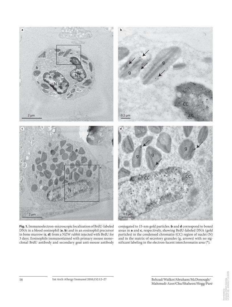
Immunoelectron Microscopic Localization and Quantification of BrdU-Labeled DNA in Bone Marrow and Blood Eosinophils of NZW Rabbits BrdU-labeled DNA was demonstrated by immuno gold
staining in the nuclei and matrix of the secretory granules of eosinophil precursors in bone marrow and blood eo-sinophils ( fig. 1 ). After 3 days of labeling with BrdU, the mean density of immunogold grains in secretory granules of bone marrow and blood eosinophils ( fig. 1 ) was signifi-cantly higher (p ! 0.0005) than that of the cytoplasm and the background staining in all compartments of blood ( ta-ble 1 ; fig. 1–3 ). Immunogold grains in the nuclei were as-sociated with condensed chromatin ( fig. 1 ). The electron-lucent interchromatin region of nucleoplasm remained de-void of any gold label after 2 or 3 days of BrdU injections ( fig. 1 ). After 2 or 3 days of labeling with BrdU, the mean density of immunogold grains in the nuclei of bone mar-row and blood-derived eosinophils was also significantly higher than that of the secretory granules (p ! 0.0001), cy-toplasm (p ! 0.0005) or background staining (p ! 0.0005; table 1 ). The mean density of immunogold grains in nuclei (p ! 0.005) and secretory granules (p ! 0.005) of bone mar-row and blood eosinophils was greater after 3 days com-pared to 2 days of labeling ( table 1 ). The density of the im-munogold in the nuclei of the bone marrow eosinophils was 36-fold higher than that of secretory granules and the nuclear staining of the blood eosinophils was 25-fold high-er than in the granules at 3 days. The mean density of im-munogold grains in the cytoplasm of bone marrow and blood eosinophils was not different when compared to background staining in all compartments from a control rabbit injected with saline ( fig. 2 ) or when isotype-matched mouse IgG ( fig. 3 ) was used as the primary antibody ( ta-ble 1 ). We also found that the staining in the cell cytoplasm did not exceed background staining on the resin outside the cells’ plasma membrane. Light microscopic examina-tion of semi-thick sections of LRP blocks from rabbits re-ceiving 2 days of BrdU showed insufficient number of white blood cells (less than 300 cells) on each section and therefore immunogold staining of ultrathin sections was not performed on sections from these blocks.
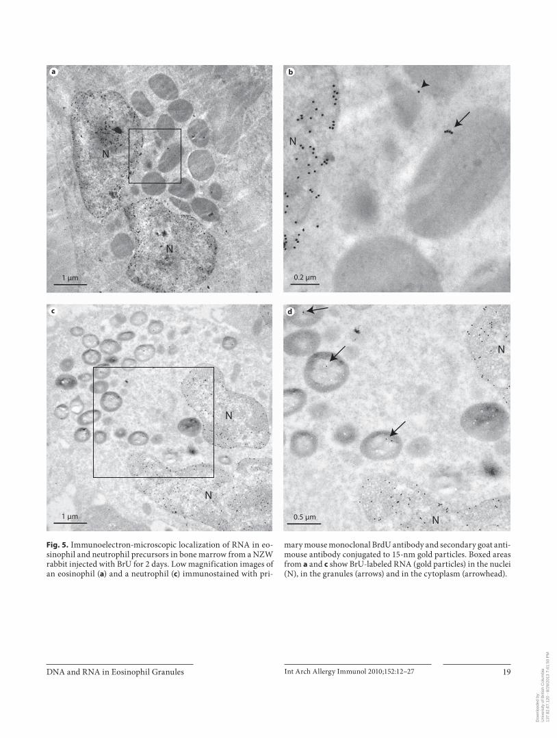
Immunoelectron-Microscopic Localization and Quantification of BrU-Labeled RNA in Bone Marrow and Blood Eosinophils of NZW Rabbits The secretory granules of blood eosinophils ( fig. 4 )
and of bone marrow eosinophil precursors ( fig. 5 a, b)incorporated BrU as demonstrated by ultrastructural im-
Dow
nloa
ded
by:
Uni
vers
ity o
f Brit
ish
Col
umbi
a
13
7.82
.67.
120
- 8/
29/2
013
7:41
:50
PM
Behzad/Walker/Abraham/McDonough/Mahmudi-Azer/Chu/Shaheen/Hogg/Paré
Int Arch Allergy Immunol 2010;152:12–27 16
g
g
g
gg
CC
CC
N
CC
N
N
*
*
*
*
2 μm
2 μm
0.2 μm
0.5 μm
a b
c d
Fig. 1. Immunoelectron-microscopic localization of BrdU-labeled DNA in a blood eosinophil ( a , b ) and in an eosinophil precursor in bone marrow ( c , d ) from a NZW rabbit injected with BrdU for 3 days. Eosinophils immunostained with primary mouse mono-clonal BrdU antibody and secondary goat anti-mouse antibody
conjugated to 15-nm gold particles. b and d correspond to boxed areas in a and c , respectively, showing BrdU-labeled DNA (gold particles) in the condensed chromatin (CC) region of nuclei (N) and in the matrix of secretory granules (g, arrows) with no sig-nificant labeling in the electron-lucent interchromatin area ( * ).
Dow
nloa
ded
by:
Uni
vers
ity o
f Brit
ish
Col
umbi
a
13
7.82
.67.
120
- 8/
29/2
013
7:41
:50
PM
DNA and RNA in Eosinophil Granules Int Arch Allergy Immunol 2010;152:12–27 17
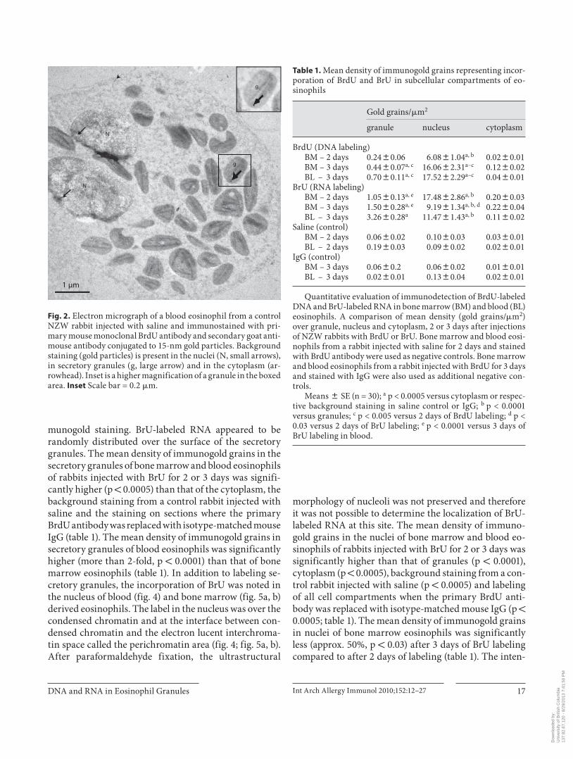
munogold staining. BrU-labeled RNA appeared to be randomly distributed over the surface of the secretory granules. The mean density of immunogold grains in the secretory granules of bone marrow and blood eosinophils of rabbits injected with BrU for 2 or 3 days was signifi-cantly higher (p ! 0.0005) than that of the cytoplasm, the background staining from a control rabbit injected with saline and the staining on sections where the primary BrdU antibody was replaced with isotype-matched mouse IgG ( table 1 ). The mean density of immunogold grains in secretory granules of blood eosinophils was significantly higher (more than 2-fold, p ! 0.0001) than that of bone marrow eosinophils ( table 1 ). In addition to labeling se-cretory granules, the incorporation of BrU was noted in the nucleus of blood ( fig. 4 ) and bone marrow ( fig. 5 a, b) derived eosinophils. The label in the nucleus was over the condensed chromatin and at the interface between con-densed chromatin and the electron lucent interchroma-tin space called the perichromatin area ( fig. 4 ; fig. 5 a, b). After paraformaldehyde fixation, the ultrastructural
morphology of nucleoli was not preserved and therefore it was not possible to determine the localization of BrU-labeled RNA at this site. The mean density of immuno-gold grains in the nuclei of bone marrow and blood eo-sinophils of rabbits injected with BrU for 2 or 3 days was significantly higher than that of granules (p ! 0.0001), cytoplasm (p ! 0.0005), background staining from a con-trol rabbit injected with saline (p ! 0.0005) and labeling of all cell compartments when the primary BrdU anti-body was replaced with isotype-matched mouse IgG (p ! 0.0005; table 1 ). The mean density of immunogold grains in nuclei of bone marrow eosinophils was significantly less (approx. 50%, p ! 0.03) after 3 days of BrU labeling compared to after 2 days of labeling ( table 1 ). The inten-
Table 1. Mean density of immunogold grains representing incor-poration of BrdU and BrU in subcellular compartments of eo-sinophils
Gold grains/�m2
granule nucleus cytoplasm
BrdU (DNA labeling)BM – 2 days 0.2480.06 6.0881.04a, b 0.0280.01BM – 3 days 0.4480.07a, c 16.0682.31a–c 0.1280.02BL – 3 days 0.7080.11a, c 17.5282.29a–c 0.0480.01
BrU (RNA labeling)BM – 2 days 1.0580.13a, e 17.4882.86a, b 0.2080.03BM – 3 days 1.5080.28a, e 9.1981.34a, b, d 0.2280.04BL – 3 days 3.2680.28a 11.4781.43a, b 0.1180.02
Saline (control)BM – 2 days 0.0680.02 0.1080.03 0.0380.01BL – 2 days 0.1980.03 0.0980.02 0.0280.01
IgG (control)BM – 3 days 0.0680.2 0.0680.02 0.0180.01BL – 3 days 0.0280.01 0.1380.04 0.0280.01
Quantitative evaluation of immunodetection of BrdU-labeled DNA and BrU-labeled RNA in bone marrow (BM) and blood (BL) eosinophils. A comparison of mean density (gold grains/�m2) over granule, nucleus and cytoplasm, 2 or 3 days after injections of NZW rabbits with BrdU or BrU. Bone marrow and blood eosi-nophils from a rabbit injected with saline for 2 days and stained with BrdU antibody were used as negative controls. Bone marrow and blood eosinophils from a rabbit injected with BrdU for 3 days and stained with IgG were also used as additional negative con-trols.
Means 8 SE (n = 30); a p < 0.0005 versus cytoplasm or respec-tive background staining in saline control or IgG; b p < 0.0001 versus granules; c p < 0.005 versus 2 days of BrdU labeling; d p < 0.03 versus 2 days of BrU labeling; e p < 0.0001 versus 3 days of BrU labeling in blood.
N
N
g
g
1 μm
Fig. 2. Electron micrograph of a blood eosinophil from a control NZW rabbit injected with saline and immunostained with pri-mary mouse monoclonal BrdU antibody and secondary goat anti-mouse antibody conjugated to 15-nm gold particles. Background staining (gold particles) is present in the nuclei (N, small arrows), in secretory granules (g, large arrow) and in the cytoplasm (ar-rowhead). Inset is a higher magnification of a granule in the boxed area. Inset Scale bar = 0.2 � m.
Dow
nloa
ded
by:
Uni
vers
ity o
f Brit
ish
Col
umbi
a
13
7.82
.67.
120
- 8/
29/2
013
7:41
:50
PM
Behzad/Walker/Abraham/McDonough/Mahmudi-Azer/Chu/Shaheen/Hogg/Paré
Int Arch Allergy Immunol 2010;152:12–27 18
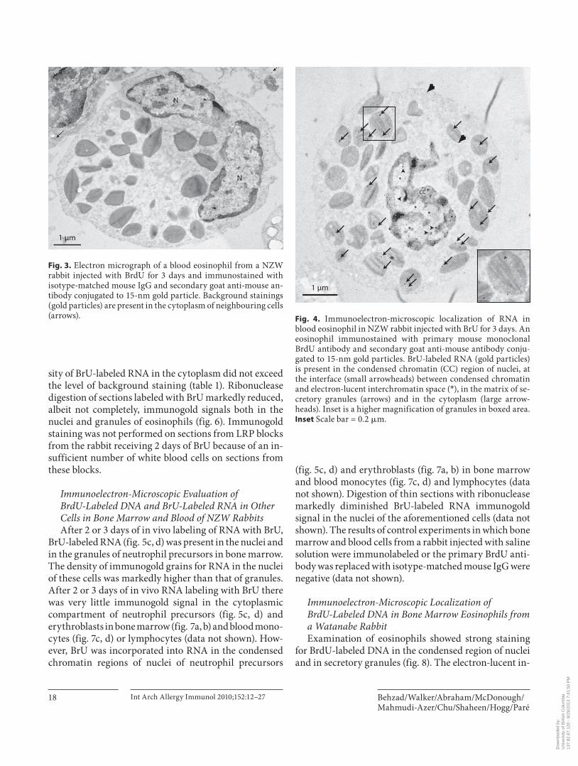
sity of BrU-labeled RNA in the cytoplasm did not exceed the level of background staining ( table 1 ). Ribonuclease digestion of sections labeled with BrU markedly reduced, albeit not completely, immunogold signals both in the nuclei and granules of eosinophils ( fig. 6 ). Immunogold staining was not performed on sections from LRP blocks from the rabbit receiving 2 days of BrU because of an in-sufficient number of white blood cells on sections from these blocks.
Immunoelectron-Microscopic Evaluation ofBrdU-Labeled DNA and BrU-Labeled RNA in Other Cells in Bone Marrow and Blood of NZW Rabbits After 2 or 3 days of in vivo labeling of RNA with BrU,
BrU-labeled RNA ( fig. 5 c, d) was present in the nuclei and in the granules of neutrophil precursors in bone marrow. The density of immunogold grains for RNA in the nuclei of these cells was markedly higher than that of granules. After 2 or 3 days of in vivo RNA labeling with BrU there was very little immunogold signal in the cytoplasmic compartment of neutrophil precursors ( fig. 5 c, d) and erythroblasts in bone marrow ( fig. 7 a, b) and blood mono-cytes ( fig. 7 c, d) or lymphocytes (data not shown). How-ever, BrU was incorporated into RNA in the condensed chromatin regions of nuclei of neutrophil precursors
( fig. 5 c, d) and erythroblasts ( fig. 7 a, b) in bone marrow and blood monocytes ( fig. 7 c, d) and lymphocytes (data not shown). Digestion of thin sections with ribonuclease markedly diminished BrU-labeled RNA immunogold signal in the nuclei of the aforementioned cells (data not shown). The results of control experiments in which bone marrow and blood cells from a rabbit injected with saline solution were immunolabeled or the primary BrdU anti-body was replaced with isotype-matched mouse IgG were negative (data not shown).
Immunoelectron-Microscopic Localization ofBrdU-Labeled DNA in Bone Marrow Eosinophils from a Watanabe Rabbit Examination of eosinophils showed strong staining
for BrdU-labeled DNA in the condensed region of nuclei and in secretory granules ( fig. 8 ). The electron-lucent in-
1 μm
Fig. 3. Electron micrograph of a blood eosinophil from a NZW rabbit injected with BrdU for 3 days and immunostained with isotype-matched mouse IgG and secondary goat anti-mouse an-tibody conjugated to 15-nm gold particle. Background stainings (gold particles) are present in the cytoplasm of neighbouring cells (arrows).
CC
**
*
*
1 μm
Fig. 4. Immunoelectron-microscopic localization of RNA in blood eosinophil in NZW rabbit injected with BrU for 3 days. An eosinophil immunostained with primary mouse monoclonal BrdU antibody and secondary goat anti-mouse antibody conju-gated to 15-nm gold particles. BrU-labeled RNA (gold particles) is present in the condensed chromatin (CC) region of nuclei, at the interface (small arrowheads) between condensed chromatin and electron-lucent interchromatin space ( * ), in the matrix of se-cretory granules (arrows) and in the cytoplasm (large arrow-heads). Inset is a higher magnification of granules in boxed area. Inset Scale bar = 0.2 � m.
Dow
nloa
ded
by:
Uni
vers
ity o
f Brit
ish
Col
umbi
a
13
7.82
.67.
120
- 8/
29/2
013
7:41
:50
PM
DNA and RNA in Eosinophil Granules Int Arch Allergy Immunol 2010;152:12–27 19
N
N
N
N
N
N
N
a b
c d
1 μm 0.2 μm
0.5 μm1 μm
Fig. 5. Immunoelectron-microscopic localization of RNA in eo-sinophil and neutrophil precursors in bone marrow from a NZW rabbit injected with BrU for 2 days. Low magnification images of an eosinophil ( a ) and a neutrophil ( c ) immunostained with pri-
mary mouse monoclonal BrdU antibody and secondary goat anti-mouse antibody conjugated to 15-nm gold particles. Boxed areas from a and c show BrU-labeled RNA (gold particles) in the nuclei (N), in the granules (arrows) and in the cytoplasm (arrowhead).
Dow
nloa
ded
by:
Uni
vers
ity o
f Brit
ish
Col
umbi
a
13
7.82
.67.
120
- 8/
29/2
013
7:41
:50
PM
Behzad/Walker/Abraham/McDonough/Mahmudi-Azer/Chu/Shaheen/Hogg/Paré
Int Arch Allergy Immunol 2010;152:12–27 20
terchromatin area of nucleoplasm remained devoid of any immunogold signal ( fig. 8 ). No staining was observed when primary BrdU antibody was replaced with isotype-matched IgG (data not shown).
DNA and CD63 Immunofluorescence Double Labeling The subcellular distribution of DNA and its colocal-
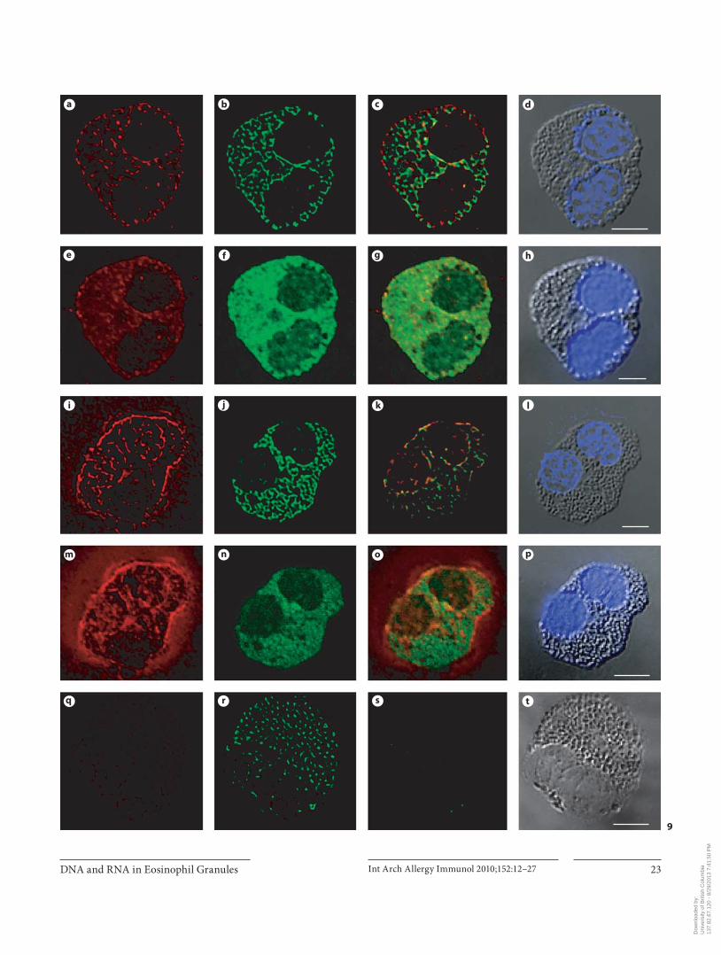
ization with CD63 were examined using confocal and DIC microscopy techniques on cytospins of purified hu-man peripheral blood eosinophils. We undertook a series of co-localization studies on the deconvolved three-di-mensional images of cells generated using multichannel confocal microscopy. Two-dimensional image slices de-rived from the deconvolved and their corresponding nondeconvolved three-dimensional image data set, are shown in figure 9 a–t. In the absence of HCl treatment, DNA ( fig. 9 a, e) and CD63 ( fig. 9 b, f) staining appeared
mainly in the cytoplasm of eosinophils. The immunore-activity against DNA and CD63 showed relative colocal-ization highlighted by the yellow color ( fig. 9 c, g). When DNA was denatured with HCl, the immunoreactivity against DNA appeared in the nucleus, cytoplasm and cell periphery ( fig. 9 i, m). The CD63 immunoreactivity ( fig. 9 j, n) and its colocalization with DNA ( fig. 9 k, o) re-mained the same as above. When cytospins were treated with DNase I for 2 h at 37 ° C prior to HCl-induced DNA denaturation, there was marked reduction in DNA im-munoreactivity ( fig. 9 q) and DNA staining with DAPI ( fig. 9 t) in the nucleus, cytoplasm and cell periphery. No DNA immunofluorescence staining or DNA staining with DAPI was observed when the denaturation step was omitted following DNase I incubation (data not shown). Incubation with DNase I did not affect CD63 staining ( fig. 9 r). No staining was seen when isotype-matched IgG was used as a negative control ( fig. 10 i).
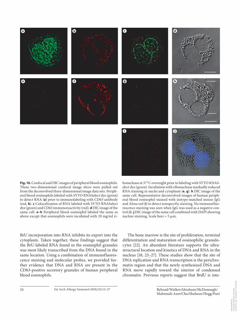
RNA and CD63 Double Staining The SYTO RNASelect dye is a cell-permeant nucleic
acid stain that exhibits bright green fluorescence when bound to RNA (absorption/emission maxima approx. 490/530 nm) and only a weak fluorescent signal when bound to DNA [20] . Using this dye alone and in combina-tion with CD63 immunofluorescence staining (red), we examined the spatial localization of RNA in eosinophils and RNA colocalization with CD63-positive granules. The two-dimensional image slices derived from the de-convolved three-dimensional image data set are shown in figure 10 a–j. In eosinophils incubated with SYTO RNA-Select dye, nuclei stained intensely and cytoplasm was la-beled in a weak granular pattern ( fig. 10 a). The immuno-reactivity against CD63 ( fig. 10 b) showed relative colocal-ization with RNA staining indicated by the yellow color ( fig. 10 c). RNA localization of the SYTO RNASelect dye in nuclei and CD63-positive granules was further sup-ported by ribonuclease treatment. Upon treatment with ribonuclease, the nuclear and cytoplasmic granular RNA-fluorescent signals were markedly reduced ( fig. 10 e, g). Incubation with ribonuclease did not affect CD63 stain-ing ( fig. 10 f). No staining was seen when isotype-matched IgG or buffer were used as a negative control ( fig. 10 i).
Discussion
These results demonstrate the localization of DNA and RNA in secretory granules of NZW rabbit and hu-man eosinophils. Using in vivo labeling of DNA and
N
g
1 μm
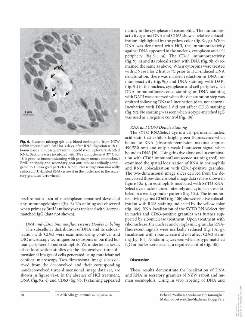
Fig. 6. Electron micrograph of a blood eosinophil, from NZW rabbit injected with BrU for 3 days, after RNA digestion with ri-bonuclease and subsequent immunogold staining for BrU-labeled RNA. Sections were incubated with 2% ribonuclease at 37 ° C for 18 h prior to immunostaining with primary mouse monoclonal BrdU antibody and secondary goat anti-mouse antibody conju-gated to 15-nm gold particles. Ribonuclease digestion markedly reduced BrU-labeled RNA (arrows) in the nuclei and in the secre-tory granules (arrowhead).
Dow
nloa
ded
by:
Uni
vers
ity o
f Brit
ish
Col
umbi
a
13
7.82
.67.
120
- 8/
29/2
013
7:41
:50
PM
DNA and RNA in Eosinophil Granules Int Arch Allergy Immunol 2010;152:12–27 21
a b
c d
CC
CC
Cy
CC
CC
Cy
N
N
*
*
*
0.5 μm
0.5 μm 0.2 μm
0.2 μm
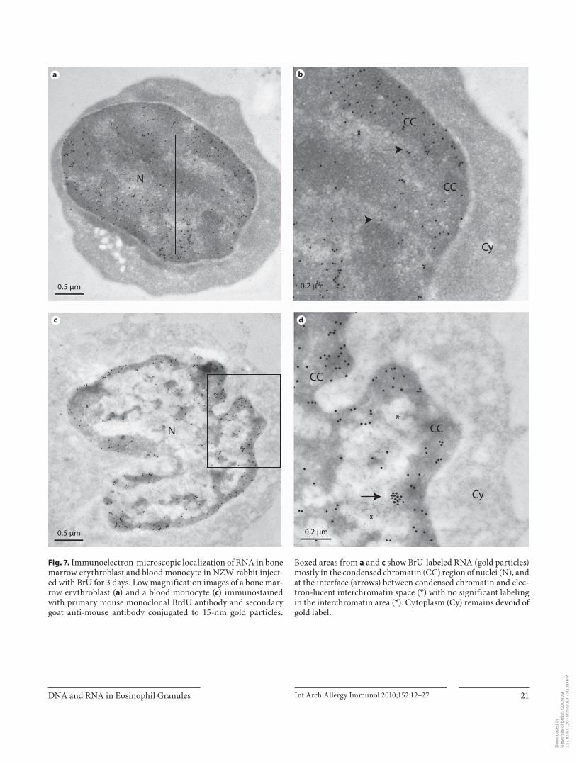
Fig. 7. Immunoelectron-microscopic localization of RNA in bone marrow erythroblast and blood monocyte in NZW rabbit inject-ed with BrU for 3 days. Low magnification images of a bone mar-row erythroblast ( a ) and a blood monocyte ( c ) immunostained with primary mouse monoclonal BrdU antibody and secondary goat anti-mouse antibody conjugated to 15-nm gold particles.
Boxed areas from a and c show BrU-labeled RNA (gold particles) mostly in the condensed chromatin (CC) region of nuclei (N), and at the interface (arrows) between condensed chromatin and elec-tron-lucent interchromatin space ( * ) with no significant labeling in the interchromatin area ( * ). Cytoplasm (Cy) remains devoid of gold label.
Dow
nloa
ded
by:
Uni
vers
ity o
f Brit
ish
Col
umbi
a
13
7.82
.67.
120
- 8/
29/2
013
7:41
:50
PM
Behzad/Walker/Abraham/McDonough/Mahmudi-Azer/Chu/Shaheen/Hogg/Paré
Int Arch Allergy Immunol 2010;152:12–27 22
RNA and subsequent postembedding immunoelectron microscopy we have shown that BrdU-labeled DNA and BrU-labeled RNA are present in the secretory granules of bone marrow and blood eosinophils in NZW rabbits. Quantitative analysis of labeled RNA indicated a signifi-cantly higher level of BrU-labeled RNA in secretory gran-ules of blood eosinophils, compared to bone marrow eo-sinophils after 3 days of labeling, suggesting a dynamic shift to RNA expression in the granule compartment of eosinophils after their release into the circulation. As ex-pected, condensed chromatin in the nuclei was positively
immunostained for BrdU-labeled DNA and BrU-labeled RNA. BrU-labeled RNA was also present in the granules of neutrophil precursors in the bone marrow, suggest-ing that RNA occurrence in granules could be a common phenomenon in granulocytes. However, BrU-labeled RNA was either absent or at the level of background stain-ing in the cytoplasmic compartment of blood eosino-phils, monocytes and lymphocytes and bone marrow erythroblasts and eosinophil and neutrophil precursors after 2 or 3 days of in vivo labeling. This observation is in agreement with previous reports [18, 21] showing that
CC*
a b
1 μm 0.2 μm
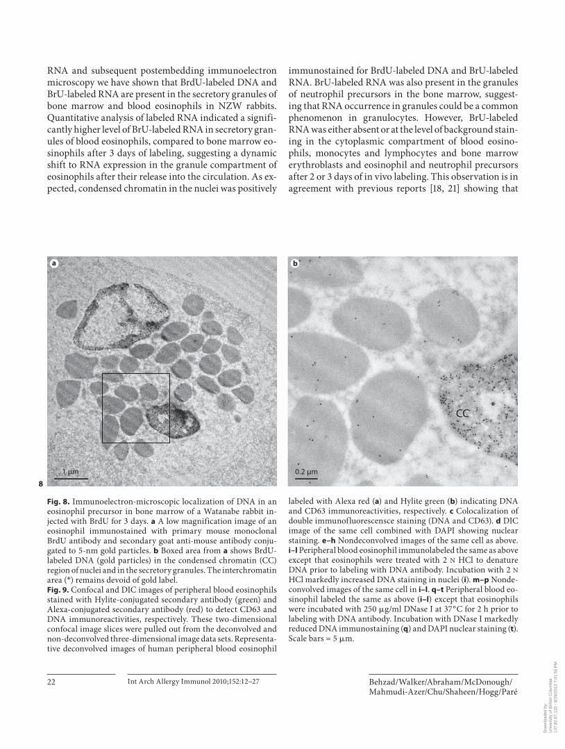
Fig. 8. Immunoelectron-microscopic localization of DNA in an eosinophil precursor in bone marrow of a Watanabe rabbit in-jected with BrdU for 3 days. a A low magnification image of an eosinophil immunostained with primary mouse monoclonal BrdU antibody and secondary goat anti-mouse antibody conju-gated to 5-nm gold particles. b Boxed area from a shows BrdU-labeled DNA (gold particles) in the condensed chromatin (CC) region of nuclei and in the secretory granules. The interchromatin area ( * ) remains devoid of gold label. Fig. 9. Confocal and DIC images of peripheral blood eosinophils stained with Hylite-conjugated secondary antibody (green) and Alexa-conjugated secondary antibody (red) to detect CD63 and DNA immunoreactivities, respectively. These two-dimensional confocal image slices were pulled out from the deconvolved and non-deconvolved three-dimensional image data sets. Representa-tive deconvolved images of human peripheral blood eosinophil
labeled with Alexa red ( a ) and Hylite green ( b ) indicating DNA and CD63 immunoreactivities, respectively. c Colocalization of double immunofluorescensce staining (DNA and CD63). d DIC image of the same cell combined with DAPI showing nuclear staining. e–h Nondeconvolved images of the same cell as above. i–l Peripheral blood eosinophil immunolabeled the same as above except that eosinophils were treated with 2 N HCl to denature DNA prior to labeling with DNA antibody. Incubation with 2 N HCl markedly increased DNA staining in nuclei ( i ). m–p Nonde-convolved images of the same cell in i–l . q–t Peripheral blood eo-sinophil labeled the same as above ( i–l ) except that eosinophils were incubated with 250 � g/ml DNase I at 37 ° C for 2 h prior to labeling with DNA antibody. Incubation with DNase I markedly reduced DNA immunostaining ( q ) and DAPI nuclear staining ( t ). Scale bars = 5 � m.
8
Dow
nloa
ded
by:
Uni
vers
ity o
f Brit
ish
Col
umbi
a
13
7.82
.67.
120
- 8/
29/2
013
7:41
:50
PM
DNA and RNA in Eosinophil Granules Int Arch Allergy Immunol 2010;152:12–27 23
a b c d
e f g h
i j k l
m n o p
q r s t
9
Dow
nloa
ded
by:
Uni
vers
ity o
f Brit
ish
Col
umbi
a
13
7.82
.67.
120
- 8/
29/2
013
7:41
:50
PM
Behzad/Walker/Abraham/McDonough/Mahmudi-Azer/Chu/Shaheen/Hogg/Paré
Int Arch Allergy Immunol 2010;152:12–27 24
BrU incorporation into RNA inhibits its export into the cytoplasm. Taken together, these findings suggest that the BrU-labeled RNA found in the eosinophil granules was most likely transcribed from the DNA found in the same location. Using a combination of immunofluores-cence staining and molecular probes, we provided fur-ther evidence that DNA and RNA are present in the CD63-positive secretory granules of human peripheral blood eosinophils.
The bone marrow is the site of proliferation, terminal differentiation and maturation of eosinophilic granulo-cytes [22] . An abundant literature supports the ultra-structural location and kinetics of DNA and RNA in the nucleus [18, 23–27] . These studies show that the site of DNA replication and RNA transcription is the perichro-matin region and that the newly synthesized DNA and RNA move rapidly toward the interior of condensed chromatin. Previous reports suggest that BrdU is inte-
a b c d
e f g h
i j
Fig. 10. Confocal and DIC images of peripheral blood eosinophils. These two-dimensional confocal image slices were pulled out from the deconvolved three-dimensional image data sets. Periph-eral blood eosinophils labeled with SYTO RNASelect dye (green) to detect RNA ( a ) prior to immunolabeling with CD63 antibody (red, b ). c Colocalization of RNA labeled with SYTO RNASelect dye (green) and CD63 immunoreactivity (red). d DIC image of the same cell. e–h Peripheral blood eosinophil labeled the same as above except that eosinophils were incubated with 20 mg/ml ri-
bonuclease at 37 ° C overnight prior to labeling with SYTO RNAS-elect dye (green). Incubation with ribonuclease markedly reduced RNA staining in nuclei and cytoplasm ( e , g ). h DIC image of the same cell. Representative deconvolved images of human periph-eral blood eosinophil stained with isotype-matched mouse IgG and Alexa red ( i ) to detect nonspecific staining. No immunofluo-rescence staining was seen when IgG was used as a negative con-trol ( i ). j DIC image of the same cell combined with DAPI showing nuclear staining. Scale bars = 5 � m.
Dow
nloa
ded
by:
Uni
vers
ity o
f Brit
ish
Col
umbi
a
13
7.82
.67.
120
- 8/
29/2
013
7:41
:50
PM
DNA and RNA in Eosinophil Granules Int Arch Allergy Immunol 2010;152:12–27 25
grated into the DNA of proliferating cells during theS phase and BrU is incorporated into RNA during tran-scription and that both can be detected by immunoelec-tron microscopy [21, 26, 28] . The localization of BrdU-labeled DNA and BrU-labeled RNA in the condensed chromatin regions of nuclei 3 or 4 days after initial ad-ministration of BrdU or BrU, which was observed in this study, is in agreement with these previous studies.
In addition to the expected finding of nuclear DNA, our results provide clear evidence for DNA localization in eosinophil secretory granules of NZW rabbits and hu-mans. In NZW rabbits, the density of BrdU-labeled DNA in the granules was significantly less than that of nuclei. While nuclear DNA is replicated only during the S phase, the mechanism of granule DNA replication is unknown. Our results showed that increasing the length of BrdU administration from 2 to 3 days had a relatively equal im-pact on the density of immunogold grains in the granules and nuclei of eosinophils, suggesting that there may be comparable dynamics for DNA replication within the granules. In humans, the double immunofluorescent staining of eosinophils with DNA and CD63 antibodies provides further proof that DNase I-sensitive DNA is present in CD63-positive secretory granules. The DNA staining in the granules did not require HCl-induced DNA denaturation, suggesting a single-stranded nature of granular DNA.
Recent reports have shown that IL-5-primed and LPS-activated eosinophils can release DNA into extracellular space [29] . This DNA was shown to be associated with eosinophil cationic protein and major basic protein and not with nuclear or mitochondrial proteins. Based on PCR on DNA released from these eosinophils, the au-thors suggested that this DNA is mitochondrial in origin [29] . However, a previous report also showed that mito-chondria in eosinophils are indeed few in number [30] . Interestingly, Yousefi et al. [29] showed that the process of DNA release occurs rapidly in a catapult-like manner in less than 1 s. These data clearly show that there is a reservoir of non-nuclear DNA in the cytoplasm of eo-sinophils that in a matter of a second can be released into the extracellular space. Taking into account the fact that secretory granules are the only organelles in live eosino-phils that are capable of releasing materials into extracel-lular space and the speed at which nonapoptotic activated live eosinophils release DNA and cationic proteins into extracellular space, the DNA present in the secretory granules is likely to be the cytoplasmic source of DNA that is released from eosinophils [29] . Localization of RNA in the same granules suggests that these granules
may support DNA replication and RNA transcription outside of the nucleus. Recent findings reported by Neves et al. [31] support the notion that eosinophil secretory granules have the potential to function autonomously outside the cell. These investigators showed that cell-free secretory granules respond to specific cytokine/chemo-kines via receptors expressed in the granule membrane and demonstrated the activation of a signal transduction pathway within granules prior to granule protein secre-tion. Such a capacity implies that secretory granules may, like mitochondria, have in situ synthetic capacity. A gran-ule with its own independent DNA replication, RNA transcription and protein synthesis could provide a basis for such a response.
The presence of RNA in secretory granules has been previously reported in mast cells and eosinophils [14, 15, 32] . Dvorak and colleagues [12–15] have used a variety of labeling methods to document the synthetic capacityof secretory granules in human mast cells. Specifically, they have documented the presence of uridine, RNA, U1snRNP (a component of spliceosomes that controls al-ternative splicing of mRNA), mRNA and ribosomes in, and attached to, the mast cell secretory granules [12–15] . These authors propose a nuclear origin for granule RNA in mast cells and they suggest that there is transport of nuclear mRNA to secretory granules as a mechanism for facilitating site-specific synthesis of granular proteins. Dvorak et al. [14] and Wickramasinghe and Hughes [32] have also reported the presence of RNA in eosinophil granules. They used ultrastructural autoradiography and found that radiolabeled uridine was incorporated into RNA in secretory granules in vitro. The present report extends those studies by using 2 different methods to demonstrate the localization of RNA in eosinophil secre-tory granules as well as its presence in the expected nu-clear sites, as a positive internal control. In rabbits, in vivo labeling of RNA with BrU and subsequent immunoelec-tron microscopy clearly showed RNA in secretory gran-ules. We also found that the incorporation of BrU into granule RNA was significantly higher in circulating than in bone marrow eosinophils, suggesting a stimulation of transcription within the granules after release from the bone marrow. Similarly, using a molecular probe that specifically binds RNA, we provide evidence of the gran-ular nature of RNA in human eosinophils. Colocaliza-tion of RNA staining with CD63-immunofluorescent staining further proves that the observed RNA is present in eosinophil-secretory granules.
It is surprising that RNA is localized to granules since this is also the storage site for eosinophil cationic protein
Dow
nloa
ded
by:
Uni
vers
ity o
f Brit
ish
Col
umbi
a
13
7.82
.67.
120
- 8/
29/2
013
7:41
:50
PM
Behzad/Walker/Abraham/McDonough/Mahmudi-Azer/Chu/Shaheen/Hogg/Paré
Int Arch Allergy Immunol 2010;152:12–27 26
and eosinophil-derived neurotoxin, both of which have been shown to be single-strand-specific RNases [33, 34] . One explanation could be that the granule-associated RNAs could form secondary structure which makes them resistant to the RNase activities of eosinophil cationic protein and eosinophil-derived neurotoxin. A number of noncoding RNAs such as transfer RNA, ribosomal RNA and ribozymes contain sequences that can fold back on themselves to form a paired double helix and stem loops [35–37] .
Although this study does not provide irrefutable evi-dence that the RNA located within the granules was tran-scribed from granular DNA, there is indirect evidence to support this assertion. Both qualitative and quantitative evaluation of BrU-labeled RNA in the cytoplasmic com-partment of bone marrow and blood cells showed either absence of immunogold signal or a level that did not ex-ceed background staining in this compartment. This finding is consistent with previous reports that showed that BrU labeling interferes with mRNA intron splicing and export into the cytoplasm [21] . This observation sug-gests that the BrU-labeled RNA in the secretory granules of eosinophils most likely originated from the extranu-clear DNA.
We can only speculate on the role that the granule-as-sociated RNA plays. In addition to protein synthesis nat-urally occurring noncoding RNA species function in a variety of cellular processes such as structural, catalytic (that is, nucleotide modification, gene splicing and pep-tide bond formation) or regulation of gene expression [38] . Various artificially produced noncoding RNAs also show enzymatic activities. This includes RNA-catalyzed carbon-carbon bond formation, nucleotide synthesis, RNA polymerization and peptide bond formation [39–43] . This accumulating evidence for multiple functions of RNAs may suggest a more comprehensive role for RNAs in secretory granules of eosinophils than previously rec-ognized. Granule RNAs could either possess catalytic ac-tivity or could function as integral components of dy-namic riboproteins. Based on our findings and those of others, it is conceivable that the synthetic capacity of se-cretory granules consists of DNA replication and RNA transcription without the need for further translation and that the RNA-based activities could complement the existing protein-based enzymatic activity in granules.
The validity of our findings regarding the granular localization of DNA and RNA is supported by a number of specificity controls. In NZW rabbit eosinophils, use of saline as a control for in vivo labeling of DNA and RNA and subsequent staining with primary anti-BrdU anti-
body or IgG resulted in a lack of label in eosinophil gran-ules and nuclei, suggesting that immunogold staining is not due to nonspecific binding of BrdU antibody with the basic proteins in the eosinophil secretory granules or with nuclear proteins. To confirm that the BrU labeling was RNA, some sections were submitted to RNA diges-tion with ribonuclease. RNase digestion of sections prior to immunogold labeling resulted in a marked reduction in the immunogold labeling in nuclei and granules, indi-cating that the labeling, for the most part, represents RNA. The failure to completely remove RNA with ribo-nuclease is consistent with previous studies [15, 44] . Sim-ilarly, in human eosinophils, use of DNase I and RNase prior to DNA immunostaining and RNA staining with molecular probe resulted in marked reduction in DNA and RNA fluorescent signals in both nuclei and granule compartments, suggesting the specificity of our staining procedures. The absence of any signals in eosinophils when isotype-matched IgG was used further supports the specificity of our immunofluorescent staining for DNA and CD63.
In summary, the presence of DNA and RNA in the se-cretory granules of eosinophils indicates the need to reas-sess the role(s) of this organelle. These data suggest that the granules may be the site of DNA and RNA synthesis. It is possible that the RNA synthesized in the granule could contribute to the overall catalytic and enzymatic activities of secretory granules.
Acknowledgement
We wish to thank Dr. Shizu Hyashi for helpful discussion and review of the manuscript.
References 1 Klementsson H: Eosinophils and the patho-physiology of allergic rhinitis. Clin Exp Al-lergy 1992; 22: 1058–1064.
2 Ovington KS, Behm CA: The enigmatic eo-sinophil: investigation of the biological role of eosinophils in parasitic helminth infec-tion. Mem Inst Oswaldo Cruz 1997; 92(suppl 2):93–104.
3 Rothenberg ME, Hogan SP: The eosinophil. Annu Rev Immunol 2006; 24: 147–174.
4 Simon D, Braathen LR, Simon HU: Eosino-phils and atopic dermatitis. Allergy 2004; 59: 561–570.
5 Gleich GJ, Adolphson CR, Leiferman KM: The biology of the eosinophilic leukocyte. Annu Rev Med 1993; 44: 85–101.
6 Lacy P, Moqbel R: Eosinophil cytokines. Chem Immunol 2000; 76: 134–155.
Dow
nloa
ded
by:
Uni
vers
ity o
f Brit
ish
Col
umbi
a
13
7.82
.67.
120
- 8/
29/2
013
7:41
:50
PM
DNA and RNA in Eosinophil Granules Int Arch Allergy Immunol 2010;152:12–27 27
7 Wardlaw AJ, Moqbel R, Kay AB: Eosino-phils: biology and role in disease. Adv Im-munol 1995; 60: 151–266.
8 Matsumoto K, Bochner BS, Wakiguchi H, Kurashige T: Functional expression of trans-membrane 4 superfamily molecules on hu-man eosinophils. Int Arch Allergy Immunol 1999; 120(suppl 1):38–44.
9 Mahmudi-Azer S, Downey GP, Moqbel R: Translocation of the tetraspanin CD63 in as-sociation with human eosinophil mediator release. Blood 2002; 99: 4039–4047.
10 Dvorak AM: Subcellular morphology and biochemistry of eosinophils; in Blood Cell Biochemistry: Megakaryocytes, Platelets, Macrophages. London, J.R. Harris, 1991.
11 Dvorak AM, Weller PF: Ultrastructural analysis of human eosinophils. Chem Im-munol 2000; 76: 1–28.
12 Dvorak AM, Morgan ES: Ultrastructural immunogold cytochemistry with autoim-mune human sera and an antibody to uri-dine implicate human mast cell granules in RNA biology. Histochem J 2000; 32: 685–696.
13 Dvorak AM, Morgan ES: Ribosomes and se-cretory granules in human mast cells: close associations demonstrated by staining with a chelating agent. Immunol Rev 2001; 179: 94–101.
14 Dvorak AM, Morgan ES, Lichtenstein LM, Schleimer R: Ultrastructural autoradio-graphic analysis of RNA in isolated human lung mast cells during secretion and recov-ery from secretion. Int Arch Allergy Immu-nol 2000; 122: 124–136.
15 Dvorak AM, Morgan ES, Lichtenstein LM, Weller PF, Schleimer RP: RNA is closely as-sociated with human mast cell secretory granules, suggesting a role(s) for granules in synthetic processes. J Histochem Cytochem 2000; 48: 1–12.
16 Goto Y, Hogg JC, Suwa T, Quinlan KB, van Eeden SF: A novel method to quantify the turnover and release of monocytes from the bone marrow using the thymidine analog 5 � -bromo-2 � -deoxyuridine. Am J Physiol Cell Physiol 2003; 285:C253–C259.
17 Terashima T, Wiggs B, English D, Hogg JC, van Eeden SF: Polymorphonuclear leuko-cyte transit times in bone marrow during streptococcal pneumonia. Am J Physiol 1996; 271:L587–L592.
18 Cmarko D, Verschure PJ, Martin TE, Dah-mus ME, Krause S, Fu XD, van Driel R, Fak-an S: Ultrastructural analysis of transcrip-tion and splicing in the cell nucleus after bromo-UTP microinjection. Mol Biol Cell 1999; 10: 211–223.
19 Ngwenya LB, Peters A, Rosene DL: Light and electron microscopic immunohistochemical detection of bromodeoxyuridine-labeled cells in the brain: Different fixation and pro-cessing protocols. J Histochem Cytochem 2005; 53: 821–832.
20 Myrdal SE, Johnson KC, Steyger PS: Cyto-plasmic and intra-nuclear binding of genta-micin does not require endocytosis. Hear Res 2005; 204: 156–169.
21 Wansink DG, Nelissen RL, de Jong L: In vi-tro splicing of pre-mRNA containing bro-mouridine. Mol Biol Rep 1994; 19: 109–113.
22 Bainton D: Phagocytic cells: developmental biology of neutrophils and eosinophils; in Inflammation: Basic Principles and Clinical Correlations. New York, Raven, 1998.
23 Ghosh S, Paweletz N: Detection of intranu-cleolar chromatin using an ultrastructural immunolabelling technique. Cell Biol Int 1998; 22: 609–614.
24 Hozak P, Cook PR, Schofer C, Mosgoller W, Wachtler F: Site of transcription of ribosom-al RNA and intranucleolar structure in HeLa cells. J Cell Sci 1994; 107: 639–648.
25 Jaunin F, Visser AE, Cmarko D, Aten JA, Fakan S: Fine structural in situ analysis of nascent DNA movement following DNA replication. Exp Cell Res 2000; 260: 313–323.
26 Momma S, Vasilyeva E, Tomilin N, Shuplia-kov O: Ultrastructural identification of di-viding cells in the adult mammalian central nervous system. J Neurosci Methods 2002; 119: 59–63.
27 Puvion E, Puvion-Dutilleul F: Ultrastruc-ture of the nucleus in relation to transcrip-tion and splicing: roles of perichromatin fi-brils and interchromatin granules. Exp Cell Res 1996; 229: 217–225.
28 Palmer TD, Willhoite AR, Gage FH: Vascu-lar niche for adult hippocampal neurogene-sis. J Comp Neurol 2000; 425: 479–494.
29 Yousefi S, Gold JA, Andina N, Lee JJ, Kelly AM, Kozlowski E, Schmid I, Straumann A, Reichenbach J, Gleich GJ, Simon HU: Cata-pult-like release of mitochondrial DNA by eosinophils contributes to antibacterial de-fense. Nat Med 2008; 14: 949–953.
30 Peachman KK, Lyles DS, Bass DA: Mito-chondria in eosinophils: functional role in apoptosis but not respiration. Proc Natl Acad Sci USA 2001; 98: 1717–1722.
31 Neves JS, Perez AC, Spencer LA, Melo RCN, Reynolds L, Ghiran L, Mahumudi-Azer S, Odemuyiwa SO, Dvorak AM, Moqbel R, Welle PF: Eosinophil granules function ex-tracellularly as receptor-mediated secretory organelles Proc Natl Acad Sci USA 2008; 105: 18478–18483.
32 Wickramasinghe SN, Hughes M: High reso-lution autoradiographic studies of RNA, protein and DNA synthesis during human eosinophil granulocytopoiesis: evidence for the presence of RNA on or within eosinophil granules. Br J Haematol 1978; 38: 179–183.
33 Sorrentino S, Glitz DG: Ribonuclease activ-ity and substrate preference of human eosin-ophil cationic protein (ECP). FEBS Lett 1991; 288: 23–26.
34 Sorrentino S, Glitz DG, Hamann KJ, Loeger-ing DA, Checkel JL, Gleich GJ: Eosinophil-derived neurotoxin and human liver ribonu-clease. Identity of structure and linkage of neurotoxicity to nuclease activity. J Biol Chem 1992; 267: 14859–14865.
35 Doherty EA, Batey RT, Masquida B, Doudna JA: A universal mode of helix packing in RNA. Nat Struct Biol 2001; 8: 339–343.
36 Doherty EA, Doudna JA: Ribozyme struc-tures and mechanisms. Annu Rev Biophys Biomol Struct 2001; 30: 457–475.
37 Omer AD, Ziesche S, Decatur WA, Fournier MJ, Dennis PP: RNA-modifying machines in archaea. Mol Microbiol 2003; 48: 617–629.
38 Szymanski M, Barciszewska MZ, Zywicki M, Barciszewski J: Noncoding RNA tran-scripts. J Appl Genet 2003; 44: 1–19.
39 Beringer M, Rodnina MV: The ribosomal peptidyl transferase. Mol Cell 2007; 26: 311–321.
40 Johnston WK, Unrau PJ, Lawrence MS, Glasner ME, Bartel DP: RNA-catalyzed RNA polymerization: accurate and general RNA-templated primer extension. Science 2001; 292: 1319–1325.
41 Tarasow TM, Tarasow SL, Eaton BE: RNA-catalysed carbon-carbon bond formation. Nature 1997; 389: 54–57.
42 Unrau PJ, Bartel DP: RNA-catalysed nucleo-tide synthesis. Nature 1998; 395: 260–263.
43 Zhang B, Cech TR: Peptide bond formation by in vitro selected ribozymes. Nature 1997; 390: 96–100.
44 Dvorak AM, Morgan ES: Ribonuclease-gold labels heparin in human mast cell granules. New use for an ultrastructural enzyme affin-ity technique. J Histochem Cytochem 1998; 46: 695–706.
Dow
nloa
ded
by:
Uni
vers
ity o
f Brit
ish
Col
umbi
a
13
7.82
.67.
120
- 8/
29/2
013
7:41
:50
PM
Related Documents