Localization and assembly of proteins comprising the outer structures of the Bacillus anthracis spore Rebecca Giorno, 1 3 Michael Mallozzi, 1 Joel Bozue, 2 Krishna- Sulayman Moody, 2 Alex Slack, 1 Dengli Qiu, 3 Rong Wang, 3 Arthur Friedlander, 4 Susan Welkos 2 and Adam Driks 1 Correspondence Adam Driks [email protected] 1 Department of Microbiology and Immunology, Loyola University Medical Center, Maywood, IL 60153, USA 2 Bacteriology Division, United States Army Medical Research Institute of Infectious Diseases, Fort Detrick, Frederick, MD 21702-5011, USA 3 Department of Biological, Chemical, and Physical Sciences, Illinois Institute of Technology, Chicago, IL 60616, USA 4 Headquarters, United States Army Medical Research Institute of Infectious Diseases, Fort Detrick, Frederick, MD 21702-5011, USA Received 15 August 2008 Revised 24 November 2008 Accepted 12 December 2008 Bacterial spores possess a series of concentrically arranged protective structures that contribute to dormancy, survival and, ultimately, germination. One of these structures, the coat, is present in all spores. In Bacillus anthracis, however, the spore is surrounded by an additional, poorly understood, morphologically complex structure called the exosporium. Here, we characterize three previously discovered exosporium proteins called ExsFA (also known as BxpB), ExsFB (a highly related paralogue of exsFA/bxpB) and IunH (similar to an inosine–uridine-preferring nucleoside hydrolase). We show that in the absence of ExsFA/BxpB, the exosporium protein BclA accumulates asymmetrically to the forespore pole closest to the midpoint of the sporangium (i.e. the mother-cell-proximal pole of the forespore), instead of uniformly encircling the exosporium. ExsFA/BxpB may also have a role in coat assembly, as mutant spore surfaces lack ridges seen in wild-type spores and have a bumpy appearance. ExsFA/BxpB also has a modest but readily detected effect on germination. Nonetheless, an exsFA/bxpB mutant strain is fully virulent in both intramuscular and aerosol challenge models in Guinea pigs. We show that the pattern of localization of ExsFA/BxpB–GFP is a ring, consistent with a location for this protein in the basal layer of the exosporium. In contrast, ExsFB–GFP fluorescence is a solid oval, suggesting a distinct subcellular location for ExsFB–GFP. We also used these fusion proteins to monitor changes in the subcellular locations of these proteins during sporulation. Early in sporulation, both fusions were present throughout the mother cell cytoplasm. As sporulation progressed, GFP fluorescence moved from the mother cell cytoplasm to the forespore surface and formed either a ring of fluorescence, in the case of ExsFA/BxpB, or a solid oval of fluorescence, in the case of ExsFB. IunH–GFP also resulted in a solid oval of fluorescence. We suggest the interpretation that at least some ExsFB–GFP and IunH–GFP resides in the region between the coat and the exosporium, called the interspace. INTRODUCTION The bacilli and clostridia encompass a large number of species that produce highly resistant dormant cells, called spores, in response to stress (Fritze, 2004; Nicholson, 2002). Prominent among these species is Bacillus anthracis, which has played a central historical role in our understanding of the transmission of disease and the mechanisms of toxin action, and which has reemerged as a serious threat to human life in light of its utility as a biological weapon (Alibek, 1999; Koch, 1876; Mock & Fouet, 2001). Because the B. anthracis spore is the infectious particle for the disease anthrax, a deeper understanding of spore composition, assembly and function could be very useful in combating this threat. Abbreviations: AFM, atomic force microscopy; IFM, immunofluorescence microscopy; IM, intramuscular; TEM, thin-section electron microscopy. A supplementary table listing PCR primers used in this study is available with the online version of this paper. 3Present address: College of Biological Sciences, Louisiana Tech University, Ruston, LA 71270, USA. Microbiology (2009), 155, 1133–1145 DOI 10.1099/mic.0.023333-0 023333 G 2009 US Government Printed in Great Britain 1133

Welcome message from author
This document is posted to help you gain knowledge. Please leave a comment to let me know what you think about it! Share it to your friends and learn new things together.
Transcript

Localization and assembly of proteins comprisingthe outer structures of the Bacillus anthracis spore
Rebecca Giorno,13 Michael Mallozzi,1 Joel Bozue,2 Krishna-Sulayman Moody,2 Alex Slack,1 Dengli Qiu,3 Rong Wang,3
Arthur Friedlander,4 Susan Welkos2 and Adam Driks1
Correspondence
Adam Driks
1Department of Microbiology and Immunology, Loyola University Medical Center, Maywood, IL60153, USA
2Bacteriology Division, United States Army Medical Research Institute of Infectious Diseases, FortDetrick, Frederick, MD 21702-5011, USA
3Department of Biological, Chemical, and Physical Sciences, Illinois Institute of Technology,Chicago, IL 60616, USA
4Headquarters, United States Army Medical Research Institute of Infectious Diseases, Fort Detrick,Frederick, MD 21702-5011, USA
Received 15 August 2008
Revised 24 November 2008
Accepted 12 December 2008
Bacterial spores possess a series of concentrically arranged protective structures that contribute
to dormancy, survival and, ultimately, germination. One of these structures, the coat, is present
in all spores. In Bacillus anthracis, however, the spore is surrounded by an additional, poorly
understood, morphologically complex structure called the exosporium. Here, we characterize
three previously discovered exosporium proteins called ExsFA (also known as BxpB), ExsFB
(a highly related paralogue of exsFA/bxpB) and IunH (similar to an inosine–uridine-preferring
nucleoside hydrolase). We show that in the absence of ExsFA/BxpB, the exosporium protein BclA
accumulates asymmetrically to the forespore pole closest to the midpoint of the sporangium
(i.e. the mother-cell-proximal pole of the forespore), instead of uniformly encircling the exosporium.
ExsFA/BxpB may also have a role in coat assembly, as mutant spore surfaces lack ridges seen
in wild-type spores and have a bumpy appearance. ExsFA/BxpB also has a modest but readily
detected effect on germination. Nonetheless, an exsFA/bxpB mutant strain is fully virulent in
both intramuscular and aerosol challenge models in Guinea pigs. We show that the pattern of
localization of ExsFA/BxpB–GFP is a ring, consistent with a location for this protein in the basal
layer of the exosporium. In contrast, ExsFB–GFP fluorescence is a solid oval, suggesting a distinct
subcellular location for ExsFB–GFP. We also used these fusion proteins to monitor changes in
the subcellular locations of these proteins during sporulation. Early in sporulation, both fusions
were present throughout the mother cell cytoplasm. As sporulation progressed, GFP fluorescence
moved from the mother cell cytoplasm to the forespore surface and formed either a ring of
fluorescence, in the case of ExsFA/BxpB, or a solid oval of fluorescence, in the case of ExsFB.
IunH–GFP also resulted in a solid oval of fluorescence. We suggest the interpretation that at least
some ExsFB–GFP and IunH–GFP resides in the region between the coat and the exosporium,
called the interspace.
INTRODUCTION
The bacilli and clostridia encompass a large number ofspecies that produce highly resistant dormant cells, called
spores, in response to stress (Fritze, 2004; Nicholson, 2002).Prominent among these species is Bacillus anthracis, whichhas played a central historical role in our understanding ofthe transmission of disease and the mechanisms of toxinaction, and which has reemerged as a serious threat to humanlife in light of its utility as a biological weapon (Alibek, 1999;Koch, 1876; Mock & Fouet, 2001). Because the B. anthracisspore is the infectious particle for the disease anthrax, adeeper understanding of spore composition, assembly andfunction could be very useful in combating this threat.
Abbreviations: AFM, atomic force microscopy; IFM, immunofluorescencemicroscopy; IM, intramuscular; TEM, thin-section electron microscopy.
A supplementary table listing PCR primers used in this study is availablewith the online version of this paper.
3Present address: College of Biological Sciences, Louisiana TechUniversity, Ruston, LA 71270, USA.
Microbiology (2009), 155, 1133–1145 DOI 10.1099/mic.0.023333-0
023333 G 2009 US Government Printed in Great Britain 1133
Spores from diverse Bacillus species possess a commonarchitecture. Surrounding the core, which houses the DNAand protective DNA-binding proteins called small acid-soluble spore proteins (SASPs) (Setlow, 2006), is a specializedpeptidoglycan called the cortex (Dowd et al., 2008; Popham,2002). The cortex, in turn, is housed in a complex multi-layered protein shell called the coat (Aronson & Fitz-James,1976; Warth et al., 1963). From studies in Bacillus subtilis, weknow that the coat protects the spore against a variety ofharmful molecules and facilitates germination, the process bywhich spores leave the dormant state and resume vegetativegrowth (Moir et al., 2002).
In some species, including B. subtilis, the coat is the outer-most spore structure. In others, such as B. anthracis, sporespossess an additional structure called the exosporium,which encases the entire spore. A thin basal layer appears tobe a structural feature shared by exosporia of most or allspecies (Aronson & Fitz-James, 1976; Holt & Leadbetter,1969). In some species, such as Bacillus megaterium, thislayer is relatively thick (Aronson & Fitz-James, 1976; A.Driks, unpublished observations; Holt & Leadbetter, 1969;Vary, 1994). In others, such as B. anthracis and Bacilluscereus, a series of fine hair-like projections (also called anap) extend a short distance from the basal layer. A strikingfeature of the B. anthracis exosporium is the significantspace between the exosporium and the coat, a region werefer to as the interspace. Although no attachment pointsbetween the two structures are evident in conventionalthin-section electron micrographs, such connections mustexist in order for the exosporium to have been able toassemble around the spore.
The function of the exosporium is currently an area ofactive investigation. Efforts to identify its purpose are com-plicated by its presence in a taxonomically diverse subset ofspecies that includes non-pathogenic organisms such asB. megaterium and Bacillus odysseyi (La Duc et al., 2004;Vary, 1994). Nonetheless, insight into possible exosporiumfunctions has come from recent studies that indicate thatthe exosporium mediates adhesion to certain eukaryoticcell types representative of host tissues, provides somedegree of resistance to the oxidative burst produced bymacrophages in culture, and helps moderate the innateimmune response to spores (Basu et al., 2007; Bozue et al.,2007b; Weaver et al., 2007). Additionally, the majorexosporial protein BclA has been shown to mediate thephagocytosis of spores via interaction with the macrophagecell-surface integrin Mac-1 (Oliva et al., 2008). Thesefunctions could provide advantages to spores duringpathogenesis and in a variety of niches other than that ofan infected host.
About 20 exosporium-associated proteins and glycopro-teins have been identified from analyses of B. anthracis andB. cereus (Redmond et al., 2004; Steichen et al., 2003;Sylvestre et al., 2002; Todd et al., 2003; Waller et al., 2005).In addition, other, less well-characterized molecules arealso present, including lipids and carbohydrates (Bozue
et al., 2005; Fox et al., 2003; Matz et al., 1970). The best-characterized B. anthracis exosporium protein is BclA,which is on the exosporium surface, is highly immuno-genic, and is a major structural component of the hair-likeprojections (referred to below as ‘projections’) (Boydstonet al., 2005; Rety et al., 2005; Steichen et al., 2003, 2005;Sylvestre et al., 2002, 2003, 2005). Several other exospor-ium proteins or candidate exosporium proteins have beencharacterized in B. anthracis. ExsFA (or BxpB) has alsobeen shown to reside in the B. anthracis basal layer, and tohave at least a partial role in the formation of projectionsand in BclA deposition (Steichen et al., 2005; Sylvestreet al., 2005). ExsFB, a paralogue of ExsFA/BxpB, also has arole in exosporium formation, since exsFA/bxpB exsFBdouble mutant spores have a more severe exosporiumdefect than exsFA/bxpB mutant spores (Sylvestre et al.,2005). The B. anthracis proteins CotY and ExsY (ortholo-gues of the B. subtilis coat proteins CotY and CotZ) havealso been found in the exosporium (Redmond et al., 2004).ExsY has a role in exosporium assembly, as exsY mutantspores possess only a fragment of this structure (Boydstonet al., 2006). The view that these proteins are together inthe exosporium is supported by the finding that ExsFA/BxpB is present as part of a complex with BclA and ExsY(Redmond et al., 2004). The exosporium glycoprotein BclBhas also been shown to play an important role inexosporium stability, as bclB mutant spores assemble onlypartial exosporia, or lack the structure entirely (Thompsonet al., 2007).
Some spore proteins play roles in assembly of both thecoat and exosporium. The B. anthracis coat protein CotEdirects assembly of at least one coat protein other thanitself (Giorno et al., 2007). In addition, in most cotEmutant sporangia, an exosporium is synthesized, butaccumulates in the mother cell without encircling theforespore. This and other data support the inference thatCotE directs the attachment of the exosporium to theforespore surface.
Some exosporium proteins are orthologues of previouslyidentified enzymes. For example, alanine racemase (AlR)and inosine hydrolase (IunH) are present in B. anthracisand B. cereus exosporia, consistent with a role for theexosporium in the modification of germinant molecules(Boydston et al., 2006; Redmond et al., 2004; Steichen et al.,2007; Todd et al., 2003). Arginase activity is also present inthe B. anthracis exosporium, and plays a role in protectionagainst oxidative bursts generated by macrophages, asalready discussed (Weaver et al., 2007).
In this work, we further characterize ExsFA/BxpB and showevidence to suggest that ExsFB and IunH do not entirelycolocalize with ExsFA/BxpB. We show that ExsFA/BxpBaffects spore surface morphology and germination, loca-lizes to the exosporium basal layer, and plays a role indistributing BclA throughout the exosporium surface.Finally, we demonstrate that, in two animal challengemodels, ExsFA/BxpB is dispensable for virulence.
R. Giorno and others
1134 Microbiology 155

METHODS
General methods. Escherichia coli strains were cultured in Luria–
Bertani (LB) medium and antibiotics were used at standard concentra-
tions (Sambrook et al., 1989). For B. anthracis, erythromycin, tetra-
cycline and kanamycin were used at concentrations of 5, 5 and 20 mg
ml21, respectively. Sporangia and spores from the Sterne 34F2 strain
and Sterne 34F2-derived stains of B. anthracis were prepared by
exhaustion in Difco sporulation medium (DSM) (Lai et al., 2003),
unless stated otherwise. Sporulation at room temperature was
performed using either 26 SG medium (Harwood & Cutting, 1990)
(for fluorescence microscopic analysis) or DSM [for immunofluores-
cence microscopy (IFM)], as indicated. We prepared Ames strain spores
by culture in Leighton–Doi broth (Ivins et al., 1990; Leighton & Doi,
1971). Time 0 (t0) of sporulation was defined as the first point at which
the culture leaves exponential-phase growth. We carried out further
purification of Ames strain spores by centrifugation through Hypaque-
76 gradients (Nycomed). All preparations possessed .95 % spores. For
germination and virulence studies, we diluted the spores in sterile water,
heated them at 65 uC for 30 min, and then kept them on ice. Electron
and atomic force microscopy (AFM) were carried out according to
Catalano et al. (2001) and Chada et al. (2003), respectively.
Recombinant DNA methods. Recombinant DNA techniques were
performed using standard methodologies (Sambrook et al., 1989) or
according to manufacturer’s instructions. Strains and plasmids are
referenced or described in Table 1. Primers are referenced in
Supplementary Table S1. To construct plasmid pMR6, we first used
PCR to amplify approximately 1 kb stretches of DNA on either side of
exsFA/bxpB, using primers OL298 and OL299 (to amplify the
upstream region), and primers OL300 and OL301 (to amplify the
downstream region). The PCR products were cloned into plasmid
pMR1, such that they flanked the plasmid-borne kanamycin-
resistance marker. To do this, we digested the upstream PCR product
and pMR1 with SacI and NotI, and ligated the products. We then
digested the resulting plasmid and the downstream PCR product with
BamHI and SalI, and ligated the resulting digestion products together.
To generate strains bearing fusions of genes to gfp, we first built
plasmids (pAS1 and pRG30) bearing gfp. To build pAS1, we PCR-
amplified gfp from plasmid pKL147 (Lemon & Grossman, 1998) with
primers OL471 and OL472, digested the PCR product and pUTE-29
with BamHI and HindIII, and ligated the resulting fragments. To
build pRG30 we digested pAS1 with HindIII and SmaI, and ligated
the gfp-bearing fragment into similarly digested pKS1. We then used
pAS1 or pRG30 to build a set of plasmids bearing gfp fusions to
different spore proteins, which were designed to integrate into the
chromosome. To build plasmid pAS4, we first PCR-amplified the 39
end of exsFA/bxpB with primers OL504 and OL505. We digested the
PCR product with BamHI and EcoRV, and pAS1 with BamHI and
PmeI, and ligated these together. To build pAS2 we first amplified the
39 end of iunH with primers OL476 and OL498. We digested the PCR
product with BamHI and EcoRV, and pAS1 with BamHI and PmeI,
and ligated the digestion products. To build pRG25 we digested pAS4
with BamHI and HindIII, and ligated it to BamHI- and HindIII-
digested plasmid pEO-3 (Mendelson et al., 2004). To build pRG31 we
first amplified the 39 end of exsFB with primers OL846 and OL847.
We digested the PCR product with NotI and EcoRV, and pRG30 with
NotI and PmeI, and ligated the digestion products.
To build plasmid pEO-3-exsFA/BxpB, we PCR-amplified an internal
fragment of exsFA/bxpB corresponding to nucleotides 105–390, using
primers ExsF-KpnI and ExsF-HindIII. We digested this product with
KpnI and HindIII, and ligated it into similarly digested plasmid
pEO-3.
Construction of B. anthracis mutant strains. We transformed the
Sterne strain by electroporation (Koehler et al., 1994), using one or
another of the plasmids described above. To build RG124 and RG130,we isolated strains bearing a chromosomally integrated copy of theplasmid by culturing transformants bearing plasmid pMR6 or pAS2in LB medium for several days, diluting the culture 1 : 10 twice a day,and plating on LB agar supplemented with either kanamycin (toisolate the exsFA/bxpB single-integration mutant) or tetracycline (toisolate the iunH–gfp-bearing mutant, strain RG130). We thenscreened individual colonies for plasmid integration by PCR. Togenerate the exsFA/bxpB deletion strain, an additional reversereciprocal excision had to take place. Therefore, we cultured akanamycin- and tetracycline-resistant colony in LB medium over thecourse of several days, as described. We screened samples of theculture until we were able to isolate a kanamycin-resistant andtetracycline-sensitive colony, which became strain RG124. To buildRG151, we introduced pRG25 into the Sterne strain and selected forchromosomally integrated plasmids by plating on solid medium witherythromycin, at the nonpermissive temperature as describedpreviously (Mendelson et al., 2004). To build RG155, we introducedpRG31 into the Sterne strain and selected for chromosomallyintegrated plasmids as described elsewhere (Shatalin & Neyfakh,2005). To build a version of the Sterne strain in which bclA isinactivated (Sterne-JAB-13), we followed the approach of Bozue et al.(2007b). Each strain was confirmed by PCR analysis.
Phage transduction. To generate strains RG157, RG158 andRG169 we used the CP51ts generalized transducing phage (Thorne,1968). Donor strains where grown overnight at 37 uC in LB mediumand 100 ml aliquots of cell culture were infected with 100 ml of 10-fold serially diluted solutions of phage. Infected cells were thenplated onto solid PA medium (Thorne, 1968) using the soft-agaroverlay technique. After incubation at 30 uC in a humidity chamberfor up to 2 days, plaques were collected and the cell debris wasremoved by centrifugation. The phage-containing supernatant wascollected and filtered through either a 0.2 or a 0.45 mm pore-sizefilter and serially diluted as before. The recipient strain was grownovernight in brain–heart infusion (BHI) broth supplemented with0.5 % (v/v) glycerol, transferred into fresh medium, and grown for6–7 h. Of this culture, 1 ml was infected with 100 ml of each phagedilution, incubated at room temperature without shaking for30 min, and then in a roller drum for a further 2 h. Finally, 0.1–1.0 ml of infected cells was plated onto LB plates supplemented withtetracycline for strains RG157 and RG158, or kanamycin for strainRG169, and incubated at 37 uC (to cure the phage and allow formarker expression).
Fluorescence and IFM. We analysed Sterne strain spores andsporangia according to Pogliano et al. (1995) using a Leica DM IRBfluorescence microscope equipped with a MagnaFire cryo-cooledcharge-coupled device (CCD) camera. Spores were water-washedprior to adherence to slides, and sporangia were treated withmethanol at 220 uC for 5 min after adherence to access interiorepitopes. Images were processed with Adobe Photoshop 7.0 software.To detect GFP fusion proteins, rabbit anti-GFP antibodies (Promega)were used at a 1 : 2000 dilution, and Alexa Fluor 568-conjugated goatanti-rabbit antibodies (Molecular Probes) were used at a 1 : 300dilution. To detect BclA, we used the anti-BclA mAb BA-MAB 5(Critical Reagents Program, Department of Defense) at a 1 : 80 000dilution and Alexa Fluor 568-conjugated goat anti-mouse antibodies(Moleular Probes) at a 1 : 300 dilution. Chromosomes were visualizedusing the DNA-specific probe Hoechst 33342 (Sigma) as previouslydescribed (Catalano et al., 2001). Data presented are representative ofmore then 150 spores observed.
Germination assays. The spectrofluorometric germination assaywas done according to Welkos et al. (2004). The tetrazolium overlayassay was done according to Giorno et al. (2007). We monitoredgermination [using spores from three independent sporulations
Bacillus anthracis spore outer structures
http://mic.sgmjournals.org 1135

(prepared by culture in DSM)] via the loss of optical density assay
according to Harwood & Cutting (1990), except that to initiate
germination, spores were resuspended at OD580 0.1 at room
temperature in RPMI–BHI medium (Steichen et al., 2005).
Animal challenges. For intramuscular (IM) challenges, female
Hartley Guinea pigs (350–400 g) (Charles River Laboratories) (n510)
were given intramuscular injections of 1000 spores, representing the
equivalent of 10 Ames LD50 (Ivins et al., 1994). We performed whole-
body aerosol challenges as described previously (Cown et al., 1957;
Friedlander et al., 1993; May, 1973) with an inhaled dose representing
approximately 5 Ames LD50 and measured survival rates.
Statistical analysis. Survival rates and mean times to death were
compared between each treatment group and control group by
Fisher’s exact test and Student’s t test, respectively, with permutation
adjustment for multiple comparisons. All analyses were conducted
using SAS version 8.2 (SAS Institute, SAS OnlineDoc, version 8). Data
collected from the loss of optical density germination assay were
analysed by the Mann–Whitney U test (Mann & Whitney, 1947).
RESULTS
Characterization of exsFA/bxpB mutant spores
In the course of a proteomic analysis designed to discoverspore coat proteins, we identified several previouslydescribed exosporium proteins (Lai et al., 2003;Redmond et al., 2004; Steichen et al., 2003; Todd et al.,2003). To better understand the role of one of these,ExsFA/BxpB, in spore function, we generated and char-acterized versions of the Ames and Sterne strains of B.anthracis in which exsFA/bxpB was inactivated (strainsAmes-JAB-5 and RG124, respectively). Both strains grewand sporulated similarly to the corresponding wild-typestrains. By thin-section electron microscopy (TEM),however, we found that the projections, or nap, extendingfrom the exosporium were missing from spores of bothmutant strains (data not shown), consistent with the
Table 1. Bacterial strains and plasmids used in this study
Strain or plasmid Genotype or description Source or reference
Strains
Sterne strain and derivatives
RG1 Sterne wild-type 34F2, attenuated P. J. Jackson*
RG124 exsFA/bxpB : : km This study
RG130 iunHVpAS2 This study
RG151 exsFA/bxpBVpRG25 This study
RG155 exsFBVpRG31 This study
RG157 cotE : : km exsFA/bxpBVpRG25 This study
RG158 cotE : : km exsFBVpRG31 This study
RG169 bclA : : km exsFBVpRG31 This study
Sterne-JAB-13 bclA : : km This study
Ames strain and derivatives
Ames strain Wild-type, fully virulent Little & Knudson (1986)
Ames-JAB-5 exsFA/bxpBVpEO3-exsFA/bxpB This study
E. coli strains
DH5a Cloning host Laboratory collection
GM1684 dam used for transformation of Sterne strain T. Koehler
GM2163 dam dcm used for transformation of Ames strain New England Biolabs
Plasmids
Bifunctional plasmids
pAS1 Apr in E. coli, Tcr in B. anthracis for construction of 39 gfp fusions This study
pAS2 iunH–gfp fusion vector This study
pAS4 exsFA/bxpB–gfp fusion vector This study
pEO3 E. coli/Bacillus shuttle vector Mendelson et al. (2004)
pEO3-bxpB/exsF exsFA/bxpB inactivation vector This study
pJRS182 Source of Vkm-2 Perez-Casal et al. (1991)
pKL147 Source of gfp mut2 Lemon & Grossman (1998)
pKS1 Temperature-sensitive vector in B. anthracis Shatalin & Neyfakh (2005)
pMR1 Derivative of pUTE-29 containing Vkm-2 from pJRS182 Giorno et al. (2007)
pMR6 exsFA/bxpB deletion vector This study
pRG25 exsFA/bxpB–gfp fusion vector This study
pRG30 Derivative of pKS1 bearing gfp This study
pRG31 exsFB–gfp fusion vector This study
pUTE-29 Apr in E. coli, Tcr in B. anthracis Koehler et al. (1994)
*Lawrence Livermore National Laboratory, Livermore, CA, USA.
R. Giorno and others
1136 Microbiology 155

findings of Steichen et al. (2005). However, Sylvestre et al.(2005) found that exsFA/bxpB mutant spores had fewerprojections.
We also characterized the mutant spore surface by AFM.This is a non-optical imaging technology that can producehigh-resolution images of cell surfaces without any fixationor other preparation (Fotiadis et al., 2002; Shao et al., 1996;Wang et al., 2007). We allowed Sterne strain or exsFA/bxpBmutant spores to air-dry and imaged at least 150 spores foreach sample in the tapping mode (Chada et al., 2003).Using AFM, the coat surface can be distinguished from theexosporium surface by distinctive ridges that run along thecoat surface (Chada et al., 2003; Plomp et al., 2004). In B.anthracis, the ridges usually appear in closely apposed pairsthat are relatively narrow, rounded and of about equalwidth (Fig. 1a) (Chada et al., 2003; Giorno et al., 2007).The surface of exsFA/bxpB mutant spores (from strainRG124) was distinct from that of wild-type, in that theylacked ridges and had a bumpy appearance (Fig. 1b, c).These results suggest that ExsFA/BxpB has a role indirecting coat surface topography. Because we rarelyobserved exosporia on exsFA/bxpB mutant spores fromstrain RG124 we were unable to obtain good qualityimages of the mutant spore exosporium surface. Ourinability to obtain images of the exosporium surface maybe related to an earlier observation that the exosporiumbasal layer is unstable in exsFA/bxpB spores (Sylvestre et al.,2005).
Earlier reports have shown that at least some BclA is spore-associated in exsFA/bxpB mutant spores (Steichen et al.,
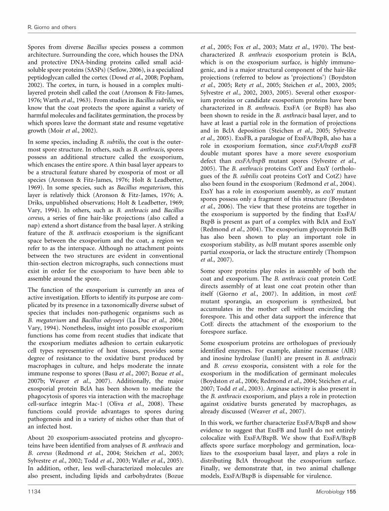
2005; Sylvestre et al., 2005). Therefore, BclA deposition canbe divided into two phases: ExsFA/BxpB-dependent andExsFA/BxpB-independent. To analyse the subcellularlocation of the ExsFA/BxpB-independent BclA duringsporulation, we used IFM, with the anti-BclA mAb BA-MAB-5, to monitor BclA localization in wild-type andexsFA/bxpB mutant cells that had completed foresporeengulfment (known as sporangia) in the Sterne strainbackground. To visualize the orientation of the foresporeand mother cell compartments, we analysed fixed andHoechst 33342-stained cells by bright-field and fluor-escence microscopy simultaneously. In these experiments,the forespore appears as an oval-shaped structure and themother cell chromosome as a bright focus (Fig. 2a, BF/DNA). We first analysed wild-type sporangia to determinewhen BclA would initially be detected by IFM. In this andsubsequent IFM experiments, we analysed a minimum of150 cells. After 4 h of sporulation, a time at which BclA isknown to be expressed (Sylvestre et al., 2002), we did notdetect BclA in sporangia (data not shown). After 7 h, BclAwas clearly localized around the forespore in wild-typesporangia (Fig. 2a). The same fluorescence pattern was seenin free spores (Fig. 2b). To confirm the specificity of themAb, we also examined a bclA mutant strain (Sterne-JAB-13). Anti-BclA fluorescence was not detected in bclAmutant sporangia or spores (data not shown).
In exsFA/bxpB mutant sporangia after 7 h of sporulation,the BclA localization pattern suggested that at least some ofthe protein was located in the mother cell (Fig. 2a). After8 h, BclA was localized exclusively around the forespore, inboth wild-type and mutant strains. However, in the exsFA/
Fig. 1. Atomic force micrographs of Sterne strain spores of B. anthracis. (a) Wild-type Sterne and (b, c) RG124 (exsFA/bxpB)spores were analysed in the tapping mode. Bars, 1 mm. In panels (aii), (bii) and (cii), the white dotted lines indicate sporeoutlines and the white dashed lines indicate surface ridges.
Bacillus anthracis spore outer structures
http://mic.sgmjournals.org 1137
bxpB mutant sporangia, BclA decoration was more intenseat the edge of the forespore closest to the midpoint of thesporangium (i.e. the mother-cell pole of the forespore)(Fig. 2a). We found that BclA also localized in a polarfashion in free spores from the exsFA/bxpB mutant (Fig.2b). This region of BclA decoration appears to coincidewith the portion of the exosporium that forms first insporulation, called the cap (Giorno et al., 2007; Ohye &Murrell, 1973; Steichen et al., 2007). These unexpectedresults indicate a previously unrecognized polarity to BclAlocalization.
Our TEM analysis indicated that released exsFA/bxpBmutant spores do not possess projections (or nap) (datanot shown), consistent with earlier results (Steichen et al.,2005). Nonetheless, as just discussed, at least some BclA is
forespore-localized in exsFA/bxpB mutant sporangia, asshown by IFM (Fig. 2a). The presence of forespore-associated BclA in mutant cells raised the possibility thatprojections were also present (presumably, if this were true,the projections are lost during or just before spore releasefrom the mother cell). To test this, we used TEM todetermine whether projections could be detected in exsFA/bxpB mutant sporangia after 6 and 8 h of sporulation,when the projections are readily visible in wild-type cells(Giorno et al., 2007; Steichen et al., 2007). We did notdetect projections in exsFA/bxpB mutant cells (data notshown). Therefore either exsFA/bxpB mutant cells do notassemble projections, even transiently, or they are unstable.Overall, these data are consistent with the view that, whilesome BclA localizes in an ExsFA/BxpB-independentmanner, this deposition is insufficient to maintain eithera uniform shell of BclA or the formation of recognizableprojections.
Role of ExsFA/BxpB in spore functions
The exosporium could have a role in germination oroutgrowth (Redmond et al., 2004; Steichen et al., 2005).Therefore, we analysed the ability of exsFA/bxpB mutantspores to germinate and resume metabolism (known asoutgrowth). We analysed early events in germination bymeasuring the rate and level of incorporation of thefluorescent dye Syto-9 (Welkos et al., 2004). We found thatthe initial dye uptake kinetics of the exsFA/bxpB sporesfrom either the Sterne or the Ames background wereidentical to those of wild-type spores (Fig. 3a). However,after 4 min, the rate of dye incorporation from the exsFA/bxpB spores was reduced, and after 60 min, the exsFA/bxpBspores had not incorporated as much dye as the wild-typespores (Fig. 3a). This effect appeared to be more severe inthe Ames background. We do not know the significance ofthis difference between the two strain backgrounds. Theeffect of the exsFA/bxpB mutation on dye uptake is not duesolely to the reduction in spore-associated BclA, sinceAmes spores entirely missing BclA (due to a bclAmutation) take up dye as well as wild-type spores (Bozueet al., 2007a). In previous work, germination of exsFA/bxpBmutant spores was measured using RPMI–BHI medium asa germinant (Steichen et al., 2005). Therefore, we alsomonitored germination by measuring the loss of opticaldensity (Harwood & Cutting, 1990) using this germinant.After 5 min, the percentage loss of optical density of byexsFA/bxpB mutant spores (in the Sterne background,strain RG124) was lower than in wild-type, and remainedso throughout the experiment (Fig. 3b). Although modest,the difference between wild-type and mutant spores in thisassay was significant as determined by the Mann–WhitneyU test (P,0.01). Taken together, these results suggest thatexsFA/bxpB spores have a defect in an early step ofgermination.
To determine whether an exsFA/bxpB mutation affects lateevents in germination and/or the resumption of metabol-
Fig. 2. Fluorescence microscopic analysis of the role of ExsFA/BxpB in BclA deposition. (a) Sporangia from the Sterne strain(wild-type; WT) and strain RG124 (exsFA/bxpB) were harvestedafter 7 and 8 h of sporulation (t7 and t8, respectively), fixed andtreated with the anti-BclA mAb BA-MAB5 (a-BclA). Bright-fieldmicroscopy and fluorescence microscopy (BF/DNA) were per-formed simultaneously so that the forespore and mother cell DNA(stained with Hoechst 33342 dye) could be visualized in a singleimage. The locations of individual cells are indicated with brackets.Mother cell chromosomes are indicated with arrowheads. The cellindicated by a dashed bracket (wild-type at t8) did not sporulate,as indicated by the chromosome morphology. (b) Spores from thewild-type or RG124 (exsFA/bxpB) strains were harvested 24 hafter the onset of sporulation and stained with the anti-BclAantibody BA-MAB-5. Corresponding bright-field (BF) images areshown for each immunohistograph (a-BclA). Data shown arerepresentative of a minimum of 150 cells observed for each sampleand time point. We judged the immunostaining to be positive if atleast 90 % of the cells showed fluorescence. All negative controlexperiments showed fluorescence in fewer than 10 % of cells.
R. Giorno and others
1138 Microbiology 155

ism (outgrowth), we used a modified version of thetetrazolium overlay assay (Harwood & Cutting, 1990;Giorno et al., 2007). Spores from exsFA/bxpB mutantstrains, in either the Sterne or the Ames background (inRG124 and Ames-JAB-5, respectively), showed wild-typelevels of outgrowth by this assay (data not shown).
To detect a role for ExsFA/BxpB in infection, we measuredthe ability of exsFA/bxpB mutant spores (from Ames-JAB-5) to infect Guinea pigs by both IM and inhalationalchallenges. The mutant strain was fully virulent in bothexperiments (Fig. 4). There were no significant differencesin mean time to death between strains by either the IMroute (P50.2878) or the aerosol route (P50.5062) asmeasured by Student’s t test, nor were there any significantdifferences in survival rates between strains after infectionby the aerosol route (P50.2105), as measured by Fisher’s
exact test. There were no survivors in the IM-exposedgroup. These results are consistent with previous results(Bozue et al., 2007a; Sylvestre et al., 2002). Our resultsindicate that neither the germination defect nor thechanges in spore composition and morphology of theexsFA/bxpB Ames mutant detectably reduce disease usingthese two routes of infection.
Subcellular localization and assembly of ExsFA/BxpB and ExsFB
To better understand the roles of ExsFA/BxpB and ExsFBin spore assembly and function, we examined theirlocations in living cells during sporulation. To do this,we built strains (in the Sterne background) bearing fusionsof each protein to GFP, and examined them byfluorescence microscopy. Cells from strains bearingExsFA/BxpB–GFP (RG151) and ExsFB–GFP (RG155) grewand sporulated normally, as judged by light microscopy(data not shown). We noted, however, that TEM showedthat exsFA/bxpB–gfp-bearing spores lacked projections,suggesting a role for the ExsFA/BxpB C terminus inprojection assembly (data not shown). Since exsFB nullmutant spores have not been shown to have strongphenotypes (Sylvestre et al., 2005), we did not furthercharacterize ExsFB–GFP-bearing cells by TEM.
Fig. 3. Germination of wild-type (WT) and mutant spores. (a)Spores from wild-type Ames (&), wild-type Sterne (m), Ames-JAB-5 (h) and RG124 (g) strains were analysed by thefluorescence dye uptake assay. The percentage increase influorescence is the difference between the fluorescence in relativefluorescence units (RFU) at a given time in germination mediumand the RFU at t0, expressed as a percentage of the latter. (b)Spores from wild-type Sterne (m) and RG124 (g) were monitoredover time for the loss of refractility by measuring the reduction inoptical density. Both assays were done in triplicate. The datashown in (a) are representative of three independent experiments,and SEM values are shown in (b).
Fig. 4. Effect of exsFA/bxpB on virulence. Spores from the wild-type (WT) Ames strain (&) or Ames-JAB-5 (h) were used in (a)Guinea pig IM or (b) Guinea pig inhalational challenge assays.Numbers of surviving animals are plotted.
Bacillus anthracis spore outer structures
http://mic.sgmjournals.org 1139
We did not detect fluorescence in cells from strain RG151(bearing the ExsFA/BxpB–GFP fusion) at 3 h after theonset of sporulation (Fig. 5a). At 5 h, we found thatfluorescence was dispersed throughout the mother cell andexcluded from the forespore (data not shown). After 6 h,we detected fluorescence in both a ring around theforespore as well as dispersed throughout the mother cell(Fig. 5a). Released spores bearing ExsFA/BxpB–GFP (fromstrain RG151) showed rings of fluorescence that sur-rounded the spores and were more intense at the caps (Fig.5a, bottom). In about 60 % of spores, we saw that one capwas brighter than the other.
To detect fluorescence due to ExsFB–GFP, we sporulatedcells bearing this fusion (strain RG155) at room temper-ature using 26 SG medium, as we did not detectfluorescence during culture at 37 uC in DSM (data notshown). Culturing cells at a decreased temperature hasbeen shown to facilitate detection of certain fusions in bothin B. subtilis and B. anthracis (Kim et al., 2006; Mallozzi etal., 2008; van Ooij et al., 2004). Similarly to the case ofExsFA/BxpB–GFP, we first detected fluorescence in ExsFB–GFP-bearing cells (RG155) as a dispersed signal in themother cell, beginning at stage IV of sporulation (wherethe stage of sporulation was determined by visualization of
chromosome morphology using a DNA stain, and byphase-contrast microscopy) (Fig. 5a and data not shown).We noted that fluorescence was excluded from theforespore during stage IV of sporulation, which is the timewhen the forespore is no longer metabolically active(Losick et al., 1986). In contrast, released spores bearingthe ExsFB–GFP fusion (from strain RG155) had either asolid oval of fluorescence or, occasionally, rings (in 17 of380 spores observed) of fluorescence that surrounded thespores (Fig. 5a, b, and data not shown). This is in strikingcontrast to what we and others have seen with fusions ofcoat protein genes to GFP, in both B. anthracis and B.subtilis, where the patterns of localization are usually rings,and sometimes caps or other structures that suggestincomplete rings, but never solid ovals (Bauer et al.,1999; Eichenberger et al., 2003; Kim et al., 2006; Mallozzi etal., 2008; McPherson et al., 2005; van Ooij et al., 2004). Wenote that the timing of the appearance of ExsFB–GFPfluorescence, just described, is inconsistent with thepossibility that ExsFB–GFP is synthesized in the sporecore. The view that ExsFB is synthesized in the mother cellis further supported by the presence of a candidate sK-driven promoter upstream of exsFB (Steichen et al., 2005).Therefore, although ExsFB–GFP fluorescence appears as asolid oval, it is highly unlikely that this fluorescenceemanates from the spore interior.
Fig. 5. Fluorescence microscopic analysis of ExsFA/BxpB–GFP,ExsFB–GFP and IunH–GFP. (a) Fluorescence microscopiclocalization of fusion proteins during sporulation. Cells from strainsRG1 (wild-type; WT), RG151 (exsFA/bxpB–gfp) and RG130(iunH–gfp) were cultured in DSM broth at 37 6C and harvested at0 (t0), 3 (t3), 6 (t6) and 24 h (Spores) after the onset ofsporulation. Cells from strain RG155 (exsFB–gfp) were cultured in2� SG broth at 25 6C, harvested at various times and staged byanalysis of chromosome morphology. DNA staining (DNA) andGFP epifluorescence (GFP) are shown for each time point, andDNA staining was used to determine whether cells hadtransitioned to sporulation [t0 or stage 0 (0)] or progressed tostage III (III) or stage IV (IV). Brackets indicate individual sporangia.GFP epifluorescence (GFP) and bright-field (BF) images ofreleased spores are also shown for all strains. (b) Larger fieldsshowing fluorescence microscopic localization of ExsFB–GFP andIunH–GFP. Additional examples of the solid oval fluorescencepattern of these two fusion proteins, without corresponding bright-field images, for spores of strain RG155 (exsFB–gfp) and RG130(iunH–gfp) are shown. (c) Immunofluorescence detection of fusionproteins in released spores from strains RG151 (exsFA/bxpB–
gfp), RG155 (exsFB–gfp), RG157 (cotE exsFA/bxpB–gfp) andRG169 (bclA exsFB–gfp). Data shown are representative of aminimum of 150 cells observed for each sample and time point.We judged the immunostaining to be positive if at least 90 % of thecells showed fluorescence. All negative control experimentsshowed fluorescence in less than 10 % of cells. Spores fromstrains RG155 and RG169 were cultured in DSM at 25 6C.Bright-field (BF) and anti-GFP immunofluorescence (a-GFP) areshown for each. Images were not acquired (ND) for RG130(iunH–gfp) after 3 h of sporulation.
R. Giorno and others
1140 Microbiology 155

To ensure that the fluorescence patterns of ExsFA/BxpB–GFP and ExsFB–GFP are truly distinct, we reexamined themore intense ExsFA/BxpB–GFP signal with the camerasensitivity reduced. The signal remained a ring at allintensities. Likewise, increasing the camera sensitivity didnot result in rings of ExsFB–GFP fluorescence. Therefore,the fluorescence patterns of ExsFA/BxpB–GFP and ExsFB–GFP are distinct.
To directly compare the timing of assembly of the twofusion proteins, in a separate experiment we also carriedout sporulation of the ExsFA/BxpB–GFP-bearing strain(RG151) and the ExsFB–GFP-bearing strain (RG155) atroom temperature (in 26 SG medium). Sporulation ofRG151 cells at room temperature did not detectably alterthe fluorescence pattern of ExsFA/BxpB–GFP, whichformed a ring, as it did during sporulation at 37 uC.From this analysis, we found that fluorescence due toExsFB–GFP appeared several hours later than fluorescencedue to ExsFA/BxpB–GFP (data not shown). Given thatsporulation is relatively slow at this temperature, thebiological significance of this difference in timing isunclear.
To learn whether the unusual solid oval localizationpattern of ExsFB–GFP might be a property of otherexosporium-associated proteins, we also analysed thelocalization of a fusion of GFP to the putative nucleosidehydrolase, IunH (encoded by a gene currently annotated asBA2888), which has also been shown to fractionate withthe exosporium (Redmond et al., 2004; Todd et al., 2003)and which we also identified in a previous proteomic studyas a spore-associated protein (Lai et al., 2003). We foundthat the pattern of fluorescence in spores bearing iunH–gfp(strain RG130) was very similar to that of the exsFB–gfp-bearing spores from strain RG155 (Fig. 5a, b), although inabout 3 % of spores the fluorescence of the solid oval wasmuch brighter than the rest (6 out of 200 spores observed)(Fig. 5a, b, and data not shown).
To interpret the solid oval patterns of ExsFB–GFP andIunH–GFP fluorescence in terms of subcellular localizationof the respective proteins, we first considered the possibilitythat the ring-like appearance of typical spore protein–GFPfusions could be ascribed to the geometry of themicroscope set-up. Because the 2D image (a ring) capturedby the camera is actually due to a projection of a 3D image(a shell), the edges of the image show a greater amount ofimage intensity, per pixel, than does the interior. Therefore,a possible explanation for the disk of fluorescence would bethat, rather than being confined to a thin shell, ExsFB–GFPand IunH–GFP form a very thick layer, as they might ifthey resided in the interspace. If this were true, then asignificant fraction (if not all) of these fusion proteinswould be hidden underneath the exosporium. Furthersupport for this interpretation comes from the observationthat GFP fluorescence is excluded from the forespore instage IV exsFB–gfp-bearing cells (Fig. 5a). These dataindicate that ExsFB–GFP is not in the forespore prior to
stage IV, when forespore metabolism has already ceased(Losick et al., 1986). Therefore, it is extremely unlikely thatthe forespore-associated GFP in these cells is due to ExsFB–GFP in the core. Much more likely, ExsFB-GFP surroundsthe core.
To clarify the locations of ExsFA/BxpB–GFP and ExsFB–GFP, we first determined whether they could be detectedon the spore surface, using IFM. We mixed anti-GFPantibodies with spores from either strain and visualized theresulting complexes using fluorescence microscopy. ExsFA/BxpB–GFP-bearing spores reacted with anti-GFP antibod-ies, as expected from the known basal-layer location ofExsFA/BxpB (Steichen et al., 2005) and the lack ofprojections, due to the exsFA/bxpB–gfp allele, see above(Fig. 5c) (Steichen et al., 2005). In contrast, anti-GFPantibodies did not react with ExsFB–GFP-bearing spores(from strain RG155, Fig. 5c).
To investigate the locations of these proteins further, weapplied anti-GFP antibodies to spores bearing eitherfusion, but in which the exosporium was also incompletelyassembled, due to a mutation in cotE (from strains RG157and RG158). As in all our IFM analyses, we examined aminimum of 150 cells. In separate work, we have shownthat deletion of cotE results in spores that lack theexosporium (Giorno et al., 2007). However, those experi-ments do not reveal whether CotE controls interspaceformation. Spores bearing ExsFA/BxpB–GFP and a muta-tion in cotE (from strain RG157) did not react with anti-GFP antibodies (and did not show fluorescence due toGFP) (Fig. 5c), as we expected. Interestingly, we also foundthat we did not detect ExsFB–GFP in spores of a strain alsobearing the cotE mutation (in strain RG158), by applica-tion of either anti-GFP antibodies or, more importantly,GFP fluorescence (data not shown). Therefore, detection ofExsFB–GFP is CotE-dependent, consistent with a locationfor the fusion in the interspace, the exosporium, or both.
These data above do not exclude the possibility that ExsFBalso is present within the exosporium basal layer. If it were,localization by IFM might be inhibited by the presence ofthe BclA projections. To address this, we analysed bclAexsFB–gfp spores (from strain RG169) by IFM. We detectedfluorescence around the spore (Fig. 5c). The pattern ofimmunofluorescence from strain RG169 spores was adiscontinuous ring, in contrast to the continuous ring ofExsFA–GFP immunofluorescence (in strain RG151). Weinterpret these data to suggest that ExsFB is present in boththe basal layer and the interspace.
DISCUSSION
An important question in B. anthracis spore assembly is thetiming and control of BclA deposition. Earlier investi-gations have shown that while most BclA assembly requiresExsFA/BxpB, a significant fraction of BclA assembles in anExsFA/BxpB-independent manner, and that ExsFA/BxpB-dependent BclA assembly is needed for formation of the
Bacillus anthracis spore outer structures
http://mic.sgmjournals.org 1141

projections (Steichen et al., 2005; Sylvestre et al., 2005). Wealso found this and, additionally, we showed that BclAaccumulates at the mother cell pole in exsFA/bxpBforespores. Therefore, the fraction of BclA that localizesindependently of ExsFA/BxpB appears on the mother-cell-proximal portion of the exosporium that forms early insporulation, and under the direction of CotE (Giorno et al.,2007; Ohye & Murrell, 1973). This is likely the first portionof the exosporium to be assembled (Giorno et al., 2007).Interestingly, this region of the exosporium is biochemi-cally distinct from the rest of the exosporium, as it lacksalanine racemase and its assembly is ExsY-independent(Steichen et al., 2007). Taken together, these data appear tosuggest that exosporium assembly is a two-step process. Inthe first step, CotE directs deposition of a biochemicallyspecialized patch of the basal layer onto the forespore poleclosest to the mother cell midpoint. In the second step,ExsY directs the remainder of the polymerization of thesheet of exosporium, an event that terminates when theexosporium shell closes on itself (Steichen et al., 2007). Thefactor(s) directing assembly of ExsFA/BxpB-independentBclA remains unknown. However, ExsFB is a plausiblecandidate, since exsFA/bxpB exsFB double mutant sporesassemble less BclA than exsFA/bxpB mutant spores(Sylvestre et al., 2005).
We detected a modest germination defect in exsFA/bxpBmutant spores by multiple assays, indicating at least onerole for this protein beyond mediating BclA deposition.The finding of a germination defect is in contrast to anearlier finding showing that both germination andoutgrowth are accelerated in mutant spores (Steichen etal., 2005). We do not know the basis for this discrepancy,but it is plausibly due to differences in the physical set-upof our experimental systems. We note that while similarmedia were used to measure germination in one case,Steichen et al. (2005) used a flow chamber system, while weused assays in which germinant was not replenished duringthe experiment (Harwood & Cutting, 1990). Possibly, theconstant flow of nutrient has a marked affect ongermination in the exsFA/bxpB mutant that cannot beobserved when the germinant is given in a single dose.
We found that ExsFA/BxpB is not needed for the typicalpresentation of B. anthracis infection in two models. This isconsistent with previous findings demonstrating that evenwithout BclA, spores from either the attenuated Sterne orthe virulent Ames backgrounds can still cause infection(Bozue et al., 2007a; Brahmbhatt et al., 2007; Sylvestre etal., 2002). Importantly, it extends those results bydemonstrating the dispensability of the projections (ornap) in an entirely separate manner, and using anadditional route of infection (the Guinea pig inhalationalchallenge). These data also are consistent with our previousfinding that spores lacking the exosporium, due to amutation in the coat protein gene cotE, are fully virulent intwo animal models (Giorno et al., 2007). These experi-ments raise an important caution; while defences andtherapeutics targeting the exosporium will be very useful,
we should not rely on them exclusively, since this structuremay be dispensable (Giorno et al., 2007). Rather, we mustprepare for events using spores both with and withoutprojections.
Although we did not detect an effect of ExsFA/BxpB ondisease, the exosporium may still have a role in interactionswith the host. For example, it has been shown recently thatthe exosporium provides a degree of protection againstmacrophages (Weaver et al., 2007). Also, the presence ofBclA may be necessary for targeting spores to macrophages,as it decreases spore adherence to epithelial, fibroblast andendothelial cells (Bozue et al., 2007b). Therefore, at leastunder certain circumstances, the exosporium could affectthe natural disease process in ways not detected in ourassays. Exosporium proteins or the genes encoding theseproteins may also be useful as subunits in an improvedanthrax vaccine, as suggested by several studies (Brossier etal., 2002; Cybulski et al., 2008; Enkhtuya et al., 2006; Hahnet al., 2006). Therefore, interactions between the host andexosporium proteins are likely to be important to futuretherapeutics.
If the projections are not always needed for disease, thenwhat is their function? It is plausible that the projectionshave roles in resistance or attachment, in the soil or someother ecological niche. B. anthracis maintains a highlycomplex series of spore protective structures, apparentlylargely dedicated to survival outside the host, in spite of itsadaptation to animal hosts. It may be challenging toidentify these additional roles. An example of an organismthat likely uses BclA for a purpose other than disease is thenon-pathogenic organism Bacillus licheniformis, a closerelative of B. subtilis that does not have an exosporium butwhich possesses an orthologue of BclA (M. Mallozzi & A.Driks, unpublished results). Preliminary phylogeneticanalysis suggests that BclA was, in this case, acquired byhorizontal gene transfer (H. Qin & A. Driks, unpublishedresults).
Most coat and exosporium genes do not have paralogues.exsFA/bxpB and exsFB are exceptions. Making this set ofgenes even more unusual, some B. cereus-group strainspossess a third paralogue that differs from exsFA/bxpBand exsFB at least as much as those two genes differ fromeach other. B. anthracis has only two paralogues, as do 19other strains, three strains have only one paralogue, and B.cereus E33L contains three paralogues. Interestingly, in B.cereus E33L two of the paralogues are on the chromosomeand one is plasmid-encoded. Possibly, these variationsresult in adaptive changes in exosporium structure andfunction.
Our data argue that ExsFB–GFP and IunH–GFP do notprecisely colocalize with the exosporium basal layer. Wesuggest the possibility that ExsFB and IunH reside in theinterspace. At present, this localization is not definitive.Future experiments to support these data and to uncoveradditional possible interspace proteins may help clarify thefunction of this poorly understood spore compartment.
R. Giorno and others
1142 Microbiology 155

ACKNOWLEDGEMENTS
The authors thank Kathy Kuehl [Pathology Department, United States
Army Medical Research Institute of Infectious Diseases (USAMRIID)]
for preparation of electron microscope samples and Sarah Norris
(USAMRIID) for statistical analysis. We thank Terri Koehler(University of Texas, Houston) for the kind gift of CP-51, plasmids
and strains, and for valuable advice. We thank Peter McKenney and
Patrick Eichenberger for important input into this study. Funding wasprovided by the NIH (AI53365, A. D.) and the US Army Medical
Research and Materiel Command under Projects 02-4-5C-018 (J. B.),
02-4-5C-023 (S. W.) and 04-0-IL-002 (S. W.) and an In-HouseLaboratory Innovative Research Award from the Department of the
Army under Project 92489 (J. B.). Research was conducted in
compliance with the Animal Welfare Act and other federal statutesand regulations relating to animals and experiments involving
animals and adheres to principles stated in the Guide for the Careand Use of Laboratory Animals (Institute of Laboratory Animal
Resources et al., 1996). The facility in which this research was
conducted is fully accredited by the Association for Assessment andAccreditation of Laboratory Animal Care International. Opinions,
interpretations, conclusions, and recommendations are those of the
authors and are not necessarily endorsed by the US Army.
REFERENCES
Alibek, K. (1999). Biohazard. New York: Random House.
Aronson, A. I. & Fitz-James, P. (1976). Structure and morphogenesis
of the bacterial spore coat. Bacteriol Rev 40, 360–402.
Basu, S., Kang, T. J., Chen, W. H., Fenton, M. J., Baillie, L., Hibbs, S. &Cross, A. S. (2007). Role of Bacillus anthracis spore structures inmacrophage cytokine responses. Infect Immun 75, 2351–2358.
Bauer, T., Little, S., Stover, A. G. & Driks, A. (1999). Functionalregions of the Bacillus subtilis spore coat morphogenetic protein CotE.
J Bacteriol 181, 7043–7051.
Boydston, J. A., Chen, P., Steichen, C. T. & Turnbough, C. L., Jr(2005). Orientation within the exosporium and structural stability of
the collagen-like glycoprotein BclA of Bacillus anthracis. J Bacteriol187, 5310–5317.
Boydston, J. A., Yue, L., Kearney, J. F. & Turnbough, C. L., Jr (2006).The ExsY protein is required for complete formation of theexosporium of Bacillus anthracis. J Bacteriol 188, 7440–7448.
Bozue, J. A., Parthasarathy, N., Phillips, L. R., Cote, C. K., Fellows,P. F., Mendelson, I., Shafferman, A. & Friedlander, A. M. (2005).Construction of a rhamnose mutation in Bacillus anthracis affects
adherence to macrophages but not virulence in guinea pigs. MicrobPathog 38, 1–12.
Bozue, J., Cote, C. K., Moody, K. L. & Welkos, S. L. (2007a). Fullyvirulent Bacillus anthracis does not require the immunodominant
protein BclA for pathogenesis. Infect Immun 75, 508–511.
Bozue, J., Moody, K. L., Cote, C. K., Stiles, B. G., Friedlander, A. M.,Welkos, S. L. & Hale, M. L. (2007b). Bacillus anthracis spores of the
bclA mutant exhibit increased adherence to epithelial, fibroblast, andendothelial cells but not macrophages. Infect Immun 75, 4498–4505.
Brahmbhatt, T. N., Janes, B. K., Stibitz, E. S., Darnell, S. C., Sanz, P.,Rasmussen, S. B. & O’Brien, A. D. (2007). Bacillus anthracisexosporium protein BclA affects spore germination, interaction with
extracellular matrix proteins, and hydrophobicity. Infect Immun 75,5233–5239.
Brossier, F., Levy, M. & Mock, M. (2002). Anthrax spores make
an essential contribution to vaccine efficacy. Infect Immun 70,661–664.
Catalano, F. A., Meador-Parton, J., Popham, D. L. & Driks, A. (2001).Amino acids in the Bacillus subtilis morphogenetic protein SpoIVA with
roles in spore coat and cortex formation. J Bacteriol 183, 1645–1654.
Chada, V. G., Sanstad, E. A., Wang, R. & Driks, A. (2003).Morphogenesis of Bacillus spore surfaces. J Bacteriol 185, 6255–6261.
Cown, W. B., Kethley, T. W. & Fincher, E. L. (1957). The critical-orifice
liquid impinger as a sampler for bacterial aerosols. Appl Microbiol 5,
119–124.
Cybulski, R. J., Jr, Sanz, P., McDaniel, D., Darnell, S., Bull, R. L. &O’Brien, A. D. (2008). Recombinant Bacillus anthracis spore proteins
enhance protection of mice primed with suboptimal amounts of
protective antigen. Vaccine 26, 4927–4939.
Dowd, M. M., Orsburn, B. & Popham, D. L. (2008). Cortex
peptidoglycan lytic activity in germinating Bacillus anthracis spores.
J Bacteriol 190, 4541–4548.
Eichenberger, P., Jensen, S. T., Conlon, E. M., van Ooij, C., Silvaggi, J.,Gonzalez-Pastor, J. E., Fujita, M., Ben-Yehuda, S., Stragier, P. & otherauthors (2003). The sE regulon and the identification of additional
sporulation genes in Bacillus subtilis. J Mol Biol 327, 945–972.
Enkhtuya, J., Kawamoto, K., Kobayashi, Y., Uchida, I., Rana, N. &Makino, S. (2006). Significant passive protective effect against anthrax
by antibody to Bacillus anthracis inactivated spores that lack two
virulence plasmids. Microbiology 152, 3103–3110.
Fotiadis, D., Scheuring, S., Muller, S. A., Engel, A. & Muller, D. J.(2002). Imaging and manipulation of biological structures with the
AFM. Micron 33, 385–397.
Fox, A., Stewart, G. C., Wallera, L. N., Fox, K. F., Harley, W. M. & Price,R. L. (2003). Carbohydrates and glycoproteins of Bacillus anthracis and
related bacilli: targets for biodetection. J Microbiol Methods 54, 143–152.
Friedlander, A. M., Welkos, S. L., Pitt, M. L., Ezzell, J. W., Worsham,P. L., Rose, K. J., Ivins, B. E., Lowe, J. R., Howe, G. B. & other authors
(1993). Postexposure prophylaxis against experimental inhalation
anthrax. J Infect Dis 167, 1239–1243.
Fritze, D. (2004). Taxonomy of the genus Bacillus and related genera: the
aerobic endospore-forming bacteria. Phytopathology 94, 1245–1248.
Giorno, R., Bozue, J., Cote, C., Wenzel, T., Moody, K. S., Mallozzi, M.,Ryan, M., Wang, R., Zielke, R. & other authors (2007). Morphogenesis
of the Bacillus anthracis spore coat. J Bacteriol 189, 691–705.
Hahn, U. K., Boehm, R. & Beyer, W. (2006). DNA vaccination against
anthrax in mice – combination of anti-spore and anti-toxin
components. Vaccine 24, 4569–4571.
Harwood, C. R. & Cutting, S. M. (1990). Molecular Biological Methods
for Bacillus. Chichester, UK: John Wiley.
Holt, S. C. & Leadbetter, E. R. (1969). Comparative ultrastructure of
selected aerobic spore-forming bacteria: a freeze-etching study.
Bacteriol Rev 33, 346–378.
Institute of Laboratory Animal Resources, Commission on LifeSciences, & National Research Council (1996). Guide for the Care and
Use of Laboratory Animals. Washington, DC: National Academy Press.
Ivins, B. E., Welkos, S. L., Knudson, G. B. & Little, S. F. (1990).Immunization against anthrax with aromatic compound-dependent
(Aro2) mutants of Bacillus anthracis and with recombinant strains of
Bacillus subtilis that produce anthrax protective antigen. Infect Immun
58, 303–308.
Ivins, B. E., Fellows, P. F. & Nelson, G. O. (1994). Efficacy of a
standard human anthrax vaccine against Bacillus anthracis spore
challenge in guinea-pigs. Vaccine 12, 872–874.
Kim, H., Hahn, M., Grabowski, P., McPherson, D. C., Wang, R.,Ferguson, C., Eichenberger, P. & Driks, A. (2006). The Bacillus subtilis
spore coat protein interaction network. Mol Microbiol 59, 487–502.
Bacillus anthracis spore outer structures
http://mic.sgmjournals.org 1143

Koch, R. (1876). The etiology of anthrax, based on the life history ofBacillus anthracis. Beitr Biol Pflanz 2, 277–310.
Koehler, T. M., Dai, Z. & Kaufman-Yarbray, M. (1994). Regulation ofthe Bacillus anthracis protective antigen gene: CO2 and a trans-actingelement activate transcription from one of two promoters. J Bacteriol176, 586–595.
La Duc, M. T., Satomi, M. & Venkateswaran, K. (2004). Bacillusodysseyi sp. nov., a round-spore-forming bacillus isolated from theMars Odyssey spacecraft. Int J Syst Evol Microbiol 54, 195–201.
Lai, E.-M., Phadke, N. D., Kachman, M. T., Giorno, R. S. V., Vazquez,J. A., Maddock, J. R. & Driks, A. (2003). Proteomic analysis of thespore coats of Bacillus subtilis and Bacillus anthracis. J Bacteriol 185,1443–1454.
Leighton, T. J. & Doi, R. H. (1971). The stability of messengerribonucleic acid during sporulation in Bacillus subtilis. J Biol Chem246, 3189–3195.
Lemon, K. P. & Grossman, A. D. (1998). Localization of bacterialDNA polymerase: evidence for a factory model of replication. Science282, 1516–1519.
Little, S. F. & Knudson, G. B. (1986). Comparative efficacy of Bacillusanthracis live spore vaccine and protective antigen vaccine againstanthrax in the guinea pig. Infect Immun 52, 509–512.
Losick, R., Youngman, P. & Piggot, P. J. (1986). Genetics ofendospore formation in Bacillus subtilis. Annu Rev Genet 20, 625–669.
Mallozzi, M., Bozue, J., Giorno, R., Moody, K. S., Slack, A., Cote, C.,Qiu, D., Wang, R., McKenney, P. & other authors (2008).Characterization of a Bacillus anthracis spore coat-surface proteinthat influences coat-surface morphology. FEMS Microbiol Lett 289,110–117.
Mann, H. B. & Whitney, D. R. (1947). On a test of whether one of tworandom variables is stochastically larger than the other. Ann MathStat 18, 50–60.
Matz, L. L., Beaman, T. C. & Gerhardt, P. (1970). Chemical composi-tion of exosporium from spores of Bacillus cereus. J Bacteriol 101,196–201.
May, K. R. (1973). The collision nebulizer, description, performance,and applications. J Aerosol Sci 4, 235–243.
McPherson, D. C., Kim, H., Hahn, M., Wang, R., Grabowski, P.,Eichenberger, P. & Driks, A. (2005). Characterization of the Bacillussubtilis spore coat morphogenetic protein CotO. J Bacteriol 187,8278–8290.
Mendelson, I., Tobery, S., Scorpio, A., Bozue, J., Shafferman, A. &Friedlander, A. M. (2004). The NheA component of the non-hemolyticenterotoxin of Bacillus cereus is produced by Bacillus anthracis but is notrequired for virulence. Microb Pathog 37, 149–154.
Mock, M. & Fouet, A. (2001). Anthrax. Annu Rev Microbiol 55, 647–671.
Moir, A., Corfe, B. M. & Behravan, J. (2002). Spore germination. CellMol Life Sci 59, 403–409.
Nicholson, W. L. (2002). Roles of Bacillus endospores in theenvironment. Cell Mol Life Sci 59, 410–416.
Ohye, D. F. & Murrell, W. G. (1973). Exosporium and spore coatformation in Bacillus cereus T. J Bacteriol 115, 1179–1190.
Oliva, C. R., Swiecki, M. K., Griguer, C. E., Lisanby, M. W., Bullard,D. C., Turnbough, C. L., Jr & Kearney, J. F. (2008). The integrinMac-1 (CR3) mediates internalization and directs Bacillus anthracisspores into professional phagocytes. Proc Natl Acad Sci U S A 105,1261–1266.
Perez-Casal, J., Caparon, M. G. & Scott, J. R. (1991). Mry, a trans-acting positive regulator of the M protein gene of Streptococcuspyogenes with similarity to the receptor proteins of two-componentregulatory systems. J Bacteriol 173, 2617–2624.
Plomp, M., Leighton, T., Wheeler, K. E. & Malkin, A. J. (2004). Thehigh-resolution architecture and structural dynamics of Bacillusspores. Biophys J 88, 603–608.
Pogliano, K., Harry, E. & Losick, R. (1995). Visualization of thesubcellular location of sporulation proteins in Bacillus subtilis usingimmunofluorescence microscopy. Mol Microbiol 18, 459–470.
Popham, D. L. (2002). Specialized peptidoglycan of the bacterialendospore: the inner wall of the lockbox. Cell Mol Life Sci 59, 426–433.
Redmond, C., Baillie, L. W., Hibbs, S., Moir, A. J. & Moir, A. (2004).Identification of proteins in the exosporium of Bacillus anthracis.Microbiology 150, 355–363.
Rety, S., Salamitou, S., Garcia-Verdugo, I., Hulmes, D. J., Le Hegarat,F., Chaby, R. & Lewit-Bentley, A. (2005). The crystal structure of theBacillus anthracis spore surface protein BclA shows remarkablesimilarity to mammalian proteins. J Biol Chem 280, 43073–43078.
Sambrook, J., Fritsch, E. F. & Maniatis, T. (1989). Molecular Cloning:a Laboratory Manual, 2nd edn. Cold Spring Harbor, NY: Cold SpringHarbor Laboratory.
Setlow, P. (2006). Spores of Bacillus subtilis: their resistance to andkilling by radiation, heat and chemicals. J Appl Microbiol 101, 514–525.
Shao, Z., Mou, J., Czajkowsky, D. M., Yang, J. & Yuan, J. Y. (1996).Biological atomic force microscopy: what is achieved and what isneeded. Adv Phys 45, 1–86.
Shatalin, K. Y. & Neyfakh, A. A. (2005). Efficient gene inactivation inBacillus anthracis. FEMS Microbiol Lett 245, 315–319.
Steichen, C., Chen, P., Kearney, J. F. & Turnbough, C. L., Jr (2003).Identification of the immunodominant protein and other proteins ofthe Bacillus anthracis exosporium. J Bacteriol 185, 1903–1910.
Steichen, C. T., Kearney, J. F. & Turnbough, C. L., Jr (2005).Characterization of the exosporium basal layer protein BxpB ofBacillus anthracis. J Bacteriol 187, 5868–5876.
Steichen, C. T., Kearney, J. F. & Turnbough, C. L., Jr (2007). Non-uniform assembly of the Bacillus anthracis exosporium and a bottle capmodel for spore germination and outgrowth. Mol Microbiol 64, 359–367.
Sylvestre, P., Couture-Tosi, E. & Mock, M. (2002). A collagen-likesurface glycoprotein is a structural component of the Bacillusanthracis exosporium. Mol Microbiol 45, 169–178.
Sylvestre, P., Couture-Tosi, E. & Mock, M. (2003). Polymorphism inthe collagen-like region of the Bacillus anthracis BclA protein leads tovariation in exosporium filament length. J Bacteriol 185, 1555–1563.
Sylvestre, P., Couture-Tosi, E. & Mock, M. (2005). Contribution ofExsFA and ExsFB proteins to the localization of BclA on the sporesurface and to the stability of the Bacillus anthracis exosporium. JBacteriol 187, 5122–5128.
Thompson, B. M., Waller, L. N., Fox, K. F., Fox, A. & Stewart, G. C.(2007). The BclB glycoprotein of Bacillus anthracis is involved inexosporium integrity. J Bacteriol 189, 6704–6713.
Thorne, C. B. (1968). Transduction in Bacillus cereus and Bacillusanthracis. Bacteriol Rev 32, 358–361.
Todd, S. J., Moir, A. J., Johnson, M. J. & Moir, A. (2003). Genes ofBacillus cereus and Bacillus anthracis encoding proteins of theexosporium. J Bacteriol 185, 3373–3378.
van Ooij, C., Eichenberger, P. & Losick, R. (2004). Dynamic patternsof subcellular protein localization during spore coat morphogenesis inBacillus subtilis. J Bacteriol 186, 4441–4448.
Vary, P. S. (1994). Prime time for Bacillus megaterium. Microbiology140, 1001–1013.
Waller, L. N., Stump, M. J., Fox, K. F., Harley, W. M., Fox, A., Stewart,G. C. & Shahgholi, M. (2005). Identification of a second collagen-likeglycoprotein produced by Bacillus anthracis and demonstration ofassociated spore-specific sugars. J Bacteriol 187, 4592–4597.
R. Giorno and others
1144 Microbiology 155

Wang, R., Krishnamurthy, S. N., Jeong, J. S., Driks, A., Mehta, M. &Gingras, B. A. (2007). Fingerprinting species and strains of Bacillispores by distinctive coat surface morphology. Langmuir 23,10230–10234.
Warth, A. D., Ohye, D. F. & Murrell, W. G. (1963). The compositionand structure of bacterial spores. J Cell Biol 16, 579–592.
Weaver, J., Kang, T. J., Raines, K. W., Cao, G. L., Hibbs, S., Tsai, P.,Baillie, L., Rosen, G. M. & Cross, A. S. (2007). Protective role of
Bacillus anthracis exosporium in macrophage-mediated killing bynitric oxide. Infect Immun 75, 3894–3901.
Welkos, S. L., Cote, C. K., Rea, K. M. & Gibbs, P. H. (2004). Amicrotiter fluorometric assay to detect the germination of Bacillusanthracis spores and the germination inhibitory effects of antibodies.J Microbiol Methods 56, 253–265.
Edited by: M. Hecker
Bacillus anthracis spore outer structures
http://mic.sgmjournals.org 1145
Related Documents


