Local overexpression of the myostatin propeptide increases glucose transporter expression and enhances skeletal muscle glucose disposal M. E. Cleasby, 1 S. Jarmin, 2 W. Eilers, 3 M. Elashry, 3 D. K. Andersen, 1 G. Dickson, 2 * and K. Foster 3 * 1 Department of Comparative Biomedical Sciences, Royal Veterinary College, University of London, London, United Kingdom; 2 School of Biological Sciences, Royal Holloway, University of London, Egham, Surrey, United Kingdom; and 3 School of Biological Sciences, University of Reading, Reading, Berkshire, United Kingdom Submitted 21 October 2013; accepted in final form 28 January 2014 Cleasby ME, Jarmin S, Eilers W, Elashry M, Andersen DK, Dickson G, Foster K. Local overexpression of the myostatin propeptide increases glucose transporter expression and enhances skeletal muscle glucose dis- posal. Am J Physiol Endocrinol Metab 306: E814 –E823, 2014. First pub- lished January 28, 2014; doi:10.1152/ajpendo.00586.2013.—Insulin resis- tance (IR) in skeletal muscle is a prerequisite for type 2 diabetes and is often associated with obesity. IR also develops alongside muscle atrophy in older individuals in sarcopenic obesity. The molecular defects that underpin this syndrome are not well characterized, and there is no licensed treatment. Deletion of the transforming growth factor- family member myostatin, or sequestration of the active peptide by overexpression of the myostatin propeptide/latency-asso- ciated peptide (ProMyo) results in both muscle hypertrophy and reduced obesity and IR. We aimed to establish whether local myo- statin inhibition would have a paracrine/autocrine effect to enhance glucose disposal beyond that simply generated by increased muscle mass, and the mechanisms involved. We directly injected adeno- associated virus expressing ProMyo in right tibialis cranialis/extensor digitorum longus muscles of rats and saline in left muscles and compared the effects after 17 days. Both test muscles were increased in size (by 7 and 11%) and showed increased radiolabeled 2-deoxy- glucose uptake (26 and 47%) and glycogen storage (28 and 41%) per unit mass during an intraperitoneal glucose tolerance test. This was likely mediated through increased membrane protein levels of GLUT1 (19% higher) and GLUT4 (63% higher). Interestingly, phosphoryla- tion of phosphoinositol 3-kinase signaling intermediates and AMP- activated kinase was slightly decreased, possibly because of reduced expression of insulin-like growth factor-I in these muscles. Thus, myostatin inhibition has direct effects to enhance glucose disposal in muscle beyond that expected of hypertrophy alone, and this approach may offer potential for the therapy of IR syndromes. hypertrophy; insulin sensitivity; glucose transporters; insulin-like growth factor-I; AMP-activated protein kinase INSULIN RESISTANCE (IR) in skeletal muscle is an essential precursor for the development of type 2 diabetes (T2D) in humans, since this tissue is responsible for a large proportion of postprandial glucose disposal. IR is frequently associated with both increasing adiposity and age, but also commonly coexists with age-associated loss of muscle mass (sarcopenia) in a syndrome referred to as “sarcopenic obesity.” This syn- drome is a substantial cause of morbidity and mortality in the elderly (affecting 4 –12% of the elderly population) (37, 47), both as a consequence of metabolic derangement but also because of frailty-related accidents and immobility. However, no licensed medications exist that are effective for sarcopenia or sarcopenic obesity; thus, it is imperative that a greater understanding is gained of the mechanisms involved in its development, such that potential therapeutic targets can be identified. Myostatin is a member of the transforming growth factor- (TGF-) family of secreted proteins that is almost exclusively expressed by skeletal muscle, and this tissue is also its princi- pal target, where it inhibits myocyte hypertrophy (24, 27). Naturally occurring mutations of myostatin have been identi- fied in a number of species, resulting in a “double muscling” phenotype (24). Similarly, myostatin null mice and mice administered with short-hairpin RNAs targeting myostatin show markedly increased muscle mass, although postnatal myostatin inhibition results in hypertrophy only, whereas null mice demonstrate both hypertrophy and hyperplasia (30, 33). Myostatin achieves its effects through activation of the Mothers against decapentaplegic homologs (25) and phosphoinositol 3-kinase (PI3K)/protein kinase B (Akt) pathways (3, 56), the latter being the site for cross talk with prohypertrophic insulin-like growth factor-I (IGF-I) signal- ing in muscle (56). However, activation of the PI3K/Akt pathway also plays an important role in insulin-stimulated glucose disposal in skeletal muscle (11, 53), and this is defective in muscle IR (9). In addition, myostatin modulates activation of AMP-activated protein kinase (AMPK) in muscle (59), the key enzyme in a second pathway that plays a role in both muscle metabolism and growth (26, 29). As might be predicted, therefore, myostatin null and mutant mice also resist diet-induced obesity and IR (17, 34, 54, 59), while conversely administration of myostatin to mice causes IR (19). In addition, myostatin mRNA is increased in muscle from ob/ob and high-fat diet-fed mice (2) and obese (35, 40) and diabetic (39) humans, but, significantly, this change is not reflected in altered plasma levels in the latter group (6). Thus, it is as yet unclear whether myostatin’s beneficial effects on metabolism are due purely to an increase in muscle mass that increases the capacity for insulin-stimulated glucose disposal, through an effect on muscle metabolism that is additional to this, or indeed whether the main beneficial effects are exerted primarily in other tissues. However, it is clear that myostatin represents a key potential molecular mediator of the pathogen- esis of IR and sarcopenic syndromes, including sarcopenic obesity. Active myostatin is generated by cleavage of a precursor protein and subsequent dimerization. The myostatin propep- tide, containing the NH 2 -terminal latency-associated peptide (ProMyo) only, sequesters the myostatin dimer and prevents its binding to the activin IIB receptor (ActRIIB) (28). Mice overexpressing the propeptide show both increased muscle * G. Dickson and and K. Foster contributed equally to this work. Address for reprint requests and other correspondence: M. E. Cleasby, Dept. of Comparative Biomedical Sciences, Royal Veterinary College, Univ. of London, Royal College St., London, NW1 0TU, UK (e-mail: [email protected]). Am J Physiol Endocrinol Metab 306: E814–E823, 2014. First published January 28, 2014; doi:10.1152/ajpendo.00586.2013. 0193-1849/14 Copyright © 2014 the American Physiological Society http://www.ajpendo.org E814

Welcome message from author
This document is posted to help you gain knowledge. Please leave a comment to let me know what you think about it! Share it to your friends and learn new things together.
Transcript
Local overexpression of the myostatin propeptide increases glucosetransporter expression and enhances skeletal muscle glucose disposal
M. E. Cleasby,1 S. Jarmin,2 W. Eilers,3 M. Elashry,3 D. K. Andersen,1 G. Dickson,2* and K. Foster3*1Department of Comparative Biomedical Sciences, Royal Veterinary College, University of London, London, United Kingdom;2School of Biological Sciences, Royal Holloway, University of London, Egham, Surrey, United Kingdom; and 3School ofBiological Sciences, University of Reading, Reading, Berkshire, United Kingdom
Submitted 21 October 2013; accepted in final form 28 January 2014
Cleasby ME, Jarmin S, Eilers W, Elashry M, Andersen DK, DicksonG, Foster K. Local overexpression of the myostatin propeptide increasesglucose transporter expression and enhances skeletal muscle glucose dis-posal. Am J Physiol Endocrinol Metab 306: E814–E823, 2014. First pub-lished January 28, 2014; doi:10.1152/ajpendo.00586.2013.—Insulin resis-tance (IR) in skeletal muscle is a prerequisite for type 2 diabetes andis often associated with obesity. IR also develops alongside muscleatrophy in older individuals in sarcopenic obesity. The moleculardefects that underpin this syndrome are not well characterized, andthere is no licensed treatment. Deletion of the transforming growthfactor-� family member myostatin, or sequestration of the activepeptide by overexpression of the myostatin propeptide/latency-asso-ciated peptide (ProMyo) results in both muscle hypertrophy andreduced obesity and IR. We aimed to establish whether local myo-statin inhibition would have a paracrine/autocrine effect to enhanceglucose disposal beyond that simply generated by increased musclemass, and the mechanisms involved. We directly injected adeno-associated virus expressing ProMyo in right tibialis cranialis/extensordigitorum longus muscles of rats and saline in left muscles andcompared the effects after 17 days. Both test muscles were increasedin size (by 7 and 11%) and showed increased radiolabeled 2-deoxy-glucose uptake (26 and 47%) and glycogen storage (28 and 41%) perunit mass during an intraperitoneal glucose tolerance test. This waslikely mediated through increased membrane protein levels of GLUT1(19% higher) and GLUT4 (63% higher). Interestingly, phosphoryla-tion of phosphoinositol 3-kinase signaling intermediates and AMP-activated kinase was slightly decreased, possibly because of reducedexpression of insulin-like growth factor-I in these muscles. Thus,myostatin inhibition has direct effects to enhance glucose disposal inmuscle beyond that expected of hypertrophy alone, and this approachmay offer potential for the therapy of IR syndromes.
hypertrophy; insulin sensitivity; glucose transporters; insulin-likegrowth factor-I; AMP-activated protein kinase
INSULIN RESISTANCE (IR) in skeletal muscle is an essentialprecursor for the development of type 2 diabetes (T2D) inhumans, since this tissue is responsible for a large proportionof postprandial glucose disposal. IR is frequently associatedwith both increasing adiposity and age, but also commonlycoexists with age-associated loss of muscle mass (sarcopenia)in a syndrome referred to as “sarcopenic obesity.” This syn-drome is a substantial cause of morbidity and mortality in theelderly (affecting 4–12% of the elderly population) (37, 47),both as a consequence of metabolic derangement but alsobecause of frailty-related accidents and immobility. However,no licensed medications exist that are effective for sarcopenia
or sarcopenic obesity; thus, it is imperative that a greaterunderstanding is gained of the mechanisms involved in itsdevelopment, such that potential therapeutic targets can beidentified.
Myostatin is a member of the transforming growth factor-�(TGF-�) family of secreted proteins that is almost exclusivelyexpressed by skeletal muscle, and this tissue is also its princi-pal target, where it inhibits myocyte hypertrophy (24, 27).Naturally occurring mutations of myostatin have been identi-fied in a number of species, resulting in a “double muscling”phenotype (24). Similarly, myostatin null mice and miceadministered with short-hairpin RNAs targeting myostatinshow markedly increased muscle mass, although postnatalmyostatin inhibition results in hypertrophy only, whereasnull mice demonstrate both hypertrophy and hyperplasia(30, 33). Myostatin achieves its effects through activation ofthe Mothers against decapentaplegic homologs (25) andphosphoinositol 3-kinase (PI3K)/protein kinase B (Akt)pathways (3, 56), the latter being the site for cross talk withprohypertrophic insulin-like growth factor-I (IGF-I) signal-ing in muscle (56). However, activation of the PI3K/Aktpathway also plays an important role in insulin-stimulatedglucose disposal in skeletal muscle (11, 53), and this isdefective in muscle IR (9). In addition, myostatin modulatesactivation of AMP-activated protein kinase (AMPK) inmuscle (59), the key enzyme in a second pathway that playsa role in both muscle metabolism and growth (26, 29).
As might be predicted, therefore, myostatin null and mutantmice also resist diet-induced obesity and IR (17, 34, 54, 59),while conversely administration of myostatin to mice causes IR(19). In addition, myostatin mRNA is increased in muscle fromob/ob and high-fat diet-fed mice (2) and obese (35, 40) anddiabetic (39) humans, but, significantly, this change is notreflected in altered plasma levels in the latter group (6). Thus,it is as yet unclear whether myostatin’s beneficial effects onmetabolism are due purely to an increase in muscle mass thatincreases the capacity for insulin-stimulated glucose disposal,through an effect on muscle metabolism that is additional tothis, or indeed whether the main beneficial effects are exertedprimarily in other tissues. However, it is clear that myostatinrepresents a key potential molecular mediator of the pathogen-esis of IR and sarcopenic syndromes, including sarcopenicobesity.
Active myostatin is generated by cleavage of a precursorprotein and subsequent dimerization. The myostatin propep-tide, containing the NH2-terminal latency-associated peptide(ProMyo) only, sequesters the myostatin dimer and prevents itsbinding to the activin IIB receptor (ActRIIB) (28). Miceoverexpressing the propeptide show both increased muscle
* G. Dickson and and K. Foster contributed equally to this work.Address for reprint requests and other correspondence: M. E. Cleasby, Dept. of
Comparative Biomedical Sciences, Royal Veterinary College, Univ. of London,Royal College St., London, NW1 0TU, UK (e-mail: [email protected]).
Am J Physiol Endocrinol Metab 306: E814–E823, 2014.First published January 28, 2014; doi:10.1152/ajpendo.00586.2013.
0193-1849/14 Copyright © 2014 the American Physiological Society http://www.ajpendo.orgE814
fiber size and number (28) and resistance to diet-inducedobesity (60), whereas we have previously shown that intrave-nous administration of adeno-associated virus (AAV)-8-myo-statin propeptide (ProMyoAAV) leads to a generalized dose-dependent increase in muscle mass (12, 32). However, studiesusing whole body genetic manipulations are unable to delineatethe importance of the autocrine/paracrine effects of myostatinon insulin sensitivity in vivo. In this study we overexpressedProMyo in a single muscle group to establish whether thiswould be sufficient to enhance insulin-stimulated glucose dis-posal in a tissue-autonomous fashion and whether any increasewould be proportional to the enhanced muscle mass, or be theresult of an additional effect of myostatin inhibition on musclesignaling or metabolism.
MATERIALS AND METHODS
Materials. General reagents were supplied by Sigma-Aldrich(Gillingham, Dorset, UK). pY608-insulin receptor substrate (IRS) 1antibody was from Biosource International (Camarillo, CA), totalIRS1, total glycogen synthase kinase (GSK) 3�/�, and GLUT1antibodies from Millipore (Billerica, MA), caveolin-1 antibody fromSanta Cruz Biotechnology (Santa Cruz, CA), actin and desmin anti-bodies from Sigma, and all others from Cell Signaling Technology(Beverly, MA).
Preparation of viral vector. The ProMyoAAV used was as previ-ously described (12). The murine propeptide sequence used shows99% homology with that of rat, whereas the mature peptide shares100% homology in its amino acid sequence. HEK293T cells werecultured in roller bottles in Dulbecco’s modified Eagle’s medium,supplemented with 10% (vol/vol) fetal bovine serum and incubated at37°C, 5% CO2. Recombinant pseudotyped AAV2/8 vector stockswere generated by using polyethylenimine and transfection with thepDD345-ProMyoFc (12) and pDP8 (46) plasmids, at a molar ratio of1:1 in HEK293T cells. After 72 h incubation, cells were lysed, andparticles were purified by using iodixanol (Sigma-Aldrich) step-gradient ultracentrifugation. The number of vector genomes wasdetermined relative to a plasmid DNA standard using dot-blot hybrid-ization.
Animals. Male Wistar rats were obtained from Charles River(Margate, UK). Animals were maintained at 22 � 0.5°C under a12:12-h day-night cycle, were fed a standard maintenance chow diet(Special Diet Services, LBS Biotechnology, Horley, Surrey, UK) adlibitum, and acclimatized to their new surroundings for 1 wk. Subse-quently, under brief isofluorane anesthesia, �7 � 1011 vector ge-nomes of ProMyoAAV were injected percutaneously in 2 � 125 �lsterile saline in the right tibialis cranialis (TC) and extensor digitorumlongus (EDL) muscles of �150-g rats, with equivalent volumes ofsaline being injected in the contralateral muscle group as control.
Seventeen days later, rats were starved overnight, and insulin-stimulated glucose clearance in paired muscles was measured usingan intraperitoneal glucose tolerance test (IPGTT), combined withadministration of 2-[1,2-3H(N)]-deoxy-D-glucose (Perkin-Elmer;Seer Green, Bucks, UK) tracer (11). Approximately 5 MBq wereadministered per rat in 50% glucose/0.9% saline at 2 g/kg body wt.Blood was collected by tail nick at 0, 15, 30, 60, and 90 minpostinjection, and blood glucose was measured immediately using anAccu-Check Advantage meter (Roche Diagnostics, Burgess Hill,West Sussex, UK). At the end of the IPGTT, rats were killed byinjection of pentobarbitone and their muscles rapidly dissected andweighed. Portions of each TC muscle were mounted in OCT com-pound (Sakura Finetech, Alphen aan den Rijn, Netherlands) andfrozen in liquid nitrogen-cooled isopentane or freeze-clamped andstored at �80°C.
All experimental procedures were approved by the Royal Veteri-nary College’s Ethics committee and were carried out under United
Kingdom Home Office license to comply with the Animals (ScientificProcedures) Act 1986.
Glucose clearance into muscle. Fresh plasma was deproteinized, itsradioactivity was determined for each time point by liquid scintillationcounting in Ultima Gold XR (Perkin-Elmer) on an LS6500 Multipur-pose scintillation counter (Beckman Coulter, High Wycombe, UK),and the area under the curve (AUC) was calculated (Sigma Plotversion 11; Systat Software, Chicago, IL). Powdered muscle washomogenized in water (Ultra-Turrax; IKA, Staufen, Germany), andthe phosphorylated deoxyglucose was separated in each homogenateby passing through an anion exchange resin (Bio-Rad Laboratories,Hemel Hempstead, UK) before �-scintillation counting. The AUCand the disintegrations per minute per unit mass of muscle were usedto calculate the clearance of deoxyglucose into each muscle. Plasmainsulin was determined using a Rat Insulin Radioimmunoassay kit(Millipore). Triglyceride and glycogen were extracted from musclesand quantified as previously described (41).
Determination of muscle fiber type distribution. Fiber type distri-bution was determined by simultaneous immunostaining for myosinheavy chain (MHC) isoforms type I, IIa, and IIb of 10-�m cryosec-tions. Transverse sections of TC muscles were air-dried and blockedin 5% fetal calf serum (vol/vol) in phosphate-buffered saline contain-ing 0.05% Triton X-100. MHC type I, IIa, and IIb fibers wereidentified using A4.840 IgM, A4.74 IgG, and BF-F3 IgM antibodies,respectively (Developmental Studies Hybridoma Bank, University ofIowa), in sections also immunostained with anti-laminin antibody (no.L9393, 1:100; Sigma). Type IIX fibers were identified by lack ofimmunostaining using these MHC antibodies. Primary antibodieswere visualized using Alexa Fluor 488 (A11029, 1:200; MolecularProbes, Invitrogen, Paisley, UK) and Alexa Fluor 633 (MolecularProbes A21046, 1:200) secondary antibodies.
Muscle lysates, SDS-PAGE, immunoblotting. Protein expressionand phosphorylation of molecules present in muscle were assessed bySDS-PAGE and quantification of Western blots, typically in dupli-cate. Whole muscle lysates were prepared as previously described(41). A crude membrane preparation was used for determination ofglucose transporter proteins, derived by homogenization using 20 mmHEPES, pH 7.4, 1 mm EDTA, and 250 mm sucrose, containing 25�g/ml leupeptin, 10 �g/ml aprotinin, 2 mmol/l sodium orthovanadate,10 mmol/l NaF, 2.5 mM sodium pyrophosphate, and 1 mmol/lpolymethylsulfonyl fluoride (HES buffer) in a Dounce homogenizer,followed by 700 g 4°C centrifugation for 10 min, centrifugation of thesupernatant at 100,000 g for 70 min at 4°C, and resuspension of thepellet in HES buffer plus protease/phosphatase inhibitors and 2%sodium dodecyl sulfate.
Protein content of lysate supernatants or membrane fractions wasquantified using the bicinchoninic acid method (Pierce Biotechnology,Rockford, IL) using a BSA standard, normalized to the lowest con-centration and denatured in Laemmli buffer for 10 min at 65°C.Aliquots containing 15–80 �g protein were resolved by SDS-PAGE,electrotransferred, and immunoblotted as previously described (10).Specific bands were detected using chemoluminescence (WesternLightning Plus; Perkin Elmer) on Fuji Super RX film (Bedford, UK),scanned and quantified using Image J software (NIH, Bethesda, MD).Data for specific protein/phosphoprotein content of whole musclelysates were normalized to the geometric mean of the protein levels of�-actin, desmin, and glyceraldehyde 3-phosphate dehydrogenase ineach lysate and then to the mean of the control muscle values.
Real-time PCR assay. TC muscles were powdered under liquidnitrogen and homogenized using an Ultra-Turrax (IKA) in Trizol(Invitrogen). Extracted total RNA concentration and purity wereassessed using a Nanodrop 1000 (Wilmington, DE), and integrity wasconfirmed by agarose gel electrophoresis. cDNA was generated usingan Omniscript kit (Qiagen, Crawley, UK) after genomic DNA diges-tion. Real-time PCR analysis was performed using Fast Start SYBRGreen Reagent (Roche Diagnostics) on a Chromo4 detector (Bio-Rad), with normalization to ROX fluorescence. Reaction mixtures
E815LOCAL MYOSTATIN INHIBITION INCREASES MUSCLE GLUCOSE DISPOSAL
AJP-Endocrinol Metab • doi:10.1152/ajpendo.00586.2013 • www.ajpendo.org
contained 20 ng of cDNA and 1.5 �M each primer and were subjectedto a 10-min hot start, followed by 40 cycles of 15 s at 95°C, 30 s at55–58°C, and 30 s at 72°C, with a final 5 min extension. Primer pairs(Invitrogen) were designed using Primer 3 (http://biotools.umassmed.edu/bioapps/primer3_www.cgi) and are listed, together with the optimizedPCR conditions for each, as Table 1. The relative abundance ofcDNAs was quantified as previously described (43). Results arequoted after normalization to the geometric mean of mRNA levels ofcyclophilin, 18S, and 36B4 expression for each muscle. Generation ofappropriate products was confirmed by melting curve analysis andagarose gel electrophoresis.
Statistics. Data are quoted as means � SE. Comparisons betweentreated and control muscles in the same animal were made usingStudent’s paired t-test. Analyses were conducted using Sigma Plotversion 11, with P 0.05 regarded as significant.
RESULTS
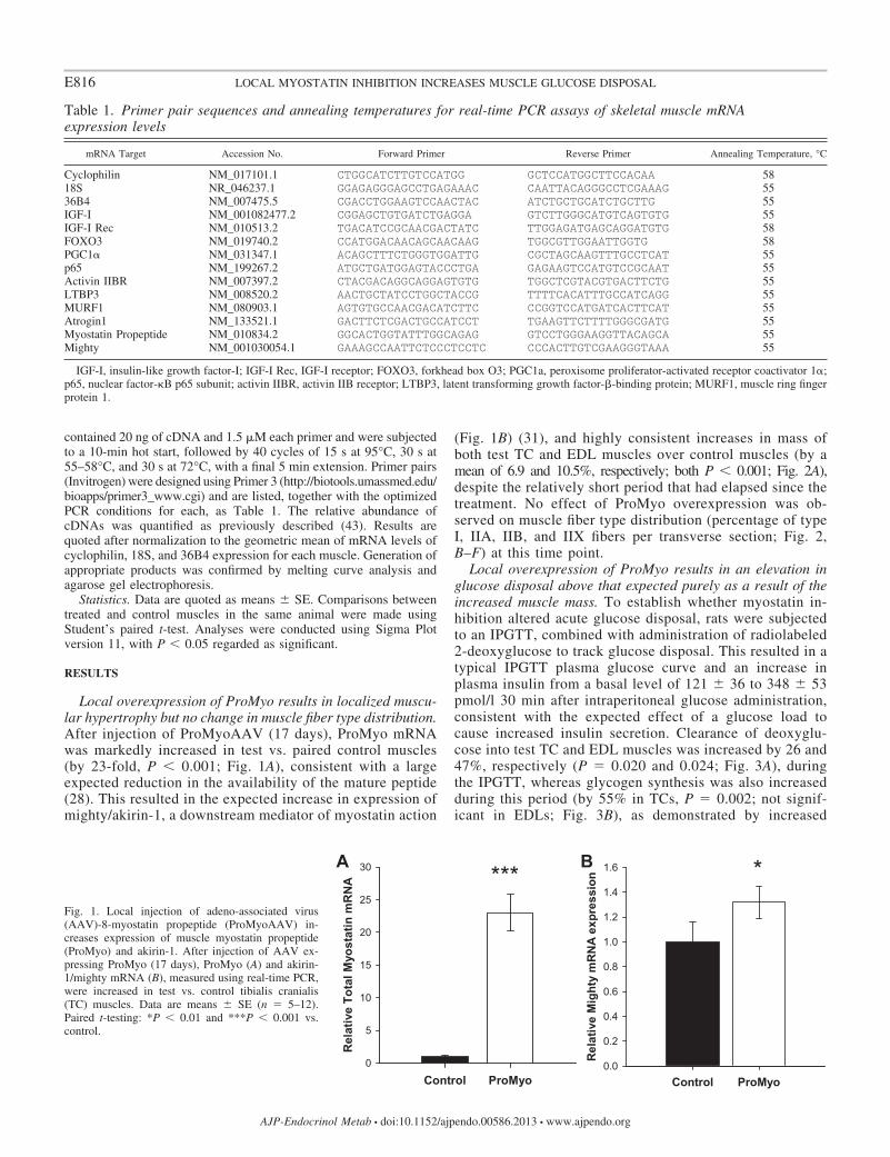
Local overexpression of ProMyo results in localized muscu-lar hypertrophy but no change in muscle fiber type distribution.After injection of ProMyoAAV (17 days), ProMyo mRNAwas markedly increased in test vs. paired control muscles(by 23-fold, P 0.001; Fig. 1A), consistent with a largeexpected reduction in the availability of the mature peptide(28). This resulted in the expected increase in expression ofmighty/akirin-1, a downstream mediator of myostatin action
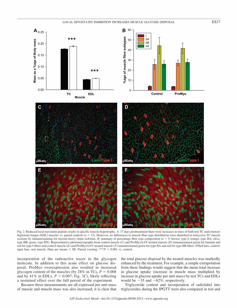
(Fig. 1B) (31), and highly consistent increases in mass ofboth test TC and EDL muscles over control muscles (by amean of 6.9 and 10.5%, respectively; both P 0.001; Fig. 2A),despite the relatively short period that had elapsed since thetreatment. No effect of ProMyo overexpression was ob-served on muscle fiber type distribution (percentage of typeI, IIA, IIB, and IIX fibers per transverse section; Fig. 2,B–F) at this time point.
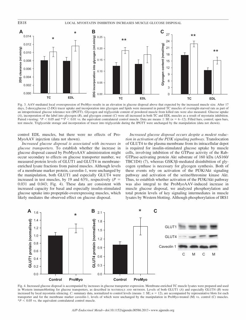
Local overexpression of ProMyo results in an elevation inglucose disposal above that expected purely as a result of theincreased muscle mass. To establish whether myostatin in-hibition altered acute glucose disposal, rats were subjectedto an IPGTT, combined with administration of radiolabeled2-deoxyglucose to track glucose disposal. This resulted in atypical IPGTT plasma glucose curve and an increase inplasma insulin from a basal level of 121 � 36 to 348 � 53pmol/l 30 min after intraperitoneal glucose administration,consistent with the expected effect of a glucose load tocause increased insulin secretion. Clearance of deoxyglu-cose into test TC and EDL muscles was increased by 26 and47%, respectively (P 0.020 and 0.024; Fig. 3A), duringthe IPGTT, whereas glycogen synthesis was also increasedduring this period (by 55% in TCs, P 0.002; not signif-icant in EDLs; Fig. 3B), as demonstrated by increased
Table 1. Primer pair sequences and annealing temperatures for real-time PCR assays of skeletal muscle mRNAexpression levels
mRNA Target Accession No. Forward Primer Reverse Primer Annealing Temperature, °C
Cyclophilin NM_017101.1 CTGGCATCTTGTCCATGG GCTCCATGGCTTCCACAA 5818S NR_046237.1 GGAGAGGGAGCCTGAGAAAC CAATTACAGGGCCTCGAAAG 5536B4 NM_007475.5 CGACCTGGAAGTCCAACTAC ATCTGCTGCATCTGCTTG 55IGF-I NM_001082477.2 CGGAGCTGTGATCTGAGGA GTCTTGGGCATGTCAGTGTG 55IGF-I Rec NM_010513.2 TGACATCCGCAACGACTATC TTGGAGATGAGCAGGATGTG 58FOXO3 NM_019740.2 CCATGGACAACAGCAACAAG TGGCGTTGGAATTGGTG 58PGC1� NM_031347.1 ACAGCTTTCTGGGTGGATTG CGCTAGCAAGTTTGCCTCAT 55p65 NM_199267.2 ATGCTGATGGAGTACCCTGA GAGAAGTCCATGTCCGCAAT 55Activin IIBR NM_007397.2 CTACGACAGGCAGGAGTGTG TGGCTCGTACGTGACTTCTG 55LTBP3 NM_008520.2 AACTGCTATCCTGGCTACCG TTTTCACATTTGCCATCAGG 55MURF1 NM_080903.1 AGTGTGCCAACGACATCTTC CCGGTCCATGATCACTTCAT 55Atrogin1 NM_133521.1 GACTTCTCGACTGCCATCCT TGAAGTTCTTTTGGGCGATG 55Myostatin Propeptide NM_010834.2 GGCACTGGTATTTGGCAGAG GTCCTGGGAAGGTTACAGCA 55Mighty NM_001030054.1 GAAAGCCAATTCTCCCTCCTC CCCACTTGTCGAAGGGTAAA 55
IGF-I, insulin-like growth factor-I; IGF-I Rec, IGF-I receptor; FOXO3, forkhead box O3; PGC1a, peroxisome proliferator-activated receptor coactivator 1�;p65, nuclear factor-�B p65 subunit; activin IIBR, activin IIB receptor; LTBP3, latent transforming growth factor-�-binding protein; MURF1, muscle ring fingerprotein 1.
Control ProMyo
Rel
ativ
e To
tal M
yost
atin
mR
NA
0
5
10
15
20
25
30 ***
Control ProMyo
Rel
ativ
e M
ight
y m
RN
A ex
pres
sion
0.0
0.2
0.4
0.6
0.8
1.0
1.2
1.4
1.6BA *Fig. 1. Local injection of adeno-associated virus(AAV)-8-myostatin propeptide (ProMyoAAV) in-creases expression of muscle myostatin propeptide(ProMyo) and akirin-1. After injection of AAV ex-pressing ProMyo (17 days), ProMyo (A) and akirin-1/mighty mRNA (B), measured using real-time PCR,were increased in test vs. control tibialis cranialis(TC) muscles. Data are means � SE (n 5–12).Paired t-testing: *P 0.01 and ***P 0.001 vs.control.
E816 LOCAL MYOSTATIN INHIBITION INCREASES MUSCLE GLUCOSE DISPOSAL
AJP-Endocrinol Metab • doi:10.1152/ajpendo.00586.2013 • www.ajpendo.org
incorporation of the radioactive tracer in the glycogenmolecule. In addition to this acute effect on glucose dis-posal, ProMyo overexpression also resulted in increasedglycogen content of the muscles (by 28% in TCs, P 0.008and by 41% in EDLs, P 0.007; Fig. 3C), likely reflectinga sustained effect over the full period of the experiment.
Because these measurements are all expressed per unit massof muscle and muscle mass was also increased, it is clear that
the total glucose disposal by the treated muscles was markedlyenhanced by the treatment. For example, a simple extrapolationfrom these findings would suggest that the mean total increasein glucose uptake (increase in muscle mass multiplied byincrease in glucose uptake per unit mass) by test TCs and EDLswould be �35 and �62%, respectively.
Triglyceride content and incorporation of radiolabel intotriglycerides during the IPGTT were also compared in test and
Control ProMyo
%ag
e of
mus
cle
fibre
sub
type
s
0
10
20
30
40
50
60
I IIA IIB IIX
MuscleTC EDL
Mas
s as
a %
age
of B
ody
mas
s
0.00
0.05
0.10
0.15
0.20
0.25
***
***BA
Lam/Type I Lam/Type I
Type IIA/Type IIB Type IIA/Type IIB
C D
FE
Fig. 2. Reduced local myostatin peptide results in specific muscle hypertrophy. A: 17 days posttreatment there were increases in mass of both test TC and extensordigitorum longus (EDL) muscles vs. paired controls (n 12). However, no differences in muscle fiber type distribution were identified in transverse TC musclesections by immunostaining for myosin heavy chain isoforms. B: summary of percentage fiber type composition (n 5; brown, type I; orange, type IIA; olive,type IIB; green, type IIX). Representative photomicrographs from control muscle (C) and ProMyoAAV-treated muscle (D) immunostained green for laminin andred for type I fibers and control muscle (E) and ProMyoAAV-treated muscle (F) immunostained green for type IIA and red for type IIB fibers. Filled bars, control;open bars, test muscle. Data are means � SE. Paired t-testing: ***P 0.001 vs. control.
E817LOCAL MYOSTATIN INHIBITION INCREASES MUSCLE GLUCOSE DISPOSAL
AJP-Endocrinol Metab • doi:10.1152/ajpendo.00586.2013 • www.ajpendo.org
control EDL muscles, but there were no effects of Pro-MyoAAV injection (data not shown).
Increased glucose disposal is associated with increases inglucose transporters. To establish whether the increase inglucose disposal caused by ProMyoAAV administration mightoccur secondary to effects on glucose transporter number, wemeasured protein levels of GLUT1 and GLUT4 in membrane-enriched lysate fractions from paired muscles. Although levelsof a membrane marker protein, caveolin-1, were unchanged bythe manipulation, both GLUT1 and especially GLUT4 wereincreased in test muscles, by 19 and 63%, respectively (P 0.031 and 0.043; Fig. 4). These data are consistent withincreased capacity for basal and especially insulin-stimulatedglucose uptake into propeptide-overexpressing muscles, whichlikely mediates the observed effect on glucose disposal.
Increased glucose disposal occurs despite a modest reduc-tion in activation of the PI3K signaling pathway. Translocationof GLUT4 to the plasma membrane from its intracellular depotis required for insulin-stimulated glucose uptake by musclecells, involving inhibition of the GTPase activity of the Rab-GTPase-activating protein Akt substrate of 160 kDa (AS160/TBC1D4) (7), whereas GSK3�-mediated disinhibition of gly-cogen synthase is necessary for glycogen synthesis. Both ofthese events rely on activation of the PI3K/Akt signalingpathway and activation of the serine/threonine kinase Akt.Thus, to establish whether activation of the PI3K/Akt pathwaywas also integral to the ProMyoAAV-induced increase inmuscle glucose disposal, we analyzed phosphorylation andtotal protein levels of key signaling intermediates in musclelysates by Western blotting. Although phosphorylation of IRS1
TC EDL
Gly
coge
n co
nten
t (nm
ol/m
g)
0
3
6
9
12
15
18
21
24
27 **
**
TC EDL2DG
inco
rpor
atio
n in
to g
lyco
gen
(ml.m
in/g
)0.00
0.05
0.10
0.15
0.20
0.25
**
TC EDL
2DG
cle
aran
ce (m
l.min
/g)
0.000
0.002
0.004
0.006
0.008
0.010
0.012
0.014*
* BA C
Fig. 3. AAV-mediated local overexpression of ProMyo results in an elevation in glucose disposal above that expected by the increased muscle size. After 17days, 2-deoxyglucose (2-DG) tracer uptake and incorporation into glycogen and lipids were measured in paired TC muscles of overnight-starved rats as part ofan intraperitoneal glucose tolerance test (IPGTT). Glycogen and triglyceride content of powdered muscle from killed rats were also measured. Glucose uptake(A), incorporation of the label into glycogen (B), and glycogen content (C) were all increased in both TC and EDL muscles as a result of myostatin inhibition.Paired t-testing: *P 0.05 and **P 0.01 vs. the equivalent contralateral control muscle. Data are means � SE (n 6–12). Filled bars, control; open bars,test muscle. Triglyceride storage and incorporation of tracer into triglyceride during the IPGTT were unchanged by the manipulation (data not shown).
Control ProMyo
GLU
T1 p
rote
in (n
orm
alis
ed)
0.0
0.2
0.4
0.6
0.8
1.0
1.2
1.4
1.6
Control ProMyo
GLU
T4 p
rote
in (n
orm
alis
ed)
0.0
0.3
0.6
0.9
1.2
1.5
1.8
2.1BA* *
GLUT1
GLUT4
Caveolin 1
C
C M C M
Fig. 4. Increased glucose disposal is accompanied by increases in glucose transporter expression. Membrane-enriched TC muscle lysates were prepared and usedin Western immunoblotting for glucose transporters, as described in MATERIALS AND METHODS. Levels of both GLUT1 (A) and especially GLUT4 (B) wereincreased by local myostatin silencing. C: summary data, normalized to control levels (means � SE; n 12), are accompanied by representative blots for eachtransporter and for the membrane marker caveolin-1, levels of which were unchanged by the manipulation in ProMyo-treated (M) vs. control (C) muscles.*P 0.05 vs. the equivalent contralateral control muscle.
E818 LOCAL MYOSTATIN INHIBITION INCREASES MUSCLE GLUCOSE DISPOSAL
AJP-Endocrinol Metab • doi:10.1152/ajpendo.00586.2013 • www.ajpendo.org
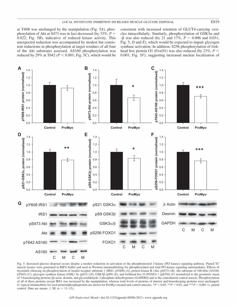
at Y608 was unchanged by the manipulation (Fig. 5A), phos-phorylation of Akt at S473 was in fact decreased (by 33%, P 0.022; Fig. 5B), indicative of reduced kinase activity. Thisunexpected reduction was accompanied by modest but consis-tent reductions in phosphorylation at target residues of all fourof the Akt substrates assessed. AS160 phosphorylation wasreduced by 29% at T642 (P 0.001; Fig. 5C), which would be
consistent with increased retention of GLUT4-carrying vesi-cles intracellularly. Similarly, phosphorylation of GSK3� and-� was also reduced (by 21 and 17%, P 0.006 and 0.031;Fig. 5, D and E), which would be expected to impair glycogensynthase activation. In addition, S256 phosphorylation of fork-head box protein O1 (FoxO1) was also reduced (by 23%, P 0.001; Fig. 5F), suggesting increased nuclear localization of
Control ProMyo
pY60
8-IR
S1 p
rote
in (n
orm
alis
ed)
0.0
0.2
0.4
0.6
0.8
1.0
1.2
1.4
Control ProMyo
pS47
3-A
kt p
rote
in (n
orm
alis
ed)
0.0
0.2
0.4
0.6
0.8
1.0
1.2
1.4
Control ProMyo
pT64
2-A
S160
pro
tein
(nor
mal
ised
)
0.0
0.2
0.4
0.6
0.8
1.0
1.2
1.4CBA
Control ProMyo
pS21
-GSK
3 α p
rote
in (n
orm
alis
ed)
0.0
0.2
0.4
0.6
0.8
1.0
1.2
1.4
Control ProMyo
pS9-
GSK
3 β p
rote
in (n
orm
alis
ed)
0.0
0.2
0.4
0.6
0.8
1.0
1.2
1.4
Control ProMyo
pS25
6-FO
XO1
prot
ein
(nor
mal
ised
)
0.0
0.2
0.4
0.6
0.8
1.0
1.2
1.4FED
******
* ***
G pY608 IRS1
IRS1
pS473 Akt
Akt
pT642 AS160
AS160
pS21 GSK3α
pS9 GSK3β
GSK3α/β
pS256 FOXO1
FOXO1
β Actin
Desmin
GAPDH
C M C M
C M C M
C M C M
Fig. 5. Increased glucose disposal occurs despite a modest reduction in activation of the phosphoinositol 3-kinase (PI3-kinase) signaling pathway. Paired TCmuscle lysates were generated in RIPA buffer and used in Western immunoblotting for phosphorylated and total PI3-kinase signaling intermediates. Effects ofmyostatin silencing on phosphorylation of insulin receptor substrate 1 (IRS1, pY608) (A), protein kinase B (Akt, pS473) (B), Akt substrate of 160 kDa (AS160,pT642) (C), glycogen synthase kinase (GSK) 3� (pS21) (D), GSK3� (pS9) (E), and forkhead box O (FOXO) 1 (pS256) (F) normalized to the geometric meanof 3 housekeeping proteins [�-actin, desmin, and glyceraldehyde-3-phosphate dehydrogenase (GAPDH)] and to the contralateral control muscle. Phosphorylationof all of these proteins except IRS1 was increased by the manipulation, whereas total levels of proteins of interest and housekeeping proteins were unchanged.G: typical immunoblots for each protein/phosphoprotein are shown for ProMyo-treated and control muscles. *P 0.05, **P 0.01, and ***P 0.001 vs. pairedcontrol. Data are means � SE (n 11–12).
E819LOCAL MYOSTATIN INHIBITION INCREASES MUSCLE GLUCOSE DISPOSAL
AJP-Endocrinol Metab • doi:10.1152/ajpendo.00586.2013 • www.ajpendo.org
this transcription factor. Total protein levels of all of thesignaling intermediates were not altered by the manipulation.Thus, ProMyo overexpression results in reduced flux throughthe PI3K/Akt pathway distal to IRS1, which would not explainthe observed effects on glucose disposal, but may instead be asecondary effect of another consequence of myostatin inhibi-tion.
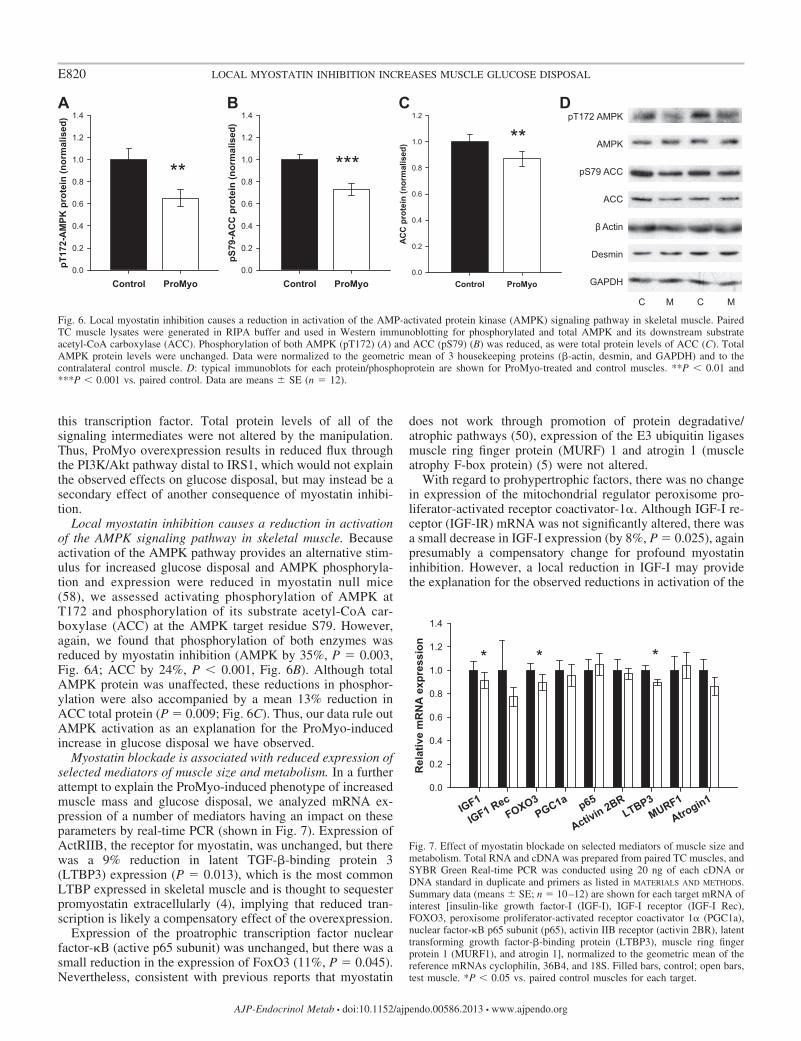
Local myostatin inhibition causes a reduction in activationof the AMPK signaling pathway in skeletal muscle. Becauseactivation of the AMPK pathway provides an alternative stim-ulus for increased glucose disposal and AMPK phosphoryla-tion and expression were reduced in myostatin null mice(58), we assessed activating phosphorylation of AMPK atT172 and phosphorylation of its substrate acetyl-CoA car-boxylase (ACC) at the AMPK target residue S79. However,again, we found that phosphorylation of both enzymes wasreduced by myostatin inhibition (AMPK by 35%, P 0.003,Fig. 6A; ACC by 24%, P 0.001, Fig. 6B). Although totalAMPK protein was unaffected, these reductions in phosphor-ylation were also accompanied by a mean 13% reduction inACC total protein (P 0.009; Fig. 6C). Thus, our data rule outAMPK activation as an explanation for the ProMyo-inducedincrease in glucose disposal we have observed.
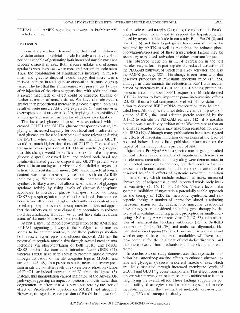
Myostatin blockade is associated with reduced expression ofselected mediators of muscle size and metabolism. In a furtherattempt to explain the ProMyo-induced phenotype of increasedmuscle mass and glucose disposal, we analyzed mRNA ex-pression of a number of mediators having an impact on theseparameters by real-time PCR (shown in Fig. 7). Expression ofActRIIB, the receptor for myostatin, was unchanged, but therewas a 9% reduction in latent TGF-�-binding protein 3(LTBP3) expression (P 0.013), which is the most commonLTBP expressed in skeletal muscle and is thought to sequesterpromyostatin extracellularly (4), implying that reduced tran-scription is likely a compensatory effect of the overexpression.
Expression of the proatrophic transcription factor nuclearfactor-�B (active p65 subunit) was unchanged, but there was asmall reduction in the expression of FoxO3 (11%, P 0.045).Nevertheless, consistent with previous reports that myostatin
does not work through promotion of protein degradative/atrophic pathways (50), expression of the E3 ubiquitin ligasesmuscle ring finger protein (MURF) 1 and atrogin 1 (muscleatrophy F-box protein) (5) were not altered.
With regard to prohypertrophic factors, there was no changein expression of the mitochondrial regulator peroxisome pro-liferator-activated receptor coactivator-1�. Although IGF-I re-ceptor (IGF-IR) mRNA was not significantly altered, there wasa small decrease in IGF-I expression (by 8%, P 0.025), againpresumably a compensatory change for profound myostatininhibition. However, a local reduction in IGF-I may providethe explanation for the observed reductions in activation of the
Control ProMyo
pT17
2-A
MPK
pro
tein
(nor
mal
ised
)
0.0
0.2
0.4
0.6
0.8
1.0
1.2
1.4
Control ProMyo
pS79
-AC
C p
rote
in (n
orm
alis
ed)
0.0
0.2
0.4
0.6
0.8
1.0
1.2
1.4
Control ProMyo
AC
C p
rote
in (n
orm
alis
ed)
0.0
0.2
0.4
0.6
0.8
1.0
1.2BA
** *****
C DpT172 AMPK
AMPK
pS79 ACC
ACC
β Actin
Desmin
GAPDH
C M C M
Fig. 6. Local myostatin inhibition causes a reduction in activation of the AMP-activated protein kinase (AMPK) signaling pathway in skeletal muscle. PairedTC muscle lysates were generated in RIPA buffer and used in Western immunoblotting for phosphorylated and total AMPK and its downstream substrateacetyl-CoA carboxylase (ACC). Phosphorylation of both AMPK (pT172) (A) and ACC (pS79) (B) was reduced, as were total protein levels of ACC (C). TotalAMPK protein levels were unchanged. Data were normalized to the geometric mean of 3 housekeeping proteins (�-actin, desmin, and GAPDH) and to thecontralateral control muscle. D: typical immunoblots for each protein/phosphoprotein are shown for ProMyo-treated and control muscles. **P 0.01 and***P 0.001 vs. paired control. Data are means � SE (n 12).
IGF1
IGF1 RecFOXO3
PGC1a p65
Activin 2BRLTBP3
MURF1
Atrogin1
Rel
ativ
e m
RN
A e
xpre
ssio
n
0.0
0.2
0.4
0.6
0.8
1.0
1.2
1.4
* **
Fig. 7. Effect of myostatin blockade on selected mediators of muscle size andmetabolism. Total RNA and cDNA was prepared from paired TC muscles, andSYBR Green Real-time PCR was conducted using 20 ng of each cDNA orDNA standard in duplicate and primers as listed in MATERIALS AND METHODS.Summary data (means � SE; n 10–12) are shown for each target mRNA ofinterest [insulin-like growth factor-I (IGF-I), IGF-I receptor (IGF-I Rec),FOXO3, peroxisome proliferator-activated receptor coactivator 1� (PGC1a),nuclear factor-�B p65 subunit (p65), activin IIB receptor (activin 2BR), latenttransforming growth factor-�-binding protein (LTBP3), muscle ring fingerprotein 1 (MURF1), and atrogin 1], normalized to the geometric mean of thereference mRNAs cyclophilin, 36B4, and 18S. Filled bars, control; open bars,test muscle. *P 0.05 vs. paired control muscles for each target.
E820 LOCAL MYOSTATIN INHIBITION INCREASES MUSCLE GLUCOSE DISPOSAL
AJP-Endocrinol Metab • doi:10.1152/ajpendo.00586.2013 • www.ajpendo.org
PI3K/Akt and AMPK signaling pathways in ProMyoAAV-injected muscles.
DISCUSSION
In our study we have demonstrated that local inhibition ofmyostatin action in skeletal muscle for only a relatively shortperiod is capable of generating both increased muscle mass andglucose disposal in rats. Both glucose uptake and glycogensynthesis were increased when measured per unit muscle mass.Thus, the combination of simultaneous increases in musclemass and glucose disposal would imply that there was amarked increase in total glucose disposal in the muscle grouptested. The fact that this enhancement was present just 17 daysafter injection of the virus suggests that, with additional time,a greater magnitude of effect could be expected because offurther accretion of muscle tissue. We have also observed agreater than proportional increase in glucose disposal both as aresult of acute muscle Akt-2 overexpression (11) and universaloverexpression of urocortin-3 (20), implying the possibility ofa more general mechanism worthy of deeper investigation.
The increased glucose disposal was associated with in-creased GLUT1 and GLUT4 glucose transporter content, im-plying an increased capacity for both basal and insulin-stimu-lated glucose uptake (the latter being of more relevance duringthe IPGTT, when total levels of plasma membrane GLUT4would be much higher than those of GLUT1). The results oftransgenic overexpression of GLUT4 in muscle (51) suggestthat this change would be sufficient to explain the improvedglucose disposal observed here, and indeed both basal andinsulin-stimulated glucose disposal and GLUT4 protein wereelevated in an analogous in vivo model of deficient myostatinaction, the myostatin null mouse (58), while muscle glycogencontent was also increased by treatment with an ActRIIBinhibitor (14). We can speculate that the increased glycogensynthesis is likely a result of allosteric stimulation of glycogensynthase activity by rising levels of glucose 6-phosphate,secondary to increased glucose transport, since we foundGSK3� phosphorylation at Ser9 to be reduced. Furthermore,because no differences in triglyceride synthesis or content werenoted in propeptide-overexpressing muscles, it does not appearthat the effects on glucose disposal are secondary to reducedlipid accumulation, although we do not have data regardingsome of the more bioactive lipid species.
At first glance, the modest downregulation of the AMPK andPI3K/Akt signaling pathways in the ProMyo-treated musclesseems to be counterintuitive, since these pathways mediateboth muscle hypertrophy and glucose disposal. Akt has thepotential to regulate muscle size through several mechanisms,including via phosphorylation of both GSK3 and FoxOs.GSK3 inhibits the translation initiation factor eIF2B (44),whereas FoxOs have been shown to promote muscle atrophythrough activation of the E3 ubiquitin ligases MURF1 andatrogin-1 (45, 48). In a previous study, myostatin overexpres-sion in rats did not alter levels of expression or phosphorylationof FoxO1, or indeed expression of E3 ubiquitin ligases (3).Instead, this manipulation caused inhibition of the Akt-mTORpathway, suggesting an impact on protein synthesis rather thandegradation, an effect that was borne out here by the lack ofeffect of ProMyoAAV injection on MURF1 and atrogin-1.However, transgenic overexpression of FoxO1 in mouse skel-
etal muscle caused atrophy (21); thus, the reduction in FoxO1phosphorylation would tend to support the hypertrophy in-duced by myostatin blockade in our study. Both FoxO1 (8) andFoxO3 (15) and their target genes have been shown to beregulated by AMPK as well as Akt; thus, the reduced phos-phorylation/expression of these transcription factors may besecondary to reduced activation of either upstream kinase.
The observed reduction in IGF-I expression in the testmuscles may at least in part explain the reduced activation ofthe PI3K/Akt pathway, of which it is a key activator, and alsothe AMPK pathway (38). This change is consistent with thatobserved previously in myostatin knockout mice (13, 55),although in these animals the reduction in IGF-I was accom-panied by increases in IGF-IR and IGF-I-binding protein ex-pression and/or increased IGF-II expression. Muscle-derivedIGF-I is known to have important paracrine/autocrine effects(20, 42); thus, a local compensatory effect of myostatin inhi-bition to decrease IGF-I mRNA transcription may be impli-cated here. Although we did not detect a change in phosphor-ylation of IRS1, the usual adaptor protein recruited by theIGF-IR to activate the PI3K/Akt pathway (42), it is possiblethat this was a sensitivity artifact of the Western blot or that analternative adaptor protein may have been recruited, for exam-ple, IRS2 (49). Although many publications have investigatedthe effects of myostatin inhibition downstream at the level ofAkt and below, there is little published information on theimpact of this manipulation upstream of Akt.
Injection of ProMyoAAV in a specific muscle group resultedin local effects, since a number of significant differences inmuscle mass, metabolism, and signaling were demonstrated inthe injected muscles. In addition, our data confirm that in-creased muscle mass alone is not the likely explanation for theobserved beneficial effects of systemic myostatin inhibitionon metabolism, which include reduced fat mass, increased“browning” of adipose tissue, and improved whole body insu-lin sensitivity (1, 16, 17, 34, 58–60). These effects makesystemic inhibition of myostatin a potentially viable approachfor the therapy of T2D, the metabolic syndrome, and sar-copenic obesity. A number of approaches aimed at reducingmyostatin action for the treatment of muscular dystrophieshave already been considered, including gene therapy by de-livery of myostatin-inhibiting genes, propeptide or small-inter-fering RNA using AAV or retrovirus (12, 18, 57), administra-tion of antimyostatin blocking antibodies (52) or ActRIIBcompetitors (1, 14, 36, 59), and antisense oligonucleotide-mediated exon skipping (22, 23). However, it is unclear as yetwhether any of these therapeutic modalities will have long-term potential for the treatment of metabolic disorders, andthus more research into mechanisms and applications is war-ranted.
In conclusion, our study demonstrates that myostatin inhi-bition has autocrine/paracrine effects to enhance glucose up-take and glycogen synthesis in skeletal muscle of rats, whichare likely mediated through increased membrane levels ofGLUT1 and GLUT4 glucose transporters. This effect occurs intandem with increased muscle mass, but is additional to it, thusmagnifying the overall effect. These findings support the po-tential utility of strategies aimed at inhibiting skeletal musclemyostatin action in the treatment of metabolic disorders, in-cluding T2D and sarcopenic obesity.
E821LOCAL MYOSTATIN INHIBITION INCREASES MUSCLE GLUCOSE DISPOSAL
AJP-Endocrinol Metab • doi:10.1152/ajpendo.00586.2013 • www.ajpendo.org
ACKNOWLEDGMENTS
We are grateful to Sejal Patel for technical assistance, to Dr. JacquelineStöckli (Garvan Institute, Sydney) for advice regarding GLUT4 immunoblot-ting, and to the staff of the Biological Services Unit at the Royal VeterinaryCollege for animal care. We acknowledge Dr. Carl Morris and Dr. PaulYaworsky of Pfizer for continued access to the mouse ProMyo construct.
GRANTS
This work was funded by the Wellcome Trust (a University Award to M. E.Cleasby) and Pfizer (previously Wyeth).
DISCLOSURES
No conflicts of interest, financial or otherwise, are declared by the authors.
AUTHOR CONTRIBUTIONS
Author contributions: M.E.C., G.D., and K.F. conception and design ofresearch; M.E.C., S.J., W.E., M.E., and D.K.A. performed experiments;M.E.C. and K.F. analyzed data; M.E.C. interpreted results of experiments;M.E.C. prepared figures; M.E.C. drafted manuscript; M.E.C., S.J., G.D., andK.F. edited and revised manuscript; M.E.C., S.J., W.E., M.E., D.K.A., G.D.,and K.F. approved final version of manuscript.
REFERENCES
1. Akpan I, Goncalves MD, Dhir R, Yin X, Pistilli EE, Bogdanovich S,Khurana TS, Ucran J, Lachey J, Ahima RS. The effects of a solubleactivin type IIB receptor on obesity and insulin sensitivity. Int J Obes(Lond) 33: 1265–1273, 2009.
2. Allen DL, Cleary AS, Speaker KJ, Lindsay SF, Uyenishi J, Reed JM,Madden MC, Mehan RS. Myostatin, activin receptor IIb, and follistatin-like-3 gene expression are altered in adipose tissue and skeletal muscle ofobese mice. Am J Physiol Endocrinol Metab 294: E918–E927, 2008.
3. Amirouche A, Durieux AC, Banzet S, Koulmann N, Bonnefoy R,Mouret C, Bigard X, Peinnequin A, Freyssenet D. Down-regulation ofAkt/mammalian target of rapamycin signaling pathway in response tomyostatin overexpression in skeletal muscle. Endocrinology 150: 286–294, 2009.
4. Anderson SB, Goldberg AL, Whitman M. Identification of a novel poolof extracellular pro-myostatin in skeletal muscle. J Biol Chem 283:7027–7035, 2008.
5. Bodine SC, Latres E, Baumhueter S, Lai VK, Nunez L, Clarke BA,Poueymirou WT, Panaro FJ, Na E, Dharmarajan K, Pan ZQ, Valen-zuela DM, DeChiara TM, Stitt TN, Yancopoulos GD, Glass DJ.Identification of ubiquitin ligases required for skeletal muscle atrophy.Science 294: 1704–1708, 2001.
6. Brandt C, Nielsen AR, Fischer CP, Hansen J, Pedersen BK, Plom-gaard P. Plasma and muscle myostatin in relation to type 2 diabetes. PLoSOne 7: e37236, 2012.
7. Bruss MD, Arias EB, Lienhard GE, Cartee GD. Increased phosphor-ylation of Akt substrate of 160 kDa (AS160) in rat skeletal muscle inresponse to insulin or contractile activity. Diabetes 54: 41–50, 2005.
8. Chen BL, Ma YD, Meng RS, Xiong ZJ, Wang HN, Zeng JY, Liu C,Dong YG. Activation of AMPK inhibits cardiomyocyte hypertrophy bymodulating of the FOXO1/MuRF1 signaling pathway in vitro. ActaPharmacol Sinica 31: 798–804, 2010.
9. Cho H, Mu J, Kim JK, Thorvaldsen JL, Chu Q, Crenshaw EB, III,Kaestner KH, Bartolomei MS, Shulman GI, Birnbaum MJ. Insulinresistance and a diabetes mellitus-like syndrome in mice lacking theprotein kinase Akt2 (PKB beta). Science 292: 1728–1731, 2001.
10. Cleasby ME, Dzamko N, Hegarty BD, Cooney GJ, Kraegen EW, YeJM. Metformin prevents the development of acute lipid-induced insulinresistance in the rat through altered hepatic signaling mechanisms. Dia-betes 53: 3258–3266, 2004.
11. Cleasby ME, Reinten TA, Cooney GJ, James DE, Kraegen EW.Functional studies of Akt isoform specificity in skeletal muscle in vivo;maintained insulin sensitivity despite reduced insulin receptor substrate-1expression. Mol Endocrinol 21: 215–228, 2007.
12. Foster K, Graham IR, Otto A, Foster H, Trollet C, Yaworsky PJ,Walsh FS, Bickham D, Curtin NA, Kawar SL, Patel K, Dickson G.Adeno-associated virus-8-mediated intravenous transfer of myostatin pro-peptide leads to systemic functional improvements of slow but not fastmuscle. Rejuvenation Res 12: 85–94, 2009.
13. Gilson H, Schakman O, Combaret L, Lause P, Grobet L, Attaix D,Ketelslegers JM, Thissen JP. Myostatin gene deletion prevents gluco-corticoid-induced muscle atrophy. Endocrinology 148: 452–460, 2007.
14. Goncalves MD, Pistilli EE, Balduzzi A, Birnbaum MJ, Lachey J,Khurana TS, Ahima RS. Akt deficiency attenuates muscle size andfunction but not the response to ActRIIB inhibition. PLoS One 5: e12707,2010.
15. Greer EL, Oskoui PR, Banko MR, Maniar JM, Gygi MP, Gygi SP,Brunet A. The energy sensor AMP-activated protein kinase directlyregulates the mammalian FOXO3 transcription factor. J Biol Chem 282:30107–30119, 2007.
16. Guo T, Bond ND, Jou W, Gavrilova O, Portas J, McPherron AC.Myostatin inhibition prevents diabetes and hyperphagia in a mouse modelof lipodystrophy. Diabetes 61: 2414–2423, 2012.
17. Guo T, Jou W, Chanturiya T, Portas J, Gavrilova O, McPherron AC.Myostatin inhibition in muscle, but not adipose tissue, decreases fat massand improves insulin sensitivity. PLoS One 4: e4937, 2009.
18. Haidet AM, Rizo L, Handy C, Umapathi P, Eagle A, Shilling C, BoueD, Martin PT, Sahenk Z, Mendell JR, Kaspar BK. Long-term enhance-ment of skeletal muscle mass and strength by single gene administrationof myostatin inhibitors. Proc Natl Acad Sci USA 105: 4318–4322, 2008.
19. Hittel DS, Axelson M, Sarna N, Shearer J, Huffman KM, Kraus WE.Myostatin decreases with aerobic exercise and associates with insulinresistance. Med Sci Sports Exerc 42: 2023–2029, 2010.
20. Jamieson PM, Cleasby ME, Kuperman Y, Morton NM, Kelly PA,Brownstein DG, Mustard KJ, Vaughan JM, Carter RN, Hahn CN,Hardie DG, Seckl JR, Chen A, Vale WW. Urocortin 3 transgenic miceexhibit a metabolically favourable phenotype resisting obesity and hyper-glycaemia on a high-fat diet. Diabetologia 54: 2392–2403, 2011.
21. Kamei Y, Miura S, Suzuki M, Kai Y, Mizukami J, Taniguchi T,Mochida K, Hata T, Matsuda J, Aburatani H, Nishino I, Ezaki O.Skeletal muscle FOXO1 (FKHR)-transgenic mice have less skeletal mus-cle mass, down-regulated type I (slow twitch/red muscle) fiber genes, andimpaired glycemic control. J Biol Chem 279: 41114–41123, 2004.
22. Kang JK, Malerba A, Popplewell L, Foster K, Dickson G. Antisense-induced myostatin exon skipping leads to muscle hypertrophy in micefollowing octa-guanidine morpholino oligomer treatment. Mol Ther 19:159–164, 2011.
23. Kinali M, Arechavala-Gomeza V, Feng L, Cirak S, Hunt D, Adkin C,Guglieri M, Ashton E, Abbs S, Nihoyannopoulos P, Garralda ME,Rutherford M, McCulley C, Popplewell L, Graham IR, Dickson G,Wood MJ, Wells DJ, Wilton SD, Kole R, Straub V, Bushby K, SewryC, Morgan JE, Muntoni F. Local restoration of dystrophin expressionwith the morpholino oligomer AVI-4658 in Duchenne muscular dystro-phy: a single-blind, placebo-controlled, dose-escalation, proof-of-conceptstudy. Lancet Neurol 8: 918–928, 2009.
24. Kollias HD, McDermott JC. Transforming growth factor-beta and myo-statin signaling in skeletal muscle. J Appl Physiol 104: 579–587, 2008.
25. Langley B, Thomas M, Bishop A, Sharma M, Gilmour S, KambadurR. Myostatin inhibits myoblast differentiation by down-regulating MyoDexpression. J Biol Chem 277: 49831–49840, 2002.
26. Lantier L, Mounier R, Leclerc J, Pende M, Foretz M, Viollet B.Coordinated maintenance of muscle cell size control by AMP-activatedprotein kinase. FASEB J 24: 3555–3561, 2010.
27. Lee SJ. Extracellular regulation of myostatin: a molecular rheostat formuscle mass. Immunol Endocr Metab Agents Med Chem 10: 183–194,2010.
28. Lee SJ, McPherron AC. Regulation of myostatin activity and musclegrowth. Proc Natl Acad Sci USA 98: 9306–9311, 2001.
29. Long YC, Zierath JR. AMP-activated protein kinase signaling in meta-bolic regulation. J Clin Invest 116: 1776–1783, 2006.
30. Magee TR, Artaza JN, Ferrini MG, Vernet D, Zuniga FI, Cantini L,Reisz-Porszasz S, Rajfer J, Gonzalez-Cadavid NF. Myostatin shortinterfering hairpin RNA gene transfer increases skeletal muscle mass. JGene Med 8: 1171–1181, 2006.
31. Marshall A, Salerno MS, Thomas M, Davies T, Berry C, Dyer K,Bracegirdle J, Watson T, Dziadek M, Kambadur R, Bower R, SharmaM. Mighty is a novel promyogenic factor in skeletal myogenesis. Exp CellRes 314: 1013–1029, 2008.
32. Matsakas A, Foster K, Otto A, Macharia R, Elashry MI, Feist S,Graham I, Foster H, Yaworsky P, Walsh F, Dickson G, Patel K.Molecular, cellular and physiological investigation of myostatin propep-tide-mediated muscle growth in adult mice. Neuromuscul Disord 19:489–499, 2009.
E822 LOCAL MYOSTATIN INHIBITION INCREASES MUSCLE GLUCOSE DISPOSAL
AJP-Endocrinol Metab • doi:10.1152/ajpendo.00586.2013 • www.ajpendo.org
33. McPherron AC, Lawler AM, Lee SJ. Regulation of skeletal musclemass in mice by a new TGF-beta superfamily member. Nature 387: 83–90,1997.
34. McPherron AC, Lee SJ. Suppression of body fat accumulation inmyostatin-deficient mice. J Clin Invest 109: 595–601, 2002.
35. Milan G, Dalla Nora E, Pilon C, Pagano C, Granzotto M, Manco M,Mingrone G, Vettor R. Changes in muscle myostatin expression in obesesubjects after weight loss. J Clin Endocrinol Metab 89: 2724–2727, 2004.
36. Morrison BM, Lachey JL, Warsing LC, Ting BL, Pullen AE, Under-wood KW, Kumar R, Sako D, Grinberg A, Wong V, Colantuoni E,Seehra JS, Wagner KR. A soluble activin type IIB receptor improvesfunction in a mouse model of amyotrophic lateral sclerosis. Exp Neurol217: 258–268, 2009.
37. Narici MV, Maffulli N. Sarcopenia: characteristics, mechanisms andfunctional significance. Br Med Bull 95: 139–159, 2010.
38. Ning J, Xi G, Clemmons DR. Suppression of AMPK activation via S485phosphorylation by IGF-I during hyperglycemia is mediated by AKTactivation in vascular smooth muscle cells. Endocrinology 152: 3143–3154, 2011.
39. Palsgaard J, Brons C, Friedrichsen M, Dominguez H, Jensen M,Storgaard H, Spohr C, Torp-Pedersen C, Borup R, De Meyts P, VaagA. Gene expression in skeletal muscle biopsies from people with type 2diabetes and relatives: differential regulation of insulin signaling path-ways. PLoS One 4: e6575, 2009.
40. Park JJ, Berggren JR, Hulver MW, Houmard JA, Hoffman EP.GRB14, GPD1, and GDF8 as potential network collaborators in weightloss-induced improvements in insulin action in human skeletal muscle.Physiol Genomics 27: 114–121, 2006.
41. Patel SA, Hoehn KL, Lawrence RT, Sawbridge L, Talbot NA, TomsigJL, Turner N, Cooney GJ, Whitehead JP, Kraegen EW, Cleasby ME.Overexpression of the adiponectin receptor AdipoR1 in rat skeletal muscleamplifies local insulin sensitivity. Endocrinology 153: 5231–5246, 2012.
42. Philippou A, Halapas A, Maridaki M, Koutsilieris M. Type I insulin-like growth factor receptor signaling in skeletal muscle regeneration andhypertrophy. J Musculoskelet Neuronal Interact 7: 208–218, 2007.
43. Polkinghorne E, Lau Q, Cooney GJ, Kraegen EW, Cleasby ME. Localactivation of the I�K-NF�B pathway in muscle does not cause insulinresistance. Am J Physiol Endocrinol Metab 294: E316–E325, 2008.
44. Rommel C, Bodine SC, Clarke BA, Rossman R, Nunez L, Stitt TN,Yancopoulos GD, Glass DJ. Mediation of IGF-1-induced skeletal myo-tube hypertrophy by PI(3)K/Akt/mTOR and PI(3)K/Akt/GSK3 pathways.Nat Cell Biol 3: 1009–1013, 2001.
45. Sandri M, Lin J, Handschin C, Yang W, Arany ZP, Lecker SH,Goldberg AL, Spiegelman BM. PGC-1alpha protects skeletal musclefrom atrophy by suppressing FoxO3 action and atrophy-specific genetranscription. Proc Natl Acad Sci USA 103: 16260–16265, 2006.
46. Sonntag F, Kother K, Schmidt K, Weghofer M, Raupp C, Nieto K,Kuck A, Gerlach B, Bottcher B, Muller OJ, Lux K, Horer M,Kleinschmidt JA. The assembly-activating protein promotes capsid as-sembly of different adeno-associated virus serotypes. J Virol 85: 12686–12697, 2011.
47. Stenholm S, Harris TB, Rantanen T, Visser M, Kritchevsky SB,Ferrucci L. Sarcopenic obesity: definition, cause and consequences. CurrOpin Clin Nutr Metab Care 11: 693–700, 2008.
48. Stitt TN, Drujan D, Clarke BA, Panaro F, Timofeyva Y, Kline WO,Gonzalez M, Yancopoulos GD, Glass DJ. The IGF-1/PI3K/Akt pathwayprevents expression of muscle atrophy-induced ubiquitin ligases by inhib-iting FOXO transcription factors. Mol Cell 14: 395–403, 2004.
49. Sun XJ, Wang LM, Zhang Y, Yenush L, Myers MG Jr, Glasheen E,Lane WS, Pierce JH, White MF. Role of IRS-2 in insulin and cytokinesignalling. Nature 377: 173–177, 1995.
50. Trendelenburg AU, Meyer A, Rohner D, Boyle J, Hatakeyama S,Glass DJ. Myostatin reduces Akt/TORC1/p70S6K signaling, inhibitingmyoblast differentiation and myotube size. Am J Physiol Cell Physiol 296:C1258–C1270, 2009.
51. Tsao TS, Burcelin R, Katz EB, Huang L, Charron MJ. Enhancedinsulin action due to targeted GLUT4 overexpression exclusively inmuscle. Diabetes 45: 28–36, 1996.
52. Wagner KR, Fleckenstein JL, Amato AA, Barohn RJ, Bushby K,Escolar DM, Flanigan KM, Pestronk A, Tawil R, Wolfe GI, EagleM, Florence JM, King WM, Pandya S, Straub V, Juneau P, MeyersK, Csimma C, Araujo T, Allen R, Parsons SA, Wozney JM,Lavallie ER, Mendell JR. A phase I/IItrial of MYO-029 in adultsubjects with muscular dystrophy. Ann Neurol 63: 561–571, 2008.
53. Whiteman EL, Cho H, Birnbaum MJ. Role of Akt/protein kinase B inmetabolism. Trends Endocrinol Metab 13: 444–451, 2002.
54. Wilkes JJ, Lloyd DJ, Gekakis N. Loss-of-function mutation in myostatinreduces tumor necrosis factor alpha production and protects liver againstobesity-induced insulin resistance. Diabetes 58: 1133–1143, 2009.
55. Williams NG, Interlichia JP, Jackson MF, Hwang D, Cohen P, Rodgers BD.Endocrine actions of myostatin: systemic regulation of the IGF and IGFbinding protein axis. Endocrinology 152: 172–180, 2011.
56. Yang W, Zhang Y, Li Y, Wu Z, Zhu D. Myostatin induces cyclin D1degradation to cause cell cycle arrest through a phosphatidylinositol3-kinase/AKT/GSK-3 beta pathway and is antagonized by insulin-likegrowth factor 1. J Biol Chem 282: 3799–3808, 2007.
57. Yang Z, Zhang J, Cong H, Huang Z, Sun L, Liu C, Tien P. Aretrovirus-based system to stably silence GDF-8 expression and enhancemyogenic differentiation in human rhabdomyosarcoma cells. J Gene Med10: 825–833, 2008.
58. Zhang C, McFarlane C, Lokireddy S, Bonala S, Ge X, Masuda S,Gluckman PD, Sharma M, Kambadur R. Myostatin-deficient miceexhibit reduced insulin resistance through activating the AMP-activatedprotein kinase signalling pathway. Diabetologia 54: 1491–1501, 2011.
59. Zhang C, McFarlane C, Lokireddy S, Masuda S, Ge X, Gluckman PD,Sharma M, Kambadur R. Inhibition of myostatin protects againstdiet-induced obesity by enhancing fatty acid oxidation and promoting abrown adipose phenotype in mice. Diabetologia 55: 183–193, 2012.
60. Zhao B, Wall RJ, Yang J. Transgenic expression of myostatin propeptideprevents diet-induced obesity and insulin resistance. Biochem Biophys ResCommun 337: 248–255, 2005.
E823LOCAL MYOSTATIN INHIBITION INCREASES MUSCLE GLUCOSE DISPOSAL
AJP-Endocrinol Metab • doi:10.1152/ajpendo.00586.2013 • www.ajpendo.org
Related Documents