Chapter 2 n Structural unit of organism – living cell and it’s biological activities n Structure of Prokaryotic Cells ¨ Single-celled, lack nucleus (nucleoid) ¨ Two types: bacteria and archaea n Structure of Eukaryotic Cells ¨ Large cells possess nucleus ¨ More complex due to Organelles n Common Features ¨ Similar chemical composition ¨ Universal use of DNA Living Cells Overview

Welcome message from author
This document is posted to help you gain knowledge. Please leave a comment to let me know what you think about it! Share it to your friends and learn new things together.
Transcript

Chapter 2
n Structural unit of organism – living cell and it’s biological activities
n Structure of Prokaryotic Cells¨ Single-celled, lack nucleus (nucleoid)¨ Two types: bacteria and archaea
n Structure of Eukaryotic Cells¨ Large cells possess nucleus¨ More complex due to
Organellesn Common Features
¨ Similar chemical composition¨ Universal use of DNA
Living Cells
Overview

Section 2.1: Basic Themes
§Water§Unique polar structure
+HO-
+H
§Hydrophilic – water loving§Hydrogen bond
§Hydrophobic - water fearing§Coalesce into droplets
Figure 2.2 Hydrophobic Interactions Between Water and a Nonpolar Substance
From McKee and McKee, Biochemistry, 5th Edition, © 2011 by Oxford University Press

Section 2.1: Basic Themes
§Biological Membranes – provide support & control flow in/out§Thin, flexible, and stable sheet-like structures enclosing
cells & some internal cellular components§Selective physical barrier between external/internal environment
§Two-dimensional supramolecular complexes consist of lipid bilayers§Held together by noncovalent intermolecular forces
§Chemically reactive§Polar surfaces; attached proteins§Phospholipid bilayer with integral and peripheral
membrane proteins§Involved in: transport, response to stimuli, cell-cell contact
catalytic functions
From McKee and McKee, Biochemistry, 5th Edition, © 2011 by Oxford University Press

Section 2.1: Basic Themes
Figure 2.3 Membrane Structure
Phospholipid bilayer§Uniquely suited for structure role
§Hydrophilic head – charged or uncharged polar group
§hydrophobic tail – fatty acid chains§Membrane Proteins
§Integral proteins – embedded within membrane
§Peripheral proteins – attached to outside of bilayer
§Functions:§Channel proteins – transport
specific ions§Carrier proteins – transport
specific molecules§Receptors – binding sites for
extracellular ligands

Section 2.1: Basic Themes
Figure 2.5
Biological Machines
§Self-Assembly§Many biomolecules spontaneously undergo self-
assembly into supermolecular structures§Molecular Machines
§Many multisubunit complexes involved in cellularprocesses function as molecular machines
From McKee and McKee, Biochemistry, 5th Edition, © 2011 by Oxford University Press

Section 2.1: Basic Themes
Figure 2.6
Volume Exclusion
Macromolecular Crowding§Lots macromolecules exist in low concentrations in a confined space
ü Excluded volume: volume occupied by macromolecules; between 20% and 40%
Signal Transduction – process for receiving & interpreting information, Ca2+ universal signaling device
§Reception – signal molecule binds to receptor§Transduction – conversion of primary message to secondary message§Response – signaling cascade§Termination – efficiency & effectiveness signal mechanisms require
timely terminationFrom McKee and McKee, Biochemistry, 5th Edition, © 2011 by Oxford University Press

Figure 2.7 Typical
Bacterial Cell
§Prokaryotes – immense/heterogeneous group§Structure: Bacillus-cylindrical/rod-like; Cocci-spheroidal§Two types: Bacteria and Archaea
§Common features: cell wall, plasma membranes, circularDNA, and no membrane-bound organelles
Section 2.2: Structure of Prokaryotic Cells
From McKee and McKee, Biochemistry, 5th Edition, © 2011 by Oxford University Press

Section 2.2: Structure of Prokaryotic Cells
Bacterial Cell
From McKee and McKee, Biochemistry, 5th Edition, © 2011 by Oxford University Press

§Cell Wall§Complex semi-rigid structure
primarily for support andprotection
•Primarily composed of peptidoglycan•Covalent complexes of short
peptide chains linking long carbohydrate chains
•Cell differentiation -retaining crystal violet stain•Gram positive – carbohydrates
take up stain•Gram negative – no
carbohydrates
Section 2.2: Structure of Prokaryotic Cells
From McKee and McKee, Biochemistry, 5th Edition, © 2011 by Oxford University Press
Bacterial Cell

Figure 2.8 Bacterial Plasma Membrane
§Plasma Membrane§Phospholipidbilayer held together by weak noncovalent forces
ü Covalent bonds would provide more stability but less flexibility & movement in and out
§Integral proteins - selectively permeable for nutrient uptake and waste disposal
§Photosynthesis – light energy to chemical energy§Respiration – oxidation of fuel molecules to generate energy
Section 2.2: Structure of Prokaryotic Cells
From McKee and McKee, Biochemistry, 5th Edition, © 2011 by Oxford University Press

Figure 2.9 Bacterial Cytoplasm
§Cytoplasm§Functional compartments
§Nucleoid – centrally located and contains the circular DNA (chromosome)
§Contains small DNA plasmids§Exist outside nucleoid; replicates
independent of chromosome§Ribosomes give uniform, grainy
appearance§RNA & proteins – synthesize
polypeptides, macromolecules, smaller metabolites
§Inclusion bodies - large granulescontain organic or inorganiccompounds
Section 2.2: Structure of Prokaryotic Cells
From McKee and McKee, Biochemistry, 5th Edition, © 2011 by Oxford University Press

Figure 2.7 Typical
Bacterial Cell
§Pili and Flagella§Many bacteria have external appendages
§Pili (pilus) are for attachment and sex§Flagella (flagellum) are used for locomotion
Section 2.2: Structure of Prokaryotic Cells
From McKee and McKee, Biochemistry, 5th Edition, © 2011 by Oxford University Press
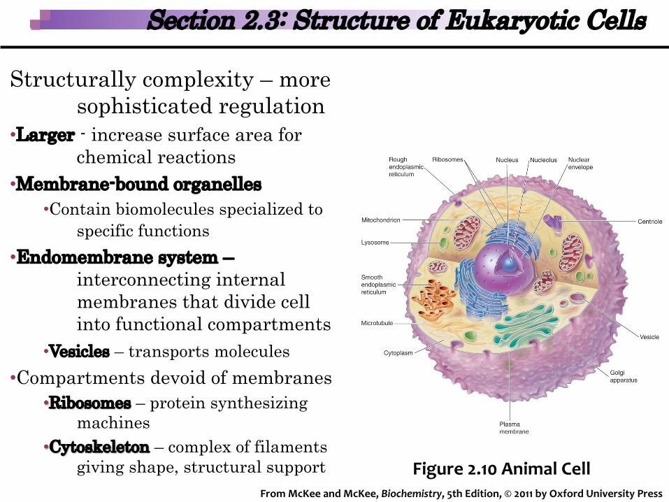
Figure 2.10 Animal Cell
Structurally complexity – more sophisticated regulation
•Larger - increase surface area for chemical reactions
•Membrane-bound organelles•Contain biomolecules specialized to
specific functions•Endomembrane system –
interconnecting internal membranes that divide cell into functional compartments
•Vesicles – transports molecules•Compartments devoid of membranes
•Ribosomes – protein synthesizing machines
•Cytoskeleton – complex of filaments giving shape, structural support
Section 2.3: Structure of Eukaryotic Cells
From McKee and McKee, Biochemistry, 5th Edition, © 2011 by Oxford University Press

§Plasma membrane §Endoplasmic reticulum§Golgi apparatus §Nucleus §Lysosomes§Mitochondria§Ribosomes,§Cytoskeleton§Chloroplasts
§Plant only
Section 2.3: Structure of Eukaryotic Cells
Figure 2.11 Plant Cell
From McKee and McKee, Biochemistry, 5th Edition, © 2011 by Oxford University Press
Figure 2.10 Animal Cell

§Plasma Membrane§Isolates the cell and is selectively permeable§Composed of lipid bilayer with associated integral &
peripheral proteins§Extracellular face contains glycocalyx – proteins & lipids
that contain covalently attached carbohydrate§Extracellular matrix protects exterior
Section 2.3: Structure of Eukaryotic Cells
From McKee and McKee, Biochemistry, 5th Edition, © 2011 by Oxford University Press
§Membrane skeleton – 3-D meshwork of proteins attached to peripheral proteins

Endoplasmic Reticulum§Series of membranous tubules, vesicles,
and flattened sacks§ER lumen -internal space enclosed in
ER membrane§Rough ER – due to ribosomes on
surface§Ribosomes – consist of 2 subunits
(40S/60S); protein synthesis; chaperones facilitate folding process; glycosylation reactions
§Smooth ER – no ribosomes; continuous with RER
§Key functions: lipid biosynthesis; Ca2+
storage
Section 2.3: Structure of Eukaryotic Cells
Figure 2.14
Endoplasmic ReticulumFrom McKee and McKee, Biochemistry, 5th Edition, © 2011 by Oxford University Press

Rough ER§ER stress – accumulation of
misfolded molecules§ER-associated protein
degradation – mechanism of degradation
Smooth ER§Hepatocytes - biotransformation
& synthesis of lipid components of very-low-density lipoproteins
§Biotransformation reactions –convert water insoluble metabolites & xenobiotics into soluble products for excretion
Section 2.3: Structure of Eukaryotic Cells
Figure 2.14
Endoplasmic ReticulumFrom McKee and McKee, Biochemistry, 5th Edition, © 2011 by Oxford University Press

§Golgi Apparatus§Golgi apparatus - large,
flattened, sac-like membranous vesicles§Processes, packages, and
distributes cell products
§Two faces: cis (cisternae) and a trans face
Section 2.3: Structure of Eukaryotic Cells
Figure 2.16 The Golgi ApparatusFrom McKee and McKee, Biochemistry, 5th Edition, © 2011 by Oxford University Press

§Cisternal maturation model vesicles are recycled back to the cis Golgi from the trans Golgi
§Secretory products concentrated at the trans Golgi into secretory vesicles
§Involved in exocytosisü Movement of membrane-bound
vesicles from Golgi apparatus to plasma membrane
Section 2.3: Structure of Eukaryotic Cells
Figure 2.15 Exocytosis
From McKee and McKee, Biochemistry, 5th Edition, © 2011 by Oxford University Press
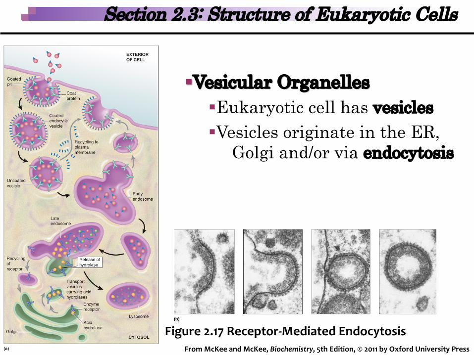
§Vesicular Organelles§Eukaryotic cell has vesicles§Vesicles originate in the ER,
Golgi and/or via endocytosis
Section 2.3: Structure of Eukaryotic Cells
Figure 2.17 Receptor-Mediated EndocytosisFrom McKee and McKee, Biochemistry, 5th Edition, © 2011 by Oxford University Press

Section 2.3: Structure of Eukaryotic Cells
Lysosomes
§Lysosomes are vesicles that contain digestive enzymes
§Enzymes are acid hydrolases§Degrade encapsulated materials§Autophagy degradation of debris
in cells
From McKee and McKee, Biochemistry, 5th Edition, © 2011 by Oxford University Press

Nucleus§Contains the hereditary information§Site of transcription§Nuclear components:
§Nucleoplasm – surrounded by membrane, contains chromatin fibers & DNA
§Chromatin (genome)§Nuclear envelope – barrier; outer/
inner nuclear membrane§Nucleolus – transcription of rRNA
genes§Nuclear matrix – scaffold of proteins
on which chromatin organized
Section 2.3: Structure of Eukaryotic Cells
Figure 2.19 Eukaryotic NucleusFrom McKee and McKee, Biochemistry, 5th Edition, © 2011 by Oxford University Press

§Nuclear envelope surrounds the nucleoplasm
§Nuclear pores (nuclear pore complexes)§Molecules enter and leave
the nucleus
Section 2.3: Structure of Eukaryotic Cells
Figure 2.20 The Nuclear Pore Complex
From McKee and McKee, Biochemistry, 5th Edition, © 2011 by Oxford University Press
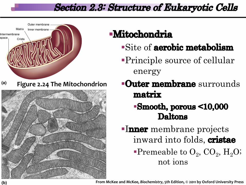
Section 2.3: Structure of Eukaryotic Cells
Figure 2.24 The Mitochondrion
§Mitochondria§Site of aerobic metabolism§Principle source of cellular
energy§Outer membrane surrounds
matrix§Smooth, porous <10,000
Daltons§Inner membrane projects
inward into folds, cristae§Premeable to O2, CO2, H2O;
not ions
From McKee and McKee, Biochemistry, 5th Edition, © 2011 by Oxford University Press

Section 2.3: Structure of Eukaryotic Cells
§Peroxisomes§Small organelle containing oxidative enzymes§Detoxifies peroxides (e.g., H2O2)
From McKee and McKee, Biochemistry, 5th Edition, © 2011 by Oxford University Press

Section 2.3: Structure of Eukaryotic Cells
§Cytoskeleton§Intricate supportive network of fibers, filaments, and associated
proteins§Three main components:
§Microtubules§Microfilaments§Intermediate filaments
§Main functions§Cell shape and structure§Large- and small-scale cell movement
§Cell movement; organelle movement§Solid-state biochemistry
§Enzymes assemble on solid surface, improves efficiency and control
§Signal transduction§Filaments facilitate & support signal transduction processes
From McKee and McKee, Biochemistry, 5th Edition, © 2011 by Oxford University Press

Section 2.3: Structure of Eukaryotic Cells
Figure 2.27 The Cytoskeleton
From McKee and McKee, Biochemistry, 5th Edition, © 2011 by Oxford University Press

Section 2.3: Structure of Eukaryotic Cells
§Cytoskeleton§Cilia and flagella, whip-like appendages encased in
plasma membrane, are highly specialized for their roles in propulsion§Undulating motion occurs via ATP-driven
structural changes in dynein molecules (arms)
From McKee and McKee, Biochemistry, 5th Edition, © 2011 by Oxford University Press
Related Documents