www.wjgnet.com TOPIC HIGHLIGHT Pediatric liver transplantation Marco Spada, Silvia Riva, Giuseppe Maggiore, Davide Cintorino, Bruno Gridelli Online Submissions: wjg.wjgnet.com World J Gastroenterol 2009 February 14; 15(6): 648-674 [email protected] World Journal of Gastroenterology ISSN 1007-9327 doi:10.3748/wjg.15.648 © 2009 The WJG Press and Baishideng. All rights reserved. Salvatore Gruttadauria, MD, Associate Professor, Series Editor Marco Spada, Silvia Riva, Davide Cintorino, Bruno Gridelli, Istituto Mediterraneo per i Trapianti e Terapie ad alta specializzazione-IsMeTT, University of Pittsburgh Medical Center, 90127 Palermo, Italy Giuseppe Maggiore, Department of Reproductive Medicine and Child Development, University of Pisa, 56100 Pisa, Italy Author contributions: Spada M and Gridelli B were the principal authors of the paper, and wrote the following sections: introduction, prioritization, the transplant operation, early post-operative period, managing immunosuppressive therapy, late allograft dysfunction, outcome following transplantation; edited the final manuscript; Riva S and Maggiore G wrote the following sections: indications for liver transplantation, contraindications to liver transplantation, evaluation of the transplant candidate, infections, post-transplant lymphoproliferative disorders; Cintorino D was involved in much of the data acquisition and participated in the writing of the surgical sections of the manuscript; all authors gave their final approval for the paper. Correspondence to: Marco Spada, MD, PhD, Istituto Mediterraneo per i Trapianti e Terapie ad alta specializzazione- IsMeTT, Via E. Tricomi 1, 90127 Palermo, Italy. [email protected]u Telephone: +39-91-2192111 Fax: +39-91-2192400 Received: July 4, 2008 Revised: December 3, 2008 Accepted: December 10, 2008 Published online: February 14, 2009 Abstract In previous decades, pediatric liver transplantation has become a state-of-the-art operation with excellent success and limited mortality. Graft and patient survival have continued to improve as a result of improvements in medical, surgical and anesthetic management, organ availability, immunosuppression, and identification and treatment of postoperative complications. The utilization of split-liver grafts and living-related donors has provided more organs for pediatric patients. Newer immunosuppression regimens, including induction therapy, have had a significant impact on graft and patient survival. Future developments of pediatric liver transplantation will deal with long-term follow- up, with prevention of immunosuppression-related complications and promotion of as normal growth as possible. This review describes the state-of-the-art in pediatric liver transplantation. © 2009 The WJG Press and Baishideng. All rights reserved. Key words: Pediatric liver transplantation; Indications; Surgical techniques; Complications Peer reviewer: Michael Torbenson, MD, Associate Professor of Pathology, Room B314 1503 E Jefferson (Bond Street Build- ing), The Johns Hopkins University School of Medicine, Balti- more, MD 21231, United States Spada M, Riva S, Maggiore G, Cintorino D, Gridelli B. Pediatric liver transplantation. World J Gastroenterol 2009; 15(6): 648-674 Available from: URL: http://www.wjgnet. com/1007-9327/15/648.asp DOI: http://dx.doi.org/10.3748/ wjg.15.648 INTRODUCTION Liver transplantation has been very successful in treating children with end-stage liver disease, and offers the opportunity for a long healthy life. Organ scarcity, which is the main limitation to the full exploitation of transplantation, is being overcome thanks to innovative surgical techniques, and all children in need, even the youngest, today have the chance of being transplanted, with almost no waiting list mortality. Split-liver and living-donor transplantation have contributed to reversing a situation in which, during the 1980s and 90s, children had greater waiting list mortality compared to that of adult patients. Several years ago, the main focus of care of children with end-stage liver disease was to find a liver transplant, but today, the main interest is in long-term follow-up, with prevention of immunosuppression-related complications and promotion of as normal growth as possible. The history of pediatric liver transplantation has clearly shown that success is dependent on strict and integrated collaboration between referring pediatricians, pediatric transplant hepatologists, transplant surgeons, nurses, transplant coordinators, psychologists and social workers. Everybody involved has the task of bringing a cure to a population of pediatric patients who present some of the most challenging clinical problems in modern medicine. INDICATIONS FOR LIVER TRANSPLANTATION The main indications for liver transplantation in the pediatric population are as follows: (1) Extra- hepatic cholestasis: biliary atresia. (2) Intra-hepatic

Liver Transplant
Nov 01, 2014
Transplant in children
Welcome message from author
This document is posted to help you gain knowledge. Please leave a comment to let me know what you think about it! Share it to your friends and learn new things together.
Transcript
www.wjgnet.com
TOPIC HIGHLIGHT
Pediatric liver transplantation
Marco Spada, Silvia Riva, Giuseppe Maggiore, Davide Cintorino, Bruno Gridelli
Online Submissions: wjg.wjgnet.com World J Gastroenterol 2009 February 14; 15(6): [email protected] World Journal of Gastroenterology ISSN 1007-9327doi:10.3748/wjg.15.648 © 2009 The WJG Press and Baishideng. All rights reserved.
Salvatore Gruttadauria, MD, Associate Professor, Series Editor
Marco Spada, Silvia Riva, Davide Cintorino, Bruno Gridelli, Istituto Mediterraneo per i Trapianti e Terapie ad alta specializzazione-IsMeTT, University of Pittsburgh Medical Center, 90127 Palermo, ItalyGiuseppe Maggiore, Department of Reproductive Medicine and Child Development, University of Pisa, 56100 Pisa, ItalyAuthor contributions: Spada M and Gridelli B were the principal authors of the paper, and wrote the following sections: introduction, prioritization, the transplant operation, early post-operative period, managing immunosuppressive therapy, late allograft dysfunction, outcome following transplantation; edited the final manuscript; Riva S and Maggiore G wrote the following sections: indications for liver transplantation, contraindications to l iver transplantation, evaluation of the transplant candidate, infections, post-transplant lymphoproliferative disorders; Cintorino D was involved in much of the data acquisition and participated in the writing of the surgical sections of the manuscript; all authors gave their final approval for the paper.Correspondence to: Marco Spada, MD, PhD, Istituto Mediterraneo per i Trapianti e Terapie ad alta specializzazione-IsMeTT, Via E. Tricomi 1, 90127 Palermo, Italy. [email protected]: +39-91-2192111 Fax: +39-91-2192400Received: July 4, 2008 Revised: December 3, 2008Accepted: December 10, 2008Published online: February 14, 2009
AbstractIn previous decades, pediatric liver transplantation has become a state-of-the-art operation with excellent success and limited mortality. Graft and patient survival have continued to improve as a result of improvements in medical, surgical and anesthetic management, organ availability, immunosuppression, and identification and treatment of postoperative complications. The utilization of split-liver grafts and living-related donors has provided more organs for pediatric patients. Newer immunosuppression regimens, including induction therapy, have had a significant impact on graft and patient survival. Future developments of pediatric liver transplantation will deal with long-term follow-up, with prevention of immunosuppression-related complications and promotion of as normal growth as possible. This review describes the state-of-the-art in pediatric liver transplantation.
© 2009 The WJG Press and Baishideng. All rights reserved.
Key words: Pediatric liver transplantation; Indications;
Surgical techniques; Complications
Peer reviewer: Michael Torbenson, MD, Associate Professor of Pathology, Room B314 1503 E Jefferson (Bond Street Build-ing), The Johns Hopkins University School of Medicine, Balti-more, MD 21231, United States
Spada M, Riva S, Maggiore G, Cintorino D, Gridelli B. Pediatric liver transplantation. World J Gastroenterol 2009; 15(6): 648-674 Available from: URL: http://www.wjgnet.com/1007-9327/15/648.asp DOI: http://dx.doi.org/10.3748/wjg.15.648
INTRODUCTIONLiver transplantation has been very successful in treating children with end-stage liver disease, and offers the opportunity for a long healthy life. Organ scarcity, which is the main limitation to the full exploitation of transplantation, is being overcome thanks to innovative surgical techniques, and all children in need, even the youngest, today have the chance of being transplanted, with almost no waiting list mortality. Split-liver and living-donor transplantation have contributed to reversing a situation in which, during the 1980s and 90s, children had greater waiting list mortality compared to that of adult patients.
Several years ago, the main focus of care of children with end-stage liver disease was to find a liver transplant, but today, the main interest is in long-term follow-up, with prevention of immunosuppression-related complications and promotion of as normal growth as possible. The history of pediatric liver transplantation has clearly shown that success is dependent on strict and integrated collaboration between referring pediatricians, pediatric transplant hepatologists, transplant surgeons, nurses, transplant coordinators, psychologists and social workers. Everybody involved has the task of bringing a cure to a population of pediatric patients who present some of the most challenging clinical problems in modern medicine.
INDICATIONS FOR LIVER TRANSPLANTATIONThe main indications for liver transplantation in the pediatric population are as follows: (1) Extra-hepatic cholestasis: biliary atresia. (2) Intra-hepatic

Spada M et al . Liver tansplantation 649
www.wjgnet.com
cholestasis: sclerosing cholangitis; Alagille’s syndrome; non-syndromic paucity of intrahepatic bile ducts; and progressive familial intrahepatic cholestasis. (3) Metabolic diseases: Wilson’s disease; α1-antitrypsin deficiency; Crigler-Najjar syndrome; inborn error of bile acid metabolism; tyrosinemia; disorders of the urea cycle; organic acidemia; acid lipase defect; oxaluria type Ⅰ; and disorders of carbohydrate metabolism. (4) Acute liver failure. (5) Others: primary liver tumor and cystic fibrosis.
Cholestatic liver diseasesTypically, the child referred to a liver transplant center is a small baby with cholestatic liver disease. Out of 1187 children transplanted in North America between 1995 and May 2002, 33.5% were ≤ 12 mo old at the time of transplantation, 55.6% had cholestatic disease, and 41.6% had biliary atresia. Of the children transplanted at < 1 year of age, 65.6% had biliary atresia[1]. Most of these children have undergone a Kasai procedure that failed to re-establish effective biliary flow, which caused rapid evolution to secondary biliary cirrhosis. When intrahepatic cholestatic diseases (Alagille’s syndrome, progressive familial intrahepatic cholestasis, and sclerosing cholangitis) or sclerosing cholangitis are diagnosed, liver transplantation is indicated to eliminate severely debilitating symptoms, such as pruritus. Children affected by these diseases are also at high risk for the development of liver cancer[2].
Metabolic diseasesMetabolic diseases are the second most common indication for liver transplantation[3]. Metabolic diseases can be divided in two groups on the basis of the presence or absence of structural damage of the liver. To the first group belong α1-antitripsin deficiency, tyrosinemia and Wilson’s disease, which have the potential to progress to end-stage liver failure, liver cancer (Figure 1) and acute liver failure, while diseases such as Crigler-Najjar syndrome type Ⅰ and ornithine transcarbamylase (OTC) deficiency belong to the second group. In primary hyperoxaluria type Ⅰ, liver and kidney transplantation is considered when irreversible kidney damage from oxalic acid accumulation has developed. Different transplantation timings have been tested, combined liver and kidney transplantation (simultaneous or sequential) and pre-emptive liver transplantation (before end-stage renal failure occurs)[4,5]. Liver transplantation has been suggested recently for the treatment of organic acidemia (propionic aciduria, methylmalonic aciduria). In patients affected by these diseases, liver transplantation does not correct the enzyme deficiency in other organs beside the liver. Although quality of life is generally improved, patients remain at risk of severe extrahepatic disease complications[6-8]. Liver cirrhosis with severe portal hypertension develops in an about 25% of the patients affected by cystic fibrosis. Liver transplantation should be considered before the development of end-stage liver failure and when pulmonary function is still preserved (FEV1 > 50%).
Acute liver failureAcute liver failure is a rare event in children; recovery without transplantation occurs in 15%-20% of patients with severe hepatic encephalopathy. A prospective study from the Pediatric Acute Liver Failure Study Group
A
B
C
D
Figure 1 Adolescent affected by tyrosinemia who developed hepatocel-lular carcinoma, despite 2-(2-nitro-4-fluoromethylbenzoyl)-1,3-cyclohex-anedione therapy. A: Magnetic resonance imaging displays a 26-mm lesion. B: After liver transplantation, the resected liver showed multiple nodules in the left lobe. C: Histological sections from the nodule revealed hepatocellular carci-noma. D: Microvascular invasion.
www.wjgnet.com
has indicated that in 49% of patients (54% of children aged 1 year), the cause of acute liver failure cannot be determined and that total bilirubin ≥ 5 mg/dL, international normalized ratio (INR) ≥ 2.55 and hepatic encephalopathy are risk factors predictive of death or liver transplantation[9]. In a large retrospective United Network for Organ Sharing (UNOS) data analysis, it has been shown that 5-year patient and graft survivals of children with acute liver failure are significantly lower than the survival of children transplanted for biliary atresia (73% and 59% vs 89% and 78%, respectively)[10].
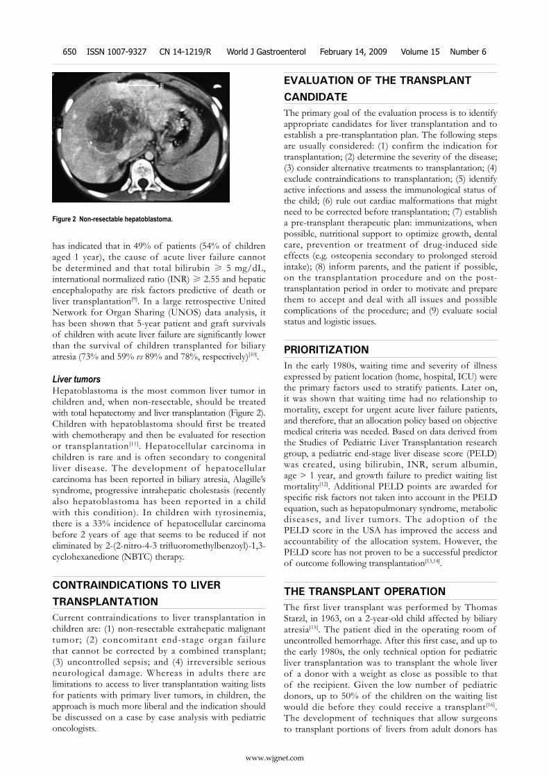
Liver tumorsHepatoblastoma is the most common liver tumor in children and, when non-resectable, should be treated with total hepatectomy and liver transplantation (Figure 2). Children with hepatoblastoma should first be treated with chemotherapy and then be evaluated for resection or transplantation[11]. Hepatocellular carcinoma in children is rare and is often secondary to congenital liver disease. The development of hepatocellular carcinoma has been reported in biliary atresia, Alagille’s syndrome, progressive intrahepatic cholestasis (recently also hepatoblastoma has been reported in a child with this condition). In children with tyrosinemia, there is a 33% incidence of hepatocellular carcinoma before 2 years of age that seems to be reduced if not eliminated by 2-(2-nitro-4-3 trifluoromethylbenzoyl)-1,3-cyclohexanedione (NBTC) therapy.
CONTRAINDICATIONS TO LIVER TRANSPLANTATIONCurrent contraindications to liver transplantation in children are: (1) non-resectable extrahepatic malignant tumor; (2) concomitant end-stage organ failure that cannot be corrected by a combined transplant; (3) uncontrolled sepsis; and (4) irreversible serious neurological damage. Whereas in adults there are limitations to access to liver transplantation waiting lists for patients with primary liver tumors, in children, the approach is much more liberal and the indication should be discussed on a case by case analysis with pediatric oncologists.
EVALUATION OF THE TRANSPLANT CANDIDATEThe primary goal of the evaluation process is to identify appropriate candidates for liver transplantation and to establish a pre-transplantation plan. The following steps are usually considered: (1) confirm the indication for transplantation; (2) determine the severity of the disease; (3) consider alternative treatments to transplantation; (4) exclude contraindications to transplantation; (5) identify active infections and assess the immunological status of the child; (6) rule out cardiac malformations that might need to be corrected before transplantation; (7) establish a pre-transplant therapeutic plan: immunizations, when possible, nutritional support to optimize growth, dental care, prevention or treatment of drug-induced side effects (e.g. osteopenia secondary to prolonged steroid intake); (8) inform parents, and the patient if possible, on the transplantation procedure and on the post-transplantation period in order to motivate and prepare them to accept and deal with all issues and possible complications of the procedure; and (9) evaluate social status and logistic issues.
PRIORITIZATIONIn the early 1980s, waiting time and severity of illness expressed by patient location (home, hospital, ICU) were the primary factors used to stratify patients. Later on, it was shown that waiting time had no relationship to mortality, except for urgent acute liver failure patients, and therefore, that an allocation policy based on objective medical criteria was needed. Based on data derived from the Studies of Pediatric Liver Transplantation research group, a pediatric end-stage liver disease score (PELD) was created, using bilirubin, INR, serum albumin, age > 1 year, and growth failure to predict waiting list mortality[12]. Additional PELD points are awarded for specific risk factors not taken into account in the PELD equation, such as hepatopulmonary syndrome, metabolic diseases, and liver tumors. The adoption of the PELD score in the USA has improved the access and accountability of the allocation system. However, the PELD score has not proven to be a successful predictor of outcome following transplantation[13,14].
THE TRANSPLANT OPERATIONThe first liver transplant was performed by Thomas Starzl, in 1963, on a 2-year-old child affected by biliary atresia[15]. The patient died in the operating room of uncontrolled hemorrhage. After this first case, and up to the early 1980s, the only technical option for pediatric liver transplantation was to transplant the whole liver of a donor with a weight as close as possible to that of the recipient. Given the low number of pediatric donors, up to 50% of the children on the waiting list would die before they could receive a transplant[16]. The development of techniques that allow surgeons to transplant portions of livers from adult donors has
Figure 2 Non-resectable hepatoblastoma.
650 ISSN 1007-9327 CN 14-1219/R World J Gastroenterol February 14, 2009 Volume 15 Number 6

www.wjgnet.com
completely changed the fate of liver transplantation in pediatric patients.
Whole-liver transplantationThe procedure of whole-liver procurement in pediatric donors can be performed exactly as in adults, applying a technique that is a combination of the initial procurement technique described by Starzl et al[17], and the most recently described rapid flush technique[18,19]. Whole-liver pediatric transplantation can be performed with two different techniques: the classic technique with inferior vena cava replacement, and the piggyback technique[20] with preservation of the native inferior vena cava. The present authors routinely use the classic technique in the vast majority of whole liver transplants. Veno-venous bypass is generally not used in pediatric liver transplantation, given that patients generally tolerate explantation well, provided that volume replacement has been adequate. Adopted techniques are almost identical to the ones used in adults recipients. In cases in which the liver is encased in adhesions, as it is in biliary atresia, we recommend that surgeons first approach the hepatic hilum from the right posterolateral aspect, identifying the Roux-en-Y jejunal limb, which is transected with a linear stapler or between ligatures. This allows better exposure and dissection of the hilum. If the portal vein is small and sclerotic, it has to be dissected proximally to the confluence of the splenic and superior mesenteric vein, dividing the coronary vein of the stomach. The portal vein anastomosis will then be performed by means of a donor interposition vein graft. In difficult dissections, the vena cava can be clamped above and below the liver before completing the mobilization of the liver itself.
Several methods of arterial reconstruction have been proposed. It is our preference to anastomose the small arterial vessels encountered in pediatric whole liver transplantation in an end-to-end manner by using the magnification loops (3.5 ×) and interrupted or running 8-0 polypropylene sutures. We generally do not use the branch patch technique, and in the case of aberrant arterial anatomy, the supraceliac aorta is the inflow vessel of choice. The use of arterial conduits anastomosed to the infrarenal aorta is avoided if possible.
In theory, biliary tract continuity can be restored through direct anastomosis between the new liver’s hepat ic duct and the rec ip ient ’s common bi le duct. However, the most common type of biliary reconstruction adopted in pediatric patients is hepaticojejunostomy. In biliary atresia patients, the reconstruction uses the previous Roux-en-Y limb of the hepatic portoenterostomy, if suitable; otherwise a 40-cm Roux-en-Y jejunal limb is created. The authors’ attitude is not to use a T tube, because no randomized trial so far has demonstrated any advantages in using it, and there are often biliary leaks when the T tube is pulled.
Occasionally in children, abdominal-wall closure may be impossible because of the large size of the transplanted liver. This may be remedied by creating a silo on the abdominal wall such that a temporary closure can be made[21].
Reduced-size liver transplantationThis procedure was first described by Bismuth et al[22] and consists in the procurement of the whole liver from an adult cadaver donor, which is reduced in its size on the back-table. In the original description, a right hepatectomy was performed on the back-table: the right lobe of the liver was discharged, while the left lobe (Couinaud liver segments 1 to 4), including the vena cava, was transplanted in a child. This technique of parenchymal reduction, very seldom used today, allows surgeons to overcome differences in size between the donor and the recipient of up to four or five times[23,24].
Following these first experiences, a more aggressive reduction that allows transplanting the liver from donors with a body weight up to 12 times the recipient’s was introduced. On the back-table, the graft undergoes an extended right hepatectomy, including segment 4 and the caudate lobe. The resulting left lateral segment graft comprises segments 2 and 3, without the vena cava. The graft is transplanted retaining the recipient’s vena cava, anastomosing the graft left hepatic vein to the recipient’s vena cava.
Reduced-size liver transplantation shows outcomes in line, if not superior, to whole-liver transplantation, and has become an essential part of the technical expertise of pediatric transplant centers[25-30] (Table 1). The development of this technique has led to almost total elimination of child mortality on the waiting list, through the utilization of an adult liver cadaver donor. Its main limitation is that it withdraws organs from the larger adult recipient pool. For this reason, after the development of living-related and split-liver transplantation, reduced-size live transplantation is used increasingly less, and should not be considered an option anymore for pediatric liver transplantation.
Living-related liver transplantation The first description of the procedure in which segments 2 and 3 were procured from a living donor (the mother), and transplanted in a child affected by biliary duct atresia, dates back to 1988[31,32]. Living-related liver transplants soon came to account for a substantial number of pediatric cases performed in many centers throughout the world, and the only possibility for liver transplants in countries where cadaveric organ procurement was not allowed until just a few years ago[33].
Living-donor procurement involves performing a left lobectomy during which segments 2 and 3 are separated from the remaining liver, and dissecting the parenchyma along a section running to the right of the round ligament. After the parenchyma dissection, the left branch of the portal vein, the hepatic artery, and the left suprahepatic vein are quickly clamped and dissected, and the left lobe perfused on the back-table. The recipient’s procedure is similar to the one described for the transplant of segments 2 and 3 from a cadaver donor, except for the fact that the arterial anastomosis can be performed only in the left branch of the hepatic artery. The branch is small and usually anastomosed directly to the recipient’s hepatic artery using the operative
Spada M et al . Liver tansplantation 651

www.wjgnet.com
microscope.Living-related liver transplantation has been widely
debated with regard to the ethics of performing major surgery on a healthy person. The validity of this procedure is broadly recognized, and over 1200 cases have been performed worldwide, with a donor mortality and morbidity of approximately 0.2% and 10%, respectively. Morbidity relates mainly to biliary fistulas, incisional hernias, and bleeding. In the majority of cases, living-related transplants register an excellent outcome for pediatric recipients, thanks to the possibility of performing the transplant before the child’s clinical condition deteriorates. Centers with most experience in this area report survival rates between 80 and 90% after 1 year[34-39] (Table 2).
Split-liver transplantationSplit-liver transplantation, as described originally by Pichlmayr, involves procuring a whole liver from a cadaver donor and dividing it into two sections along the round ligament, leaving the vascular structures for the two portions of hepatic parenchyma intact[40]. In this way, two partial organs are obtained from a single liver: the left lateral segment (segments 2 and 3), which can be transplanted in a child, and the extended right liver (segments 1 and 4-8), which can be transplanted into an adult. This procedure involves a much longer ischemia time, which, at the beginning of its adoption, led to unsatisfactory results, with a high incidence of primary dysfunction and technical complications[41-55] (Table 3). In 1994, Rogiers described a technical variation in the split-liver technique, derived from the living-related transplant experience that consisted in dividing the liver in situ
during the procurement procedure[56]. The technique has shown outcomes comparable to those obtained with conventional techniques[57-62] (Table 4).
The donor operationA section of the liver is made along the falciform ligament to obtain a left graft, composed of segments 2 and 3, including the left hepatic vein, the left branch of the portal vein, and the left branch of the hepatic artery, along with the common hepatic artery and the celiac tripod, and a right graft, composed of segments 1 and 4 to 8, including the vena cava, the right branch of the hepatic artery, and the portal vein along with the origin of the mesenteric and splenic veins (Figure 3).
At the beginning of the split procedure, the hepatogastric ligament is inspected to detect an accessory left hepatic artery originating from the left gastric artery, which must be preserved. When this vessel is not detected, the ligament is sectioned. The common hepatic artery is then identified and dissected from the gastroduodenal artery up to its division into the right and left hepatic arteries. The left hepatic artery is then encircled (Figure 4A). If present, branches for the fourth segment originating from the left hepatic artery should be identified and divided. The base of the round ligament is exposed by dividing the small bridge of parenchyma that connects the lower portion of segment 4 to the left lateral section of the liver. The round ligament is dissected and completely mobilized with isolation and division of its venous connections to the fourth segment. Once the round ligament is dissected, the extrahepatic portion of the left branch of the portal vein can be identified just below the left hepatic artery.
Series Period n Survival (%) ReTX (%) Complications (%)
Patient Organ HAT PVT BC PNF
Broelsch et al[25] 1984-1987 9 44 33 11 0 0 11 11Otte et al[26] 1984-1988 42 68 54 28 7 0 NA 5Bismuth et al[22] 1984-1988 14 50 44 14 7 7 14 7Houssin et al[27] 1986-1989 40 75 73 - 5 5 5 5Kalayoglu et al[28] 1988-1989 12 83 67 25 16 8 0 0Esquivel et al[29] 1988-1990 20 81 75 12 0 3 5 0Langnas et al[30] 1988-1991 29 68 65 3 7 0 20 10
Table 1 Series of pediatric reduced-size liver transplantation
ReTX: Retransplantation; HAT: Hepatic artery thrombosis; PVT: Portal vein thrombosis; BC: Biliary complication; PNF: Primary non-function; NA: Not available.
Series Period n Survival (%) ReTX (%) Complications (%)
Patient Organ HAT PVT BC PNF
Tanaka et al[33] 1990-1992 37 E 90 U 57 E 90 U 57 0 U 14 E 3 E 10 0Emond et al[34] 1991-1992 18 94 84 16 11 6 16 0Broelsch et al[35] 1991 20 85 75 20 25 20 35 0Malagò et al[36] 1991-1994 36 72 72 8 2.8 3 25 -Otte et al[37] 1993-1995 30 97 93 - 20Haberal et al[38] 1990-1997 19 58 58 0 5 0 0 0Darwish et al[39] 1993-2002 100 94 92 3 1 14 27 0
Table 2 Series of pediatric living-related liver transplantation
E: Elective cases; U: Urgent cases; ReTX: Retransplantation; HAT: Hepatic artery thrombosis; PVT: Portal vein thrombosis; BC: Biliary complication; PNF: Primary non-function.
652 ISSN 1007-9327 CN 14-1219/R World J Gastroenterol February 14, 2009 Volume 15 Number 6

www.wjgnet.com
This vein must be carefully dissected and encircled (Figure 4B). The left lateral section is rotated laterally on
the right side and the ligamentum venosum is dissected up to left lateral hepatic vein, which can be isolated and encircled (Figure 4C). The bile ducts of the left lateral segment are included in the porta hepatis and should not be dissected. On the contrary, the porta hepatis must be encircled and divided sharply (Figure 4D).
The section of the parenchyma can now be performed along the falciform ligament (Figure 4E). It is helpful when identifying the plane of the dissection to pass the cotton tape, which encircles the left hepatic vein on the posterior surface of the liver in the fossa of the ductus venosus, laterally to the left branch of the hepatic artery and of the portal vein (Figure 4F and G). Pulling up on this tape, the dissection of the parenchyma is usually easy. At this point, the procedure continues as a standard donor operation with heparinization, cannulation and cross-clamping of the aorta, perfusion, and cooling of the abdominal cavity. The left hepatic vein is then sectioned close to the vena cava. Care must be taken to identify a distal bifurcation of this vein. A double left hepatic vein significantly increases the technical difficulty of the implantation of the graft. In this case, the vessel should be removed with a cuff of vena cava to allow a single vascular anastomosis with
Series Year ADU (n) PED (n) Urgent (%) Patient survival (%) Graft survival (%) Complications (%)
ADU PED ADU PED HAT PVT BC PNF
Pichlmayr et al[40] 1989 2 0 0 50 - 50 - 0 0 0 0Bismuth et al[41] 1989 2 0 100 0 - 0 - 0 0 0 0Otte et al[42] 1990 1 3 75 0 66 0 66 0 0 0 0Emond et al[16] 1990 5 13 38 40 63 40 53 6 6 27 24Broelsch et al[24] 1990 4 21 40 25 66 20 48 NA NA 27 NALangnas et al[30] 1992 1 9 73 NA NA NA NA 7 0 20 17Houssin et al[43] 1993 6 10 50 83 70 83 60 13 25 25 0Otte et al[44] 1994 11 18 27 NA NA NA NA 10 0 17 10Kalayoglu et al[45] 1996 5 7 8 100 85 80 71 8 0 17 0Rogiers et al[46] 1996 5 7 44 57 100 42 100 15 0 15 0Azoulay et al[47] 1996 26 1 14 80 100 76 100 15 0 22 4Dunn et al[48] 1997 0 12 50 75 66 0 0 0 0Rela et al[49] 1998 15 26 12 93 89 93 84 3 0 15 0Mirza et al[50] 1998 10 14 58 80 78 NA NA 8 0 8 16Chardot et al[51] 1999 0 15 31 - 66 - 62 12 19 25 0Reyes et al[52] 2000 13 12 66 69 66 61 50 12 0 8 NADeshpande et al[53] 2002 0 80 20 - 89 - 86 5 1 9 0Noujaim et al[54] 2003 24 36 25 NA NA NA NA 3 0 20 3Oswari et al[55] 2005 0 30 13 - 70 - 67 2 5 7 NA
Table 3 Series of ex situ split-liver transplantation
ADU: Adults; PED: Children.
Series Year ADU (n) PED (n) Urgent (%) Patient survival (%) Graft survival (%) Complications (%)
ADU PED ADU PED HAT PVT BC PNF
Rogiers et al[56] 1996 7 7 35 100 85 85 71 0 0 0 0Goss et al[57] 1997 14 12 58 85 100 78 91 0 0 14 11Busuttil et al[58] 1999 NA NA 66 85 96 86 75 3 1 3 8Ghobrial et al[59] 2000 51 51 49 83 78 NA NA 2 2 NA 8Reyes et al[52] 2000 NA NA NA 93 100 79 83 3 0 3 7Spada et al[60] 2000 36 35 25 84 85 79 76 5 10 28 2Gridelli et al[61] 2003 0 90 28 - 90 - 80 7 6 33 1Yersiz et al[62] 2003 57 104 - 78 75 69 64 13 11 19 26
Table 4 Series of in situ split-liver transplantation
75%-80%
20%-25%
Figure 3 Spit liver allows for the procurement of two separate grafts of different size. A section of the liver is made along the falciform ligament and divides the left lateral segment from the extended right liver. The left graft, composed of segments 2 and 3, and representing 20%-25% of the total liver volume, includes the left hepatic vein, the left branch of the portal vein, and the left branch of the hepatic artery, along with the common hepatic artery and the celiac tripod. The right graft composed of segments 1 and 4-8, and representing 75%-80% of the total liver volume, includes the vena cava, the right branch of the hepatic artery, and the portal vein.
Spada M et al . Liver tansplantation 653

www.wjgnet.com
the recipient vena cava. The left branch of the portal vein is sectioned close to the parenchyma. The right hepatic artery is sectioned close to its origin, and the hepatic artery is dissected up to the celiac trunk, which is removed along with an aortic cuff.
The recipient operationRecipient hepatectomy is performed, as previously described for whole-liver transplantation, with the piggy-back technique[63]. Implantation of the left lateral segment is substantially different from a whole-sized graft. Assuring an adequate venous outflow requires a careful technique of anastomosis between the left hepatic vein of the graft and the inferior vena cava of the recipient and a proper positioning of the graft itself, which is rotated clockwise 45° on a transversal plane and slightly on a frontal plane. The final position of the cut surface of the parenchyma, including the new hilum of the graft, is high and posterior, so that the portal vein and hepatic artery have a course that is curved and longer than usual.
The outflow anastomosis is end-to-side between
the left hepatic vein of the graft and the inferior vena cava of the recipient, with the triangulation technique described by Emond et al[64]. The bridge between the ostia of the right and middle hepatic veins is cut to obtain a single opening. The ostium of the left hepatic vein may be treated in the same fashion, to obtain a further enlargement of the opening, or suture-closed. The opening is then enlarged by cutting the anterior face of the vena cava to obtain a wide reversed triangular orifice. The cuff of the left hepatic vein of the graft is trimmed as short as possible, to avoid kinking. Three 5/0 vascular monofilament sutures are placed, taking the three corners of the graft and recipient orifices (Figure 5). The graft is then placed in the hepatic fossa of the recipient and the triangular anastomosis performed with three running sutures.
The second anastomosis is the portal one, performed in an end-to-end fashion with running sutures of 6/0 or 7/0 vascular monofilament. Both the length and the section of the vessels are crucial. As already mentioned, the length should be sufficient for the vessel to make a gentle curve that reaches the hilum of the graft; as for
A B C
D E F
G H
Figure 4 Main phases of split liver procurement. A: Dissection of the hepatogastric ligament and encircling of the left hepatic artery; B: Identification and encircl-ing of the extrahepatic portion of the left branch of the portal vein; C: Isolation and encircling of the left hepatic vein; D: Division with a scalpel of the porta hepatis containing the bile duct(s) of the left lateral segment; E: Section of the parenchyma started along the falciform ligament; F: Identification of the plane of parenchymal dissection by passing the cotton tape, which encircled the left hepatic vein, on the posterior surface of the liver in the fossa of the ductus venosus; G: Laterally to the left branch of the hepatic artery and of the portal vein; H: The two partial grafts at the end of the procedure.
654 ISSN 1007-9327 CN 14-1219/R World J Gastroenterol February 14, 2009 Volume 15 Number 6

www.wjgnet.com
the section, the limiting factor is the size of the graft cuff. In the majority of cases, the recipient’s vessel matches this size rather well. If not, it can be cut at its bifurcation, to obtain a branch patch. In case of real hypoplasia of the recipient’s portal vein, the confluence of the mesenteric and splenic vein can be clamped, the vessel sectioned at this level and a venous graft from the donor (usually the splenic or the external iliac vein) interposed between the confluence and the portal vein of the new liver. After completion of the anastomosis the graft is reperfused.
The arterial anastomosis comes next. The arterial axis of the graft usually includes the proper and common hepatic artery, in continuity with the celiac artery, and a patch of the aorta. The level of the anastomosis is chosen at any place along the recipient’s arterial axis, and
the two vessels are trimmed to obtain a similar section and an adequate length, according to what has already been stated concerning the portal vein. The anastomosis is performed end-to-end with a running suture of 7/0 or 8/0 vascular monofilament. If the recipient’s arterial axis is deemed inadequate, the aorta can be clamped at the origin of the celiac artery or just below the renal arteries, and an end-to-side anastomosis can be performed at one of these sites. In the latter case, the interposition of an arterial graft from the donor, usually represented by an iliac artery, may be necessary.
The final stage is biliary reconstruction, which is always a hepaticojejunostomy with a Roux-en-Y loop. The bile duct of the graft may be single or double, although in the latter case two different anastomoses are performed (Figure 6).
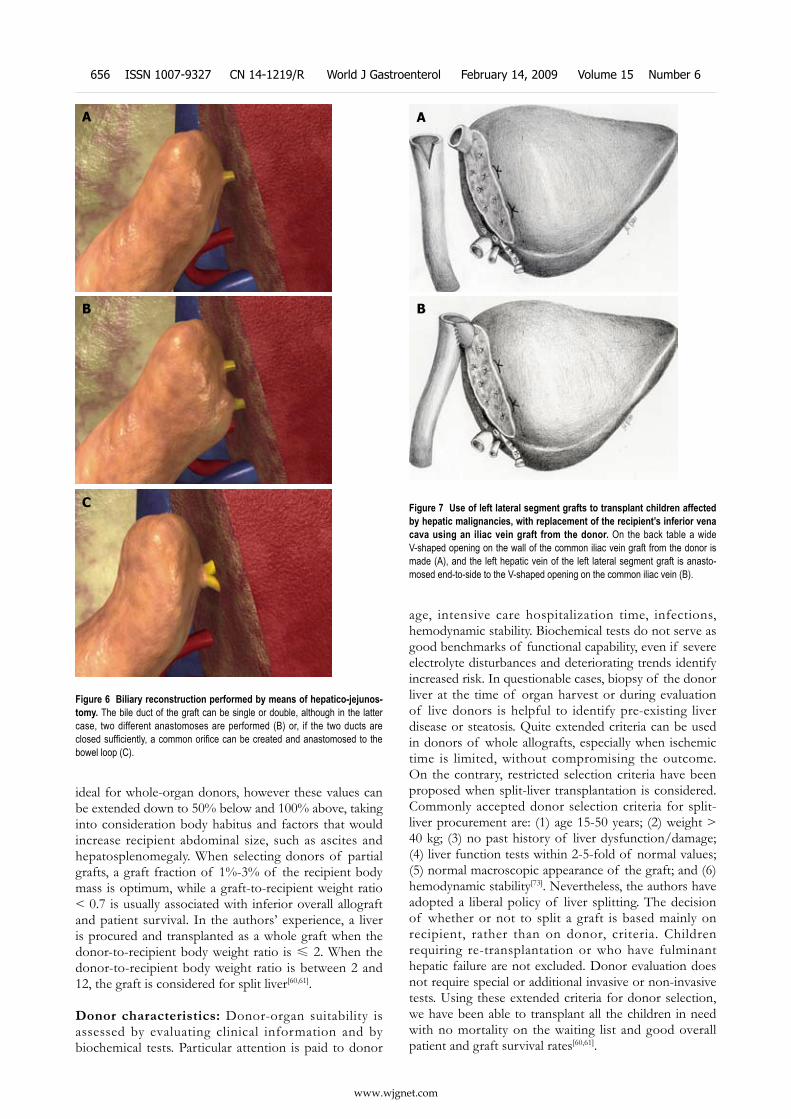
Chi ldhood hepat ic mal ignancies have been considered a contraindication to the use of split-liver transplantation, since the need for the retention of the recipient’s inferior vena cava potentially precludes obtaining a tumor-free margin[65]. A technical variation, which has allowed us and others to successfully use left lateral segment grafts to transplant children affected by hepatic malignancies, involves the replacement of the recipient’s inferior vena cava using an iliac vein graft from the donor[66]. On the back-table, a wide V-shaped opening on the wall of the common iliac vein graft from the donor is made. The left hepatic vein of the left lateral segment graft is anastomosed end-to-side to the V-shaped opening on the common iliac vein with two 5/0 polypropylene running sutures (Figure 7). On the recipient, a total hepatectomy is usually performed using the standard technique of removing the liver together with the retrohepatic vena cava. At this point, the left lateral segment graft with the iliac vein graft is anastomosed to the suprahepatic vena cava in an end-to-end fashion with a 4/0 polypropylene running suture. The inferior edge of the iliac graft is then anastomosed to the infrahepatic vena cava with a 5/0 polypropylene running suture.
Donor selection The following factors must be considered when a donor is evaluated for a specific patient.
Dimensional matching: The selection of a graft with an adequate parenchymal mass is critical to success. The minimal hepatic mass necessary for recovery is not clearly established, and its calculation must take into account the temporary loss of hepatocytes caused by the donor’s injury or treatment, as well as preservation injury, acute rejection, or technical problems. Several formulas have been proposed to estimate adult and pediatric normal liver volume[67-72].
Considering that preservation injury is greater in organs from deceased donors, the hepatic mass of a graft procured from a cadaver donor should be greater than the calculated mass necessary using a living-donor liver segment. In the authors’ experience, a donor weight range 20%-30% above or below that of the recipient is
A
B
C
Figure 5 Anastomosis between the left hepatic vein of the graft and the inferior vena cava of the recipient, performed with the triangulation tech-nique. A: The bridge between the ostia of the right, middle, and left hepatic veins is cut to obtain a single opening; B: The opening is further enlarged by cutting the anterior face of the vena cava to obtain a wide triangular orifice; C: Three 5/0 vascular monofilament sutures are placed, taking the three corners of the graft and recipient orifices.
Spada M et al . Liver tansplantation 655
www.wjgnet.com
ideal for whole-organ donors, however these values can be extended down to 50% below and 100% above, taking into consideration body habitus and factors that would increase recipient abdominal size, such as ascites and hepatosplenomegaly. When selecting donors of partial grafts, a graft fraction of 1%-3% of the recipient body mass is optimum, while a graft-to-recipient weight ratio < 0.7 is usually associated with inferior overall allograft and patient survival. In the authors’ experience, a liver is procured and transplanted as a whole graft when the donor-to-recipient body weight ratio is ≤ 2. When the donor-to-recipient body weight ratio is between 2 and 12, the graft is considered for split liver[60,61].
Donor characteristics: Donor-organ suitability is assessed by evaluating clinical information and by biochemical tests. Particular attention is paid to donor
age, intensive care hospitalization time, infections, hemodynamic stability. Biochemical tests do not serve as good benchmarks of functional capability, even if severe electrolyte disturbances and deteriorating trends identify increased risk. In questionable cases, biopsy of the donor liver at the time of organ harvest or during evaluation of live donors is helpful to identify pre-existing liver disease or steatosis. Quite extended criteria can be used in donors of whole allografts, especially when ischemic time is limited, without compromising the outcome. On the contrary, restricted selection criteria have been proposed when split-liver transplantation is considered. Commonly accepted donor selection criteria for split-liver procurement are: (1) age 15-50 years; (2) weight > 40 kg; (3) no past history of liver dysfunction/damage; (4) liver function tests within 2-5-fold of normal values; (5) normal macroscopic appearance of the graft; and (6) hemodynamic stability[73]. Nevertheless, the authors have adopted a liberal policy of liver splitting. The decision of whether or not to split a graft is based mainly on recipient, rather than on donor, criteria. Children requiring re-transplantation or who have fulminant hepatic failure are not excluded. Donor evaluation does not require special or additional invasive or non-invasive tests. Using these extended criteria for donor selection, we have been able to transplant all the children in need with no mortality on the waiting list and good overall patient and graft survival rates[60,61].
A
B
C
Figure 6 Biliary reconstruction performed by means of hepatico-jejunos-tomy. The bile duct of the graft can be single or double, although in the latter case, two different anastomoses are performed (B) or, if the two ducts are closed sufficiently, a common orifice can be created and anastomosed to the bowel loop (C).
A
B
Figure 7 Use of left lateral segment grafts to transplant children affected by hepatic malignancies, with replacement of the recipient’s inferior vena cava using an iliac vein graft from the donor. On the back table a wide V-shaped opening on the wall of the common iliac vein graft from the donor is made (A), and the left hepatic vein of the left lateral segment graft is anasto-mosed end-to-side to the V-shaped opening on the common iliac vein (B).
656 ISSN 1007-9327 CN 14-1219/R World J Gastroenterol February 14, 2009 Volume 15 Number 6

www.wjgnet.com
No consistent data exist on the effect of donor age on the long-term results of pediatric l iver transplantation. Data from multicenter registries have shown that pediatric patients receiving livers from pediatric-age donors have significantly better graft survival compared to those receiving livers from donors aged > 18 years[74,75]. These data strongly support the primary use of pediatric donors for pediatric recipients, but are not to be considered a contraindication to the use of adult donors in pediatric transplantation. The limited availability of pediatric donor organs does not allow us to satisfy the need of an increased waiting list population. Moreover, the results obtained using adult donors are biased by the policy to use older donors only in high-risk urgent cases. For split-liver transplantation, the authors used donors over the age of 50 years without affecting the 3-year patient and graft survival[76]. In addition, pediatric donors can be safely used for split-liver procurement and transplantation: left lateral segment is transplanted in a small child, while the extended right lobe can be used in larger children, adolescents or adults[77,78].
Living-donor selection: In living-donor transplantation, the evaluation and selection of a donor, usually a parent or first-degree relative is performed on the assumption that donor safety can be assured and that the donor’s liver function is normal. Donors should be 18-55 years of age, and have an ABO-compatible blood type. Following a satisfactory medical and psychological examination by physicians who are not directly involved with the transplantation program, vascular imaging is performed to assess the hepatic arterial anatomy. Donor safety has been excellent in all living donor series.
EARLY POSTOPERATIVE PERIODThe early postoperative period consists of managing problems related to technical complications and to the prevention, diagnosis, and treatment of acute rejection and infection episodes. Postoperative complications usually present with a combination of cholestasis, rising hepatocellular enzyme levels, and variable fever, lethargy and anorexia. This non-specific symptom complex requires specific diagnostic evaluation before establishing treatment, and empiric therapy may result in misdiagnosis, morbidity and mortality.
Primary non-functionThe lack of graft functional recovery can be seen in the first hours following transplantation, with high lactate levels, increased prothrombin time and partial thromboplastin time, and failure of the patient to wake despite sedation suspension. This extremely serious complication must be treated aggressively and immediately by infusing prostaglandin E1, adopting the necessary measures to prevent a brain edema (mannitol infusion, hyperventilation), and addressing the effects of the liver failure by infusing plasma and glucose. If the signs of lack of functional recovery persist for more
than a few hours, the patient needs a new transplant as soon as possible. Lesser degrees of allograft dysfunction occur more frequently but are usually reversible. The status of the donor liver contributes significantly to the potential for primary non-function because of ischemic injury secondary to anemia, hypotension, hypoxia, or direct tissue injury. A possible cause of primary non-function is hyperacute rejection, a rare phenomenon characterized by rapid intraparenchymal vascular thrombosis, mediated by pre-formed antibodies that bind to the vascular endothelium and trigger the complement system. Antibodies are generally directed against protein alloantigens such as foreign MHC molecules or less differentiated alloantigens expressed on endothelial cells.
Vascular complicationsThe hepatic artery anastomosis carries the highest risk of thrombosis (5%-18%) and leads to massive graft necrosis in cases of early onset. Hepatic artery thrombosis occurs in children three to four times more frequently than in adult transplant patients, and occurs most often within the first 30 d after transplantation and in small babies transplanted with whole livers[62,79]. When hepatic artery thrombosis is identified early (Figure 8), reconstruction can be attempted to avoid allograft necrosis[80]. When allograft failure develops, urgent re-transplantation is the only option. Late thromboses (occurring some weeks after the transplant) can manifest with biliary complications (stenosis or dehiscence of the biliary anastomosis, intrahepatic bilomas) or sepsis. Rarely, allograft necrosis occurs. Stenosis of the hepatic artery usually occurs at the anastomosis and in many cases may progress to complete thrombosis. Clinical manifestations include cholestasis or graft failure caused by diminution in hepatic blood flow. Non-invasive diagnosis relies on Doppler ultrasound with calculation of resistive indices and systolic acceleration time. Treatment modalities include revision of the anastomosis or balloon angioplasty (Figure 9).
A typical complication of a left lateral segment graft is stenosis at the level of the anastomosis between the left hepatic vein of the graft and the native vena cava, which in the worst cases can lead to acute Budd-Chiari syndrome. However, since the introduction of the triangulation technique, this complication has become quite rare[68]. When present, outflow venous obstruction can be treated by cavography and balloon angioplasty (Figure 10).
Finally, portal vein thrombosis occurs in 5%-10% of recipients. It is more frequent in children transplanted for biliary atresia, because of pre-existing portal vein hypoplasia, which requires replacing the entire portal vein down to the confluence of the superior mesenteric vein with the splenic vein to avoid low-flow-related thrombosis. Early thrombosis following transplantation, detected by ultrasound screening, requires immediate anastomotic revision and thrombectomy[81]. Later thrombosis is usually detected by decreased platelet counts and increasing spleen size or gastrointestinal bleeding (Figure 11). Interventional radiographic stent
Spada M et al . Liver tansplantation 657

www.wjgnet.com
placement or balloon dilation has been successful in patients who have portal anastomotic stenosis but is less successful when complete thrombosis has occurred[82]. Portal venous shunting may be needed in patients who have progressive portal hypertensive complications.
Biliary complicationsBiliary complications occur in approximately 10%-30% of pediatric liver transplant recipients, depending on the type of allograft used[62,83-85]. In the early postoperative period, the presence of bile-like fluid in the abdominal drainage is strongly suggestive of a bile leak. Ultrasound evidence of intrahepatic biliary ducts dilatation, elevated alkaline phosphatase and γ-glutamyl transferase (GT), and/or recurrent cholangitis suggest anastomotic or intrahepatic biliary stricture or small bowel obstruction at or distal to the Roux-en-Y anastomosis. Sometimes, non-specifically elevated liver function tests may be caused by a biliary stricture; in these cases a liver biopsy showing biliary duct proliferation and portal tract enlargement may help in differential diagnosis (Figure 12). Complications after duct-to-duct biliary reconstruction can be treated by dilation and internal stenting. With recur rent stenosis or persistent postoperative leak, Roux-en-Y choledochojejunostomy is the preferred treatment. In small children and in all patients transplanted for biliary atresia or with a partial graft, Roux-en-Y choledochojejunostomy is the reconstruction method of choice. In these patients,
A
B
Thrombosed hepatic artery
Figure 8 Selective celiac angiography showing early hepatic artery throm-bosis after left lateral segment transplantation. A: Conventional angiogra-phy is the gold standard for radiographic diagnosis of hepatic artery thrombosis. B: Nowadays, the sensitivity of multiphase, multislice computed tomographic angiography with multidetector reconstruction approaches that of conventional angiography.
A
B
C
Hepatic arterystenosis
Figure 9 A case of hepatic artery stenosis. Reconstructed computed tomo-graphic angiography demonstrating severe hepatic artery stenosis in an extended right graft recipient (A), and complete resolution of the stenosis 6 mo later (B), after stenosis treatment by early interventionally guided balloon angioplasty (C).
658 ISSN 1007-9327 CN 14-1219/R World J Gastroenterol February 14, 2009 Volume 15 Number 6

www.wjgnet.com
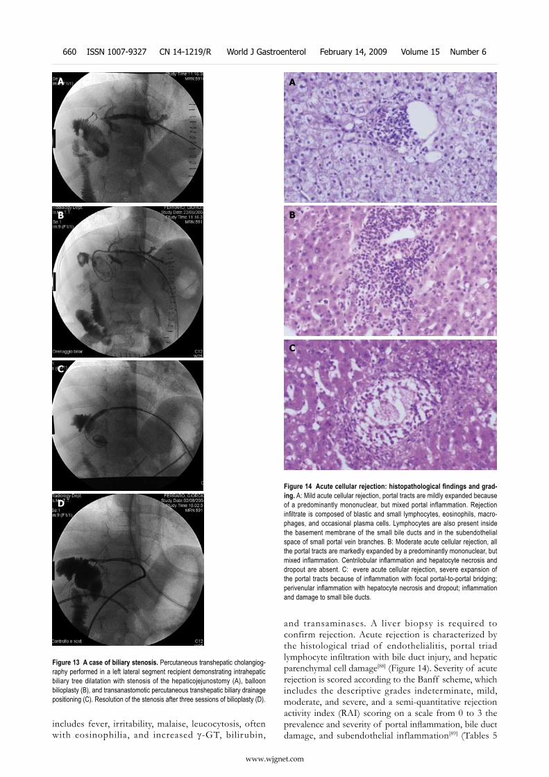
dilatation and stenting are performed by percutaneous transhepatic cholangiography (Figure 13). The presence of multiple bile ducts has a documented increased risk for biliary complications[86].
Reoperation and re-transplantationEarly second-look reoperation is commonly used in several centers for the best diagnosis and treatment of bile leakage, hemorrhage, bowel injury secondary to multiple intra-abdominal adhesions, and sepsis. Infants and small children who have had only initial skin closure require secondary laparotomy for musculofascial closure in 5-7 d[87].
The overall incidence of re-transplantation ranges from 8% to 29%. The incidence of re-transplantation is similar for whole-organ allografts and partial allografts. The majority of re-transplantations result from acute allograft damage caused by either hepatic artery
thrombosis or primary non-function; chronic rejection and biliary complications are uncommon causes. When re-transplantation for acute organ failure is undertaken in a timely manner, patient survival exceeds 80%. When re-transplantation is performed after prolonged immunosuppression for chronic allograft failure, often complicated by multiorgan insufficiency, the survival is only 50%.
Acute rejectionAbout 20%-50% of patients develop at least one episode of acute rejection in the first weeks after liver transplantation. The clinical picture of rejection
Figure 10 Venogram of hepatic venous outflow obstruction after left lat-eral segment split-liver transplantation. Venogram demonstrates a stenosis at the left hepatic vein anastomosis (A). Balloon angioplasty is performed (B), with resolution of the stenosis (C).
A
B
C
A
B
Figure 11 Portal vein thrombosis. Computed tomographic angiography with evidence of portal vein thrombosis and cavernomatous degeneration with collateral drainage through the left gastric vein (A), and evidence of intrahepatic portal flux restoration (B).
Figure 12 Liver biopsy performed in a left lateral segment recipient be-cause of non-specifically elevated liver function tests. Histology shows biliary duct proliferation and portal tract enlargement suggestive of mechanic cholestasis.
Spada M et al . Liver tansplantation 659
www.wjgnet.com
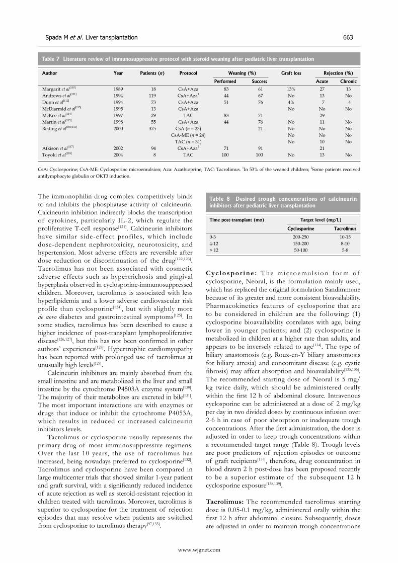
includes fever, irritability, malaise, leucocytosis, often with eosinophilia, and increased γ-GT, bilirubin,
and transaminases. A liver biopsy is required to confirm rejection. Acute rejection is characterized by the histological triad of endothelialitis, portal triad lymphocyte infiltration with bile duct injury, and hepatic parenchymal cell damage[88] (Figure 14). Severity of acute rejection is scored according to the Banff scheme, which includes the descriptive grades indeterminate, mild, moderate, and severe, and a semi-quantitative rejection activity index (RAI) scoring on a scale from 0 to 3 the prevalence and severity of portal inflammation, bile duct damage, and subendothelial inflammation[89] (Tables 5
A
B
C
D
Figure 13 A case of biliary stenosis. Percutaneous transhepatic cholangiog-raphy performed in a left lateral segment recipient demonstrating intrahepatic biliary tree dilatation with stenosis of the hepaticojejunostomy (A), balloon bilioplasty (B), and transanastomotic percutaneous transhepatic biliary drainage positioning (C). Resolution of the stenosis after three sessions of bilioplasty (D).
A
B
C
Figure 14 Acute cellular rejection: histopathological findings and grad-ing. A: Mild acute cellular rejection, portal tracts are mildly expanded because of a predominantly mononuclear, but mixed portal inflammation. Rejection infiltrate is composed of blastic and small lymphocytes, eosinophils, macro-phages, and occasional plasma cells. Lymphocytes are also present inside the basement membrane of the small bile ducts and in the subendothelial space of small portal vein branches. B: Moderate acute cellular rejection, all the portal tracts are markedly expanded by a predominantly mononuclear, but mixed inflammation. Centrilobular inflammation and hepatocyte necrosis and dropout are absent. C: evere acute cellular rejection, severe expansion of the portal tracts because of inflammation with focal portal-to-portal bridging; perivenular inflammation with hepatocyte necrosis and dropout; inflammation and damage to small bile ducts.
660 ISSN 1007-9327 CN 14-1219/R World J Gastroenterol February 14, 2009 Volume 15 Number 6

www.wjgnet.com
and 6).The primary treatment of rejection is a short course
of high-dose steroids. Bolus doses administered over a 3-6-day period with a rapid taper to baseline therapy are successful in the majority of cases. When refractory or recurrent rejection occurs, conversion from cyclosporine to tacrolimus, or antilymphocyte therapy using the monoclonal antibody, ornithine-ketoacid transaminase orthoclone, have been successfully used[90,91].
INFECTIONSImmunosuppressive drugs used to prevent rejection inhibit activation of T lymphocytes, medullar cell proliferation and macrophage function, therefore creating an optimal environment for the development of infections. Infectious complications now represent the most common source of morbidity and mortality after transplantation.
Bacterial infections occur in the immediate post-transplantation period and are most often caused by Gram-negative enteric organisms, enterococci, or staphylococci. Sepsis originating at sites of invasive monitoring lines can be minimized by replacing or removing all of the intraoperative lines soon after transplantation. The use of prophylactic antibacterial antibiotics is discontinued as soon as possible to avoid the development of resistant organisms.
Fungal infection is a potential problem in the early post-transplantation period. To prevent fungal infection, aggressive protocols for pre-transplantation
prophylaxis have been proposed[92]. Fungal infection most often occurs in high-risk patients requiring multiple operative procedures, re-transplantation, hemodialysis or continuous hemofiltration, pre-transplant chemotherapy, and multiple antibiotic courses. The authors use antifungal postoperative prophylaxis with liposomal amphotericin B only in high-risk patients undergoing liver transplantation.
Early and severe viral infections are caused by viruses of the herpes family, including Epstein-Barr virus (EBV), cytomegalovirus (CMV), and herpes simplex virus[93]. The risk of developing either CMV or EBV infection is influenced by the preoperative serological status of the transplant donor and recipient[94,95]. Seronegative recipients receiving seropositive donor organs are at greatest risk. Various prophylactic protocols, including intravenous IgG and hyperimmune anti-CMV IgG, associated with acyclovir or ganciclovir have been used to decrease the incidence of symptomatic CMV and EBV infection, although seroconversion in naive recipients inevitably occurs[94,96]. The suspicion of CMV infection is suggested by the presence of fever, leukopenia, maculopapular rash and hepatocellular abnormalities, respiratory insufficiency, or gastrointestinal hemorrhage. Hepatic biopsy or endoscopic biopsy of colonic or gastroduodenal sites allows early diagnosis with immunohistochemical recognition. Nowadays, the availability of specific antiviral drugs like ganciclovir, foscarnet and more recently valaciclovir, have radically modified the prognosis of CMV infection. At the start of the 1990s, the concept of pre-symptomatic therapy
Assessment Criteria RAIIndeterminate Portal inflammatory infiltrate that fails to meet criteria for the diagnosis of acute rejection 1-2Mild Rejection infiltrate in a minority of the triads that is generally mild and confined within the portal spaces 3-4Moderate Rejection infiltrate expanding most or all of the triads 5-6
Severe As above for moderate, with spillover into the periportal areas and moderate to severe perivenular inflammation that extends into the hepatic perenchyma and is associated with perivenular hepatocyte necrosis
> 6
Table 5 Banff grading of acute liver allograft rejection
Category Criteria Score
Portal inflammation Mostly lymphocytic inflammation involving, but not noticeably expanding, a minority of the triads 1
Expansion of most or all of the triads by a mixed infiltrate containing lymphocytes with occasional blasts, neutrophils, and eosinophils
2
Marked expansion of most or all of the triads by a mixed infiltrate containing numerous blasts and eosinophils with inflammatory spillover into the periportal parenchyma
3
Bile duct inflammation damage
A minority of the ducts are cuffed and infiltrated by inflammatory cells and show only mild reactive changes such as an increased nuclear-to-cytoplasmatic ratio of the epithelial cells
1
Most or all of the ducts infiltrated by inflammatory cells. More than an occasional duct shows degenerative changes such as nuclear pleomorphism, disordered polarity, and cytoplasmatic vacuolization of the epithelium
2
As above for the 2nd criterion, with most or all of the ducts showing degenerative changes or focal luminal disruption 3Venous endothelial inflammation
Subendothelial lymphocytic infiltration involving some, but not a majority, of the portal and/or hepatic venules 1
Subendothelial infiltration involving most or all of the portal and/or hepatic venules 2As above for the 2nd criterion, with moderate or severe perivenular inflammation that extends into the perivenular parenchyma and is associated with perivenular hepatocyte necrosis
3
Table 6 Rejection activity index (RAI)
Spada M et al . Liver tansplantation 661

www.wjgnet.com
was introduced as a strategy to prevent the incidence of CMV-related disease, based on the principle of not administering antiviral medications up to the point when these will have maximum effect, and monitoring CMV antigenemia (pp65) or viremia (CMV DNA)[97,98].
Herpes simplex virus infections, similar to those seen in non-transplant patients, require treatment with acyclovir when diagnosed.
EBV infection represents a potential risk for the pediatric transplant recipient. EBV infection has a variable clinical picture including a mononucleosis-like syndrome, hepatitis-simulating rejection, extranodal lymphoproliferative infiltration, peritonsillar or lymph node enlargement, or encephalopathy. Monitoring of EBV blood viral load by quantitative polymerase chain reaction (PCR) is the best predictor of risk. When evidence of active infection exists, an acute reduction in immunosuppression is mandatory. The authors recommend monthly EBV-DNA PCR counts and more frequent monitoring in case of increasing viral load levels. As a result of the lack of a standardized EBV DNA count methodology, no common cutoff exists. In the authors’ experience, more than 500 genomes/105 peripheral blood leukocytes identify patients who benefit from reduction in primary immunosuppression[99]. Antiviral therapy with ganciclovir and CMV-IgG is also used, although no definitive data support their use[100,101].
Other post-transplantation infectious complications include adenovirus hepatitis, varicella, and enterovirus-induced gastroenteritis. Pneumocystis carinii infection has been nearly eliminated by the prophylactic administration of sulfisoxazole and trimethoprim or aerosolized pentamidine.
MANAGING IMMUNOSUPPRESSIVE THERAPYThe immune system recognizes the graft as foreign and begins a destructive immune response mediated principally by the T lymphocytes. In order to avoid destruction of the graft, immunosuppressive drugs must be administered. Progress in transplant surgery in the last 20 years has been characterized in large part by the introduction of calcineurin inhibitors that today represent the keystone of most immunosuppressive protocols[102,103]. In the last decade, new drugs that selectively target various cellular activation pathways have been proposed and used. The following are the most commonly used drugs in pediatric liver recipients.
CorticosteroidsCorticosteroids were the first drugs to be used to control rejection and are still an essential element of the immunosuppressive regimen; they are effective in both the prevention and treatment of graft rejection. They act through intracellular receptors expressed in all cells of the body. Their immunosuppressive action mechanism, not fully clarified yet, is linked to the suppression of antibody production; inhibition of synthesis of
cytokines such as interleukin-2 (IL-2) and interferon-γ; reduction in the proliferation of helper and suppressor T cells, cytotoxic T cells, and B cells; and the migration and activity of neutrophils.
Long-term clinical experience with steroid use has documented a host of adverse effects. Over-immunosuppression is associated with increased incidence of bacterial, fungal and viral infections. In addition, patients taking steroids carry an increased risk for developing malignancies, especially lymphomas and skin cancers[104]. Detrimental metabolic effects of steroids are wide ranging and are of particular concern for the pediatric transplant patient[105-107]. In terms of hospital costs, the calculated 10-year cumulative expense for steroid-related complications in adult kidney recipients has been shown to be 5300 $ per patient per year[108]. Efforts are underway to develop immunotherapy regimens in which steroids can be withdrawn early, or not used at all.
The experience of steroid weaning after pediatric liver transplantation was summarized in 2000 by Reding[109]. There are a total of nine recent studies, not all of which were non-randomized and uncontrolled. Steroid treatment could be successfully stopped in 21%-100% of the transplanted patients. The risk of rejection was not significantly increased, and varied from 7% to 29%. Chronic rejection did not seem to be increased[110-118] (Table 7). The conclusions of this review are the following: (1) weaning of steroids after pediatric liver transplanatation is safe and, most of the time, beneficial; and (2) in many patients, calcineurin inhibitor monotherapy can be achieved, suggesting that the next step could be the adoption of steroid-free immunosuppressive protocols.
In a non-randomized study, Reding et al[119] compared pediatric liver transplantation under steroid-free immunosuppression in children who received combined tacrolimus and antibody to the IL-2 receptor of T cells (basiliximab), with matched historical recipients taking tacrolimus and steroids. Twelve-month rejection-free survival was similar in the steroid-free group compared with the corticosteroid group. The authors performed the first prospective, controlled, randomized study designed for children undergoing liver transplantation to test the possibility of avoiding the use of corticosteroids under baseline tacrolimus immunosuppression plus basiliximab induction, which confirmed no harmful effect of steroid avoidance on graft acceptance[120].
Corticosteroid withdrawal or avoidance can be difficult in patients with autoimmune hepatitis, primary biliary cirrhosis, or primary sclerosing cholangitis. In these patients it might be desirable to include steroids in the immunosuppressive protocol as a principle, although definitive and convincing data are not available.
Calcineurin inhibitorsCyclosporine and tacrolimus are classified as calcineurin inhibitors because they inhibit T-cell responses and bind to intracellular proteins called immunophilins.
662 ISSN 1007-9327 CN 14-1219/R World J Gastroenterol February 14, 2009 Volume 15 Number 6
www.wjgnet.com
The immunophilin-drug complex competitively binds to and inhibits the phosphatase activity of calcineurin. Calcineurin inhibition indirectly blocks the transcription of cytokines, particularly IL-2, which regulate the proliferative T-cell response[121]. Calcineurin inhibitors have similar side-effect profiles, which include dose-dependent nephrotoxicity, neurotoxicity, and hypertension. Most adverse effects are reversible after dose reduction or discontinuation of the drug[122,123]. Tacrolimus has not been associated with cosmetic adverse effects such as hypertrichosis and gingival hyperplasia observed in cyclosporine-immunosuppressed children. Moreover, tacrolimus is associated with less hyperlipidemia and a lower adverse cardiovascular risk profile than cyclosporine[124], but with slightly more de novo diabetes and gastrointestinal symptoms[125]. In some studies, tacrolimus has been described to cause a higher incidence of post-transplant lymphoproliferative disease[126,127], but this has not been confirmed in other authors’ experiences[128]. Hypertrophic cardiomyopathy has been reported with prolonged use of tacrolimus at unusually high levels[129].
Calcineurin inhibitors are mainly absorbed from the small intestine and are metabolized in the liver and small intestine by the cytochrome P4503A enzyme system[130]. The majority of their metabolites are excreted in bile[131]. The most important interactions are with enzymes or drugs that induce or inhibit the cytochrome P4053A, which results in reduced or increased calcineurin inhibitors levels.
Tacrolimus or cyclosporine usually represents the primary drug of most immunosuppressive regimens. Over the last 10 years, the use of tacrolimus has increased, being nowadays preferred to cyclosporine[132]. Tacrolimus and cyclosporine have been compared in large multicenter trials that showed similar 1-year patient and graft survival, with a significantly reduced incidence of acute rejection as well as steroid-resistant rejection in children treated with tacrolimus. Moreover, tacrolimus is superior to cyclosporine for the treatment of rejection episodes that may resolve when patients are switched from cyclosporine to tacrolimus therapy[97,133].
Cyc lospor ine : T he m ic roemu l s i on fo r m o f cyclosporine, Neoral, is the formulation mainly used, which has replaced the original formulation Sandimmune because of its greater and more consistent bioavailability. Pharmacokinetics features of cyclosporine that are to be considered in children are the following: (1) cyclosporine bioavailability correlates with age, being lower in younger patients; and (2) cyclosporine is metabolized in children at a higher rate than adults, and appears to be inversely related to age[134]. The type of biliary anastomosis (e.g. Roux-en-Y biliary anastomosis for biliary atresia) and concomitant disease (e.g. cystic fibrosis) may affect absorption and bioavailability[135,136]. The recommended starting dose of Neoral is 5 mg/kg twice daily, which should be administered orally within the first 12 h of abdominal closure. Intravenous cyclosporine can be administered at a dose of 2 mg/kg per day in two divided doses by continuous infusion over 2-6 h in case of poor absorption or inadequate trough concentrations. After the first administration, the dose is adjusted in order to keep trough concentrations within a recommended target range (Table 8). Trough levels are poor predictors of rejection episodes or outcome of graft recipients[137], therefore, drug concentration in blood drawn 2 h post-dose has been proposed recently to be a superior estimate of the subsequent 12 h cyclosporine exposure[138,139].
Tacrolimus: The recommended tacrolimus starting dose is 0.05-0.1 mg/kg, administered orally within the first 12 h after abdominal closure. Subsequently, doses are adjusted in order to maintain trough concentrations
Time post-transplant (mo) Target level (mg/L)
Cyclosporine Tacrolimus
0-3 200-250 10-154-12 150-200 8-10> 12 50-100 5-8
Table 8 Desired trough concentrations of calcineurin inhibitors after pediatric liver transplantation
Spada M et al . Liver tansplantation 663
CsA: Cyclosporine; CsA-ME: Cyclosporine microemulsion; Aza: Azathioprine; TAC: Tacrolimus. 1In 53% of the weaned children; 2Some patients received antilymphocyte globulin or OKT3 induction.
Author Year Patients (n ) Protocol Weaning (%) Graft loss Rejection (%)
Performed Success Acute Chronic
Margarit et al[110] 1989 18 CsA+Aza 83 61 13% 27 13Andrews et al[111] 1994 119 CsA+Aza1 44 67 No 13 NoDunn et al[112] 1994 73 CsA+Aza 51 76 4% 7 4McDiarmid et al[113] 1995 13 CsA+Aza No No NoMcKee et al[114] 1997 29 TAC 83 71 29Martin et al[115] 1998 55 CsA+Aza 44 76 No 11 NoReding et al[109,116] 2000 375 CsA (n = 23) 21 No No No
CsA-ME (n = 24) No No NoTAC (n = 31) No 10 No
Atkison et al[117] 2002 94 CsA+Aza2 71 91 21Toyoki et al[118] 2004 8 TAC 100 100 No 13 No
Table 7 Literature review of immunosuppressive protocol with steroid weaning after pediatric liver transplantation

www.wjgnet.com
within a recommended target range (Table 8). The trough level is widely accepted for routine tacrolimus drug level monitoring. Large inter- and intra-individual differences in pharmacokinetics exist. The elimination half-life of tacrolimus in children is 50% of that in adults, and clearance is correspondingly two to four times faster[140,141]. Therefore, children require higher doses to achieve similar tacrolimus concentrations.
Mycophenolate mofetilThe active metabolite of mycophenolate mofetil, mycophenolic acid, is a selective inhibitor of the enzyme inosine monophosphate dehydrogenase, which is essential for the de novo pathway of purine synthesis[142]. Inhibition of the de novo pathway results in the depletion of guanosine nucleotides and arrested lymphocytes replication because they are unable to use the alternative pathway for nucleotide production[143].
Mycophenolate mofetil has been used successfully as an alternative immunosuppressive agent in patients with chronic rejection, refractory rejection, or severe calcineurin inhibitor toxicity[144,145]. Mycophenolate mofetil has also been used in calcineurin-inhibitor and corticosteroid-sparing immunosuppressive protocols, without increasing the risk of rejection[146,147]. The suggested dose for pediatric liver transplant recipients is 15 mg/kg twice daily[148]. Pharmacokinetic studies showed large inter-individual variations in mycophenolic acid parameters[149,150], which indicates the need for therapeutic drug monitoring and individualized dosing. The most relevant adverse effects of mycophenolate mofetil are dose-dependent gastrointestinal symptoms and bone marrow suppression[147,151]. Acyclovir and ganciclovir increase mycophenolic acid efficacy, whereas cholestyramine, oral antibiotics, antacids, cyclosporine, and high tacrol imus concentrat ions reduce i ts concentration[148-150].
SirolimusSirolimus (rapamycin) is a macrolide antibiotic with potent immunosuppressive properties that acts by blocking T-cell activation by way of IL-2R post-receptor signal transduction[152]. Sirolimus has been used in small, uncontrolled studies in liver transplant recipients (Table 9) and reduces rate of acute rejection, when used in combination with calcineurin inhibitors, even at low doses, or facilitates early steroid withdrawal, while
maintaining low rates of acute rejection[153-157].Sirolimus has also been used as rescue treatment in
chronic rejection and calcineurin inhibitor toxicity[157-159], whereas attempts to use sirolimus as a single primary immunosuppressive agent have resulted in a high rate of acute rejection[160]. Sirolimus has not yet been approved by the US Food and Drug Administration for use in liver transplantation. One trial to evaluate sirolimus in liver transplant recipients was halted because of an increased incidence of hepatic artery thrombosis. In contrast, other studies have not confirmed this finding[154,161,162], and a possible benefit of sirolimus in the prevention of coronary artery restenosis after percutaneous coronary revascularization has been described[163]. Sirolimus has shown antineoplastic activity, inhibiting angiogenesis in malignant tissue through reduction of vascular endothelial growth factor secretion, which may provide a specific indication for using of the drug in patients transplanted for primary liver malignancy[164].
Sirolimus drug interactions are similar to those of calcineurin inhibitors. It has a long half-life (40-86 h) and intra- and inter-individual variation[152,165]. Therefore, daily sirolimus monitoring is not necessary and monitoring trough level twice weekly for the first month and weekly for the next month is recommended, targeting a 5-15 mg/L range. Sirolimus levels increase during simultaneous administration of cyclosporine[166]. The most relevant dose-related side effects of sirolimus are hyperlipidemia, thrombocytopenia and leukopenia[153,157].
IL-2 receptor antibodiesT cells involved in acute rejection act by exposing activation markers such as the IL-2 receptors. Therefore, anti-IL-2 receptor therapy appears to be a promising option for specific immunosuppression. IL-2 receptor antibodies have been used primarily in children as induction agents in double or triple immunosuppression protocols. Preliminary experience in pediatric liver recipients is encouraging: pooled data from the available papers from the literature encompassed 79 patients treated with daclizumab, 165 with basiliximab, and 209 no-induction controls; incidence of acute rejection was lower in the induction groups[119,120,167-172] (Table 10).
A multicenter trial studied basiliximab pharmacokinetics and pharmacodynamics in children. It demonstrated that to achieve efficacious results, pediatric patients less than 35 kg in weight should receive two intravenous 10-mg
Author Immunosuppression No. of patients
Survival (%) Acute rejection (%)
Follow-up (mo)Patient Graft
McAlister et al[153] TRL, SRL, STER1 32 92 3 8McAlister et al[154] TRL, SRL, STER1 56 93 91 14 23Peltekian et al[155] TRL, SRL, STER1 42 93 90 10 14Pridöhl et al[156] TRL, SRL, STER 22 91 78 14 14Sindhi et al[157] TRL, early SRL, STER 6 17 15
TRL, late SRL, ATG 9 332 3
Table 9 Use of sirolimus in primary immunosuppressive regimens in liver transplantation
ATG: Antithymoglobulin; SRL: Sirolimus; STER: Corticosteroids; TRL: Tacrolimus; 1Corticosteroids withdrawal 3 mo after trans-plantation; 2Rejection episodes observed before sirolimus was introduced in the immunosuppressive regimen.
664 ISSN 1007-9327 CN 14-1219/R World J Gastroenterol February 14, 2009 Volume 15 Number 6
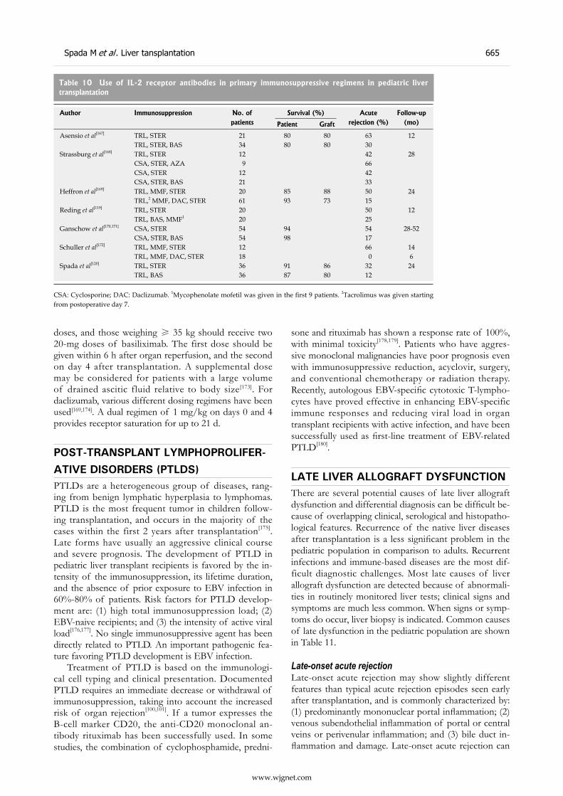
www.wjgnet.com
doses, and those weighing ≥ 35 kg should receive two 20-mg doses of basiliximab. The first dose should be given within 6 h after organ reperfusion, and the second on day 4 after transplantation. A supplemental dose may be considered for patients with a large volume of drained ascitic fluid relative to body size[173]. For daclizumab, various different dosing regimens have been used[169,174]. A dual regimen of 1 mg/kg on days 0 and 4 provides receptor saturation for up to 21 d.
POST-TRANSPLANT LYMPHOPROLIFER-ATIVE DISORDERS (PTLDS)PTLDs are a heterogeneous group of diseases, rang-ing from benign lymphatic hyperplasia to lymphomas. PTLD is the most frequent tumor in children follow-ing transplantation, and occurs in the majority of the cases within the first 2 years after transplantation[175]. Late forms have usually an aggressive clinical course and severe prognosis. The development of PTLD in pediatric liver transplant recipients is favored by the in-tensity of the immunosuppression, its lifetime duration, and the absence of prior exposure to EBV infection in 60%-80% of patients. Risk factors for PTLD develop-ment are: (1) high total immunosuppression load; (2) EBV-naive recipients; and (3) the intensity of active viral load[176,177]. No single immunosuppressive agent has been directly related to PTLD. An important pathogenic fea-ture favoring PTLD development is EBV infection.
Treatment of PTLD is based on the immunologi-cal cell typing and clinical presentation. Documented PTLD requires an immediate decrease or withdrawal of immunosuppression, taking into account the increased risk of organ rejection[100,101]. If a tumor expresses the B-cell marker CD20, the anti-CD20 monoclonal an-tibody rituximab has been successfully used. In some studies, the combination of cyclophosphamide, predni-
sone and rituximab has shown a response rate of 100%, with minimal toxicity[178,179]. Patients who have aggres-sive monoclonal malignancies have poor prognosis even with immunosuppressive reduction, acyclovir, surgery, and conventional chemotherapy or radiation therapy. Recently, autologous EBV-specific cytotoxic T-lympho-cytes have proved effective in enhancing EBV-specific immune responses and reducing viral load in organ transplant recipients with active infection, and have been successfully used as first-line treatment of EBV-related PTLD[180].
LATE LIVER ALLOGRAFT DYSFUNCTIONThere are several potential causes of late liver allograft dysfunction and differential diagnosis can be difficult be-cause of overlapping clinical, serological and histopatho-logical features. Recurrence of the native liver diseases after transplantation is a less significant problem in the pediatric population in comparison to adults. Recurrent infections and immune-based diseases are the most dif-ficult diagnostic challenges. Most late causes of liver allograft dysfunction are detected because of abnormali-ties in routinely monitored liver tests; clinical signs and symptoms are much less common. When signs or symp-toms do occur, liver biopsy is indicated. Common causes of late dysfunction in the pediatric population are shown in Table 11.
Late-onset acute rejectionLate-onset acute rejection may show slightly different features than typical acute rejection episodes seen early after transplantation, and is commonly characterized by: (1) predominantly mononuclear portal inflammation; (2) venous subendothelial inflammation of portal or central veins or perivenular inflammation; and (3) bile duct in-flammation and damage. Late-onset acute rejection can
CSA: Cyclosporine; DAC: Daclizumab. 1Mycophenolate mofetil was given in the first 9 patients. 2Tacrolimus was given starting from postoperative day 7.
Author Immunosuppression No. of patients
Survival (%) Acute rejection (%)
Follow-up (mo)Patient Graft
Asensio et al[167] TRL, STER 21 80 80 63 12TRL, STER, BAS 34 80 80 30
Strassburg et al[168] TRL, STER 12 42 28CSA, STER, AZA 9 66CSA, STER 12 42CSA, STER, BAS 21 33
Heffron et al[169] TRL, MMF, STER 20 85 88 50 24TRL,2 MMF, DAC, STER 61 93 73 15
Reding et al[119] TRL, STER 20 50 12TRL, BAS, MMF1 20 25
Ganschow et al[170,171] CSA, STER 54 94 54 28-52CSA, STER, BAS 54 98 17
Schuller et al[172] TRL, MMF, STER 12 66 14TRL, MMF, DAC, STER 18 0 6
Spada et al[120] TRL, STER 36 91 86 32 24TRL, BAS 36 87 80 12
Table 10 Use of IL-2 receptor antibodies in primary immunosuppressive regimens in pediatric liver transplantation
Spada M et al . Liver tansplantation 665

www.wjgnet.com
also manifest as so-called central perivenulitis [181-183], or may resemble chronic hepatitis[184,185]. Mild cases may re-solve spontaneously[183], but more severe forms warrant more aggressive treatment.
Chronic rejectionChronic rejection develops in 5%-10% of transplanted patients. The primary clinical manifestation is progres-sive cholestasis. This course can occur within weeks
from transplantation or later, and can be asymptomatic or follow persistent and/or unresponsive acute rejec-tion and/or inadequate immunosuppression. Two clini-cal forms have been described[186]. In the first, named vanishing bile duct syndrome, the biliary epithelium is primarily injured with changes ranging from senescence (early stage) to severe ductopenia in at least 50% of the portal tracts (late stage)[187]. This form can be success-fully treated by conversion from cyclosporine to tacroli-mus immunosuppression protocols. Re-transplantation is necessary in non-responding children. The second subtype is characterized by the development of progres-sive ischemic injury to bile ducts and hepatocytes, which causes ductopenia and ischemic necrosis with fibrosis (Figure 15). In this setting, the diagnosis is rarely based on histology alone, because arteries with pathognomonic changes are rarely present in needle biopsy specimens. Bile duct injury and ductopenia, however, can be caused by biliary strictures, hepatic artery pathology, adverse drug reactions, and CMV. Selective hepatic angiography showing pruning of the intrahepatic arteries with poor peripheral filling and segmental narrowing supports a diagnosis of chronic rejection[188,189]. This form nearly always requires retransplantation.
Recurrent and new-onset or de novo autoimmune hepa-titisTheoretically all forms of autoimmune hepatitis after transplantation can be classified as rejection[190-192]. No conventional clinical tests differentiate an autoimmune response from rejection. The diagnosis of autoimmune hepatitis is established by a combination of serologi-cal, molecular biological and histopathological findings. Non-organ-specific autoantibodies are a requisite for the diagnosis, and they typically include smooth muscle antibodies (SMAs), antinuclear antibodies (ANAs), and antibodies to liver kidney microsome (anti-LKM)[193]. Minimal diagnostic criteria for recurrent or de novo au-
Figure 15 Histological findings in chronic rejection: Little portal inflam-mation in conjunction with bile duct loss affecting > 50% of the portal tracts and moderate or severe perivenular fibrosis.
Incidence at 5 yr (%) Risk factors
Acute rejection Variable (< 30) Inadequate immunosuppression Treatment with immune activating drugs (e.g. interferon)History of autoimmune liver disease
Chronic rejection -3 Inadequate immunosuppressionTreatment with immune-activating drugs (e.g. interferon)Refractory acute rejection Chronic rejection in a previous failed allograft
Recurrent AIH -30 Suboptimal immunosuppressionAIH type ⅠSevere inflammation in native liverHLA DR3 or DR4
De novo AIH < 5Recurrent PBC 20-30 Tacrolimus as baseline immunosuppression
Living-related donorSteroid and other immunosuppression withdrawal
Recurrent PSC 20-30 Male sex; donor-recipient gender mismatchIntact colon at time of transplantation
Idiopathic post-transplant hepatitis 5-60
Table 11 Common causes of late dysfunction in the pediatric population
AIH: Autoimmune hepatitis; PBC: Primary biliary cirrhosis; PSC: Primary sclerosing cholangitis.
666 ISSN 1007-9327 CN 14-1219/R World J Gastroenterol February 14, 2009 Volume 15 Number 6
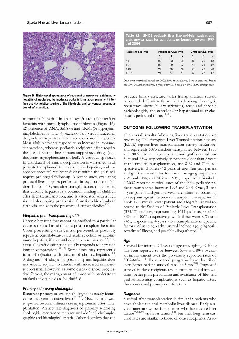
www.wjgnet.com
toimmune hepatitis in an allograft are: (1) interface hepatitis with portal lymphocytic infiltrates (Figure 16); (2) presence of ANA, SMA or anti-LKM; (3) hypergam-maglobulinemia; and (4) exclusion of virus-induced or drug-related hepatitis and late acute or chronic rejection. Most adult recipients respond to an increase in immuno-suppression, whereas pediatric recipients often require the use of second-line immunosuppressive drugs (aza-thioprine, mycophenolate mofetil). A cautious approach to withdrawal of immunosuppression is warranted in all patients transplanted for autoimmune hepatitis, and the consequences of recurrent disease within the graft will require prolonged follow-up. A recent study, evaluating protocol liver biopsies performed in asymptomatic chil-dren 1, 5 and 10 years after transplantation, documented that chronic hepatitis is a common finding in children after liver transplantation, and is associated with a high risk of developing progressive fibrosis, which leads to cirrhosis, and with the presence of autoantibodies[194].
Idiopathic post-transplant hepatitisChronic hepatitis that cannot be ascribed to a particular cause is defined as idiopathic post-transplant hepatitis. Cases presenting with central perivenulitis probably represent centrilobular-based acute rejection or autoim-mune hepatitis, if autoantibodies are also present[185], be-cause allograft dysfunction usually responds to increased immunosuppression[185,195]. Some cases may represent a form of rejection with features of chronic hepatitis[195]. A diagnosis of idiopathic post-transplant hepatitis does not usually require treatment with increased immuno-suppression. However, as some cases do show progres-sive fibrosis, the management of those with moderate to marked activity needs to be clarified.
Primary sclerosing cholangitisRecurrent primary sclerosing cholangitis is nearly identi-cal to that seen in native livers[196,197]. Most patients with suspected recurrent disease are asymptomatic after trans-plantation. An accurate diagnosis of primary sclerosing cholangitis recurrence requires well-defined cholangio-graphic and histological criteria. Other disorders that can
produce biliary strictures after transplantation should be excluded. Graft with primary sclerosing cholangitis recurrence shows biliary strictures, acute and chronic pericholangitis, and centrilobular hepatocanalicular cho-lestasis periductal fibrosis[198].
OUTCOME FOLLOWING TRANSPLANTATION
The overall results following liver transplantation are rewarding. The European Liver Transplantation Registry (ELTR) reports liver transplantation activity in Europe, and represents 5895 children transplanted between 1988 and 2005. Overall 1-year patient and graft survival was 84% and 73%, respectively, in patients older than 2 years at the time of transplantation, and 81% and 71%, re-spectively, in children < 2 years of age. Ten-year patient and graft survival rates for the same age groups were 75% and 61%, and 74% and 60%, respectively. Similarly, UNOS reported survival rates of the 9064 pediatric pa-tients transplanted between 1997 and 2004. One-, 3- and 5-year patient and graft survival rates stratified according to recipient age at the time of transplant are reported in Table 12. Overall 1-year patient and allograft survival re-ported to the Studies of Pediatric Liver Transplantation (SPLIT) registry, representing 1611 patients, reached 88% and 82%, respectively, while these were 83% and 74%, respectively, 4 years after transplantation. Specific factors influencing early survival include age, diagnosis, severity of illness, and possibly allograft type[199].
AgeSurvival for infants < 1 year of age or weighing < 10 kg has been reported to be between 65% and 80% overall, an improvement over the previously reported rates of 50%-60%[200]. Experienced programs have described even better patient survival rates at 3 mo[201]. Improved survival in these recipients results from technical innova-tions, better graft preparation and avoidance of life- and graft-threatening complications such as hepatic artery thrombosis and primary non-function.
DiagnosisSurvival after transplantation is similar in patients who have cholestatic and metabolic liver disease. Early sur-vival rates are worse for patients who have acute liver failure[9,202,203] and liver tumors[11], but their long-term sur-vival rates are similar to those of other recipients. Asso-
Recipient age (yr) Patient survival (yr) Graft survival (yr)
1 3 5 1 3 5
< 1 89 82 78 81 70 631-5 86 80 77 78 71 676-10 91 86 86 84 76 7511-17 93 87 81 87 77 67
Table 12 UNOS pediatric liver Kaplan-Meier patient and graft survival rates for transplants performed between 1997 and 2004
One-year survival based on 2002-2004 transplants, 3-year survival based on 1999-2002 transplants, 5-year survival based on 1997-2000 transplants.
Figure 16 Histological appearance of recurrent or new-onset autoimmune hepatitis characterized by moderate portal inflammation, prominent inter-face activity, relative sparing of the bile ducts, and perivenular accumula-tion of inflammation.
Spada M et al . Liver tansplantation 667
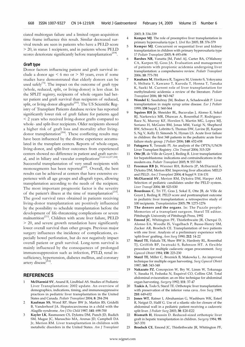
www.wjgnet.com
ciated multiorgan failure and a limited organ-acquisition time frame influence this result. Similar decreased sur-vival trends are seen in patients who have a PELD score > 20, in status 1 recipients, and in patients whose PELD scores deteriorate significantly before transplantation[204].
Graft typeDonor factors influencing patient and graft survival in-clude a donor age < 6 mo or > 50 years, even if some studies have demonstrated that elderly donors can be used safely[76]. The impact on the outcome of graft type (whole, reduced, split, or living-donor) is less clear. In the SPLIT registry, recipients of whole organs had bet-ter patient and graft survival than recipients of reduced, split, or living-donor allografts[205]. The US Scientific Reg-istry of Transplant Patients database review has reported significantly lower risk of graft failure for patients aged < 2 years who received living-donor grafts compared to whole- and split-liver recipients. Older recipients showed a higher risk of graft loss and mortality after living-donor transplantation[206]. These conflicting results may have been influenced by the diverse experience accumu-lated in the transplant centers. Reports of whole-organ, living-donor, and split-liver outcomes from experienced centers showed no difference in patient and graft surviv-al, and in biliary and vascular complications[53,60-62,207,208]. Successful transplantation of very small recipients with monosegments has been reported[209]. Overall, the best results can be achieved at centers that have extensive ex-perience with all age groups and allograft types, allowing transplantation according to the needs of the recipient. The most important prognostic factor is the severity of the patient’s illness at the time of transplantation[210]. The good survival rates obtained in patients receiving living-donor transplantation are positively influenced by the possibility to schedule transplantation before the development of life-threatening complications or severe malnutrition[211]. Children with acute liver failure, PELD > 20, and severe growth retardation have significantly lower overall survival than other groups. Previous major surgery influences the incidence of complications, es-pecially bowel perforation, but do not negatively impact overall patient or graft survival. Long-term survival is mainly influenced by the consequences of prolonged immunosuppression such as infection, PTLD, renal in-sufficiency, hypertension, diabetes mellitus, and coronary artery disease[212].
REFERENCES1 McDiarmid SV, Anand R, Lindblad AS. Studies of Pediatric
Liver Transplantation: 2002 update. An overview of demographics, indications, timing, and immunosuppressive practices in pediatric liver transplantation in the United States and Canada. Pediatr Transplant 2004; 8: 284-294
2 Kaufman SS, Wood RP, Shaw BW Jr, Markin RS, Gridelli B, Vanderhoof JA. Hepatocarcinoma in a child with the Alagille syndrome. Am J Dis Child 1987; 141: 698-700
3 Kayler LK, Rasmussen CS, Dykstra DM, Punch JD, Rudich SM, Magee JC, Maraschio MA, Arenas JD, Campbell DA Jr, Merion RM. Liver transplantation in children with metabolic disorders in the United States. Am J Transplant
2003; 3: 334-3394 Kemper MJ. The role of preemptive liver transplantation in
primary hyperoxaluria type 1. Urol Res 2005; 33: 376-3795 Kemper MJ. Concurrent or sequential liver and kidney
transplantation in children with primary hyperoxaluria type 1? Pediatr Transplant 2005; 9: 693-696
6 Barshes NR, Vanatta JM, Patel AJ, Carter BA, O'Mahony CA, Karpen SJ, Goss JA. Evaluation and management of patients with propionic acidemia undergoing liver transplantation: a comprehensive review. Pediatr Transplant 2006; 10: 773-781
7 Kasahara M, Horikawa R, Tagawa M, Uemoto S, Yokoyama S, Shibata Y, Kawano T, Kuroda T, Honna T, Tanaka K, Saeki M. Current role of liver transplantation for methylmalonic acidemia: a review of the literature. Pediatr Transplant 2006; 10: 943-947
8 Wendel U, Saudubray JM, Bodner A, Schadewaldt P. Liver transplantation in maple syrup urine disease. Eur J Pediatr 1999; 158 Suppl 2: S60-S64
9 Squires RH Jr, Shneider BL, Bucuvalas J, Alonso E, Sokol RJ, Narkewicz MR, Dhawan A, Rosenthal P, Rodriguez-Baez N, Murray KF, Horslen S, Martin MG, Lopez MJ, Soriano H, McGuire BM, Jonas MM, Yazigi N, Shepherd RW, Schwarz K, Lobritto S, Thomas DW, Lavine JE, Karpen S, Ng V, Kelly D, Simonds N, Hynan LS. Acute liver failure in children: the first 348 patients in the pediatric acute liver failure study group. J Pediatr 2006; 148: 652-658
10 Futagawa Y, Terasaki PI. An analysis of the OPTN/UNOS Liver Transplant Registry. Clin Transpl 2004; 315-329
11 Otte JB, de Ville de Goyet J, Reding R. Liver transplantation for hepatoblastoma: indications and contraindications in the modern era. Pediatr Transplant 2005; 9: 557-565
12 Freeman RB Jr, Wiesner RH, Roberts JP, McDiarmid S, Dykstra DM, Merion RM. Improving liver allocation: MELD and PELD. Am J Transplant 2004; 4 Suppl 9: 114-131
13 McDiarmid SV, Merion RM, Dykstra DM, Harper AM. Selection of pediatric candidates under the PELD system. Liver Transpl 2004; 10: S23-S30
14 Bourdeaux C, Tri TT, Gras J, Sokal E, Otte JB, de Ville de Goyet J, Reding R. PELD score and posttransplant outcome in pediatric liver transplantation: a retrospective study of 100 recipients. Transplantation 2005; 79: 1273-1276
15 The donors and the organs . In: The Puzzle people: Memories of a transplant surgeon. Starzl TE editor. Pittsburgh: University of Pittsburgh Press, 1992
16 Emond JC, Whitington PF, Thistlethwaite JR, Cherqui D, Alonso EA, Woodle IS, Vogelbach P, Busse-Henry SM, Zucker AR, Broelsch CE. Transplantation of two patients with one liver. Analysis of a preliminary experience with 'split-liver' grafting. Ann Surg 1990; 212: 14-22
17 Starzl TE, Hakala TR, Shaw BW Jr, Hardesty RL, Rosenthal TJ, Griffith BP, Iwatsuki S, Bahnson HT. A flexible procedure for multiple cadaveric organ procurement. Surg Gynecol Obstet 1984; 158: 223-230
18 Starzl TE, Miller C, Broznick B, Makowka L. An improved technique for multiple organ harvesting. Surg Gynecol Obstet 1987; 165: 343-348
19 Nakazato PZ, Concepcion W, Bry W, Limm W, Tokunaga Y, Itasaka H, Feduska N, Esquivel CO, Collins GM. Total abdominal evisceration: an en bloc technique for abdominal organ harvesting. Surgery 1992; 111: 37-47
20 Tzakis A, Todo S, Starzl TE. Orthotopic liver transplantation with preservation of the inferior vena cava. Ann Surg 1989; 210: 649-652
21 Jones WT, Ratner I, Abrahamian G, Washburn WK, Esterl R, Neigut D, Halff G. Use of a silastic silo for closure of the abdominal wall in a pediatric patient receiving a cadaveric split liver. J Pediatr Surg 2003; 38: E20-E22
22 Bismuth H, Houssin D. Reduced-sized orthotopic liver graft in hepatic transplantation in children. Surgery 1984; 95: 367-370
23 Broelsch CE, Emond JC, Thistlethwaite JR, Whitington PF,
668 ISSN 1007-9327 CN 14-1219/R World J Gastroenterol February 14, 2009 Volume 15 Number 6
www.wjgnet.com
Zucker AR, Baker AL, Aran PF, Rouch DA, Lichtor JL. Liver transplantation, including the concept of reduced-size liver transplants in children. Ann Surg 1988; 208: 410-420
24 Broelsch CE, Emond JC, Whitington PF, Thistlethwaite JR, Baker AL, Lichtor JL. Application of reduced-size liver transplants as split grafts, auxiliary orthotopic grafts, and living related segmental transplants. Ann Surg 1990; 212: 368-375; discussion 375-377
25 Broelsch CE, Emond JC, Thistlethwaite JR, Rouch DA, Whitington PF, Lichtor JL. Liver transplantation with reduced-size donor organs. Transplantation 1988; 45: 519-524
26 Otte JB, de Ville de Goyet J, Sokal E, Alberti D, Moulin D, de Hemptinne B, Veyckemans F, van Obbergh L, Carlier M, Clapuyt P. Size reduction of the donor liver is a safe way to alleviate the shortage of size-matched organs in pediatric liver transplantation. Ann Surg 1990; 211: 146-157
27 Houssin D, Soubrane O, Boillot O, Dousset B, Ozier Y, Devictor D, Bernard O, Chapuis Y. Orthotopic liver transplantation with a reduced-size graft: an ideal compromise in pediatrics? Surgery 1992; 111: 532-542
28 Kalayoglu M, D'Alessandro AM, Sollinger HW, Hoffman RM, Pirsch JD, Belzer FO. Experience with reduced-size liver transplantation. Surg Gynecol Obstet 1990; 171: 139-147
29 Esquivel CO, Nakazato P, Cox K, Concepcion W, Berquist W, Russell TR. The impact of liver reductions in pediatric liver transplantation. Arch Surg 1991; 126: 1278-1285; discussion 1285-1286
30 Langnas AN, Marujo WC, Inagaki M, Stratta RJ, Wood RP, Shaw BW Jr . The results of reduced-size l iver transplantation, including split livers, in patients with end-stage liver disease. Transplantation 1992; 53: 387-391
31 Raia S, Nery JR, Mies S. Liver transplantation from live donors. Lancet 1989; 2: 497
32 Strong RW, Lynch SV, Ong TH, Matsunami H, Koido Y, Balderson GA. Successful liver transplantation from a living donor to her son. N Engl J Med 1990; 322: 1505-1507
33 Tanaka K , Uemoto S, Tokunaga Y, Fujita S, Sano K, Yamamoto E, Sugano M, Awane M, Yamaoka Y, Kumada K. Living related liver transplantation in children. Am J Surg 1994; 168: 41-48
34 Emond JC, Heffron TG, Kortz EO, Gonzalez-Vallina R, Contis JC, Black DD, Whitington PF. Improved results of living-related liver transplantation with routine application in a pediatric program. Transplantation 1993; 55: 835-840
35 Broelsch CE, Whitington PF, Emond JC, Heffron TG, Thistlethwaite JR, Stevens L, Piper J, Whitington SH, Lichtor JL. Liver transplantation in children from living related donors. Surgical techniques and results. Ann Surg 1991; 214: 428-437; discussion 437-439
36 Malagó M, Rogiers X, Burdelski M, Broelsch CE. Living related liver transplantation: 36 cases at the University of Hamburg. Transplant Proc 1994; 26: 3620-3621
37 Otte JB, de Ville de Goyet J, Reding R, Sokal E, Lerut J, Vanormelingen P, Janssen M. Living related donor liver transplantation in children: the Brussels experience. Transplant Proc 1996; 28: 2378-2379
38 Haberal M , Bilgin N, Büyükpamukçu N, Karakayali H, Moray G, Arslan G. Living-related partial l iver transplantation in pediatric patients. Transplant Proc 1998; 30: 706-707
39 Darwish AA, Bourdeaux C, Kader HA, Janssen M, Sokal E, Lerut J, Ciccarelli O, Veyckemans F, Otte JB, de Goyet Jde V, Reding R. Pediatric liver transplantation using left hepatic segments from living related donors: surgical experience in 100 recipients at Saint-Luc University Clinics. Pediatr Transplant 2006; 10: 345-353
40 Pichlmayr R, Ringe B, Gubernatis G, Hauss J, Bunzendahl H. [Transplantation of a donor liver to 2 recipients (splitting transplantation)--a new method in the further development of segmental liver transplantation] Langenbecks Arch Chir 1988; 373: 127-130
41 Bismuth H, Morino M, Castaing D, Gillon MC, Descorps
Declere A, Saliba F, Samuel D. Emergency orthotopic liver transplantation in two patients using one donor liver. Br J Surg 1989; 76: 722-724
42 Otte JB, de Ville de Goyet J, Alberti D, Balladur P, de Hemptinne B. The concept and technique of the split liver in clinical transplantation. Surgery 1990; 107: 605-612
43 Houssin D, Boillot O, Soubrane O, Couinaud C, Pitre J, Ozier Y, Devictor D, Bernard O, Chapuis Y. Controlled liver splitting for transplantation in two recipients: technique, results and perspectives. Br J Surg 1993; 80: 75-80
44 Otte JB . Is i t r ight to develop l iving related l iver transplantation? Do reduced and split livers not suffice to cover the needs? Transpl Int 1995; 8: 69-73
45 Kalayoglu M, D'Alessandro AM, Knechtle SJ, Hoffmann RM, Pirsch JD, Judd RH, Armbrust M, Spaith E, Pilli G, Young CJ, Geffner SR, Odorico JS, Sollinger HW, Belzer FO. Preliminary experience with split liver transplantation. J Am Coll Surg 1996; 182: 381-387
46 Rogiers X, Malagó M, Gawad KA, Kuhlencordt R, Fröschle G, Sturm E, Sterneck M, Pothmann W, Schulte am Esch J, Burdelski M, Broelsch C. One year of experience with extended application and modified techniques of split liver transplantation. Transplantation 1996; 61: 1059-1061
47 Azoulay D, Astarcioglu I, Bismuth H, Castaing D, Majno P, Adam R, Johann M. Split-liver transplantation. The Paul Brousse policy. Ann Surg 1996; 224: 737-746; discussion 746-748
48 Dunn SP, Haynes JH, Nicolette LA, Falkenstein K, Pierson A, Billmire DF, Vinocur CD, Weintraub W. Split liver transplantation benefits the recipient of the 'leftover liver'. J Pediatr Surg 1997; 32: 252-254; discussion 254-255
49 Rela M , Vougas V, Muiesan P, Vilca-Melendez H, Smyrniotis V, Gibbs P, Karani J, Williams R, Heaton N. Split liver transplantation: King's College Hospital experience. Ann Surg 1998; 227: 282-288
50 Mirza DF, Achilleos O, Pirenne J, Buckels JA, McMaster P , M a y e r A D . E n c o u r a g i n g r e s u l t s o f s p l i t - l i v e r transplantation. Br J Surg 1998; 85: 494-497
51 Chardot C, Branchereau S, de Dreuzy O, Dubuisson C, Le Pommelet C, Waguet J, Vellutini G, Gauthier F, Valayer J. Paediatric liver transplantation with a split graft: experience at Bicêtre. Eur J Pediatr Surg 1999; 9: 146-152
52 Reyes J, Gerber D, Mazariegos GV, Casavilla A, Sindhi R, Bueno J, Madariaga J, Fung JJ. Split-liver transplantation: a comparison of ex vivo and in situ techniques. J Pediatr Surg 2000; 35: 283-289; discussion 289-290
53 Deshpande RR, Bowles MJ, Vilca-Melendez H, Srinivasan P, Girlanda R, Dhawan A, Mieli-Vergani G, Muiesan P, Heaton ND, Rela M. Results of split liver transplantation in children. Ann Surg 2002; 236: 248-253
54 Noujaim HM, Gunson B, Mayer DA, Mirza DF, Buckels JA, Candinas D, McMaster P, de Ville de Goyet J. Worth continuing doing ex situ liver graft splitting? A single-center analysis. Am J Transplant 2003; 3: 318-323
55 Oswari H , Lynch SV, Fawcett J , Strong RW, Ee LC. Outcomes of split versus reduced-size grafts in pediatric liver transplantation. J Gastroenterol Hepatol 2005; 20: 1850-1854
56 Rogiers X, Malagó M, Gawad K, Jauch KW, Olausson M, Knoefel WT, Gundlach M, Bassas A, Fischer L, Sterneck M, Burdelski M, Broelsch CE. In situ splitting of cadaveric livers. The ultimate expansion of a limited donor pool. Ann Surg 1996; 224: 331-339; discussion 339-341
57 Goss JA, Yersiz H, Shackleton CR, Seu P, Smith CV, Markowitz JS, Farmer DG, Ghobrial RM, Markmann JF, Arnaout WS, Imagawa DK, Colquhoun SD, Fraiman MH, McDiarmid SV, Busuttil RW. In situ splitting of the cadaveric liver for transplantation. Transplantation 1997; 64: 871-877
58 Busuttil RW, Goss JA. Split liver transplantation. Ann Surg 1999; 229: 313-321
59 Ghobrial RM, Yersiz H, Farmer DG, Amersi F, Goss J, Chen
Spada M et al . Liver tansplantation 669

www.wjgnet.com
P, Dawson S, Lerner S, Nissen N, Imagawa D, Colquhoun S, Arnout W, McDiarmid SV, Busuttil RW. Predictors of survival after In vivo split liver transplantation: analysis of 110 consecutive patients. Ann Surg 2000; 232: 312-323
60 Spada M, Gridelli B, Colledan M, Segalin A, Lucianetti A, Petz W, Riva S, Torre G. Extensive use of split liver for pediatric liver transplantation: a single-center experience. Liver Transpl 2000; 6: 415-428
61 Gridelli B, Spada M, Petz W, Bertani A, Lucianetti A, Colledan M, Altobelli M, Alberti D, Guizzetti M, Riva S, Melzi ML, Stroppa P, Torre G. Split-liver transplantation eliminates the need for living-donor liver transplantation in children with end-stage cholestatic liver disease. Transplantation 2003; 75: 1197-1203
62 Yersiz H, Renz JF, Farmer DG, Hisatake GM, McDiarmid SV, Busut t i l RW. One hundred in s i tu spl i t - l iver transplantations: a single-center experience. Ann Surg 2003; 238: 496-505; discussion 506-507
63 Rogiers X, Malagó M, Habib N, Broelsch CE. An easy technique for inferior vena cava control in pediatric liver transplantation. J Am Coll Surg 1996; 182: 555-556
64 Emond JC, Heffron TG, Whitington PF, Broelsch CE. Reconstruction of the hepatic vein in reduced size hepatic transplantation. Surg Gynecol Obstet 1993; 176: 11-17
65 Chardot C, Saint Martin C, Gilles A, Brichard B, Janssen M, Sokal E, Clapuyt P, Lerut J, Reding R, Otte JB. Living-related liver transplantation and vena cava reconstruction after total hepatectomy including the vena cava for hepatoblastoma. Transplantation 2002; 73: 90-92
66 Corno V, Colledan M, Segalin A, Lucianetti A, Spada M, Gridelli B. Recostrction of inferior vena cava in pediatric liver trasplantation for malignancy. Liver Trasplant Surg 1999; 5: 170
67 Urata K, Kawasaki S, Matsunami H, Hashikura Y, Ikegami T, Ishizone S, Momose Y, Komiyama A, Makuuchi M. Calculation of child and adult standard liver volume for liver transplantation. Hepatology 1995; 21: 1317-1321
68 Heinemann A , Wischhusen F, Püschel K, Rogiers X. Standard liver volume in the Caucasian population. Liver Transpl Surg 1999; 5: 366-368
69 Noda T, Todani T, Watanabe Y, Yamamoto S. Liver volume in children measured by computed tomography. Pediatr Radiol 1997; 27: 250-252
70 Yoshizumi T, Gondolesi GE, Bodian CA, Jeon H, Schwartz ME, Fishbein TM, Miller CM, Emre S. A simple new formula to assess liver weight. Transplant Proc 2003; 35: 1415-1420
71 Vauthey JN , Abdalla EK, Doherty DA, Gertsch P, Fenstermacher MJ, Loyer EM, Lerut J, Materne R, Wang X, Encarnacion A, Herron D, Mathey C, Ferrari G, Charnsangavej C, Do KA, Denys A. Body surface area and body weight predict total liver volume in Western adults. Liver Transpl 2002; 8: 233-240
72 DeLand FH, North WA. Relationship between liver size and body size. Radiology 1968; 91: 1195-1198
73 Emond JC, Freeman RB Jr, Renz JF, Yersiz H, Rogiers X, Busuttil RW. Optimizing the use of donated cadaver livers: analysis and policy development to increase the application of split-liver transplantation. Liver Transpl 2002; 8: 863-872
74 McDiarmid SV, Davies DB, Edwards EB. Improved graft survival of pediatric liver recipients transplanted with pediatric-aged liver donors. Transplantation 2000; 70: 1283-1291
75 Adam R, Cailliez V, Majno P, Karam V, McMaster P, Caine RY, O'Grady J, Pichlmayr R, Neuhaus P, Otte JB, Hoeckerstedt K, Bismuth H. Normalised intrinsic mortality risk in liver transplantation: European Liver Transplant Registry study. Lancet 2000; 356: 621-627
76 Petz W, Spada M, Sonzogni A, Colledan M, Segalin A, Lucianetti A, Bertani A, Guizzetti M, Peloni G, Gridelli B. Pediatric split liver transplantation using elderly donors. Transplant Proc 2001; 33: 1361-1363
77 Cescon M, Spada M, Colledan M, Andorno E, Valente U,
Rossi G, Reggiani P, Grazi GL, Tisone G, Majno P, Rogiers X, Santamaria ML, Baccarani U, Ettorre GM, Cillo U, Rossi M, Scalamogna M, Gridelli B. Split-liver transplantation with pediatric donors: a multicenter experience. Transplantation 2005; 79: 1148-1153
78 Cescon M, Spada M, Colledan M, Torre G, Andorno E, Valente U, Rossi G, Reggiani P, Cillo U, Baccarani U, Grazi GL, Tisone G, Filipponi F, Rossi M, Ettorre GM, Salizzoni M, Cuomo O, De Feo T, Gridelli B. Feasibility and limits of split liver transplantation from pediatric donors: an italian multicenter experience. Ann Surg 2006; 244: 805-814
79 Renz JF, Yersiz H, Reichert PR, Hisatake GM, Farmer DG, Emond JC, Busuttil RW. Split-liver transplantation: a review. Am J Transplant 2003; 3: 1323-1335
80 Langnas AN, Marujo W, Stratta RJ, Wood RP, Li SJ, Shaw BW. Hepatic allograft rescue following arterial thrombosis. Role of urgent revascularization. Transplantation 1991; 51: 86-90
81 Corno V, Torri E, Bertani A, Guizzetti M, Lucianetti A, Maldini G, Pinelli D, Zambelli M, Aluffi A, Alberti D, Spada M, Gridelli B, Torre G, Colledan M. Early portal vein thrombosis after pediatric split liver transplantation with left lateral segment graft. Transplant Proc 2005; 37: 1141-1142
82 Ueda M, Egawa H, Ogawa K, Uryuhara K, Fujimoto Y, Kasahara M, Ogura Y, Kozaki K, Takada Y, Tanaka K. Portal vein complications in the long-term course after pediatric living donor liver transplantation. Transplant Proc 2005; 37: 1138-1140
83 Heffron TG, Emond JC, Whitington PF, Thistlethwaite JR Jr, Stevens L, Piper J, Whitington S, Broelsch CE. Biliary complications in pediatric liver transplantation. A compar ison of reduced-s ize and whole gra f t s . Transplantation 1992; 53: 391-395
84 Peclet MH, Ryckman FC, Pedersen SH, Dittrich VS, Heubi JE, Farrell M, Balistreri WF, Ziegler MM. The spectrum of bile duct complications in pediatric liver transplantation. J Pediatr Surg 1994; 29: 214-219; discussion 219-220
85 Sunku B, Salvalaggio PR, Donaldson JS, Rigsby CK, Neighbors K, Superina RA, Alonso EM. Outcomes and risk factors for failure of radiologic treatment of biliary strictures in pediatric liver transplantation recipients. Liver Transpl 2006; 12: 821-826
86 Salvalaggio PR, Whitington PF, Alonso EM, Superina RA. Presence of multiple bile ducts in the liver graft increases the incidence of biliary complications in pediatric liver transplantation. Liver Transpl 2005; 11: 161-166
87 Seaman DS, Newell KA, Piper JB, Bruce DS, Woodle ES, Cronin DC 2nd, Alonso EM, Whitington PF, Thistlethwaite JR, Millis JM. Use of polytetrafluoroethylene patch for temporary wound closure after pediatric l iver transplantation. Transplantation 1996; 62: 1034-1036
88 Terminology for hepatic allograft rejection. International Working Party. Hepatology 1995; 22: 648-654
89 Banff schema for grading liver allograft rejection: an international consensus document. Hepatology 1997; 25: 658-663
90 Ryckman FC, Schroeder TJ, Pedersen SH, Fisher RA, Farrell MK, Heubi JE, Ziegler MM, Balistreri WF. The use of monoclonal antibody immunosuppressive therapy in pediatric renal and liver transplantation. Clin Transplant 1991; 5: 186-190
91 Spada M, Corno V, Colledan M, Segalin A, Lucianetti A, Torre G, Riva S, Sonzogni A, Petz W, Gridelli B. Rejection and tacrolimus conversion therapy in paediatric liver transplantation. Transpl Int 2000; 13 Suppl 1: S341-S344
92 Wiesner RH , Hermans PE, Rakela J, Washington JA 2nd, Perkins JD, DiCecco S, Krom R. Selective bowel decontamination to decrease gram-negative aerobic bacterial and Candida colonization and prevent infection after orthotopic liver transplantation. Transplantation 1988; 45: 570-574
93 Singh N, Carrigan DR, Gayowski T, Marino IR. Human
670 ISSN 1007-9327 CN 14-1219/R World J Gastroenterol February 14, 2009 Volume 15 Number 6
www.wjgnet.com
herpesvirus-6 infection in liver transplant recipients: documentation of pathogenicity. Transplantation 1997; 64: 674-678
94 Patel R, Snydman DR, Rubin RH, Ho M, Pescovitz M, Martin M, Paya CV. Cytomegalovirus prophylaxis in solid organ transplant recipients. Transplantation 1996; 61: 1279-1289
95 Fox AS, Tolpin MD, Baker AL, Broelsch CE, Whittington PF, Jackson T, Thistlethwaite JR, Stuart FP. Seropositivity in liver transplant recipients as a predictor of cytomegalovirus disease. J Infect Dis 1988; 157: 383-385
96 Darenkov IA, Marcarelli MA, Basadonna GP, Friedman AL, Lorber KM, Howe JG, Crouch J, Bia MJ, Kliger AS, Lorber MI. Reduced incidence of Epstein-Barr virus-associated posttransplant lymphoproliferative disorder using preemptive antiviral therapy. Transplantation 1997; 64: 848-852
97 Manez R, Kusne S, Rinaldo C, Aguado JM, St George K, Grossi P, Frye B, Fung JJ, Ehrlich GD. Time to detection of cytomegalovirus (CMV) DNA in blood leukocytes is a predictor for the development of CMV disease in CMV-seronegative recipients of allografts from CMV-seropositive donors following liver transplantation. J Infect Dis 1996; 173: 1072-1076
98 Kusne S, Grossi P, Irish W, St George K, Rinaldo C, Rakela J, Fung J. Cytomegalovirus PP65 antigenemia monitoring as a guide for preemptive therapy: a cost effective strategy for prevention of cytomegalovirus disease in adult liver transplant recipients. Transplantation 1999; 68: 1125-1131
99 Spada M, Guizzetti M, Petz W, Colledan M, Segalin A, Lucianetti A, Bertani A, Peloni G, Sonzogni A, Alberti D, Riva S, Melzi M, Gridelli B. Circulating EBV-DNA in the monitoring of EBV infection in pediatric liver transplant recipients. Transplant Proc 2001; 33: 1835-1837
100 Holmes RD , Orban-El ler K, Karrer FR, Rowe DT, Narkewicz MR, Sokol RJ. Response of elevated Epstein-Barr virus DNA levels to therapeutic changes in pediatric liver transplant patients: 56-month follow up and outcome. Transplantation 2002; 74: 367-372
101 Holmes RD , Sokol RJ. Epstein-Barr virus and post-transplant lymphoproliferative disease. Pediatr Transplant 2002; 6: 456-464
102 Starzl TE, Klintmalm GB, Porter KA, Iwatsuki S, Schröter GP. Liver transplantation with use of cyclosporin a and prednisone. N Engl J Med 1981; 305: 266-269
103 Starzl TE, Todo S, Fung J, Demetris AJ, Venkataramman R, Ja in A. FK 506 for l iver , k idney, and pancreas transplantation. Lancet 1989; 2: 1000-1004
104 Fryer JP , Granger DK, Leventhal JR, Gillingham K, Najarian JS, Matas AJ. Steroid-related complications in the cyclosporine era. Clin Transplant 1994; 8: 224-229
105 Viner RM, Forton JT, Cole TJ, Clark IH, Noble-Jamieson G, Barnes ND. Growth of long-term survivors of liver transplantation. Arch Dis Child 1999; 80: 235-240
106 Bartosh SM, Thomas SE, Sutton MM, Brady LM, Whitington PF. Linear growth after pediatric liver transplantation. J Pediatr 1999; 135: 624-631
107 Hyams JS, Carey DE. Corticosteroids and growth. J Pediatr 1988; 113: 249-254
108 Veenstra DL, Best JH, Hornberger J, Sullivan SD, Hricik DE. Incidence and long-term cost of steroid-related side effects after renal transplantation. Am J Kidney Dis 1999; 33: 829-839
109 Reding R. Steroid withdrawal in liver transplantation: benefits, risks, and unanswered questions. Transplantation 2000; 70: 405-410
110 Margarit C, Martínez Ibañez V, Tormo R, Infante D, Iglesias H. Maintenance immunosuppression without steroids in pediatric liver transplantation. Transplant Proc 1989; 21: 2230-2231
111 Andrews WS , Shimaoka S, Sommerauer J, Moore P, Hudgins P. Steroid withdrawal after pediatric liver transplantation. Transplant Proc 1994; 26: 159-160
112 Dunn SP, Falkenstein K, Lawrence JP, Meyers R, Vinocur CD, Billmire DF, Weintraub WH. Monotherapy with cyclosporine for chronic immunosuppression in pediatric liver transplant recipients. Transplantation 1994; 57: 544-547
113 McDiarmid SV, Farmer DA, Goldstein LI, Martin P, Vargas J, Tipton JR, Simmons F, Busuttil RW. A randomized prospective tr ial of steroid withdrawal after l iver transplantation. Transplantation 1995; 60: 1443-1450
114 McKee M, Mattei P, Schwarz K, Wise B, Colombani P. Steroid withdrawal in tacrolimus (FK506)-treated pediatric liver transplant recipients. J Pediatr Surg 1997; 32: 973-975
115 M a r t i n S R , P a r a d i s K , A l v a r e z F . C y c l o s p o r i n e monotherapy in long-term pediatric liver transplant recipients. Transplant Proc 1998; 30: 1424-1426
116 Diem HV, Sokal EM, Janssen M, Otte JB, Reding R. Steroid withdrawal after pediatric liver transplantation: a long-term follow-up study in 109 recipients. Transplantation 2003; 75: 1664-1670
117 Atkison PR, Ross BC, Williams S, Howard J, Sommerauer J, Quan D, Wall W. Long-term results of pediatric liver transplantation in a combined pediatric and adult transplant program. CMAJ 2002; 166: 1663-1671
118 Toyoki Y, Hakamada K, Narumi S, Totsuka E, Nara M, Ono H, Ishizawa Y, Sasaki M. Primary immunosuppression regimen of rapid steroid withdrawal after living related liver transplantation: a single-center experience. Transplant Proc 2004; 36: 2279-2281
119 Reding R, Gras J, Sokal E, Otte JB, Davies HF. Steroid-free liver transplantation in children. Lancet 2003; 362: 2068-2070
120 Spada M, Petz W, Bertani A, Riva S, Sonzogni A, Giovannelli M, Torri E, Torre G, Colledan M, Gridelli B. Randomized trial of basiliximab induction versus steroid therapy in pediatric liver allograft recipients under tacrolimus immunosuppression. Am J Transplant 2006; 6: 1913-1921
121 Liu J, Farmer JD Jr, Lane WS, Friedman J, Weissman I, Schreiber SL. Calcineurin is a common target of cyclophilin-cyclosporin A and FKBP-FK506 complexes. Cell 1991; 66: 807-815
122 Staatz CE, Taylor PJ, Lynch SV, Tett SE. A pharmacodynamic investigation of tacrolimus in pediatric liver transplantation. Liver Transpl 2004; 10: 506-512
123 Aw MM, Samaroo B, Baker AJ, Verma A, Rela M, Heaton ND, Mieli-Vergani G, Dhawan A. Calcineurin-inhibitor related nephrotoxicity- reversibility in paediatric liver transplant recipients. Transplantation 2001; 72: 746-749
124 Manzarbeitia C, Reich DJ, Rothstein KD, Braitman LE, Levin S, Munoz SJ. Tacrolimus conversion improves hyperlipidemic states in stable liver transplant recipients. Liver Transpl 2001; 7: 93-99
125 Van Thiel DH, Iqbal M, Jain A, Fung J, Todo S, Starzl TE. Gastrointestinal and metabolic problems associated with immunosuppression with either CyA or FK 506 in liver transplantation. Transplant Proc 1990; 22: 37-40
126 Cox KL , Lawrence-Miyasaki LS, Garcia-Kennedy R, Lennette ET, Martinez OM, Krams SM, Berquist WE, So SK, Esquivel CO. An increased incidence of Epstein-Barr virus infection and lymphoproliferative disorder in young children on FK506 after liver transplantation. Transplantation 1995; 59: 524-529
127 Younes BS, McDiarmid SV, Martin MG, Vargas JH, Goss JA, Busuttil RW, Ament ME. The effect of immunosuppression on posttransplant lymphoproliferative disease in pediatric liver transplant patients. Transplantation 2000; 70: 94-99
128 Jain A, Mazariegos G, Kashyap R, Green M, Gronsky C, Starzl TE, Fung J, Reyes J. Comparative long-term evaluation of tacrolimus and cyclosporine in pediatric liver transplantation. Transplantation 2000; 70: 617-625
129 Atkison P, Joubert G, Barron A, Grant D, Paradis K, Seidman E, Wall W, Rosenberg H, Howard J, Williams S. Hypertrophic cardiomyopathy associated with tacrolimus in paediatric transplant patients. Lancet 1995; 345: 894-896
130 Drewe J, Beglinger C, Kissel T. The absorption site of
Spada M et al . Liver tansplantation 671
www.wjgnet.com
cyclosporin in the human gastrointestinal tract. Br J Clin Pharmacol 1992; 33: 39-43
131 Faulds D, Goa KL, Benfield P. Cyclosporin. A review of its pharmacodynamic and pharmacokinetic properties, and therapeutic use in immunoregulatory disorders. Drugs 1993; 45: 953-1040
132 Martin SR, Atkison P, Anand R, Lindblad AS. Studies of Pediatric Liver Transplantation 2002: patient and graft survival and rejection in pediatric recipients of a first liver transplant in the United States and Canada. Pediatr Transplant 2004; 8: 273-283
133 Millis JM, Cronin DC, Newell KA, Bruce DS, Woodle ES, Grewal HP, Loss GE, Lissoos T, Conjeevaram H, Schiano T, O'Laughlin R, Charette J, McNaughton M, Baker AL, Thistlethwaite JR Jr. Successful use of tacrolimus for initial rejection episodes after liver transplantation. Transplant Proc 1998; 30: 1407-1408
134 C o o n e y G F , H a b u c k y K , H o p p u K . C y c l o s p o r i n pharmacokinetics in paediatric transplant recipients. Clin Pharmacokinet 1997; 32: 481-495
135 Trull AK, Tan KK, Tan L, Alexander GJ, Jamieson NV. Absorption of cyclosporin from conventional and new microemulsion oral formulations in liver transplant recipients with external biliary diversion. Br J Clin Pharmacol 1995; 39: 627-631
136 Pescovitz MD , Puente JG, J indal RM, Fitzgerald J , Chong SK, Milgrom ML, Leapman SB, Filo RS. Improved absorption of cyclosporine for microemulsion in a pediatric liver transplant recipient with cystic fibrosis. Transplantation 1996; 61: 331-333
137 Lindholm A , Kahan BD. Influence of cyclosporine pharmacokinetics, trough concentrations, and AUC monitoring on outcome after kidney transplantation. Clin Pharmacol Ther 1993; 54: 205-218
138 Cantarovich M, Barkun JS, Tchervenkov JI, Besner JG, Aspeslet L, Metrakos P. Comparison of neoral dose monitoring with cyclosporine through levels versus 2-hr postdose levels in stable liver transplant patients. Transplantation 1998; 66: 1621-1627
139 Grant D, Kneteman N, Tchervenkov J, Roy A, Murphy G, Tan A, Hendricks L, Guilbault N, Levy G. Peak cyclosporine levels (Cmax) correlate with freedom from liver graft rejection: results of a prospective, randomized comparison of neoral and sandimmune for liver transplantation (NOF-8). Transplantation 1999; 67: 1133-1137
140 McDiarmid SV, Colonna JO 2nd, Shaked A, Vargas J, Ament ME, Busuttil RW. Differences in oral FK506 dose requirements between adult and pediatric liver transplant patients. Transplantation 1993; 55: 1328-1332
141 McDiarmid SV. The use of tacrolimus in pediatric liver transplantation. J Pediatr Gastroenterol Nutr 1998; 26: 90-102
142 Sollinger HW. Mycophenolate mofetil. Kidney Int Suppl 1995; 52: S14-S17
143 Dayton JS, Lindsten T, Thompson CB, Mitchell BS. Effects of human T lymphocyte activation on inosine monophosphate dehydrogenase expression. J Immunol 1994; 152: 984-991
144 Chardot C, Nicoluzzi JE, Janssen M, Sokal E, Lerut J, Otte JB, Reding R. Use of mycophenolate mofetil as rescue therapy after pediatric liver transplantation. Transplantation 2001; 71: 224-229
145 Evans HM, McKiernan PJ, Kelly DA. Mycophenolate mofetil for renal dysfunction after pediatric liver transplantation. Transplantation 2005; 79: 1575-1580
146 Stegall MD, Wachs ME, Everson G, Steinberg T, Bilir B, Shrestha R, Karrer F, Kam I. Prednisone withdrawal 14 days after liver transplantation with mycophenolate: a prospective trial of cyclosporine and tacrolimus. Transplantation 1997; 64: 1755-1760
147 Eckhoff DE , McGuire BM, Frenette LR, Contreras JL, Hudson SL, Bynon JS. Tacrolimus (FK506) and mycophenolate mofetil combination therapy versus tacrolimus in adult liver transplantation. Transplantation
1998; 65: 180-187148 Aw MM, Brown NW, Itsuka T, Gonde CE, Adams JE,
Heaton ND, Tredger JM, Mieli-Vergani G, Dhawan A. Mycophenolic acid pharmacokinetics in pediatric liver transplant recipients. Liver Transpl 2003; 9: 383-388
149 Brown NW, Aw MM, Mieli-Vergani G, Dhawan A, Tredger JM. Mycophenolic acid and mycophenolic acid glucuronide pharmacokinetics in pediatric liver transplant recipients: effect of cyclosporine and tacrolimus comedication. Ther Drug Monit 2002; 24: 598-606
150 Tredger JM, Brown NW, Adams J, Gonde CE, Dhawan A, Rela M, Heaton N. Monitoring mycophenolate in liver transplant recipients: toward a therapeutic range. Liver Transpl 2004; 10: 492-502
151 Fulton B, Markham A. Mycophenolate mofetil. A review of its pharmacodynamic and pharmacokinetic properties and clinical efficacy in renal transplantation. Drugs 1996; 51: 278-298
152 Napoli KL, Taylor PJ. From beach to bedside: history of the development of sirolimus. Ther Drug Monit 2001; 23: 559-586
153 McAlister VC, Gao Z, Peltekian K, Domingues J, Mahalati K, MacDonald AS. Sirolimus-tacrolimus combination immunosuppression. Lancet 2000; 355: 376-377
154 McAlister VC, Peltekian KM, Malatjalian DA, Colohan S, MacDonald S, Bitter-Suermann H, MacDonald AS. Orthotopic liver transplantation using low-dose tacrolimus and sirolimus. Liver Transpl 2001; 7: 701-708
155 Peltekian K, McAlister VC, Colohan S, Gao Z, Salazar AB, Bitter-Suermann H, MacDonald AS. De novo use of low-dose tacrolimus and sirolimus in liver transplantation. Transplant Proc 2001; 33: 1341
156 Pridöhl O, Heinemann K, Hartwig T, Witzigmann H, Lamesch P, Fangmann J, Berr F, Hauss J, Kohlhaw K. Low-dose immunosuppression with FK 506 and sirolimus after liver transplantation: 1-year results. Transplant Proc 2001; 33: 3229-3231
157 Sindhi R, Ganjoo J, McGhee W, Mazariegos G, Reyes J. Preliminary immunosuppression withdrawal strategies with sirolimus in children with liver transplants. Transplant Proc 2002; 34: 1972-1973
158 Cotterell AH, Fisher RA, King AL, Gehr TW, Dawson S, Sterling RK, Stravitz RT, Luketic VA, Sanyal AJ, Shiffman ML, Posner MP. Calcineurin inhibitor-induced chronic nephrotoxicity in liver transplant patients is reversible using rapamycin as the primary immunosuppressive agent. Clin Transplant 2002; 16 Suppl 7: 49-51
159 Neff GW, Montalbano M, Slapak-Green G, Berney T, Bejarano PA, Joshi A, Icardi M, Nery J, Seigo N, Levi D, Weppler D, Pappas P, Ruiz J, Schiff ER, Tzakis AG. A retrospective review of sirolimus (Rapamune) therapy in orthotopic liver transplant recipients diagnosed with chronic rejection. Liver Transpl 2003; 9: 477-483
160 W a t s o n C J , F r i e n d P J , J a m i e s o n N V , F r i c k T W , Alexander G, Gimson AE, Calne R. Sirolimus: a potent new immunosuppressant for l iver transplantation. Transplantation 1999; 67: 505-509
161 MacDonald AS. A worldwide, phase III, randomized, controlled, safety and efficacy study of a sirolimus/cyclosporine regimen for prevention of acute rejection in recipients of primary mismatched renal allografts. Transplantation 2001; 71: 271-280
162 Dunkelberg JC, Trotter JF, Wachs M, Bak T, Kugelmas M, Steinberg T, Everson GT, Kam I. Sirolimus as primary immunosuppression in liver transplantation is not associated with hepatic artery or wound complications. Liver Transpl 2003; 9: 463-468
163 Morice MC, Serruys PW, Sousa JE, Fajadet J, Ban Hayashi E, Perin M, Colombo A, Schuler G, Barragan P, Guagliumi G, Molnàr F, Falotico R. A randomized comparison of a sirolimus-eluting stent with a standard stent for coronary revascularization. N Engl J Med 2002; 346: 1773-1780
164 Groth CG, Bäckman L, Morales JM, Calne R, Kreis H,
672 ISSN 1007-9327 CN 14-1219/R World J Gastroenterol February 14, 2009 Volume 15 Number 6

www.wjgnet.com
Lang P, Touraine JL, Claesson K, Campistol JM, Durand D, Wramner L, Brattström C, Charpentier B. Sirolimus (rapamycin)-based therapy in human renal transplantation: similar efficacy and different toxicity compared with cyclosporine. Sirolimus European Renal Transplant Study Group. Transplantation 1999; 67: 1036-1042
165 Kahan BD, Napoli KL, Kelly PA, Podbielski J, Hussein I, Urbauer DL, Katz SH, Van Buren CT. Therapeutic drug monitoring of sirolimus: correlations with efficacy and toxicity. Clin Transplant 2000; 14: 97-109
166 Kaplan B, Meier-Kriesche HU, Napoli KL, Kahan BD. The effects of relative timing of sirolimus and cyclosporine microemulsion formulation coadministration on the pharmacokinetics of each agent. Clin Pharmacol Ther 1998; 63: 48-53
167 Asensio M, Margarit C, Chavez R, Ortega J, Charco R, Iglesias J. Induction with basiliximab reduces acute rejection in pediatric liver transplant patients treated with tacrolimus and steroids. Transplant Proc 2002; 34: 1970-1971
168 Strassburg A, Pfister ED, Arning A, Nashan B, Ehrich JH, Melter M. Basiliximab reduces acute liver allograft rejection in pediatric patients. Transplant Proc 2002; 34: 2374-2375
169 Heffron TG, Pillen T, Smallwood GA, Welch D, Oakley B, Romero R. Pediatric liver transplantation with daclizumab induction. Transplantation 2003; 75: 2040-2043
170 Ganschow R , Broering DC, Stuerenburg I , Rogiers X, Hellwege HH, Burdelski M. First experience with basiliximab in pediatric liver graft recipients. Pediatr Transplant 2001; 5: 353-358
171 Ganschow R, Grabhorn E, Schulz A, Von Hugo A, Rogiers X, Burdelski M. Long-term results of basiliximab induction immunosuppression in pediatric liver transplant recipients. Pediatr Transplant 2005; 9: 741-745
172 Schuller S, Wiederkehr JC, Coelho-Lemos IM, Avilla SG, Schultz C. Daclizumab induction therapy associated with tacrolimus-MMF has better outcome compared with tacrolimus-MMF alone in pediatric living donor liver transplantation. Transplant Proc 2005; 37: 1151-1152
173 Kovarik JM, Gridelli BG, Martin S, Rodeck B, Melter M, Dunn SP, Merion RM, Tzakis AG, Alonso E, Bucuvalas J, Sharp H, Gerbeau C, Chodoff L, Korn A, Hall M. Basiliximab in pediatric liver transplantation: a pharmacokinetic-derived dosing algorithm. Pediatr Transplant 2002; 6: 224-230
174 Eckhoff DE, McGuire B, Sellers M, Contreras J, Frenette L, Young C, Hudson S, Bynon JS. The safety and efficacy of a two-dose daclizumab (zenapax) induction therapy in liver transplant recipients. Transplantation 2000; 69: 1867-1872
175 Smets F, Sokal EM. Lymphoproliferation in children after liver transplantation. J Pediatr Gastroenterol Nutr 2002; 34: 499-505
176 G u t h e r y S L , H e u b i J E , B u c u v a l a s J C , G r o s s T G , Ryckman FC, Alonso MH, Balistreri WF, Hornung RW. Determination of risk factors for Epstein-Barr virus-associated posttransplant lymphoproliferative disorder in pediatric liver transplant recipients using objective case ascertainment. Transplantation 2003; 75: 987-993
177 P e n n I . P o s t - t r a n s p l a n t m a l i g n a n c y : t h e r o l e o f immunosuppression. Drug Saf 2000; 23: 101-113
178 Orjuela M, Gross TG, Cheung YK, Alobeid B, Morris E, Cairo MS. A pilot study of chemoimmunotherapy (cyclophosphamide, prednisone, and rituximab) in patients with post-transplant lymphoproliferative disorder following solid organ transplantation. Clin Cancer Res 2003; 9: 3945S-3952S
179 Gross TG. Low-dose chemotherapy for children with post-transplant lymphoproliferative disease. Recent Results Cancer Res 2002; 159: 96-103
180 Comoli P, Ginevri F, Maccario R, Frasson C, Valente U, Basso S, Labirio M, Huang GC, Verrina E, Baldanti F, Perfumo F, Locatelli F. Successful in vitro priming of EBV-specific CD8+ T cells endowed with strong cytotoxic function from T cells of EBV-seronegative children. Am J Transplant 2006; 6: 2169-2176
181 Demetris AJ, Fung JJ, Todo S, McCauley J, Jain A, Takaya S, Alessiani M, Abu-Elmagd K, Van Thiel DH, Starzl TE. Conversion of liver allograft recipients from cyclosporine to FK506 immunosuppressive therapy--a clinicopathologic study of 96 patients. Transplantation 1992; 53: 1056-1062
182 Tsamandas AC, Jain AB, Felekouras ES, Fung JJ, Demetris AJ, Lee RG. Central venulitis in the allograft liver: a clinicopathologic study. Transplantation 1997; 64: 252-257
183 Krasinskas AM, Ruchelli ED, Rand EB, Chittams JL, Furth EE. Central venulitis in pediatric liver allografts. Hepatology 2001; 33: 1141-1147
184 Pappo O, Ramos H, Starzl TE, Fung JJ, Demetris AJ. Structural integrity and identification of causes of liver allograft dysfunction occurring more than 5 years after transplantation. Am J Surg Pathol 1995; 19: 192-206
185 Hübscher SG. Recurrent autoimmune hepatitis after liver transplantation: diagnostic criteria, risk factors, and outcome. Liver Transpl 2001; 7: 285-291
186 Freese DK, Snover DC, Sharp HL, Gross CR, Savick SK, Payne WD. Chronic rejection after liver transplantation: a study of clinical, histopathological and immunological features. Hepatology 1991; 13: 882-891
187 Demetris A, Adams D, Bellamy C, Blakolmer K, Clouston A, Dhillon AP, Fung J, Gouw A, Gustafsson B, Haga H, Harrison D, Hart J, Hubscher S, Jaffe R, Khettry U, Lassman C, Lewin K, Martinez O, Nakazawa Y, Neil D, Pappo O, Parizhskaya M, Randhawa P, Rasoul-Rockenschaub S, Reinholt F, Reynes M, Robert M, Tsamandas A, Wanless I, Wiesner R, Wernerson A, Wrba F, Wyatt J, Yamabe H. Update of the International Banff Schema for Liver Allograft Rejection: working recommendations for the histopathologic staging and reporting of chronic rejection. An International Panel. Hepatology 2000; 31: 792-799
188 White RM, Zajko AB, Demetris AJ, Bron KM, Dekker A, Starzl TE. Liver transplant rejection: angiographic findings in 35 patients. AJR Am J Roentgenol 1987; 148: 1095-1098
189 Devlin J, Page AC, O'Grady J, Portmann B, Karani J, Williams R. Angiographically determined arteriopathy in liver graft dysfunction and survival. J Hepatol 1993; 18: 68-73
190 Kerkar N, Hadzić N, Davies ET, Portmann B, Donaldson PT, Rela M, Heaton ND, Vergani D, Mieli-Vergani G. De-novo autoimmune hepatitis after liver transplantation. Lancet 1998; 351: 409-413
191 Aguilera I, Wichmann I, Sousa JM, Bernardos A, Franco E, García-Lozano JR, Núñez-Roldán A. Antibodies against glutathione S-transferase T1 (GSTT1) in patients with de novo immune hepatitis following liver transplantation. Clin Exp Immunol 2001; 126: 535-539
192 Czaja AJ. Autoimmune hepatitis after liver transplantation and other lessons of self-intolerance. Liver Transpl 2002; 8: 505-513
193 Czaja AJ , Freese DK. Diagnosis and treatment of autoimmune hepatitis. Hepatology 2002; 36: 479-497
194 Evans HM , Kel ly DA, McKiernan PJ , Hübscher S . Progressive histological damage in liver allografts following pediatric liver transplantation. Hepatology 2006; 43 : 1109-1117
195 Kemnitz J , Gubernatis G, Bunzendahl H, Ringe B, Pichlmayr R, Georgii A. Criteria for the histopathological classification of liver allograft rejection and their clinical relevance. Transplant Proc 1989; 21: 2208-2210
196 Hubscher SG, Elias E, Buckels JA, Mayer AD, McMaster P, Neuberger JM. Primary biliary cirrhosis. Histological evidence of disease recurrence after liver transplantation. J Hepatol 1993; 18: 173-184
197 Neuberger J, Portmann B, Calne R, Williams R. Recurrence of autoimmune chronic active hepatitis following orthotopic liver grafting. Transplantation 1984; 37: 363-365
198 Sebagh M, Yilmaz F, Karam V, Falissard B, Roche B, Azoulay D, Samuel D, Guettier C. The histologic pattern of "biliary tract pathology" is accurate for the diagnosis of biliary complications. Am J Surg Pathol 2005; 29: 318-323
Spada M et al . Liver tansplantation 673

www.wjgnet.com
199 Studies of Pediatric Liver Transplantation ( SPLIT) Annual Report. Rockville (MD): SPLIT, 2004: 1-27
200 Sokal EM, Veyckemans F, de Ville de Goyet J, Moulin D, Van Hoorebeeck N, Alberti D, Buts JP, Rahier J, Van Obbergh L, Clapuyt P. Liver transplantation in children less than 1 year of age. J Pediatr 1990; 117: 205-210
201 Lucianetti A, Guizzetti M, Bertani A, Corno V, Maldini G, Pinelli D, Aluffi A, Codazzi D, Spotti A, Spada M, Gridelli B, Torre G, Colledan M. Liver transplantation in children weighting less than 6 kg: the Bergamo experience. Transplant Proc 2005; 37: 1143-1145
202 Pinelli D, Spada M, Lucianetti A, Riva S, Guizzetti M, Giovanelli M, Maldini G, Corno V, Sonzogni V, Vedovati S , Bertani A, Zambell i M, Gridel l i B, Colledan M. Transplantation for acute liver failure in children. Transplant Proc 2005; 37: 1146-1148
203 Baliga P, Alvarez S, Lindblad A, Zeng L. Posttransplant survival in pediatric fulminant hepatic failure: the SPLIT experience. Liver Transpl 2004; 10: 1364-1371
204 Bourdeaux C, Tri TT, Gras J, Sokal E, Otte JB, de Ville de Goyet J, Reding R. PELD score and posttransplant outcome in pediatric liver transplantation: a retrospective study of 100 recipients. Transplantation 2005; 79: 1273-1276
205 Lozanov J, Millis JM, Anand R. Surgical outcomes in primary pediatric liver transplantation: split database report. Am J Transplant 2005; 5: 525
206 Roberts JP, Hulbert-Shearon TE, Merion RM, Wolfe RA, Port FK. Influence of graft type on outcomes after pediatric liver transplantation. Am J Transplant 2004; 4: 373-377
207 Kim JS, Broering DC, Tustas RY, Fischer L, Ganschow R, Burdelski M, Rogiers X. Split liver transplantation: past, present and future. Pediatr Transplant 2004; 8: 644-648
208 Busuttil RW, Farmer DG, Yersiz H, Hiatt JR, McDiarmid SV, Goldstein LI, Saab S, Han S, Durazo F, Weaver M, Cao C, Chen T, Lipshutz GS, Holt C, Gordon S, Gornbein J, Amersi F, Ghobrial RM. Analysis of long-term outcomes of 3200 liver transplantations over two decades: a single-center experience. Ann Surg 2005; 241: 905-916; discussion 916-918
209 Enne M , Pacheco-Moreira L, Balbi E, Cerqueira A, Santalucia G, Martinho JM. Liver transplantation with monosegments. Technical aspects and outcome: a meta-analysis. Liver Transpl 2005; 11: 564-569
210 Bilik R, Greig P, Langer B, Superina RA. Survival after reduced-size liver transplantation is dependent on pretransplant status. J Pediatr Surg 1993; 28: 1307-1311
211 Austin MT, Feurer ID, Chari RS, Gorden DL, Wright JK, Pinson CW. Survival after pediatric liver transplantation: why does living donation offer an advantage? Arch Surg 2005; 140: 465-470; discussion 470-471
212 Ryckman FC, Alonso MH, Bucuvalas JC, Balistreri WF. Long-term survival after liver transplantation. J Pediatr Surg 1999; 34: 845-849; discussion 849-850
S- Editor Li JL L- Editor Kerr C E- Editor Yin DH
674 ISSN 1007-9327 CN 14-1219/R World J Gastroenterol February 14, 2009 Volume 15 Number 6
Related Documents