Liver Pathology – Lab 1 Shannon Martinson, 2017 http://people.upei.ca/smartinson/

Welcome message from author
This document is posted to help you gain knowledge. Please leave a comment to let me know what you think about it! Share it to your friends and learn new things together.
Transcript

Liver Pathology – Lab 1
Shannon Martinson, 2017 http://people.upei.ca/smartinson/
Case 1
Signalment: • 10 year old MC DSH cat History: • Inappetence and weight loss • Fluid in the abdomen noted on US • Esophageal feeding tube placed and treated with ampicillin, ursodiol, denosyl • Began wandering aimlessly, drooling, and seizures - owners opted to euthanize
Case 1
• The liver is diffusely pale yellow, soft, greasy, and markedly enlarged with prominent rounding of the margins
• There is a faint reticular pattern
Description
Normal
Case 1
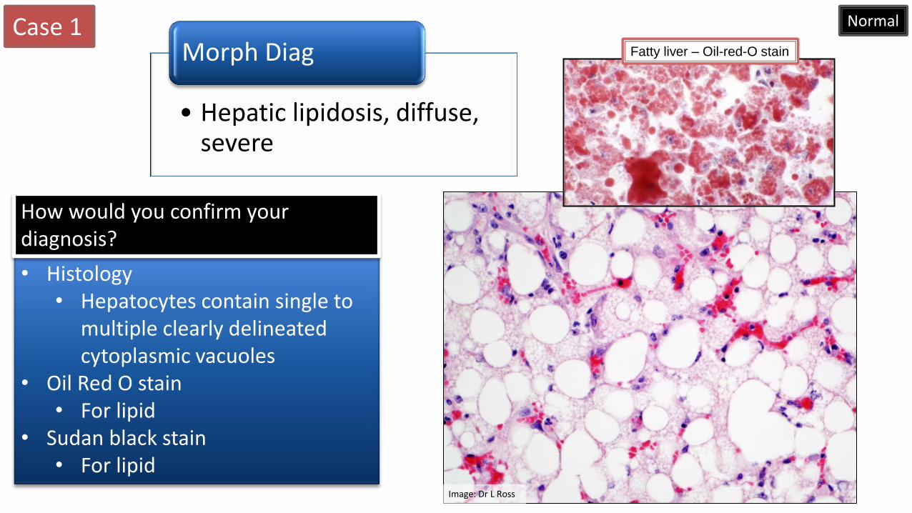
• Hepatic lipidosis, diffuse, severe
Morph Diag
• Histology • Hepatocytes contain single to
multiple clearly delineated cytoplasmic vacuoles
• Oil Red O stain • For lipid
• Sudan black stain • For lipid
How would you confirm your diagnosis?
Normal
Image: Dr L Ross
Fatty liver – Oil-red-O stain
Case 1
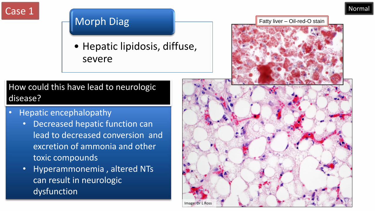
• Hepatic lipidosis, diffuse, severe
Morph Diag
• Hepatic encephalopathy • Decreased hepatic function can
lead to decreased conversion and excretion of ammonia and other toxic compounds
• Hyperammonemia , altered NTs can result in neurologic dysfunction
How could this have lead to neurologic disease?
Normal
Image: Dr L Ross
Fatty liver – Oil-red-O stain

Case 1 What are some causes for this change (sheep, cattle, cats, dogs, and horses)?
Sheep
• Ketosis
• Cobalt deficiency
• Dietary excess
• Toxins
Cattle
• Ketosis
• Fatty liver syndrome
• Toxins
• Dietary excess
Feline
• Dietary excess
• Fatty liver syndrome
• Diabetes
• Toxins
Dog
• Dietary excess
• Diabetes
• Hypothyroidism
• Toxins
Equine
• Equine hepatic lipidosis
• Dietary excess
• Toxins
Case 2
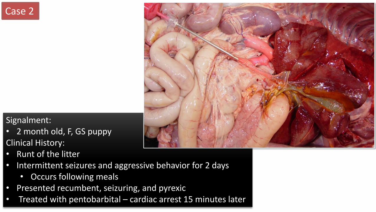
Signalment: • 2 month old, F, GS puppy Clinical History: • Runt of the litter • Intermittent seizures and aggressive behavior for 2 days
• Occurs following meals • Presented recumbent, seizuring, and pyrexic • Treated with pentobarbital – cardiac arrest 15 minutes later
Case 2

Case 2
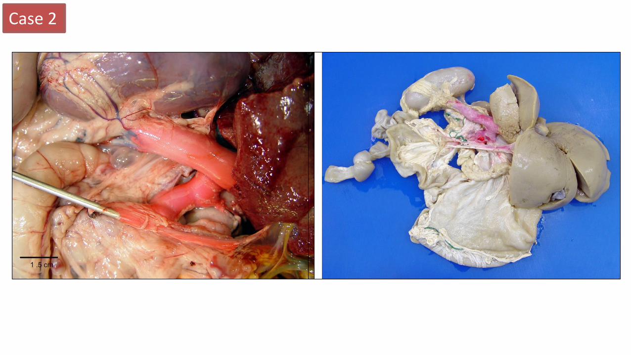
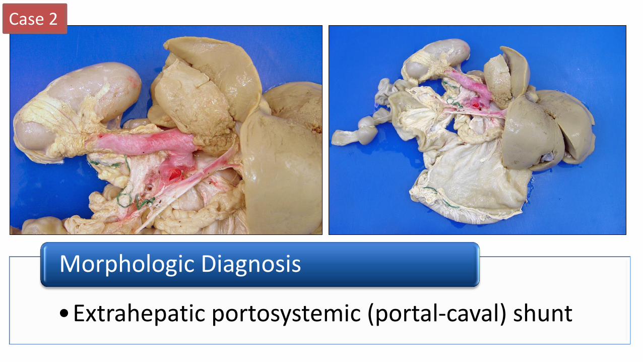
• The portal vein is markedly reduced in dimeter and is connected to the caudal vena cava via a large (1 cm diameter) vessel
Description
Case 2
•Extrahepatic portosystemic (portal-caval) shunt
Morphologic Diagnosis
Case 2
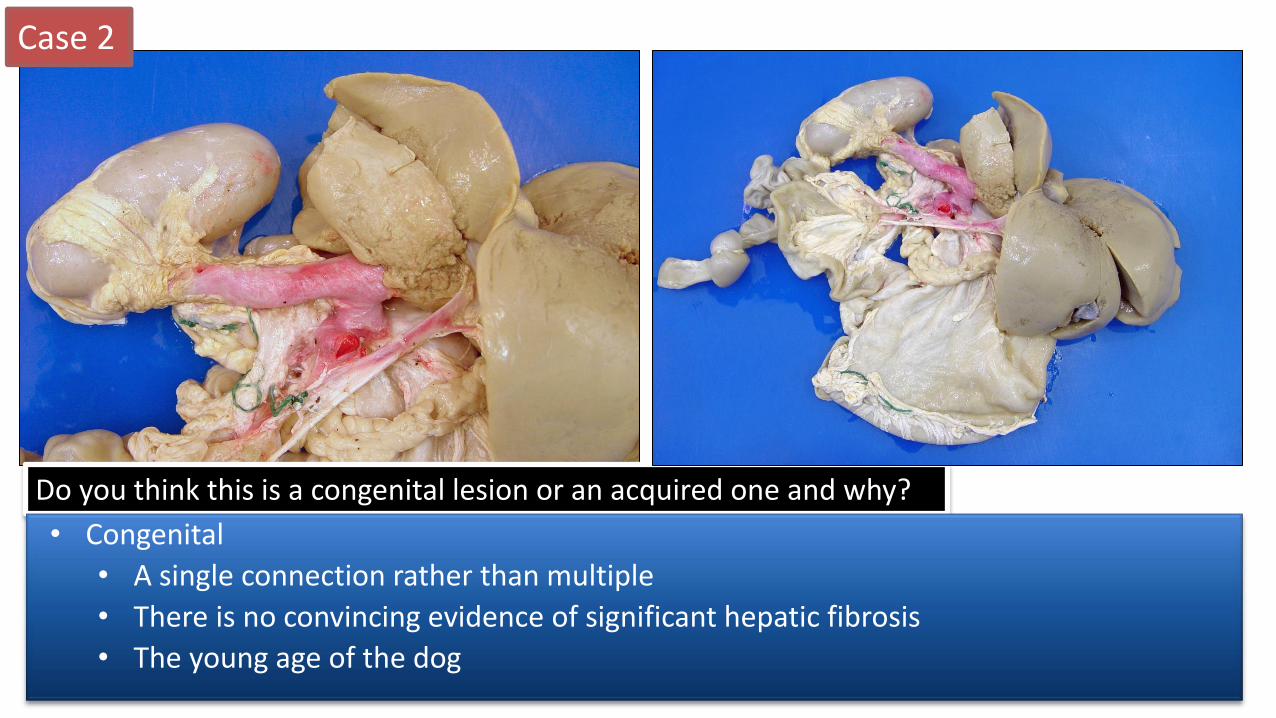
Do you think this is a congenital lesion or an acquired one and why?
• Congenital
• A single connection rather than multiple
• There is no convincing evidence of significant hepatic fibrosis
• The young age of the dog
Case 2
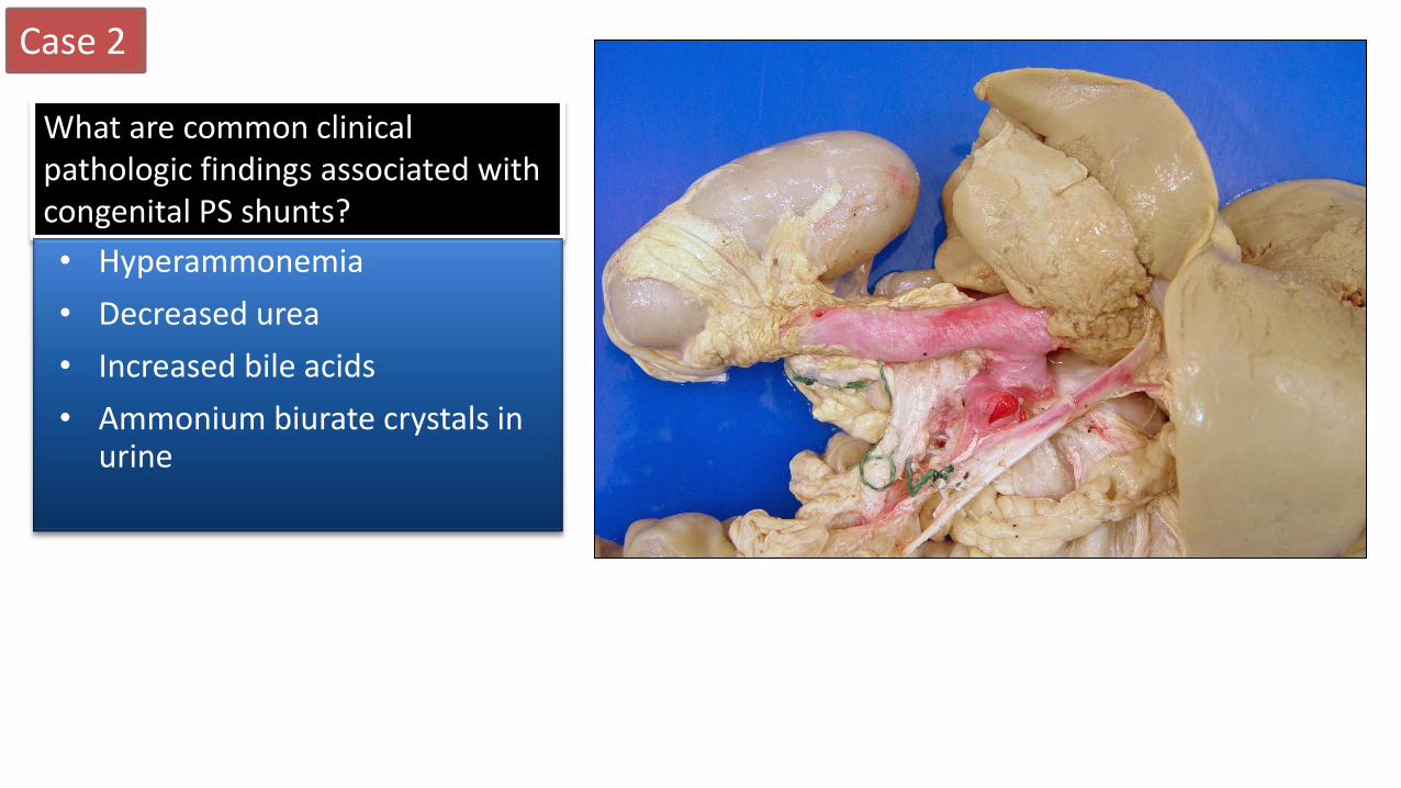
What are common clinical pathologic findings associated with congenital PS shunts?
• Hyperammonemia
• Decreased urea
• Increased bile acids
• Ammonium biurate crystals in urine
Case 2
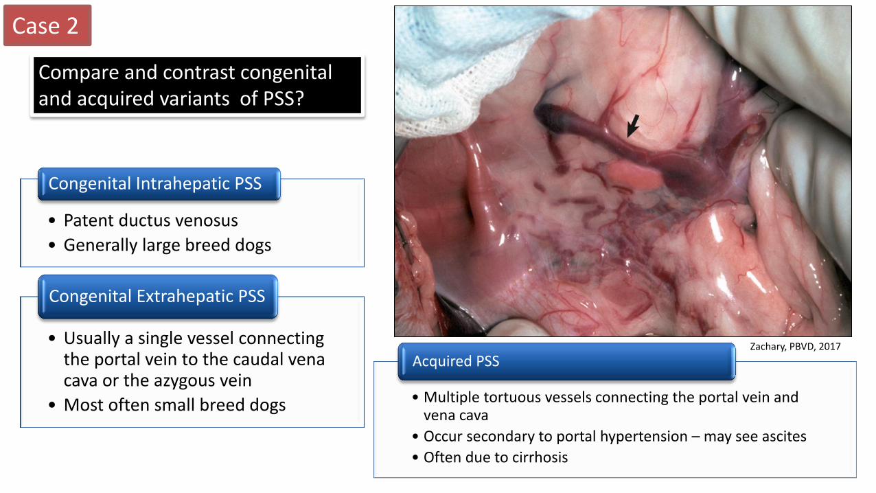
Compare and contrast congenital and acquired variants of PSS?
• Multiple tortuous vessels connecting the portal vein and vena cava
• Occur secondary to portal hypertension – may see ascites
• Often due to cirrhosis
Acquired PSS
• Patent ductus venosus
• Generally large breed dogs
Congenital Intrahepatic PSS
• Usually a single vessel connecting the portal vein to the caudal vena cava or the azygous vein
• Most often small breed dogs
Congenital Extrahepatic PSS
Zachary, PBVD, 2017
Case 3
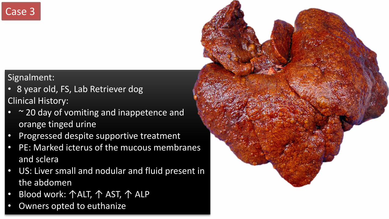
Signalment: • 8 year old, FS, Lab Retriever dog Clinical History: • ~ 20 day of vomiting and inappetence and
orange tinged urine • Progressed despite supportive treatment • PE: Marked icterus of the mucous membranes
and sclera • US: Liver small and nodular and fluid present in
the abdomen • Blood work: ↑ALT, ↑ AST, ↑ ALP • Owners opted to euthanize
Case 3
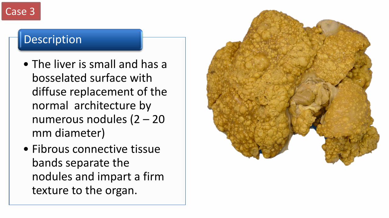
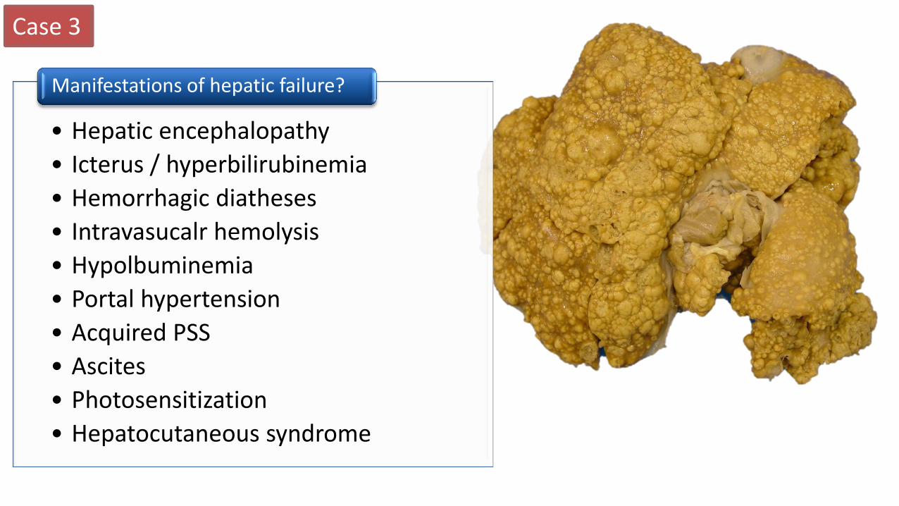
• The liver is small and has a bosselated surface with diffuse replacement of the normal architecture by numerous nodules (2 – 20 mm diameter)
• Fibrous connective tissue bands separate the nodules and impart a firm texture to the organ.
Description
Case 3
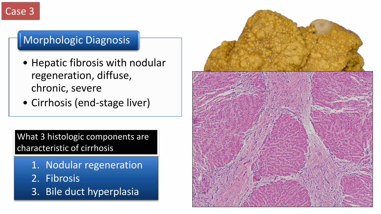
• Hepatic fibrosis with nodular regeneration, diffuse, chronic, severe
• Cirrhosis (end-stage liver)
Morphologic Diagnosis
1. Nodular regeneration 2. Fibrosis 3. Bile duct hyperplasia
What 3 histologic components are characteristic of cirrhosis
Case 3
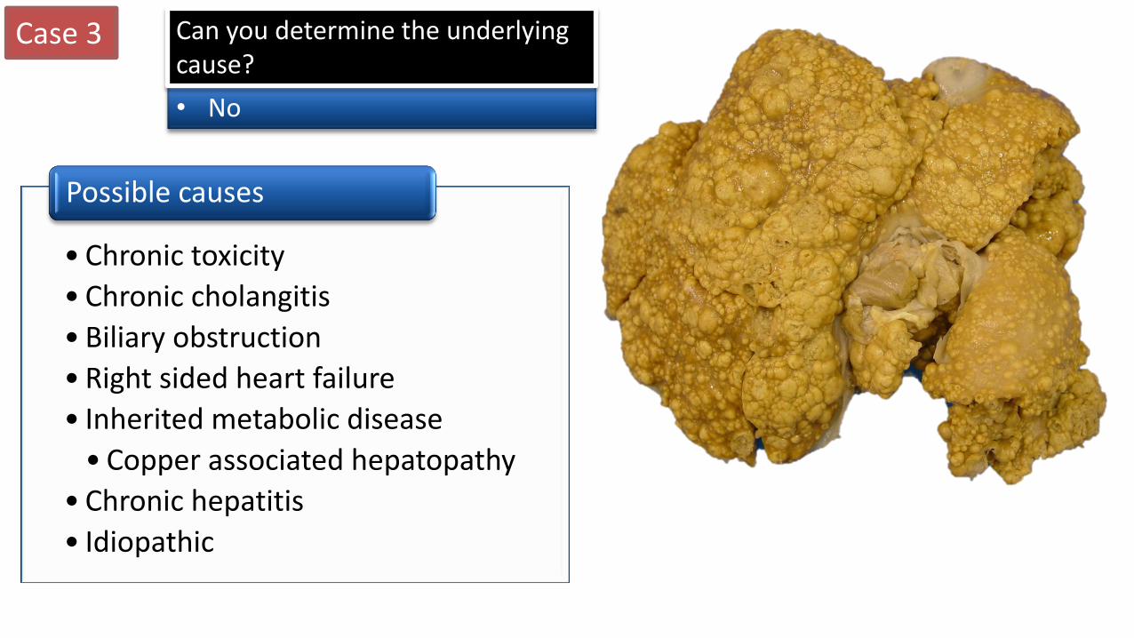
• No
Can you determine the underlying cause?
• Chronic toxicity
• Chronic cholangitis
• Biliary obstruction
• Right sided heart failure
• Inherited metabolic disease
• Copper associated hepatopathy
• Chronic hepatitis
• Idiopathic
Possible causes
Case 3
• Hepatic encephalopathy
• Icterus / hyperbilirubinemia
• Hemorrhagic diatheses
• Intravasucalr hemolysis
• Hypolbuminemia
• Portal hypertension
• Acquired PSS
• Ascites
• Photosensitization
• Hepatocutaneous syndrome
Manifestations of hepatic failure?
Case 4
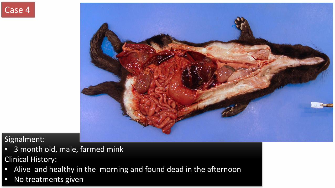
Signalment: • 3 month old, male, farmed mink Clinical History: • Alive and healthy in the morning and found dead in the afternoon • No treatments given
Case 4

Case 4
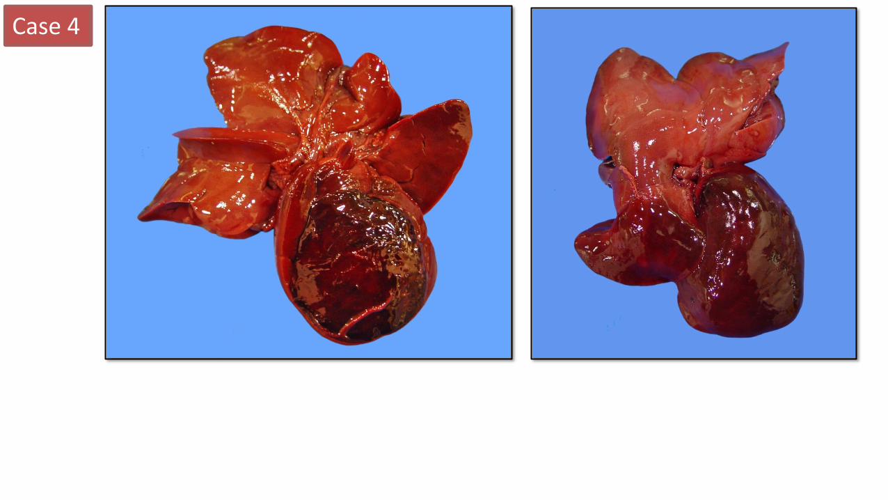
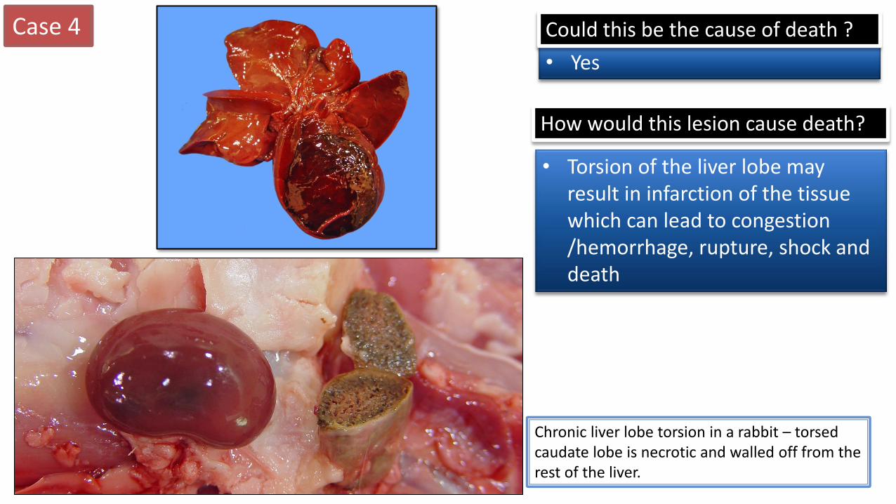
• The left lateral lobe of the liver is markedly enlarged, dark brown (red), with a roughened irregular surface and is twisted ~ 360 degree around its base
• Indentations are present on the capsular surface of many lobes (artefact).
Description
Case 4
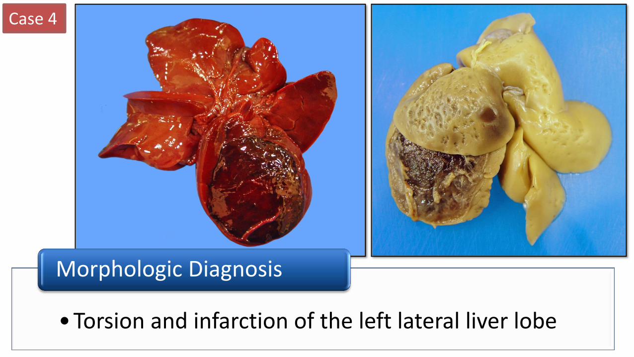
•Torsion and infarction of the left lateral liver lobe
Morphologic Diagnosis
Case 4
• Yes
Could this be the cause of death ?
• Torsion of the liver lobe may result in infarction of the tissue which can lead to congestion /hemorrhage, rupture, shock and death
How would this lesion cause death?
Chronic liver lobe torsion in a rabbit – torsed caudate lobe is necrotic and walled off from the rest of the liver.
Case 5
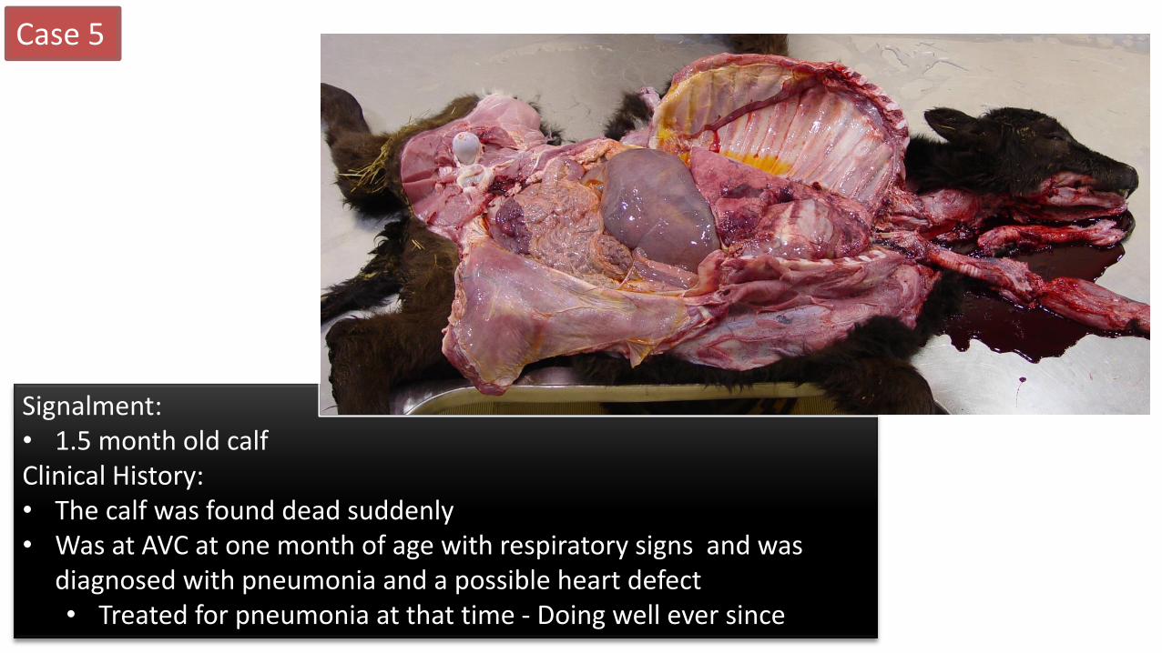
Signalment: • 1.5 month old calf Clinical History: • The calf was found dead suddenly • Was at AVC at one month of age with respiratory signs and was
diagnosed with pneumonia and a possible heart defect • Treated for pneumonia at that time - Doing well ever since
Case 5
Case 5
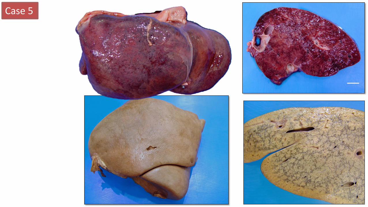
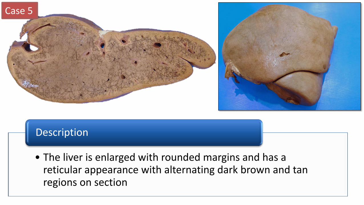
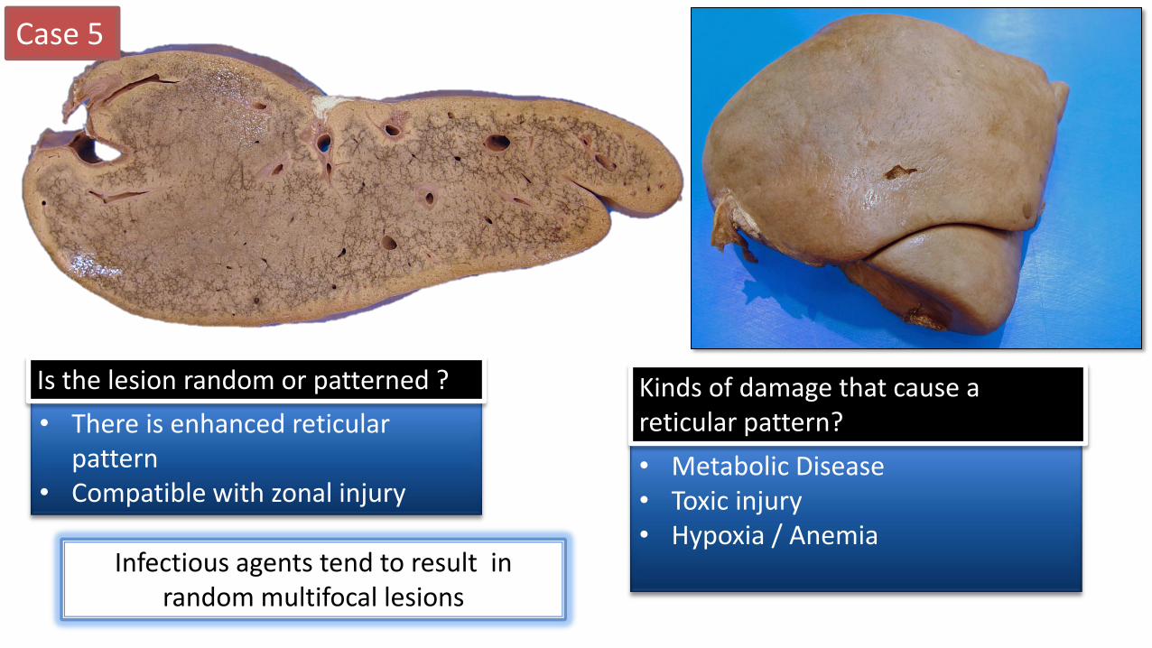
• The liver is enlarged with rounded margins and has a reticular appearance with alternating dark brown and tan regions on section
Description
Case 5
• There is enhanced reticular pattern
• Compatible with zonal injury
Is the lesion random or patterned ?
• Metabolic Disease • Toxic injury • Hypoxia / Anemia
Kinds of damage that cause a reticular pattern?
Infectious agents tend to result in random multifocal lesions
Case 5
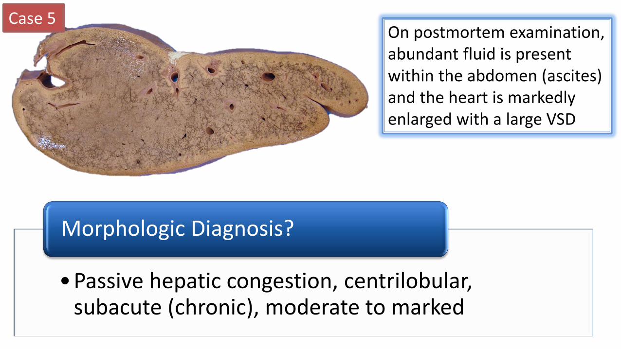
•Passive hepatic congestion, centrilobular, subacute (chronic), moderate to marked
Morphologic Diagnosis?
On postmortem examination, abundant fluid is present within the abdomen (ascites) and the heart is markedly enlarged with a large VSD
Case 5
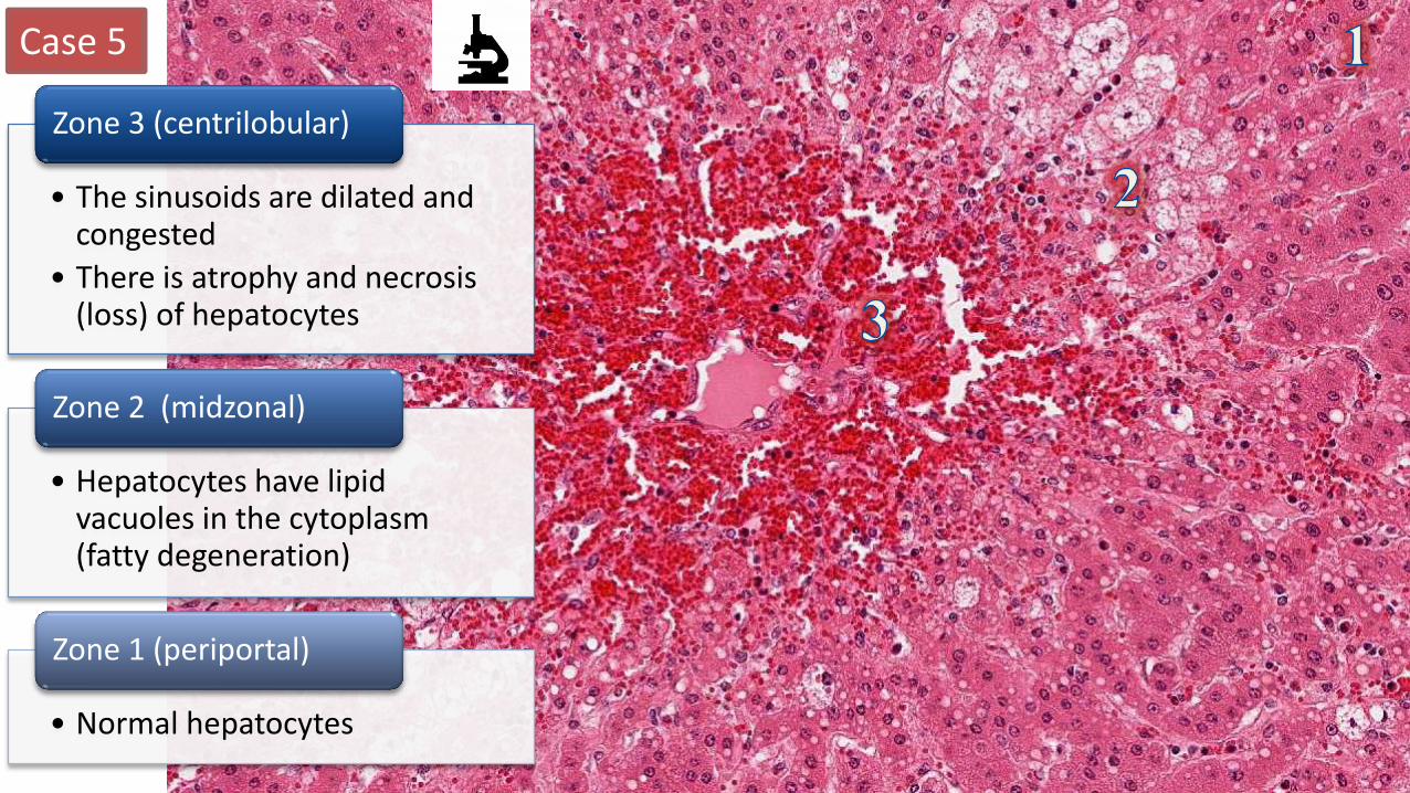
• The sinusoids are dilated and congested
• There is atrophy and necrosis (loss) of hepatocytes
Zone 3 (centrilobular)
• Hepatocytes have lipid vacuoles in the cytoplasm (fatty degeneration)
Zone 2 (midzonal)
• Normal hepatocytes
Zone 1 (periportal)
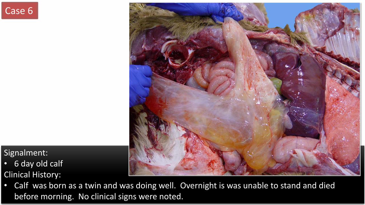
Signalment: • 6 day old calf Clinical History: • Calf was born as a twin and was doing well. Overnight is was unable to stand and died
before morning. No clinical signs were noted.
Case 6
Case 6
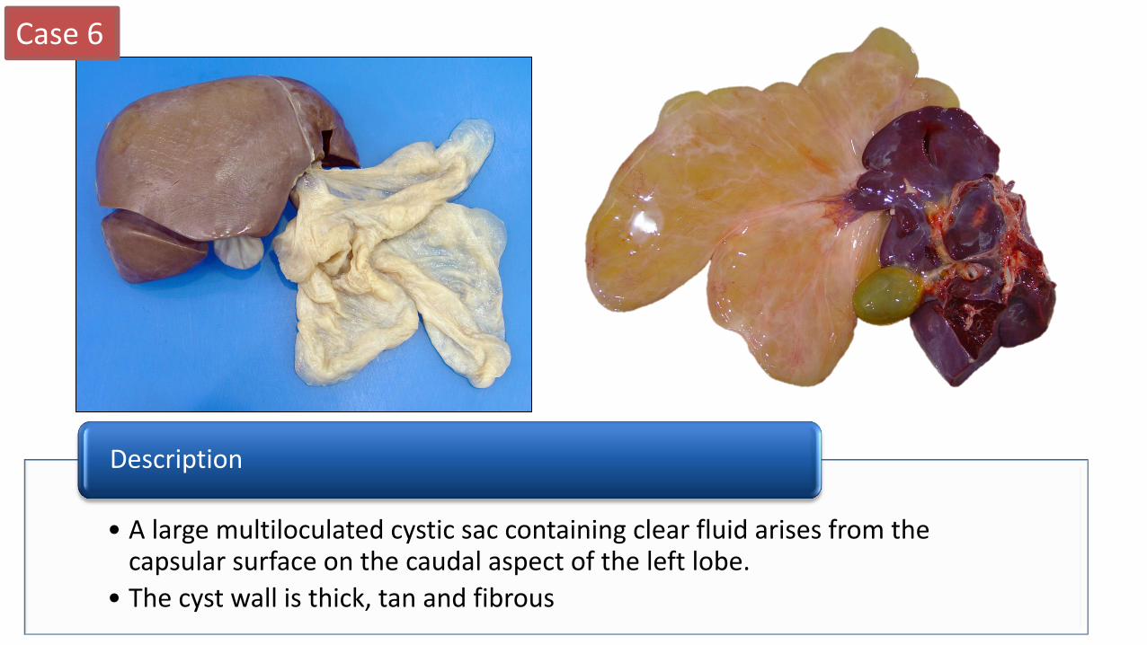
• A large multiloculated cystic sac containing clear fluid arises from the capsular surface on the caudal aspect of the left lobe.
• The cyst wall is thick, tan and fibrous
Description
Case 6
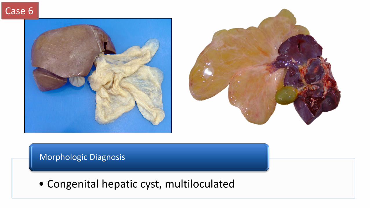
• Congenital hepatic cyst, multiloculated
Morphologic Diagnosis
Case 6
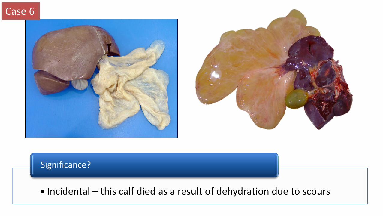
• Incidental – this calf died as a result of dehydration due to scours
Significance?
Case 6
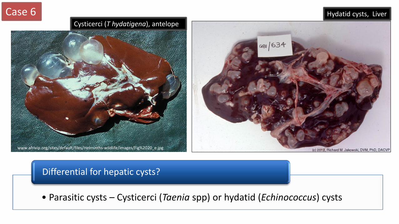
• Parasitic cysts – Cysticerci (Taenia spp) or hydatid (Echinococcus) cysts
Differential for hepatic cysts?
Hydatid cysts, Liver
Cysticerci (T hydatigena), antelope
www.afrivip.org/sites/default/files/Helminths-wildlife/images/Fig%2020_e.jpg
Related Documents