IOP PUBLISHING PHYSICS IN MEDICINE AND BIOLOGY Phys. Med. Biol. 52 (2007) 5187–5204 doi:10.1088/0031-9155/52/17/006 List-mode-based reconstruction for respiratory motion correction in PET using non-rigid body transformations F Lamare 1,2 , M J Ledesma Carbayo 3 , T Cresson 1 , G Kontaxakis 3 , A Santos 3 , C Cheze Le Rest 1 , A J Reader 4 and D Visvikis 1 1 INSERM, U650, Laboratoire du Traitement de l’Information M´ edicale (LaTIM), Brest, F-29200 France 2 MRC Clinical Sciences Centre, Imperial College, Faculty of Medicine, Hammersmith Hospital, London, UK 3 ETSI Telecomunicacion Universidad Politecnica de Madrid, Ciudad Universitaria s/n 28040, Madrid, Spain 4 School of Chemical Engineering & Analytical Science, The University of Manchester, Manchester, UK Received 14 March 2007, in final form 2 July 2007 Published 9 August 2007 Online at stacks.iop.org/PMB/52/5187 Abstract Respiratory motion in emission tomography leads to reduced image quality. Developed correction methodology has been concentrating on the use of respiratory synchronized acquisitions leading to gated frames. Such frames, however, are of low signal-to-noise ratio as a result of containing reduced statistics. In this work, we describe the implementation of an elastic transformation within a list-mode-based reconstruction for the correction of respiratory motion over the thorax, allowing the use of all data available throughout a respiratory motion average acquisition. The developed algorithm was evaluated using datasets of the NCAT phantom generated at different points throughout the respiratory cycle. List-mode-data-based PET-simulated frames were subsequently produced by combining the NCAT datasets with Monte Carlo simulation. A non-rigid registration algorithm based on B-spline basis functions was employed to derive transformation parameters accounting for the respiratory motion using the NCAT dynamic CT images. The displacement matrices derived were subsequently applied during the image reconstruction of the original emission list mode data. Two different implementations for the incorporation of the elastic transformations within the one-pass list mode EM (OPL-EM) algorithm were developed and evaluated. The corrected images were compared with those produced using an affine transformation of list mode data prior to reconstruction, as well as with uncorrected respiratory motion average images. Results demonstrate that although both correction techniques considered lead to significant improvements in accounting for respiratory motion artefacts in the lung fields, the elastic-transformation-based correction 0031-9155/07/175187+18$30.00 © 2007 IOP Publishing Ltd Printed in the UK 5187

Welcome message from author
This document is posted to help you gain knowledge. Please leave a comment to let me know what you think about it! Share it to your friends and learn new things together.
Transcript

IOP PUBLISHING PHYSICS IN MEDICINE AND BIOLOGY
Phys. Med. Biol. 52 (2007) 5187–5204 doi:10.1088/0031-9155/52/17/006
List-mode-based reconstruction for respiratorymotion correction in PET using non-rigid bodytransformations
F Lamare1,2, M J Ledesma Carbayo3, T Cresson1, G Kontaxakis3,A Santos3, C Cheze Le Rest1, A J Reader4 and D Visvikis1
1 INSERM, U650, Laboratoire du Traitement de l’Information Medicale (LaTIM),Brest, F-29200 France2 MRC Clinical Sciences Centre, Imperial College, Faculty of Medicine, Hammersmith Hospital,London, UK3 ETSI Telecomunicacion Universidad Politecnica de Madrid, Ciudad Universitaria s/n 28040,Madrid, Spain4 School of Chemical Engineering & Analytical Science, The University of Manchester,Manchester, UK
Received 14 March 2007, in final form 2 July 2007Published 9 August 2007Online at stacks.iop.org/PMB/52/5187
AbstractRespiratory motion in emission tomography leads to reduced image quality.Developed correction methodology has been concentrating on the use ofrespiratory synchronized acquisitions leading to gated frames. Such frames,however, are of low signal-to-noise ratio as a result of containing reducedstatistics. In this work, we describe the implementation of an elastictransformation within a list-mode-based reconstruction for the correction ofrespiratory motion over the thorax, allowing the use of all data availablethroughout a respiratory motion average acquisition. The developed algorithmwas evaluated using datasets of the NCAT phantom generated at different pointsthroughout the respiratory cycle. List-mode-data-based PET-simulated frameswere subsequently produced by combining the NCAT datasets with MonteCarlo simulation. A non-rigid registration algorithm based on B-spline basisfunctions was employed to derive transformation parameters accounting forthe respiratory motion using the NCAT dynamic CT images. The displacementmatrices derived were subsequently applied during the image reconstructionof the original emission list mode data. Two different implementations forthe incorporation of the elastic transformations within the one-pass list modeEM (OPL-EM) algorithm were developed and evaluated. The corrected imageswere compared with those produced using an affine transformation of list modedata prior to reconstruction, as well as with uncorrected respiratory motionaverage images. Results demonstrate that although both correction techniquesconsidered lead to significant improvements in accounting for respiratorymotion artefacts in the lung fields, the elastic-transformation-based correction
0031-9155/07/175187+18$30.00 © 2007 IOP Publishing Ltd Printed in the UK 5187

5188 F Lamare et al
leads to a more uniform improvement across the lungs for different lesion sizesand locations.
1. Introduction
One of the parameters affecting quantitation in emission tomography (ET) imaging ofthe thoracic and abdominal regions is respiratory motion. Respiratory motion has beenshown to reduce the accuracy of determining functional lesion volumes and associatedrecovered activity concentrations (Nehmeh et al 2002, Boucher et al 2004) influencingpositron emission tomography (PET) applications such as radiotherapy treatment planningand response to therapy monitoring, respectively. Furthermore, the introduction of scanningdevices combining anatomical and functional imaging has revealed various artefacts in thefunctional images caused by the use of the anatomical datasets for attenuation correctionin combination with associated differences in the respiratory motion conditions during theacquisition of the CT and ET datasets (Goerres et al 2002, Visvikis et al 2004, Erdi et al2004).
In order to account for respiratory motion effects the gated acquisition of both CT andPET datasets has been suggested as a potential solution. Pan et al have demonstrated that theacquisition of a 4D CT can be equally used to derive a truly respiratory average CT that canbe subsequently used to correct a PET respiratory average dataset for attenuation effects inthe thoracic region (Pan et al 2005). On the other hand, using the 4D CT frames to correct arespiratory average PET dataset has been shown to lead to an activity concentration variationof over 30% depending on the phase of the CT frame used for the attenuation correction ofthe PET images (Erdi et al 2004). Similarly, an increase of 36% in the standardized uptakevalues (SUV) was observed by Nehmeh et al when using phase-matched 4D CT frames forthe attenuation correction of the corresponding 4D PET frames relative to the use of a staticCT scan (Nehmeh et al 2004). However, the result of such multi-frame acquisitions leads togated PET images suffering from poor signal-to-noise ratio since each of the frames containsonly part of the counts available throughout the acquisition of a respiration average PET study(Visvikis et al 2005). Therefore, the need exists for the development of methodology thatcorrects for respiratory motion effects between individual gated frames in order to allow theuse of the data available throughout a respiratory cycle.
Different authors have attempted to correct the effects of respiratory motion in cardiacET imaging through the use of either a rigid body transformation of list mode PET datasets(Livieratos et al 2005) or through tracking the centre of mass in single photon emissiontomography (SPECT) projections (Bruyant et al 2003). Although such relatively simplerespiratory motion models could be sufficient focusing on single organs such as the heart,it may be inadequate in oncology cases where the thoracic and diaphragmatic areas are ofinterest. In an attempt to make use of all data available throughout a respiratory gatedacquisition for such applications, numerous authors have previously suggested the use of 4DCT datasets to derive transformation maps that could be subsequently used to shift the detectedlines of response in the corresponding PET gated frames (Lamare et al 2005, 2007, Qiaoet al 2006, Li et al 2006). This work, based on the use of an affine transformation of list-modedata prior to reconstruction, has demonstrated that although this approach leads to significantimprovements in lesion contrast and position in the lung fields, it is impossible to use such amodel to account at the same time for respiratory motion effects in both the lung fields andorgans under the diaphragm. On the other hand, several authors have in the past explored
Elastic respiratory motion correction in PET list-mode reconstruction 5189
the use of elastic deformation algorithms to realign individual gated frames suggesting thatthe resulting deformed gated images could be subsequently summed together making use ofall the available data (Klein and Huesman 1997, Dawood et al 2006, Visvikis et al 2006).However, the summing together of individual gated frames following their reconstruction willobviously lead to inferior quality images than the incorporation of such deformations into thereconstruction process, particularly in the case of using iterative reconstruction algorithmswhich have become a standard in current clinical practice (Visvikis et al 2001, Schoder et al2004, Asma et al 2006).
The use of list mode data acquisitions for gated PET may facilitate the a posterioribinning of the acquired data. This in turn allows fine temporal sampling and the potentialof implementing different binning methodologies reducing the effect of generally observedirregular respiratory motion patterns (Bruyant et al 2006). Although the application of a rigid oraffine transformation in the raw data domain is feasible considering individual lines of response(Livieratos et al 2005, Lamare et al 2007), a similar approach for elastic transformation posesobvious challenges. In the past, different approaches for the incorporation of transformationsin the system matrix during the reconstruction process have been described (Qi and Huesman2002, Jacobson and Fessler 2003, Rahmim et al 2004, Lamare et al 2005, Qiao et al 2006,Li et al 2006). While the work of Qi and Huesman as well as Rahmim et al consideredonly rigid body transformations, Jacobson and Fessler described the theoretical framework ofincorporating non-rigid transformations in the reconstruction algorithm without evaluating theproposed methodology. In addition, Qiao et al and Li et al evaluated their proposed algorithmon a phantom study simulating only rigid body motion, therefore not allowing the evaluationof elastic transformations in the performance of their algorithm implementation. Finally, thepatient study included in Li et al was at the level of the pancreas with limited respiratorymotion extend of approximately 1 cm and of limited non-rigid nature. The objectives of thisstudy are multiple. Firstly, two different implementations of a list-mode-based reconstructionalgorithm incorporating elastic deformations in the system matrix are described and comparedusing an anthropomorphic phantom, which includes the non-rigid effects of physiologicalmotion at the level of the lung and the diaphragm. Lesions at different locations throughoutthe lung and liver were included to assess the impact of this correction approach for oncologyapplications. Secondly, the performance for respiratory motion correction of incorporatingelastic transformations in the reconstruction process is compared to the use of an affinetransformation applied prior to the reconstruction process (Lamare et al 2007). Finally, weestablish the qualitative and quantitative image improvements associated with applying thedeformation during the reconstruction process of the raw data rather than applying them inindividual gated frames which are subsequently summed.
2. Materials and methods
2.1. Simulation study
A digital NURBS-based 4D cardiac-torso phantom (NCAT) was used in the simulation ofPET respiratory gated acquisitions (Segars et al 2001). A number of different size lesions(7, 11, 15 and 21 mm) were included at different locations throughout the lungs. Normal FDGactivity levels were placed in the lung and liver fields, while a tumour to background ratioof 8 to 1 was used. Eight NCAT emission images were produced, corresponding to 0.625 sconsidering a normal respiratory cycle of 5 s. The first frame represented full exhalation,while the maximum magnitude of respiratory motion (full inspiration) is occurring betweenthe 4th and 5th frames. The model of a clinical PET system (Lamare et al 2006) developed

5190 F Lamare et al
with GATE (Geant4 Application for Tomographic Emission) was combined with the generatedNCAT phantom frames in order to obtain dynamic emission images throughout the respiratorycycle. The attenuation images of the NCAT phantom were also integrated in order to simulatethe effects of attenuation. Finally, the data simulated for each individual frame was saved in alist mode format.
2.2. Transformation fields
In the clinical case, 3D spatial transformations to be used for the correction of respiratorymotion may be obtained through the use of dynamic (respiratory gated) PET/CT datasets,as a number of different authors have already demonstrated (Nehmeh et al 2004, Klein andHuesman 1997, Dawood et al 2006, Visvikis et al 2006). In the simulation study presentedhere, the generated NCAT attenuation/CT images without any blurring effects (resolution,image statistics effects, etc) were used in combination with an elastic or affine registrationalgorithm to define the transformation fields to be used during the reconstruction of thesimulated list mode emission datasets. Deformation matrices were derived between allindividual frames (frames 2–8) and that corresponding to full exhalation (i.e. frame 1, seesection 2.1), referred to from here onwards as the reference frame. In the case of the affineregistration a normalized mutual information algorithm was employed (Studholme et al 1999,Lamare et al 2007). On the other hand, respiratory motion compensation fields were alsoobtained performing independent non-rigid registration processes (Ledesma-Carbayo et al2006) of the NCAT CT frames. The transformation gt(x) between frames 2–8 f (x, t) andthe reference f (x, 0) frame was defined as a linear combination of B-spline basis functions,located in a rectangular grid (Kybic and Unser 2003, Ledesma-Carbayo et al 2006, Sorzanoet al 2005):
Dt ≡ gt(x) = x +∑j∈Z
N
cjβr (x/h − j) (1)
where βr(x) is a tensor product of centred B-splines of degree r and j are the indices of thegrid locations. The spacing between grid points h determines the number of parameters cj
to be optimized and the final rigidity of the solution. The registration is then formulatedas an optimization procedure that minimizes the sum of squared differences metric to findthe best transformation parameters cj. The optimization used a variation of the Marquart–Levenverg nonlinear least-squares optimization in combination with an efficient estimationof the Hessian matrix (Broyden–Fletcher–Goldfarb–Shanno (BFGS)) (Sorzano et al 2005).Spline interpolation provides a continuous function of the discrete images, allowing anexcellent framework for finding a subpixel solution and to compute analytically the derivativesneeded in the optimization process. Speed and robustness are improved by the use of amultiresolution approach in both the image and the transformation space. The multiresolutionmethodology used creates a pyramid of subsampled images optimal in the L2-sense takingadvantage of the spline representation (Unser et al 1993). The problem is solved starting atthe coarser level of the pyramid (the most subsampled image) and proceeding to the finestlevel. For each image level two levels of deformation are optimized also following a coarse tofine strategy.
In order to choose the optimal parameters for the particular application in this paper,we performed a set of initial tests. Three different transformation grid spacings were tested:h = 8, h = 16 and h = 32 pixels. As a result, cubic B-splines with a grid spacing set to8 × 8 × 8 pixels (8 pixels ∼25 mm) provided the best results and were used to representthe deformation. Cubic B-splines were also used for image interpolation and for providing
Elastic respiratory motion correction in PET list-mode reconstruction 5191
a continuous representation. The multi-scale processing scheme was set at four levels withsuccessive levels having half size in each dimension.
2.3. Image reconstruction on list-mode data
The one-pass list mode EM (OPL-EM) algorithm (Reader et al 2002) was implemented forthe reconstruction of the transformed LORs:
nk+1j = nk
j
sj
∑i∈T k
pij
1
qki
for k = 1, . . . , K (2)
where qki = ∑J
j=1 pijnkj is the expected count in LOR i, pij is the purely geometric term and
represents the geometric probability of detecting at LOR i an event generated in voxel j, nj
is the intensity of voxel j, J is the total number of voxels, sj is the voxel j of the sensitivityimage including the normalization and attenuation corrections and K is the number of timesubsets. k is both the iteration number and the subset used in that iteration. T k is the set oflist mode events in the kth subset. The forward projection step was implemented using theaccelerated version of the Siddon ray tracing (Siddon 1986, Han et al 1999, Zhao and Reader2002).
An overall sensitivity image S including the normalization and attenuation correctionsfor each of the individual simulated frames was produced through a forward projection andbackprojection of the corresponding NCAT attenuation images. Each voxel sj of the sensitivityimage S is computed as follows:
sj = 1
Nframes
∑Nframes
∑i∈I
pijNiAi , with Ai = exp
−
J∑j=1
pijµj
(3)
where Ni is the normalization term of the LOR i, Ai is the attenuation correction factor of theLOR i, I is the total number of detectable LORs, J is the total number of voxels and µj isthe linear attenuation coefficient at the energy of 511 keV (µj is the intensity of the voxel j inthe NCAT attenuation image) and Nframes is the number of temporal gated frames.
2.4. Implementation of elastic transformations in the system matrix
The elastic motion correction can be integrated in a mathematical representation of the systemmatrix in the PET reconstruction process. If one notes P the system matrix usually describingthe PET system, and whose elements pij represent the geometric probability of detecting atLOR i an event generated in voxel j , the data acquisition in PET can be represented by theequation
m = Pf (4)
where m are the measured datasets and f is the radioactive distribution. If t0 corresponds to thereference frame time, the system matrix can be modified to take into account the deformationof the radioactive distribution from time t to time t0 giving
mt = Ptf (5)
where mt are the measured datasets at time t and f remains the radioactive distribution hadthe object not moved (i.e corresponding to the reference frame time). The number of systemmatrices, Pt , corresponds to the number of available PET/CT synchronized frames.
This elastic-based respiratory motion correction can be integrated in any reconstructionprocess using a representation of the system matrix as described in equation (4), allowing
5192 F Lamare et al
(a) (b)
(c) (d) (e)
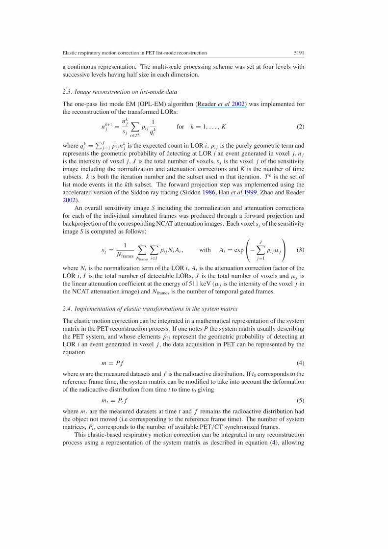
Figure 1. Comparison of the Frame 1 (a), Non-Corrected (b), LORs-Affine (c), Elastic Method 1(d) and Elastic Method 2 (e) images. Visual differences can be clearly seen at the levels of the liverand the heart.
CompensationMotion
LOR i
Motion duringacquisition
atij
a0ij ′
(a)
CompensationMotion
acquisitionMotion during
LOR i
atij
a0ij ′
(b)
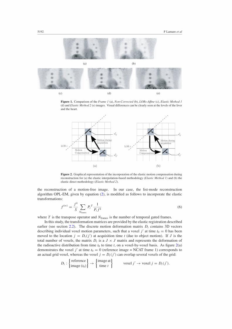
Figure 2. Graphical representation of the incorporation of the elastic motion compensation duringreconstruction for (a) the elastic interpolation-based methodology (Elastic Method 1) and (b) theelastic direct methodology (Elastic Method 2).
the reconstruction of a motion-free image. In our case, the list-mode reconstructionalgorithm OPL-EM, given by equation (2), is modified as follows to incorporate the elastictransformations:
f k+1 = f k
S
∑Nframes
PtT 1
Ptf k(6)
where T is the transpose operator and Nframes is the number of temporal gated frames.In this study, the transformation matrices are provided by the elastic registration described
earlier (see section 2.2). The discrete motion deformation matrix Dt contains 3D vectorsdescribing individual voxel motion parameters, such that a voxel j ′ at time t0 = 0 has beenmoved to the location j = Dt(j
′) at acquisition time t (due to object motion). If J is thetotal number of voxels, the matrix Dt is a J × J matrix and represents the deformation ofthe radioactive distribution from time t0 to time t, on a voxel-by-voxel basis. As figure 2(a)demonstrates the voxel j ′ at time t0 = 0 (reference image = NCAT frame 1) corresponds toan actual grid voxel, whereas the voxel j = Dt(j
′) can overlap several voxels of the grid:
Dt :
{referenceimage (t0)
}→
{image attime t
}voxel j ′ → voxel j = Dt(j
′).
Elastic respiratory motion correction in PET list-mode reconstruction 5193
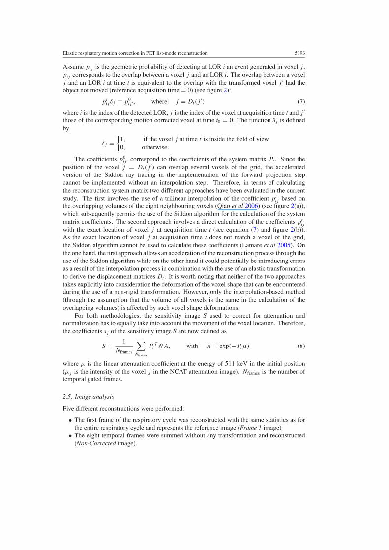
Assume pij is the geometric probability of detecting at LOR i an event generated in voxel j .pij corresponds to the overlap between a voxel j and an LOR i. The overlap between a voxelj and an LOR i at time t is equivalent to the overlap with the transformed voxel j ′ had theobject not moved (reference acquisition time = 0) (see figure 2):
ptij δj ≡ p0
ij ′ , where j = Dt(j′) (7)
where i is the index of the detected LOR, j is the index of the voxel at acquisition time t and j ′
those of the corresponding motion corrected voxel at time t0 = 0. The function δj is definedby
δj ={
1, if the voxel j at time t is inside the field of view0, otherwise.
The coefficients p0ij ′ correspond to the coefficients of the system matrix Pt . Since the
position of the voxel j = Dt(j′) can overlap several voxels of the grid, the accelerated
version of the Siddon ray tracing in the implementation of the forward projection stepcannot be implemented without an interpolation step. Therefore, in terms of calculatingthe reconstruction system matrix two different approaches have been evaluated in the currentstudy. The first involves the use of a trilinear interpolation of the coefficient pt
ij based onthe overlapping volumes of the eight neighbouring voxels (Qiao et al 2006) (see figure 2(a)),which subsequently permits the use of the Siddon algorithm for the calculation of the systemmatrix coefficients. The second approach involves a direct calculation of the coefficients pt
ij
with the exact location of voxel j at acquisition time t (see equation (7) and figure 2(b)).As the exact location of voxel j at acquisition time t does not match a voxel of the grid,the Siddon algorithm cannot be used to calculate these coefficients (Lamare et al 2005). Onthe one hand, the first approach allows an acceleration of the reconstruction process through theuse of the Siddon algorithm while on the other hand it could potentially be introducing errorsas a result of the interpolation process in combination with the use of an elastic transformationto derive the displacement matrices Dt . It is worth noting that neither of the two approachestakes explicitly into consideration the deformation of the voxel shape that can be encounteredduring the use of a non-rigid transformation. However, only the interpolation-based method(through the assumption that the volume of all voxels is the same in the calculation of theoverlapping volumes) is affected by such voxel shape deformations.
For both methodologies, the sensitivity image S used to correct for attenuation andnormalization has to equally take into account the movement of the voxel location. Therefore,the coefficients sj of the sensitivity image S are now defined as
S = 1
Nframes
∑Nframes
PtT NA, with A = exp(−Ptµ) (8)
where µ is the linear attenuation coefficient at the energy of 511 keV in the initial position(µj is the intensity of the voxel j in the NCAT attenuation image). Nframes is the number oftemporal gated frames.
2.5. Image analysis
Five different reconstructions were performed:
• The first frame of the respiratory cycle was reconstructed with the same statistics as forthe entire respiratory cycle and represents the reference image (Frame 1 image)
• The eight temporal frames were summed without any transformation and reconstructed(Non-Corrected image).

5194 F Lamare et al
• Each of the list-mode files corresponding to the last seven frames were correctedusing the affine registration parameters. They were subsequently summed together andreconstructed using the standard OPL-EM algorithm (LORs-Affine image) (Lamare et al2007).
• The elastic transformation fields were included during the reconstruction of thecorresponding list-mode datasets (Elastic Method 1 and Elastic Method 2 images,corresponding to the implementation of the methodology with and without interpolation,respectively, as described in section 2.4).
• Each of the individual temporal gated frames were reconstructed. The reconstructedimages corresponding to the last seven frames were corrected using the elastic parameterscited above and were subsequently summed together (gated-Frames image).
Two reconstructed image sizes were considered for the different lesion diameters. Imagesof 128 × 128 × 60 (voxel size of 3.125 × 3.125 × 3.125 mm3) and 256 × 256 × 120 (voxelsize of 1.5625 × 1.5625 × 1.5625 mm3) were obtained for each of the reconstructed temporalNCAT frames with the lung lesions of 15, 21 mm and 7, 11 mm in diameter, respectively. Fora list mode dataset of 10 million detected coincidences, an iteration of the OPL-EM algorithmwas <4 min for an image of the above dimensions (Intel Xeon 3 GHz dual core). A total ofseven iterations were found to be optimum for the reconstruction of the NCAT images. Usingthe interpolation- and the non-interpolation-based approaches in the implementation for theincorporation of the non-rigid transformations in the reconstruction system matrix increasedthe time of acquisition by less than 5% and over a factor of 10, respectively.
The motion corrected images using the affine model and the two different implementationsof the elastic model were compared to the first temporal frame (Frame 1 = reference image).The total number of coincidences were kept the same for all images in order to distinguishthe effects purely associated with motion, rather than including those arising from differencesin statistical quality between the single temporal frame (including only part of the data) andthe corrected frames (including all of the available data). In order to better quantify motioncompensation, we have also compared the motion corrected images with the respiratoryaverage image (Non-Corrected).
The percent relative difference (PRD) of the radioactivity concentration (Lamare et al2007) was used to quantify the improvement in terms of contrast, position and FWHM of thesimulated lesions. The PRD is computed as
%PRD =∣∣∣∣Evaluated − Frame 1
Frame 1
∣∣∣∣ × 100 (9)
where the variable Evaluated can be either the LORs-Affine, the gated-Frames, the Non-Corrected or the Elastic image. Based on the calculated PRD, the contrast and FWHMimprovements as a result of the respiratory correction are computed using
%improvement = PRDCorrected − PRDNon-Corrected
PRDNon-Corrected× 100 (10)
where the variable Corrected can be either the LORs-Affine, the gated-Frames or the Elasticimage.
To assess the improvement in terms of contrast in the reconstructed images, regions ofinterest (ROI) were placed in each of the lung lesions and in the background lung. The slicewith the maximum count density over the lesion was identified for the ROI analysis. Averagecount densities were subsequently derived for each lesion.
On the other hand, to quantify the position and FWHM improvement as a result of themotion compensation, line profiles were drawn in the x-, y- and z-directions for each lesion.
Elastic respiratory motion correction in PET list-mode reconstruction 5195
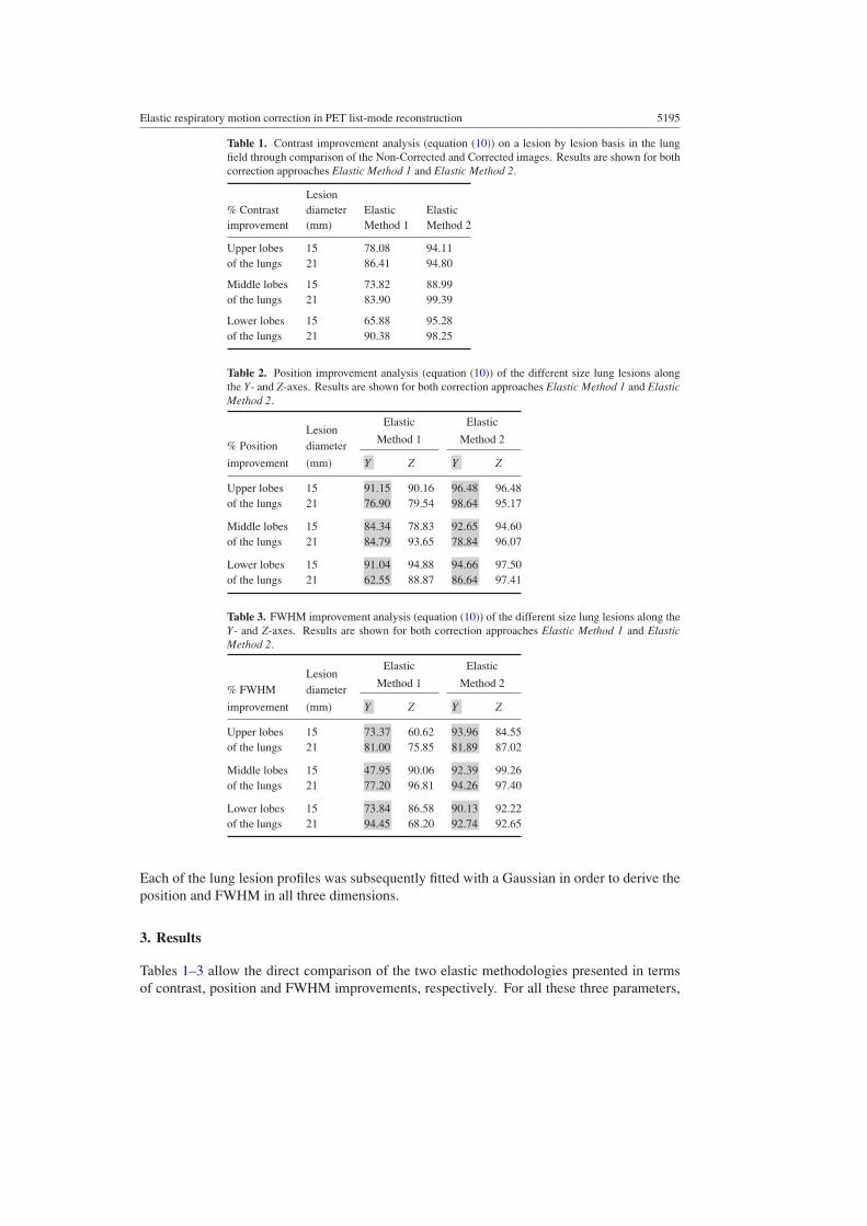
Table 1. Contrast improvement analysis (equation (10)) on a lesion by lesion basis in the lungfield through comparison of the Non-Corrected and Corrected images. Results are shown for bothcorrection approaches Elastic Method 1 and Elastic Method 2.
Lesion% Contrast diameter Elastic Elasticimprovement (mm) Method 1 Method 2
Upper lobes 15 78.08 94.11of the lungs 21 86.41 94.80
Middle lobes 15 73.82 88.99of the lungs 21 83.90 99.39
Lower lobes 15 65.88 95.28of the lungs 21 90.38 98.25
Table 2. Position improvement analysis (equation (10)) of the different size lung lesions alongthe Y- and Z-axes. Results are shown for both correction approaches Elastic Method 1 and ElasticMethod 2.
Elastic ElasticLesion
Method 1 Method 2% Position diameter
improvement (mm) Y Z Y Z
Upper lobes 15 91.15 90.16 96.48 96.48of the lungs 21 76.90 79.54 98.64 95.17
Middle lobes 15 84.34 78.83 92.65 94.60of the lungs 21 84.79 93.65 78.84 96.07
Lower lobes 15 91.04 94.88 94.66 97.50of the lungs 21 62.55 88.87 86.64 97.41
Table 3. FWHM improvement analysis (equation (10)) of the different size lung lesions along theY- and Z-axes. Results are shown for both correction approaches Elastic Method 1 and ElasticMethod 2.
Elastic ElasticLesion
Method 1 Method 2% FWHM diameter
improvement (mm) Y Z Y Z
Upper lobes 15 73.37 60.62 93.96 84.55of the lungs 21 81.00 75.85 81.89 87.02
Middle lobes 15 47.95 90.06 92.39 99.26of the lungs 21 77.20 96.81 94.26 97.40
Lower lobes 15 73.84 86.58 90.13 92.22of the lungs 21 94.45 68.20 92.74 92.65
Each of the lung lesion profiles was subsequently fitted with a Gaussian in order to derive theposition and FWHM in all three dimensions.
3. Results
Tables 1–3 allow the direct comparison of the two elastic methodologies presented in termsof contrast, position and FWHM improvements, respectively. For all these three parameters,
5196 F Lamare et al
0
20
40
60
80
100
211511721151172115117
% C
ontr
ast i
mpr
ovem
ent
Lesion diameter (mm)
gated-FramesLORs-Affine
Elastic Method 2
Upper lobes of the lungs Lower lobes of the lungsMiddle lobes of the lungs︸ ︷︷ ︸ ︸ ︷︷ ︸ ︸ ︷︷ ︸
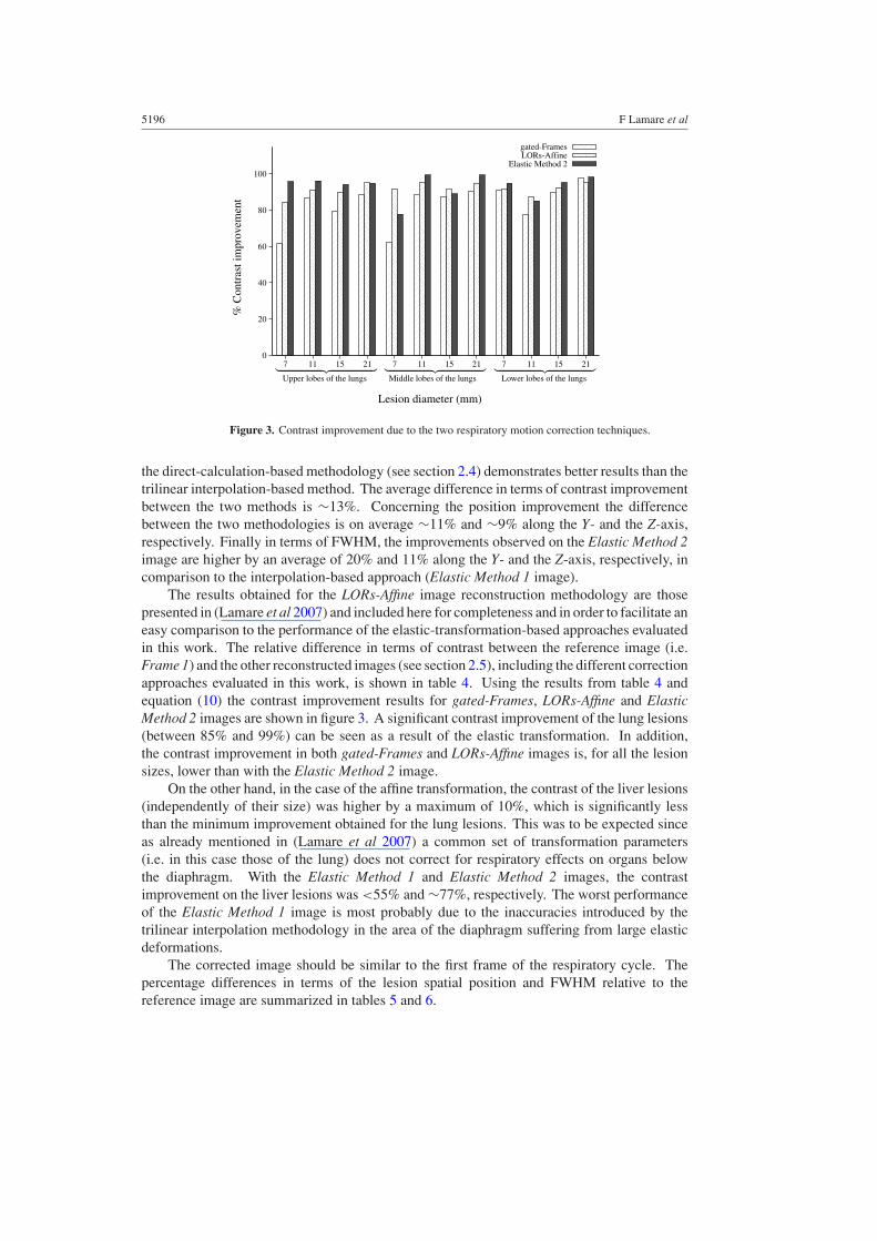
Figure 3. Contrast improvement due to the two respiratory motion correction techniques.
the direct-calculation-based methodology (see section 2.4) demonstrates better results than thetrilinear interpolation-based method. The average difference in terms of contrast improvementbetween the two methods is ∼13%. Concerning the position improvement the differencebetween the two methodologies is on average ∼11% and ∼9% along the Y- and the Z-axis,respectively. Finally in terms of FWHM, the improvements observed on the Elastic Method 2image are higher by an average of 20% and 11% along the Y- and the Z-axis, respectively, incomparison to the interpolation-based approach (Elastic Method 1 image).
The results obtained for the LORs-Affine image reconstruction methodology are thosepresented in (Lamare et al 2007) and included here for completeness and in order to facilitate aneasy comparison to the performance of the elastic-transformation-based approaches evaluatedin this work. The relative difference in terms of contrast between the reference image (i.e.Frame 1) and the other reconstructed images (see section 2.5), including the different correctionapproaches evaluated in this work, is shown in table 4. Using the results from table 4 andequation (10) the contrast improvement results for gated-Frames, LORs-Affine and ElasticMethod 2 images are shown in figure 3. A significant contrast improvement of the lung lesions(between 85% and 99%) can be seen as a result of the elastic transformation. In addition,the contrast improvement in both gated-Frames and LORs-Affine images is, for all the lesionsizes, lower than with the Elastic Method 2 image.
On the other hand, in the case of the affine transformation, the contrast of the liver lesions(independently of their size) was higher by a maximum of 10%, which is significantly lessthan the minimum improvement obtained for the lung lesions. This was to be expected sinceas already mentioned in (Lamare et al 2007) a common set of transformation parameters(i.e. in this case those of the lung) does not correct for respiratory effects on organs belowthe diaphragm. With the Elastic Method 1 and Elastic Method 2 images, the contrastimprovement on the liver lesions was <55% and ∼77%, respectively. The worst performanceof the Elastic Method 1 image is most probably due to the inaccuracies introduced by thetrilinear interpolation methodology in the area of the diaphragm suffering from large elasticdeformations.
The corrected image should be similar to the first frame of the respiratory cycle. Thepercentage differences in terms of the lesion spatial position and FWHM relative to thereference image are summarized in tables 5 and 6.

Elastic respiratory motion correction in PET list-mode reconstruction 5197
Table 4. Contrast per cent relative difference analysis (equation (9)) on a lesion by lesion basis inthe lung field through comparison of the Non-Corrected and Corrected images. Results are shownfor both approaches of applying the transformation in the raw data prior to the reconstructionprocess (LORs-Affine) as well as in the images and subsequently summing them together (gated-Frames) or integrating the elastic transformation parameters within the reconstruction process(Elastic Method 1).
Lesiondiameter Non- gated- LORs- Elastic
Contrast (mm) Corrected Frames Affine Method 2
Upper lobes 7 28.30 10.83 4.39 1.21of the lungs 11 31.48 4.23 2.82 1.21
15 30.33 6.20 3.05 1.7921 12.96 1.50 0.65 0.67
Middle lobes 7 18.87 7.14 1.56 4.20of the lungs 11 28.86 3.35 1.38 0.19
15 30.87 3.89 2.66 3.4021 21.16 2.09 1.14 0.13
Lower lobes 7 32.42 2.92 2.63 1.66of the lungs 11 35.35 7.89 4.45 5.28
15 38.68 4.01 3.07 1.8221 27.56 0.69 1.30 0.48
Table 5. Position per cent relative difference analysis (equation (9)) of the different size lung lesionsalong the Y- and Z-axes. Results are shown for both approaches of applying the transformationin the raw data prior to the reconstruction process (LORs-Affine) as well as in the images andsubsequently summing them together (gated-Frames) or integrating the elastic transformationparameters within the reconstruction process (Elastic Method 1).
Non- gated- LORs- ElasticLesion
Corrected Frames Affine Method 2Position diameter
Y/Z (mm) Y Z Y Z Y Z Y Z
Upper lobes 7 0.76 4.67 0.83 0.79 0.52 1.06 0.36 0.40of the lungs 11 0.65 3.37 0.57 0.51 0.37 1.40 0.18 0.28
15 2.69 7.38 0.08 0.36 0.11 0.43 0.09 0.2621 1.51 5.62 0.14 0.33 0.08 0.59 0.02 0.27
Middle lobes 7 1.19 3.87 0.38 0.49 0.36 1.81 0.27 0.61of the lungs 11 2.16 3.83 0.10 0.61 0.20 0.60 0.06 0.59
15 0.84 4.27 0.09 0.36 0.13 0.74 0.06 0.2321 2.02 5.76 0.68 0.71 0.54 0.34 0.43 0.23
Lower lobes 7 3.35 9.34 0.38 0.29 0.27 0.57 0.09 0.17of the lungs 11 2.28 2.47 1.43 0.43 1.09 0.43 0.66 0.38
15 1.71 4.63 0.15 0.24 0.28 0.65 0.09 0.1221 2.23 5.18 0.33 0.16 0.34 0.12 0.30 0.13
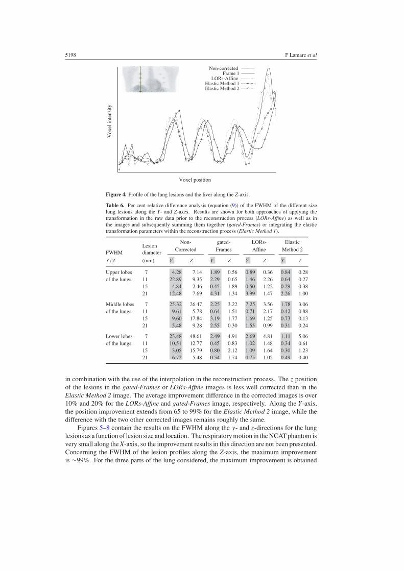
Figure 4 shows the profile response of the lesions in the respiratory corrected images,compared with the NCAT reference frame image. According to the profile in the Z-direction,one can appreciate that the displacement of the lesions is well corrected in the Elastic Method2 image in comparison to the NCAT gated Frame 1 and the uncorrected respiration averageimage, resulting in a spatial location improvement of between 85% and 99%. It is worthnoting the drop in the voxel intensity in the corrected Elastic Method 1 image at the positionof the diaphragm that can be once more attributed to the large elastic deformations in this area
5198 F Lamare et al
Vox
el in
tens
ity
Voxel position
Non-correctedFrame 1
LORs-AffineElastic Method 1Elastic Method 2
Figure 4. Profile of the lung lesions and the liver along the Z-axis.
Table 6. Per cent relative difference analysis (equation (9)) of the FWHM of the different sizelung lesions along the Y- and Z-axes. Results are shown for both approaches of applying thetransformation in the raw data prior to the reconstruction process (LORs-Affine) as well as inthe images and subsequently summing them together (gated-Frames) or integrating the elastictransformation parameters within the reconstruction process (Elastic Method 1).
Non- gated- LORs- ElasticLesion
Corrected Frames Affine Method 2FWHM diameter
Y/Z (mm) Y Z Y Z Y Z Y Z
Upper lobes 7 4.28 7.14 1.89 0.56 0.89 0.36 0.84 0.28of the lungs 11 22.89 9.35 2.29 0.65 1.46 2.26 0.64 0.27
15 4.84 2.46 0.45 1.89 0.50 1.22 0.29 0.3821 12.48 7.69 4.31 1.34 3.99 1.47 2.26 1.00
Middle lobes 7 25.32 26.47 2.25 3.22 7.25 3.56 1.78 3.06of the lungs 11 9.61 5.78 0.64 1.51 0.71 2.17 0.42 0.88
15 9.60 17.84 3.19 1.77 1.69 1.25 0.73 0.1321 5.48 9.28 2.55 0.30 1.55 0.99 0.31 0.24
Lower lobes 7 23.48 48.61 2.49 4.91 2.69 4.81 1.11 5.06of the lungs 11 10.51 12.77 0.45 0.83 1.02 1.48 0.34 0.61
15 3.05 15.79 0.80 2.12 1.09 1.64 0.30 1.2321 6.72 5.48 0.54 1.74 0.75 1.02 0.49 0.40
in combination with the use of the interpolation in the reconstruction process. The z positionof the lesions in the gated-Frames or LORs-Affine images is less well corrected than in theElastic Method 2 image. The average improvement difference in the corrected images is over10% and 20% for the LORs-Affine and gated-Frames image, respectively. Along the Y-axis,the position improvement extends from 65 to 99% for the Elastic Method 2 image, while thedifference with the two other corrected images remains roughly the same.
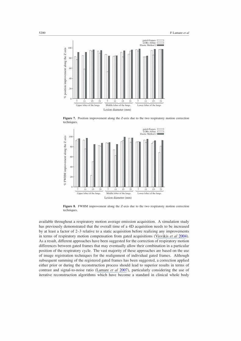
Figures 5–8 contain the results on the FWHM along the y- and z-directions for the lunglesions as a function of lesion size and location. The respiratory motion in the NCAT phantom isvery small along the X-axis, so the improvement results in this direction are not been presented.Concerning the FWHM of the lesion profiles along the Z-axis, the maximum improvementis ∼99%. For the three parts of the lung considered, the maximum improvement is obtained
Elastic respiratory motion correction in PET list-mode reconstruction 5199
0
20
40
60
80
100
211511721151172115117
% p
ositi
on im
prov
emen
t alo
ng th
e Y
axi
s
Lesion diameter (mm)
gated-FramesLORs-Affine
Elastic Method 2
Upper lobes of the lungs Lower lobes of the lungsMiddle lobes of the lungs︸ ︷︷ ︸ ︸ ︷︷ ︸︸ ︷︷ ︸
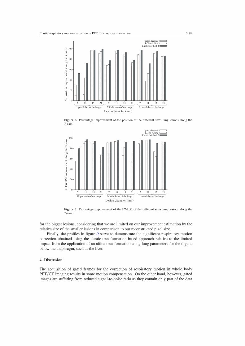
Figure 5. Percentage improvement of the position of the different sizes lung lesions along theY-axis.
0
20
40
60
80
100
211511721151172115117
% F
WH
M im
prov
emen
t alo
ng th
e Y
axi
s
Lesion diameter (mm)
gated-FramesLORs-Affine
Elastic Method 2
Upper lobes of the lungs Lower lobes of the lungsMiddle lobes of the lungs︸ ︷︷ ︸ ︸ ︷︷ ︸︸ ︷︷ ︸
Figure 6. Percentage improvement of the FWHM of the different sizes lung lesions along theY-axis.
for the bigger lesions, considering that we are limited on our improvement estimation by therelative size of the smaller lesions in comparison to our reconstructed pixel size.
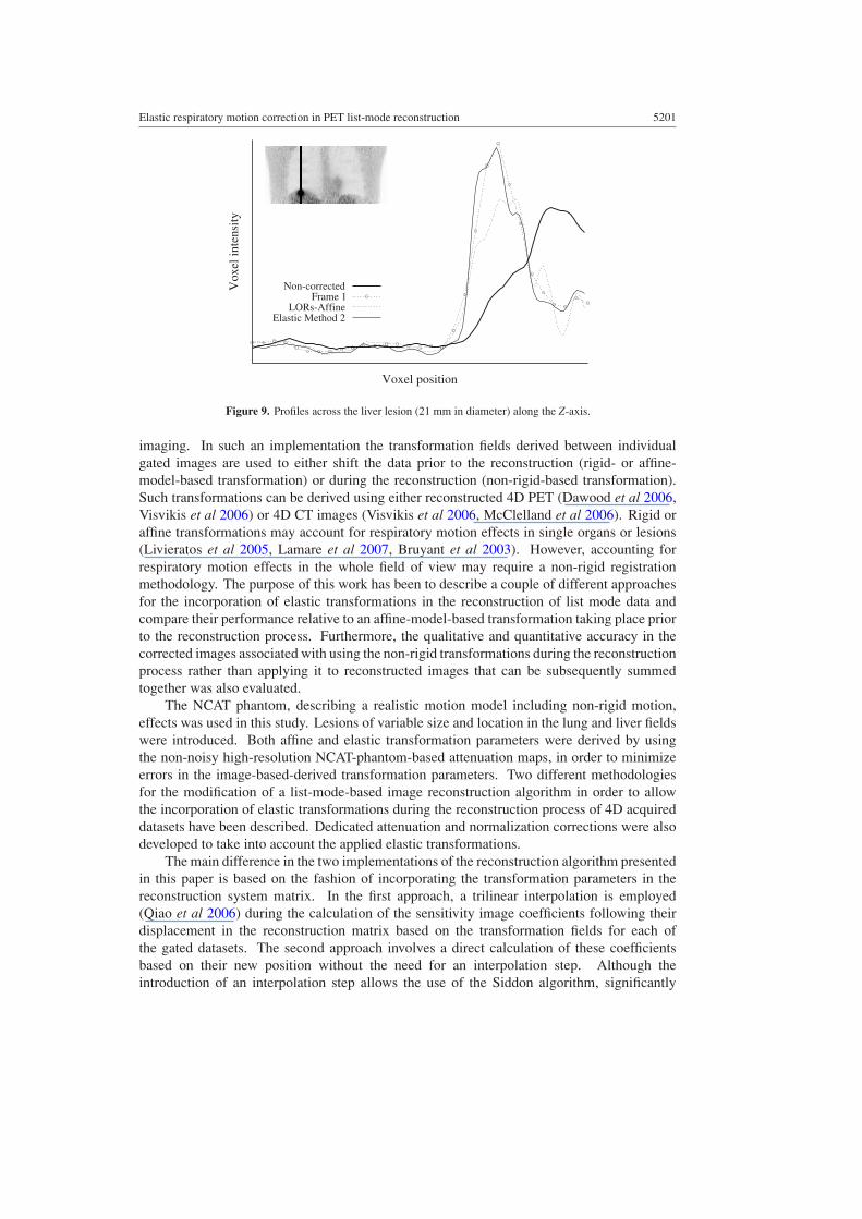
Finally, the profiles in figure 9 serve to demonstrate the significant respiratory motioncorrection obtained using the elastic-transformation-based approach relative to the limitedimpact from the application of an affine transformation using lung parameters for the organsbelow the diaphragm, such as the liver.
4. Discussion
The acquisition of gated frames for the correction of respiratory motion in whole bodyPET/CT imaging results in some motion compensation. On the other hand, however, gatedimages are suffering from reduced signal-to-noise ratio as they contain only part of the data
5200 F Lamare et al
0
20
40
60
80
100
211511721151172115117
% p
ositi
on im
prov
emen
t alo
ng th
e Z
axi
s
Lesion diameter (mm)
gated-FramesLORs-Affine
Elastic Method 2
Upper lobes of the lungs Lower lobes of the lungsMiddle lobes of the lungs︸ ︷︷ ︸ ︸ ︷︷ ︸︸ ︷︷ ︸
Figure 7. Position improvement along the Z-axis due to the two respiratory motion correctiontechniques.
0
20
40
60
80
100
211511721151172115117
% F
WH
M im
prov
emen
t alo
ng th
e Z
axi
s
Lesion diameter (mm)
gated-FramesLORs-Affine
Elastic Method 2
Upper lobes of the lungs Lower lobes of the lungsMiddle lobes of the lungs︸ ︷︷ ︸ ︸ ︷︷ ︸︸ ︷︷ ︸
Figure 8. FWHM improvement along the Z-axis due to the two respiratory motion correctiontechniques.
available throughout a respiratory motion average emission acquisition. A simulation studyhas previously demonstrated that the overall time of a 4D acquisition needs to be increasedby at least a factor of 2–3 relative to a static acquisition before realizing any improvementsin terms of respiratory motion compensation from gated acquisitions (Visvikis et al 2004).As a result, different approaches have been suggested for the correction of respiratory motiondifferences between gated frames that may eventually allow their combination in a particularposition of the respiratory cycle. The vast majority of these approaches are based on the useof image registration techniques for the realignment of individual gated frames. Althoughsubsequent summing of the registered gated frames has been suggested, a correction appliedeither prior or during the reconstruction process should lead to superior results in terms ofcontrast and signal-to-noise ratio (Lamare et al 2007), particularly considering the use ofiterative reconstruction algorithms which have become a standard in clinical whole body
Elastic respiratory motion correction in PET list-mode reconstruction 5201
Vox
el in
tens
ity
Voxel position
Non-correctedFrame 1
LORs-AffineElastic Method 2
Figure 9. Profiles across the liver lesion (21 mm in diameter) along the Z-axis.
imaging. In such an implementation the transformation fields derived between individualgated images are used to either shift the data prior to the reconstruction (rigid- or affine-model-based transformation) or during the reconstruction (non-rigid-based transformation).Such transformations can be derived using either reconstructed 4D PET (Dawood et al 2006,Visvikis et al 2006) or 4D CT images (Visvikis et al 2006, McClelland et al 2006). Rigid oraffine transformations may account for respiratory motion effects in single organs or lesions(Livieratos et al 2005, Lamare et al 2007, Bruyant et al 2003). However, accounting forrespiratory motion effects in the whole field of view may require a non-rigid registrationmethodology. The purpose of this work has been to describe a couple of different approachesfor the incorporation of elastic transformations in the reconstruction of list mode data andcompare their performance relative to an affine-model-based transformation taking place priorto the reconstruction process. Furthermore, the qualitative and quantitative accuracy in thecorrected images associated with using the non-rigid transformations during the reconstructionprocess rather than applying it to reconstructed images that can be subsequently summedtogether was also evaluated.
The NCAT phantom, describing a realistic motion model including non-rigid motion,effects was used in this study. Lesions of variable size and location in the lung and liver fieldswere introduced. Both affine and elastic transformation parameters were derived by usingthe non-noisy high-resolution NCAT-phantom-based attenuation maps, in order to minimizeerrors in the image-based-derived transformation parameters. Two different methodologiesfor the modification of a list-mode-based image reconstruction algorithm in order to allowthe incorporation of elastic transformations during the reconstruction process of 4D acquireddatasets have been described. Dedicated attenuation and normalization corrections were alsodeveloped to take into account the applied elastic transformations.
The main difference in the two implementations of the reconstruction algorithm presentedin this paper is based on the fashion of incorporating the transformation parameters in thereconstruction system matrix. In the first approach, a trilinear interpolation is employed(Qiao et al 2006) during the calculation of the sensitivity image coefficients following theirdisplacement in the reconstruction matrix based on the transformation fields for each ofthe gated datasets. The second approach involves a direct calculation of these coefficientsbased on their new position without the need for an interpolation step. Although theintroduction of an interpolation step allows the use of the Siddon algorithm, significantly
5202 F Lamare et al
reducing reconstruction times, it suffers by the assumption governing the interpolation step.This interpolation is based on the overlapping volumes of the eight neighbouring voxels (seesection 2.2), the assumption being that the non-rigid transformation operations performed toobtain the necessary displacement vectors do not significantly alter the shape of the voxels.Such an assumption, although valid for small displacements, can be questioned for areaswhere significant non-rigid body movements take place. In our investigation with the NCATphantom, the area of the diaphragm presents the largest non-rigid deformations induced bythe respiratory motion. As demonstrated in figure 1(e), white band artefacts appear in thearea of the diaphragm. The corresponding loss of voxel intensity is also shown in figure 4.In addition, as far as quantitative results are concerned, the interpolation-based methodologyperformed globally worse than the direct-calculation-based implementation by <13% as faras lesion contrast and position recovery are concerned and up to 20% in the lesion sizeimprovements. These differences occurred in their large majority considering the lesionscloser to the diaphragm. At the same time, the direct-based method renders the reconstructionprocess slower by over a factor of 10.
A comparison of the reconstructed images with and without transformation applied onthe raw list mode datasets revealed significant respiratory motion compensation in the lungs.As figure 3 demonstrates an improvement of ∼85–99% in terms of contrast on lung lesionsin the corrected images was obtained. In terms of improvements on lesion location and sizethere was a more non-uniform recovery depending on the placement of the lesion in the lungfield varying from 70% to 95% and from 80% to 97%, respectively.
The corrected images based on the use of elastic transformations in the reconstructionprocess were compared to the results obtained through the use of a previously described affine-based model, where the transformation of list mode data takes place prior to the reconstruction(Lamare et al 2007). In terms of contrast improvements of up to 30% in comparison to theaffine-transformation-based correction were obtained. A larger improvement was generallyobserved for the smaller lesions placed in the lower part of the lung fields, leading to anoverall more uniform correction throughout the lungs relative to the affine-transformation-based approach, irrespective of the lesion size and location.
Finally, a superior contrast by on average between 20% and 30% was observed in therecovered lesion contrast as a result of performing the correction prior to the reconstructionprocess rather than using the spatial transformation parameters for adjusting individual gatedimages and subsequently summing them together. On the other hand, differences obtainedbetween the recovered position and size of the lesions between the image-based and theraw-data-based solution were on average less than 10%.
In summary, the developed methodology has demonstrated that the application of an elasticspatio-temporal transformation during the reconstruction process of gated PET datasets leadsto significant improvements in overall image qualitative and quantitative accuracy, makinguse of all available data throughout a respiratory gated acquisition. In addition, our resultsdemonstrate that the application of the spatial transformation in the raw data domain withinthe reconstruction leads to superior contrast in comparison to simply adding together alreadyreconstructed and realigned images of the individual gated frames.
5. Conclusion
A list-mode-data-based respiratory motion correction using elastic transformations duringimage reconstruction has been implemented and its performance evaluated. The developedalgorithm includes the implementation of modified normalization and attenuation correctionstaking into consideration the applied transformations. Our results with a digital phantom,
Elastic respiratory motion correction in PET list-mode reconstruction 5203
including realistic non-rigid respiratory motion, demonstrate that the use of an interpolationstep in the calculation of the sensitivity matrix coefficients leads to artefacts and a reducedquantitative improvement in the motion corrected reconstructed images.
A comparison of the reconstructed images with and without correction revealed significantrespiratory motion compensation in the lung lesions. In comparison to the use of an affinemodel, the elastic-transformation-based solution leads to a more uniform improvement acrossthe lung fields for the different lesion sizes considered. In addition, the application of thedisplacements in the image space followed by summing the realigned gated frames leads to<30% contrast loss relative to the incorporation of the displacements in the reconstructionprocess.
References
Asma E, Manjeshwar R and Thielemans K 2006 Theoretical comparison of motion correction techniques for PETimage reconstruction IEEE Med. Image Conf. Rec. 3 1762–7
Boucher L, Rodrigue S, Lecomte R and Benard F 2004 Respiratory gating for 3-dimensional PET of the thorax:feasibility and initial results J. Nucl. Med. 45 214–9
Bruyant P, King M and Pretorius P 2003 Correction of the respiratory motion of the heart by tracking of the center ofmass of thresholded projections: a simulation study using the dynamic MCAT phantom IEEE Trans. Nucl. Sci.49 2159–66
Bruyant P, Turzo A, Bizais Y, Cheze Le Rest C and Visvikis D 2006 A comparison of three respiratory gating methodsin PET imaging for oncology J. Nucl. Med. 47 183P
Dawood M, Lang N, Jiang X and Schafers K P 2006 Lung motion correction on respiratory gated 3-D PET/CTimages IEEE Trans. Med. Imaging 25 476–85
Erdi Y E et al 2004 The CT motion quantitation of lung lesions and its impact on PET-measured SUVs J. Nucl.Med. 45 1287–92
Goerres G W, Kamel E, Heidelberg T N, Schwitter M R, Burger C and von Schulthess G K 2002 PET-CT imageco-registration in the thorax: influence of respiration Eur. J. Nucl. Med. Mol. Imaging 29 351–60
Han G, Liang Z and You J 1999 A fast raytracing technique for TCT and ECT studies IEEE Nucl. Sci. Symp. Conf.Rec. 3 1515–8
Jacobson M and Fessler J 2003 Joint estimation of image and deformation parameters in motion-corrected PET IEEENucl. Sci. Symp. Conf. Rec. 5 3290–4
Klein G and Huesman R 1997 A 3D optical flow approach to addition of deformable PET volumes Proc. IEEENonrigid and Articulated Motion Workshop (Puerto Rico) pp 136–43
Kybic J and Unser M 2003 Fast parametric elastic image registration IEEE Trans. Image Process 12 1427–42Lamare F, Cresson T, Savean J, Cheze Le Rest C, Reader A J and Visvikis D 2007 Respiratory motion correction for
PET oncology applications using affine transformation of list mode data Phys. Med. Biol. 52 121–40Lamare F, Ledesma Carbayo M, Kontaxakis G, Santos A, Turzo A, Bizais Y, Cheze Le Rest C and Visvikis D 2005
Incorporation of elastic transformations in list-mode based reconstruction for respiratory motion correction inPET IEEE Nucl. Sci. Symp. Conf. Rec. 3 1740–4
Lamare F, Turzo A, Bizais Y, Le Rest C C and Visvikis D 2006 Validation of a Monte Carlo simulation of the PhilipsAllegro/GEMINI PET systems using GATE Phys. Med. Biol. 51 943–62
Ledesma-Carbayo M J, Mahia-Casado P, Santos A, Perez-David E, Garcia-Fernandez M A and Desco M 2006Cardiac motion analysis from ultrasound sequences using nonrigid registration: validation against Dopplertissue velocity Ultrasound Med. Biol. 32 483–90
Li T, Thorndyke B, Schreibmann E, Yang Y and Xing L 2006 Model-based image reconstruction for four-dimensionalPET Med. Phys. 33 1288–98
Livieratos L, Stegger L, Bloomfield P M, Schafers K, Bailey D L and Camici P G 2005 Rigid-body transformationof list-mode projection data for respiratory motion correction in cardiac PET Phys. Med. Biol. 50 3313–22
McClelland J R, Blackall J M, Tarte S, Chandler A C, Hughes S, Ahmad S, Landau D B and Hawkes D J 2006A continuous 4D motion model from multiple respiratory cycles for use in lung radiotherapy Med. Phys.33 3348–58
Nehmeh S A et al 2002 Effect of respiratory gating on reducing lung motion artefacts in PET imaging of lung cancerMed. Phys. 29 366–71
Nehmeh S A et al 2004 Four-dimensional (4D) PET/CT imaging of the thorax Med. Phys. 31 3179–86

5204 F Lamare et al
Pan T, Mawlawi O, Nehmeh S A, Erdi Y E, Luo D, Liu H H, Castillo R, Mohan R, Liao Z andMacapinlac H A 2005 Attenuation correction of PET images with respiration-averaged CT images in PET/CTJ. Nucl. Med. 46 1481–7
Qi J and Huesman R 2002 List Mode reconstruction for PET with motion compensation: a simulation study Proc.IEEE Int. Symp. Biomed. Imaging pp 413–6
Qiao F, Pan T, Clark Jr W J and Mawlawi O R 2006 A motion-incorporated reconstruction method for gated PETstudies Phys. Med. Biol. 51 3769–83
Rahmim A, Bloomfield P, Houle S, Lenox M, Michel C, Buckley K, Ruth T and Sossi V 2004 Motion compensationin histogram-mode and list-mode reconstructions: beyond the event-driven approach IEEE Trans. Nucl. Sci.51 2588–96
Reader A, Ally S, Bakatselos F, Manavaki R, Walledge R, Jeavons A, Julyan P, Zhao S, Hastings D and Zweit J2002 One-pass list-mode EM algorithm for high-resolution 3D PET image reconstruction into large array IEEETrans. Nucl. Sci. 49 693–9
Schoder H, Erdi Y E, Chao K, Gonen M, Larson S M and Yeung H W 2004 Clinical implications of differentimage reconstruction parameters for interpretation of whole-body PET studies in cancer patients J. Nucl. Med.45 559–66
Segars W, Lalush D and Tsui B 2001 Modelling respiratory mechanics in the MCAT and spline-based MCATphantoms IEEE Trans. Nucl. Sci. 48 89–97
Siddon R 1986 Fast calculation of the exact radiological path for a three-dimensional CT array Med. Phys. 12 252–5Sorzano C, Thevenaz P and Unser M 2005 Elastic registration of biological images using vector-spline regularization
IEEE Trans. Biomed. Eng. 52 652–63Studholme C, Hill D and Hawkes D 1999 An overlap invariant entropy measure of 3D medical image alignment
Pattern Recognit. 32 71–86Unser M, Aldroubi A and Eden M 1993 The L2 polynomial spline pyramid IEEE Trans. Pattern Anal. Mach. Intell.
15 364–79Visvikis D, Barret O, Fryer T, Lamare F, Turzo A, Bizais Y and Cheze-LeRest C 2004 Evaluation of respiratory
motion effects in comparison with other parameters affecting PET image quality IEEE Nucl. Sci. Symp. Conf. Rec.6 3668–72
Visvikis D, Cheze-LeRest C, Costa D C, Bomanji J, Gacinovic S and Ell P J 2001 Influence of OSEM andsegmented attenuation correction in the calculation of standardised uptake values for [18F]FDG PET Eur.J. Nucl. Med. 28 1326–35
Visvikis D, Lamare F, Turzo A, Barrett O, Fryer T, Bizais Y and Cheze Le Rest C 2005 Efficiency of respiratorygating for motion correction in PET J. Nucl. Med. 46 163P
Visvikis D, Ledesma Carbayo M, Lamare F, Mawlawi O, Jarritt P, Bruyant P, Santos A, Kontaxakis G, Boussion Nand Cheze-LeRest C 2006 A spatiotemporal image registration algorithm for correction of respiratory motionin PET/CT J. Nucl. Med. 47 234P
Zhao H and Reader A 2002 Fast Projection algorithm for voxel arrays with object dependent boundaries IEEE Nucl.Sci. Symp. Conf. Rec. 2 1490–4
Related Documents