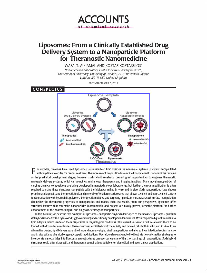
www.pubs.acs.org/accounts Vol. XXX, No. XX ’ XXXX ’ 000–000 ’ ACCOUNTS OF CHEMICAL RESEARCH ’ A 10.1021/ar200105p & XXXX American Chemical Society Liposomes: From a Clinically Established Drug Delivery System to a Nanoparticle Platform for Theranostic Nanomedicine WAFA' T. AL-JAMAL AND KOSTAS KOSTARELOS* Nanomedicine Laboratory, Centre for Drug Delivery Research, The School of Pharmacy, University of London, 29-39 Brunswick Square, London WC1N 1AX, United Kingdom RECEIVED ON APRIL 5, 2011 CONSPECTUS F or decades, clinicians have used liposomes, self-assembled lipid vesicles, as nanoscale systems to deliver encapsulated anthracycline molecules for cancer treatment. The more recent proposition to combine liposomes with nanoparticles remains at the preclinical development stages; however, such hybrid constructs present great opportunities to engineer theranostic nanoscale delivery systems, which can combine simultaneous therapeutic and imaging functions. Many novel nanoparticles of varying chemical compositions are being developed in nanotechnology laboratories, but further chemical modification is often required to make these structures compatible with the biological milieu in vitro and in vivo. Such nanoparticles have shown promise as diagnostic and therapeutic tools and generally offer a large surface area that allows covalent and non-covalent surface functionalization with hydrophilic polymers, therapeutic moieties, and targeting ligands. In most cases, such surface manipulation diminishes the theranostic properties of nanoparticles and makes them less stable. From our perspective, liposomes offer structural features that can make nanoparticles biocompatible and present a clinically proven, versatile platform for further enhancement of the pharmacological and diagnostic efficacy of nanoparticles. In this Account, we describe two examples of liposomenanoparticle hybrids developed as theranostics: liposomequantum dot hybrids loaded with a cytotoxic drug (doxorubicin) and artificially enveloped adenoviruses. We incorporated quantum dots into lipid bilayers, which rendered them dispersible in physiological conditions. This overall vesicular structure allowed them to be loaded with doxorubicin molecules. These structures exhibited cytotoxic activity and labeled cells both in vitro and in vivo. In an alternative design, lipid bilayers assembled around non-enveloped viral nanoparticles and altered their infection tropism in vitro and in vivo with no chemical or genetic capsid modifications. Overall, we have attempted to illustrate how alternative strategies to incorporate nanoparticles into liposomal nanostructures can overcome some of the shortcomings of nanoparticles. Such hybrid structures could offer diagnostic and therapeutic combinations suitable for biomedical and even clinical applications.

Welcome message from author
This document is posted to help you gain knowledge. Please leave a comment to let me know what you think about it! Share it to your friends and learn new things together.
Transcript
www.pubs.acs.org/accounts Vol. XXX, No. XX ’ XXXX ’ 000–000 ’ ACCOUNTS OF CHEMICAL RESEARCH ’ A10.1021/ar200105p & XXXX American Chemical Society
Liposomes: From a Clinically Established DrugDelivery System to a Nanoparticle Platform
for Theranostic NanomedicineWAFA' T. AL-JAMAL AND KOSTAS KOSTARELOS*Nanomedicine Laboratory, Centre for Drug Delivery Research,
The School of Pharmacy, University of London, 29-39 Brunswick Square,London WC1N 1AX, United Kingdom
RECEIVED ON APRIL 5, 2011
CONS P EC TU S
F or decades, clinicians have used liposomes, self-assembled lipid vesicles, as nanoscale systems to deliver encapsulatedanthracycline molecules for cancer treatment. The more recent proposition to combine liposomes with nanoparticles remains
at the preclinical development stages; however, such hybrid constructs present great opportunities to engineer theranosticnanoscale delivery systems, which can combine simultaneous therapeutic and imaging functions. Many novel nanoparticles ofvarying chemical compositions are being developed in nanotechnology laboratories, but further chemical modification is oftenrequired to make these structures compatible with the biological milieu in vitro and in vivo. Such nanoparticles have shownpromise as diagnostic and therapeutic tools and generally offer a large surface area that allows covalent and non-covalent surfacefunctionalization with hydrophilic polymers, therapeutic moieties, and targeting ligands. In most cases, such surface manipulationdiminishes the theranostic properties of nanoparticles and makes them less stable. From our perspective, liposomes offerstructural features that can make nanoparticles biocompatible and present a clinically proven, versatile platform for furtherenhancement of the pharmacological and diagnostic efficacy of nanoparticles.
In this Account, we describe two examples of liposome�nanoparticle hybrids developed as theranostics: liposome�quantumdot hybrids loaded with a cytotoxic drug (doxorubicin) and artificially enveloped adenoviruses. We incorporated quantum dots intolipid bilayers, which rendered them dispersible in physiological conditions. This overall vesicular structure allowed them to beloaded with doxorubicin molecules. These structures exhibited cytotoxic activity and labeled cells both in vitro and in vivo. In analternative design, lipid bilayers assembled around non-enveloped viral nanoparticles and altered their infection tropism in vitroand in vivo with no chemical or genetic capsid modifications. Overall, we have attempted to illustrate how alternative strategies toincorporate nanoparticles into liposomal nanostructures can overcome some of the shortcomings of nanoparticles. Such hybridstructures could offer diagnostic and therapeutic combinations suitable for biomedical and even clinical applications.

B ’ ACCOUNTS OF CHEMICAL RESEARCH ’ 000–000 ’ XXXX ’ Vol. XXX, No. XX
Liposome�Nanoparticle Hybrids as Theranostics Al-Jamal and Kostarelos
IntroductionLiposomes are the most clinically established nanometer-
scale systems that are used to deliver cytotoxic and anti-
fungal drugs, genes, vaccines, as well as imaging agents.1
Liposome vesicles consist of single or multiple concentric
lipid bilayers (called lamellae) encapsulating an aqueous
compartment. Biocompatibility, biodegradability, reduced
toxicity, and capacity for size and surface manipulations
comprise the outstanding profile that liposomes offer com-
pared to other delivery systems.
The past decade has witnessed tremendous advances in
the field of nanoparticle engineering technologies. Particles
with a wide range of consistency, composition, and func-
tionality have been developed. Nanoparticles such as iron
oxide, quantum dots, and gold are recognized to offer
extraordinary features for diagnostics as well as therapeu-
tics. Imaging modalities, such as optical imaging, magnetic
resonance imaging (MRI), and surface plasmon resonance
(SPR), are rapid, noninvasive techniques that have beenused
in cancer therapy where early detection and disease prog-
nosis have improved. Iron oxide nanoparticles, for example,
are clinically approved MRI contrast agents and have also
been proposed as a platform for development of targeted
contrast agents, due to their large surface area that can be
functionalized with different targeting ligands, their blood
circulation can be modulated according to their physico-
chemical properties, and contrast agents and drugs can be
included at predetermined ratios either in the interior or on
the surface of the nanoparticles.
“Theranostic” is a term which defines ongoing efforts to
develop more specific, individualized therapies for various
diseases, combined with diagnostic capabilities into a single
pharmaceutical agent. Merging diagnosis with therapeutic
intervention is aiming to provide clinical protocols that are
more specific to individuals and, therefore, more likely to
offer improved prognoses and monitoring of disease. The
field of theranostics is growing especially with the develop-
ment of various delivery systems, such as liposomes, mi-
celles, dendrimers, carbon nanotubes, in combination with
imaging agents. The concept of simultaneous transport of
therapeutic anddiagnostic agents in a single delivery system
is being intensively explored, either by upgrading theranos-
tic nanoparticles to deliver drugs or by simply incorporating
two moieties (therapeutic and diagnostic) in the same deliv-
ery system.2,3 The present work is based only on the second
concept, illustrating how therapeutic and imaging agents
can be codelivered using liposomal carriers.
An overview of liposomes as delivery systems and as soft
nanoscale platforms will be offered, highlighting on selec-
tive liposome�nanoparticle hybrids and their applications
developed for the theranostic field. Furthermore, we will
describe two examples of liposome�nanoparticle hybrid
theranostics developed in our laboratory. The first study will
report engineering artificial envelopes for non-enveloped,
fluorescently labeled adenoviruses. In the second study, we
will discuss doxorubicin- loaded liposome�quantum dot
(L-QD-Dox) hybrids.
Liposome:AClinically EstablishedDrugDeliverySystemLiposomes are proven candidates for delivery of a wide
range of therapeutics, since their payload can be encapsu-
lated in their internal aqueous compartment or embedded
within the phospholipid bilayer. Clinical applications of
liposomes in the delivery of anticancer agents for the treat-
ment of different cancer indications are well-established.
Stealth liposomes can passively accumulate in solid tumors
due to their inherently leaky vasculature and defective
lymphatic drainage. Doxil, Caelyx, and Myocet are nano-
meter-sized liposome systems (encapsulating doxorubicin in
their aqueous core) which have been used for Kaposi's
sarcoma, ovarian cancer, and multiple myeloma.4�6 DepoCyt
(cytarabine-containing multivesicular liposomes) with a sus-
tained-release profile has also been approved for cancer
therapy.7
New generations of liposome systems, including those
responsive to external or environmental stimuli (e.g., pH,
temperature, enzymes) to trigger drug release at specific and
controlled sites, have been developed. ThermoDox is a
temperature-sensitive nanometer-sized liposome that is
currently in clinical trials to be used in combination with
hyperthermia treatment in oncology.8
Liposome: A Soft Spherical NanoscalePlatformLipid bilayer vesicles were first used as a model mimicking
biological membranes.9 The flexibility offered in manipulat-
ing the size and composition of lipid vesicles has led to their
use as model plasma membranes, subsequently used to
mimic living cells and to understand a variety of physiolo-
gical phenomena, such as diffusion across membranes and
responses to various biological andpharmacological agents.
Moreover, the possibility to reconstruct proteins in the lipid
membrane has been heavily exploited in the design of

Vol. XXX, No. XX ’ XXXX ’ 000–000 ’ ACCOUNTS OF CHEMICAL RESEARCH ’ C
Liposome�Nanoparticle Hybrids as Theranostics Al-Jamal and Kostarelos
biosensors to detect and study the interaction between small
molecules and ligands with plasma membrane receptors.10
Liposomes have also been explored as “chemical con-
tainers”, designed to serve as nanoscale reaction vessels for
remotely controlled reactions.11 The ultrasmall dimensions
of the reaction volume can lead to rapid diffusional mixing
that permits the study of fast chemical kinetics. These
technologies are also well suited for the study of reaction
dynamics between biological molecules within lipid-en-
closed environments that mimic cell membranes.
Liposome�Nanoparticle HybridsThe versatility of the liposomal structure lies in its capacity to
cargo drug molecules and biological macromolecules that
are either hydrophilic, therefore entrapped in the liposome
inner aqueous core, or hydrophobic, therefore incorporated
within the lipid bilayer. Early forms of liposomal hybrid
systems were introduced by Ringsdorf with the description
of liposome�polymer hybrids and their ensuing assembled
structures.12 In the past few years, with the advent of
nanotechnology, there has been a dramatic increase in the
development of novel nanoparticulate materials. Nano-
meter-sized particles such as superparamagnetic iron oxides
(SPIOs), gold, and semiconducting nanocrystals (quantum
dots, QD) possess novel magnetic and optical properties that
can be used as imaging probes. However, their surface hydro-
phobicity or poor colloidal stability at physiological conditions
frequently renders them inappropriate for clinical use. A few
years ago, we presented the concept of liposome�nanoparticle
hybrids as a general methodology, taking advantage of the
much more developed and sophisticated liposome technol-
ogy, to be used as a platform for the delivery of novel
nanoparticles.13 Encapsulationofvarious typesofnanoparticles
within liposomes can lead to enhanced nanoparticle hydrophi-
licity, stability in plasma, better control of the pharmacological
fate, and an overall improvement in their biocompatibility.
Encapsulation of SPIOs, gold, silver nanoparticles, polystyrene
nanospheres, lipidvesicles, andmanyothers into liposomeshas
beenachieved.The threedifferentapproaches for theengineer-
ing of liposome�nanoparticle hybrids are schematically shown
in Figure 1. Hydrophobic nanoparticles can be embedded in the
lipid bilayer, whereas hydrophilic nanoparticles can be encap-
sulated within the internal liposome aqueous core. Alterna-
tively, various types of nanoparticles can be chemically or
physically adsorbed onto the external liposome surface.
Table 1 summarizes the liposome�nanoparticle hybrid
systems that have been described today, classified
according to these three engineering approaches, highlight-
ing their intended applications. As can be seen, most of the
liposome�nanoparticle hybrids have been designed for use
as diagnostic probes. In addition, various types of liposome�nanoparticle hybrids have shown promise in significantly sta-
bilizing colloidal dispersions of otherwise unstable nanoparticle
systems invitroand invivo.Recently,manystudieshaveshown
that incorporation of metallic nanoparticles in liposomes can
also trigger release of encapsulated contents using external
stimuli, such asmagnetic fields, laser irradiation, or electromag-
netic radiation at different radiofrequencies. Lastly, only a few
studies have described loading of liposome�nanoparticle hy-
brids with therapeutic agents to be used as theranostics. Two
such examples from our own research will be described in
following sections.
Liposome�NanoparticleHybrids forTheranosticApplicationsThere are multiple examples of liposome systems with
diverse characteristics and capabilities that incorporate ther-
apeutics or imaging agents.14 However, only a few studies
have described combinatory systems with therapeutic and
imaging capacity using liposomes. Grange et al. showed
combined delivery and magnetic resonance imaging (MRI)
of doxorubicin-containing liposomes in a Kaposi's sarcoma
model in vivo.15 MRI was exploited not only to track the
liposome tissue distribution but also to monitor drug
FIGURE 1. Schematic diagram of three different approaches to engi-neer liposome�nanoparticle hybrids. Hydrophobic nanoparticles em-bedded in the lipid bilayer (left); hydrophilic nanoparticles encapsulatedin the aqueous core (right); and nanoparticles chemically conjugated orphysically adsorbed/complexed to the liposome surface (bottom). Dia-grams are not drawn to scale.

D ’ ACCOUNTS OF CHEMICAL RESEARCH ’ 000–000 ’ XXXX ’ Vol. XXX, No. XX
Liposome�Nanoparticle Hybrids as Theranostics Al-Jamal and Kostarelos
TABLE 1. Liposome�Nanoparticle Hybridsa
aTMAG, N-(R-trimethylammonio-acetyl)-didodecyl-D-glutamate chloride; DLPC, dilauroylphosphatidylcholine; DOPE, dioleoylphosphatidylethanolamine;SPC, soy phosphatidylcholine; PS, phosphatidylserine; DPPC, dipalmitoylphosphatidylcholine; DSPE-PEG2000, distearoylphosphatidylethanolamine poly-ethylene glycol of MW 2000; EYPC, egg yolk phosphatidylcholine; NIR, near infrared; DOTAP, 1,2-dioleoyl-3-trimethylammonium-propane; DOPC,dioleoylphosphocholine; DOPS, dioleoylphosphatidylserine; DHP, dihexadecylphosphate; DODAB, dioctadecyldimethylammonium bromide; DODAC,dioctadecyldimethylammonium chloride; PC, phosphatidylcholine; DMPC, dimyristoylphosphatidylcholine; Chol, cholesterol; DSPC, distearoylphosphati-dylcholine; DPPG, dipalmitoylphosphatidylglycerol; DC-Chol, 3 [N-(N,N-dimethylaminoethane)-carbamoyl] cholesterol; SOPC,1-stearoyl-2-oleoylphospha-tidylcholine; Y2O3,Erþ3, rare earth dopped ceramic; DDAB, dimethyl-dioctadecyl ammonium bromide; SPIO, superparamagnetic iron oxides; TOPO,trioctylphosphine oxide; CdSe, cadmium selenide.

Vol. XXX, No. XX ’ XXXX ’ 000–000 ’ ACCOUNTS OF CHEMICAL RESEARCH ’ E
Liposome�Nanoparticle Hybrids as Theranostics Al-Jamal and Kostarelos
delivery and release. Encapsulation of doxorubicin and MRI
contrast agents in temperature-sensitive liposomes allowed
noninvasive and dynamic imaging of drug release during
hyperthermia application.16,17 In these studies, the MRI
imaging agents were either ProHance (Gd(HPDO3A)-
(H(2)O)) or manganese sulfate (MnSO4); therefore, no nanopar-
ticles were used. Previously, cationic magnetoliposomes (iron
oxide nanoparticle-containing liposomes) were used for gene
delivery, since they were able to complex nucleic acids
(plasmid DNA), and consequently to allow the isolation of
the transfected cells using a magnetic field.18 In other studies,
magnetolipsomes were used to accumulate the vesicles to a
desired tissue or to induce hyperthermia in response to
a magnetic field . Despite the fact that such liposome�nanoparticle hybrids exhibited therapeutic activity, the
magnetic nanoparticles were not used as imaging agents. In
our research, we sought to combine liposomes with nanopar-
ticles with the intention to design liposome�nanoparticle
hybrids that could achieve therapeutic and imaging
capabilities.
Liposome�Viral Nanoparticle Hybrids: Engi-neering Artificial Viral EnvelopesViruses traditionally have a major role in the study of
infection and disease, whilemore recently genetic engineer-
ing technologies have allowed their utilization as com-
pelling gene therapy vectors.47 With the advent of nano-
technology, viruses have also been explored in the
context of nanomedicine, specifically due to their nanoscale
dimension (most virions are between 20 and 100 nm in
diameter), unique size distribution, well-characterized surface
structure, and high surface area-to-volume ratio that allows
for attachment of multiple moieties at specific sites on the
viral particle surface.48,49 Recently, several investigations
have recognized the viral capsid as a versatile building
block that can serve as a scaffold for the fabrication of
novel nanomaterials. A range of minerals, such as cobalt,
nickel, and gallium, have been deposited on the viral
template.50 Besides their use as a template for mineraliza-
tion, viruses have been used as a scaffold for fluorescent
molecules or other nanoparticles exploiting the conjuga-
tion capabilities offered by the lysines and/or cysteines of the
viral capsid. Quantum dots (QD),51 super paramagnetic iron
oxide particles (SPIOs),52 platinum53 and gold54 nanoparticles
have all been chemically conjugated to virus nanoparticles to
design novel biosensors, electronic memory devices, and
multimodal imaging agents.
Apart from designing biosensor devices and diagnostic
agents, adenovirus (Ad) and adeno-associated virus (AAV)
are human viruses that have been heavily explored in
gene therapy.55 Despite their high gene transfer and
expression efficacy, Ad and AAV suffer from a variety of
issues that have precluded their widespread clinical trans-
lation, including rapid blood clearance (requiring multiple
administrations), tissue toxicity (liver in the case of Ad),
and activation of severe and complex immune responses.
In order to alleviate some of these side effects, many
reports have described genetic modifications of viral
capsids56 and chemical conjugation of hydrophilic poly-
mers, such as polyethylene glycol (PEG) and poly N-(2-
hydroxypropyl)methacrylamide (HPMA) on both Ad and
AAV, to prolong their blood circulation and reduce their
immunogenicity and toxicity.57 Despite some success in
partially shielding the virus from the immune system, all
such strategies suffered a significant reduction in gene
transfer efficacy and a dramatic increase in the overall size
of these vectors that affected their pharmacokinetic
profile.
Our group has described an alternative approach by
engineering a liposome�virus hybrid system. The Ad
virions are seen as nanoparticles that can be entirely
encapsulated within liposomes, in that way allowing the
construction of artificial (lipid bilayer) viral envelopes by
self-assembly (Figure 2A).58,59 In these studies, viral par-
ticles could be enveloped with cationic, zwitterionic, and
PEGylated lipid bilayer envelopes. That illustrates the
design flexibility offered by such a hybrid system in terms
of the possible resultingphysicochemicalandpharmacokinetic
properties. The lipid envelope tightly wrapped around the Ad
surfaceas shownby transmissionelectronmicroscopy (TEM) and
atomic forcemicroscopy (AFM) (Figure2BandC,middle), and the
presence of artificial envelopes altered the biological
behavior of the virus in vitro and in vivo. Cationic lipid
envelopes dramatically reduced the transfection capability
of Ad in vitro (Figure 2E) due to their failure to escape
the endosomal compartments following endocytosis
(Figure 2D, middle). In contrast, PEGylated lipid envelopes
prolonged the Ad blood circulation and reduced their
liver gene expression and toxicity following systemic
administration, allowing for passive targeting to solid
tumors.59
More recently, in order to improve on the poor gene
transfer efficiency of the artificially enveloped viruses, a pH-
sensitive envelope (DOPE:CHEMS) was allowed to self-as-
semble around the Ad nanoparticles and pH-sensitive

F ’ ACCOUNTS OF CHEMICAL RESEARCH ’ 000–000 ’ XXXX ’ Vol. XXX, No. XX
Liposome�Nanoparticle Hybrids as Theranostics Al-Jamal and Kostarelos
enveloped Ad hybrids were successfully engineered
(Figure 2B and C, right).60 These liposome�virus hybrid
systems showed similar levels of gene expression as those
of naked Ad (Figure 2E) in vitro. Intracellular trafficking of
fluorescently labeled Ad confirmed that both Ad and pH-
sensitive enveloped Ad successfully escaped the endo-
somes and trafficked to the perinuclear region, in contrast
to Ad enveloped in cationic envelopes where clear endoso-
mal localization was observed (Figure 2D). The high level of
gene expression of pH-sensitive lipid enveloped Adwas also
maintained in vivo following intratumoral injection, which
offers encouragement for further development. This
hybrid system has been designed with potential thera-
nostic capabilities, whereby the encapsulated virion is
FIGURE 2. Liposome�virus hybrids: artificially enveloped adenoviruses (Ad). (A) Schematic depiction; (B) transmission electron microscopy; (C)atomic forcemicroscopy (height images) of naked Ad, enveloped Ad in cationic (DOTAP:Chol), and pH-sensitive (DOPE:CHEMS) bilayers (left to right).(D) Intracellular trafficking of fluorescently labeled Ad in A549 (CARþ) cells. Confocal laser scanning microscopy images of Cy3-labeled, naked, andenveloped Ad in cationic and pH-sensitive envelopes (left to right). (E) In vitro (β-gal) gene expression of cationic and pH-sensitive enveloped Adcompared to naked Ad.58�60

Vol. XXX, No. XX ’ XXXX ’ 000–000 ’ ACCOUNTS OF CHEMICAL RESEARCH ’ G
Liposome�Nanoparticle Hybrids as Theranostics Al-Jamal and Kostarelos
the (gene) therapeutic component and imaging
probes can be incorporated either on the viral capsid or
on the lipid bilayer wrapping it. So far, we have only
shown that the Ad capsid can be conjugated with organic
fluorescent probes (Cy3) to allow optical tracking of
the Ad; however, further modifications with a variety of
diagnostic probes49 prior to liposome encapsulation are
deemed feasible with minimum disruption of the
overall hybrid structure. Such engineering flexibility
can allow the theranostic applications of artificially en-
veloped Ad to be easily tailored and should be further
investigated.
Liposome�Quantum Dot Hybrids for CancerTheranosticsSemiconducting nanocrystals known as quantum dots (QD)
are fluorescent nanoparticles, which offer distinct spectro-
fluorometric advantages over traditional fluorescent
organic molecules. QD exhibit fluorescence characteris-
tics that are 10�20 times brighter than conventional
organic dyes and offer greater photostability. Due to
these photophysical characteristics, they are being ex-
plored as potential imaging agents primarily in optical
(fluorescence-based) diagnostic applications.61
Most QD types are originally prepared in organic sol-
vents; therefore, their hydrophobic shells compromise
their water solubility and consequently their compatibility
with the biological milieu. The most successful approach
has been to functionalize QD with polar moieties and
ligands with specific receptor-recognition signals (such as
peptides and monoclonal antibodies or their fragments).62
However, this surface modification often leads to signifi-
cant reduction in QD fluorescence intensity and photo-
stability. We have recently proposed engineering of
liposome�QD hybrids that follows the approach shown
in Figure 1 (left) by embedding hydrophobic QD within a
variety of lipid bilayers (L-QD hybrids) (Figure 3A; right).24
Structural characterization by cryo-transmission electron
microscopy (cryo-TEM) and atomic force microscopy (AFM)
of such L-QD hybrid bilayer vesicles confirmed the incor-
poration of QD in the liposome bilayer (Figure 3B and C;
middle). L-QD hybrids allowed hydrophobic QD nanopar-
ticles to be used in aqueous (i.e., biological) environments,
while their incorporation in the acyl environment of the
lipid bilayer significantly enhanced QD optical stability
during storage and exposure to UV light.
The hybrid L-QD bilayer vesicles are thought to constitute
a novel delivery system that offers the potential for transport
of combinatory therapeutic and diagnostic agents to cancer
cells in vitro and in vivo. To prove this concept, doxorubicin
(Dox) was loaded inside L-QD hybrids of different lipid
compostions (Figure 3A; right).25 Successful Dox loading
was confirmed by observation of doxorubicin crystals inside
the hybrids (Figure 3B and C; right). The effect of QD on the
vesicle integrity was shown to be highly dependent on the
lipid components used. Minimum drug leakage was ob-
tained using high phase transition temperature (Tm) bilayers
(consisting mainly of DSPC lipids) compared to a low Tmbilayer (mademainly of EPC) (Figure 3D). More interestingly,
these stable, Dox-loaded L-QD (L-QD-Dox) hybrids were
uptakenby cells andwere able to releaseDox intracellularly,
as evidenced by a significant enhancement in the cytotoxi-
city obtained, almost to the same level as the free drug
(Figure 3E). In an alternative approach (according to Figure 1;
bottom) to design liposome�QD hybrid systems with ther-
anostic capabilities, Weng et al. covalently conjugated hy-
drophilic, polymer-functionalized QD at the outer tips of the
polyethylene glycol chains coating the surface of liposomes
and then loaded Dox into the aqueous core of these
vesicles.37 Prolonged blood circulation half-life and ther-
apeutic efficacy compared to Dox alone was reported for
this hybrid system; however, conjugation of the polymer-
coated QD onto the outer liposome surface almost
doubled the average size of the hybrid (from 112 to 212
nm) and greatly lowered Dox loading efficiency (30%). In
comparison to previous attempts to design drug-loaded
liposome-QD conjugates, we have demonstrated that
incorporation of hydrophobic QD into the lipid bilayer of
liposomes maintains the mean vesicle diameter consis-
tently between the desirable 100�120 nm range, allows
high loading efficiencies of Dox (>95%) using the estab-
lished osmotic gradient technique, and has minimal effect
on the Dox fluorescence characteristics.25 Preservation of
high Dox loading efficiencies, in particular, that can be
achieved with this L-QD-Dox hybrid vesicle system are
considered essential to maintain therapeutic efficacy
comparable to the clinically used liposomal doxorubicin
(DOXIL).
Following the design, engineering, and characterization of
various L-QD and L-QD-Dox hybrid systems in vitro, we
further studied the in vivo behavior of L-QDhybrids following
local and systemic administration. Similar to in vitro results,
the profile of L-QD in vivo was determined by the molecular
structure of the lipids used. For instance, hybrids made of

H ’ ACCOUNTS OF CHEMICAL RESEARCH ’ 000–000 ’ XXXX ’ Vol. XXX, No. XX
Liposome�Nanoparticle Hybrids as Theranostics Al-Jamal and Kostarelos
cationic lipids efficiently labeled tumor cells compared to
zwitterionic L-QDhybrids after intratumoral administration.24
In addition, simply bymanipulating the lipid composition, we
targeted L-QD hybrids to different organs following systemic
administration;63 sterically stabilized (DSPE-PEG-containing)
L-QD exhibited prolonged blood circulation compared to
cationic L-QD hybrids. In contrast, cationic L-QD hybrids
showed high transient lung accumulation post-injection
which makes themmore suitable for pulmonary targeting
and imaging.
In accordance to the approach in Figure 1 (right), our
group has also reported the engineering of functionalized
FIGURE 3. Liposome�quantum dot hybrids: lipid bilayer-embedded hydrophobic QD vesicles loaded with doxorubicin. (A) Schematic depiction ofhybrids; (B) cryo-transmission electronmicroscopy; (C) atomic forcemicroscopy (3D images) of empty liposomes, L-QDhybrids, and L-QD-Doxhybrids(left to right). (D) Serum stability of L-QD-Dox hybrids incubated in 50%mouse serum. QDwere embedded in EPC:Chol:DSPE-PEG2000 andDSPC:Chol:DSPE-PEG2000 liposomes. Dox was loaded using the pH-gradient technique and Dox release was assessed by measuring Dox fluorescence. (E)Cytotoxicity of L-QD-Dox hybrids. MCF-7 cells were incubated with free Dox, L-QD-Dox, and L-QD hybrids, and cell viability was assessed using MTTassay.25

Vol. XXX, No. XX ’ XXXX ’ 000–000 ’ ACCOUNTS OF CHEMICAL RESEARCH ’ I
Liposome�Nanoparticle Hybrids as Theranostics Al-Jamal and Kostarelos
quantum dot�liposome hybrids (f-QD-L) by encapsulation
of hydrophilic f-QD in the internal liposome aqueous
compartment.29 We proposed the design of such hybrids
for the delivery of hydrophobic therapeutic agents (small
drug molecules, photosensitizers, etc.) along with hydro-
philic imaging probes (such as f-QDs) using a single vesicle.
In addition, we showed that such an approach can enhance
the cellular uptake and retention of polymer-coated QD in
vivo and accelerate their tumor accumulation following
systemic administration.30 Further work is being under-
taken to combine imaging and therapy in vivo of both
types of L-QD and f-QD-L hybrids by our laboratories
and those of others. In addition, such vesicle systems
can be further modified with different targeting ligands
on the liposome surface to offer binding specificity and
target cancer cell receptors. In that way, construct
liposome�nanoparticle hybrid systems with triple capabil-
ities (active targeting, therapy, and imaging) can be
obtained.
ConclusionLiposome (or lipid bilayer vesicle) science has developed
into a mature and rich technology base, offering us a wide
range of tools and components for the construction of
more complex and multifunctional devices in a variety of
industrial applications. In medicine, liposomes constitute
one of the most successfully translated delivery systems
that is currently in clinical use for a variety of indications
against cancer, inflammatory, dermatological diseases and
in various types of vaccines. Such established clinical
profiles along with the engineering versatility of the lipo-
some structure are considered key advantages for the
further development of liposome-based systemswith com-
binatory therapeutic-diagnostic (theranostic) capabilities.
In this Account, we have elaborated on the concept of
utilizing liposomes as templates for the incorporation
of a wide variety of nanoparticles toward engineering
liposome�nanoparticle hybrid systems of advanced, pre-
viously unattainable functionality. The rapid develop-
ments in nanomaterials synthesis and fabrication now
offer a tremendous range of nanoparticles with different
characteristics and capabilities that are very commonly
incompatible with the biological milieu. Liposome�nanoparticle hybrids can be designed by embedding, en-
capsulation, or conjugation of nanoparticles onto various
types of liposomes. The theranostic potential of such
hybrids has been illustrated with two examples from our
work: liposome-encapsulated viral nanoparticle hybrids cap-
able of gene therapy (viral gene transfer) and optical
imaging (probe molecules on the viral capsid), and doxo-
rubicin-loaded, lipid bilayer-embedded quantum dot vesicle
hybrids capable of chemotherapy (cytotoxic activity of
doxorubicin) and optical imaging (embedded quantum
dots). Such liposome�nanoparticle hybrid systems also
offer unprecedented versatility and flexibility in the com-
bination of physicochemical characteristics (emission/
absorbance wavelength; surface; size; responsiveness to
external stimuli) and biological activity profiles (gene trans-
fer; cytotoxicity; pharmacokinetics) that can possibly be
achieved with the right selection of lipid compositions and
nanoparticle types to best suit a given biomedical applica-
tion and pathological condition. More research from var-
ious laboratories is needed to explore this concept further
in order to truly exploit our knowledge and clinical experi-
ence of liposomes in combination with novel nano-
materials toward more capable and “smarter” theranostic
devices.
BIOGRAPHICAL INFORMATION
Dr.Wafa' T. Al-Jamal received her Ph.D. in drug delivery fromThe School of Pharmacy, University of London in 2008. She wasawarded an Overseas (ORS) Scholarship from the University ofLondon to focus on the development of novel liposome-basedstructures for tumor imaging, targeting, and therapy. She is aSenior Research Fellow at the Nanomedicine Lab, Centre forDrug Delivery working on the engineering and pharmacoki-netics of organic/inorganic hybrids for theranostic applicationsfor cancer. She also focuses on the development and study oftemperature- and pressure-sensitive delivery systems for cancerimaging and therapy and is the Scientific Manager of theSONODRUGS project sponsored by the FP7 European Commis-sion programme.
ProfessorKostasKostarelos is Chair of Nanomedicine at theSchool of Pharmacy, Head of the Centre for Drug DeliveryResearch, and Leader of the Nanomedicine Lab (www.nanome-dicinelab.org) all at the University of London. He has beeninvited as a Fellow of the Royal Society of Medicine, the Instituteof Nanotechnology, and the Royal Society of Arts. He obtainedhis Diploma and Ph.D. in Chemical Engineering from ImperialCollege London. His previous academic appointments includeAssistant Professor of Genetic Medicine & Chemical Engineeringin Medicine, Cornell University Weill Medical College, NY, USA;Instructor, Pulmonary & Critical Care Medicine, New York-Pres-byterian Hospital, NY, USA; Manager, Bioengineering Core, BelferGene Therapy Center, Cornell University Weill Medical College,NY, USA; Deputy Director, Imperial College Genetic TherapiesCentre, Imperial College London, UK. He is the Founding andSenior Editor of the journal Nanomedicine. In 2010 he was

J ’ ACCOUNTS OF CHEMICAL RESEARCH ’ 000–000 ’ XXXX ’ Vol. XXX, No. XX
Liposome�Nanoparticle Hybrids as Theranostics Al-Jamal and Kostarelos
awarded an Invitation Professorial Fellowship from the JapaneseSociety for the Promotion of Science (JSPS).
ABBREVIATIONS
DOPE, dioleoylphosphatidylethanolamine; CHEMS, choles-
teryl hemisuccinate; DOTAP, 1,2-dioleoyl-3-trimethylam-
monium-propane; EPC, egg phosphatidylcholine; Chol,
cholesterol; DSPC, distearoylphosphatidylcholine; DSPE-
PEG2000, distearoylphosphatidylethanolamine polyethylene
glycol of MW 2000; Cy3, cyanine dye 3; A549, human lung
adenocarcinoma epithelial cell line; β-gal, β-galactosidase;
Dox, doxorubicin; L-QD, liposome�quantumdot; L-QD-Dox,
doxorubicin-loaded liposome�quantumdot;MCF-7, human
breast adenocarcinoma cell line.
FOOTNOTES
*To whom correspondence should be addressed. Telephone: þþ44-207-753 5861.E-mail: [email protected].
REFERENCES
1 Lasic, D. D.; Papahadjopoulos, D. Medical Applications of Liposomes; Elsevier Science:Amsterdam, 1998.
2 Janib, S. M.; Moses, A. S.; MacKay, J. A. Imaging and Drug Delivery Using TheranosticNanoparticles. Adv. Drug Delivery Rev. 2010, 62, 1052–1063.
3 Xie, J.; Lee, S.; Chen, X. Nanoparticle-Based Theranostic Agents. Adv. Drug Delivery Rev.2010, 62, 1064–1079.
4 http://www.doxil.com/common/prescribing_information/DOXIL/PDF/DOXIL_PI_Booklet.pdf.5 Minisini, A. M.; Andreetta, C.; Fasola, G.; Puglisi, F. Pegylated Liposomal Doxorubicin in Elderly
Patients With Metastatic Breast Cancer. Expert. Rev. Anticancer Ther. 2008, 8, 331–342.6 Verma, S.; Dent, S.; Chow, B. J.; Rayson, D.; Safra, T. Metastatic Breast Cancer: the Role of
Pegylated Liposomal Doxorubicin After Conventional Anthracyclines. Cancer Treat. Rev.2008, 34, 391–406.
7 Mantripragada, S. A Lipid Based Depot (DepoFoam Technology) for Sustained Release DrugDelivery. Prog. Lipid Res. 2002, 41, 392–406.
8 Celsion Coporation. Clinical trials. http:www/celsion.com/trials.cfm.9 Bangham, A. D. Surrogate Cells or Trojan Horses. The Discovery of Liposomes. Bioessays
1995, 17, 1081–1088.10 Sicchierolli, S. M.; Carmona-Ribeiro, A. M. Incorporation of the Cholera Toxin Receptor in
Phospholipid-Covered Polystyrene Microspheres. Colloids Surf., B 1995, 5, 57–61.11 Sun, B.; Lim, D. S.; Kuo, J. S.; Kuyper, C. L.; Chiu, D. T. Fast Initiation of Chemical
Reactions With Laser-Induced Breakdown of a Nanoscale Partition. Langmuir 2004,20, 9437–9440.
12 Ringsdorf, H.; Sackmann, E.; Simon, J.; Winnik, F. M. Interactions of Liposomes andHydrophobically-Modified Poly(N-Isopropylacrylamides): an Attempt to Model the Cytos-keleton. Biochim. Biophys. Acta 1993, 1153, 335–344.
13 Al-Jamal, W. T.; Kostarelos, K. Liposome-Nanoparticle Hybrids for Multimodal Diagnosticand Therapeutic Applications. Nanomedicine 2007, 2, 85–98.
14 Sajja, H. K.; East, M. P.; Mao, H.; Wang, Y. A.; Nie, S.; Yang, L. Development ofMultifunctional Nanoparticles for Targeted Drug Delivery and Noninvasive Imaging ofTherapeutic Effect. Curr. Drug Discovery Technol. 2009, 6, 43–51.
15 Grange, C.; Geninatti-Crich, S.; Esposito, G.; Alberti, D.; Tei, L.; Bussolati, B.; Aime, S.;Camussi, G. Combined Delivery and Magnetic Resonance Imaging of Neural Cell AdhesionMolecule-Targeted Doxorubicin-Containing Liposomes in Experimentally Induced Kaposi'sSarcoma. Cancer Res. 2010, 70, 2180–2190.
16 deSmet,M.; Heijman, E.; Langereis, S.; Hijnen, N.M.; Grull, H.Magnetic Resonance Imaging ofHigh Intensity Focused Ultrasound Mediated Drug Delivery From Temperature-SensitiveLiposomes: An in Vivo Proof-of-Concept Study. J. Controlled Release 2011, 150, 102–110.
17 Viglianti, B. L.; Abraham, S. A.; Michelich, C. R.; Yarmolenko, P. S.; MacFall, J. R.; Bally,M. B.; Dewhirst, M. W. In Vivo Monitoring of Tissue Pharmacokinetics of Liposome/Drug UsingMRI: Illustration of Targeted Delivery. Magn Reson.Med. 2004, 51, 1153–1162.
18 Margolis, L. B.; Namiot, V. A.; Kljukin, L. M. Magnetoliposomes: Another Principle of CellSorting. Biochim. Biophys. Acta 1983, 735, 193–195.
19 Rasch, M. R.; Rossinyol, E.; Hueso, J. L.; Goodfellow, B. W.; Arbiol, J.; Korgel, B. A.Hydrophobic Gold Nanoparticle Self-Assembly With Phosphatidylcholine Lipid: Membrane-Loaded and Janus Vesicles. Nano Lett. 2010, 10, 3733–3739.
20 Paasonen, L.; Laaksonen, T.; Johans, C.; Yliperttula, M.; Kontturi, K.; Urtti, A. GoldNanoparticles Enable Selective Light-Induced Contents Release From Liposomes.J. Controlled Release 2007, 122, 86–93.
21 Park, S. H.; Oh, S. G.; Mun, J. Y.; Han, S. S. Loading of Gold Nanoparticles Inside the DPPCBilayers of Liposome and Their Effects on Membrane Fluidities. Colloids Surf., B 2006, 48,112–118.
22 Chen, Y.; Bose, A.; Bothun, G. D. Controlled Release From Bilayer-Decorated Magneto-liposomes Via Electromagnetic Heating. ACS Nano 2010, 4, 3215–3221.
23 Gopalakrishnan, G; Danelon, C; Izewska, P; Prummer, M; Bolinger, P. Y.; Geissbuhler, I;Demurtas, D; Dubochet, J; Vogel, H Multifunctional Lipid/Quantum Dot Hybrid Nanocontainersfor Controlled Targeting of Live Cells. Angew. Chem., Int. Ed. 2006, 45, 5478–5483.
24 Al-Jamal, W. T.; Al-Jamal, K. T.; Tian, B.; Lacerda, L.; Bomans, P. H.; Frederik, P. M.;Kostarelos, K. Lipid-Quantum Dot Bilayer Vesicles Enhance Tumor Cell Uptake andRetention in Vitro and in Vivo. ACS Nano 2008, 2, 408–418.
25 Tian, B.; Al-Jamal, W. T.; Al-Jamal, K. T.; Kostarelos, K. Doxorubicin-Loaded Lipid-Quantum Dot Hybrids: Surface Topography and Release Properties. Int. J. Pharm. 2011,[Epub ahead of print].
26 Kasili, P. M.; Vo-Dinh, T. Liposome Encapsulated Gold Nanoshells for NanoPhototherapyInduced Hyperthermia. lnt. J. Nanotechnol. 2005, 2, 397–410.
27 Wu, G.; Mikhailovsky, A.; Khant, H. A.; Fu, C.; Chiu, W.; Zasadzinski, J. A. RemotelyTriggered Liposome Release by Near-Infrared Light Absorption Via Hollow Gold Nanoshells.J. Am. Chem. Soc. 2008, 130, 8175–8177.
28 Soga, K.; Tokuzen, K.; Tsuji, K.; Yamano, T.; Hyodo, H.; Kishimoto, H. NIR Bioimaging:Development of Liposome-Encapsulated, Rare-Earth-Doped Y2O3 Nanoparticles AsFluorescent Probes. Eur. J. Inorg. Chem. 2010, 2673–2677.
29 Al-Jamal, W. T.; Al-Jamal, K. T.; Bomans, P. H.; Frederik, P. M.; Kostarelos, K.Functionalized-Quantum-Dot-Liposome Hybrids As Multimodal Nanoparticles for Cancer.Small 2008, 4, 1406–1415.
30 Al-Jamal, W. T.; Al-Jamal, K. T.; Tian, B.; Cakebread, A.; Halket, J. M.; Kostarelos, K.Tumor Targeting of Functionalized Quantum Dot-Liposome Hybrids by IntravenousAdministration. Mol. Pharmaceutics 2009, 6, 520–530.
31 Nagatani, N.; Shinkai, M.; Honda, H.; Kobayashi, T. Development of a New TransformationMethod Using Magnetite Cationic Liposomes and Magnetic Selection of Transformed Cells.Biotechnology Techniques 1998, 12, 525–528.
32 Jain, S.; Mishra, V.; Singh, P.; Dubey, P. K.; Saraf, D. K.; Vyas, S. P. RGD-AnchoredMagnetic Liposomes for Monocytes/Neutrophils-Mediated Brain Targeting. Int. J. Pharm.2003, 261, 43–55.
33 Martina, M. S.; Fortin, J. P.; Menager, C.; Clement, O.; Barratt, G.; Grabielle-Madelmont, C.;Gazeau, F.; Cabuil, V.; Lesieur, S. Generation of Superparamagnetic Liposomes RevealedAs Highly Efficient MRI Contrast Agents for in Vivo Imaging. J. Am. Chem. Soc. 2005, 127,10676–10685.
34 Kisak, E. T.; Coldren, B.; Zasadzinski, J. A. Nanocompartments Enclosing Vesicles, Colloids,and Macromolecules Via Interdigitated Lipid Bilayers. Langmuir 2002, 18, 284–288.
35 Carmona-Ribeiro, A. M.; Midmore, B. R. Synthetic Bilayer Adsorption Onto PolystyreneMicrospheres. Langmuir 1991, 8, 801–806.
36 Sigot, V.; Arndt-Jovin, D. J.; Jovin, T. M. Targeted Cellular Delivery of QuantumDots Loadedon and in Biotinylated Liposomes. Bioconjugate Chem. 2010, 21, 1465–1472.
37 Weng, K. C.; Noble, C. O.; Papahadjopoulos-Sternberg, B.; Chen, F. F.; Drummond, D. C.;Kirpotin, D. B.; Wang, D.; Hom, Y. K.; Hann, B.; Park, J. W. Targeted Tumor CellInternalization and Imaging of Multifunctional Quantum Dot-Conjugated Immunoliposomesin Vitro and in Vivo. Nano Lett. 2008, 8, 2851–2857.
38 Kojima, C.; Hirano, Y.; Yuba, E.; Harada, A.; Kono, K. Preparation and Characterization ofComplexes of Liposomes With Gold Nanoparticles. Colloids Surf., B 2008, 66, 246–252.
39 Chithrani, D. B.; Dunne, M.; Stewart, J.; Allen, C.; Jaffray, D. A. Cellular Uptake andTransport of Gold Nanoparticles Incorporated in a Liposomal Carrier. Nanomedicine. 2010,6, 161–169.
40 Ba, H.; Rodriguez-Fernandez, J.; Stefani, F. D.; Feldmann, J. Immobilization of GoldNanoparticles on Living Cell Membranes Upon Controlled Lipid Binding. Nano Lett. 2010,10, 3006–3012.
41 Srinivasan, C.; Lee, J.; Papadimitrakopoulos, F.; Silbart, L. K.; Zhao, M.; Burgess, D. J.Labeling and Intracellular Tracking of Functionally Active Plasmid DNA With SemiconductorQuantum Dots. Mol. Ther. 2006, 14, 192–201.
42 Chen, A. A.; Derfus, A. M.; Khetani, S. R.; Bhatia, S. N. Quantum Dots to Monitor RNAiDelivery and Improve Gene Silencing. Nucleic Acids Res. 2005, 33, e190.
43 Pornpattananangkul, D.; Olson, S.; Aryal, S.; Sartor, M.; Huang, C. M.; Vecchio, K.; Zhang,L. Stimuli-Responsive Liposome Fusion Mediated by Gold Nanoparticles. ACS Nano 2010,4, 1935–1942.
44 Volodkin, D. V.; Skirtach, A. G.; Mohwald, H. Near-IR Remote Release From Assemblies ofLiposomes and Nanoparticles. Angew. Chem., Int. Ed. 2009, 48, 1807–1809.

Vol. XXX, No. XX ’ XXXX ’ 000–000 ’ ACCOUNTS OF CHEMICAL RESEARCH ’ K
Liposome�Nanoparticle Hybrids as Theranostics Al-Jamal and Kostarelos
45 Li, D.; Li, G.; Li, P.; Zhang, L.; Liu, Z.; Wang, J.; Wang, E. The Enhancement of TransfectionEfficiency of Cationic Liposomes by Didodecyldimethylammonium Bromide Coated GoldNanoparticles. Biomaterials 2010, 31, 1850–1857.
46 Zhang, L.; Granick, S. How to Stabilize Phospholipid Liposomes (Using Nanoparticles). NanoLett. 2006, 6, 694–698.
47 Davidson, B. L.; Breakefield, X. O. Viral Vectors for Gene Delivery to the Nervous System.Nat. Rev. Neurosci. 2003, 4, 353–364.
48 Steinmetz, N. F. Viral Nanoparticles As Platforms for Next-Generation Therapeutics andImaging Devices. Nanomedicine 2010, 6, 634–641.
49 Singh, R.; Kostarelos, K. Designer Adenoviruses for Nanomedicine and Nanodiagnostics.Trends Biotechnol. 2009, 27, 220–229.
50 Royston, E.; Ghosh, A.; Kofinas, P.; Harris, M. T.; Culver, J. N. Self-Assembly of Virus-Structured High Surface Area Nanomaterials and Their Application As Battery Electrodes.Langmuir 2008, 24, 906–912.
51 Portney, N. G.; Singh, K.; Chaudhary, S.; Destito, G.; Schneemann, A.; Manchester, M.;Ozkan, M. Organic and Inorganic Nanoparticle Hybrids. Langmuir 2005, 21, 2098–2103.
52 Huh, Y. M.; Lee, E. S.; Lee, J. H.; Jun, Y. W.; Kim, P. H.; Yun, C. O.; Kim, J. H.; Suh, J. S.;Cheon, J. Hybrid Nanoparticles for Magnetic Resonance Imaging of Target-Specific ViralGene Delivery. Adv. Mater. 2007, 19, 3109–3112.
53 Tseng, R. J.; Tsai, C.; Ma, L.; Ouyang, J.; Ozkan, C. S.; Yang, Y. Digital Memory DeviceBased on Tobacco Mosaic Virus Conjugated With Nanoparticles. Nat. Nanotechnol. 2006,1, 72–77.
54 Everts, M.; Saini, V.; Leddon, J. L.; Kok, R. J.; Stoff-Khalili, M.; Preuss, M. A.;Millican, C. L.;Perkins, G.; Brown, J. M.; Bagaria, H.; Nikles, D. E.; Johnson, D. T.; Zharov, V. P.; Curiel,
D. T. Covalently Linked Au Nanoparticles to a Viral Vector: Potential for CombinedPhotothermal and Gene Cancer Therapy. Nano Lett. 2006, 6, 587–591.
55 Lu, Y.; Madu, C. O. Viral-Based Gene Delivery and Regulated Gene Expression for TargetedCancer Therapy. Expert. Opin. Drug Delivery 2010, 7, 19–35.
56 Seregin, S. S.; Amalfitano, A. Overcoming Pre-Existing Adenovirus Immunity by GeneticEngineering of Adenovirus-Based Vectors. Expert. Opin. Biol. Ther. 2009, 9, 1521–1531.
57 Kreppel, F.; Kochanek, S. Modification of Adenovirus Gene Transfer Vectors WithSynthetic Polymers: a Scientific Review and Technical Guide. Mol. Ther. 2008, 16, 16–29.
58 Singh, R.; Al-Jamal, K. T.; Lacerda, L.; Kostarelos, K. Nanoengineering Artificial LipidEnvelopes Around Adenovirus by Self-Assembly. ACS Nano 2008, 2, 1040–1050.
59 Singh, R.; Tian, B.; Kostarelos, K. Artificial Envelopment of NonenvelopedViruses: Enhancing Adenovirus Tumor Targeting in Vivo. FASEB J. 2008, 22, 3389–3402.
60 Van den, B. J.; Al-Jamal, W. T.; Yilmazer, A.; Bizzarri, E.; Tian, B.; Kostarelos, K. IntracellularTrafficking and Gene Expression of PH-Sensitive, Artificially Enveloped Adenoviruses in Vitroand in Vivo. Biomaterials 2011, 32, 3085–3093.
61 Michalet, X.; Pinaud, F. F.; Bentolila, L. A.; Tsay, J. M.; Doose, S.; Li, J. J.; Sundaresan, G.;Wu, A. M.; Gambhir, S. S.; Weiss, S. Quantum Dots for Live Cells, in Vivo Imaging, andDiagnostics. Science 2005, 307, 538–544.
62 Akerman, M. E.; Chan, W. C.; Laakkonen, P.; Bhatia, S. N.; Ruoslahti, E. NanocrystalTargeting in Vivo. Proc. Natl. Acad. Sci. U.S.A. 2002, 99, 12617–12621.
63 Al-Jamal, W. T.; Al-Jamal, K. T.; Cakebread, A.; Halket, J. M.; Kostarelos, K. BloodCirculation and Tissue Biodistribution of Lipid�Quantum Dot (L-QD) HybridVesicles Intravenously Administered in Mice. Bioconjugate Chem. 2009, 20,1696–1702.
Related Documents