Liposomes Liposomes Supervised by : Dr. Roshan Isarani H.O.D of Pharmaceutics Submitted by: Sunil M.pharm(P`ceut ics) Sem-2 nd LACHOO MEMORIAL COLLEGE OF SCIENCE AND TECHNOLOGY (PHARMACY WING) JODHPUR JAI NARAYAN VYAS UNIVERSITY, JODHPUR 1

Welcome message from author
This document is posted to help you gain knowledge. Please leave a comment to let me know what you think about it! Share it to your friends and learn new things together.
Transcript
LiposomesLiposomesSupervised by :
Dr. Roshan IsaraniH.O.D of Pharmaceutics
Submitted by:
SunilM.pharm(P`ceutics) Sem-2nd
LACHOO MEMORIAL COLLEGE OF SCIENCE AND TECHNOLOGY (PHARMACY WING)
JODHPUR
JAI NARAYAN VYAS UNIVERSITY, JODHPUR 1
OUTLINE
• liposomes• Basic liposome structure.• Structural Components of Liposomes• Advantages of liposomes.• Classification of liposomes.• Preparation of liposomes.• Evaluation of liposomes • Applications
2
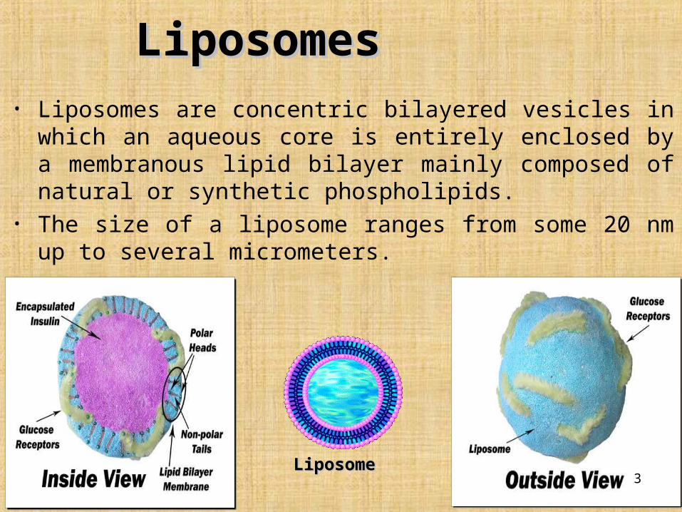
• Liposomes are concentric bilayered vesicles in which an aqueous core is entirely enclosed by a membranous lipid bilayer mainly composed of natural or synthetic phospholipids.
• The size of a liposome ranges from some 20 nm up to several micrometers.
Liposomes Liposomes
Liposome Liposome 3
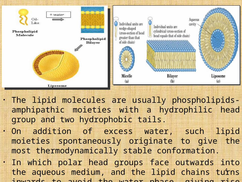
• The lipid molecules are usually phospholipids- amphipathic moieties with a hydrophilic head group and two hydrophobic tails.
• On addition of excess water, such lipid moieties spontaneously originate to give the most thermodynamically stable conformation.
• In which polar head groups face outwards into the aqueous medium, and the lipid chains turns inwards to avoid the water phase, giving rise to double layer or bilayer lamellar structures.
4
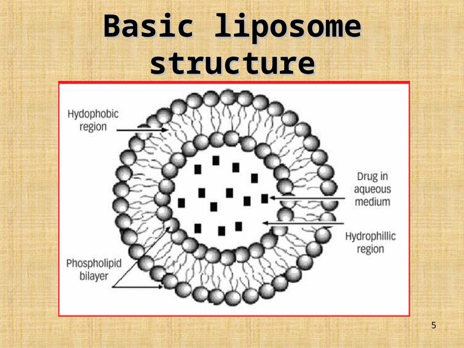
Basic liposome structureBasic liposome structure
5
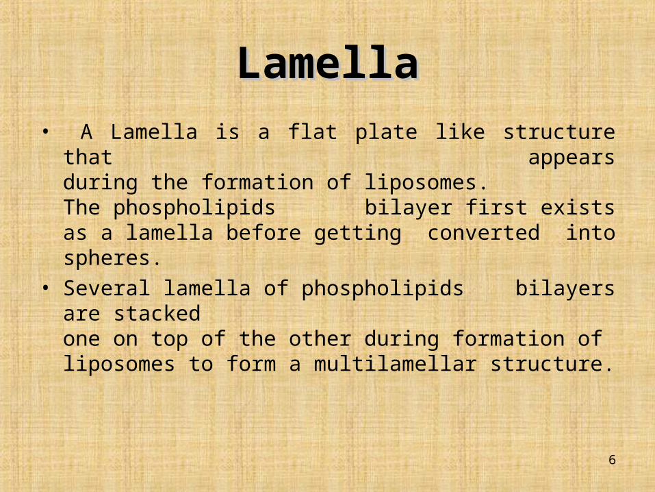
LamellaLamella• A Lamella is a flat plate like structure that appears
during the formation of liposomes. The phospholipids bilayer first exists as a lamella before getting converted into spheres.
• Several lamella of phospholipids bilayers are stacked one on top of the other during formation of liposomes to form a multilamellar structure.
6
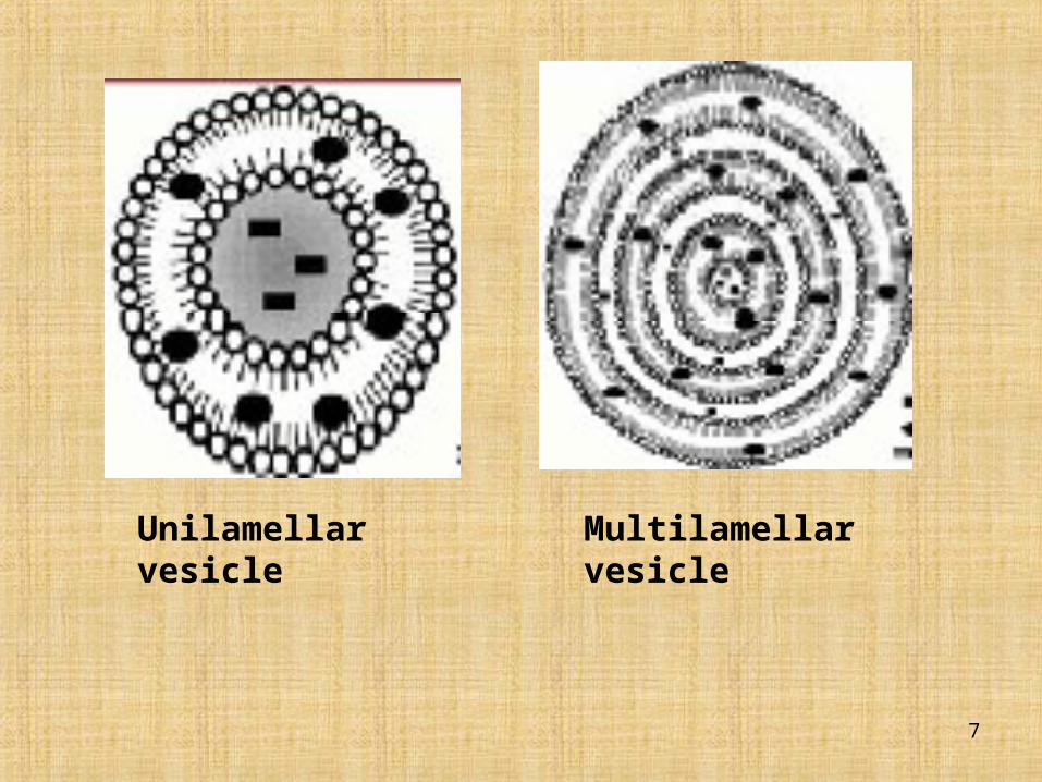
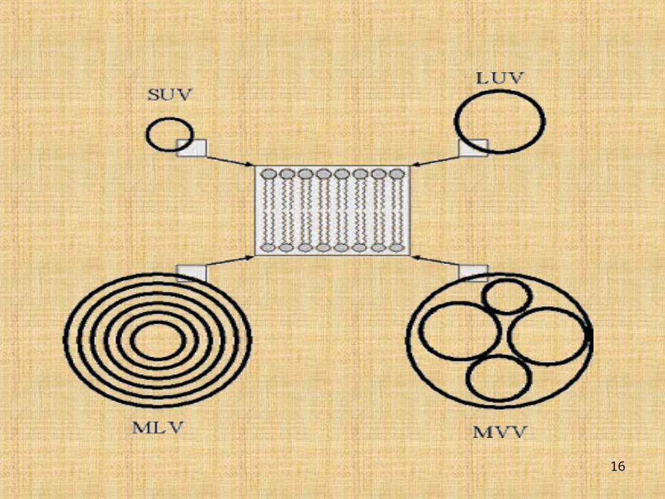
Multilamellar vesicle Unilamellar vesicle
7
Structural Components of Structural Components of LiposomesLiposomes
• The main components of liposomes are :-
1. Phospholipids
2. Cholesterol
8
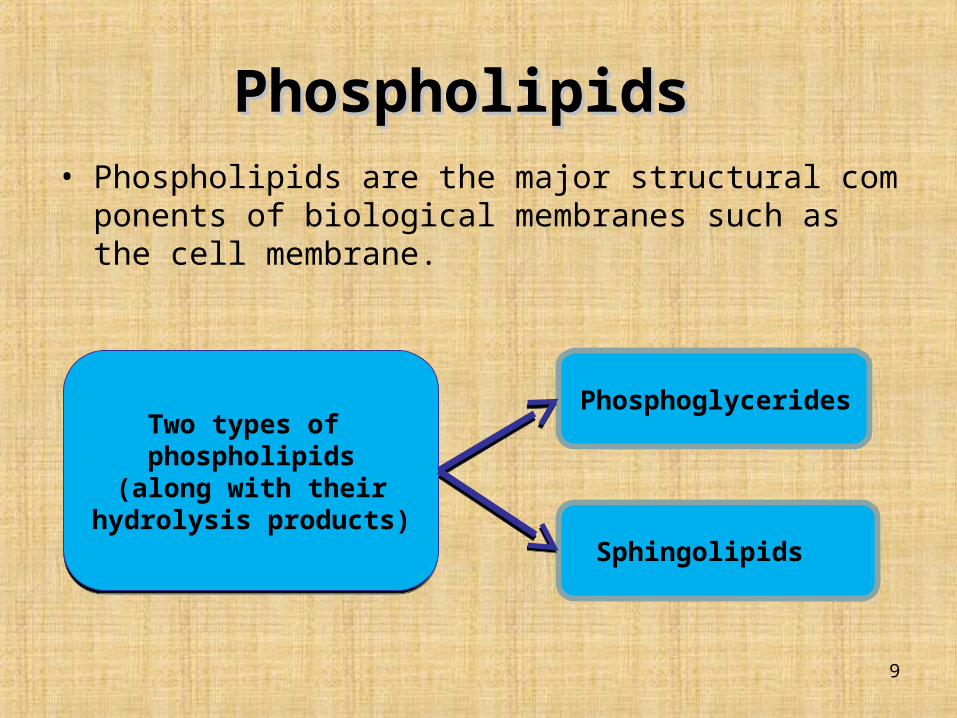
Phospholipids Phospholipids • Phospholipids are the major structural components of biological
membranes such as the cell membrane.
Phosphoglycerides
Two types of phospholipids(along with their hydrolysis
products)
Two types of phospholipids(along with their hydrolysis
products)
Sphingolipids
9

PhosphatidylcholinePhosphatidylcholine
• Most common phospholipids used is
phosphatidylcholine (PC).
• Phosphatidylcholine is an amphipathic
molecule in which exists:-
– a hydrophilic polar head group,
phosphocholine.
– a glycerol bridge.
– a pair of hydrophobic acyl hydrocarbon
chains.
10
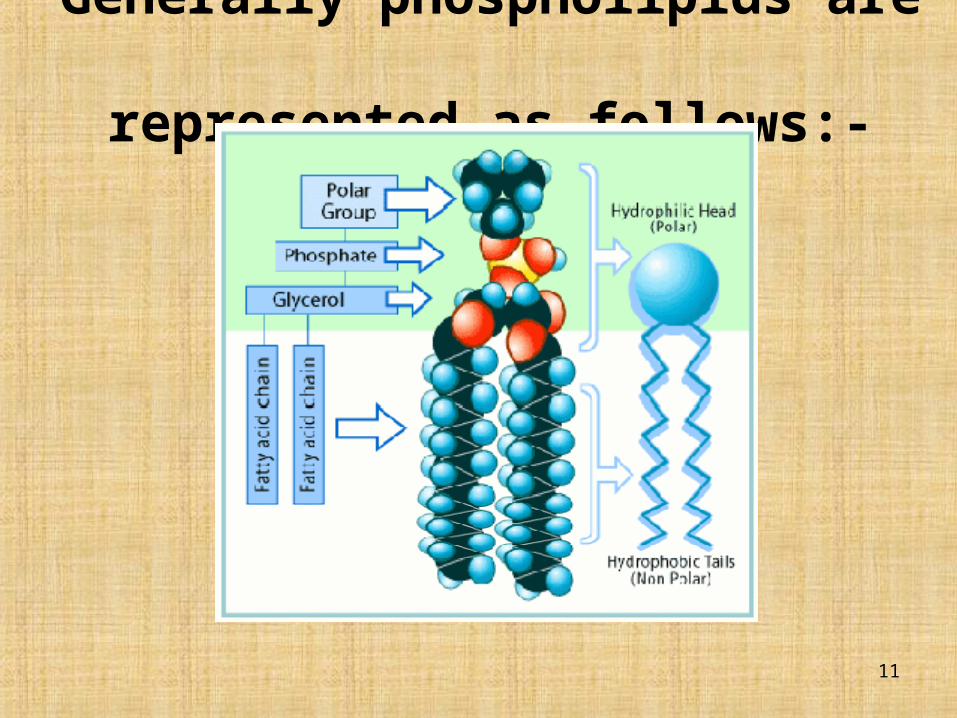
Generally phospholipids are represented as follows:-
11
CholesterolCholesterol
• Cholesterol by itself does not form bilayer structure.
• Cholesterol act as fluidity buffer
• After intercalation with phospholipid molecules alter the freedom of motion of carbon molecules in the acyl chain
• Restricts the transformations of trans to gauche conformations
• Cholesterol incorporation increases the separation between choline head group & eliminates normal electrostatic & hydrogen bonding interactions
12
Advantages of liposomesAdvantages of liposomes
• Provides selective passive targeting to tumor tissues.
• Increased efficacy and therapeutic index.
• Increased stability of encapsulated drug.
• Reduction in toxicity of the encapsulated agent.
• Site avoidance effect (avoids non-target tissues).
• Improved pharmacokinetic effects (reduced elimination increased circulation life times).
• Flexibility to couple with site specific ligands to achieve active targetting.
13
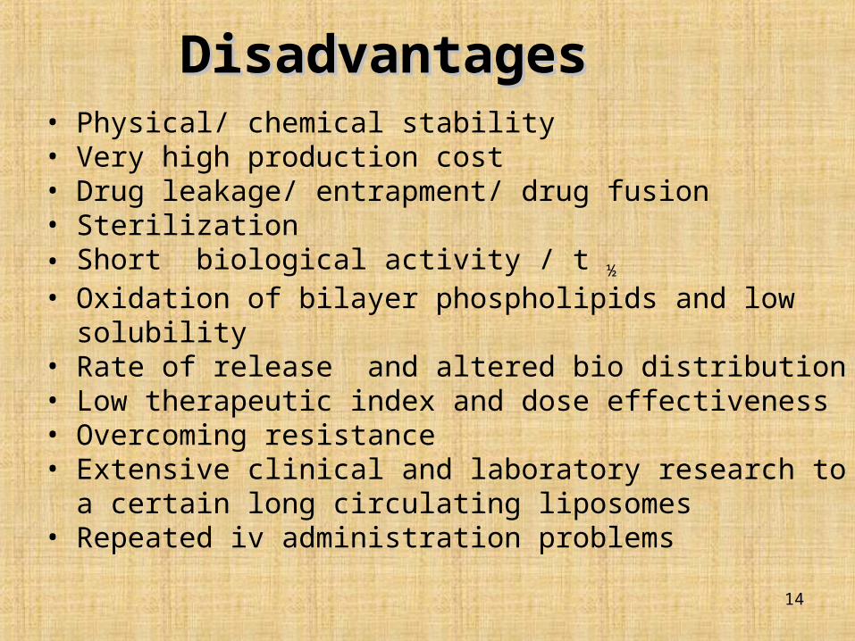
DisadvantagesDisadvantages• Physical/ chemical stability• Very high production cost• Drug leakage/ entrapment/ drug fusion• Sterilization • Short biological activity / t ½
• Oxidation of bilayer phospholipids and low solubility• Rate of release and altered bio distribution• Low therapeutic index and dose effectiveness• Overcoming resistance• Extensive clinical and laboratory research to a certain long
circulating liposomes• Repeated iv administration problems
14
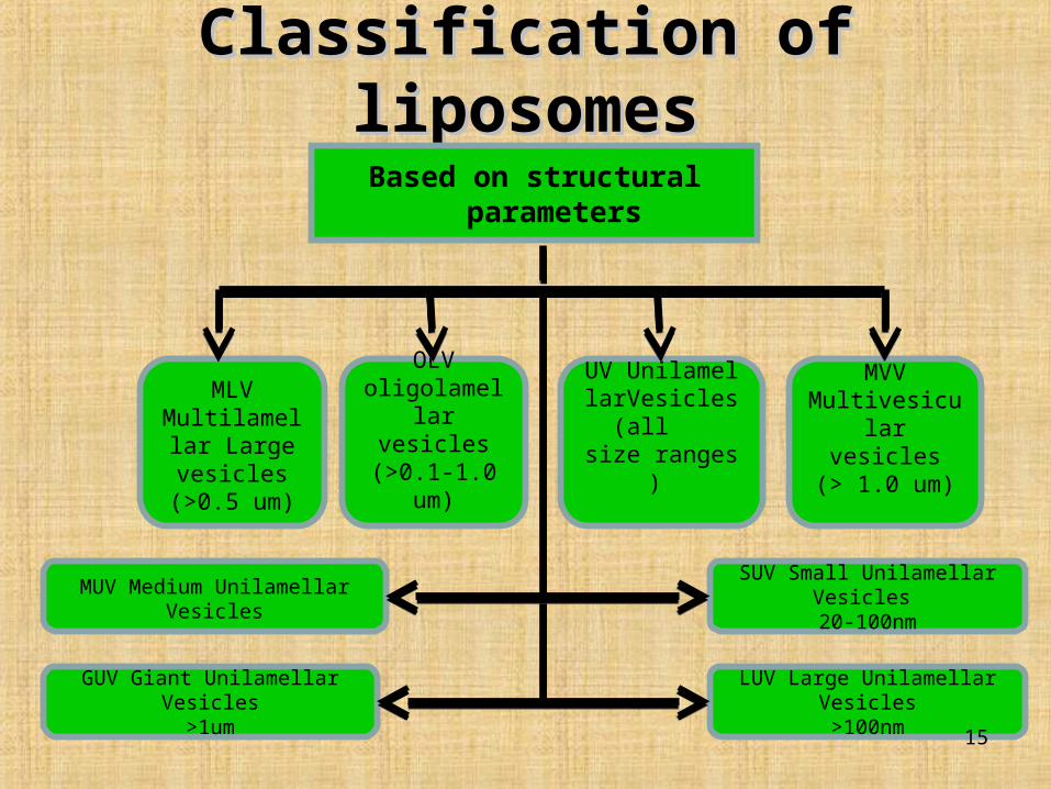
Classification of liposomesClassification of liposomes
MLVMultilamellar
Large vesicles
(>0.5 um)
OLV oligolamellar
vesicles(>0.1-1.0 um)
UV UnilamellarVesicles (all size ranges)
MVVMultivesicular
vesicles(> 1.0 um)
MUV Medium Unilamellar Vesicles
GUV Giant Unilamellar Vesicles>1um
SUV Small Unilamellar Vesicles 20-100nm
LUV Large Unilamellar Vesicles>100nm
Based on structural parameters
15
16
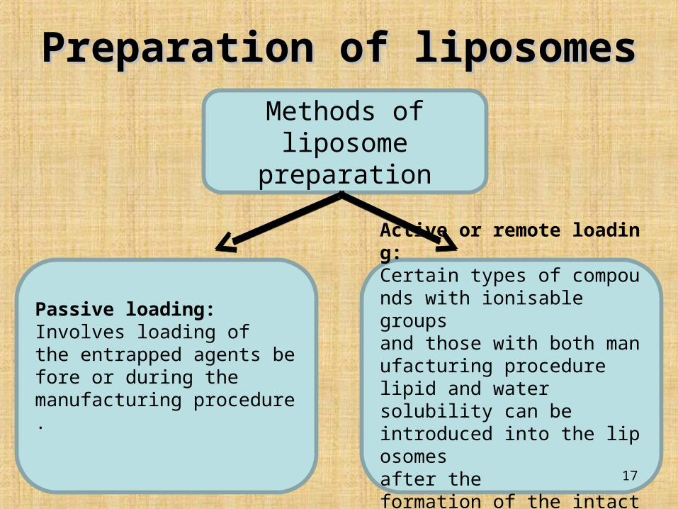
Preparation of liposomesPreparation of liposomes
Methods of liposome preparation
Passive loading:Involves loading of the entrapped agents before or during the manufacturing procedure.
Active or remote loading:Certain types of compounds with ionisable groups and those with both manufacturing procedure lipid and water solubility can be introduced into the liposomes after the formation of the intact vesicles 17

Methods of liposome preparation
Solvent dispersion
methods
Ethanol injectionEther injectionDouble emulsion vesiclesStable plurilamellar VesiclesReverse phase evaporation vesicles
Detergent removal methods
Passive loading techniques
Detergent(Cholate, Alkyl glycoside, Triton X-100) removal from mixed micelles byDialysisColumn chromatographyDilutionReconstituted sendai virus enveloped vesicles
Active loading techniques
Lipid film hydration by hand shaking non-hand shaking and freeze dryingMicro emulsificationSonicationFrench pressure cellMembrane extrusionDried reconstituted vesiclesFreeze thawed liposomes
Mechanical dispersion
methods
18

Evaluation of liposomes Evaluation of liposomes The liposomes prepared by various techniques are to be evaluated
for their physical properties, has these influence the behavior of liposomes in vivo.
Physical properties
1. Particle size
Both particle size and particle size distribution of liposomes influence their physical stability. These can be determined by the following method.
a) Laser light scattering
b) Transmission electron microscopy
19
2. Surface charge The positive, negative or neutral charge on the surface of the
liposomes is due to the composition of the head groups. The surface charge of liposomes governs the kinetic and extent of
distribution in vivo, as well as interaction with the target cells. The method involved in the measurement of surface charge is
based on free-flow electrophoresis of MLVs.• It utilizes a cellulose acetate plate dipped in sodium borate buffer
of pH 8.8.• About 5N moles of lipid samples are applied on to the plate,
which is then subjected to electrophoresis at 4 ? c for 30 mins.• The liposomes get bifurcated depending on their surface charge.
This technique can be used for determining the heterogeneity of charges in the liposome suspension as well as to detect any impurities such as fatty acids.
20
3. Percent drug encapsulated.
• Quantity of drug entrapped in the liposomes helps to estimate the behavior of the drug in biological system
• Liposomes are mixture of encapsulated and unencapsulated drug fractions
• The % of drug encapsulation is done by first separating the free drug fraction from encapsulated drug fraction
• The encapsulated fraction is then made to leak off the liposome into aqueous solution using suitable detergents
• The methods used to separate the free drug from the sample are:
a. Mini column centrifugation method
b. Protamine aggregated method
21

4. Phase behavior• At transition temperature liposomes undergo reversible phase
transition• The transition temperature is the indication of stability
permeability and also indicates the region of drug entrapment• Done by DSC
5. Drug Release Rate The rate of drug release from the liposomes can be determined
by in vivo assays which helps to predict the pharmacokinetics and bioavailability of the drug. However in vivo studies are found to be more complete.
Liposome encapsulating the tracer [ᵌH] insulin are employed for the study. This [ᵌH] insulin is preferred, as it is released only in the ECF and undergoes rapid renal excretion of the face tracer coupled to the degradation rate constant o the tracer released from the liposomes. 22

ApplicationsApplications
• Liposomes as drug or protein delivery vehicles.
• Liposome in antimicrobial, antifungal(lung therapeutics) and antiviral (anti HIV) therapy.
• In tumor therapy.
• In gene therapy.
• In immunology.
• Liposomes as artificial blood surrogates.
• Liposomes as radiopharmaceutical and radio diagnostic carriers.
• Liposomes in cosmetics and dermatology.
23

24
Related Documents