Lipids as cofactors in protein folding: Stereo-specific lipid–protein interactions are required to form HAMLET (human -lactalbumin made lethal to tumor cells) MALIN SVENSSON, 1 ANN-KRISTIN MOSSBERG, 1 JENNY PETTERSSON, 1,2 SARA LINSE, 2 AND CATHARINA SVANBORG 1 1 Department of Microbiology, Immunology and Glycobiology (MIG), Institute of Laboratory Medicine and 2 Department of Biophysical Chemistry, Lund University, Lund, Sweden (RECEIVED September 25, 2002; FINAL REVISION July 10, 2003; ACCEPTED September 12, 2003) Abstract Proteins can adjust their structure and function in response to shifting environments. Functional diversity is created not only by the sequence but by changes in tertiary structure. Here we present evidence that lipid cofactors may enable otherwise unstable protein folding variants to maintain their conformation and to form novel, biologically active complexes. We have identified unsaturated C18 fatty acids in the cis conformation as the cofactors that bind apo -lactalbumin and form HAMLET (human -lactalbumin made lethal to tumor cells). The complexes were formed on an ion exchange column, were stable in a molten globule-like conformation, and had attained the novel biological activity. The protein–fatty acid interaction was specific, as saturated C18 fatty acids, or unsaturated C18:1trans conformers were unable to form complexes with apo -lactalbumin, as were fatty acids with shorter or longer carbon chains. Unsaturated cis fatty acids other than C18:1:9cis were able to form stable complexes, but these were not active in the apoptosis assay. The results demonstrate that stereo-specific lipid–protein interactions can stabilize partially unfolded conformations and form molecular complexes with novel biological activity. The results offer a new mechanism for the functional diversity of proteins, by exploiting lipids as essential, tissue-specific cofactors in this process. Keywords: -Lactalbumin; HAMLET; fatty acids; apoptosis; cancer; protein folding The human genome sequence was recently found to contain much fewer distinctive genes than previously expected (Green and Chakravarti 2001). This finding emphasizes the need to identify mechanisms by which individual proteins may attain functional diversity. One may predict that pro- teins must be able to sense different environments and ad- just their structure and function to meet local demands. Structural diversity may be achieved by various chemical modifications such as methylation, sulphation, or acetyla- tion, but in addition, changes in tertiary structure are be- coming recognized as a mechanism potentially allowing proteins to achieve diversity of function (Jeffery 1999). The most striking example is the prion protein, which changes from a mixed -helical and -sheet conformation to the -sheet–rich, disease-causing iso-form Prp sc (Pan et al. 1993; Safar et al. 1993; Pergami et al. 1996). It has been postulated that this shift requires tissue specific cofactors that stabilize Prp sc , but the “factor x” has not yet been identified (Booth et al. 1997; Cohen and Prusiner 1998; Jackson and Collinge 2000). Reprint requests to: Catharina Svanborg, Department of Microbiology, Immunology and Glycobiology (MIG), Institute of Laboratory Medicine, Lund University, Sölvegatan 23, S-223 62 Lund, Sweden; e-mail: [email protected]; fax: 46-46-137468. Abbreviations: GC/MS, gas chromatography/mass spectrometry; EDTA, ethylenediamine tetra acetic acid; Tris, tris (hydroxymethyl) aminometh- ane; ANS, 8-anilinonaphtalene-1-sulfonic acid; CD, circular dichroism; DEAE, diethylaminoethyl; EGTA, ethylene-bis(oxyethyleneitriol)tetraace- tic acid; PBS, phosphate-buffered saline. Article and publication are at http://www.proteinscience.org/cgi/doi/ 10.1110/ps.0231103. Protein Science (2003), 12:2805–2814. Published by Cold Spring Harbor Laboratory Press. Copyright © 2003 The Protein Society 2805

Welcome message from author
This document is posted to help you gain knowledge. Please leave a comment to let me know what you think about it! Share it to your friends and learn new things together.
Transcript
Lipids as cofactors in protein folding: Stereo-specificlipid–protein interactions are required to formHAMLET (human �-lactalbumin made lethal totumor cells)
MALIN SVENSSON,1 ANN-KRISTIN MOSSBERG,1 JENNY PETTERSSON,1,2
SARA LINSE,2 AND CATHARINA SVANBORG1
1Department of Microbiology, Immunology and Glycobiology (MIG), Institute of Laboratory Medicine and2Department of Biophysical Chemistry, Lund University, Lund, Sweden
(RECEIVED September 25, 2002; FINAL REVISION July 10, 2003; ACCEPTED September 12, 2003)
Abstract
Proteins can adjust their structure and function in response to shifting environments. Functional diversity iscreated not only by the sequence but by changes in tertiary structure. Here we present evidence that lipidcofactors may enable otherwise unstable protein folding variants to maintain their conformation and to formnovel, biologically active complexes. We have identified unsaturated C18 fatty acids in the cis conformationas the cofactors that bind apo �-lactalbumin and form HAMLET (human �-lactalbumin made lethal totumor cells). The complexes were formed on an ion exchange column, were stable in a molten globule-likeconformation, and had attained the novel biological activity. The protein–fatty acid interaction was specific,as saturated C18 fatty acids, or unsaturated C18:1trans conformers were unable to form complexes with apo�-lactalbumin, as were fatty acids with shorter or longer carbon chains. Unsaturated cis fatty acids other thanC18:1:9cis were able to form stable complexes, but these were not active in the apoptosis assay. The resultsdemonstrate that stereo-specific lipid–protein interactions can stabilize partially unfolded conformations andform molecular complexes with novel biological activity. The results offer a new mechanism for thefunctional diversity of proteins, by exploiting lipids as essential, tissue-specific cofactors in this process.
Keywords: �-Lactalbumin; HAMLET; fatty acids; apoptosis; cancer; protein folding
The human genome sequence was recently found to containmuch fewer distinctive genes than previously expected(Green and Chakravarti 2001). This finding emphasizes theneed to identify mechanisms by which individual proteinsmay attain functional diversity. One may predict that pro-
teins must be able to sense different environments and ad-just their structure and function to meet local demands.Structural diversity may be achieved by various chemicalmodifications such as methylation, sulphation, or acetyla-tion, but in addition, changes in tertiary structure are be-coming recognized as a mechanism potentially allowingproteins to achieve diversity of function (Jeffery 1999). Themost striking example is the prion protein, which changesfrom a mixed �-helical and �-sheet conformation to the�-sheet–rich, disease-causing iso-form Prpsc (Pan et al.1993; Safar et al. 1993; Pergami et al. 1996). It has beenpostulated that this shift requires tissue specific cofactorsthat stabilize Prpsc, but the “factor x” has not yet beenidentified (Booth et al. 1997; Cohen and Prusiner 1998;Jackson and Collinge 2000).
Reprint requests to: Catharina Svanborg, Department of Microbiology,Immunology and Glycobiology (MIG), Institute of Laboratory Medicine,Lund University, Sölvegatan 23, S-223 62 Lund, Sweden; e-mail:[email protected]; fax: 46-46-137468.
Abbreviations: GC/MS, gas chromatography/mass spectrometry; EDTA,ethylenediamine tetra acetic acid; Tris, tris (hydroxymethyl) aminometh-ane; ANS, 8-anilinonaphtalene-1-sulfonic acid; CD, circular dichroism;DEAE, diethylaminoethyl; EGTA, ethylene-bis(oxyethyleneitriol)tetraace-tic acid; PBS, phosphate-buffered saline.
Article and publication are at http://www.proteinscience.org/cgi/doi/10.1110/ps.0231103.
Protein Science (2003), 12:2805–2814. Published by Cold Spring Harbor Laboratory Press. Copyright © 2003 The Protein Society 2805
This study addressed the role of specific lipids as cofac-tors that may enable protein folding variants to attain newfunctions. HAMLET (human �-lactalbumin made lethal totumor cells) was used as a model. HAMLET is a molecularcomplex that induces in vitro apoptosis in tumor cells butnot in healthy differentiated cells (Hakansson et al. 1995;Svanborg et al. 2003). The apoptotic activity of this variantfold was discovered by serendipity, in a fraction of humanmilk casein obtained by precipitation at low pH (Hakanssonet al. 1995). By spectroscopy, the active fraction was shownto contain partially unfolded �-lactalbumin (Svensson et al.1999), with native-like secondary structure, but lacked spe-cific tertiary packing of the side chains. Unlike apo �-lact-albumin, however, the protein was stable in this unfoldedstate at physiologic pH and in the presence of Ca2+. The linkbetween apoptosis induction and the folding change wasproven by deliberate conversion of native �-lactalbumin tothe apoptosis-inducing form (Svensson et al. 2000). Theconversion was shown require both the folding change anda lipid cofactor (Svensson et al. 2000).
The aim of the present study was to compare structuralvariants of oleic acid, and other fatty acids differing in thedegree of saturation, carbon chain length and cis/trans con-formation. We demonstrate that highly specific interactionsguide the formation of HAMLET and that only the cis-unsaturated C18 fatty acids serve as suitable cofactors.
Results
Conversion of �-lactalbumin to HAMLET requires theC18:1 fatty acid cofactor
To form HAMLET from native �-lactalbumin, the stronglybound Ca2+ ion was removed with etylenediamintetra aceticacid (EDTA), and the conformational change was con-firmed by spectroscopy. Apo �-lactalbumin was then sub-jected to ion exchange chromatography on a matrix that hadbeen preconditioned with oleic acid, C18:1:9cis. The pro-tein was retained on the conditioned column, forming acomplex that eluted as a sharp peak after 1 M NaCl. Theeluted protein maintained a molten globule-like conforma-tion at neutral pH and in the presence of Ca2+, as shown bycircular dichroism (CD) and 8-anilinonaphtalene-1-sulfonicacid (ANS) spectroscopy (Svensson et al. 2000). TheHAMLET complex induced apoptosis in many types of tu-mor cells, including the L1210 lymphoma cell line (Svan-borg et al. 2003). HAMLET was thus defined as the con-version product of apo �-lactalbumin and oleic acid, withthe ability to induce apoptosis in tumor cells.
Comparison with other fatty acids
To understand the specificity of the protein–lipid interac-tion, we used structural variants of oleic acid and other fattyacids. Fatty acids differing in the degree of saturation, car-
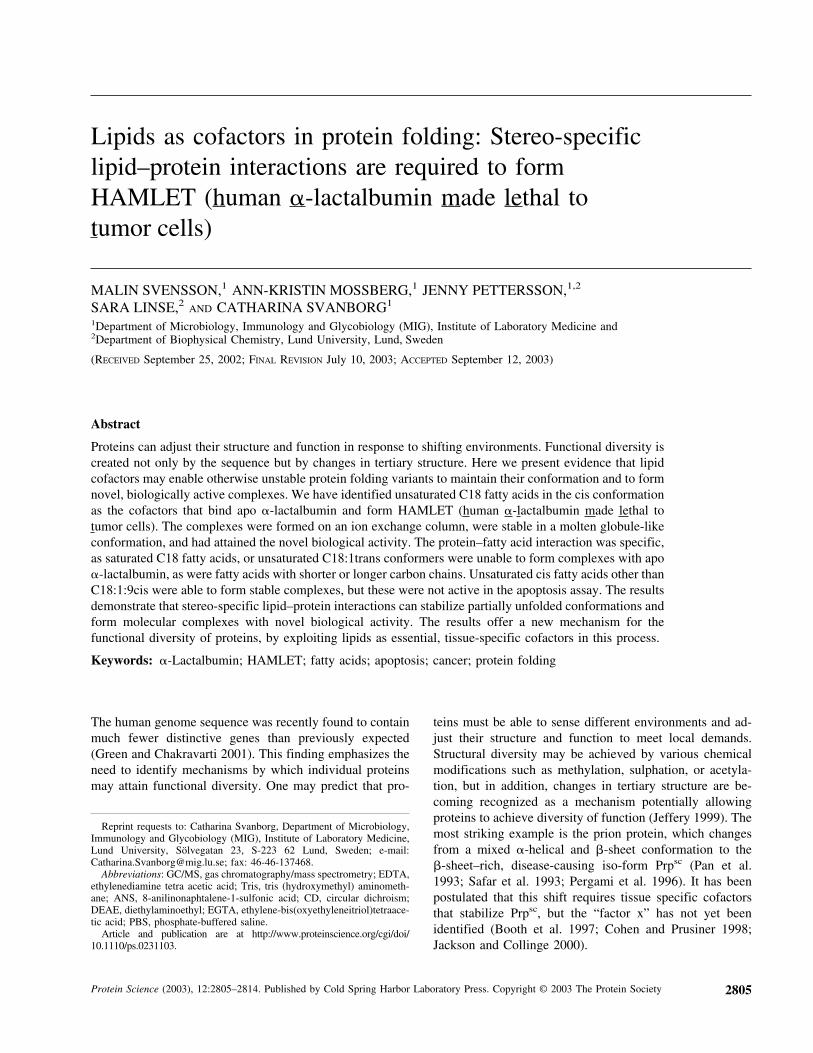
bon chain length, and cis/trans conformation were assayedfor their ability to form HAMLET-like complexes with apo�-lactalbumin (Fig. 1). Clean column matrices were condi-tioned with the individual fatty acids, apo �-lactalbuminwas applied to the columns, and the material eluting after 1M NaCl was collected for analysis.
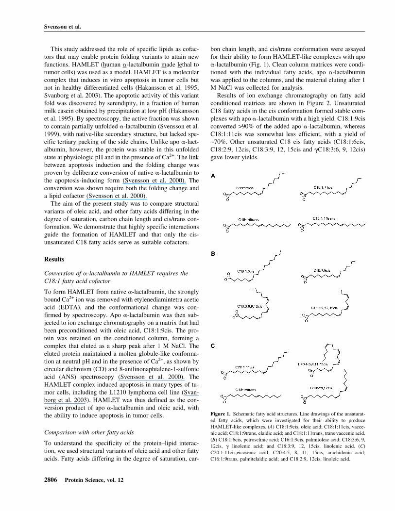
Results of ion exchange chromatography on fatty acidconditioned matrices are shown in Figure 2. UnsaturatedC18 fatty acids in the cis conformation formed stable com-plexes with apo �-lactalbumin with a high yield. C18:1:9cisconverted >90% of the added apo �-lactalbumin, whereasC18:1:11cis was somewhat less efficient, with a yield of∼70%. Other unsaturated C18 cis fatty acids (C18:1:6cis,C18:2:9, 12cis, C18:3:9, 12, 15cis and �C18:3:6, 9, 12cis)gave lower yields.
Figure 1. Schematic fatty acid structures. Line drawings of the unsaturat-ed fatty acids, which were investigated for their ability to produceHAMLET-like complexes. (A) C18:1:9cis, oleic acid; C18:1:11cis, vacce-nic acid; C18:1:9trans, elaidic acid; and C18:1:11trans, trans vaccenic acid.(B) C18:1:6cis, petroselinic acid; C16:1:9cis, palmitoleic acid; C18:3:6, 9,12cis, � linolenic acid; and C18:3:9, 12, 15cis, linolenic acid. (C)C20:1:11cis,eicosenic acid; C20:4:5, 8, 11, 15cis, arachidonic acid;C16:1:9trans, palmitelaidic acid; and C18:2:9, 12cis, linoleic acid.
Svensson et al.
2806 Protein Science, vol. 12
When exposed to columns conditioned with the transconformers of C18:1 or with saturated fatty acids, the pro-tein remained largely unconverted. Only small amountseluted with high salt from columns conditioned withC18:1:9trans, C18:1:11trans, or the saturated C18:0 fattyacid and the eluted complexes were inactive in the apoptosisassay.
Unsaturated cis fatty acids with shorter (C16:1:9cis) orlonger (C20:1:11cis and C20:4:5, 8, 11, 15cis) carbonchains formed stable complexes, but with yields lower thanC18:1:9cis. The columns conditioned with the saturatedfatty acids C6:0, C14:0, or C16:0 retained no apo �-lact-
albumin. The results demonstrate that apo �-lactalbumininteracts in a stereo-specific manner with fatty acids in thecis conformation, and the C18 cis fatty acids give the high-est yield.
Conformation analysis by CD spectroscopy
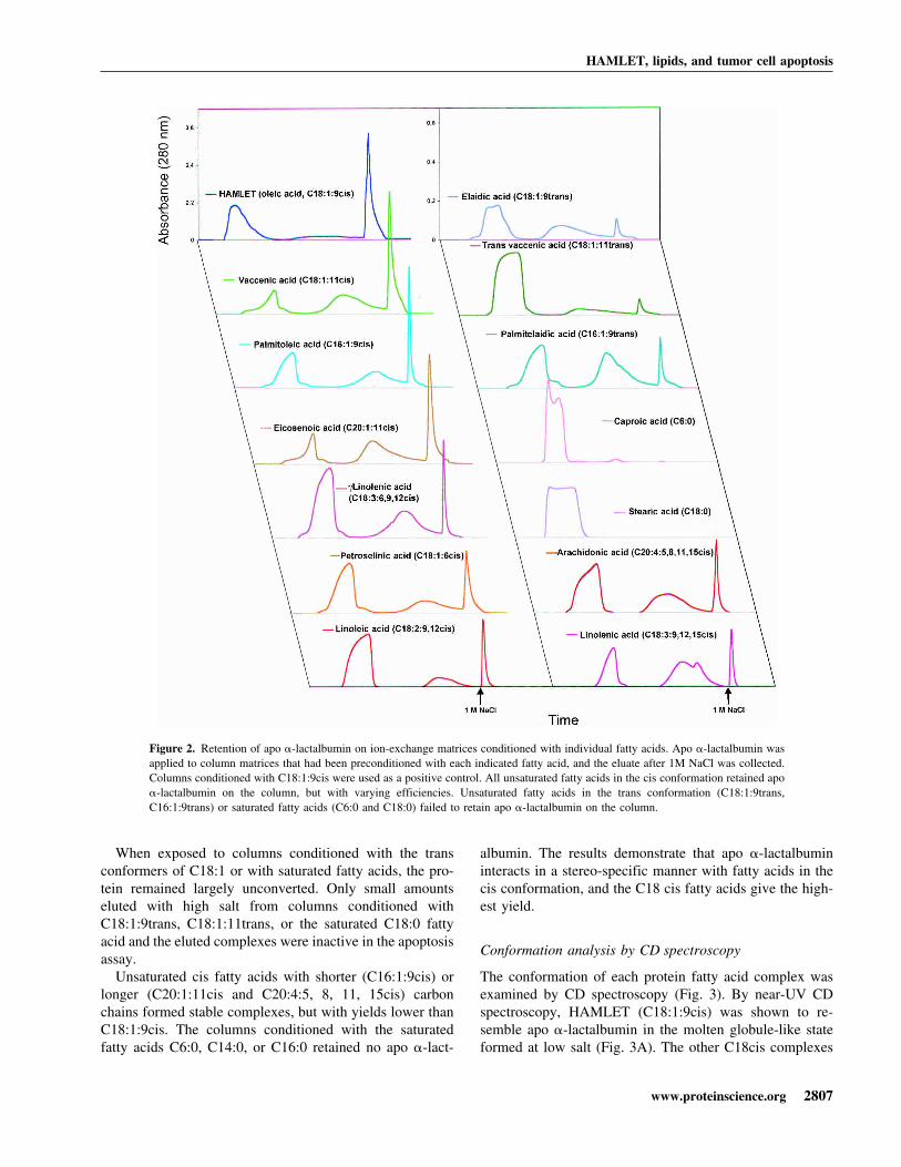
The conformation of each protein fatty acid complex wasexamined by CD spectroscopy (Fig. 3). By near-UV CDspectroscopy, HAMLET (C18:1:9cis) was shown to re-semble apo �-lactalbumin in the molten globule-like stateformed at low salt (Fig. 3A). The other C18cis complexes
Figure 2. Retention of apo �-lactalbumin on ion-exchange matrices conditioned with individual fatty acids. Apo �-lactalbumin wasapplied to column matrices that had been preconditioned with each indicated fatty acid, and the eluate after 1M NaCl was collected.Columns conditioned with C18:1:9cis were used as a positive control. All unsaturated fatty acids in the cis conformation retained apo�-lactalbumin on the column, but with varying efficiencies. Unsaturated fatty acids in the trans conformation (C18:1:9trans,C16:1:9trans) or saturated fatty acids (C6:0 and C18:0) failed to retain apo �-lactalbumin on the column.
HAMLET, lipids, and tumor cell apoptosis
www.proteinscience.org 2807
Fig
ure
3.T
ertia
ryst
ruct
ure
and
hydr
opho
bici
tyof
the
fatty
acid
-pro
tein
com
plex
esex
amin
edby
CD
and
AN
Ssp
ectr
osco
py.(
A)
CD
spec
trum
ofna
tive
�-l
acta
lbum
insh
owin
gth
ech
arac
teri
stic
sof
aw
ell-
fold
edpr
otei
nat
25°C
,with
am
inim
umat
270
nmar
isin
gfr
omty
rosi
nere
sidu
esan
da
max
imum
at29
4nm
aris
ing
from
tryp
toph
anre
sidu
es.T
heap
o�
-lac
talb
umin
cont
rol
had
lost
mos
tof
the
char
acte
rist
icsi
gnal
s,in
dica
ting
less
rest
rain
edty
rosi
nes
and
tryp
toph
ans.
(B)
The
C18
com
plex
esre
sem
bled
HA
ML
ET
with
alo
ssof
sign
alin
the
tyro
sine
and
tryp
toph
anre
gion
s.(C
)T
heC
20:4
:5,
8,11
,15c
isan
dth
eC
16:1
:9fa
ttyac
idco
mpl
exes
rese
mbl
edH
AM
LE
T,w
here
asth
eC
20:1
:11c
isco
mpl
exsh
owed
am
ore
nativ
elik
eco
nfor
mat
ion.
(D)N
ativ
e�
-lac
talb
umin
did
notb
ind
AN
Sas
show
nby
the
flat
curv
e,an
dth
elo
wsi
gnal
at49
0nm
at25
°C,
b ut
apo-
�-l
acta
lbum
insh
owed
sign
ific
ant
AN
Sbi
ndin
gw
ithen
hanc
edin
tens
ityan
da
max
imum
at47
0nm
,as
expe
cted
from
the
incr
ease
dhy
drop
hobi
city
ofth
isfo
ld.A
llof
the
�-l
acta
lbum
in-
fatty
acid
com
plex
esex
cept
C20
:1:1
1cis
boun
dA
NS.
(E)
The
C18
:1:6
cis,
C18
:3:9
,12,
15ci
s,an
d�
C18
:3:6
,9,1
2cis
fatty
acid
com
plex
es,s
how
eda
wav
elen
gth
shif
tsi
mila
rto
HA
ML
ET
,w
here
asC
18:1
:11c
is,
C18
:2:9
,12
cis,
C18
:1:9
tran
s,an
dC
18:1
:11t
rans
com
plex
essh
owed
asl
ight
lylo
wer
wav
elen
gth
shif
t.(F
)T
heot
her
com
plex
es(C
16:1
:9ci
s,C
20:4
:5,
8,11
,15
cis,
C20
:1:1
1cis
)sh
owed
alo
wer
shif
tm
ore
sim
ilar
toth
eap
o�
-lac
talb
umin
cont
rol.
Svensson et al.
2808 Protein Science, vol. 12
(C18:1:11cis, �C18:3:6, 9, 12cis, C18:1:6cis, C18:2:9,12cis, C18:3:9, 12, 15cis) showed a similar spectral pattern(Fig. 3B). Interestingly, the C18:1:9trans complex showed asimilar CD spectrum as that of the C18 cis fatty acids com-plexes, even though very little material was formed. TheC20:4:5, 8, 11, 15cis and the C16:1:9cis fatty acid com-plexes resembled HAMLET, whereas the C20:1:11cis ortrans complexes had adopted a more native-like conforma-tion, resembling the apo control at high salt (Fig. 3C). Weconclude that the unsaturated fatty acids, which retained apo�-lactalbumin on the column, also stabilized the partiallyunfolded conformation. By CD spectroscopy, there was nodiscernable difference between the active HAMLET com-plex and the inactive complexes (see below).
ANS fluorescence spectroscopy
Exposure of hydrophobic surfaces was probed by ANSspectroscopy (Fig. 3D–F). Native �-lactalbumin failed tobind ANS, as shown by the low intensity with a maximumat 510 nm. HAMLET showed enhanced intensity and amaximum at 470 nm (Fig. 3D). The C18:1:6cis, C18:3:9,12, 15cis, and �C18:3:6, 9, 12cis fatty acid complexes,bound ANS with similar wavelength shift as HAMLET(Fig. 3E). C18:1:9trans, C18:1:11cis, C18:1:11trans, andC18:2:9, 12cis complexes showed a slightly lower wave-length shift than did HAMLET (Fig. 3E). The other com-plexes (C16:1:9cis, C20:4:5, 8, 11, 15cis, C20:1:11cis) alsoshowed lower ANS fluorescence intensity than the apo�-lactalbumin control, as well as a lower shift. Despite thesedifferences, all of the fatty acid complexes were blue-shiftedcompared with native �-lactalbumin. To exclude the directbinding of ANS to the fatty acids in the protein–lipid com-plexes, mixtures of ANS and fatty acids were subjected tospectroscopy. No ANS–fatty acid interaction was observed(data not shown). We conclude that all eluted fatty acidcomplexes bind ANS more efficiently than native protein,indicating increased exposure of hydrophobic surfaces.
Biological activity
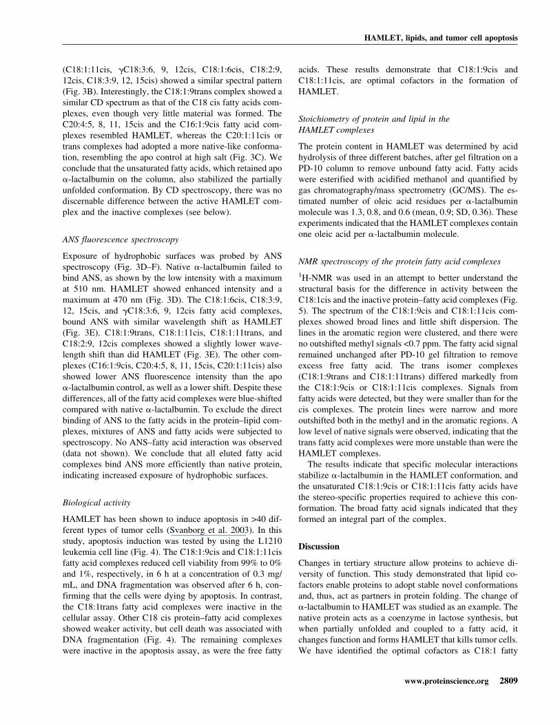
HAMLET has been shown to induce apoptosis in >40 dif-ferent types of tumor cells (Svanborg et al. 2003). In thisstudy, apoptosis induction was tested by using the L1210leukemia cell line (Fig. 4). The C18:1:9cis and C18:1:11cisfatty acid complexes reduced cell viability from 99% to 0%and 1%, respectively, in 6 h at a concentration of 0.3 mg/mL, and DNA fragmentation was observed after 6 h, con-firming that the cells were dying by apoptosis. In contrast,the C18:1trans fatty acid complexes were inactive in thecellular assay. Other C18 cis protein–fatty acid complexesshowed weaker activity, but cell death was associated withDNA fragmentation (Fig. 4). The remaining complexeswere inactive in the apoptosis assay, as were the free fatty
acids. These results demonstrate that C18:1:9cis andC18:1:11cis, are optimal cofactors in the formation ofHAMLET.
Stoichiometry of protein and lipid in theHAMLET complexes
The protein content in HAMLET was determined by acidhydrolysis of three different batches, after gel filtration on aPD-10 column to remove unbound fatty acid. Fatty acidswere esterified with acidified methanol and quantified bygas chromatography/mass spectrometry (GC/MS). The es-timated number of oleic acid residues per �-lactalbuminmolecule was 1.3, 0.8, and 0.6 (mean, 0.9; SD, 0.36). Theseexperiments indicated that the HAMLET complexes containone oleic acid per �-lactalbumin molecule.
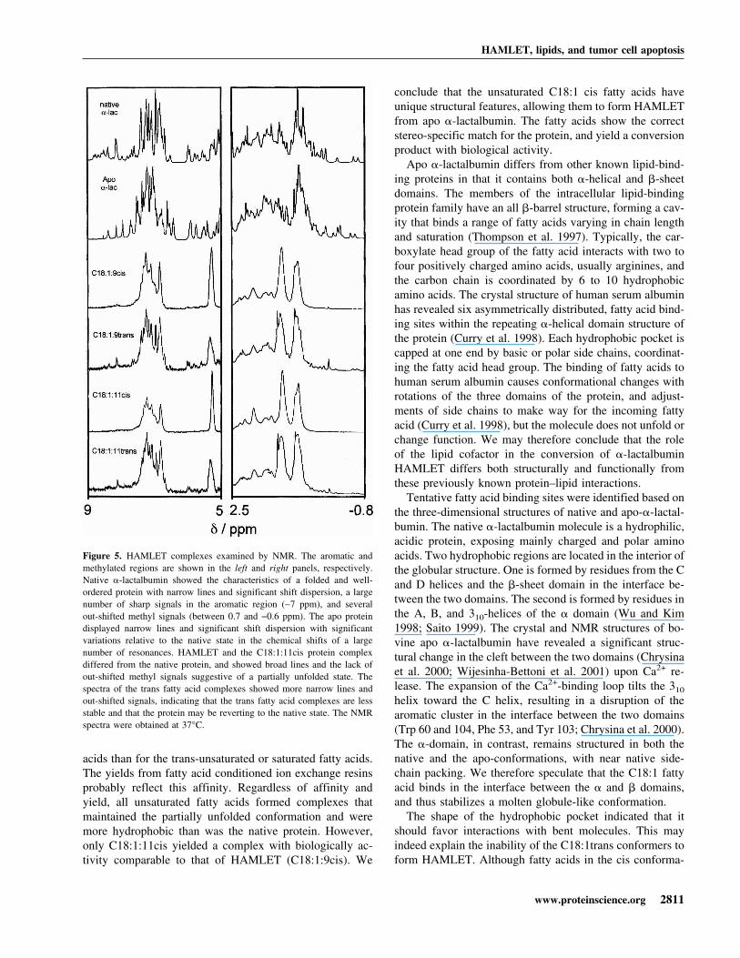
NMR spectroscopy of the protein fatty acid complexes1H-NMR was used in an attempt to better understand thestructural basis for the difference in activity between theC18:1cis and the inactive protein–fatty acid complexes (Fig.5). The spectrum of the C18:1:9cis and C18:1:11cis com-plexes showed broad lines and little shift dispersion. Thelines in the aromatic region were clustered, and there wereno outshifted methyl signals <0.7 ppm. The fatty acid signalremained unchanged after PD-10 gel filtration to removeexcess free fatty acid. The trans isomer complexes(C18:1:9trans and C18:1:11trans) differed markedly fromthe C18:1:9cis or C18:1:11cis complexes. Signals fromfatty acids were detected, but they were smaller than for thecis complexes. The protein lines were narrow and moreoutshifted both in the methyl and in the aromatic regions. Alow level of native signals were observed, indicating that thetrans fatty acid complexes were more unstable than were theHAMLET complexes.
The results indicate that specific molecular interactionsstabilize �-lactalbumin in the HAMLET conformation, andthe unsaturated C18:1:9cis or C18:1:11cis fatty acids havethe stereo-specific properties required to achieve this con-formation. The broad fatty acid signals indicated that theyformed an integral part of the complex.
Discussion
Changes in tertiary structure allow proteins to achieve di-versity of function. This study demonstrated that lipid co-factors enable proteins to adopt stable novel conformationsand, thus, act as partners in protein folding. The change of�-lactalbumin to HAMLET was studied as an example. Thenative protein acts as a coenzyme in lactose synthesis, butwhen partially unfolded and coupled to a fatty acid, itchanges function and forms HAMLET that kills tumor cells.We have identified the optimal cofactors as C18:1 fatty
HAMLET, lipids, and tumor cell apoptosis
www.proteinscience.org 2809
acids with a double bond in the cis conformation at position9 or 11. Saturated C18 fatty acids, unsaturated fatty acids inthe trans conformation, or fatty acids with shorter carbonchains could not form HAMLET. The results show that theunfolded protein interacts specifically with certain fatty acids,resulting in complex formation and new biological activity.
HAMLET is defined as the conversion product of apo�-lactalbumin and oleic acid, with the ability to induceapoptosis in tumor cells. Three molecular events are in-volved in the conversion of �-lactalbumin to HAMLET:Protein unfolding, fatty acid binding, and formation of thebiologically active complex. First, the protein must be par-tially unfolded and/or destabilized, as the native proteincannot be converted. This is achieved experimentally byadding EDTA (pH 8.5) to remove the tightly bound Ca2+
ion. Second, the apo-protein must bind the lipid cofactor,
and this occurs with high efficiency on an ion exchangematrix preconditioned with oleic acid. Fatty acids bind tothe apo protein also in solution, but the yield of HAMLETin such mixtures of �-lactalbumin and oleic acid are muchlower than on the column. Third, the protein and the lipidform a complex with apoptosis-inducing activity. This thirdstep may occur on the column, during elution with high salt,or during the dialysis step. In the active complex, �-lactal-bumin has a molten globule-like conformation and has ex-posed hydrophobic surfaces, but additional molecular modi-fications may have occurred.
The structural basis for biologic activity is not quite clear,but several important observations in this study allow us tospeculate about the unique molecular features of cis-unsat-urated C18 fatty acids as cofactors in HAMLET. Clearly,�-lactalbumin has a higher affinity for cis-unsaturated fatty
Figure 4. Tumor cell apoptosis induced by the lipid–protein complexes. Apoptosis induction in L1210 leukemia cells exposed to the different protein–lipidcomplexes, including the HAMLET control. HAMLET and the C18:1:11cis complex had killed 99% to 100% of the cells after 6 h and induced DNAfragmentation, but the C18:1trans fatty acid complexes were inactive. The C16 and C20 unsaturated fatty acid complexes caused a small loss of cell viabilityand intermediate DNA fragmentation. Lipid extracts derived from HAMLET or from each of the other complexes showed no effect on cell viability or DNAfragmentation. The lipid concentrations corresponded to the amount present in 1.0 mg of each complex. At very high lipid concentrations, the cells diedof necrosis, but at no time were there evidence of apoptosis in response to lipids.
Svensson et al.
2810 Protein Science, vol. 12
acids than for the trans-unsaturated or saturated fatty acids.The yields from fatty acid conditioned ion exchange resinsprobably reflect this affinity. Regardless of affinity andyield, all unsaturated fatty acids formed complexes thatmaintained the partially unfolded conformation and weremore hydrophobic than was the native protein. However,only C18:1:11cis yielded a complex with biologically ac-tivity comparable to that of HAMLET (C18:1:9cis). We
conclude that the unsaturated C18:1 cis fatty acids haveunique structural features, allowing them to form HAMLETfrom apo �-lactalbumin. The fatty acids show the correctstereo-specific match for the protein, and yield a conversionproduct with biological activity.
Apo �-lactalbumin differs from other known lipid-bind-ing proteins in that it contains both �-helical and �-sheetdomains. The members of the intracellular lipid-bindingprotein family have an all �-barrel structure, forming a cav-ity that binds a range of fatty acids varying in chain lengthand saturation (Thompson et al. 1997). Typically, the car-boxylate head group of the fatty acid interacts with two tofour positively charged amino acids, usually arginines, andthe carbon chain is coordinated by 6 to 10 hydrophobicamino acids. The crystal structure of human serum albuminhas revealed six asymmetrically distributed, fatty acid bind-ing sites within the repeating �-helical domain structure ofthe protein (Curry et al. 1998). Each hydrophobic pocket iscapped at one end by basic or polar side chains, coordinat-ing the fatty acid head group. The binding of fatty acids tohuman serum albumin causes conformational changes withrotations of the three domains of the protein, and adjust-ments of side chains to make way for the incoming fattyacid (Curry et al. 1998), but the molecule does not unfold orchange function. We may therefore conclude that the roleof the lipid cofactor in the conversion of �-lactalbuminHAMLET differs both structurally and functionally fromthese previously known protein–lipid interactions.
Tentative fatty acid binding sites were identified based onthe three-dimensional structures of native and apo-�-lactal-bumin. The native �-lactalbumin molecule is a hydrophilic,acidic protein, exposing mainly charged and polar aminoacids. Two hydrophobic regions are located in the interior ofthe globular structure. One is formed by residues from the Cand D helices and the �-sheet domain in the interface be-tween the two domains. The second is formed by residues inthe A, B, and 310-helices of the � domain (Wu and Kim1998; Saito 1999). The crystal and NMR structures of bo-vine apo �-lactalbumin have revealed a significant struc-tural change in the cleft between the two domains (Chrysinaet al. 2000; Wijesinha-Bettoni et al. 2001) upon Ca2+ re-lease. The expansion of the Ca2+-binding loop tilts the 310
helix toward the C helix, resulting in a disruption of thearomatic cluster in the interface between the two domains(Trp 60 and 104, Phe 53, and Tyr 103; Chrysina et al. 2000).The �-domain, in contrast, remains structured in both thenative and the apo-conformations, with near native side-chain packing. We therefore speculate that the C18:1 fattyacid binds in the interface between the � and � domains,and thus stabilizes a molten globule-like conformation.
The shape of the hydrophobic pocket indicated that itshould favor interactions with bent molecules. This mayindeed explain the inability of the C18:1trans conformers toform HAMLET. Although fatty acids in the cis conforma-
Figure 5. HAMLET complexes examined by NMR. The aromatic andmethylated regions are shown in the left and right panels, respectively.Native �-lactalbumin showed the characteristics of a folded and well-ordered protein with narrow lines and significant shift dispersion, a largenumber of sharp signals in the aromatic region (∼7 ppm), and severalout-shifted methyl signals (between 0.7 and −0.6 ppm). The apo proteindisplayed narrow lines and significant shift dispersion with significantvariations relative to the native state in the chemical shifts of a largenumber of resonances. HAMLET and the C18:1:11cis protein complexdiffered from the native protein, and showed broad lines and the lack ofout-shifted methyl signals suggestive of a partially unfolded state. Thespectra of the trans fatty acid complexes showed more narrow lines andout-shifted signals, indicating that the trans fatty acid complexes are lessstable and that the protein may be reverting to the native state. The NMRspectra were obtained at 37°C.
HAMLET, lipids, and tumor cell apoptosis
www.proteinscience.org 2811
tion are U-shaped around the double bond, with both carbonchains projecting in one direction, trans fatty acids are rodshaped around the double bond due to the carbon chains onopposite sides of the double bond. The saturated fatty acidsare most flexible, with no structural constraints due to thelack of double bonds. The results thus indicate that only thecis conformation allows fatty acids a close stereo-specificfit, and the additional critical feature of the fatty acid isthe carbon chain length. In addition, the pocket is capped bybasic residues, which may coordinate the polar head groupsof the fatty acids, thus orienting the lipid. This interactionis, however, not sufficient for activation as the trans andsaturated fatty acids, which possess the same charged headgroup failed to form the active complex. It is highly likelythat the stereo-specific fit involves both hydrophobic inter-actions with the lipid tail and electrostatic interactions ofthe negatively charged head group with basic side chains.Based on the analogy with other fatty acid binding proteins,the fatty acid may bind to HAMLET by electrostatic in-teractions between its negatively charged head group andbasic side chains in the protein, due to the hydrophobiceffect, as well as by van der Waal’s contacts with the tail,which are optimized for the preferred stereo-specific match(C18:1:9cis).
The present study raises the possibility that lipids may actas stabilizing cofactors in a variety of protein folding pro-cesses. The prions and amyloid fibrils are examples of un-successful protein processing, resulting in protein accumu-lation in peripheral tissues in which damage is done(McLaurin et al. 2000; Dobson 2001; Pepys 2001). It hasbeen postulated that a cofactor, factor x or “protein x” isrequired for the transmission of human prions to transgenicmice, to form the nascent scrapie isoforms during prionpropagation (Telling et al. 1994; Billeter et al. 1997). Bymutational analysis, the interaction with protein x wasshown to depend on a discontinuous epitope formed by theC-terminal � helix, with residues 167 and 171 in an adjacentloop (Kaneko et al. 1997), but the molecular nature of factorx has remained elusive. There is evidence that amyloid fi-bers contain a mixture of lipid species, but their role in theformation of fibrils remains to be defined (Kim et al. 1967;McLaurin et al. 2000). The present study indicates that lip-ids should be explored as cofactors locking the prions andamyloid proteins in their �-sheet–rich conformations.
Finally, the availability of fatty acid cofactors may facili-tate and determine if proteins will adapt to different envi-ronments. In case of �-lactalbumin, this mechanism mayallow the molecule to be modified in the nursing child. Thelow pH in the stomach is, for example, known to causepartial unfolding of �-lactalbumin, and lipids are hydro-lyzed by acid lipases to release oleic acid (Bernback et al.1990; Sarles et al. 1992). It is interesting to speculate thatthe beneficial function of HAMLET explains the specialabundance of both �-lactalbumin (2 mg/mL [140 �M];
Heine et al. 1991) and oleic acid (>50% of the fatty acidchains of triglycerides; Jensen 1996) in human milk. Breast-fed children show an overall lower incidence of childhoodcancer with an especially strong epidemiological associa-tion for lymphomas (crude odds ratio, 8.19; Davis et al.1988) that might result from the selective purging of un-wanted cells in the nursing child.
Materials and methods
�-Lactalbumin purification
Native human �-lactalbumin was purified from human milk byammonium sulphate precipitation and phenyl-Sepharose chroma-tography as described (Svensson et al. 2000).
Anion-exchange chromatography
Columns (14 × 1.6 cm) packed with diethylaminoethyl (DEAE)-Trisacryl M (BioSepra) were attached to a Bio-Logic chromatog-raphy system (Bio-Rad Laboratories), and eluted with a NaCl gra-dient (buffer A, 10 mM tris (hydroxymethyl) aminomethane [Tris]/HCl at pH 8.5; buffer B, buffer A containing 1 M NaCl). Thematrices were each conditioned with one of the different fattyacids used in the study (Larodan Biochemicals). Ten milligramswas dissolved in 500 �L 99.5% ethanol by sonication using aBranson 2200 bath sonicator (Branson). After addition of 10 mL of10 mM Tris/HCl (pH 8.5), the lipid solution was applied to anewly packed DEAE-Trisacryl M matrix and dispersed throughoutthe matrix by using a NaCl gradient.
Ten milligrams of human �-lactalbumin was dissolved in 20 mLof 10 mM Tris/HCl (pH 8.5) with 0.08 mM EDTA and added tothe column. The protein fraction eluting after high salt was de-salted by dialysis (Spectra/Pore, Spectrum Medical Industries;membrane cutoff, 3.5 kD), against distilled water with at least fourchanges of water in 100-fold volume excess, and then lyophilized.
Spectroscopic analysis
Stock solutions were prepared by dissolving the lyophilized ma-terial in 10 mM potassium phosphate buffer (pH 7.5), and con-centrations determined based on the absorbance at 280 nm. CDspectra were obtained on a JASCO J-720 spectro-polarimeter witha JASCO PTC-343 Peltier type thermostated cell holder. Quartzcuvettes were used with 1-cm path length, and spectra were re-corded between 240 and 320 nm at 25°C. The wavelength step was1 nm; the response time, 8 sec; and the scan rate, 10 nm/min. Sixscans were recorded and averaged for each spectrum. Baselinespectra were recorded with pure buffer in the cuvette and sub-tracted from the protein spectra.
The mean residue ellipticity �m was calculated from the re-corded ellipticity, �, as
�m � � / (c · n · l)
where c is the protein concentration in M; n, the number of resi-dues in the protein (123 in this case); l, the path length in m; and�, the ellipticity in degrees.
ANS fluorescence emission spectra were recorded at 25°C on aPerkin Elmer LS-50B spectrometer by using a quartz cuvette with1-cm excitation path length, between 400 and 600 nm (step 1 nm)
Svensson et al.
2812 Protein Science, vol. 12
with excitation at 385 nm. Both the excitation and emission band-pass were set to 5 nm. ANS ammonium salt (Fluka) was addedstepwise, and the spectra at 1.5 M equivalents are shown.
The 1H NMR spectra were recorded by using an Omega 500spectrometer at 500 MHz in D2O with 0.15 M NaCl at 37°C, for1 to 2 mM solutions of protein. Lyophilized HAMLET, native orapo �-lactalbumin (5 mg), or the protein–lipid complexes weredissolved in 500 �L of D2O, and the pH was set to 7.0 by usingNaOD. Apo �-lactalbumin was generated by dissolving �-lactal-bumin in doubly distilled water containing 10-fold molar excess ofethylene-bis(oxyethyleneitriol)tetraacetic acid (EGTA) at pH 8.0.The sample was applied to a PD-10 gel filtration column after analiquot of saturated NaCl (calcium-depleted) and eluted by doublydistilled water. The sample was passed through the saturated NaClto reduce binding of EGTA to the protein, and EGTA-free apoprotein was eluted in the water.
Stoichiometry of protein and lipid in theHAMLET complexes
Three batches of HAMLET were subjected to gel filtration on aPD-10 column (Pharmacia AB) for removal of unbound free fattyacids. Ten milligrams of lyophilized HAMLET was dissolved in0.5 mL phosphate-buffered saline (PBS; 0.14 M NaCl, 8.1 mMNa2HPO4, 2.7 mM KCl, 1.5 mM KH2PO4 at pH 7.4) and appliedon the gel filtration column. Fractions containing protein wereeluted with PBS and pooled (∼1 mL). The protein concentrationwas determined by the Amino Acid Analysis Centre at UppsalaUniversity on 100 �L of the sample. The fatty acid content wasdetermined by GC/MS on 200 �L of the sample, using C17:0 andC19:0 as internal standards. The sample solutions were evaporatedwith nitrogen gas. Internal standards and 1 mL of 2 M acidifiedmethanol were added, and the samples were methanolysed for 1 hat 85°C. Heptane was added, and the samples were centrifuged.The heptane phase, containing the fatty acids methyl esters, wasisolated. The nonpolar phases were dried with nitrogen gas, and 1mL heptane was added to the samples. Two �L was further ana-lyzed on a Varian gas chromatograph (model 3500) equipped witha split/split-less injector and a flame ionization detector, separatedon a (0.25-mm inner diameter) fused silica capillary column coatedwith FFAP (Chrompack). The column temperature was pro-grammed from 140°C/min to 240°C/min at 8°C/min. Chromato-grams were evaluated by using a Varian integrator model 4290.
Bioassays of apoptosis
The L1210 (ATCC, CCL 219) cell line was cultured in suspension,as described (Svensson et al. 1999). The cells were harvested bycentrifugation (200g for 10 min), resuspended in cell culture me-dium (RPMI 1640 supplemented without fetal calf serum, nones-sential amino acids, sodium pyruvate, and 50 �g gentamicin/mL;Life Technologies, GIBCO BRL), and seeded into 24-well plates(Falcon, Becton Dickinson) at a density of 2 × 10⁶/well. The dif-ferent substances were dissolved in cell culture medium, withoutfetal calf serum, and added to the cells (final volume 1 mL/well).Plates were incubated at 37°C in 5% CO2 atmosphere, and 100 �Lof fetal calf serum was added to each well only after 30 min ofincubation with HAMLET. Cell culture medium served as a con-trol. Cell viability was determined by trypan blue exclusion after 6h of incubation.
DNA fragmentation
Oligonucleosome length DNA fragments were detected by agarosegel electrophoresis. The cell suspension remaining after trypan
blue (970 �L, 2 × 10⁶/mL) was lysed in 5 mM Tris, 20 mMEDTA, 0.5% Triton X-100 (pH 8.0) for 1 h at 4°C and centrifugedat 13,000g for 15 min. DNA was ethanol precipitated over night in−20°C, treated with proteinase K and RNAse, loaded on 1.8%agarose gels, and electrophoresed with constant voltage set at 50 Vover night. DNA fragments were visualized with ethidium bromideby using a 305-nm UV-light source and photographed by usingPolaroid type 55 positive-negative film.
Acknowledgments
We thank Hanna Nilsson for the purification of �-lactalbumin andLennart Larsson for the GC/MS analysis of the lipid content inHAMLET. This work was supported by The Swedish Cancer So-ciety (grant number 3807-B97-01XAB [1997–2002] to C.S.),American Cancer Society (grant number SPG-97-157 [1997–2003] toC.S.), The Swedish Medical Research Council (grant number K97-03X-11552-02BK, to S.L.), The Swedish Pediatric Cancer Society,The Segerfalk Foundation, The Österlund Foundation, The LundHospital Foundation, and the Swedish Natural Science ResearchCouncil (grant number K-AA/KU 10178-300, to S.L.).
The publication costs of this article were defrayed in part bypayment of page charges. This article must therefore be herebymarked “advertisement” in accordance with 18 USC section 1734solely to indicate this fact.
References
Bernback, S., Blackberg, L., and Hernell, O. 1990. The complete digestion ofhuman milk triacylglycerol in vitro requires gastric lipase, pancreatic coli-pase-dependent lipase, and bile salt- stimulated lipase. J. Clin. Invest. 85:1221–1226.
Billeter, M., Riek, R., Wider, G., Hornemann, S., Glockshuber, R., andWuthrich, K. 1997. Prion protein NMR structure and species barrier forprion diseases. Proc. Natl. Acad. Sci. 94: 7281–7285.
Booth, D.R., Sunde, M., Bellotti, V., Robinson, C.V., Hutchinson, W.L., Fraser,P.E., Hawkins, P.N., Dobson, C.M., Radford, S.E., Blake, C.C., et al. 1997.Instability, unfolding and aggregation of human lysozyme variants under-lying amyloid fibrillogenesis. Nature 385: 787–793.
Chrysina, E.D., Brew, K., and Acharya, K.R. 2000. Crystal structures of apo-and holo-bovine �-lactalbumin at 2.2 Å resolution reveal an effect of cal-cium on inter-lobe interactions. J. Biol. Chem. 275: 37021–37029.
Cohen, F.E. and Prusiner, S.B. 1998. Pathologic conformations of prion pro-teins. Annu. Rev. Biochem. 67: 793–819.
Curry, S., Mandelkow, H., Brick, P., and Franks, N. 1998. Crystal structure ofhuman serum albumin complexed with fatty acid reveals an asymmetricdistribution of binding sites. Nat. Struct. Biol. 5: 827–835.
Davis, M.K., Savitz, D.A., and Graubard, B.I. 1988. Infant feeding and child-hood cancer. Lancet 2: 365–368.
Dobson, C.M. 2001. The structural basis of protein folding and its links withhuman disease. Philos. Trans. R. Soc. Lond. B Biol. Sci. 356: 133–145.
Green, E.D. and Chakravarti, A. 2001. The human genome sequence expedition:Views from the “base camp.” Genome Res. 11: 645–651.
Hakansson, A., Zhivotovsky, B., Orrenius, S., Sabharwal, H., and Svanborg, C.1995. Apoptosis induced by a human milk protein. Proc. Natl. Acad. Sci.92: 8064–8068.
Heine, W.E., Klein, P.D., and Reeds, P.J. 1991. The importance of �-lactalbu-min in infant nutrition. J. Nutr. 121: 277–283.
Jackson, G.S. and Collinge, J. 2000. Prion disease: The propagation of infec-tious protein topologies. Microbes Infect. 2: 1445–1449.
Jeffery, C.J. 1999. Moonlighting proteins. Trends Biochem. Sci. 24: 8–11.Jensen, R.G. 1996. The lipids in human milk. Prog. Lipid Res. 35: 53–92.Kaneko, K., Zulianello, L., Scott, M., Cooper, C.M., Wallace, A.C., James, T.L.,
Cohen, F.E., and Prusiner, S.B. 1997. Evidence for protein X binding to adiscontinuous epitope on the cellular prion protein during scrapie prionpropagation. Proc. Natl. Acad. Sci. 94: 10069–10074.
Kim, I.C., Shirahama, T., and Cohen, A.S. 1967. The lipid content of amyloidfibrils purified by a variety of methods. Am. J. Pathol. 50: 869–886.
HAMLET, lipids, and tumor cell apoptosis
www.proteinscience.org 2813
McLaurin, J., Yang, D., Yip, C.M., and Fraser, P.E. 2000. Review: Modulatingfactors in amyloid-� fibril formation. J. Struct. Biol. 130: 259–270.
Pan, K.M., Baldwin, M., Nguyen, J., Gasset, M., Serban, A., Groth, D., Mehl-horn, I., Huang, Z., Fletterick, R.J., Cohen, F.E., et al. 1993. Conversion of�-helices into �-sheets features in the formation of the scrapie prion pro-teins. Proc. Natl. Acad. Sci. 90: 10962–10966.
Pepys, M.B. 2001. Pathogenesis, diagnosis and treatment of systemic amyloid-osis. Philos. Trans. R. Soc. Lond. B Biol. Sci. 356: 203–210.
Pergami, P., Jaffe, H., and Safar, J. 1996. Semipreparative chromatographicmethod to purify the normal cellular isoform of the prion protein in non-denatured form. Anal. Biochem. 236: 63–73.
Safar, J., Roller, P., Gajdusek, D., and Gibbs, C.J. 1993. Thermal-stability andconformational transitions of scrapie amyloid (prion) protein correlate withinfectivity. Protein Sci. 2: 2206–2216.
Saito, M. 1999. Molecular dynamics model structures for the molten globulestate of �-lactalbumin: Aromatic residue clusters I and II. Protein Eng. 12:1097–1104.
Sarles, J., Moreau, H., and Verger, R. 1992. Human gastric lipase: Ontogenyand variations in children. Acta Paediatr. 81: 511–513.
Svanborg, C., Agerstam, H., Aronson, A., Bjerkvig, R., Duringer, C., Fischer,W., Gustafsson, L., Hallgren, O., Leijonhuvud, I., Linse, S., et al. 2003.
HAMLET kills tumor cells by an apoptosis-like mechanism: Cellular, mo-lecular, and therapeutic aspects. Adv. Cancer Res. 88: 1–29.
Svensson, M., Sabharwal, H., Hakansson, A., Mossberg, A.K., Lipniunas, P.,Leffler, H., Svanborg, C., and Linse, S. 1999. Molecular characterization of�-lactalbumin folding variants that induce apoptosis in tumor cells. J. Biol.Chem. 274: 6388–6396.
Svensson, M., Hakansson, A., Mossberg, A.K., Linse, S., and Svanborg, C.2000. Conversion of �-lactalbumin to a protein inducing apoptosis. Proc.Natl. Acad. Sci. 97: 4221–4226.
Telling, G.C., Scott, M., Hsiao, K.K., Foster, D., Yang, S.L., Torchia, M., Sidle,K.C., Collinge, J., DeArmond, S.J., and Prusiner, S.B. 1994. Transmissionof Creutzfeldt-Jakob disease from humans to transgenic mice expressingchimeric human-mouse prion protein. Proc. Natl. Acad. Sci. 91: 9936–9940.
Thompson, J., Winter, N., Terwey, D., Bratt, J., and Banaszak, L. 1997. Thecrystal structure of the liver fatty acid–binding protein: A complex with twobound oleates. J. Biol. Chem. 272: 7140–7150.
Wijesinha-Bettoni, R., Dobson, C.M., and Redfield, C. 2001. Comparison of thestructural and dynamical properties of holo and apo bovine �-lactalbuminby NMR spectroscopy. J. Mol. Biol. 307: 885–898.
Wu, L.C. and Kim, P.S. 1998. A specific hydrophobic core in the �-lactalbuminmolten globule. J. Mol. Biol. 280: 175–182.
Svensson et al.
2814 Protein Science, vol. 12
Related Documents