Lipid Binding Defects and Perturbed Synaptogenic Activity of a Collybistin R290H Mutant That Causes Epilepsy and Intellectual Disability * Received for publication, December 15, 2014, and in revised form, January 24, 2015 Published, JBC Papers in Press, February 12, 2015, DOI 10.1074/jbc.M114.633024 Theofilos Papadopoulos ‡ , Rudolf Schemm § , Helmut Grubmüller § , and Nils Brose ‡1 From the ‡ Department of Molecular Neurobiology, Max Planck Institute for Experimental Medicine, 37075 Göttingen, Germany, and the § Department of Theoretical and Computational Biophysics, Max Planck Institute for Biophysical Chemistry, 37077 Göttingen, Germany Background: A Collybistin R290H mutation causes epilepsy and intellectual disability in humans. Results: Collybistin R290H is defective in PI3P binding and Gephyrin clustering activity during neuronal synaptogenesis. Conclusion: Deficient lipid binding by Collybistin R290H is a likely pathomechanism in epilepsy and intellectual disability. Significance: The study describes a new mechanism of Collybistin dysfunction during synapse formation that likely causes epilepsy and mental retardation. Signaling at nerve cell synapses is a key determinant of proper brain function, and synaptic defects— or synaptopathies—are at the basis of many neurological and psychiatric disorders. In key areas of the mammalian brain, such as the hippocampus or the basolateral amygdala, the clustering of the scaffolding protein Gephyrin and of -aminobutyric acid type A receptors at inhib- itory neuronal synapses is critically dependent upon the brain- specific guanine nucleotide exchange factor Collybistin (Cb). Accordingly, it was discovered recently that an R290H missense mutation in the diffuse B-cell lymphoma homology domain of Cb, which carries the guanine nucleotide exchange factor activ- ity, leads to epilepsy and intellectual disability in human patients. In the present study, we determined the mechanism by which the Cb R290H mutation perturbs inhibitory synapse forma- tion and causes brain dysfunction. Based on a combination of biochemical, cell biological, and molecular dynamics simulation approaches, we demonstrate that the R290H mutation alters the strength of intramolecular interactions between the diffuse B-cell lymphoma homology domain and the pleckstrin homol- ogy domain of Cb. This defect reduces the phosphatidylinositol 3-phosphate binding affinity of Cb, which limits its normal syn- aptogenic activity. Our data indicate that impairment of the membrane lipid binding activity of Cb and a consequent defect in inhibitory synapse maturation represent a likely molecular pathomechanism of epilepsy and mental retardation in humans. Efficient signal transfer at chemical synapses between nerve cells is a key determinant of all brain functions. Correspond- ingly, defects in synapse formation or function, which are often referred to as synaptopathies, are at the basis of numerous neu- rological and psychiatric disorders. In view of this paramount importance of synapses for brain function and dysfunction, a detailed understanding of the molecular mechanisms of syn- apse formation and function is essential for basic neuroscience and translational neuroscience alike and must be considered a prerequisite for the development of diagnoses and treatment strategies for synaptopathies. Synaptic inhibition in the brain is essential for the regulation of neuronal excitability, for the maintenance of the excitation/ inhibition balance in neuronal circuits, and for the determina- tion of the input-output relationship in nerve cells. Fast synap- tic inhibition in the brain is mostly mediated by GABA acting upon GABA A receptors (GABA A Rs), 2 which are ligand-gated chloride channels that play a key role in the control of neuronal and network function in the brain (1). Correspondingly, defects at inhibitory GABAergic synapses, e.g. caused by mutations of their core protein components, cause very diverse brain dis- eases, such as epilepsy, mental retardation, autism spectrum disorders, or schizophrenia (2– 4). Core components of many inhibitory GABAergic postsyn- apses are the cell adhesion protein Neuroligin-2 (NL2), the scaf- folding protein Gephyrin, the guanine nucleotide exchange fac- tor (GEF) Collybistin (Cb), and GABA A Rs (5). Cb is required for the postsynaptic clustering of Gephyrin and GABA A Rs in var- ious regions of the mammalian forebrain, including the hip- pocampus and the basolateral amydgala (6). Accordingly, Cb KO mice exhibit a region-specific loss of Gephyrin and Gephy- rin-dependent GABA A Rs at inhibitory synapses and a conse- quent increase in anxiety scores, impaired spatial learning, and generalized tonic-clonic convulsions (5, 6). In humans, several mutations of the Cb gene (ARHGEF9; OMIM 300429) have been linked to epilepsy, X-linked mental * This work was supported by the Max Planck Society (to N. B.), the German Research Foundation (Center of Nanoscale Microscopy and Molecular Physiology of the Brain grant to N. B. and Grant PA2087/1-1 to T. P.) and European Commission Innovative Medicines Initiative FP7-115300 (to N. B.). 1 To whom correspondence should be addressed: Dept. of Molecular Neuro- biology, Max Planck Inst. for Experimental Medicine, Hermann-Rein Str. 3, 37075 Göttingen, Germany. Tel.: 49-551-3899-725/705; Fax: 49-551-3899- 715; E-mail: [email protected]. 2 The abbreviations used are: GABA A R, GABA A receptor; aa, amino acid(s); CA, constitutively active; Cb, Collybistin; Dbl, diffuse B-cell lymphoma; DH, Dbl homology; GEF, guanine nucleotide exchange factor; HMD, molecular dynamics; NL2, Neuroligin-2; PBD, p21-binding domain; PH, pleckstrin homology; PI3P, phosphatidylinositol-3-phosphate; SH3, Src homology 3; R/H, Arg 3 His; mut, mutant(s); PDB, Protein Data Bank; DIV, day in vitro. THE JOURNAL OF BIOLOGICAL CHEMISTRY VOL. 290, NO. 13, pp. 8256 –8270, March 27, 2015 © 2015 by The American Society for Biochemistry and Molecular Biology, Inc. Published in the U.S.A. 8256 JOURNAL OF BIOLOGICAL CHEMISTRY VOLUME 290 • NUMBER 13 • MARCH 27, 2015 at Max Planck Inst.Biophysikalische Chemie,Otto Hahn Bibl,Pf.2841,37018 Goettingen on April 27, 2015 http://www.jbc.org/ Downloaded from

Welcome message from author
This document is posted to help you gain knowledge. Please leave a comment to let me know what you think about it! Share it to your friends and learn new things together.
Transcript

Lipid Binding Defects and Perturbed Synaptogenic Activity ofa Collybistin R290H Mutant That Causes Epilepsy andIntellectual Disability*
Received for publication, December 15, 2014, and in revised form, January 24, 2015 Published, JBC Papers in Press, February 12, 2015, DOI 10.1074/jbc.M114.633024
Theofilos Papadopoulos‡, Rudolf Schemm§, Helmut Grubmüller§, and Nils Brose‡1
From the ‡Department of Molecular Neurobiology, Max Planck Institute for Experimental Medicine, 37075 Göttingen, Germany,and the §Department of Theoretical and Computational Biophysics, Max Planck Institute for Biophysical Chemistry,37077 Göttingen, Germany
Background: A Collybistin R290H mutation causes epilepsy and intellectual disability in humans.Results: Collybistin R290H is defective in PI3P binding and Gephyrin clustering activity during neuronal synaptogenesis.Conclusion: Deficient lipid binding by Collybistin R290H is a likely pathomechanism in epilepsy and intellectual disability.Significance: The study describes a new mechanism of Collybistin dysfunction during synapse formation that likely causesepilepsy and mental retardation.
Signaling at nerve cell synapses is a key determinant of properbrain function, and synaptic defects— or synaptopathies—are atthe basis of many neurological and psychiatric disorders. In keyareas of the mammalian brain, such as the hippocampus or thebasolateral amygdala, the clustering of the scaffolding proteinGephyrin and of �-aminobutyric acid type A receptors at inhib-itory neuronal synapses is critically dependent upon the brain-specific guanine nucleotide exchange factor Collybistin (Cb).Accordingly, it was discovered recently that an R290H missensemutation in the diffuse B-cell lymphoma homology domain ofCb, which carries the guanine nucleotide exchange factor activ-ity, leads to epilepsy and intellectual disability in humanpatients. In the present study, we determined the mechanism bywhich the CbR290H mutation perturbs inhibitory synapse forma-tion and causes brain dysfunction. Based on a combination ofbiochemical, cell biological, and molecular dynamics simulationapproaches, we demonstrate that the R290H mutation alters thestrength of intramolecular interactions between the diffuseB-cell lymphoma homology domain and the pleckstrin homol-ogy domain of Cb. This defect reduces the phosphatidylinositol3-phosphate binding affinity of Cb, which limits its normal syn-aptogenic activity. Our data indicate that impairment of themembrane lipid binding activity of Cb and a consequent defectin inhibitory synapse maturation represent a likely molecularpathomechanism of epilepsy and mental retardation in humans.
Efficient signal transfer at chemical synapses between nervecells is a key determinant of all brain functions. Correspond-ingly, defects in synapse formation or function, which are often
referred to as synaptopathies, are at the basis of numerous neu-rological and psychiatric disorders. In view of this paramountimportance of synapses for brain function and dysfunction, adetailed understanding of the molecular mechanisms of syn-apse formation and function is essential for basic neuroscienceand translational neuroscience alike and must be considered aprerequisite for the development of diagnoses and treatmentstrategies for synaptopathies.
Synaptic inhibition in the brain is essential for the regulationof neuronal excitability, for the maintenance of the excitation/inhibition balance in neuronal circuits, and for the determina-tion of the input-output relationship in nerve cells. Fast synap-tic inhibition in the brain is mostly mediated by GABA actingupon GABAA receptors (GABAARs),2 which are ligand-gatedchloride channels that play a key role in the control of neuronaland network function in the brain (1). Correspondingly, defectsat inhibitory GABAergic synapses, e.g. caused by mutations oftheir core protein components, cause very diverse brain dis-eases, such as epilepsy, mental retardation, autism spectrumdisorders, or schizophrenia (2– 4).
Core components of many inhibitory GABAergic postsyn-apses are the cell adhesion protein Neuroligin-2 (NL2), the scaf-folding protein Gephyrin, the guanine nucleotide exchange fac-tor (GEF) Collybistin (Cb), and GABAARs (5). Cb is required forthe postsynaptic clustering of Gephyrin and GABAARs in var-ious regions of the mammalian forebrain, including the hip-pocampus and the basolateral amydgala (6). Accordingly, CbKO mice exhibit a region-specific loss of Gephyrin and Gephy-rin-dependent GABAARs at inhibitory synapses and a conse-quent increase in anxiety scores, impaired spatial learning, andgeneralized tonic-clonic convulsions (5, 6).
In humans, several mutations of the Cb gene (ARHGEF9;OMIM 300429) have been linked to epilepsy, X-linked mental* This work was supported by the Max Planck Society (to N. B.), the German
Research Foundation (Center of Nanoscale Microscopy and MolecularPhysiology of the Brain grant to N. B. and Grant PA2087/1-1 to T. P.) andEuropean Commission Innovative Medicines Initiative FP7-115300 (toN. B.).
1 To whom correspondence should be addressed: Dept. of Molecular Neuro-biology, Max Planck Inst. for Experimental Medicine, Hermann-Rein Str. 3,37075 Göttingen, Germany. Tel.: 49-551-3899-725/705; Fax: 49-551-3899-715; E-mail: [email protected].
2 The abbreviations used are: GABAAR, GABAA receptor; aa, amino acid(s); CA,constitutively active; Cb, Collybistin; Dbl, diffuse B-cell lymphoma; DH, Dblhomology; GEF, guanine nucleotide exchange factor; HMD, moleculardynamics; NL2, Neuroligin-2; PBD, p21-binding domain; PH, pleckstrinhomology; PI3P, phosphatidylinositol-3-phosphate; SH3, Src homology 3;R/H, Arg3 His; mut, mutant(s); PDB, Protein Data Bank; DIV, day in vitro.
THE JOURNAL OF BIOLOGICAL CHEMISTRY VOL. 290, NO. 13, pp. 8256 –8270, March 27, 2015© 2015 by The American Society for Biochemistry and Molecular Biology, Inc. Published in the U.S.A.
8256 JOURNAL OF BIOLOGICAL CHEMISTRY VOLUME 290 • NUMBER 13 • MARCH 27, 2015
at Max Planck Inst.B
iophysikalische Chem
ie,Otto H
ahn Bibl,Pf.2841,37018 G
oettingen on April 27, 2015
http://ww
w.jbc.org/
Dow
nloaded from

retardation, aggressive behavior, anxiety, and hyperekplexia(7–13). The corresponding patients show microdeletions (10,11), nonsense mutations (9, 11), chromosomal disruptions (8),or missense mutations (7, 12, 13) in the ARHGEF9 gene. Com-mon clinical features of all these ARHGEF9 mutations are epi-leptic seizures and intellectual disability, whereas other symp-toms appear to be dependent upon the different mutationtypes.
The clarification of the pathomechanisms that cause epilepsyand intellectual disability in human patients with Cb mutationsrequires a detailed understanding of the organization and inter-actions of protein domains in Cb. Most Cb splice variants thatare detectable in vivo contain an N-terminal Src homology 3(SH3) domain, followed by a diffuse B-cell lymphoma (Dbl)homology (DH) domain and a C-terminal pleckstrin homol-ogy (PH) domain (14). The DH domain of Cb acts as a GEFthat activates the Rho-like GTPases Cdc42 and TC10 in vivo,albeit with very low efficacy (15, 16). In accord with its lowGEF activity, GEF-deficient Cb mutants have an almost nor-mal Gephyrin and GABAAR clustering activity, which indi-cates that the catalytic activity of the DH domain of Cb is notrequired for the Cb-dependent maturation of inhibitory syn-apses (17).
In contrast, loss of function mutation of the PH domain res-idues that are essential for the binding of Cb to phosphatidyli-nositol 3-phosphate (PI3P), the major phosphoinositide ligandof the Cb PH domain (9, 14), abolishes the Gephyrin andGABAAR clustering activity of Cb (17). In addition to phos-phoinositide binding, the PH domain of Cb interacts with GTP-TC10, and likely also with GTP-Cdc42, in an effector-like man-ner, which enhances Cb-mediated Gephyrin clustering andGABAergic synaptic transmission in cultured neurons (16).
The N-terminal SH3 domain of Cb, which is present in mostCb splice variants detectable in vivo (7, 14, 18), mediates intra-molecular interactions with the DH/PH tandem domain andthereby inhibits the ability of Cb to bind PI3P and to clusterGephyin at nascent synapses (14). This intramolecular interac-tion and the consequent autoinhibition are absent in Cb splicevariants lacking the SH3 domain (e.g. �SH3CbII), and they canbe functionally relieved by NL2 binding to the Cb SH3 domain,which triggers the maturation of inhibitory postsynapses atNL2-containing nascent synaptic contacts (7, 14, 17, 19). Inessence, depending on whether SH3 domain-containing Cbisoforms interact with NL2 or not, they can adopt an open/active conformation, like �SH3Cb, or a closed/inactive confor-mation and thus act as switchable adaptors that bind PI3P andcluster Gephyrin at inhibitory postsynaptic sites (14).
In the present study, we characterized an R290H missensemutation in Cb, which was discovered in three adult brotherssuffering from epileptic seizures and intellectual disability (12).Based on the very high amino acid sequence identity betweenhuman and rat Cb and on the available high resolution crystalstructure of the constitutively active �SH3CbII splice variant,which lacks the autoinhibitory SH3 domain, we used site-di-rected mutagenesis to substitute the corresponding arginineresidue by a histidine in the sequences of two rat Cb splicevariants, i.e. �SH3CbII and an autoinhibited full-length Cb var-iant carrying an N-terminal SH3 domain (SH3(�)CbII). Our
biochemical and cell biological analyses show that the Arg3His (R/H) mutation leads to a significant impairment of Cb-de-pendent Gephyrin clustering in COS7 cells and cultured rathippocampal neurons, as compared with neurons expressingWT CbII isoforms. This impairment of the mutant Cb proteinsto efficiently cluster Gephyrin can be attributed to a PI3P bind-ing defect caused by the R/H mutation. Based on complemen-tary molecular dynamics (MD) simulations of the WT and R/Hmutant Cb variants, we show further that the R/H mutationaffects the strength of intermolecular interactions between theDH and PH domains of Cb, which leads to a conformation ofthe PH domain relative to the DH domain that significantlyperturbs the interaction of Cb with PI3P.
EXPERIMENTAL PROCEDURES
Antibodies—The following primary antibodies were used forimmunocytochemistry: monoclonal mouse anti-Gephyrin(mAb7a, 1:3,000; Connex, Martinsried, Germany), polyclonalrabbit anti-HA (1:2,000; Zymed Laboratories Inc., Invitrogen),polyclonal anti-c-Myc (C3956), and monoclonal mouse anti-c-Myc clone 9E10 (1:1,000; Sigma-Aldrich). The following sec-ondary antibodies were used for immunocytochemistry: AlexaFluor 488, 555, or 633 goat anti-mouse or goat anti-rabbit IgG(1:2,000; Invitrogen). The following primary antibodies wereused for Western blotting: monoclonal mouse anti-Gephyrin(3B11, 1:3,000; Synaptic Systems, Göttingen, Germany), rabbitanti-TC10 T8950 (Sigma-Aldrich; 1:4,000), rat monoclonalanti-HA conjugated with peroxidase (Roche; 1:10,000), andpolyclonal anti-c-Myc (C3956, 1:2,000; Sigma-Aldrich). Thefollowing secondary antibodies were used for Western blotting:HRP-conjugated AffiniPure goat anti-mouse or goat anti-rab-bit IgGs (Jackson ImmunoResearch Laboratories, Inc., Ham-burg, Germany, 1:10,000). For protein-lipid overlay assays, agoat anti-GST-HRP conjugate (1:10,000; GE Healthcare) wasused.
cDNA Constructs—The pEGFP-C2-Gephyrin plasmid (20)and the N-terminally Myc-tagged CbII constructs (7) weredescribed previously. The HA-tagged NL2 construct wasdescribed previously (14). The Myc-tagged SH3(�)- or�SH3CbII R/H mutants were generated by oligonucleotide-di-rected mutagenesis using the QuikChange mutagenesis kit(Stratagene). The N-terminally HA-tagged TC10 constructs(WT or constitutively active (CA)) were a generous gift fromDr. Jeffrey E. Pessin (New York, NY). His-TC10 in pRSET-Awas kindly provided by Dr. Perihan Nalbant (Cardiff, UK).GST-tagged SH3(�)- and �SH3CbII were created by cloningthe CbII cDNAs (18) into the EcoRI/XhoI sites of the pGEX-4T-1 vector (GE Healthcare). The GST-�SH3CbII R/H cDNAwas generated in pGEX-4T-1 by oligonucleotide-directedmutagenesis using the QuikChange mutagenesis kit (Strat-agene). All constructs used in the present study were confirmedby sequencing.
Purification of GST- and His6-tagged Proteins—GST-taggedand His6-tagged proteins were expressed and purified asdescribed previously (16).
In Vivo GTPase Activation Assay—Glutathione-Sepharose-immobilized GST-PAK1 PBD, a GST fusion protein containingthe p21-binding domain (PBD, residues 67–150) of human
Lipid Binding Defects in Epileptogenic Collybistin R290H
MARCH 27, 2015 • VOLUME 290 • NUMBER 13 JOURNAL OF BIOLOGICAL CHEMISTRY 8257
at Max Planck Inst.B
iophysikalische Chem
ie,Otto H
ahn Bibl,Pf.2841,37018 G
oettingen on April 27, 2015
http://ww
w.jbc.org/
Dow
nloaded from

PAK-1, was purchased from Cytoskeleton (PAK02; Tebu-bio,Frankfurt, Germany). COS7 cells were grown and transfectedas described previously (16). At 24 h after transfection, cellswere washed twice with ice-cold PBS and lysed on the dish for20 min by adding 0.8 ml of lysis buffer (25 mM HEPES, pH 7.5,150 mM NaCl, 1% (w/v) Igepal CA-630, 2% (v/v) glycerol, 25 mM
NaF, 10 mM MgCl2, 1 mM EDTA, 1 mM sodium orthovanadate,10 �g/ml leupeptin, 10 �g/ml aprotinin). After centrifugationat 14,000 � g for 10 min at 4 °C, the supernatants of the lysateswere incubated at 4 °C for 1 h with 15 �g of GST-PAK1 PBDunder constant agitation. The beads were washed four timeswith lysis buffer and eluted directly with SDS-PAGE samplebuffer. The input and bead-coupled fractions were separated ona 12% polyacrylamide gel. The amounts of total and boundrecombinant HA-TC10 were detected by Western blottingusing an antibody against the HA epitope tag. Western blottingwas performed as described previously (6).
GST Pulldown Assays with Tissue Extracts—Adult (8 weeksold) mice were anesthetized by isoflurane and decapitatedaccording to legal guidelines. Brains were removed and homog-enized with a Potter homogenizer (Braun Melsungen, Melsun-gen, Germany) after addition of 5 volumes of cold 25 mM Tris/HCl, pH 7.5, 150 mM NaCl containing 1 �g/ml aprotinin, 0.5�g/ml leupeptin, and 1.74 �g/ml PMSF. The homogenate wasthen centrifuged at 2,000 � g for 10 min at 4 °C. To the super-natant, Triton X-100 was added to a final concentration of 1%(v/v). After 30 min under mild mixing at 4 °C, samples werecentrifuged at 12,000 � g for 30 min at 4 °C. The supernatantwas used directly in the GST pulldown assays. The GST pull-down procedure was performed by incubating purified GST-tagged proteins coupled to glutathione-Sepharose beadswith 0.5 ml of Triton X-100 extracts for 2 h at 4 °C. Afterwashing four times with 0.5 ml of Triton X-100 extractionbuffer, proteins were resuspended in 50 �l of SDS-PAGEsample buffer, heated at 95 °C for 10 min, and analyzed bySDS-PAGE and Western blotting. A MemCode staining(MemCodeTM reversible protein stain kit for nitrocellulosemembrane; Thermo Scientific, Bonn, Germany) was per-formed to visualize proteins in the input (2% of total) andGST-tagged proteins. Native Gephyrin was visualized byimmunostaining.
In Vitro GTPase Binding Assays—TC10 binding assays wereperformed by incubating purified GST-tagged proteins coupledto glutathione-Sepharose beads for 2 h at 4 °C in a buffer con-taining a final concentration of 50 mM Tris/HCl, pH 7.5, 1 mM
DTT, 0.5% Triton X-100, 200 mM NaCl, and, for nucleotide-free conditions, 5 mM EDTA; for nucleotide-bound conditions5 mM MgCl2 was used. After washing four times with at least20 bead volumes, proteins were resuspended in 50 �l of SDS-PAGE sample buffer, heated at 60 °C for 30 min, and ana-lyzed by SDS-PAGE and Western blotting. A MemCodestaining (MemCodeTM reversible protein stain kit for nitro-cellulose membrane; Thermo Scientific) was performed tovisualize GST-tagged proteins, and TC10 was visualized byimmunostaining.
Protein-Lipid Overlay Assays—To test relative PI3P bindingaffinity of the GST-�SH3CbII R/H mutant as compared withGST-�SH3CbII WT, custom-made PI3P strips were prepared
by spotting 200, 100, 25, and 12.5 pmol of PI3P-diC16 (P-3016;Echelon, Mobitec, Göttingen, Germany) onto Hybond-C-extramembranes (GE Healthcare). Membranes were blocked with3% (w/v) fatty acid-free BSA (A7030; Sigma-Aldrich) in TBS,pH 7.5, and incubated with 0.5 �g/ml (2.5 �g of total protein in5 ml of TBST (0.1% Tween 20 in TBS), 3% BSA) of GST-taggedCb proteins for 2 h at room temperature. After four washes withTBST and two washes with TBS, bound proteins were detectedwith standard dot-blot techniques using a goat anti-GST-HRPconjugate (GE Healthcare).
Transfection and Immunostaining of COS-7 Cells—COS7cells were cultured in DMEM (Gibco, Life Technologies), 10%(v/v) FCS (Gibco, Life Technologies), 50 units/ml penicillin,and 50 units/ml streptomycin at 37 °C and 5% CO2. In the past,we have optimized the transfection conditions and post-trans-fection analysis time points for the optimal readout of Cb func-tion in submembraneous Gephyrin clustering (14, 16, 19). Fortransfection, cells were plated in 24-well plates on 12-mm cov-erslips. Sterile coverslips were coated with poly-L-lysine (Sigma;0.001%) diluted from 0.01% stock in Dulbecco’s PBS (PAA Lab-oratories, Cölbe, Germany) for at least 2 h, washed three timeswith Dulbecco’s PBS before plating COS-7 cells. Medium wasexchanged to DMEM prior to transfection at �80% confluency.For transfection, 200 ng of GFP-Gephyrin, 100 ng of HA-TC10(WT, CA), and 100 ng of Myc-Cb plasmids were used per well.pcDNA 3.1 was used to equalize amounts of DNA per transfec-tion to 400 ng. Cells were transfected using Lipofectamine 2000(Invitrogen) following the manufacturer’s protocol. DMEMcontaining 10% FCS and antibiotics were added 4 h after trans-fection. Cells were fixed in 4% PFA in PBS for 10 min at 10 hafter transfection. After three washes in PBS, cells were per-meabilized for 20 min using 0.2% Triton X-100 in PBS.Blocking was performed in 10% goat serum in PBS for at least60 min. For the detection of epitopes, antibodies werediluted as indicated above in 10% goat serum in PBS andincubated for 1.5 h (primary antibodies) or 45 min (second-ary antibodies). Cells were washed three times with PBSbefore mounting with Aqua Poly/Mount (Polysciences,Warrington PA). For microscopy, a Zeiss Imager Z1 with aZeiss apochromat 63� objective was used. GFP-Gephyrinexposure was kept constant at 170 ms.
Transfection and Immunostaining of Cultured Rat Hip-pocampal Neurons—Rat hippocampal neurons were preparedfrom embryonic day 18 rats. Hippocampi were treated withtrypsin in Hanks’ balanced salt solution (Gibco, Life Technolo-gies) for 15 min at 37 °C and mechanically triturated. Cells wereplated on poly-L-lysine-coated glass coverslips at a density of120,000 cells/ml in Neurobasal medium supplemented withB27 (Gibco, Life Technologies), GlutaMAX (Gibco, Life Tech-nologies), and penicillin-streptomycin (Roche). Neurons weretransfected at DIV 4 using the CalPhosTM mammalian trans-fection kit (Clontech) and 2 �g of plasmid DNA per well of a12-well plate. Pictures of transfected hippocampal neuronswere taken on an inverse Leica DMIRE2 microscope equippedwith a 63� oil immersion objective and connected to a LeicaTCS SP2 AOBS confocal laser scanning setup (Leica Microsys-tems, Bensheim, Germany). Acquired images were processedidentically using the ImageJ software package. Single channels
Lipid Binding Defects in Epileptogenic Collybistin R290H
8258 JOURNAL OF BIOLOGICAL CHEMISTRY VOLUME 290 • NUMBER 13 • MARCH 27, 2015
at Max Planck Inst.B
iophysikalische Chem
ie,Otto H
ahn Bibl,Pf.2841,37018 G
oettingen on April 27, 2015
http://ww
w.jbc.org/
Dow
nloaded from

were recorded using exactly the same standardized thresholdlevels. Subsequently, a binary image was generated, and punctawere counted automatically by the ImageJ software. For colo-calization analysis, perisomatic and dendritic areas wereselected within the image, image channels (green, Gephyrinlabel; red, Myc label) were automatically thresholded and pro-cessed using the colocalization threshold algorithm of ImageJ,and a standard Pearson’s correlation coefficient between thechannels was calculated. The values are expressed as means �S.E., and statistical significance was evaluated with Student’s ttest.
MD Simulations—MD simulations of the WT and the R/Hmutant proteins were performed using the Gromacs softwarepackage (version 4.6) (21–23) and the amber99sb*ildn forcefield (24) using the tip3p water model (25). Electrostatic inter-actions were calculated using particle mesh Ewald (26). Simu-lations were carried out in an ensemble with constant pressure,temperature, and particle number, using the velocity rescalingmethod (27) for temperature coupling with a coupling timeconstant of 0.1 ps and a reference temperature of 300 K, and theBerendsen pressure coupling (28) with a 1-ps coupling timeconstant. All bonds were constrained using the LINCS algo-rithm (29). Initial coordinates were taken from the 2.3 Å crystalstructure of the Cdc42-Cb II complex (PDB code 2DFK) (30).The R/H mutation in the DH domain was introduced by Sybyl(Sybyl 8.0.1; Tripos, St. Louis, MO). Protonation states werechosen according to pH 7, meaning that His-237 is not proto-nated. Because His-237 is largely solvated and has negatively aswell as positively charged residues next to it, we assume that thepKa of His-237 remains nearly unchanged. Both the WT andthe mutant protein complex were placed in a cubic simulationbox (box size, �97 nm) with periodic boundary conditions. Thebox was filled with water and NaCl with a concentration of 0.15M. Each system was then equilibrated for 5 ns applying positionrestraints (force constant, 1000 kJ/(mol*nm2)) on the non-hy-drogen protein atoms. Subsequently, five �300-ns uncon-strained simulations of WT and mutant protein were carriedout. To characterize structural changes caused by the mutation,PH domain root mean square deviations of the PH domainbackbone relative to the DH domain backbone were calculated.Additionally, we performed principal component analyses ofthe PH domain backbone relative to the DH domain backbone,of the PH domain backbone itself, as well as of the �3�4 loopregion of the PH domain. For each simulation, the averagestructure was obtained by discarding the first 100 ns. To analyzethe impact of the mutation on interdomain energetics, interac-tion enthalpies (Coulomb and Lennard-Jones interactionswithin 4 nm) between selected Cb groups were calculated for alltrajectories, discarding the first 100 ns.
RESULTS
The R/H Mutation Results in Defective Cb-mediated Sub-membrane Clustering of GFP-Gephyrin in COS7 Cells—TheR290H missense mutation was identified in three adult broth-ers suffering from epileptic seizures and mental retardation(12). We analyzed the effects of this R/H mutation in the ratCbII isoform because the three-dimensional structures of ratSH3(�)CbII (Fig. 1B; Ref. 14; PDB code 4MT6) and �SH3CbII
(Fig. 1C; Ref. 30; PDB code 2DFK) are known, which supportsdata interpretation and MD simulations, and because thehuman Cb isoform (hPEM-2 or CB3; also present in rats andmice) and the SH3(�)CbII isoform (also named CB2SH3�; pres-ent in rats and mice but not in humans) are very similar, exceptfor their very C termini and seven additional amino acid (aa)residues in the initial N-terminal sequence of SH3(�)CbII (Fig.1A). Indeed, there is currently no evidence that the additionalN-terminal aa and the C terminus of Cb are involved in Cb-me-diated Gephyrin clustering at inhibitory postsynapses, and inhippocampal neurons, both isoforms target to GABAergicpostsynapses and induce a similar increase in Gephyrin cluster-ing (31).
The arginine residue affected in the R/H mutation is locatedwithin the catalytic core of the DH domain, in close proximityto the highly conserved Asn-285/Glu-286 residues that areessential for exchange activity (16, 17). However, according tothe crystal structure of the rat CbII splice variant lacking theN-terminal SH3 domain (�SH3CbII) in complex with Cdc42(Ref. 30; PDB code 2DFK), the Arg-290 residue (Arg-237 in thesequence of rat �SH3CbII and Arg-297 in the sequence ofSH3(�)CbII; Fig. 1, A and B) does not interact directly with theGTPase but contacts instead Asp-377 in the PH domain of�SH3CbII (the corresponding residue in SH3(�)CbII is Asp-437; Fig. 1, A and B).
To assess the functional consequences of the R/H mutation,we first expressed Myc-tagged WT CbII splice variants withor without an N-terminal SH3 domain (SH3(�)CbII and�SH3CbII) or their corresponding R/H mutants (R/H mut)together with GFP-Gephyrin in COS7 cells. As described pre-viously (18), in the majority of the double-transfected cells,�SH3CbII, but not SH3(�)CbII, induced the redistribution ofGFP-Gephyrin from large intracellular aggregates to small,submembranous microclusters (�50 GFP-Gephyrin positivepuncta per cell; Fig. 1, D and F). In the presence of the SH3domain, SH3-DH/PH intramolecular interactions in Cb (Fig.1B) favor a closed/inactive conformation of the protein, whichprevents its interaction with membrane-bound PI3P (14), anessential step in Cb-mediated clustering of Gephyrin (17).Hence, the percentage of transfected cells displaying micro-clusters was strongly reduced in cells expressing SH3(�)CbII,as compared with those expressing the constitutively active,�SH3CbII isoform (19.66 � 1.76% versus 93 � 0.57%, respec-tively; Fig. 1L, top panel).
Coexpression of the �SH3CbII R/H or the SH3(�)CbII R/Hmutant with GFP-Gephyrin in COS7 cells resulted in the for-mation of intracellular aggregates, in which both Gephyrin andthe mutant proteins were perfectly colocalized (Fig. 1, E and G).The percentage of �SH3CbII R/H-expressing cells showingsubmembraneous microclusters was strongly reduced, as com-pared with cells expressing WT �SH3CbII (5.33 � 1.76% versus93 � 0.57%, respectively; Fig. 1L, top panel). Furthermore, thequantifications indicated that the SH3(�)CbII R/H mutant wascompletely defective in redistributing GFP-Gephyrin intomicroclusters because only 0.66 � 0.33% of transfected cellsformed microclusters (Fig. 1L, top panel).
We showed previously that NL2 or active, GTP-loaded TC10relieve SH3-DH/PH domain-mediated autoinhibition and
Lipid Binding Defects in Epileptogenic Collybistin R290H
MARCH 27, 2015 • VOLUME 290 • NUMBER 13 JOURNAL OF BIOLOGICAL CHEMISTRY 8259
at Max Planck Inst.B
iophysikalische Chem
ie,Otto H
ahn Bibl,Pf.2841,37018 G
oettingen on April 27, 2015
http://ww
w.jbc.org/
Dow
nloaded from

enhance SH3(�)CbII-mediated Gephyrin clustering via aninteraction of NL2 with the SH3 domain or of the activeGTPase TC10 with the PH domain of Cb (16, 19). Accordingly,cotransfection of GFP-Gephyrin together with SH3(�)CbIIand NL2 or together with SH3(�)CbII and the constitutivelyactive GTP-bound TC10 Q75L mutant (TC10 CA) in COS7cells induced the formation of Gephyrin microclusters, similar
to those observed with the �SH3 isoform (Fig. 1, H and J). Thefraction of cells displaying GFP-Gephyrin microclusters wassignificantly higher in the presence of TC10 CA or NL2 (42 �1.53 and 61 � 3.46%, respectively; Fig. 1L, bottom panel) ascompared with cells expressing only GFP-Gephyrin andSH3(�)CbII (19.66 � 1.76%). In contrast, both TC10 CA andNL2 failed to trigger the redistribution of Gephyrin into micro-
FIGURE 1. The R/H mutation impairs Cb-mediated submembraneous clustering of GFP-Gephyrin in COS7 cells. A, schematic representation of the humanSH3(�)Cb R290H mutant and the corresponding mutants of rat �SH3CbII (R237H) and rat SH3(�)CbII (R297H) used in this study. Amino acids surroundingArg-290 (red) in the primary sequence of human Cb and the corresponding residues in the rat isoforms are shown. The affected arginine residue is locatedwithin the catalytic core of the DH domain, in close proximity to the highly conserved Asn-285/Glu-286 residues (bold) that are essential for exchange activity.B and C, three-dimensional structures of rat SH3(�)CbII (B) according to Soykan et al. (Ref. 14; PDB code 4MT6) and �SH3CbII (C) according to Xiang et al. (Ref.30; PDB code 2DFK). The arginine affected in the R/H mutation (red) interacts with Asp-377 in the PH domain of �SH3CbII (C). The corresponding aspartatewithin the PH domain of SH3(�)CbII is Asp-437 (B, red). Arginine residues within the �3-�4 loop of the PH domain (B and C, magenta) that were shown to beinvolved in PI3P binding (17) are indicated. The SH3 domain (B, orange) forms an intramolecular interface within the DH and PH domains that has been shownto interfere with PI3P binding (14). Cyan and gray residues within the SH3 domain contribute to SH3-DH/PH and SH3-NL2 interactions, respectively (14). Notethat the locations of Asp-437 and Arg-297 are on opposite sides of the SH3-DH/PH interface (B). D–K, images of COS7 cells transfected as indicated. GFP-Gephyrin accumulates in large cytoplasmic aggregates when expressed together with Myc-SH3(�)CbII (F). In contrast, in the presence of Myc-�SH3CbII (D),GFP-Gephyrin forms microclusters at the plasma membrane. Similarly, Myc-SH3(�)CbII and constitutively active HA-TC10 (TC10 CA), or Myc-SH3(�)CbII andHA-NL2 jointly trigger GFP-Gephyrin microcluster formation (H and J), as described previously (16, 19). Notably, the R/H mutation prevents both the ability of�SH3CbII to induce GFP-Gephyrin microclusters (E) and the ability of TC10 CA and NL2 to trigger SH3(�)CbII-mediated Gephyrin redistribution (I and K). Thefollowing antibodies were used: polyclonal rabbit anti-HA (1:2,000; Zymed Laboratories Inc., Invitrogen) and monoclonal mouse anti-c-Myc clone 9E10(1:1,000; Sigma-Aldrich). Dotted lines indicate the cell borders. Scale bar, 10 �m. L, percentages of GFP-Gephyrin cotransfected cells classified as displayingGFP-Gephyrin microclusters (� 50 puncta per cell; n � 3 independent transfections; n � 300 cells). The data represent means � S.E. ***, p 0.001; unpaired,two-tailed Student’s t test.
Lipid Binding Defects in Epileptogenic Collybistin R290H
8260 JOURNAL OF BIOLOGICAL CHEMISTRY VOLUME 290 • NUMBER 13 • MARCH 27, 2015
at Max Planck Inst.B
iophysikalische Chem
ie,Otto H
ahn Bibl,Pf.2841,37018 G
oettingen on April 27, 2015
http://ww
w.jbc.org/
Dow
nloaded from

clusters when coexpressed with the SH3(�)CbII R/H mutant(Fig. 1, I, K, and L, bottom panel).
The R/H Mutation Does Not Affect the GEF Activity of Cb—The arginine residue affected in the R/H mutation is locatedwithin the catalytic core of the DH domain of Cb (Fig. 1A). Theproximal asparagine residue in the sequence NERKRR forms ahydrogen bond with D65 of Cdc42 (30), which is located withinthe highly conserved YDRLRPL switch II-�2 motif of Cdc42(32). The corresponding residue of TC10 within the YDRLRPLmotif is Asp-79, and we recently showed that, similar to Cdc42,TC10 can be activated in cells by WT �SH3CbII WT but not bythe GEF-deficient �SH3CbII NE/AA mutant (16).
To analyze whether the R/H mutation affects the GEF activ-ity of Cb, we used an in vivo activation assay described previ-ously (16). TC10 was expressed either alone or together withWT �SH3CbII or the R/H mutant in COS7 cells, and after celllysis, the amount of activated TC10 was determined by cosedi-mentation with immobilized GST-PAK1-PBD, a known effec-tor that specifically binds GTP-bound GTPases (33). Notably,GTP-TC10 levels did not differ significantly between extractsfrom cells coexpressing TC10 along with either WT �SH3CbIIor the R/H mutant (Fig. 2, A and B). These data show that the
affected arginine residue in the R/H mutant of Cb is notinvolved in catalysis.
The R/H Mutation Does Not Affect Binding of Cb toGephyrin—A previous study indicated that the DH domain ofCb is crucial for Gephyrin binding (34). To investigate whetherthe R/H mutation affects the interaction of Cb with Gephyrin,we performed a GST pulldown assay using Triton X-100extracts from adult mouse brain and bacterially expressed andpurified GST, GST-tagged WT �SH3CbII, or its R/H mutantcoupled to glutathione-Sepharose beads. Western blotting fol-lowed by immunostaining with a Gephyrin-specific antibodyindicated that native Gephyrin coprecipitated with the Cb R/Hmutant at levels similar to those observed with Cb WT (Fig. 3, Aand B). Thus, the R/H Cb mutant retains its affinity forGephyrin.
The R/H Mutation Does Not Affect the Preferential Binding ofCb to GTP-loaded TC10 —As shown in Fig. 1 (I and K), Cb-me-diated Gephyrin recruitment to the plasma membrane is defec-tive upon coexpression of GFP-Gephyrin with the SH3(�)CbIIR/H mutant and TC10 CA or with the SH3(�)CbII R/H mutantand NL2. We showed recently (14) that the SH3 domain of Cbbinds autonomously to NL2 and that the SH3 domain residuesthat are involved in NL2 binding are on the opposite side of theDH domain helix that contains the mutated arginine within theR/H site (Fig. 1B), so that the R/H mutation is not expected toaffect the interaction of the SH3 domain with NL2. In contrast,the interaction sites involved in the interaction between TC10and the PH domain of Cb (16) have not yet been characterizedin detail, and the Cb PH domain residues involved in TC10binding are currently unknown. These data and considerationsprompted us to test whether the R/H mutation affects the prop-erties of TC10 binding by Cb.
To this end, we performed in vitro binding assays, using puri-fied His-TC10 (preloaded with GTP�S, preloaded with GDP, ortrapped in its nucleotide-free state) and GST-tagged WT�SH3CbII or its R/H mutant coupled to glutathione-Sepharosebeads, as described previously (16). In agreement with our pre-vious study on the Cb-TC10 interaction (16), we found a stronginteraction of WT �SH3CbII with GTP�S-loaded TC10, butonly weak interactions with GDP-loaded or nucleotide-freeTC10 (Fig. 3C, right panel). Notably, the �SH3CbII R/H mutantalso bound preferentially to GTP�S-loaded TC10 and exhibitedWT-like binding efficacy (Fig. 3, C, right panel, and D). Controlincubations with immobilized GST-PAK1-PBD, the bindingdomain of a known downstream effector of TC10, and GSTalone confirmed the specificity of the binding (Fig. 3C, leftpanel). These results indicate that the R/H mutation does notaffect the noncatalytic site in the PH domain of Cb that isresponsible for the effector-like interaction with TC10 (16).
The R/H Mutation Impairs Cb Binding to PI3P—Given thatthe R/H mutation does not affect the GEF activity of Cb towardTC10 or the preferential binding of Cb to GTP-loaded TC10,we extended the functional characterization of this missensemutation by investigating the importance of the interactionbetween the DH domain helix bearing the mutated arginineresidue and the PH domain of Cb for proper membranetargeting.
FIGURE 2. The R/H mutation does not affect the GEF activity of Cb. A, COS7cells were transfected with HA-TC10 either alone () or together with Myc-�SH3CbII (WT or R/H mut). Cell lysates were used for cosedimentation withimmobilized GST-PAK1-PBD. GTP-bound TC10 (middle panel) was detectedby Western blotting with an anti-HA antibody coupled to HRP (1:10,000;Roche). MemCode staining (bottom panel) of the same membrane prior toTC10 immunoblotting was used to confirm that equal amounts of GST-PAK1-PBD had been added to each lysate. To measure HA-TC10 and Myc-�SH3CbIIexpression, 2% of the cell lysates used for GST-PAK1-PBD binding assays werein addition subjected to anti-HA (as described above) and anti-Myc (1:2,000,polyclonal anti-c-Myc C3956; Sigma-Aldrich) blotting, respectively. B, relativeband intensities of TC10 bound to GST-PAK1-PBD (n � 3 experiments). Sta-tistical significance was compared between �SH3CbII WT and �SH3CbII R/Hmutant. The data represent means � S.E. n.s., not significant (unpaired, two-tailed Student’s t test).
Lipid Binding Defects in Epileptogenic Collybistin R290H
MARCH 27, 2015 • VOLUME 290 • NUMBER 13 JOURNAL OF BIOLOGICAL CHEMISTRY 8261
at Max Planck Inst.B
iophysikalische Chem
ie,Otto H
ahn Bibl,Pf.2841,37018 G
oettingen on April 27, 2015
http://ww
w.jbc.org/
Dow
nloaded from

According to the crystal structure of �SH3CbII (30), the DHdomain residue Arg-237 is in contact with the PH domain res-idue Asp-377 (Fig. 1C). The crystal structure of �SH3CbII wasthe first example where structural differences in the PH domainof a Dbl protein caused by variable interactions between the �6region of the DH domain and the PH domain were observed(30). Depending on whether these variable interactions providea less or more extensive interface, �SH3CbII adopts two differ-ent conformations (30). However, the functional implicationsof this structural flexibility have remained unclear. A recently
published study of ours (14) indicated that in the presence ofthe SH3 domain, Cb displays an even more compact conforma-tion, in which the SH3 domain is packed against the DH and PHdomains (Fig. 1B). In this closed conformation, SH3(�)CbIIbinding to PI3P is strongly reduced, indicating that the inabilityof SH3(�)CbII to induce microcluster formation in cells is dueto an orientation of the PH domain that impedes anchoring tomembrane-bound PI3P (Fig. 1B). Binding of NL2 to the SH3domain of Cb stabilizes the open/active conformation of Cb bycompeting with intramolecular SH3-DH/PH interactions
FIGURE 3. The �SH3CbII R/H mutant retains its binding to Gephyrin and to active TC10. A, Triton X-100 extracts from adult mouse brain were incubatedwith the indicated recombinant proteins bound to glutathione-Sepharose beads. Bound native Gephyrin was detected by Western blotting using the 3B11antibody (1:3,000; Synaptic Systems). Top right panel, note that Gephyrin bound similarly well to GST-�SH3CbII WT and the GST-�SH3CbII R/H mutant, but notto GST alone. Bottom right panel, MemCode stainings of the same membrane prior to Gephyrin immunoblotting was used to assess the amounts of purified GSTand GST-tagged bait proteins used. Left, MemCode staining (bottom panel) and immunoblotting (top panel) of the same membrane was used to ensure thatsimilar amounts of total protein and total Gephyrin were present in all reaction mixtures. B, relative band intensities (Gephyrin pulled down/total Gephyrin,normalized to Cb in MemCode) of Gephyrin bound to GST, GST-�SH3CbII WT, or the GST-�SH3CbII R/H mutant (n � 3 experiments). Statistical significance wascompared between �SH3CbII WT and �SH3CbII R/H mutant. No binding of Gephyrin to GST was observed. The data represent means � S.E. n.d., notdetectable; n.s., not significant (unpaired, two-tailed Student’s t test). C, purified His-TC10, either nucleotide-free or preloaded with GTP�S or GDP, wasincubated with the indicated recombinant proteins bound to glutathione-Sepharose beads. Bound His-TC10 was detected by Western blotting using aTC10-specific polyclonal antibody (T8950, 1:4,000; Sigma-Aldrich). Note that GST-�SH3CbII WT and the GST-�SH3CbII R/H mutant bound preferentially toGTP�S-TC10, as did GST-PAK1-PBD but not GST alone (top panel). Bottom panel, MemCode stainings of the same membranes prior to TC10 immunoblottingindicating the amounts of the GST-tagged bait proteins used in the actual reaction mixture. Middle panel, to ensure that similar amounts of His-TC10 wereincluded in all reaction mixtures, 2% of the premixed reactions were stored and subsequently subjected to anti-TC10 Western blotting using the T8950antibody. D, relative band intensities (pulled down His-TC10/total His-TC10, normalized to GST-tagged bait proteins in MemCode) of TC10 bound to GST or theGST-�SH3CbII R/H mutant, as compared with those of the corresponding TC10 (nucleotide-free, GDP�S, or GDP-loaded) bound to GST-�SH3CbII WT (n � 3experiments). Statistical significance was compared between �SH3CbII WT and �SH3CbII R/H mutant. No binding of His-TC10 to GST was observed. The datarepresent means � S.E. n.d., not detectable; n.s., not significant (unpaired, two-tailed Student’s t test).
Lipid Binding Defects in Epileptogenic Collybistin R290H
8262 JOURNAL OF BIOLOGICAL CHEMISTRY VOLUME 290 • NUMBER 13 • MARCH 27, 2015
at Max Planck Inst.B
iophysikalische Chem
ie,Otto H
ahn Bibl,Pf.2841,37018 G
oettingen on April 27, 2015
http://ww
w.jbc.org/
Dow
nloaded from

within Cb. In this open/active conformation, the orientation ofthe �3-�4 loop of the PH domain, bearing the arginine residuesthat are involved in PI3P binding, is different from the one seenin the absence of NL2 (Fig. 1, B and C), which facilitates theinteraction of the Cb PH domain with PI3P (14). In addition, weshowed recently that NL2-induced activation of Cb and PI3Pbinding to the PH domain are both required for Gephyrin clus-tering, as indicated by the fact that SH3(�)CbII mutants defi-cient in PI3P binding (R363A/R364A and K358A/K359A; bothaa pairs located in the �3-�4 loop) fail to redistribute Gephyrinto the plasma membrane in the presence of NL2 and remaintightly coclustered with intracellular Gephyrin aggregates (14).As also shown previously for �SH3CbII, which permanentlyadopts an open/active conformation (17), PI3P binding of Cb isessential for Gephyrin clustering, which is indicated by thefinding that a PI3P binding-deficient �SH3CbII R303N/R304Nmutant cannot redistribute GFP-Gephyrin into microclustersin heterologous cells and is unable to recruit Gephyrin to inhib-itory postsynaptic sites in cultured neurons.
Because PI3P binding is essential for Cb function (17),because the activation of SH3 domain containing Cb variantsby NL2, TC10, and functionally related proteins appears tooperate upstream of PI3P binding and mainly results in anincrease of PI3P binding by exposing the PH domain (14, 16),and because our results so far indicated a strong impairment ofthe �SH3CbII R/H mutant in mediating submembraneousGFP-Gephyrin clustering in COS7 cells (Fig. 1E), we next testedwhether the R/H mutations affects the binding of �SH3CbII toPI3P. To this end, we used increasing amounts of immobilizedPI3P (12.5–200 pmol) and bacterially expressed and purifiedWT GST-�SH3CbII or the GST-�SH3CbII R/H mutant in aprotein-lipid overlay assay, as described previously (9, 17). Thisassay showed that WT �SH3CbII bound PI3P much morestrongly than the �SH3CbII R/H mutant at all PI3P amountstested (Fig. 4, B and C). Hence, the R/H mutation perturbs theinteraction of the PH domain of Cb with PI3P, likely leading toa reduced tethering of Cb to lipid membranes. Furthermore,the impaired PI3P binding indicates that the R/H mutation mayalter the conformation of Cb such that the PH domain shows adifferent degree of rotation relative to the DH domain as com-pared with that shown for WT �SH3CbII (Fig. 1C).
MD Simulations Show That the R/H Mutation Causes anIncreased Conformational Heterogeneity in the Cb Structure—As mentioned above, previous studies (14, 30) support the viewthat altered interactions between the DH and PH domains ofCb can lead to changes in protein conformation, specifically inthe orientation of the PH domain relative to the DH domain,which affect PI3P binding by the PH domain.
Our finding that the R/H mutation in the DH domain impairsthe interaction of Cb with PI3P prompted us to examine WT�SH3CbII WT and the �SH3CbII R/H mutant using MD sim-ulations. We decided to perform our MD simulations usingthe three-dimensional structure of the shorter, open/active,�SH3CbII isoform and not that of the SH3(�)CbII isoform,which is more closely related to human Cb, because we foundpreviously that the SH3 domain moves freely in solution, result-ing in an equilibrium of closed and open Cb states, with theclosed/inactive state being favored in the presence of the SH3
domain (14). In contrast, �SH3CbII adopts permanently theopen and active state. Furthermore, the intrinsic Gephyrinclustering activity of Cb increases upon release of the intramo-lecular SH3-DH/PH domain interactions (which is the case (i)in the presence of NL2, (ii) upon introduction of mutations thatperturb SH3-DH/PH interactions (14), or (iii) upon removal ofthe SH3 domain). Based on these data and considerations, wedecided that the structure of the �SH3CbII isoform would bethe most appropriate for MD simulation-based analyses of theeffects of the R/H mutation on the active Cb conformation.
Initial coordinates were taken from the 2.3 Å crystal struc-ture of the Cdc42-�SH3CbII complex (Ref. 30; PDB code2DFK), and MD simulations for the WT and the R/H mutantprotein were performed as described under “Experimental Pro-cedures.” Alignment of the average structures over five inde-pendent simulations for WT �SH3CbII and the �SH3CbII R/Hmutant, respectively, show an increased conformational flexi-bility of the PH domain of the R/H mutant relative to the DHdomain, as compared with the WT protein (Fig. 5A).
The slight shift in the relative DH/PH domain orientationsbetween the average structures of the R/H mutant and the WTprotein prompted us to measure the root mean square devia-tions of the whole PH domains relative to the whole DHdomains for all five trajectories, as well as for the average struc-tures of the WT protein and the R/H mutant. Although theaverage changes of the PH domain relative to the DH domainremained nearly unaltered for the WT protein, the trajectoriesof the R/H mutant showed larger changes and stronger fluctu-ations (Fig. 5B). To quantify the collective fluctuations of the
FIGURE 4. The R/H mutation changes the properties of PI3P binding by�SH3CbII. A, Coomassie Blue staining of SDS-PAGE of equal amounts of thepurified GST-�SH3CbII WT and the GST-�SH3CbII R/H mutant preparationsused in the protein-lipid overlay assay. B, protein-lipid overlay assay with GST-�SH3CbII WT (left panel) and the GST-�SH3CbII R/H mutant (right panel; 0.5�g/�l protein, respectively). The amounts (in pmol) of PI3P spotted onto themembrane are indicated. Bound GST-tagged proteins were detected by incu-bating the membranes with a goat anti-GST-HRP conjugate (1:10,000; GEHealthcare). Note that PI3P binding of the R/H mutant is strongly reduced, ascompared with WT Cb. C, relative binding of each protein (WT Cb and R/Hmutant) for each PI3P amount, determined by measuring the intensity of thechemiluminescence. The data represent means � S.E. of n � 3 experiments. *,p 0.05; ***, p 0.001 (unpaired, two-tailed Student’s t test).
Lipid Binding Defects in Epileptogenic Collybistin R290H
MARCH 27, 2015 • VOLUME 290 • NUMBER 13 JOURNAL OF BIOLOGICAL CHEMISTRY 8263
at Max Planck Inst.B
iophysikalische Chem
ie,Otto H
ahn Bibl,Pf.2841,37018 G
oettingen on April 27, 2015
http://ww
w.jbc.org/
Dow
nloaded from

R/H mutant relative to the WT protein and to decide whetherthese fluctuations are mainly interdomain (PH/DH domain) orintradomain (PH domain) motions, we concatenated all trajec-tories of the WT protein and the R/H mutant and performed aprincipal component analysis of both the motion of the PHdomains relative to the DH domains (Fig. 5C) and of the inter-nal PH domain motions (Fig. 5D) and the projected WT and
R/H mutant protein motions on the first two eigenvectors ofthe principle component analysis. We found that the PHdomain of the R/H mutant samples only few additional internalconformations, as compared with the WT protein (Fig. 5D).Moreover, a principle component analysis of the �3-�4 loop ofthe PH domain, bearing the Arg-303 and Arg-304 residues thatare involved in PI3P binding (17), indicated that the �3-�4 loop
FIGURE 5. The R/H mutation affects the orientation of the PH domain relative to the DH domain. A, ribbon backbone presentation of the superimposedaverage structures over five independent simulations of �SH3CbII WT (green) and the corresponding R/H mutant (red). For simplicity, the ribbon backbone ofCdc42 is omitted. The �SH3CbII DH and PH domains are indicated. The black dot and arrow indicate the position of the R/H mutation within the DH domain�-helix (left blue arrowed line). The right blue arrowed line indicates the neighboring PH domain �-helix. Note that the superimposed DH domains perfectlyoverlap, whereas the average PH domain of the R/H mutant shows a different orientation relative to the DH domain, as compared with WT. B, root mean squaredeviation (RMSD; WT, green; R/H mutant, red) of the whole PH domain backbone to the starting structure using the DH domain as the fitting group. Averagestructures of trajectories 1–5 (open triangles) and the average structures thereof (Av., closed triangles) are indicated. C, two-dimensional projections of theconcatenated WT and R/H mutant trajectories, respectively, on the first and second eigenvector of the PH domain motion relative to the DH domain. D,two-dimensional projections on the first and second eigenvector of the internal PH domain motion. E, ribbon backbone presentation of the DH/PH �-helix (aa213–251) and the PH domain �-helix (aa 357–381) of the average structures of the five independent �SH3CbII WT trajectories (colored) and the averagestructure thereof (black). F, Ribbon backbone presentation of the DH/PH �-helix (aa 213–251) and the PH domain �-helix (aa 357–381) of the average structuresof the five independent �SH3CbII R/H mutant trajectories (colored) and the average structure thereof (black).
Lipid Binding Defects in Epileptogenic Collybistin R290H
8264 JOURNAL OF BIOLOGICAL CHEMISTRY VOLUME 290 • NUMBER 13 • MARCH 27, 2015
at Max Planck Inst.B
iophysikalische Chem
ie,Otto H
ahn Bibl,Pf.2841,37018 G
oettingen on April 27, 2015
http://ww
w.jbc.org/
Dow
nloaded from
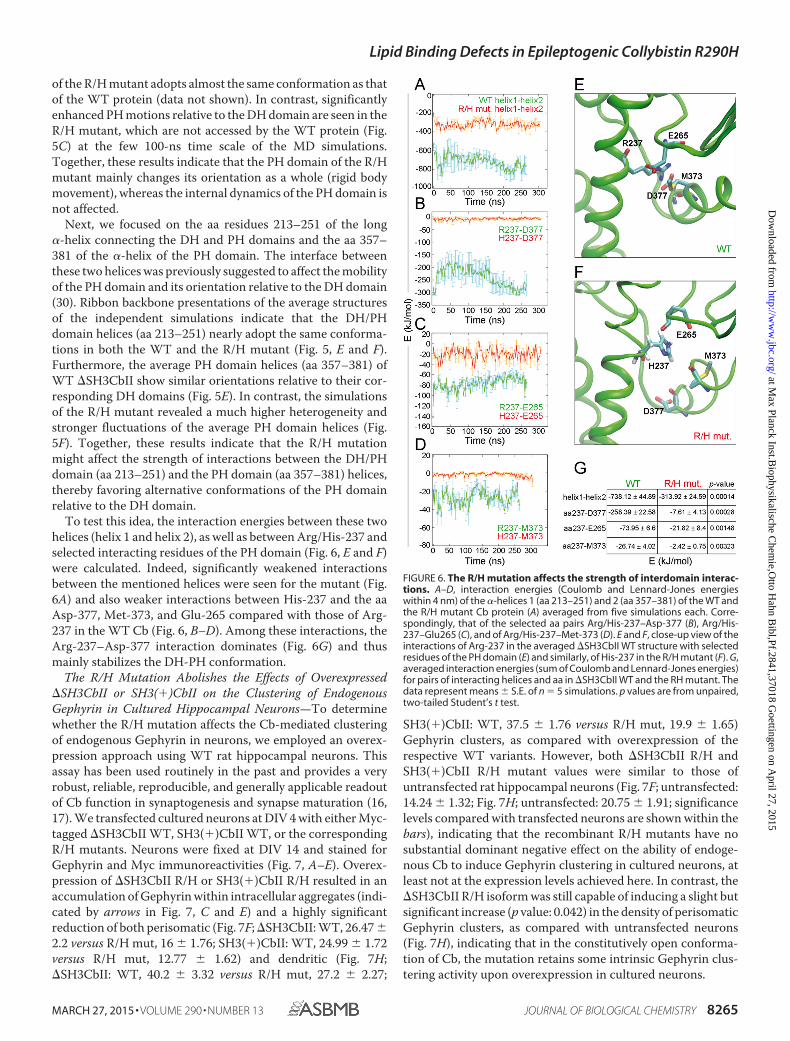
of the R/H mutant adopts almost the same conformation as thatof the WT protein (data not shown). In contrast, significantlyenhanced PH motions relative to the DH domain are seen in theR/H mutant, which are not accessed by the WT protein (Fig.5C) at the few 100-ns time scale of the MD simulations.Together, these results indicate that the PH domain of the R/Hmutant mainly changes its orientation as a whole (rigid bodymovement), whereas the internal dynamics of the PH domain isnot affected.
Next, we focused on the aa residues 213–251 of the long�-helix connecting the DH and PH domains and the aa 357–381 of the �-helix of the PH domain. The interface betweenthese two helices was previously suggested to affect the mobilityof the PH domain and its orientation relative to the DH domain(30). Ribbon backbone presentations of the average structuresof the independent simulations indicate that the DH/PHdomain helices (aa 213–251) nearly adopt the same conforma-tions in both the WT and the R/H mutant (Fig. 5, E and F).Furthermore, the average PH domain helices (aa 357–381) ofWT �SH3CbII show similar orientations relative to their cor-responding DH domains (Fig. 5E). In contrast, the simulationsof the R/H mutant revealed a much higher heterogeneity andstronger fluctuations of the average PH domain helices (Fig.5F). Together, these results indicate that the R/H mutationmight affect the strength of interactions between the DH/PHdomain (aa 213–251) and the PH domain (aa 357–381) helices,thereby favoring alternative conformations of the PH domainrelative to the DH domain.
To test this idea, the interaction energies between these twohelices (helix 1 and helix 2), as well as between Arg/His-237 andselected interacting residues of the PH domain (Fig. 6, E and F)were calculated. Indeed, significantly weakened interactionsbetween the mentioned helices were seen for the mutant (Fig.6A) and also weaker interactions between His-237 and the aaAsp-377, Met-373, and Glu-265 compared with those of Arg-237 in the WT Cb (Fig. 6, B–D). Among these interactions, theArg-237–Asp-377 interaction dominates (Fig. 6G) and thusmainly stabilizes the DH-PH conformation.
The R/H Mutation Abolishes the Effects of Overexpressed�SH3CbII or SH3(�)CbII on the Clustering of EndogenousGephyrin in Cultured Hippocampal Neurons—To determinewhether the R/H mutation affects the Cb-mediated clusteringof endogenous Gephyrin in neurons, we employed an overex-pression approach using WT rat hippocampal neurons. Thisassay has been used routinely in the past and provides a veryrobust, reliable, reproducible, and generally applicable readoutof Cb function in synaptogenesis and synapse maturation (16,17). We transfected cultured neurons at DIV 4 with either Myc-tagged �SH3CbII WT, SH3(�)CbII WT, or the correspondingR/H mutants. Neurons were fixed at DIV 14 and stained forGephyrin and Myc immunoreactivities (Fig. 7, A–E). Overex-pression of �SH3CbII R/H or SH3(�)CbII R/H resulted in anaccumulation of Gephyrin within intracellular aggregates (indi-cated by arrows in Fig. 7, C and E) and a highly significantreduction of both perisomatic (Fig. 7F; �SH3CbII: WT, 26.47 �2.2 versus R/H mut, 16 � 1.76; SH3(�)CbII: WT, 24.99 � 1.72versus R/H mut, 12.77 � 1.62) and dendritic (Fig. 7H;�SH3CbII: WT, 40.2 � 3.32 versus R/H mut, 27.2 � 2.27;
SH3(�)CbII: WT, 37.5 � 1.76 versus R/H mut, 19.9 � 1.65)Gephyrin clusters, as compared with overexpression of therespective WT variants. However, both �SH3CbII R/H andSH3(�)CbII R/H mutant values were similar to those ofuntransfected rat hippocampal neurons (Fig. 7F; untransfected:14.24 � 1.32; Fig. 7H; untransfected: 20.75 � 1.91; significancelevels compared with transfected neurons are shown within thebars), indicating that the recombinant R/H mutants have nosubstantial dominant negative effect on the ability of endoge-nous Cb to induce Gephyrin clustering in cultured neurons, atleast not at the expression levels achieved here. In contrast, the�SH3CbII R/H isoform was still capable of inducing a slight butsignificant increase (p value: 0.042) in the density of perisomaticGephyrin clusters, as compared with untransfected neurons(Fig. 7H), indicating that in the constitutively open conforma-tion of Cb, the mutation retains some intrinsic Gephyrin clus-tering activity upon overexpression in cultured neurons.
FIGURE 6. The R/H mutation affects the strength of interdomain interac-tions. A–D, interaction energies (Coulomb and Lennard-Jones energieswithin 4 nm) of the �-helices 1 (aa 213–251) and 2 (aa 357–381) of the WT andthe R/H mutant Cb protein (A) averaged from five simulations each. Corre-spondingly, that of the selected aa pairs Arg/His-237–Asp-377 (B), Arg/His-237–Glu265 (C), and of Arg/His-237–Met-373 (D). E and F, close-up view of theinteractions of Arg-237 in the averaged �SH3CbII WT structure with selectedresidues of the PH domain (E) and similarly, of His-237 in the R/H mutant (F). G,averaged interaction energies (sum of Coulomb and Lennard-Jones energies)for pairs of interacting helices and aa in �SH3CbII WT and the RH mutant. Thedata represent means � S.E. of n � 5 simulations. p values are from unpaired,two-tailed Student’s t test.
Lipid Binding Defects in Epileptogenic Collybistin R290H
MARCH 27, 2015 • VOLUME 290 • NUMBER 13 JOURNAL OF BIOLOGICAL CHEMISTRY 8265
at Max Planck Inst.B
iophysikalische Chem
ie,Otto H
ahn Bibl,Pf.2841,37018 G
oettingen on April 27, 2015
http://ww
w.jbc.org/
Dow
nloaded from

FIGURE 7. The R/H mutation abolishes the enhancing effects of overexpressed �SH3CbII or SH3(�)CbII on the clustering of endogenous Gephyrin incultured hippocampal neurons. A–E, cultured rat hippocampal neurons were transfected at DIV 4 with Myc-�SH3CbII (B), Myc-SH3(�)CbII (D), or theircorresponding R/H mutants (C and E). Untransfected neurons (A) served as control. At DIV 14, the cells were fixed and immunostained for Gephyrin (green)and Myc (red) using the mAb7a (1:3,000; Connex) and the polyclonal rabbit antibody C3956 (1:1,000; Sigma-Aldrich), respectively. Silhouettes of somataand dendrites of untransfected neurons (indicated by dotted lines in A) were identified by massively increasing the brightness of the Gephyrin stainingusing ImageJ so that the cellular background signal became visible. Note the accumulation of Gephyrin in somatic aggregates (indicated by arrows) inneurons expressing the R/H mutants, as compared with untransfected neurons and to neurons expressing the corresponding WT isoforms. Scale bar, 10�m. F and G, bar diagrams of perisomatic Gephyrin cluster densities per 100-�m2 surface area (F) and average sizes of perisomatic Gephyrin clusters (G).H and I, bar diagrams of the number of Gephyrin immunoreactive clusters per 40-�m dendritic length (H) and average sizes of dendritic Gephyrinclusters (I). Bars correspond to values obtained from the perisomatic surface area and one randomly selected second order dendrite (60 –100 �m distalto the soma) (F) per neuron, respectively (n � 10 –20 individual neurons from three independent transfection experiments). The data represent means �S.E. n.s., not significant; *, p 0.05; **, p 0.01; ***, p 0.001 (unpaired, two-tailed Student’s t test). Significance levels compared with untransfectedneurons are shown within the bars. J–M, exemplary overlapping pixel regions of selected somatic (top panels) and dendritic (bottom panels) areas fromimages of neurons expressing Myc-�SH3CbII (J), Myc-SH3(�)CbII (L), or their corresponding R/H mutants (K and M). Green (Gephyrin staining) and red(Myc staining) channels of selected image areas (as indicated by dotted lines) were automatically thresholded and processed using the colocalizationthreshold algorithm of the ImageJ software package The overlapping pixel regions were converted into a binary threshold mask (white) and superim-posed on the original images as a colocalization map. Note the strong colocalization of the Myc-SH3(�)CbII R/H mutant with somatic Gephyrinaggregates (M, indicated by arrows) and the reduced colocalization of the Myc-�SH3CbII R/H mutant in both somatic and dendritic areas (K), ascompared with WT �SH3CbII (J). N and O, Pearson’s correlation coefficients were determined to quantify the levels of colocalization of each CbII variant.Red bars, Myc-�SH3CbII WT; orange bars, Myc-�SH3CbII R/H mutant; green bars, Myc-SH3(�)CbII WT; lime green bars, Myc-SH3(�)CbII R/H mutant) withGephyrin in somatic/perisomatic (N) and dendritic (O) areas. The data represent means � S.E. of n � 10 somata or dendrites of individual neurons. n.s.,not significant; *, p 0.05; **, p 0.01 (unpaired, two-tailed Student’s t test).
Lipid Binding Defects in Epileptogenic Collybistin R290H
8266 JOURNAL OF BIOLOGICAL CHEMISTRY VOLUME 290 • NUMBER 13 • MARCH 27, 2015
at Max Planck Inst.B
iophysikalische Chem
ie,Otto H
ahn Bibl,Pf.2841,37018 G
oettingen on April 27, 2015
http://ww
w.jbc.org/
Dow
nloaded from

Previous studies demonstrated a significant increase in thesize of Gephyrin clusters upon overexpression of �SH3CbII incultured neurons (31, 35). In agreement with these studies, wefound that the average sizes of perisomatic and dendritic clus-ters of endogenous Gephyrin were significantly increased inneurons expressing WT �SH3CbII as compared with untrans-fected neurons (Fig. 7G; untransfected: 0.12 � 0.005 versus�SH3CbII WT: 0.3 � 0.027; Fig. 7I; untransfected: 0.13 � 0.007versus �SH3CbII WT: 0.25 � 0.022). Overexpression of the�SH3CbII R/H mutant, on the other hand, caused hardly anyincrease in the size of endogenous Gephyrin clusters (Fig. 7G;�SH3CbII R/H mut: 0.15 � 0.016; Fig. 7I; �SH3CbII R/H mut:0.16 � 0.013). In contrast, the average sizes of Gephyrin punctain neurons expressing the SH3(�)CbII isoform were notaffected by the R/H mutation and were similar to those ofuntransfected neurons at both perisomatic and dendritic areas(Fig. 7, G and I). These results indicate that the increase in thesize of Gephyrin clusters in neurons overexpressing the open/active �SH3CbII isoform might be a feature associated with theability of this isoform to bind efficiently to PI3P-enriched mem-branes, because the impairment of Cb binding to PI3P was theonly defect observed in the corresponding R/H mutant. In con-trast, in SH3(�)CbII overexpressing neurons, the availability ofendogenous NL2, GABAAR �2 subunit, or active TC10 andCdc42, i.e. proteins that interact with either the SH3 domain orthe PH domain of Cb and relieve autoinhibition (16, 19, 36),might be limiting factors that explain why the SH3(�)CbII iso-form is not inducing a similar effect on the size of Gephyrinclusters as the effect observed with the �SH3CbII isoform.
We next tested whether the R/H mutation affects the colo-calization of recombinant �SH3CbII and SH3(�)CbII withendogenous Gephyrin. To this end, we selected somatic/peri-somatic and dendritic areas of neurons expressing the Myc-tagged CbII variants and their corresponding R/H mutants atsimilar levels (Fig. 7, J–M, and data not shown) and performedan analysis of colocalization between endogenous Gephyrinand the recombinant Myc-Cb labels, using the colocalizationthreshold algorithm provided by ImageJ. We found that theR/H mutants remained tightly coclustered with large intracel-lular Gephyrin aggregates, which were more frequent in neu-rons expressing the SH3(�)CbII R/H isoform (indicated byarrows in Fig. 7M). In contrast, Gephyrin aggregates were notobserved in neurons expressing the WT CbII isoforms (Fig. 7, Jand L). Furthermore, compared with WT �SH3CbII, the�SH3CbII R/H mutant showed a significantly reduced colocal-ization with Gephyrin in both somatic/perisomatic (Fig. 7N;WT: 0.34 � 0.038 versus R/H mut: 0.17 � 0.019;) and dendritic(Fig. 7O; WT: 0.39 � 0.045 versus 0.2 � 0.031) areas. In den-drites, also the SH3(�)CbII R/H mutant showed a significantreduction in its colocalization with Gephyrin, as compared withWT SH3(�)CbII (Fig. 7O; WT: 0.27 � 0.019 versus R/H mut:0.16 � 0.043). In somatic/perisomatic areas, the colocalizationof the SH3(�)CbII R/H mutant with Gephyrin was reduced(Fig. 7N; 0.26 � 0.027), but this change did not reach statisticalsignificance, which is probably due to the overlapping distribu-tions of the Myc-tagged SH3(�)CbII R/H mutant and Gephy-rin immunoreactivities in large somatic aggregates (Fig. 7, Eand M). Taken together, these data indicate that the R/H muta-
tion affects the ability of Cb to efficiently cocluster with post-synaptically enriched Gephyrin in the somata and dendrites ofhippocampal neurons.
DISCUSSION
In the present study, we employed biochemical, cell biologi-cal, and MD simulation approaches to elucidate the patho-mechanism of an R290H missense mutation in human Cb,which was identified in three adult brothers suffering from epi-leptic seizures and intellectual disability (12). For this purpose,we used the CbII (rat- and mouse-specific) isoform and not thehuman (hPEM-2) Cb isoform. This choice was made for to thefollowing reasons: (i) three-dimensional structures are cur-rently available for the two CbII (�SH3 and SH3(�)) isoforms(14, 30), but not for hPEM-2, which allowed us to complementour cell biological and biochemical studies with MD simula-tions and detailed analyses of the DH/PH interface bearing theR/H mutations; (ii) a previous study indicated that hPEM-2 andSH3(�)CbII behave very similarly in targeting to GABAergicpostsynapses and in inducing increases in Gephyrin clustering,when overexpressed in cultured neurons (31); and (iii) the twoisoforms are identical except for their very C termini, which areextremely unlikely to influence the effects of the R/H mutationstudied here and which do not appear to differentially affectCb-mediated Gephyrin clustering.
Based on three lines of evidence, our present data define theR/H mutant of Cb as a loss of function mutant: (i) Whenexpressed in COS7 cells together with GFP-Gephyrin, the�SH3CbII R/H mutant cannot mediate the redistribution ofGephyrin from large intracellular aggregates to membrane-as-sociated microclusters. Indeed, the R/H mutation is the firstknown single mutation in the DH domain of the open/active�SH3CbII isoform that renders the protein completely inactivein redistributing Gephyrin in cells. Although the vast majorityof Cb isoforms in mouse brain contains an SH3 domain (14) andthe mRNAs encoding SH3 domain-containing isoforms aremuch more abundant than mRNAs encoding �SH3Cb iso-forms in the rodent brain and spinal cord (7), some �SH3Cbsplice variants appear to exist in vivo and may play an importantrole in synaptogenesis and synapse maturation (7). In this con-text, our approach to overexpress �SH3CbII variants in non-neuronal cells and hippocampal neurons proved to be veryvaluable for analyses of the functional importance of single aaresidues for Cb-mediated Gephyrin clustering. The use of�SH3CbII variants circumvents the need for overexpressingadditional Cb activators (i.e. NL2, the �2 subunit of GABAARsor active GTPases TC10 and Cdc42 (16, 19, 36)), which interactwith the SH3 domain or the PH domain of Cb and therebyrevert autoinhibition. In essence, �SH3CbII isoforms perfectlyimitate the open/active states of SH3-containing Cb variantsthat are induced upon the interaction of the SH3 domain withNL2, the �2 subunit of GABAARs, or active TC10 or Cdc42. (ii)�SH3CbII R/H, as well as the longer, SH3 domain-containing,variant SH3(�)CbII R/H are unable to enhance perisomaticand dendritic clustering of endogenous Gephyrin in culturedneurons and instead lead to the somatic accumulation ofGephyrin. Interestingly, the clustering of endogenous Gephyrinin neurons overexpressing the R/H mutants was similar to the
Lipid Binding Defects in Epileptogenic Collybistin R290H
MARCH 27, 2015 • VOLUME 290 • NUMBER 13 JOURNAL OF BIOLOGICAL CHEMISTRY 8267
at Max Planck Inst.B
iophysikalische Chem
ie,Otto H
ahn Bibl,Pf.2841,37018 G
oettingen on April 27, 2015
http://ww
w.jbc.org/
Dow
nloaded from

Gephyrin clustering seen in untransfected neurons, indicatingthat the overexpressed R/H mutants had no dominant negativeeffect on Gephyrin clustering in cultured neurons, at least not atthe expression levels achieved here. (iii) Both NL2 and TC10CA cannot induce the GFP-Gephyrin redistribution to sub-membraneous microclusters when coexpressed together withthe SH3(�)CbII R/H mutant.
The arginine residue affected in the R/H mutation is locatedwithin the catalytic core of the DH domain of Cb, which medi-ates its GEF activity. However, according to the crystal struc-ture of �SH3CbII in complex with Cdc42 (30), the affectedarginine does not interact directly with the GTPase but con-tacts instead Asp-377 in the PH domain of Cb. The PH domainwas previously shown to be involved in intramolecular interac-tions with the SH3 and DH domains (14) and to mediate theinteraction with active, GTP-loaded TC10 (16) and with PI3P(9). The SH3 domain of Cb binds autonomously to NL2 (14),the SH3 domain residues involved in NL2 and PH/DH domainbinding are on opposite sides of the SH3 structure (Fig. 1B), andthe R/H site and the DH domain residues involved in SH3domain interactions are facing in opposite directions, all ofwhich indicates that the R/H mutation cannot affect the inter-action of the SH3 domain with NL2. Whereas the residues ofthe PH domain that are involved in the GTP-TC10 interactionare currently not known, the interaction with PI3P is mediatedby two arginines (Arg-303/Arg-304) located in the �3-�4 loopof the PH domain (17). Thus, the particular location and inter-acting residues of the affected arginine would be compatiblewith the notion that the R/H mutation might cause changes inthe conformation of both the DH and PH domains of Cb, lead-ing to altered GEF activity or altered binding to protein inter-actors. However, our biochemical and cell biological experi-ments showed that the R/H mutation affects neither the GEFactivity of Cb nor its binding to Gephyrin or GTP-loaded TC10,as demonstrated by the in vivo activation assays, the GST pull-down assays, and the in vitro TC10 binding assays, respectively.Consistent with these findings, our MD simulations predictedno significant changes in the internal conformations of theaverage structures of the entire DH and PH domains of the�SH3CbII R/H mutant, as compared with WT �SH3CbII.
A key finding of the present study is that the R/H mutationimpairs Cb binding to PI3P, as assessed in a protein-lipid over-lay assay. Consequently, and in agreement with results obtainedwith the PI3P binding-deficient R303N/R304N mutant (17),the R/H mutation studied here abolishes the ability of the�SH3CbII splice variant, which adopts an intrinsically open/active conformation, to induce membrane clustering of Gephy-rin in COS7 cells and synaptic clustering of Gephyrin in cul-tured neurons. Furthermore, GTP-TC10, which binds to thePH domain of Cb and thereby enables the SH3(�)CbII variantto adopt an open/active conformation (16), cannot induceGephyrin targeting to the plasma membrane when coexpressedwith the SH3(�)CbII R/H mutant. This finding is reminiscentof data obtained with the PI3P binding-deficient SH3(�)CbIIRR/AA mutant (16) and indicates that the interaction of the PHdomain of Cb with PI3P is an essential step in the Gephyrinclustering cascade that cannot be bypassed by GTPaseoverexpression.
But how can a single mutation in the DH domain of Cb affectPI3P binding to the distal �3-�4 loop of the PH domain? Asmentioned above, the arginine residue mutated in the R/Hmutant contacts an acidic side chain in the PH domain (30).Depending on whether the R/H mutation provides a less ormore extensive interface between the DH and PH domains, themutant might then adopt a more open or more closed confor-mation, respectively, thus increasing or decreasing the accessi-bility of the PH domain for membrane lipid binding. Indeed, arecently published study of ours (14) indicated that the autoin-hibited SH3(�)CbII, which adopts a closed/inactive conforma-tion, binds to PI3P only weakly, similar to the PI3P binding-deficient SH3(�)CbII RR/AA mutant. In contrast, anSH3(�)CbII W24A/E262A mutant, with two substitutions inthe SH3-DH/PH interface that lead to a more open conforma-tion of the protein, displayed an increased binding to PI3P andincreased plasma-membrane targeting of Gephyrin as com-pared with SH3(�)CbII WT (14). Thus, conformational activa-tion of Cb facilitates PI3P binding and subsequent Gephyrinclustering, and the R/H mutation might exert its effects in thiscontext.
The PH domains of Dbl family proteins adopt multiple ori-entations with regard to the respective DH domains. Depend-ing on variable interactions between the DH and PH domains, aflexibility in the orientation of the PH domain relative to theDH domain of �SH3CbII was suggested previously (30). How-ever, the functional implications of this flexibility haveremained unclear. Our MD simulations of WT �SH3CbII andits corresponding R/H mutant in complex with Cdc42 predictthat the R/H mutation markedly increases orientational fluctu-ations of the entire PH domain relative to the DH domain, pre-sumably because of changes in the strength of interactionsbetween residues of the DH/PH �-helix (aa 213–251) and res-idues in the PH domain �-helix (aa 357–381). Together, theseresults indicate that the PH domain conformation that isfavored in the WT Cb protein is the most efficient in mediatingthe binding to PI3P.
Although many mutations in the Cb gene have been identi-fied in patients with epilepsy and intellectual disability, onlyvery few of them have been characterized functionally. Ourpresent analysis of the R/H mutation in Cb (12), together with apreviously described balanced chromosomal translocationresulting in mRNAs that no longer encode the PH domain of Cb(9), provides evidence for the notion that an impairment ofPI3P binding by Cb is a likely common pathomechanism for Cbdysfunction in the human brain.
Despite their low abundance, phosphoinositides are criticalregulators of intracellular signaling and membrane compart-mentalization. The functional roles of phosphoinositidemetabolism have been studied in substantial detail at the pre-synaptic terminal, where phosphoinositide turnover is of criti-cal importance for synaptic vesicle recycling and synapse func-tion (37). Further, there is abundant evidence for the relevanceof phosphoinositide pathways in synaptic plasticity (38). How-ever, much less is known about specific roles of phosphoinositi-des at inhibitory postsynapses. PI3P is considered a lipid that isconstitutively present on endosomes where it is mainly gener-ated by class III phosphatidylinositol 3-kinase (39, 40). The spe-
Lipid Binding Defects in Epileptogenic Collybistin R290H
8268 JOURNAL OF BIOLOGICAL CHEMISTRY VOLUME 290 • NUMBER 13 • MARCH 27, 2015
at Max Planck Inst.B
iophysikalische Chem
ie,Otto H
ahn Bibl,Pf.2841,37018 G
oettingen on April 27, 2015
http://ww
w.jbc.org/
Dow
nloaded from

cific recruitment of class III PI3K to endosomes and the localproduction of PI3P on endosomal membranes provide a mech-anism for the recruitment of a specific group of proteins toendosomes, and most PI3P-binding proteins that have beenfunctionally characterized so far are associated with differentaspects of endosomal membrane sorting and trafficking (forreview see Ref. 41). Thus, the accumulation of the Cb R/Hmutants in large somatic aggregates together with endogenousGephyrin might reflect a defect in the cotransport of both pro-teins via PI3P-enriched endosomal compartments to theplasma membrane, with consequent effects on inhibitory post-synapse formation. Alternatively, a signaling process involvingplasma membrane PI3P might be involved in the control of Cbfunction. Over the past few years, it has become increasinglyevident that pools of PI3P can be specifically generated at theplasma membrane upon cellular stimulation, and PI3P hasemerged as a critical intracellular second messenger in varioussignaling pathways (42). Notably, PI3P is specifically produceddownstream of TC10 activation to promote the plasma mem-brane translocation of the glucose transporter 4 (43). Moreover,TC10-mediated activation of a member of the class II PI3Ks,PI3K-C2�, has been shown to regulate the generation of PI3P atthe plasma membrane (44). These findings, together with thepreviously described TC10-induced increase in GABAergicpostsynaptic strength (16), strongly support the notion that themolecular mechanisms involved in local PI3P production onendomembranes or the plasma membrane play an importantrole in triggering inhibitory postsynapse formation and matu-ration. The present study indicates that the CbR290H mutantmight cause epilepsy and intellectual disability because it is notresponsive to the types of PI3P signaling described above, con-sequently lacks Gephyrin clustering activity, and therefore can-not support the proper formation and maturation of functionalinhibitory synapses in the brain.
Acknowledgments—We thank Foteini Paraskevopoulou and OzgeDemet Ozcete for help with some of the experiments.
REFERENCES1. Luscher, B., Fuchs, T., and Kilpatrick, C. L. (2011) GABAA receptor traf-
ficking-mediated plasticity of inhibitory synapses. Neuron 70, 385– 4092. Smith, K. R., and Kittler, J. T. (2010) The cell biology of synaptic inhibition
in health and disease. Curr. Opin. Neurobiol. 20, 550 –5563. Ramamoorthi, K., and Lin, Y. (2011) The contribution of GABAergic dys-
function to neurodevelopmental disorders. Trends Mol. Med. 17,452– 462
4. Verpelli, C., Galimberti, I., Gomez-Mancilla, B., and Sala, C. (2014) Mo-lecular basis for prospective pharmacological treatment strategies in in-tellectual disability syndromes. Dev. Neurobiol. 74, 197–206
5. Papadopoulos, T., and Soykan, T. (2011) The role of collybistin in gephyrinclustering at inhibitory synapses: facts and open questions. Front. Cell.Neurosci. 5, 11
6. Papadopoulos, T., Korte, M., Eulenburg, V., Kubota, H., Retiounskaia, M.,Harvey, R. J., Harvey, K., O’Sullivan, G. A., Laube, B., Hülsmann, S., Geiger,J. R., and Betz, H. (2007) Impaired GABAergic transmission and alteredhippocampal synaptic plasticity in collybistin-deficient mice. EMBO J. 26,3888 –3899
7. Harvey, K., Duguid, I. C., Alldred, M. J., Beatty, S. E., Ward, H., Keep, N. H.,Lingenfelter, S. E., Pearce, B. R., Lundgren, J., Owen, M. J., Smart, T. G.,Lüscher, B., Rees, M. I., and Harvey, R. J. (2004) The GDP-GTP exchange
factor collybistin: An essential determinant of neuronal gephyrin cluster-ing. J. Neurosci. 24, 5816 –5826
8. Marco, E. J., Abidi, F. E., Bristow, J., Dean, W. B., Cotter, P., Jeremy, R. J.,Schwartz, C. E., and Sherr, E. H. (2008) ARHGEF9 disruption in a femalepatient is associated with X linked mental retardation and sensory hyper-arousal. J. Med. Genet. 45, 100 –105
9. Kalscheuer, V. M., Musante, L., Fang, C., Hoffmann, K., Fuchs, C., Carta,E., Deas, E., Venkateswarlu, K., Menzel, C., Ullmann, R., Tommerup, N.,Dalprà, L., Tzschach, A., Selicorni, A., Lüscher, B., Ropers, H. H., Harvey,K., and Harvey, R. J. (2009) A balanced chromosomal translocation dis-rupting ARHGEF9 is associated with epilepsy, anxiety, aggression, andmental retardation. Hum. Mutat. 30, 61– 68
10. Lesca, G., Till, M., Labalme, A., Vallee, D., Hugonenq, C., Philip, N., Edery,P., and Sanlaville, D. (2011) De novo Xq11.11 microdeletion includingARHGEF9 in a boy with mental retardation, epilepsy, macrosomia, anddysmorphic features. Am. J. Med. Genet. 155A, 1706 –1711
11. Shimojima, K., Sugawara, M., Shichiji, M., Mukaida, S., Takayama, R.,Imai, K., and Yamamoto, T. (2011) Loss-of-function mutation of collybis-tin is responsible for X-linked mental retardation associated with epilepsy.J. Hum. Genet. 56, 561–565
12. Lemke, J. R., Riesch, E., Scheurenbrand, T., Schubach, M., Wilhelm, C.,Steiner, I., Hansen, J., Courage, C., Gallati, S., Bürki, S., Strozzi, S., Simo-netti, B. G., Grunt, S., Steinlin, M., Alber, M., Wolff, M., Klopstock, T.,Prott, E. C., Lorenz, R., Spaich, C., Rona, S., Lakshminarasimhan, M., Kröll,J., Dorn, T., Krämer, G., Synofzik, M., Becker, F., Weber, Y. G., Lerche, H.,Böhm, D., and Biskup, S. (2012) Targeted next generation sequencing as adiagnostic tool in epileptic disorders. Epilepsia 53, 1387–1398
13. de Ligt, J., Willemsen, M. H., van Bon, B. W., Kleefstra, T., Yntema, H. G.,Kroes, T., Vulto-van Silfhout, A. T., Koolen, D. A., de Vries, P., Gilissen, C.,del Rosario, M., Hoischen, A., Scheffer, H., de Vries, B. B., Brunner, H. G.,Veltman, J. A., and Vissers, L. E. (2012) Diagnostic exome sequencing inpersons with severe intellectual disability. N. Engl. J. Med. 367, 1921–1929
14. Soykan, T., Schneeberger, D., Tria, G., Buechner, C., Bader, N., Svergun,D., Tessmer, I., Poulopoulos, A., Papadopoulos, T., Varoqueaux, F., Schin-delin, H., and Brose, N. (2014) A conformational switch in collybistindetermines the differentiation of inhibitory postsynapses. EMBO J. 33,2113–2133
15. Reid, T., Bathoorn, A., Ahmadian, M. R., and Collard, J. G. (1999) Identi-fication and characterization of hPEM-2, a guanine nucleotide exchangefactor specific for Cdc42. J. Biol. Chem. 274, 33587–33593
16. Mayer, S., Kumar, R., Jaiswal, M., Soykan, T., Ahmadian, M. R., Brose, N.,Betz, H., Rhee, J. S., and Papadopoulos, T. (2013) Collybistin activation byGTP-TC10 enhances postsynaptic gephyrin clustering and hippocampalGABAergic neurotransmission. Proc. Natl. Acad. Sci. U.S.A. 110,20795–20800
17. Reddy-Alla, S., Schmitt, B., Birkenfeld, J., Eulenburg, V., Dutertre, S.,Böhringer, C., Götz, M., Betz, H., and Papadopoulos, T. (2010) PH-do-main-driven targeting of collybistin but not Cdc42 activation is requiredfor synaptic gephyrin clustering. Eur. J. Neurosci. 31, 1173–1184
18. Kins, S., Betz, H., and Kirsch, J. (2000) Collybistin, a newly identified brain-specific GEF, induces submembrane clustering of gephyrin. Nat. Neurosci.3, 22–29
19. Poulopoulos, A., Aramuni, G., Meyer, G., Soykan, T., Hoon, M., Papado-poulos, T., Zhang, M., Paarmann, I., Fuchs, C., Harvey, K., Jedlicka, P.,Schwarzacher, S. W., Betz, H., Harvey, R. J., Brose, N., Zhang, W., andVaroqueaux, F. (2009) Neuroligin 2 drives postsynaptic assembly at peri-somatic inhibitory synapses through gephyrin and collybistin. Neuron 63,628 – 642
20. Fuhrmann, J. C., Kins, S., Rostaing, P., El Far, O., Kirsch, J., Sheng, M.,Triller, A., Betz, H., and Kneussel, M. (2002) Gephyrin interacts with dy-nein light chains 1 and 2, components of motor protein complexes. J. Neu-rosci. 22, 5393–5402
21. Van Der Spoel, D., Lindahl, E., Hess, B., Groenhof, G., Mark, A. E., andBerendsen, H. J. (2005) GROMACS: fast, flexible, and free. J. Comput.Chem. 26, 1701–1718
22. Hess, B., Kutzner, C., van der Spoel, D., and Lindahl, E. (2008) GROMACS4: Algorithms for highly efficient, load-balanced, and scalable molecularsimulation. J. Chem. Theory Comput. 4, 435– 447
Lipid Binding Defects in Epileptogenic Collybistin R290H
MARCH 27, 2015 • VOLUME 290 • NUMBER 13 JOURNAL OF BIOLOGICAL CHEMISTRY 8269
at Max Planck Inst.B
iophysikalische Chem
ie,Otto H
ahn Bibl,Pf.2841,37018 G
oettingen on April 27, 2015
http://ww
w.jbc.org/
Dow
nloaded from

23. Pronk, S., Páll, S., Schulz, R., Larsson, P., Bjelkmar, P., Apostolov, R., Shirts,M. R., Smith, J. C., Kasson, P. M., van der Spoel, D., Hess, B., and Lindahl,E. (2013) GROMACS 4.5: a high-throughput and highly parallel opensource molecular simulation toolkit. Bioinformatics 29, 845– 854
24. Hornak, V., Abel, R., Okur, A., Strockbine, B., Roitberg, A., and Simmer-ling, C. (2006) Comparison of multiple Amber force fields and develop-ment of improved protein backbone parameters. Proteins 65, 712–725
25. Jorgensen, W. L., Chandrasekhar, J., Madura, J. D., Impey, R. W., andKlein, M. L. (1983) Comparison of simple potential functions for simulat-ing liquid water. J. Chem. Phys. 79, 926 –935
26. Darden, T. A., and Pedersen, L. G. (1993) Molecular modeling: an exper-imental tool. Environ. Health Perspect. 101, 410 – 412
27. Bussi, G., Donadio, D., and Parrinello, M. (2007) Canonical samplingthrough velocity rescaling. J. Chem. Phys. 126, 014101
28. Berendsen, H. J. C., Postma, J. P. M., Vangunsteren, W. F., Dinola, A., andHaak, J. R. (1984) Molecular dynamics with coupling to an external bath.J. Chem. Phys. 81, 3684 –3690
29. Hess, B., Bekker, H., Berendsen, H. J. C., and Fraaije, J. G. E. M. (1997)LINCS: A linear constraint solver for molecular simulations. J. Comput.Chem. 18, 1463–1472
30. Xiang, S., Kim, E. Y., Connelly, J. J., Nassar, N., Kirsch, J., Winking, J.,Schwarz, G., and Schindelin, H. (2006) The crystal structure of Cdc42 incomplex with collybistin II, a gephyrin-interacting guanine nucleotide ex-change factor. J. Mol. Biol. 359, 35– 46
31. Chiou, T. T., Bonhomme, B., Jin, H., Miralles, C. P., Xiao, H., Fu, Z.,Harvey, R. J., Harvey, K., Vicini, S., and De Blas, A. L. (2011) Differentialregulation of the postsynaptic clustering of �-aminobutyric acid type A(GABAA) receptors by collybistin isoforms. J. Biol. Chem. 286,22456 –22468
32. Hemsath, L., Dvorsky, R., Fiegen, D., Carlier, M. F., and Ahmadian, M. R.(2005) An electrostatic steering mechanism of Cdc42 recognition by Wis-kott-Aldrich syndrome proteins. Mol. Cell 20, 313–324
33. Neudauer, C. L., Joberty, G., Tatsis, N., and Macara, I. G. (1998) Distinctcellular effects and interactions of the Rho-family GTPase TC10. Curr.Biol. 8, 1151–1160
34. Grosskreutz, Y., Hermann, A., Kins, S., Fuhrmann, J. C., Betz, H., andKneussel, M. (2001) Identification of a gephyrin-binding motif in the
GDP/GTP exchange factor collybistin. Biol. Chem. 382, 1455–146235. Tyagarajan, S. K., Ghosh, H., Harvey, K., and Fritschy, J. M. (2011) Colly-
bistin splice variants differentially interact with gephyrin and Cdc42 toregulate gephyrin clustering at GABAergic synapses. J. Cell Sci. 124,2786 –2796
36. Saiepour, L., Fuchs, C., Patrizi, A., Sassoè-Pognetto, M., Harvey, R. J., andHarvey, K. (2010) Complex role of collybistin and gephyrin in GABAA
receptor clustering. J. Biol. Chem. 285, 29623–2963137. Wenk, M. R., and De Camilli, P. (2004) Protein-lipid interactions and
phosphoinositide metabolism in membrane traffic: insights from vesiclerecycling in nerve terminals. Proc. Natl. Acad. Sci. U.S.A. 101, 8262– 8269
38. Man, H. Y., Wang, Q., Lu, W. Y., Ju, W., Ahmadian, G., Liu, L., D’Souza, S.,Wong, T. P., Taghibiglou, C., Lu, J., Becker, L. E., Pei, L., Liu, F., Wymann,M. P., MacDonald, J. F., and Wang, Y. T. (2003) Activation of PI3-kinase isrequired for AMPA receptor insertion during LTP of mEPSCs in culturedhippocampal neurons. Neuron 38, 611– 624
39. Gillooly, D. J., Morrow, I. C., Lindsay, M., Gould, R., Bryant, N. J., Gaullier,J. M., Parton, R. G., and Stenmark, H. (2000) Localization of phosphatidy-linositol 3-phosphate in yeast and mammalian cells. EMBO J. 19,4577– 4588
40. Vicinanza, M., D’Angelo, G., Di Campli, A., and De Matteis, M. A. (2008)Function and dysfunction of the PI system in membrane trafficking.EMBO J. 27, 2457–2470
41. Raiborg, C., Schink, K. O., and Stenmark, H. (2013) Class III phosphatidy-linositol 3-kinase and its catalytic product PtdIns3P in regulation of endo-cytic membrane traffic. FEBS J. 280, 2730 –2742
42. Falasca, M., and Maffucci, T. (2006) Emerging roles of phosphatidylinosi-tol 3-monophosphate as a dynamic lipid second messenger. Arch. Physiol.Biochem. 112, 274 –284
43. Maffucci, T., Brancaccio, A., Piccolo, E., Stein, R. C., and Falasca, M. (2003)Insulin induces phosphatidylinositol-3-phosphate formation throughTC10 activation. EMBO J. 22, 4178 – 4189
44. Falasca, M., Hughes, W. E., Dominguez, V., Sala, G., Fostira, F., Fang,M. Q., Cazzolli, R., Shepherd, P. R., James, D. E., and Maffucci, T. (2007)The role of phosphoinositide 3-kinase C2alpha in insulin signaling. J. Biol.Chem. 282, 28226 –28236
Lipid Binding Defects in Epileptogenic Collybistin R290H
8270 JOURNAL OF BIOLOGICAL CHEMISTRY VOLUME 290 • NUMBER 13 • MARCH 27, 2015
at Max Planck Inst.B
iophysikalische Chem
ie,Otto H
ahn Bibl,Pf.2841,37018 G
oettingen on April 27, 2015
http://ww
w.jbc.org/
Dow
nloaded from

Helmut Grubmüller and Nils BroseTheofilos Papadopoulos, Rudolf Schemm, Intellectual DisabilityR290H Mutant That Causes Epilepsy andSynaptogenic Activity of a Collybistin Lipid Binding Defects and PerturbedNeurobiology:
doi: 10.1074/jbc.M114.633024 originally published online February 12, 20152015, 290:8256-8270.J. Biol. Chem.
10.1074/jbc.M114.633024Access the most updated version of this article at doi:
.JBC Affinity SitesFind articles, minireviews, Reflections and Classics on similar topics on the
Alerts:
When a correction for this article is posted•
When this article is cited•
to choose from all of JBC's e-mail alertsClick here
http://www.jbc.org/content/290/13/8256.full.html#ref-list-1
This article cites 43 references, 16 of which can be accessed free at
at Max Planck Inst.B
iophysikalische Chem
ie,Otto H
ahn Bibl,Pf.2841,37018 G
oettingen on April 27, 2015
http://ww
w.jbc.org/
Dow
nloaded from
Related Documents