Lipid Transport between the Endoplasmic Reticulum and Mitochondria Vid V. Flis and Gu ¨ nther Daum Institute of Biochemistry, Graz University of Technology, A-8010 Graz, Austria Correspondence: [email protected] Mitochondria are partially autonomous organelles that depend on the import of certain proteins and lipids to maintain cell survival and membrane formation. Although phospha- tidylglycerol, cardiolipin, and phosphatidylethanolamine are synthesized by mitochondrial enzymes, phosphatidylcholine, phosphatidylinositol, phosphatidylserine, and sterols need to be imported from other organelles. The origin of most lipids imported into mitochondria is the endoplasmic reticulum, which requires interaction of these two subcellular compart- ments. Recently, protein complexesthat are involved in membrane contact between endo- plasmic reticulum and mitochondria were identified, but their role in lipid transport is still unclear. In the present review, we describe components involved in lipid translocation between the endoplasmic reticulum and mitochondria and discuss functional as well as regulatory aspects that are important for lipid homeostasis. B iological membranes are major structural components of all cell types. They protect the cell from external influences, organize the interior in distinct compartments and allow bal- anced flux of components. Besides their specific proteome, organelles exhibit unique lipid com- positions, which influence their shape, physical properties, and function. Major lipid classes found in biological membranes are phospholip- ids, sterols, and sphingolipids. The major “lipid factory” within the cell is the endoplasmic reticulum (ER). It is able to synthesize the bulk of structural phospholipids, sterols, and storage lipids such as triacylglycer- ols and steryl esters (van Meer et al. 2008). Fur- thermore, initial steps of ceramide synthesis occur in the ER providing precursors for the formation of complex sphingolipids in other organelles (Futerman 2006). Besides the export of ceramides, the ER supplies a large portion of lipids to other organelles, which cannot pro- duce their own lipids or have a limited capacity to do so. Organelle interaction and transport of lipids require specific carrier proteins, mem- brane contact sites, tethering complexes, and/ or vesicle flux. These processes are highly im- portant for the maintenance of cell structure and survival but are still a matter of dispute. Most prominent organelle interaction partners are the ER and mitochondria. A subfraction of the ER named mitochondria-associated mem- brane (MAM) (Vance 1990) was described to be involved in lipid translocation to mitochon- dria. MAM is part of the ER network, which was shown to be in contact or close proximity to the outer mitochondrial membrane (OMM). Editors: Susan Ferro-Novick, Tom A. Rapoport, and Randy Schekman Additional Perspectives on The Endoplasmic Reticulum available at www.cshperspectives.org Copyright # 2013 Cold Spring Harbor Laboratory Press; all rights reserved; doi: 10.1101/cshperspect.a013235 Cite this article as Cold Spring Harb Perspect Biol 2013;5:a013235 1 on March 23, 2022 - Published by Cold Spring Harbor Laboratory Press http://cshperspectives.cshlp.org/ Downloaded from

Welcome message from author
This document is posted to help you gain knowledge. Please leave a comment to let me know what you think about it! Share it to your friends and learn new things together.
Transcript

Lipid Transport between the EndoplasmicReticulum and Mitochondria
Vid V. Flis and Gunther Daum
Institute of Biochemistry, Graz University of Technology, A-8010 Graz, Austria
Correspondence: [email protected]
Mitochondria are partially autonomous organelles that depend on the import of certainproteins and lipids to maintain cell survival and membrane formation. Although phospha-tidylglycerol, cardiolipin, and phosphatidylethanolamine are synthesized by mitochondrialenzymes, phosphatidylcholine, phosphatidylinositol, phosphatidylserine, and sterols needto be imported from other organelles. The origin of most lipids imported into mitochondria isthe endoplasmic reticulum, which requires interaction of these two subcellular compart-ments. Recently, protein complexes that are involved in membrane contact between endo-plasmic reticulum and mitochondria were identified, but their role in lipid transport is stillunclear. In the present review, we describe components involved in lipid translocationbetween the endoplasmic reticulum and mitochondria and discuss functional as well asregulatory aspects that are important for lipid homeostasis.
Biological membranes are major structuralcomponents of all cell types. They protect
the cell from external influences, organize theinterior in distinct compartments and allow bal-anced flux of components. Besides their specificproteome, organelles exhibit unique lipid com-positions, which influence their shape, physicalproperties, and function. Major lipid classesfound in biological membranes are phospholip-ids, sterols, and sphingolipids.
The major “lipid factory” within the cell isthe endoplasmic reticulum (ER). It is able tosynthesize the bulk of structural phospholipids,sterols, and storage lipids such as triacylglycer-ols and steryl esters (van Meer et al. 2008). Fur-thermore, initial steps of ceramide synthesisoccur in the ER providing precursors for theformation of complex sphingolipids in other
organelles (Futerman 2006). Besides the exportof ceramides, the ER supplies a large portion oflipids to other organelles, which cannot pro-duce their own lipids or have a limited capacityto do so. Organelle interaction and transport oflipids require specific carrier proteins, mem-brane contact sites, tethering complexes, and/or vesicle flux. These processes are highly im-portant for the maintenance of cell structureand survival but are still a matter of dispute.Most prominent organelle interaction partnersare the ER and mitochondria. A subfraction ofthe ER named mitochondria-associated mem-brane (MAM) (Vance 1990) was described tobe involved in lipid translocation to mitochon-dria. MAM is part of the ER network, which wasshown to be in contact or close proximity tothe outer mitochondrial membrane (OMM).
Editors: Susan Ferro-Novick, Tom A. Rapoport, and Randy Schekman
Additional Perspectives on The Endoplasmic Reticulum available at www.cshperspectives.org
Copyright # 2013 Cold Spring Harbor Laboratory Press; all rights reserved; doi: 10.1101/cshperspect.a013235
Cite this article as Cold Spring Harb Perspect Biol 2013;5:a013235
1
on March 23, 2022 - Published by Cold Spring Harbor Laboratory Press http://cshperspectives.cshlp.org/Downloaded from

Contact sites between MAM and mitochondriawere assumed to facilitate exchange of compo-nents between the two compartments. Interest-ingly, MAM harbor a number of lipid synthe-sizing enzymes (Gaigg et al. 1994). Recently,molecular components governing membranecontact between the two organelles were identi-fied (Dolman et al. 2005; Csordas et al. 2006; deBrito and Scorrano 2008; Kornmann et al. 2009;Friedman et al. 2010; Lavieu et al. 2010), al-though the specific role of these componentsin lipid translocation is not yet clear.
MAJOR PHOSPHOLIPID CLASSES OFMITOCHONDRIA
Cardiolipin (CL) and/or phosphatidylglycerol(PG) are considered as mitochondria-specificphospholipids (Zinser et al. 1991). Both lipidsare synthesized by mitochondria themselves(Davidson and Stanacev 1971). The importanceof CL for ATP-production has been shown withdifferent membrane types, e.g., bacteria, hydro-genosomes, and mitochondria (Mileykovskayaet al. 2005; Schlame 2008; Acehan et al. 2011).In mitochondria, the majority of CL is localizedto the inner membrane (IMM) (Zinser et al.1991), but substantial amounts were also de-tected in the OMM (Gebert et al. 2009). Withits uncommon, dimeric structure (Fig. 1) CLtogether with phosphatidylethanolamine (PE)interacts with many mitochondrial proteins(Osman et al. 2011) and stabilizes their confor-mation (Joshi et al. 2012). Mutants lacking CLand PE biosynthesis are synthetic lethal in yeastand bacteria (Gohil et al. 2005). In bacteria,both lipids are organized in membrane clusters(Matsumoto et al. 2006). Specific interactionpartners of CL are proteins of ATP production,mitochondrial transport systems (Bogdanovet al. 2008a; Schlame and Ren 2009), and pro-teins required for mitochondrial structure andfusion (Joshi et al. 2012).
Another prominent mitochondrial phos-pholipid is PE. Mitochondria are able to synthe-size a large portion of PE by decarboxylation ofphosphatidylserine (PS) (Vial et al. 1982; Trotteret al. 1993; Emoto et al. 1999; Birner et al. 2001;Nerlich et al. 2007). With its small hydrophilic
head group and large hydrophobic tail, PE is atypical nonbilayer-forming phospholipid (seeFig. 1). This structure appears to be importantfor some peripheral and integral membrane pro-teins (van den Brink-van der Laan et al. 2004).Together with CL, PE plays acrucial role in main-taining mitochondrial morphology. Loss ofboth PE and CL is lethal probably due to defectsin mitochondrial fusion (Joshi et al. 2012) and/or protein destabilizing (Bogdanov et al. 2008b;Osman et al. 2009b). Lack of PE synthesized inthe IMM cannot be fully compensated by PEimported from extramitochondrial sites (Birneret al. 2001; Burgermeister et al. 2004; Joshi et al.2012). However, PE derived from exogenouslyso-PE can restore both intra- and extramito-chondrial PE deficiencies by remodeling pro-cesses (Riekhof and Voelker 2006). Noteworthy,mitochondria are the major supplier of PE toother organelles (Voelker 1984).
Mitochondrial enzymes of CL and PG syn-thesis, as well as the major PS decarboxylase, aresynthesized on cytosolic ribosomes, importedinto mitochondria, and assembled into theIMM (Minskoff and Greenberg 1997; Jianget al. 2000; Nowicki et al. 2005; Nebauer et al.2007; Choi et al. 2012). Only recently were weable to demonstrate experimentally the involve-ment of components of the mitochondrialTOM complex and of mitochondrial proteasesin import and processing of the mitochondrialPS decarboxylase Psd1p from the yeast (Hor-vath et al. 2012).
Phosphatidylcholine (PC) has a big hydro-philic head group and a long hydrophobic tail(see Fig. 1). Its cylindrical shape makes it a per-fect component of bilayer membranes and itsrole as a structural component is essential (vanMeer et al. 2008). Specific functions of PC inmitochondria are not well defined. Similar toPC, specific functions of phosphatidylinositol(PI) in mitochondria are not known, but itsessential role in total cellular metabolism makesit directly or indirectly indispensible for themaintenance of mitochondria. The bulkheadgroup of PI makes this phospholipid a specificmembrane bilayer component.
Phosphatidylserine (PS) has also a cylin-drical shape that allows integration into bilayer
V.V. Flis and G. Daum
2 Cite this article as Cold Spring Harb Perspect Biol 2013;5:a013235
on March 23, 2022 - Published by Cold Spring Harbor Laboratory Press http://cshperspectives.cshlp.org/Downloaded from

membranes (see Fig. 1). Although PS is presentin low concentrations in organelles of eukary-otic cells (Zinser and Daum 1995), it is impor-tant as a precursor for the two major phospho-lipids, PE and PC. Supply of PS to mitochondriais essential because in many cells the majority ofPE is formed by decarboxylation of PS in mito-chondria. The metabolic conversion of PS to PEupon import of PS into mitochondria providesa convenient biochemical method to study thisimport process (Voelker 1988, 1989b, 1991, 1992,1993; Achleitner et al. 1995, 1999).
Typically, the lipid composition of mito-chondria shows as major components 40%–44% PC, 27%–34% PE, 1%–3% PS, 5%–15% PI, and 13%–14% CL depending on thecell type (Zinser and Daum 1995; Daum and
Vance 1997; van Meer 2008). OMM and IMMare strongly different with respect to their lipidequipment. Whereas the IMM is strongly en-riched in proteins and contains only 20% lipidsof total mass, the OMM is a lipid rich membrane(Zinser and Daum 1995; Dolis et al. 1996; Daumand Vance 1997). Accumulation of CL, PG, andalso PE in the IMM appears to be related tofunctions mentioned above.
SYNTHESIS OFAMINOGLYCEROPHOSPHOLIPIDSINVOLVES INTERACTION OF ORGANELLES
The biosynthetic sequence of aminoglycero-phospholipid formation starts with the syn-thesis of PS in the ER (Fig. 2). The highest
CH3
CH2
CH3
Phosphatidylcholine
Phosphatidylethanolamine
Phosphatidylserine
CardiolipinConical
nonbilayerConical
nonbilayer
Phosphatidylinositol Cylindricalbilayer
NH3CO2
+– +
CH2
CH2CH2 CH
R2
O
O
O
P O*O
NCH3
O
P O*O
CH2CH
OH
CH2
O
O
PP OO O**O
CH2
CH2
NH3
+
O
O
O
R1
O
CHCH2CH2 CH2
R1
O
R2
O
CH
R2
O
CH2
R1
O CH2CH
R2
O
CH2
R1
O
CH2CH2 CH
R2
O
O
HO
OH
OHHO
HO
O
P O*O
R1
O
CH
CH2
CH2CH2 CH
R2
O
O
O
P O*O
R1
O
Figure 1. Major phospholipid classes of mitochondria. Chemical formulas and geometrical forms of the majormitochondrial phospholipids are shown. The shape-structure concept of lipids compares the area of the head groupwith the area of their acyl chains. If the cross-section area of the head group is similar to that of the acyl chains, lipidshave an overall cylindrical shape and have a strong tendency of self-assembly into bilayer phases of biologicalmembranes. A typical example for such geometry is PC. If the cross-section area of the head group is smaller thanthat of the acyl chains, lipids have a conical shape and form structures with negative curvature such as hexagonalphase. Examples of this type are PE and CL. While PC, PS, and PI exhibit cylindrical shape and self-organize intobilayers, PE and CL with their conical shape induce hexagonal phases and disturb bilayer arrangement.
Lipid Transport between the ER and Mitochondria
Cite this article as Cold Spring Harb Perspect Biol 2013;5:a013235 3
on March 23, 2022 - Published by Cold Spring Harbor Laboratory Press http://cshperspectives.cshlp.org/Downloaded from

Eki1p
Dpl1pSAM Cho2p/Pem1p
Pss1p/Cho1p
Mitochondria
Mitochondria
Endoplasmic reticulum
Endoplasmic reticulum
Serine
Golgi network Psd1p–CO2Psd2p –CO2
Opi3p/Pem2p
Opi3p/Pem2p
SAM
SAM
ATP
Ect1p
CTP
CCT
CDP-Cho
CDP-Etn
PL
PLA2
PL CDP-Etn/Cho
Lyso-PL
Land’scycle
C
B
A
Methylation pathway
Acyl-CoA:lysophospholipidacyltransferase
Fatty acid Acyl-CoA
Decarboxylation pathway
CTP
CPTChoSer
Ser
Etn
PSS1
PSS2
DAG
EPT
PEMT
3 × SAM
DAG
ECT
CTP
EK
ATP
CK
ATP
Ept1p
DAGPE
PSPSCDP-DAG
PE
PSD –CO2
PS
PE
SL
Etn
Cki1p
ATP
Cct1p
CTP
Cpt1p
DAGPC
PC ChoPS
PE Etn
Cho
Figure 2. Biosynthetic pathways of aminoglycerophospholipids. (A) Biosynthesis of aminoglycerophospholipidsin yeast. Formation of PS is accomplished in the ER and catalyzed in a CDP-DAG-dependent reaction by the PSsynthase, Pss1p. Decarboxylation of PS yielding PE occurs in mitochondria (via Psd1p) and a Golgi/vacuolarcompartment (via Psd2p). Three-step methylation of PE in the ER catalyzed by Cho2p/Pem1p and Opi3p/Pem2p leads to formation of PC with S-adenosine methionine (SAM) as methyl donor. PC and PE can beformed also by the CDP-ethanolamine and CDP-choline branches of the so-called Kennedy pathway making useof exogenous or endogenous choline (Cho) and ethanolamine (Etn), respectively. SL, sphingolipids. (B) Amino-glycerophospholipid biosynthesis in mammalian cells. The major mechanism of PS production in mammals isbase exchange with PC (PSS1) and PE (PSS2) as substrates. A major route of PE and PC production are the CDP-ethanolamine and CDP-choline pathways. In mammalian cells, PE can also be produced by decarboxylation ofPS. In hepatocytes, a single methyltransferase catalyzes methylation of PE to PC. (C) The Land’s cycle describes asequence of deacylation and reacylation. PE and PC are converted to lyso-PE and lyso-PC and vice versa.
V.V. Flis and G. Daum
4 Cite this article as Cold Spring Harb Perspect Biol 2013;5:a013235
on March 23, 2022 - Published by Cold Spring Harbor Laboratory Press http://cshperspectives.cshlp.org/Downloaded from

concentration of PS synthesizing enzymes hasbeen detected in the MAM (mitochondria-as-sociated membrane) (Kuchler et al. 1986; Vance1990). Interestingly, mammalian cells and yeasthave different pathways to produce PS. In mam-malian cells, two phosphatidylserine synthases(PSS-1 and PSS-2) produce PS by base exchangeat the head group of PE or PC (Vance 2008) in aCa2þ-dependent reaction (Fig. 2B). This releaseof Ca2þ into the lumen of the ER is energydependent. In yeast, formation of PS is cata-lyzed by the gene product of PSS1/CHO1 (Lettset al. 1983), which requires CDP-diacylglycerol(CDP-DAG) and serine (Nikawa and Yamashita1981) as substrates and depends on Mg2þ orMn2þ (Fig. 2A). Cellular energy is required forthe formation of CDP-DAG. In plants, bothpathways described above are active for PS pro-duction (Delhaize et al. 1999; Manoharan et al.2000; Rontein et al. 2003). In all types of cells,PS synthesized in the ER is exported to otherorganelles, including mitochondria where itserves as a substrate for PE synthesis.
PE is the second most abundant lipid ofeukaryotic cells. It can be synthesized by fourdifferent pathways: (i) the CDP-ethanolaminepathway (also named Kennedy pathway) (Kan-fer and Kennedy 1964), (ii) decarboxylation ofPS to PE, (iii) base exchange between differentphospholipids, and (iv) acylation of lyso-PE(see Fig. 2). Yeast has two PS decarboxylases(PSD) with overlapping functions (Trotter etal. 1993). Psd1p is localized to mitochondria(Trotter et al. 1993), and Psd2p has been foundin a Golgi/vacuolar compartment (Trotter andVoelker 1995). Psd1p is a component of theIMM/intermembrane space and produces themajority of total cellular and mitochondrial PE(Trotter et al. 1993; Birner et al. 2001).
Yeast can also produce PE through the CDP-ethanolamine branch of the Kennedy pathway(Kennedy and Weiss 1956). The CDP-ethanol-amine pathway incorporates externally addedor endogenous ethanolamine through stepwisephosphorylation, activation with CTP, and at-tachment to diacylglycerol (DAG). In the finalstep, phosphoethanolamine is transferred fromCDP-ethanolamine to a DAG acceptor form-ing PE. However, PE synthesized through this
pathway cannot fully complement for the mi-tochondrial requirement of PE in cells deletedof both PSD1 and PSD2 (Trotter and Voelker1995). Ethanolamine phosphate, which is anintermediate in this biosynthetic sequence,can also be derived from sphingolipid degrada-tion providing a link between sphingolipidand PE metabolism (Mao et al. 1997; Sabaet al. 1997). Finally, yeast harbors enzymes thatcatalyze acylation of lyso-PE (Riekhof andVoelker 2006; Riekhof et al. 2007b; Deng et al.2010). This type of reaction named Land’s cycleincludes deacylation/reacylation of phospho-lipids (Lands 1958) and appears to be importantfor remodeling processes. The reason for the ex-istence of overlapping pathways is still a matterof dispute, although evidence for distinct poolsof PE has been presented (Burgermeister et al.2004).
Mammalian cells have the capacity to syn-thesize their entire PE through the PSD pathwayin mitochondria (Voelker and Frazier 1986),but in the presence of ethanolamine the CDP-ethanolamine pathway can fulfill most of theextramitochondrial requirements for this lipid(Vance 2008). The Land’s cycle is importantfor remodeling phospholipids that have beenformed by other pathways. As an example, for-mation of PE from lyso-PE can be accom-plished by LPEAT2 (acyl-CoA:lysophosphatidyl-ethanolamine acyltransferase 2) in the braincells. Ethanolamine phospholipids are majorconstituents of the myelin sheath increasingthe signal transmission speed along the axons(Cao et al. 2008). In plants, remodeling of PE tolyso-PE was reported to be involved in plantgrowth promotion and leaf senescence (Cowan2009; Hong et al. 2009). In the parasites, Trypa-nosoma brucei and Plasmodium berghei, PE isexclusively produced through the CDP-etha-nolamine branch of the Kennedy pathway (Ser-ricchio and Butikofer 2011).
PC is the most abundant phospholipid ineukaryotic cells (van Meer et al. 2008). Becausemitochondria lack PC synthesizing enzymes,this phospholipid has to be imported from theER. PC can be produced via three pathways:(i) the CDP-choline branch of the Kennedy path-way (Gibellini and Smith 2010), (ii) methylation
Lipid Transport between the ER and Mitochondria
Cite this article as Cold Spring Harb Perspect Biol 2013;5:a013235 5
on March 23, 2022 - Published by Cold Spring Harbor Laboratory Press http://cshperspectives.cshlp.org/Downloaded from

of PE (Sundler and Akesson 1975; Li et al. 2005),and (iii) the Land’s cycle, in which LPCAT (lyso-phosphocholine acyltransferase) produces PCfrom lyso-PC and fatty acids (Riekhof et al.2007a; Hishikawa et al. 2008; Shindou et al.2009) (see Fig. 2). In most mammalian cells,the majority of PC is formed through the CDP-choline pathway (Kennedy 1956; Gibellini andSmith 2010; Hermansson et al. 2011), andonly in hepatocytes PE methylation is the pre-dominant pathway (Sundler and Akesson 1975;Li et al. 2005). Reactions of the CDP-cho-line pathway are similar to the CDP-ethanol-amine pathway and catalyzed by choline kinase,phosphocholine cytidyltransferase, and cholinephosphotransferase. In the final step, phospho-choline is transferred from CDP-choline to DAGforming PC. In yeast (Boumann et al. 2004) aswell as in mammals (Henneberry and McMaster1999), enzymes of the both branches of theKennedy pathway have overlapping substratespecificities.
The second pathway of PC production inmammalian cells (Van Pilsum and Carlson1970) and yeast (Kodaki and Yamashita 1987)is PE methylation. In this sequence, S-adenosyl-methionine (SAM) serves as methyl donor forthree steps of methyltransferase reactions. In thefirst step, PE is methylated to monomethyl-PEand then further converted to dimethyl-PE inthe second step. The biosynthetic sequence iscompleted by a third step of methylation yield-ing the final product PC. In yeast, the geneproducts of CHO2/PEM1 and OPI3/PEM2are involved (Kodaki and Yamashita 1987),whereas hepatocytes harbor only one N-meth-yltransferase, which catalyzes all three steps ofPE methylation (Sundler and Akesson 1975;Ridgway et al. 1989). The lower eukaryote, Try-panosoma brucei, lacks genes coding for PEN-methyltransferases (Gibellini et al. 2009).
CARDIOLIPIN ANDPHOSPHATIDYLGLYCEROL SYNTHESISIN MITOCHONDRIA
Synthesis of PG and CL in mitochondria occursby a multistep reaction sequence (Schlame et al.1993). First, PA is converted to CDP-DAG by
Cds1p, either in the ER/MAM or in mitochon-dria. Then, conversion of CDP-DAG to phos-phatidylglycerol phosphate (PGP) catalyzed byPgs1p occurs (Chang et al. 1998a). PGP is de-phosphorylated to PG in a reaction that is ac-complished by the PGP phosphatase Gep4p(Osman et al. 2010). In the final step of CLsynthesis, a phosphatidate (PA) moiety is trans-ferred from CDP-DAG to the hydroxyl group inthe head group of PG. The cleavage of the py-rophosphate group provides the chemical ener-gy for the latter reaction. In yeast (Jiang et al.1997; Chang et al. 1998b; Tuller et al. 1998),Arabidopsis (Katayama et al. 2004; Nowicki et al.2005), and human cells (Chen et al. 2006; Hout-kooper et al. 2006; Lu et al. 2006), CL synthasewas localized to mitochondria. Importantly, CLundergoes remodeling processes (Joshi et al.2009; Schlame and Ren 2009) with so-calledtafazzins (Malhotra et al. 2009) involved. Tafaz-zins are phospholipid transacylases that transferacyl groups from phospholipids, preferentiallyPC, to monolysocardiolipin (MLCL). The re-verse reaction of this modification is the transferof an acyl group from CL to lyso-PC. The reac-tion does not require activation of fatty acidsbut occurs in a lysophospholipid–phospholip-id complex by deprotonation and nucleophilicattack on the ester bond of the acyl donor (Xuet al. 2006).
PA and CDP-DAG are important interme-diates not only for PG/CL synthesis but also forthe formation of PI and PS (Athenstaedt andDaum 1999). In the yeast, PA is synthesizedfrom glycerol-3-phosphate and/or dihydroxy-acetone phosphate (DHAP) in the ER and lipiddroplets by two steps of acylation. The first acet-ylation reaction with DHAP as a substrate leadsto acyl-DHAP, which is then reduced by the1-acyl-DHAP reductase Ayr1p in an NADPH-dependent reaction to lyso-PA. Alternatively,acylation of glycerol-3-phosphate leads to for-mation of lyso-PA. In a final acylation step, lyso-PA is converted to PA (Athenstaedt et al. 1999a;Sorger and Daum 2003). PA can also be gener-ated by phospholipase D (Osman et al. 2011)in mitochondria. In plants, PA synthesis oc-curs in plastids, mitochondria, and micro-somes. Dephosphorylation of PA is catalyzed
V.V. Flis and G. Daum
6 Cite this article as Cold Spring Harb Perspect Biol 2013;5:a013235
on March 23, 2022 - Published by Cold Spring Harbor Laboratory Press http://cshperspectives.cshlp.org/Downloaded from

by mammalian lipin and the yeast orthologPah1p (Carman and Han 2006, 2009; Brindleyet al. 2009; Harris and Finck 2011), which wererecently shown to be pacemakers and switchpoints in lipid metabolism. Dephosphorylationof PA is a crucial step, because the product of thedephosphorylation reaction, DAG, becomessubstrate for triacylglycerol synthases and thuspromotes biogenesis of lipid droplets (Adeyoet al. 2011). PA, which is not dephosphorylated,is preferentially converted to CDP-DAG, whichis further used for the synthesis of phospholip-ids. CDP-DAG synthase activity in yeast, aswell as in mammals, was localized to the ERand mitochondria (Kuchler et al. 1986; Kelleyand Carman 1987; Chen et al. 2006).
PHOSPHATIDYLINOSITOL SYNTHESIS
PI is produced in the ER and has to be importedinto mitochondria. PI is synthesized from CDP-DAG and inositol (Gardocki et al. 2005). In theyeast, deletion of the only phosphatidylinositolsynthase gene PIS1 is lethal (Nikawa et al. 1987).Furthermore, PI serves as precursor for cell sig-naling molecules such as phosphatidylinosi-tol phosphates (PIP), phosphatidylinositol bis-phosphates (PIP2) and phosphatidylinositoltriphosphates (PIP3) and for the biosynthesisof GPI anchors (Serricchio and Butikofer 2011).
TRANSPORT OF PHOSPHOLIPIDS BETWEENTHE ENDOPLASMIC RETICULUM ANDMITOCHONDRIA
As the biosynthetic sequence of PS-PE-PC syn-thesis occurs in different subcellular compart-ments (see above), the amounts of intermedi-ates and products are indication and measurefor transport between organelles. Therefore,interorganelle translocation of aminoglycero-phospholipids can be investigated by followingthe incorporation of serine into PS in the ER,decarboxylation of PS to PE in the mitochon-dria, and methylation of PE to PC upon thereturn of PE to the ER without isolation of or-ganelles. Making use of this experimental strat-egy, phospholipid transport studies with in-tact cells, isolated organelles, and permeabilized
cells have been performed (Butler and Thomp-son 1975; Voelker 1985, 1989a,b, 1990, 1991;Vance 1990; Achleitner et al. 1995; Shiao et al.1995; Emoto et al. 1999; Kuge et al. 2001; Schu-macher et al. 2002; Wu and Voelker 2004; Car-rasco et al. 2006; Riekhof and Voelker 2006;Kornmann et al. 2009; Nguyen et al. 2012; Padi-lla-Lopez et al. 2012; Tamura et al. 2012). Thesestudies showed that in mammalian cells PStransport from MAM to the OMM requiresATP. Further import of PS to the IMM yieldsPE (Voelker and Frazier 1986; Voelker 1990,1991; Shiao et al. 1995).
Experiments with isolated organelles pro-vided more detailed information about theprocess of phospholipid import into mitochon-dria. Reconstitution of transport systems usingisolated mitochondria and microsomes/MAM(Kuchler et al. 1986; Simbeni et al. 1990; Vance1990; Achleitner et al. 1999; Emoto et al. 1999)revealed that uptake of PS by mitochondria de-pended on the PS concentration in the do-nor membranes (ER, Golgi) (Wu and Voelker2004). Although ATP was required for transportof PS in intact and permeabilized cells (see be-low), no such observation was made in the re-constituted in vitro system. It was argued thatclose apposition of donor (ER/MAM) and ac-ceptor (mitochondria) membranes was suffi-cient for transport, but that ATP was probablyrequired to provide appropriate conditions fortransport of this phospholipid in intact cells.Experiments making use of the coisolation ofMAM with mitochondria suggested that thecontact of these two cellular fractions is fairlytight (Vance 1990; Achleitner et al. 1999). Alto-gether, it was concluded that membrane contactbetween ER and mitochondria is important forlipid translocation between these two organ-elles.
Experiments performed with permeabilizedcells provided a useful alternative to the exper-iments described above (Lim et al. 1986; Voelker1992; Achleitner et al. 1995; Kuge et al. 2001; Wuand Voelker 2001). Permeabilized cells are ob-tained by mild chemical or mechanical treat-ment of whole cells, resulting in the internalorganelle structures remaining largely intact,while access to the interior is possible for
Lipid Transport between the ER and Mitochondria
Cite this article as Cold Spring Harb Perspect Biol 2013;5:a013235 7
on March 23, 2022 - Published by Cold Spring Harbor Laboratory Press http://cshperspectives.cshlp.org/Downloaded from
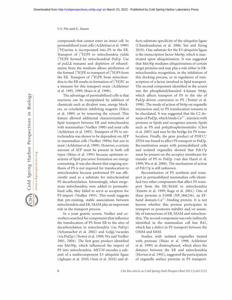
compounds that cannot enter an intact cell. Inpermeabilized yeast cells (Achleitner et al. 1999)[3H]serine is incorporated into PS in the ER.Transport of [3H]PS to mitochondria yields[3H]PE formed by mitochondrial Psd1p. Useof psd2D mutants and depletion of ethanol-amine from the medium allows attribution ofthe formed [3H]PE to transport of [3H]PS fromthe ER. Transport of [3H]PE from mitochon-dria to the ER results in formation of [3H]PC asa measure for this transport route (Achleitneret al. 1995, 1999; Shiao et al. 1998).
The advantage of permeabilized cells is thatreactions can be manipulated by addition ofchemicals such as divalent ions, energy block-ers, or cytoskeleton inhibiting reagents (Eilerset al. 1989) or by removing the cytosol. Thisfeature allowed additional characterization oflipid transport between ER and mitochondriawith mammalian (Voelker 1990) and yeast cells(Achleitner et al. 1995). Transport of PS to mi-tochondria was shown to be dependent on ATPin mammalian cells (Voelker 1989a) but not inyeast (Achleitner et al. 1999). However, a certainamount of ATP must be present in both celltypes (Shiao et al. 1995) because upstream re-actions of lipid precursor formation are energyconsuming. It was also shown that ongoing syn-thesis of PS is not required for translocation tomitochondria because preformed PS was effi-ciently used as a substrate for mitochondrialPS decarboxylation. Interestingly, when exoge-nous mitochondria were added to permeabi-lized cells, they failed to serve as acceptors forPS import (Voelker 1993). This result suggeststhat pre-existing, stable associations betweenmitochondria and ER/MAM play an importantrole in the transport process.
In a yeast genetic screen, Voelker and co-workers searched for components that influencethe translocation of PS from ER to the sites ofdecarboxylation in mitochondria (via Psd1p)(Schumacher et al. 2002) and Golgi/vacuoles(via Psd2p) (Trotter et al. 1998; Wu and Voelker2001, 2004). The first gene product identifiedwas Met30p, which influenced the import ofPS into mitochondria. MET30 encodes a sub-unit of a multicomponent E3 ubiquitin ligase(Aghajan et al. 2010; Ouni et al. 2010) and af-
fects substrate specificity of the ubiquitin ligase(Chandrasekaran et al. 2006; Yan and Xiong2010). One substrate for the E3 ubiquitin ligaseis the transcription factor Met4p, which is inac-tivated upon ubiquitination. It was suggestedthat Met30p mediates ubiquitination of certaintarget proteins and may play a role either in ER-mitochondria recognition, in the inhibition ofthis docking process, or in regulation of tran-scription of a factor involved in lipid transport.The second component identified in the screenwas the phosphatidylinositol 4-kinase Stt4p,which affects transport of PS to the site ofPsd2p-driven conversion to PE (Trotter et al.1998). The mode of action of Stt4p on organelleinteraction and/or PS translocation remains tobe elucidated. It was suggested that the C2 do-main of Psd2p, which binds Ca2þ, interacts withproteins or lipids and recognizes anionic lipidssuch as PS and polyphosphoinositides (Choiet al. 2005) and may be the bridge for PS trans-location. Finally, the gene product of PDR17/SFH4 was found to affect PS transport to Psd2p.Reconstitution assays with permeabilized cellsand isolated organelles showed that Pdr17pmust be present on the acceptor membrane fortransfer of PS to Psd2p (van den Hazel et al.1999; Wu et al. 2000). The mechanism of actionof Pdr17p is still unknown.
Reconstitution of PS synthesis and trans-port in permeabilized mammalian cells identi-fied two other components that affect PS trans-port from the ER/MAM to mitochondria(Emoto et al. 1999; Kuge et al. 2001). One ofthese proteins is S100B (NP_006236), an EF-hand domain-Ca2þ-binding protein. It is notknown whether this protein participates intransport or promotes stability and/or assem-bly of interactions of ER/MAM and mitochon-dria. The second component was only indirectlyidentified in the mammalian cell line R41,which has a defect in PS transport between theOMM and IMM.
Studies with isolated organelles treatedwith protease (Shiao et al. 1998; Achleitneret al. 1999) or dinitrophenol, which alters thedistance between the ER and mitochondria(Hovius et al. 1992), suggested the participationof organelle surface proteins in PS transport.
V.V. Flis and G. Daum
8 Cite this article as Cold Spring Harb Perspect Biol 2013;5:a013235
on March 23, 2022 - Published by Cold Spring Harbor Laboratory Press http://cshperspectives.cshlp.org/Downloaded from

Electron microscopy supported this view bydemonstrating close contact zones betweenER/MAM and mitochondria, which mightserve as bridges for lipid translocation (Csordaset al. 2006). Furthermore, the involvement ofacetylated microtubules in ER-mitochondriadynamics was demonstrated, a mechanism bywhich membrane contact can be establishedand/or maintained (Friedman et al. 2010).Moreover, mitochondrial division sites pro-duced by Dnm1p and Drp1p were found to bephysically connected with the ER (Friedmanet al. 2011).
To shed more light on the role of ER-mito-chondria contact, genetic and synthetic geneticinteractions analysis mainly with the yeast, Sac-charomyces cerevisiae, were performed (Birneret al. 2003; Kornmann et al. 2009; Osman et al.2009a). Such genetic screens were designedin a way that defects in two or more interactinggene products become lethal. Recently, Korn-mann et al. (2009) identified components ofER-mitochondria contact in yeast at the molec-ular level. They showed that in wild-type cells, aprotein complex tethers the ER/MAM and mi-tochondria. Mutations in this protein complexcaused slow growth or were lethal for the cells,but could be suppressed by an artificial tether-ing construct called ChiMERA, which carried aGFP molecule. When staining the mitochon-dria with mito-tracker and expressing the Chi-MERA, association was visualized confirmingthat ChiMERA acted as a bridge between theER and mitochondria. The authentic complexwas named ERMES (ER mitochondria encoun-tered structures) and contains the OMM pro-teins, Mdm10p, Mdm12p, and Mdm34p, whichinteract with the ER membrane anchored pro-tein Mmm1p. In the absence of ERMES, the CLlevel and the PS/PC conversion rates were de-creased, suggesting the involvement of ERMESin phospholipids transport between ER andmitochondria (Fig. 3). Moreover, genetic inter-actions between PSD1, GEM1, and the ERMEScomplex were shown (Kornmann et al. 2009).The Ca2þMiro (mitochondria rho-like) GTPaseGem1p is a regulatory component of ERMES(Kornmann et al. 2011; Stroud et al. 2011),maintains mitochondrial morphology and in-
heritance (Frederick et al. 2004, 2008) andmost likely connects ERMES to the actin cyto-skeleton (Kornmann et al. 2011; Michel andKornmann 2012). It was assumed that Gem1pshuttles between a free and ERMES bound form(see Fig. 3), although the molecular mechanismof Gem1p remains unknown. Physical interac-tion of Psd1p with ERMES or Gem1p was notdemonstrated. Nevertheless, it was reported thata gem1Dpsd2Ddpl1D yeast mutant grown on anonfermentable carbon source was defective inits PC synthesis (Kornmann et al. 2011), mostlikely by impaired PE and PS transport. A similargrowth phenotype was reported before for thegem1D single deletion strain grown on syntheticglycerol media (Frederick et al. 2008). GEM1also shows strong synthetic lethality withGEP4, encoding a PGP phosphatase (Kornmannet al. 2011). Gem1p is well conserved in theeukaryotic kingdom, suggesting that MiroGTPases are general components of ER-mito-chondria connections. Miro-1, the mammalianhomolog of Gem1p, interacts with mitofusin 1and 2, the human homologs of yeast Fzo1p.Mfn-2 was suggested to tether mitochondria toER and being involved in mitochondrial move-ment along axons (de Brito and Scorrano 2008;Misko et al. 2010). Most recently, however, therole of ERMES and Gem1p in the import of PSinto mitochondria and its conversion to PE waschallenged (Nguyen et al. 2012). It was arguedthat, despite their genetic and physical inter-action, ERMES and Gem1p function in dis-tinct pathways, and the absence of ERMES andGem1p had only little effect on PS import intomitochondria and conversion to PE. The effecton PC formation as described by Kornmannet al. (2009) was regarded as minor. Nguyenet al. (2012) argued that changes in the lipidprofile of cells lacking ERMES were side effectsof defects in mitochondrial morphology.
Recently, another mitochondrial complexnamed MitOS, MICOS, or MINOS was identi-fied (Harner et al. 2011; Hoppins et al. 2011;von der Malsburg et al. 2011). This IMM-asso-ciated complex functions in cristae formationand morphology (Rabl et al. 2009). It was pro-posed (van der Laan et al. 2012) that MINOSforms the central core of a large and complex
Lipid Transport between the ER and Mitochondria
Cite this article as Cold Spring Harb Perspect Biol 2013;5:a013235 9
on March 23, 2022 - Published by Cold Spring Harbor Laboratory Press http://cshperspectives.cshlp.org/Downloaded from

organizing system named ERMIONE, whichincludes the ERMES complex, the prohibitinringlike structures, the TOM and TIM com-plexes, and Mdm31/32 proteins required formtDNA maintenance. MINOS was also foundto interact with the protein VDAC and with thefusion protein Ugo1 (van der Laan et al. 2012).Both ERMES and MINOS are genetically linkedto the prohibitin ring complexes of the IMMthat are integrated into the lipid network me-tabolism (Psd1, Ups1/Ups2) (Birner et al. 2003;Gohil et al. 2005; Kornmann et al. 2009; Osman
et al. 2009a; Tamura et al. 2009, 2012; Pottinget al. 2010). However, direct involvement ofMINOS in lipid transport and/or assemblyinto mitochondrial membrane has not beendemonstrated.
LINK BETWEEN CARDIOLIPIN FORMATIONAND INTRAMITOCHONDRIAL TRANSPORTOF AMINOGLYCEROPHOSPHOLIPIDS
A link between CL and PE assembly in mito-chondrial membranes, PE synthesis by Psd1p,
PHB1/PHB2
MLCLCL
CL
PG
CDP-DAG
CDP-DAG
UPS1/UPS2
PSD1
MDM35
PA
PA
PA CDP-DAG PCPEM2 PEM1
PE PSERMES
ER/MAM
10–4
0 nm
PC PE PS UPS2
MD
M34
MD
M10
MD
M12
MM
M1
UPS1
YME1 ATP23
GEM1
OMM
IMM
Nonbilayer P-lipids
Bilayer P-lipids
PGP
CLD1
CRD1
CDS1
CDS1
GEP4PGS1
TAZ1
Figure 3. Components involved in translocation of phospholipids to and within mitochondria of yeast. Thedirections of lipid translocation are indicated byarrows. While PS and PC are imported into mitochondria, a largeportion PE is exported. Upon import the majority of PS is converted to PE by Psd1p. Decarboxylation of PS to PEin mitochondria and methylation of PE to PC in the ER are sufficient for supplying cells with PE and PCeven in theabsence of external ethanolamine or choline. Similar to PS and PC, PA and CDP-DAG also have to be importedinto mitochondria where they are required for CL synthesis. The pathway of CL synthesis in the IMM starts withCDP-DAG and is completed during several steps by a multienzyme cascade. A small portion of CL is exported tothe OMM. The ERMES complexcontaining gene products of MDM10, MDM34, MDM12, and MMM1 tethers ERand mitochondria. In mammalian cells, Mfn1/2 seem to have a similar function. ERMES associates with theGTPase GEM1, cycling between ERMES bound and free form. UPS1/UPS2 interact with Mdm35p and regulatethe mitochondrial levels of PE and CL. UPS1 and UPS2 are degraded by ATP23 and YEM1, respectively. Prohibitinringlike structures made from two proteins PHB1/2 contribute to the formation of PE and CL clusters in theIMM. For a detailed description of biosynthetic and translocation processes and components involved, see text.
V.V. Flis and G. Daum
10 Cite this article as Cold Spring Harb Perspect Biol 2013;5:a013235
on March 23, 2022 - Published by Cold Spring Harbor Laboratory Press http://cshperspectives.cshlp.org/Downloaded from

and the role of prohibitins was uncovered insynthetic lethal screens with the yeast (Birneret al. 2003; Gohil et al. 2005). It was shownthat a phb1Dphb2Dpsd1D triple mutation waslethal due to reduced PE levels and loss ofmtDNA. Osman et al. (2009a) demonstratedthat ringlike prohibitin complexes organize CLand PE in clusters in the IMM and become es-sential in strains with low levels of PE and CL.This finding supported the view of physical sim-ilarities of CL and PE and their importance formitochondria morphology (Osman et al. 2011;Joshi et al. 2012). In subsequent work, 35 genet-ic interactions of prohibitins (GEP) were iden-tified, which were required for cell survival inthe absence of prohibitins. Among the genesdetected, UPS1, GEP1/UPS2, PSD1, MDM35,MMM1, GEP4, and CRD1 were prominent (Os-man et al. 2009b) (see Fig. 3). Prohibitins seemto control IMM organization and integrity byacting as lipid scaffolds for PE and CL (Osmanet al. 2009a,b). Ups1p and Ups2p antagonisti-cally regulate the CL (Tamura et al. 2009) level,and Ups2p also regulates the PE level (Pottinget al. 2010) in the IMM. Most recently, it wasshown that Ups1p promotes conversion of PE toPC, whereas Ups2p suppresses this process (Ta-mura et al. 2012). It was concluded that UPSproteins affect export of PE from the IMM, al-though the mechanism of this regulation is un-known. The authors showed that loss of Ups1,the ERMES complex, and Mdm31p caused sim-ilardefects in mitochondria, especially in CL andPE homeostasis in the IMM. Yeast Ups1p andUps2p are per se unstable proteins and degradedby the proteases Yme1p and Atp23p in the IMM(Potting et al. 2010). The newly identified pro-tein Mdm35p protects them from proteolyticdegradation (Tamura et al. 2009, 2012; Pottinget al. 2010).
IMPORT OF PHOSPHATIDYLCHOLINE ANDPHOSPHATIDYLINOSITOL INTOMITOCHONDRIA
Although PC and PI are major phospholipidsof mitochondrial membranes, little is knownabout their import into this organelle. Bothphospholipids are synthesized in the ER from
where they are translocated to mitochondriaand assembled into membranes. Lampl et al.(1994) designed an in vitro assay to study theimport of [3H]-labeled PI and PC from uni-lamellar donor vesicles to isolated yeast mito-chondria. Both phospholipids ended up in theIMM. During import, they were detected incontact sites between OMM and IMM, sup-porting the notion that these sites are involvedin intramitochondrial phospholipid transport.The uncoupler CCCP, the antibiotic adriamy-cin, and energy depletion did not inhibit thisprocess. Janssen et al. (1999) described trans-bilayer movement of PC in isolated OMMvesicles from yeast. They showed that this trans-location was rapid and bidirectional. Pretreat-ment of the OMM with proteinase K or sulf-hydryl reagents had no effect on PC transport.
STEROLS AND SPHINGOLIPIDS OFMITOCHONDRIA
Sterols and sphingolipids are minor lipid com-ponents of mitochondria. Both lipid classes aresynthesized in extramitochondrial compart-ments and need to be imported and assembledinto mitochondrial membranes. Sterol biosyn-thesis is accomplished by a complex sequence ofreactions that are localized to the ER and perox-isomes of mammalian cells (Ikonen 2008; Millerand Auchus 2011; Miller and Bose 2011). Inmammalian cells, cholesterol is the end productof this biosynthetic pathway (Soccio and Bres-low 2004; Brown and Goldstein 2009), whereasin yeast ergosterol is the major sterol (Wagnerand Daum 2005; Jacquier and Schneiter 2012).In yeast, the ER and lipid droplets contributeto sterol biosynthesis (Leber et al. 1994; Athen-staedt et al. 1999b). Plants produce a varietyof sterols such as phytosterols (Suzuki andMuranaka 2007). In mammalian mitochondriathe majority of cholesterol is localized to theOMM (Daum 1985), whereas in yeast (Sperka-Gottlieb et al. 1988; Wriessnegger et al. 2009)most of the mitochondrial sterol was found inthe IMM.
Biosynthesis of sphingolipids occurs in theER and in organelles along the protein secretorypathway, mainly the Golgi (Perry and Ridgway
Lipid Transport between the ER and Mitochondria
Cite this article as Cold Spring Harb Perspect Biol 2013;5:a013235 11
on March 23, 2022 - Published by Cold Spring Harbor Laboratory Press http://cshperspectives.cshlp.org/Downloaded from
2005; Breslow and Weissman 2010; Levy andFuterman 2010; Mencarelli et al. 2010). Howev-er, recent work showed the presence of ceramidemetabolic enzymes in mitochondria frommammals and yeast (Kitagaki et al. 2007; Nov-gorodov et al. 2011). The majority of this lipidclass is located to the plasma membrane, where-as amounts in mitochondria are minor (Bres-low and Weissman 2010; Stiban et al. 2010; vanMeer and Hoetzl 2010). In the OMM, the con-centration of ceramide is threefold higher thanin the IMM (Mencarelli et al. 2010).
IMPORT OF STEROLS INTOMITOCHONDRIA
Sterols in mitochondria may serve as structuralcomponents of membranes and/or as precur-sors for hormone synthesis. For both purposes,they have to be imported into mitochondria.Transport of sterols from the ER to mitochon-dria appears to occur by machinery, whichis different from that described for phospho-lipids. A nonvesicular and a vesicular pathwayhave been proposed for cholesterol transloca-tion to mitochondria (Maxfield and Wustner2002).
A number of components appear tocontribute to sterol transport to mitochondria(Fig. 4). The best studied protein of this kind isthe so-called steroidogenic acute regulatoryprotein (StAR) (Clark et al. 1994). StAR is al-most exclusively expressed in steroid-producingcells and regulated by cAMP (Strauss et al. 1999;Benmessahel et al. 2002). StAR requires ATPand an electrochemical gradient for maximal ac-tivity (King and Stocco 1996). Import of StARinto mitochondria results in cleavage of an ami-no-terminal mitochondrial targeting sequence(Arakane et al. 1998). The carboxyl-terminusof StAR displays cholesterol transferring capa-city (Strauss et al. 1999; Stocco 2000). Ligandbinding to StAR changes its secondary structure;the protein itself alters the domain structureof the OMM to facilitate rapid sterol transfer(Petrescu et al. 2001). StAR interacts with thecAMP-dependent protein kinase (PKA) and aperipheral-type benzodiazepine receptor (PBR)(Hauet et al. 2002), whichwas later named TSPO
(translocator protein) (Mukhin et al. 1989;Krueger and Papadopoulos 1990). A proteinnamed PAP7 was identified as a regulator ofPBR and PKA. PBR/TSPO (translocator pro-tein) was shown to support import and process-ing of StAR in the mitochondria (Hauet et al.2005). VDAC (voltage-dependent anion chan-nel) and ANT (adenine nucleotide transporter)were identified as interaction partners of PBR/TSPO (McEnery et al. 1992; Rone et al. 2009).VDAC binds cholesterol and influences its mito-chondrial distribution (Campbell and Chan2007). It appears that TSPO and VDAC togetherwithTOM(translocaseof theOMM) function inStAR assembly into mitochondria. Also mem-bers of the ACBD family, ACBD1 and ACBD3/PAP7, seem to participate in the function of thetransduceosome (Midzak et al. 2011). Finally, aprotein named MLN64 (metastatic lymph nodeprotein 64) with a carboxy-terminal START do-main for cholesterol binding was identified as amediator for cholesterol transport from late en-dosomes to the plasma membrane and also tomitochondria (Zhang et al. 2002; Charman et al.2010; Liapis et al. 2012). It was shown that inNiemann-Pick Type C (NPC) mutated cells,MLN64 supplies cholesterol from endosomesto mitochondria. In analogy to StAR, MLN64needs direct contact with mitochondria to trans-fer cholesterol, indicating that direct contact be-tween endosomes and mitochondria may occur(Charman et al. 2010).
Once cholesterol has been imported intomitochondria, it is further converted to steroids(Miller and Auchus 2011) (see Fig. 4). The firstand rate-limiting step in steroidogenesis is theconversion of cholesterol to pregnenolone by acytochrome P450. Further conversion requiresthe action of hydroxysteroid dehydrogenases.Steroidogenic cells in humans are localized to avariety of tissues, most prominently to adrenalglands and gonads. In the adrenal glands, main-ly the conversion of cholesterol to aldosterol,dehydroepiandrosterone (DHEA), and corti-sol-like is catalyzed. In the gonal glands, choles-terol is converted to steroid hormones, namelyestradiol in the ovarian granulose cells andto testosterone by the testicular leyding cells(Miller and Auchus 2011).
V.V. Flis and G. Daum
12 Cite this article as Cold Spring Harb Perspect Biol 2013;5:a013235
on March 23, 2022 - Published by Cold Spring Harbor Laboratory Press http://cshperspectives.cshlp.org/Downloaded from

Evidence about import of sterols in non-steroidogenic cells is rare. Import of radiola-beled ergosterol and cholesterol from unilamel-lar vesicles into isolated mitochondria has beenstudied in Saccharomyces cerevisiae (Tuller and
Daum 1995). Supply of ergosterol to the mito-chondrial surface was enhanced by a cytosolicfraction, whereas no such additive was requiredfor cholesterol transport. Both sterols reachedthe IMM in an energy-independent process
N C NCcAMP
Cytosol
Cytosol
cAMP
OMM IMM
ANT
ANT
Matrix
Cytosol OMM IMM Matrix
A
B
C
StAR StAR
TSPO
TSPO
VDACStAR
ACBD1
ACBD1
PKA PAP7
PKA PAP7
StAR
CY
P11
A1
CY
P11
A1
VDAC
HO
HO
O
O
Figure 4. Import of sterols into mitochondria for steroidogenesis. (A) Upon binding of cholesterol the StARprecursor protein changes its conformation. StAR expression is regulated by cAMP. (B) StAR comes in contactwith PKA, PAP7/ACBD3, and ACBD1 and is imported into the mitochondria with sterols bound. Uponinteraction with TSPO, StAR induces changes in the OMM and a transduceosome is formed. This structure,which includes TSPO/VDAC of the OMM and ANT in the IMM, facilitates imports and maturation of StAR.(C) Upon arrival in the mitochondrial matrix, the mature StAR protein delivers cholesterol to CPY11A1 where itis converted to pregnenolone.
Lipid Transport between the ER and Mitochondria
Cite this article as Cold Spring Harb Perspect Biol 2013;5:a013235 13
on March 23, 2022 - Published by Cold Spring Harbor Laboratory Press http://cshperspectives.cshlp.org/Downloaded from
but accumulated to some extent in contact sitesbetween OMM and IMM, supporting the ideathat these zones are sites of intramitochondriallipid translocation.
SPHINGOLIPID INCORPORATION INTOMITOCHONDRIA
Little is known about import and function ofsphingolipids in mitochondria. There is evi-dence that sphingolipid metabolism affects themitochondrial pathway of apoptosis (Chipuket al. 2012). Ceramides with their short andlong acyl-chains could form large protein-per-meable channels in the OMM (Siskind and Co-lombini 2000), and such channels may contrib-ute to the release of proapoptotic proteins frommitochondria (Futerman 2006). It was pro-posed that ceramide is produced by hydrolysisof sphingomyelin by a neutral sphingomyeli-nase in MAM (Chipuk et al. 2012; Mullenet al. 2012) and then transferred to mitochon-dria where it is converted to sphingosine-1-phosphate and hexadecenal. It has also beenproposed that in liver mitochondria, thioester-ase and neutral ceramidase produce ceramidefrom sphingosine and acyl-CoA (Novgorodovand Gudz 2009; Novgorodov et al. 2011). Invitro, ceramide synthase, or reverse ceramidaseactivity was detected in mitochondria (Biondaet al. 2004). In yeast, Isc1p has been suggested togenerate ceramides in mitochondria (Kitagakiet al. 2007, 2009).
Evidence for the existence of two ceramidetransporting proteins has been presented: theGoodpasture antigen-binding protein (GPBP)and its splice variant, the ceramide transporter(CERT) (Mencarelli et al. 2010). It was pro-posed that these proteins might be involved inmitochondrial lipid homeostasis, although thisview is still a matter of debate.
CONCLUSIONS
Among the different subcellular compartments,mitochondria are one of the most unique whenconsidering lipid composition. Presence of PGand CL, as well as the large amount of PE, ischaracteristic for mitochondrial membranes.
Recent studies have shown that especially thesenonbilayer forming lipids are important for mi-tochondrial function (Birner et al. 2003; Gohilet al. 2005; Nebauer et al. 2007; Osman et al.2009a; Acehan et al. 2011; Joshi et al. 2012; Ta-mura et al. 2012). However, low abundant lipidsin mitochondria such as sterols or sphingolipidsmust not be ignored, although at present ascrib-ing specific functions to these components inmitochondria is difficult.
Large amounts of lipids have to be import-ed into mitochondria. Different mechanisms ofintracellular lipid transport have been discussedfor several decades (Daum and Vance 1997;Holthuis and Levine 2005; Voelker 2005; Schulzand Prinz 2007; Osman et al. 2011). The role oflipid transfer proteins, which were enthusiasti-cally studied in the beginning of these investiga-tions, has been scrutinized more recently. Suchtransfer proteins may be more relevant for lipidsorting or sensing than bulk lipid transport.Vesicle flux was recognized as a more potentmechanism of lipid transport because it allowsrapid, robust, and efficient translocation to tar-get membranes. It appears that at least signifi-cant portions of lipids are cotransported to thecell periphery by the classical pathway of proteinsecretion (Salama and Schekman 1995; Kohl-wein et al. 1996). However, other types of vesi-cles may also be relevant although experimen-tal evidence for such processes is missing. Lastbut not least, membrane contact has becomean attractive alternative for intracellular lipidtranslocation (Levine and Loewen 2006; Le-biedzinska et al. 2009; Voelker 2009; de Britoand Scorrano 2010; Elbaz and Schuldiner2011; Osman et al. 2011). Contact of the ERwith mitochondria through the MAM fraction,association of the ER with the plasma mem-brane or nuclear–vacuolar junctions becameprominent examples of this kind. Finally, trans-membrane movements of lipids by translocasesand flippases contribute to appropriate lipiddistribution in organelles (Holthuis and Levine2005).
Close proximity of ER and mitochondriaas shown by electron microscopy (Csordaset al. 2006; Friedman et al. 2011) supports theidea of lipid transport by membrane contact.
V.V. Flis and G. Daum
14 Cite this article as Cold Spring Harb Perspect Biol 2013;5:a013235
on March 23, 2022 - Published by Cold Spring Harbor Laboratory Press http://cshperspectives.cshlp.org/Downloaded from

The distance is sufficient for a protein complexto bridge the gap between organelles and toconnect membranes without causing fusion(Achleitner et al. 1999). A hemistalk structurecaused by high amounts of mitochondrial PEand CL, which provide negative membranecurvature, may support attachment and pointfusion events (Chernomordik and Kozlov2005, 2008; Chernomordik et al. 2006). Proteinbridges like ERMES stabilize the energy-unfa-vorable state (Kozlov et al. 2010). Microtubulesand actin filaments may help by bridging ERand mitochondria and establishing or main-taining protein contact between membranes(Friedman et al. 2010, 2011). Although ERMESand associated proteins were shown to create ormaintain contact between ER and mitochon-dria (Kornmann et al. 2009; Stroud et al.2011), direct functions of these components inlipid transport were scrutinized (Nguyen et al.2012). It was argued that ERMES facilitatesmembrane contact, but other components maybe more directly involved in lipid translocation.MAM-OMM contact sites appear to be furtherlinked to complexes of the IMM (de Brito andScorrano 2008; Kornmann et al. 2009). Trans-port of lipids from OMM to IMM may occureither by membrane contact or by translocationthrough the intermembrane space. Despite itsrole in IMM organization the cristae-formingMINOS complex (Harner et al. 2011; Hoppinset al. 2011; van der Laan et al. 2012) is most likelynot involved in this process. The role of Gem1pand Ups-proteins in OMM-IMM lipid translo-cation has to be taken into account (Tamuraet al. 2012). Interaction of prohibitins with themitochondrial lipid synthesizing machineryalso appears to be important for lipid homeo-stasis in this organelle (Birner et al. 2003; Gohilet al. 2005; Gohil and Greenberg 2009; Osmanet al. 2009a,b).
An intriguing although unsolved questionis regulation of lipid transport to/from andwithin mitochondria. Fluidity of the membraneor membrane curvature affected by PE and CLare parameters that may influence biophysicalproperties of the membrane environment. Alsothe acyl chain length of specific phospholipidspecies could have an effect (Dolis et al. 1996;
Heikinheimo and Somerharju 1998; Kevala andKim 2001). A balanced concentration of phos-pholipids, sterols, and sphingolipids appears tobe important for membrane fusion or fission. Inthat respect, the low-abundant lipids in mito-chondria such as sterols or sphingolipids maybe relevant. Regulation of the mitochondriallipid assembly by Ups1p and Ups2p, which arein turn subject to proteolytic degradation/pro-tection by Yme1p, Atp23p, and Mdm35p (Pot-ting et al. 2010; Tamura et al. 2012) are examplesfor the widespread network of lipid assemblyand homeostasis in mitochondrial membranes,which we are just beginning to understand inmore detail at the molecular level.
ACKNOWLEDGMENTS
We thank Susanne Horvath and Edina Harsayfor critically reading this manuscript. This workis financially supported by the FWF (DK Mo-lecular Enzymology W901-B05 and projectP21429 to G.D.).
REFERENCES
Acehan D, Malhotra A, Xu Y, Ren M, Stokes DL, Schlame M.2011. Cardiolipin affects the supramolecular organiza-tion of ATP synthase in mitochondria. Biophys J 100:2184–2192.
Achleitner G, Zweytick D, Trotter PJ, Voelker DR, DaumG. 1995. Synthesis and intracellular transport of ami-noglycerophospholipids in permeabilized cells of theyeast, Saccharomyces cerevisiae. J Biol Chem 270: 29836–29842.
Achleitner G, Gaigg B, Krasser A, Kainersdorfer E, KohlweinSD, Perktold A, Zellnig G, Daum G. 1999. Associa-tion between the endoplasmic reticulum and mitochon-dria of yeast facilitates interorganelle transport of phos-pholipids through membrane contact. Eur J Biochem 264:545–553.
Adeyo O, Horn PJ, Lee S, Binns DD, Chandrahas A,Chapman KD, Goodman JM. 2011. The yeast lipin or-thologue Pah1p is important for biogenesis of lipid drop-lets. J Cell Biol 192: 1043–1055.
Aghajan M, Jonai N, Flick K, Fu F, Luo M, Cai X, Ouni I,Pierce N, Tang X, Lomenick B, et al. 2010. Chemicalgenetics screen for enhancers of rapamycin identifies aspecific inhibitor of an SCF family E3 ubiquitin ligase.Nat Biotechnol 28: 738–742.
Arakane F, Kallen CB, Watari H, Stayrook SE, Lewis M,Strauss JF 3rd. 1998. Steroidogenic acute regulatory pro-tein (StAR) acts on the outside of mitochondria to stim-ulate steroidogenesis. Endocr Res 24: 463–468.
Lipid Transport between the ER and Mitochondria
Cite this article as Cold Spring Harb Perspect Biol 2013;5:a013235 15
on March 23, 2022 - Published by Cold Spring Harbor Laboratory Press http://cshperspectives.cshlp.org/Downloaded from

Athenstaedt K, Daum G. 1999. Phosphatidic acid, a keyintermediate in lipid metabolism. Eur J Biochem 266:1–16.
Athenstaedt K, Weys S, Paltauf F, Daum G. 1999a. Redun-dant systems of phosphatidic acid biosynthesis via acyl-ation of glycerol-3-phosphate or dihydroxyacetone phos-phate in the yeast Saccharomyces cerevisiae. J Bacteriol181: 1458–1463.
Athenstaedt K, Zweytick D, Jandrositz A, Kohlwein SD,Daum G. 1999b. Identification and characterization ofmajor lipid particle proteins of the yeast Saccharomycescerevisiae. J Bacteriol 181: 6441–6448.
Benmessahel Y, Guennoun R, Cadepond F, Baulieu EE,Schumacher M, Groyer G. 2002. Expression of steroido-genic acute regulatory protein in cultured Schwann cellsand its regulation by cAMP. Ann NYAcad Sci 973: 83–87.
Bionda C, Portoukalian J, Schmitt D, Rodriguez-LafrasseC, Ardail D. 2004. Subcellular compartmentalization ofceramide metabolism: MAM (mitochondria-associat-ed membrane) and/or mitochondria? Biochem J 382:527–533.
Birner R, Burgermeister M, Schneiter R, Daum G. 2001.Roles of phosphatidylethanolamine and of its several bio-synthetic pathways in Saccharomyces cerevisiae. Mol BiolCell 12: 997–1007.
Birner R, Nebauer R, Schneiter R, Daum G. 2003. Syntheticlethal interaction of the mitochondrial phosphatidyleth-anolamine biosynthetic machinery with the prohibitincomplex of Saccharomyces cerevisiae. Mol Biol Cell 14:370–383.
Bogdanov M, Mileykovskaya E, Dowhan W. 2008a. Lipids inthe assembly of membrane proteins and organization ofprotein supercomplexes: Implications for lipid-linkeddisorders. Subcell Biochem 49: 197–239.
Bogdanov M, Xie J, Heacock P, Dowhan W. 2008b. To flip ornot to flip: Lipid-protein charge interactions are a deter-minant of final membrane protein topology. J Cell Biol182: 925–935.
Boumann HA, de Kruijff B, Heck AJR, de Kroon AIPM.2004. The selective utilization of substrates in vivo bythe phosphatidylethanolamine and phosphatidylcholinebiosynthetic enzymes Ept1p and Cpt1p in yeast. FEBSLett 569: 173–177.
Breslow DK, Weissman JS. 2010. Membranes in balance:Mechanisms of sphingolipid homeostasis. Mol Cell 40:267–279.
Brindley DN, Pilquil C, Sariahmetoglu M, Reue K. 2009.Phosphatidate degradation: Phosphatidate phosphatases(lipins) and lipid phosphate phosphatases. Biochim Bio-phys Acta 1791: 956–961.
Brown MS, Goldstein JL. 2009. Cholesterol feedback: FromSchoenheimer’s bottle to Scap’s MELADL. J Lipid Res 50(Suppl): S15–S27.
Burgermeister M, Birner-Grunberger R, Nebauer R, DaumG. 2004. Contribution of different pathways to the supplyof phosphatidylethanolamine and phosphatidylcholineto mitochondrial membranes of the yeast Saccharomycescerevisiae. Biochim Biophys Acta 1686: 161–168.
Butler MM, Thompson W. 1975. Transfer of phosphatidyl-serine from liposomes or microsomes to mitochondria.Stimulation by a cell supernatant factor. Biochim BiophysActa 388: 52–57.
Campbell AM, Chan SHP. 2007. The voltage dependentanion channel affects mitochondrial cholesterol distribu-tion and function. Arch Biochem Biophys 466: 203–210.
Cao J, Shan D, Revett T, Li D, Wu L, Liu W, Tobin JF,Gimeno RE. 2008. Molecular identification of a novelmammalian brain isoform of acyl-CoA:lysophospholipidacyltransferase with prominent ethanolamine lysophos-pholipid acylating activity, LPEAT2. J Biol Chem 283:19049–19057.
Carman GM, Han G-S. 2006. Roles of phosphatidate phos-phatase enzymes in lipid metabolism. Trends Biochem Sci31: 694–699.
Carman GM, Han G-S. 2009. Phosphatidic acid phospha-tase, a key enzyme in the regulation of lipid synthesis.J Biol Chem 284: 2593–2597.
Carrasco MP, Jimenez-Lopez JM, Martinez-Duenas L,Ubina S, Segovia JL, Marco C. 2006. Ethanol specificallyalters the synthesis, acylation and transbilayer movementof aminophospholipids in rat-liver microsomes. Life Sci78: 2781–2786.
Chandrasekaran S, Deffenbaugh AE, Ford DA, Bailly E,Mathias N, Skowyra D. 2006. Destabilization of bindingto cofactors and SCFMet30 is the rate-limiting regulatorystep in degradation of polyubiquitinated Met4. Mol Cell24: 689–699.
Chang SC, Heacock PN, Clancey CJ, Dowhan W. 1998a. ThePEL1 gene (renamed PGS1) encodes the phosphatidyl-glycerophosphate synthase of Saccharomyces cerevisiae.J Biol Chem 273: 9829–9836.
Chang SC, Heacock PN, Mileykovskaya E, Voelker DR,Dowhan W. 1998b. Isolation and characterization of thegene (CLS1) encoding cardiolipin synthase in Saccharo-myces cerevisiae. J Biol Chem 273: 14933–14941.
Charman M, Kennedy BE, Osborne N, Karten B. 2010.MLN64 mediates egress of cholesterol from endosomesto mitochondria in the absence of functional Niemann-Pick Type C1 protein. J Lipid Res 51: 1023–1034.
Chen D, Zhang X-Y, Shi Y. 2006. Identification and func-tional characterization of hCLS1, a human cardiolipinsynthase localized in mitochondria. Biochem J 398: 169–176.
Chernomordik LV, Kozlov MM. 2005. Membrane hemifu-sion: Crossing a chasm in two leaps. Cell 123: 375–382.
Chernomordik LV, Kozlov MM. 2008. Mechanics of mem-brane fusion. Nat Struct Mol Biol 15: 675–683.
Chernomordik LV, Zimmerberg J, Kozlov MM. 2006. Mem-branes of the world unite! J Cell Biol 175: 201–207.
Chipuk JE, McStay GP, Bharti A, Kuwana T, Clarke CJ,Siskind LJ, Obeid LM, Green DR. 2012. Sphingolipidmetabolism cooperates with BAK and BAX to promotethe mitochondrial pathway of apoptosis. Cell 148: 988–1000.
Choi J-Y, Wu W-I, Voelker DR. 2005. Phosphatidylserinedecarboxylases as genetic and biochemical tools forstudying phospholipid traffic. Anal Biochem 347: 165–175.
Choi J-Y, Augagneur Y, Ben MC, Voelker DR. 2012. Identi-fication of gene encoding Plasmodium knowlesi phospha-tidylserine decarboxylase by genetic complementation inyeast and characterization of in vitro maturation of en-coded enzyme. J Biol Chem 287: 222–232.
V.V. Flis and G. Daum
16 Cite this article as Cold Spring Harb Perspect Biol 2013;5:a013235
on March 23, 2022 - Published by Cold Spring Harbor Laboratory Press http://cshperspectives.cshlp.org/Downloaded from

Clark BJ, Wells J, King SR, Stocco DM. 1994. The purifica-tion, cloning, and expression of a novel luteinizing hor-mone-induced mitochondrial protein in MA-10 mouseLeydig tumor cells. Characterization of the steroidogenicacute regulatory protein (StAR). J Biol Chem 269: 28314–28322.
Cowan AK. 2009. Plant growth promotion by 18:0-lyso-phosphatidylethanolamine involves senescence delay.Plant Signal Behav 4: 324–327.
Csordas G, Renken C, Varnai P, Walter L, Weaver D,Buttle KF, Balla T, Mannella CA, Hajnoczky G. 2006.Structural and functional features and significance ofthe physical linkage between ER and mitochondria. JCell Biol 174: 915–921.
Daum G. 1985. Lipids of mitochondria. Biochim BiophysActa 822: 1–42.
Daum G, Vance JE. 1997. Import of lipids into mitochon-dria. Prog Lipid Res 36: 103–130.
Davidson JB, Stanacev NZ. 1971. Biosynthesis of cardiolipinin mitochondria isolated from guinea pig liver. BiochemBiophys Res Commun 42: 1191–1199.
de Brito OM, Scorrano L. 2008. Mitofusin 2 tethers endo-plasmic reticulum to mitochondria. Nature 456: 605–610.
de Brito OM, Scorrano L. 2010. An intimate liaison: Spatialorganization of the endoplasmic reticulum-mitochon-dria relationship. EMBO J 29: 2715–2723.
Delhaize E, Hebb DM, Richards KD, Lin JM, Ryan PR,Gardner RC. 1999. Cloning and expression of a wheat(Triticum aestivum L.) phosphatidylserine synthasecDNA. Overexpression in plants alters the compositionof phospholipids. J Biol Chem 274: 7082–7088.
Deng L, Fukuda R, Kakihara T, Narita K, Ohta A. 2010.Incorporation and remodeling of phosphatidylethanol-amine containing short acyl residues in yeast. BiochimBiophys Acta 1801: 635–645.
Dolis D, de Kroon AI, de Kruijff B. 1996. Transmembranemovement of phosphatidylcholine in mitochondrial out-er membrane vesicles. J Biol Chem 271: 11879–11883.
Dolman NJ, Gerasimenko JV, Gerasimenko OV, VoroninaSG, Petersen OH, Tepikin AV. 2005. Stable Golgi-mito-chondria complexes and formation of Golgi Ca2þ gradi-ents in pancreatic acinar cells. J Biol Chem 280: 15794–15799.
Eilers M, Endo T, Schatz G. 1989. Adriamycin, a drug inter-acting with acidic phospholipids, blocks import of pre-cursor proteins by isolated yeast mitochondria. J BiolChem 264: 2945–2950.
Elbaz Y, Schuldiner M. 2011. Staying in touch: The molec-ular era of organelle contact sites. Trends Biochem Sci 36:616–623.
Emoto K, Kuge O, Nishijima M, Umeda M. 1999. Isolationof a Chinese hamster ovary cell mutant defective in intra-mitochondrial transport of phosphatidylserine. Proc NatlAcad Sci 96: 12400–12405.
Frederick RL, McCaffery JM, Cunningham KW, OkamotoK, Shaw JM. 2004. Yeast Miro GTPase, Gem1p, regulatesmitochondrial morphology via a novel pathway. J CellBiol 167: 87–98.
Frederick RL, Okamoto K, Shaw JM. 2008. Multiple path-ways influence mitochondrial inheritance in buddingyeast. Genetics 178: 825–837.
Friedman JR, Webster BM, Mastronarde DN, Verhey KJ,Voeltz GK. 2010. ER sliding dynamics and ER-mitochon-drial contacts occur on acetylated microtubules. J CellBiol 190: 363–375.
Friedman JR, Lackner LL, West M, DiBenedetto JR,Nunnari J, Voeltz GK. 2011. ER tubules mark sites ofmitochondrial division. Science 334: 358–362.
Futerman AH. 2006. Intracellular trafficking of sphingo-lipids: Relationship to biosynthesis. Biochim BiophysActa 1758: 1885–1892.
Gaigg B, Simbeni R, Hrastnik C, Paltauf F, Daum G. 1994.Characterization of a microsomal subfraction associatedwith mitochondria of the yeast, Saccharomyces cerevisiae.Involvement in synthesis and import of phospholipidsinto mitochondria. Biochim Biophys Acta 1234: 214–220.
Gardocki ME, Jani N, Lopes JM. 2005. Phosphatidylinositolbiosynthesis: Biochemistry and regulation. Biochim Bio-phys Acta 1735: 89–100.
Gebert N, Joshi AS, Kutik S, Becker T, McKenzie M, GuanXL, Mooga VP, Stroud DA, Kulkarni G, Wenk MR, et al.2009. Mitochondrial cardiolipin involved in outer-mem-brane protein biogenesis: Implications for Barth syn-drome. Curr Biol 19: 2133–2139.
Gibellini F, Smith TK. 2010. The Kennedy pathway—Denovo synthesis of phosphatidylethanolamine and phos-phatidylcholine. IUBMB Life 62: 414–428.
Gibellini F, Hunter WN, Smith TK. 2009. The ethanolaminebranch of the Kennedy pathway is essential in the blood-stream form of Trypanosoma brucei. Mol Microbiol 73:826–843.
Gohil VM, Greenberg ML. 2009. Mitochondrial membranebiogenesis: Phospholipids and proteins go hand in hand.J Cell Biol 184: 469–472.
Gohil VM, Thompson MN, Greenberg ML. 2005. Syntheticlethal interaction of the mitochondrial phosphatidyleth-anolamine and cardiolipin biosynthetic pathways in Sac-charomyces cerevisiae. J Biol Chem 280: 35410–35416.
Gulshan K, Schmidt JA, Shahi P, Moye-Rowley WS. 2008.Evidence for the bifunctional nature of mitochondrialphosphatidylserine decarboxylase: Role in Pdr3-depen-dent retrograde regulation of PDR5 expression. Mol CellBiol 28: 5851–5864.
Harner M, Korner C, Walther D, Mokranjac D, KaesmacherJ, Welsch U, Griffith J, Mann M, Reggiori F, Neupert W.2011. The mitochondrial contact site complex, a determi-nant of mitochondrial architecture. EMBO J 30: 4356–4370.
Harris TE, Finck BN. 2011. Dual function lipin proteins andglycerolipid metabolism. Trends Endocrinol Metab 22:226–233.
Hauet T, Liu J, Li H, Gazouli M, Culty M, Papadopoulos V.2002. PBR, StAR, and PKA: Partners in cholesterol trans-port in steroidogenic cells. Endocr Res 28: 395–401.
Hauet T, Yao Z-X, Bose HS, Wall CT, Han Z, Li W, HalesDB, Miller WL, Culty M, Papadopoulos V. 2005. Periph-eral-type benzodiazepine receptor-mediated action ofsteroidogenic acute regulatory protein on cholesterol
Lipid Transport between the ER and Mitochondria
Cite this article as Cold Spring Harb Perspect Biol 2013;5:a013235 17
on March 23, 2022 - Published by Cold Spring Harbor Laboratory Press http://cshperspectives.cshlp.org/Downloaded from

entry into leydig cell mitochondria. Mol Endocrinol 19:540–554.
Heikinheimo L, Somerharju P. 1998. Preferential decarbox-ylation of hydrophilic phosphatidylserine species in cul-tured cells. Implications on the mechanism of transportto mitochondria and cellular aminophospholipid speciescompositions. J Biol Chem 273: 3327–3335.
Henneberry AL, McMaster CR. 1999. Cloning and expres-sion of a human choline/ethanolaminephosphotransfer-ase: Synthesis of phosphatidylcholine and phosphatidyl-ethanolamine. Biochem J 339(Pt 2): 291–298.
Hermansson M, Hokynar K, Somerharju P. 2011. Mecha-nisms of glycerophospholipid homeostasis in mammali-an cells. Prog Lipid Res 50: 240–257.
Hishikawa D, Shindou H, Kobayashi S, Nakanishi H,Taguchi R, Shimizu T. 2008. Discovery of a lysophospho-lipid acyltransferase family essential for membrane asym-metry and diversity. Proc Natl Acad Sci 105: 2830–2835.
Holthuis JCM, Levine TP. 2005. Lipid traffic: Floppy drivesand a superhighway. Nat Rev Mol Cell Biol 6: 209–220.
Hong JH, Chung G, Cowan AK. 2009. Delayed leaf senes-cence by exogenous lyso-phosphatidylethanolamine:Towards a mechanism of action. Plant Physiol Biochem47: 526–534.
Hoppins S, Collins SR, Cassidy-Stone A, Hummel E,Devay RM, Lackner LL, Westermann B, Schuldiner M,Weissman JS, Nunnari J. 2011. A mitochondrial-focusedgenetic interaction map reveals a scaffold-like complexrequired for inner membrane organization in mitochon-dria. J Cell Biol 195: 323–340.
Horvath SE, Bottinger L, Vogtle F-N, Wiedeman N,Meisinger C, Becker T, Daum G. 2012. Processing andtopology of the yeast mitochondrial phosphatidylserinedecarboxylase 1. J Biol Chem 287: 36744–36755.
Houtkooper RH, Akbari H, van Lenthe H, Kulik W,Wanders RJA, Frentzen M, Vaz FM. 2006. Identificationand characterization of human cardiolipin synthase.FEBS Lett 580: 3059–3064.
Hovius R, Faber B, Brigot B, Nicolay K, de Kruijff B. 1992.On the mechanism of the mitochondrial decarboxylationof phosphatidylserine. J Biol Chem 267: 16790–16795.
Ikonen E. 2008. Cellular cholesterol trafficking and com-partmentalization. Nat Rev Mol Cell Biol 9: 125–138.
Jacquier N, Schneiter R. 2012. Mechanisms of sterol uptakeand transport in yeast. J Steroid Biochem Mol Biol 129:70–78.
Janssen MJ, Koorengevel MC, de Kruijff B, de Kroon AI.1999. Transbilayer movement of phosphatidylcholine inthe mitochondrial outer membrane of Saccharomyces cer-evisiae is rapid and bidirectional. Biochim Biophys Acta1421: 64–76.
Jiang F, Rizavi HS, Greenberg ML. 1997. Cardiolipin is notessential for the growth of Saccharomyces cerevisiae onfermentable or non-fermentable carbon sources. MolMicrobiol 26: 481–491.
Jiang F, Ryan MT, Schlame M, Zhao M, Gu Z, KlingenbergM, Pfanner N, Greenberg ML. 2000. Absence of cardio-lipin in the crd1 null mutant results in decreased mito-chondrial membrane potential and reduced mitochon-drial function. J Biol Chem 275: 22387–22394.
Joshi AS, Zhou J, Gohil VM, Chen S, Greenberg ML. 2009.Cellular functions of cardiolipin in yeast. Biochim BiophysActa 1793: 212–218.
Joshi AS, Thompson MN, Fei N, Huttemann M, Green-berg ML. 2012. Cardiolipin and mitochondrial phospha-tidylethanolamine have overlapping functions in mito-chondrial fusion in Saccharomyces cerevisiae. J Biol Chem287: 17589–17597.
Kanfer J, Kennedy EP. 1964. Metabolism and function ofbacterial lipids. II. Biosynthesis of phospholipids inescherichia coli. J Biol Chem 239: 1720–1726.
Katayama K, Sakurai I, Wada H. 2004. Identification of anArabidopsis thaliana gene for cardiolipin synthase locatedin mitochondria. FEBS Lett 577: 193–198.
Kelley MJ, Carman GM. 1987. Purification and characteri-zation of CDP-diacylglycerol synthase from Saccharomy-ces cerevisiae. J Biol Chem 262: 14563–14570.
Kennedy EP. 1956. The synthesis of cytidine diphosphatecholine, cytidine diphosphate ethanolamine, and relatedcompounds. J Biol Chem 222: 185–191.
Kennedy EP, Weiss SB. 1956. The function of cytidine coen-zymes in the biosynthesis of phospholipides. J Biol Chem222: 193–214.
Kevala JH, Kim HY. 2001. Determination of substrate pref-erence in phosphatidylserine decarboxylation by liquidchromatography-electrospray ionization mass spectrom-etry. Anal Biochem 292: 130–138.
King SR, Stocco DM. 1996. ATP and a mitochondrial elec-trochemical gradient are required for functional activityof the steroidogenic acute regulatory (StAR) protein inisolated mitochondria. Endocr Res 22: 505–514.
Kitagaki H, Cowart LA, Matmati N, Vaena de Avalos S,Novgorodov SA, Zeidan YH, Bielawski J, Obeid LM,Hannun YA. 2007. Isc1 regulates sphingolipid metabo-lism in yeast mitochondria. Biochim Biophys Acta 1768:2849–2861.
Kitagaki H, Cowart LA, Matmati N, Montefusco D, GandyJ, de Avalos SV, Novgorodov SA, Zheng J, Obeid LM,Hannun YA. 2009. ISC1-dependent metabolic adapta-tion reveals an indispensable role for mitochondria ininduction of nuclear genes during the diauxic shift inSaccharomyces cerevisiae. J Biol Chem 284: 10818–10830.
Kodaki T, Yamashita S. 1987. Yeast phosphatidylethanol-amine methylation pathway. Cloning and characteriza-tion of two distinct methyltransferase genes. J Biol Chem262: 15428–15435.
Kohlwein SD, Daum G, Schneiter R, Paltauf F. 1996. Phos-pholipids: Synthesis, sorting, subcellular traffic—theyeast approach. Trends Cell Biol 6: 260–266.
Kornmann B, Currie E, Collins SR, Schuldiner M,Nunnari J, Weissman JS, Walter P. 2009. An ER-mito-chondria tethering complex revealed by a synthetic biol-ogy screen. Science 325: 477–481.
Kornmann B, Osman C, Walter P. 2011. The conservedGTPase Gem1 regulates endoplasmic reticulum-mito-chondria connections. Proc Natl Acad Sci 108: 14151–14156.
Kozlov MM, McMahon HT, Chernomordik LV. 2010. Pro-tein-driven membrane stresses in fusion and fission.Trends Biochem Sci 35: 699–706.
V.V. Flis and G. Daum
18 Cite this article as Cold Spring Harb Perspect Biol 2013;5:a013235
on March 23, 2022 - Published by Cold Spring Harbor Laboratory Press http://cshperspectives.cshlp.org/Downloaded from

Krueger KE, Papadopoulos V. 1990. Peripheral-type benzo-diazepine receptors mediate translocation of cholesterolfrom outer to inner mitochondrial membranes in adre-nocortical cells. J Biol Chem 265: 15015–15022.
Kuchler K, Daum G, Paltauf F. 1986. Subcellular and sub-mitochondrial localization of phospholipid-synthesiz-ing enzymes in Saccharomyces cerevisiae. J Bacteriol 165:901–910.
Kuge O, Yamakawa Y, Nishijima M. 2001. Enhancement oftransport-dependent decarboxylation of phosphatidyl-serine by S100B protein in permeabilized Chinese ham-ster ovary cells. J Biol Chem 276: 23700–23706.
Lampl M, Leber A, Paltauf F, Daum G. 1994. Import ofphosphatidylinositol and phosphatidylcholine into mi-tochondria of the yeast, Saccharomyces cerevisiae. FEBSLett 356: 1–4.
Lands WE. 1958. Metabolism of glycerolipides; a compari-son of lecithin and triglyceride synthesis. J Biol Chem 231:883–888.
Lavieu G, Orci L, Shi L, Geiling M, Ravazzola M, Wieland F,Cosson P, Rothman JE. 2010. Induction of cortical endo-plasmic reticulum by dimerization of a coatomer-binding peptide anchored to endoplasmic reticulummembranes. Proc Natl Acad Sci 107: 6876–6881.
Leber R, Zinser E, Zellnig G, Paltauf F, Daum G. 1994. Char-acterization of lipid particles of the yeast, Saccharomycescerevisiae. Yeast 10: 1421–1428.
Lebiedzinska M, Szabadkai G, Jones AWE, Duszynski J,Wieckowski MR. 2009. Interactions between the endo-plasmic reticulum, mitochondria, plasma membraneand other subcellular organelles. Int J Biochem Cell Biol41: 1805–1816.
Letts VA, Klig LS, Bae-Lee M, Carman GM, Henry SA. 1983.Isolation of the yeast structural gene for the membrane-associated enzyme phosphatidylserine synthase. ProcNatl Acad Sci 80: 7279–7283.
Levine T, Loewen C. 2006. Inter-organelle membrane con-tact sites: Through a glass, darkly. Curr Opin Cell Biol 18:371–378.
Levy M, Futerman AH. 2010. Mammalian ceramide syn-thases. IUBMB Life 62: 347–356.
Li Z, Agellon LB, Vance DE. 2005. Phosphatidylcholine ho-meostasis and liver failure. J Biol Chem 280: 37798–37802.
Liapis A, Chen FW, Davies JP, Wang R, Ioannou YA. 2012.MLN64 transport to the late endosome is regulated bybinding to 14-3-3 via a non-canonical binding site. PLoSONE 7: e34424.
Lim P, Cornell R, Vance DE. 1986. The supply of both CDP-choline and diacylglycerol can regulate the rate of phos-phatidylcholine synthesis in HeLa cells. Biochem Cell Biol64: 692–698.
Lu B, Xu FY, Jiang YJ, Choy PC, Hatch GM, Grunfeld C,Feingold KR. 2006. Cloning and characterization of acDNA encoding human cardiolipin synthase (hCLS1).J Lipid Res 47: 1140–1145.
Malhotra A, Xu Y, Ren M, Schlame M. 2009. Formation ofmolecular species of mitochondrial cardiolipin. 1. A nov-el transacylation mechanism to shuttle fatty acids be-tween sn-1 and sn-2 positions of multiple phospholipidspecies. Biochim Biophys Acta 1791: 314–320.
Manoharan K, Chae HS, Cha JM, Cho SH, Shin SH,Cho BH, Lee WS. 2000. Synthesis of phosphatidylserinein carrot cells cultured under carbon-source starvation.Plant Cell Physiol 41: 1143–1148.
Mao C, Wadleigh M, Jenkins GM, Hannun YA, Obeid LM.1997. Identification and characterization of Saccharomy-ces cerevisiae dihydrosphingosine-1-phosphate phospha-tase. J Biol Chem 272: 28690–28694.
Matsumoto K, Kusaka J, Nishibori A, Hara H. 2006. Lipiddomains in bacterial membranes. Mol Microbiol 61:1110–1117.
Maxfield FR, Wustner D. 2002. Intracellular cholesteroltransport. J Clin Invest 110: 891–898.
McEnery MW, Snowman AM, Trifiletti RR, Snyder SH.1992. Isolation of the mitochondrial benzodiazepine re-ceptor: Association with the voltage-dependent anionchannel and the adenine nucleotide carrier. Proc NatlAcad Sci 89: 3170–3174.
Mencarelli C, Losen M, Hammels C, De Vry J, HesselinkMKC, Steinbusch HWM, De Baets MH, Martınez-Martınez P. 2010. The ceramide transporter and theGoodpasture antigen binding protein: One protein–one function? J Neurochem 113: 1369–1386.
Michel AH, Kornmann B. 2012. The ERMES complex andER-mitochondria connections. Biochem Soc Trans 40:445–450.
Midzak A, Rone M, Aghazadeh Y, Culty M, Papadopoulos V.2011. Mitochondrial protein import and the genesis ofsteroidogenic mitochondria. Mol Cell Endocrinol 336:70–79.
Mileykovskaya E, Zhang M, Dowhan W. 2005. Cardiolipinin energy transducing membranes. Biochemistry Mosc 70:154–158.
Miller WL, Auchus RJ. 2011. The molecular biology, bio-chemistry, and physiology of human steroidogenesis andits disorders. Endocr Rev 32: 81–151.
Miller WL, Bose HS. 2011. Early steps in steroidogenesis:Intracellular cholesterol trafficking. J Lipid Res 52: 2111–2135.
Minskoff SA, Greenberg ML. 1997. Phosphatidylglycero-phosphate synthase from yeast. Biochim Biophys Acta1348: 187–191.
Misko A, Jiang S, Wegorzewska I, Milbrandt J, Baloh RH.2010. Mitofusin 2 is necessary for transport of axonalmitochondria and interacts with the Miro/Milton com-plex. J Neurosci 30: 4232–4240.
Mukhin AG, Papadopoulos V, Costa E, Krueger KE. 1989.Mitochondrial benzodiazepine receptors regulate steroidbiosynthesis. Proc Natl Acad Sci 86: 9813–9816.
Mullen TD, Hannun YA, Obeid LM. 2012. Ceramide syn-thases at the centre of sphingolipid metabolism and biol-ogy. Biochem J 441: 789–802.
Nebauer R, Schuiki I, Kulterer B, Trajanoski Z, Daum G.2007. The phosphatidylethanolamine level of yeast mito-chondria is affected by the mitochondrial componentsOxa1p and Yme1p. FEBS J 274: 6180–6190.
Nerlich A, von Orlow M, Rontein D, Hanson AD, Dor-mann P. 2007. Deficiency in phosphatidylserine decar-boxylase activity in the psd1 psd2 psd3 triple mutant ofArabidopsis affects phosphatidylethanolamine accumu-lation in mitochondria. Plant Physiol 144: 904–914.
Lipid Transport between the ER and Mitochondria
Cite this article as Cold Spring Harb Perspect Biol 2013;5:a013235 19
on March 23, 2022 - Published by Cold Spring Harbor Laboratory Press http://cshperspectives.cshlp.org/Downloaded from

Nguyen TT, Lewandowska A, Choi J-Y, Markgraf DF, JunkerM, Bilgin M, Ejsing CS, Voelker DR, Rapoport TA,Shaw JM. 2012. Gem1 and ERMES do not directly affectphosphatidylserine transport from ER to mitochondriaor mitochondrial inheritance. Traffic 13: 880–890.
Nikawa JI, Yamashita S. 1981. Characterization of phospha-tidylserine synthase from Saccharomyces cerevisiae and amutant defective in the enzyme. Biochim Biophys Acta665: 420–426.
Nikawa J, Kodaki T, Yamashita S. 1987. Primary structureand disruption of the phosphatidylinositol synthase geneof Saccharomyces cerevisiae. J Biol Chem 262: 4876–4881.
Novgorodov SA, Gudz TI. 2009. Ceramide and mitochon-dria in ischemia/reperfusion. J Cardiovasc Pharmacol53: 198–208.
Novgorodov SA, Wu BX, Gudz TI, Bielawski J, Ovchinni-kova TV, Hannun YA, Obeid LM. 2011. Novel pathway ofceramide production in mitochondria: Thioesterase andneutral ceramidase produce ceramide from sphingosineand acyl-CoA. J Biol Chem 286: 25352–25362.
Nowicki M, Muller F, Frentzen M. 2005. Cardiolipin syn-thase of Arabidopsis thaliana. FEBS Lett 579: 2161–2165.
Osman C, Haag M, Potting C, Rodenfels J, Dip PV, Wie-land FT, Brugger B, Westermann B, Langer T. 2009a. Thegenetic interactome of prohibitins: Coordinated controlof cardiolipin and phosphatidylethanolamine by con-served regulators in mitochondria. J Cell Biol 184:583–596.
Osman C, Merkwirth C, Langer T. 2009b. Prohibitins andthe functional compartmentalization of mitochondrialmembranes. J Cell Sci 122: 3823–3830.
Osman C, Haag M, Wieland FT, Brugger B, Langer T.2010. A mitochondrial phosphatase required for cardio-lipin biosynthesis: The PGP phosphatase Gep4. EMBO J29: 1976–1987.
Osman C, Voelker DR, Langer T. 2011. Making heads or tailsof phospholipids in mitochondria. J Cell Biol 192: 7–16.
Ouni I, Flick K, Kaiser P. 2010. A transcriptional activator ispart of an SCF ubiquitin ligase to control degradation ofits cofactors. Mol Cell 40: 954–964.
Padilla-Lopez S, Langager D, Chan C-H, Pearce DA. 2012.BTN1, the Saccharomyces cerevisiae homolog to the hu-man Batten disease gene, is involved in phospholipiddistribution. Dis Model Mech 5: 191–199.
Perry RJ, Ridgway ND. 2005. Molecular mechanisms andregulation of ceramide transport. Biochim Biophys Acta1734: 220–234.
Petrescu AD, Gallegos AM, Okamura Y, Strauss JF 3rd,Schroeder F. 2001. Steroidogenic acute regulatory proteinbinds cholesterol and modulates mitochondrial mem-brane sterol domain dynamics. J Biol Chem 276: 36970–36982.
Potting C, Wilmes C, Engmann T, Osman C, Langer T. 2010.Regulation of mitochondrial phospholipids by Ups1/PRELI-like proteins depends on proteolysis and Mdm35.EMBO J 29: 2888–2898.
Rabl R, Soubannier V, Scholz R, Vogel F, Mendl N, Vasiljev-Neumeyer A, Korner C, Jagasia R, Keil T, Baumeister W,et al. 2009. Formation of cristae and crista junctions inmitochondria depends on antagonism between Fcj1 andSu e/g. J Cell Biol 185: 1047–1063.
Ridgway ND, Yao Z, Vance DE. 1989. Phosphatidylethanol-amine levels and regulation of phosphatidylethanol-amine N-methyltransferase. J Biol Chem 264: 1203–1207.
Riekhof WR, Voelker DR. 2006. Uptake and utilization oflyso-phosphatidylethanolamine by Saccharomyces cerevi-siae. J Biol Chem 281: 36588–36596.
Riekhof WR, Wu J, Gijon MA, Zarini S, Murphy RC,Voelker DR. 2007a. Lysophosphatidylcholine metabo-lism in Saccharomyces cerevisiae: The role of P-typeATPases in transport and a broad specificity acyltransfer-ase in acylation. J Biol Chem 282: 36853–36861.
Riekhof WR, Wu J, Jones JL, Voelker DR. 2007b. Identifica-tion and characterization of the major lysophosphatidyl-ethanolamine acyltransferase in Saccharomyces cerevi-siae. J Biol Chem 282: 28344–28352.
Rone MB, Fan J, Papadopoulos V. 2009. Cholesterol trans-port in steroid biosynthesis: Role of protein–protein in-teractions and implications in disease states. BiochimBiophys Acta 1791: 646–658.
Rontein D, Wu W-I, Voelker DR, Hanson AD. 2003. Mito-chondrial phosphatidylserine decarboxylase from higherplants. Functional complementation in yeast, localiza-tion in plants, and overexpression in Arabidopsis. PlantPhysiol 132: 1678–1687.
Saba JD, Nara F, Bielawska A, Garrett S, Hannun YA. 1997.The BST1 gene of Saccharomyces cerevisiae is the sphin-gosine-1-phosphate lyase. J Biol Chem 272: 26087–26090.
Salama NR, Schekman RW. 1995. The role of coat proteinsin the biosynthesis of secretory proteins. Curr Opin CellBiol 7: 536–543.
Schlame M. 2008. Cardiolipin synthesis for the assembly ofbacterial and mitochondrial membranes. J Lipid Res 49:1607–1620.
Schlame M, Ren M. 2009. The role of cardiolipin in thestructural organization of mitochondrial membranes.Biochim Biophys Acta 1788: 2080–2083.
Schlame M, Brody S, Hostetler KY. 1993. Mitochondrialcardiolipin in diverse eukaryotes. Comparison of biosyn-thetic reactions and molecular acyl species. Eur J Biochem212: 727–735.
Schulz TA, Prinz WA. 2007. Sterol transport in yeast and theoxysterol binding protein homologue (OSH) family. Bio-chim Biophys Acta 1771: 769–780.
Schumacher MM, Choi J-Y, Voelker DR. 2002. Phosphati-dylserine transport to the mitochondria is regulated byubiquitination. J Biol Chem 277: 51033–51042.
Serricchio M, Butikofer P. 2011. Trypanosoma brucei: Amodel microorganism to study eukaryotic phospholipidbiosynthesis. FEBS J 278: 1035–1046.
Shiao YJ, Lupo G, Vance JE. 1995. Evidence that phospha-tidylserine is imported into mitochondria via a mito-chondria-associated membrane and that the majorityof mitochondrial phosphatidylethanolamine is derivedfrom decarboxylation of phosphatidylserine. J Biol Chem270: 11190–11198.
Shiao YJ, Balcerzak B, Vance JE. 1998. A mitochondrialmembrane protein is required for translocation of phos-phatidylserine from mitochondria-associated mem-branes to mitochondria. Biochem J 331: 217–223.
V.V. Flis and G. Daum
20 Cite this article as Cold Spring Harb Perspect Biol 2013;5:a013235
on March 23, 2022 - Published by Cold Spring Harbor Laboratory Press http://cshperspectives.cshlp.org/Downloaded from

Shindou H, Hishikawa D, Harayama T, Yuki K, Shimizu T.2009. Recent progress on acyl CoA: Lysophospholipidacyltransferase research. J Lipid Res 50 (Suppl): S46–S51.
Simbeni R, Paltauf F, Daum G. 1990. Intramitochondrialtransfer of phospholipids in the yeast, Saccharomyces cer-evisiae. J Biol Chem 265: 281–285.
Siskind LJ, Colombini M. 2000. The lipids C2- and C16-ceramide form large stable channels. Implications forapoptosis. J Biol Chem 275: 38640–38644.
Soccio RE, Breslow JL. 2004. Intracellular cholesterol trans-port. Arterioscler Thromb Vasc Biol 24: 1150–1160.
Sorger D, Daum G. 2003. Triacylglycerol biosynthesis inyeast. Appl Microbiol Biotechnol 61: 289–299.
Sperka-Gottlieb CD, Hermetter A, Paltauf F, Daum G. 1988.Lipid topology and physical properties of the outermitochondrial membrane of the yeast, Saccharomycescerevisiae. Biochim Biophys Acta 946: 227–234.
Stiban J, Tidhar R, Futerman AH. 2010. Ceramide synthases:Roles in cell physiology and signaling. Adv Exp Med Biol688: 60–71.
Stocco DM. 2000. Intramitochondrial cholesterol transfer.Biochim Biophys Acta 1486: 184–197.
Strauss JF 3rd, Kallen CB, Christenson LK, Watari H,Devoto L, Arakane F, Kiriakidou M, Sugawara T. 1999.The steroidogenic acute regulatory protein (StAR): Awindow into the complexities of intracellular cholesteroltrafficking. Recent Prog Horm Res 54: 394–395.
Stroud DA, Oeljeklaus S, Wiese S, Bohnert M, Lewan-drowski U, Sickmann A, Guiard B, van der Laan M,Warscheid B, Wiedemann N. 2011. Composition andtopology of the endoplasmic reticulum-mitochondriaencounter structure. J Mol Biol 413: 743–750.
Sundler R, Akesson B. 1975. Regulation of phospholipidbiosynthesis in isolated rat hepatocytes. Effect of differentsubstrates. J Biol Chem 250: 3359–3367.
Suzuki M, Muranaka T. 2007. Molecular genetics of plantsterol backbone synthesis. Lipids 42: 47–54.
Tamura Y, Endo T, Iijima M, Sesaki H. 2009. Ups1p andUps2p antagonistically regulate cardiolipin metabolismin mitochondria. J Cell Biol 185: 1029–1045.
Tamura Y, Onguka O, Hobbs AEA, Jensen RE, Iijima M,Claypool SM, Sesaki H. 2012. Role for two conservedintermembrane space proteins, Ups1p and Up2p, in in-tra-mitochondrial phospholipid trafficking. J Biol Chem287: 15205–15218.
Trotter PJ, Voelker DR. 1995. Identification of a non-mito-chondrial phosphatidylserine decarboxylase activity(PSD2) in the yeast Saccharomyces cerevisiae. J Biol Chem270: 6062–6070.
Trotter PJ, Pedretti J, Voelker DR. 1993. Phosphatidylserinedecarboxylase from Saccharomyces cerevisiae. Isolation ofmutants, cloning of the gene, and creation of a null allele.J Biol Chem 268: 21416–21424.
Trotter PJ, Pedretti J, Yates R, Voelker DR. 1995. Phosphati-dylserine decarboxylase 2 of Saccharomyces cerevisiae.Cloning and mapping of the gene, heterologous expres-sion, and creation of the null allele. J Biol Chem 270:6071–6080.
Trotter PJ, Wu WI, Pedretti J, Yates R, Voelker DR. 1998. Agenetic screen for aminophospholipid transport mutantsidentifies the phosphatidylinositol 4-kinase, STT4p, as an
essential component in phosphatidylserine metabolism.J Biol Chem 273: 13189–13196.
Tuller G, Daum G. 1995. Import of sterols into mitochon-dria of the yeast Saccharomyces cerevisiae. FEBS Lett 372:29–32.
Tuller G, Hrastnik C, Achleitner G, Schiefthaler U, Klein F,Daum G. 1998. YDL142c encodes cardiolipin synthase(Cls1p) and is non-essential for aerobic growth of Sac-charomyces cerevisiae. FEBS Lett 421: 15–18.
Vance JE. 1990. Phospholipid synthesis in a membrane frac-tion associated with mitochondria. J Biol Chem 265:7248–7256.
Vance JE. 2008. Phosphatidylserine and phosphatidyletha-nolamine in mammalian cells: Two metabolically relatedaminophospholipids. J Lipid Res 49: 1377–1387.
van den Brink-van der Laan E, Killian JA, de Kruijff B. 2004.Nonbilayer lipids affect peripheral and integral mem-brane proteins via changes in the lateral pressure profile.Biochim Biophys Acta 1666: 275–288.
van den Hazel HB, Pichler H, do Valle Matta MA, Leitner E,Goffeau A, Daum G. 1999. PDR16 and PDR17, two ho-mologous genes of Saccharomyces cerevisiae, affect lipidbiosynthesis and resistance to multiple drugs. J Biol Chem274: 1934–1941.
van der Laan M, Bohnert M, Wiedemann N, Pfanner N.2012. Role of MINOS in mitochondrial membranearchitecture and biogenesis. Trends Cell Biol 22: 185–192.
van der Veen JN, Lingrell S, Vance DE. 2012. The membranelipid, phosphatidylcholine, is an unexpected sourceof triacylglycerol in the liver. J Biol Chem 287: 23418–23426.
van Meer G, Hoetzl S. 2010. Sphingolipid topology and thedynamic organization and function of membrane pro-teins. FEBS Lett 584: 1800–1805.
van Meer G, Voelker DR, Feigenson GW. 2008. Membranelipids: Where they are and how they behave. Nat Rev MolCell Biol 9: 112–124.
Vial HJ, Thuet MJ, Philippot JR. 1982. Phospholipid bio-synthesis in synchronous Plasmodium falciparum cul-tures. J Protozool 29: 258–263.
Voelker DR. 1984. Phosphatidylserine functions as the ma-jor precursor of phosphatidylethanolamine in culturedBHK-21 cells. Proc Natl Acad Sci 81: 2669–2673.
Voelker DR. 1985. Disruption of phosphatidylserine trans-location to the mitochondria in baby hamster kidneycells. J Biol Chem 260: 14671–14676.
Voelker DR. 1988. Phosphatidylserine translocation in ani-mal cells. Prog Clin Biol Res 282: 153–164.
Voelker DR. 1989a. Phosphatidylserine translocation to themitochondrion is an ATP-dependent process in permea-bilized animal cells. Proc Natl Acad Sci 86: 9921–9925.
Voelker DR. 1989b. Reconstitution of phosphatidylserineimport into rat liver mitochondria. J Biol Chem 264:8019–8025.
Voelker DR. 1990. Characterization of phosphatidylserinesynthesis and translocation in permeabilized animal cells.J Biol Chem 265: 14340–14346.
Voelker DR. 1991. Adriamycin disrupts phosphatidylserineimport into the mitochondria of permeabilized CHO-K1cells. J Biol Chem 266: 12185–12188.
Lipid Transport between the ER and Mitochondria
Cite this article as Cold Spring Harb Perspect Biol 2013;5:a013235 21
on March 23, 2022 - Published by Cold Spring Harbor Laboratory Press http://cshperspectives.cshlp.org/Downloaded from

Voelker DR. 1992. Phosphatidylserine synthesis and trans-port to mitochondria in permeabilized animal cells.Methods Enzymol 209: 530–534.
Voelker DR. 1993. The ATP-dependent translocation ofphosphatidylserine to the mitochondria is a processthat is restricted to the autologous organelle. J BiolChem 268: 7069–7074.
Voelker DR. 2005. Bridging gaps in phospholipid transport.Trends Biochem Sci 30: 396–404.
Voelker DR. 2009. Genetic and biochemical analysis of non-vesicular lipid traffic. Annu Rev Biochem 78: 827–856.
Voelker DR, Frazier JL. 1986. Isolation and characterizationof a Chinese hamster ovary cell line requiring ethanol-amine or phosphatidylserine for growth and exhibitingdefective phosphatidylserine synthase activity. J BiolChem 261: 1002–1008.
von der Malsburg K, Muller JM, Bohnert M, Oeljeklaus S,Kwiatkowska P, Becker T, Loniewska-Lwowska A, Wiese S,Rao S, Milenkovic D, et al. 2011. Dual role of mitofilin inmitochondrial membrane organization and proteinbiogenesis. Dev Cell 21: 694–707.
Wagner A, Daum G. 2005. Formation and mobilization ofneutral lipids in the yeast Saccharomyces cerevisiae. Bio-chem Soc Trans 33: 1174–1177.
Wriessnegger T, Leitner E, Belegratis MR, Ingolic E,Daum G. 2009. Lipid analysis of mitochondrial mem-branes from the yeast Pichia pastoris. Biochim BiophysActa 1791: 166–172.
Wu WI, Voelker DR. 2001. Characterization of phosphati-dylserine transport to the locus of phosphatidylserine
decarboxylase 2 in permeabilized yeast. J Biol Chem276: 7114–7121.
Wu W-I, Voelker DR. 2004. Reconstitution of phosphatidyl-serine transport from chemically defined donor mem-branes to phosphatidylserine decarboxylase 2 implicatesspecific lipid domains in the process. J Biol Chem 279:6635–6642.
Wu WI, Routt S, Bankaitis VA, Voelker DR. 2000. A new geneinvolved in the transport-dependent metabolism ofphosphatidylserine, PSTB2/PDR17, shares sequencesimilarity with the gene encoding the phosphatidylino-sitol/phosphatidylcholine transfer protein, SEC14. J BiolChem 275: 14446–14456.
Xu Y, Malhotra A, Ren M, Schlame M. 2006. The enzymaticfunction of tafazzin. J Biol Chem 281: 39217–39224.
Yan J, Xiong Y. 2010. Targeted ubiquitylation: The preybecomes predator. Mol Cell 40: 853–855.
Zhang M, Liu P, Dwyer NK, Christenson LK, Fujimoto T,Martinez F, Comly M, Hanover JA, Blanchette-MackieEJ, Strauss JF 3rd. 2002. MLN64 mediates mobilizationof lysosomal cholesterol to steroidogenic mitochondria.J Biol Chem 277: 33300–33310.
Zinser E, Daum G. 1995. Isolation and biochemical charac-terization of organelles from the yeast, Saccharomycescerevisiae. Yeast 11: 493–536.
Zinser E, Sperka-Gottlieb CD, Fasch EV, Kohlwein SD,Paltauf F, Daum G. 1991. Phospholipid synthesis andlipid composition of subcellular membranes in the uni-cellular eukaryote Saccharomyces cerevisiae. J Bacteriol173: 2026–2034.
V.V. Flis and G. Daum
22 Cite this article as Cold Spring Harb Perspect Biol 2013;5:a013235
on March 23, 2022 - Published by Cold Spring Harbor Laboratory Press http://cshperspectives.cshlp.org/Downloaded from

2013; doi: 10.1101/cshperspect.a013235Cold Spring Harb Perspect Biol Vid V. Flis and Günther Daum Lipid Transport between the Endoplasmic Reticulum and Mitochondria
Subject Collection The Endoplasmic Reticulum
the Golgi Apparatus through ProteomicsCell Biology of the Endoplasmic Reticulum and
Jeffrey Smirle, Catherine E. Au, Michael Jain, et al.Entry, Replication, and AssemblyHow Viruses Use the Endoplasmic Reticulum for
Takamasa Inoue and Billy Tsai
Reticulum-Associated Degradation SystemThe Mammalian Endoplasmic
ChristiansonJames A. Olzmann, Ron R. Kopito and John C.
NetworksExpanding Proteostasis by Membrane Trafficking
Darren M. Hutt and William E. Balch
of the Nuclear Pore and Coat Protein ComplexesFunctional Insights from Studies on the Structure
Thomas SchwartzEndoplasmic Reticulum
-Linked Protein Glycosylation in theN
Jörg Breitling and Markus Aebi
PathwayTail-Anchored Membrane Proteins by the GET Endoplasmic Reticulum Targeting and Insertion of
Vladimir Denic, Volker Dötsch and Irmgard Sinning
Endoplasmic ReticulumRetrograde Traffic from the Golgi to the
Anne Spang
Reticulum and MitochondriaLipid Transport between the Endoplasmic
Vid V. Flis and Günther DaumPeroxisome BiogenesisThe Role of the Endoplasmic Reticulum in
SchekmanLazar Dimitrov, Sheung Kwan Lam and Randy
Protein Folding in the Endoplasmic ReticulumIneke Braakman and Daniel N. Hebert Reticulum and Beyond
Sphingolipid Homeostasis in the Endoplasmic
David K. Breslow
Interconnections with Other OrganellesEndoplasmic Reticulum Structure and
Amber R. English and Gia K. Voeltz Endoplasmic ReticulumCharacterizing the Proteins and Functions of the The Contribution of Systematic Approaches to
Maya Schuldiner and Jonathan S. Weissman
Unfolded Protein ResponseEndoplasmic Reticulum Stress Sensing in the
et al.Brooke M. Gardner, David Pincus, Katja Gotthardt,
Endoplasmic ReticulumProtein Translocation across the Rough
GilmoreElisabet C. Mandon, Steven F. Trueman and Reid
http://cshperspectives.cshlp.org/cgi/collection/ For additional articles in this collection, see
Copyright © 2013 Cold Spring Harbor Laboratory Press; all rights reserved
on March 23, 2022 - Published by Cold Spring Harbor Laboratory Press http://cshperspectives.cshlp.org/Downloaded from

http://cshperspectives.cshlp.org/cgi/collection/ For additional articles in this collection, see
Copyright © 2013 Cold Spring Harbor Laboratory Press; all rights reserved
on March 23, 2022 - Published by Cold Spring Harbor Laboratory Press http://cshperspectives.cshlp.org/Downloaded from
Related Documents


