Lipid reorganization induced by membrane-active peptides probed using differential scanning calorimetry Pierre Joanne a,b, 1 , Cécile Galanth a,b, 1 , Nicole Goasdoué c,d , Pierre Nicolas a,b , Sandrine Sagan c,d , Solange Lavielle c,d , Gérard Chassaing c,d , Chahrazade El Amri a,b , Isabel D. Alves c,d, ⁎ a UPMC Univ Paris 06, FRE 2852 Peptidome de la Peau d'Amphibiens, Paris F-75005, France b CNRS, FRE2852, Paris F-75005, France c UPMC Univ Paris 06, UMR 7203 Laboratoire des Biomolécules, FR 2769 Chimie Moléculaire, Paris F-75005, France d CNRS, UMR 7203, Paris F-75005 abstract article info Article history: Received 6 January 2009 Received in revised form 2 April 2009 Accepted 4 May 2009 Available online 7 May 2009 Keywords: Cell penetrating peptide Antimicrobial peptide Membrane active peptide Peptide–membrane interaction Lipid lateral reorganization Differential scanning calorimetry Circular dichroism The overlapping biological behaviors between some cell penetrating peptides (CPPs) and antimicrobial peptides (AMPs) suggest both common and different membrane interaction mechanisms. We thus explore the capacity of selected CPPs and AMPs to reorganize the planar distribution of binary lipid mixtures by means of differential scanning calorimetry (DSC). Additionally, membrane integrity assays and circular dichroism (CD) experiments were performed. Two CPPs (Penetratin and RL16) and AMPs belonging to the dermaseptin superfamily (Drs B2 and C-terminal truncated analog [1–23]-Drs B2 and two plasticins DRP- PBN2 and DRP-PD36KF) were selected. Herein we probed the impact of headgroup charges and acyl chain composition (length and unsaturation) on the peptide/lipid interaction by using binary lipid mixtures. All peptides were shown to be α-helical in all the lipid mixtures investigated, except for the two CPPs and [1– 23]-Drs B2 in the presence of zwitterionic lipid mixtures where they were rather unstructured. Depending on the lipid composition and peptide sequence, simple binding to the lipid surface that occur without affecting the lipid distribution is observed in particular in the case of AMPs. Recruitments and segregation of lipids were observed, essentially for CPPs, without a clear relationship between peptide conformation and their effect in the lipid lateral organization. Nonetheless, in most cases after initial electrostatic recognition between the peptide charged amino acids and the lipid headgroups, the lipids with the lowest phase transition temperature were selectively recruited by cationic peptides while those with the highest phase transition were segregated. Membrane activities of CPPs and AMPs could be thus related to their preferential interactions with membrane defects that correspond to areas with marked fluidity. Moreover, due to the distinct membrane composition of prokaryotes and eukaryotes, lateral heterogeneity may be differently affected by cationic peptides leading to either uptake or/and antimicrobial activities. © 2009 Elsevier B.V. All rights reserved. 1. Introduction Membranotropic peptides are of crucial importance in many biological processes such as membrane fusion, cell traffic or immunity. Particularly, two classes of membrane-active peptides are currently extensively studied: antimicrobial peptides (AMPs) and cell penetrat- ing peptides (CPPs). Membrane translocation and the ability to reach the inner leaflet of the bilayer are critical for both CPPs and AMPs. Moreover, it has been reported that, at high concentrations, CPPs perturb membranes and may behave as AMPs [1], whereas AMPs may reach membrane targets before membrane permeabilization. As pointed out recently by Castanho [2], potentially all CPPs are AMPs and all AMPs are CPPs and, from a mechanistic point of view, there is no reason to study these two classes separately. Different strategies have been employed to cross the membrane barrier and deliver hydrophilic molecules inside the cell for experi- mental and therapeutic purposes [3]. In this regard, a new class of Biochimica et Biophysica Acta 1788 (2009) 1772–1781 Abbreviations: AMP, antimicrobial peptide; CD, circular dichroism; CHO, Chinese hamster ovary; CL, cardiolipin; CPP, cell penetrating peptide; DMEM, Dulbecco's modified Eagle's medium; DMPC,1,2-Dimyristoyl-sn-Glycero-3-Phosphocholine; DMPG,1,2-Dimyr- istoyl-sn-Glycero-3-[Phospho-rac-(1-glycerol)]; DPPC, 1,2-Dipalmitoyl-sn-Glycero-3- Phosphocholine; DSC, differential scanning calorimetry; DSPC, 1,2-Distearoyl-sn-Gly- cero-3-Phosphocholine; DSPG, 1,2-Distearoyl-sn-Glycero-3-[Phospho-rac-(1-glycerol)]; EDTA, Ethylenediamine-tetraacetic acid; IPTG, isopropyl-β-D-thiogalactopyranoside; LUV, large unilamellar vesicle; MIC, minimal inhibitory concentration; MLV, multilamellar vesicle; ONPG, Ortho-NitroPhenyl-β-Galactoside; ONP, Ortho-NitroPhenol; PBS, phos- phate buffer saline; POPG, 1-Palmitoyl-2-Oleoyl-sn-Glycero-3-[Phospho-rac-(1-gly- cerol)]; POPC, 1-Palmitoyl-2-Oleoyl-sn-Glycero-3-Phosphocholine; RCB, red blood cells; LDH, lactate dehydrogenase; P/L, peptide/lipid ratio ⁎ Corresponding author. UPMC, UMR 7613 CNRS, case courrier 182, 4 place Jussieu, 75005 Paris, France. Tel.: +33 1 44275509; fax: +33 1 44277150. E-mail address: [email protected] (I.D. Alves). 1 The authors have equally contributed to this work. 0005-2736/$ – see front matter © 2009 Elsevier B.V. All rights reserved. doi:10.1016/j.bbamem.2009.05.001 Contents lists available at ScienceDirect Biochimica et Biophysica Acta journal homepage: www.elsevier.com/locate/bbamem

Welcome message from author
This document is posted to help you gain knowledge. Please leave a comment to let me know what you think about it! Share it to your friends and learn new things together.
Transcript

Biochimica et Biophysica Acta 1788 (2009) 1772–1781
Contents lists available at ScienceDirect
Biochimica et Biophysica Acta
j ourna l homepage: www.e lsev ie r.com/ locate /bbamem
Lipid reorganization induced by membrane-active peptides probed using differentialscanning calorimetry
Pierre Joanne a,b,1, Cécile Galanth a,b,1, Nicole Goasdoué c,d, Pierre Nicolas a,b, Sandrine Sagan c,d,Solange Lavielle c,d, Gérard Chassaing c,d, Chahrazade El Amri a,b, Isabel D. Alves c,d,⁎a UPMC Univ Paris 06, FRE 2852 Peptidome de la Peau d'Amphibiens, Paris F-75005, Franceb CNRS, FRE2852, Paris F-75005, Francec UPMC Univ Paris 06, UMR 7203 Laboratoire des Biomolécules, FR 2769 Chimie Moléculaire, Paris F-75005, Franced CNRS, UMR 7203, Paris F-75005
Abbreviations: AMP, antimicrobial peptide; CD, circhamster ovary; CL, cardiolipin; CPP, cell penetrating peptidEagle'smedium;DMPC,1,2-Dimyristoyl-sn-Glycero-3-Phoistoyl-sn-Glycero-3-[Phospho-rac-(1-glycerol)]; DPPC,Phosphocholine; DSC, differential scanning calorimetrycero-3-Phosphocholine; DSPG, 1,2-Distearoyl-sn-GlyceroEDTA, Ethylenediamine-tetraacetic acid; IPTG, isopropyl-βlarge unilamellar vesicle; MIC, minimal inhibitory concvesicle; ONPG, Ortho-NitroPhenyl-β-Galactoside; ONP,phate buffer saline; POPG, 1-Palmitoyl-2-Oleoyl-sn-Gcerol)]; POPC, 1-Palmitoyl-2-Oleoyl-sn-Glycero-3-PhosphLDH, lactate dehydrogenase; P/L, peptide/lipid ratio⁎ Corresponding author. UPMC, UMR 7613 CNRS, cas
75005 Paris, France. Tel.: +33 1 44275509; fax: +33 1 4E-mail address: [email protected] (I.D. Alves).
1 The authors have equally contributed to this work.
0005-2736/$ – see front matter © 2009 Elsevier B.V. Adoi:10.1016/j.bbamem.2009.05.001
a b s t r a c t
a r t i c l e i n f oArticle history:Received 6 January 2009Received in revised form 2 April 2009Accepted 4 May 2009Available online 7 May 2009
Keywords:Cell penetrating peptideAntimicrobial peptideMembrane active peptidePeptide–membrane interactionLipid lateral reorganizationDifferential scanning calorimetryCircular dichroism
The overlapping biological behaviors between some cell penetrating peptides (CPPs) and antimicrobialpeptides (AMPs) suggest both common and different membrane interaction mechanisms. We thus explorethe capacity of selected CPPs and AMPs to reorganize the planar distribution of binary lipid mixtures bymeans of differential scanning calorimetry (DSC). Additionally, membrane integrity assays and circulardichroism (CD) experiments were performed. Two CPPs (Penetratin and RL16) and AMPs belonging to thedermaseptin superfamily (Drs B2 and C-terminal truncated analog [1–23]-Drs B2 and two plasticins DRP-PBN2 and DRP-PD36KF) were selected. Herein we probed the impact of headgroup charges and acyl chaincomposition (length and unsaturation) on the peptide/lipid interaction by using binary lipid mixtures. Allpeptides were shown to be α-helical in all the lipid mixtures investigated, except for the two CPPs and [1–23]-Drs B2 in the presence of zwitterionic lipid mixtures where they were rather unstructured. Depending onthe lipid composition and peptide sequence, simple binding to the lipid surface that occur without affectingthe lipid distribution is observed in particular in the case of AMPs. Recruitments and segregation of lipidswere observed, essentially for CPPs, without a clear relationship between peptide conformation and theireffect in the lipid lateral organization. Nonetheless, in most cases after initial electrostatic recognitionbetween the peptide charged amino acids and the lipid headgroups, the lipids with the lowest phasetransition temperature were selectively recruited by cationic peptides while those with the highest phasetransition were segregated. Membrane activities of CPPs and AMPs could be thus related to their preferentialinteractions with membrane defects that correspond to areas with marked fluidity. Moreover, due to thedistinct membrane composition of prokaryotes and eukaryotes, lateral heterogeneity may be differentlyaffected by cationic peptides leading to either uptake or/and antimicrobial activities.
© 2009 Elsevier B.V. All rights reserved.
ular dichroism; CHO, Chinesee; DMEM, Dulbecco'smodifiedsphocholine;DMPG,1,2-Dimyr-1,2-Dipalmitoyl-sn-Glycero-3-; DSPC, 1,2-Distearoyl-sn-Gly--3-[Phospho-rac-(1-glycerol)];-D-thiogalactopyranoside; LUV,entration; MLV, multilamellarOrtho-NitroPhenol; PBS, phos-lycero-3-[Phospho-rac-(1-gly-ocholine; RCB, red blood cells;
e courrier 182, 4 place Jussieu,4277150.
ll rights reserved.
1. Introduction
Membranotropic peptides are of crucial importance in manybiological processes such asmembrane fusion, cell traffic or immunity.Particularly, two classes of membrane-active peptides are currentlyextensively studied: antimicrobial peptides (AMPs) and cell penetrat-ing peptides (CPPs). Membrane translocation and the ability to reachthe inner leaflet of the bilayer are critical for both CPPs and AMPs.Moreover, it has been reported that, at high concentrations, CPPsperturb membranes and may behave as AMPs [1], whereas AMPs mayreach membrane targets before membrane permeabilization. Aspointed out recently by Castanho [2], potentially all CPPs are AMPsand all AMPs are CPPs and, from a mechanistic point of view, there isno reason to study these two classes separately.
Different strategies have been employed to cross the membranebarrier and deliver hydrophilic molecules inside the cell for experi-mental and therapeutic purposes [3]. In this regard, a new class of

1773P. Joanne et al. / Biochimica et Biophysica Acta 1788 (2009) 1772–1781
peptides, named cell penetrating peptides have been identified afterthe observation that some intracellular exogenous or endogenousproteins (Tat and pAnt) when added to the extracellular medium,were able to cross the membrane [4,5]. The CPP family includes allthe peptides (natural, synthetic and chimeric) that are able topenetrate and transport cargos into the cell. Their general uptakemechanism is far from being understood, although the internaliza-tion was first considered to be endocytosis-independent [6,7], recentevidence suggests that both endocytic and non-endocytic routes areused [8,9].
The AMPs are characterized by a microbicidal activity against alarge variety of pathogens [10,11]. These peptides share commonproperties with CPPs since they are usually short and cationic,interact strongly with cell membranes and selectively attackpathogens without disturbing host cell integrity [12,13]. Regardingthe mechanism of membrane damage, AMPs can disrupt themembrane bilayer organization by forming pores composed ofpeptides alone or peptides and lipids, or by a carpet mechanism[13–16]. CPPs and AMPs represent two extreme behaviors of cationicpeptides and there is probably a continuum between them regardingtheir membrane activity. Following the latter hypothesis, we havechosen to investigate in a systematic manner pure cell penetratingpeptides (penetratin), peptides with both CPP and antimicrobialactivities (RL16), classical antimicrobial peptides (dermaseptins) [17]and AMPs with more exotic properties such as chameleon-like(plasticins) [18].
Lateral heterogeneity in lipid distribution such as gel/fluid phaseseparation has been intensively investigated (e.g. [19]) and demon-strated to be relevant in biological functions ([20–23] to cite a fewexamples). Most interestingly, recent studies suggest a novelmechanism for breaching membrane permeability based on lateralmembrane lipid segregation [24–26]. We investigated the effects ofCPPs and AMPs on the lateral lipid distribution using DSC. Differentialscanning calorimetry has emerged as a valuable tool to study peptide/lipid interactions and has been routinely employed to obtainimportant information regarding the level of interaction of variouspeptides with lipids [27]. This technique has been recently used todemonstrate the induction of lipid domain by AMPs [26,28–32].
Herein, the capacity of the selected CPPs and AMPs to reorganizebinary lipid mixtures and to selectively recruit specific lipids isinvestigated by DSC together with membrane integrity assays andCD experiments. The lipid partners in the binary lipid mixture wereselected on the basis of distinct and sufficient difference in phasetransition temperatures (while still miscible) to be able to follow thepossible preferential interaction or segregation of a given lipidcomponent after peptide addition. Therefore the studies focus on: (i)the role of electrostatic forces on the peptide/lipid interaction by usingDPPC and cardiolipin (7/3 mol/mol) vesicles, (ii) the role of the fattyacid chain length using DMPC/DSPC or DMPG/DSPG (1/1 mol/mol)vesicles and (iii) the role of the fatty acid chain unsaturation, whichwas investigated using DMPC/POPC and DMPG/POPG (1/1 mol/mol)vesicles, which although non-ideal, are completely miscible in bothliquid and crystalline states.
2. Materials and methods
2.1. Materials
DMPC, DMPG, DPPC, DPPG, POPC, POPG, DSPC and DSPG werepurchased from Genzyme (Switzerland) and were used withoutfurther purification. Cardiolipin from bovine heart was obtained fromAvanti Polar Lipids (Alabaster, AL). The synthesis of the dermaseptinand plasticins analogs: DRP-PBN2, [K8, 12, F18]-DRP-PD3–6 (DRP-PD36KF), [33]; Drs B2, [1–23]-Drs B2 [34,35] was performed by Fmoc-solid phase strategy, while penetratin and RL16 were synthesizedusing Boc-solid phase strategy [36].
2.2. Cell culture
Chinese hamster ovary (CHO) K1 cells were cultured in Dulbecco'smodified Eagle's medium (DMEM) supplemented with 10% fetal calfserum, penicillin (100000 IU/L), streptomycin (100000 IU/L), andamphotericin B (1 mg/L) in a humidified atmosphere containing 5%CO2 at 37 °C.
2.3. Cytotoxicity assays
Cytotoxicity assays were conducted using the Cell Counting Kit 8(CCK-8) from Dojindo Molecular Technologies (Gaithersburg, Mary-land, USA) with Chinese Hamster Ovary (CHO) cells. Briefly, a 96-wellplate was inoculated with 100 μL/well of a suspension of CHO cells(10×103 cells/well). After 24 h of incubation (37 °C, 5% CO2) 10 μL ofdifferent concentration of each peptide (final concentrations in thewell being of 10 and 50 μM) were added and the plate was incubatedovernight. Then, 10 μL of CCK-8 solution were added in each well andafter 2 h of incubation the absorbance at 450 nmwas measured usinga microplate reader. Controls corresponded to untreated cells(negative control) and cells treated with 0.2% of Triton X-100 (positivecontrol).
2.4. Antimicrobial activity
Gram-positive eubacteria (Staphylococcus aureus) and Gram-negative eubacteria (Escherichia coli 363) were cultured as describedpreviously [33]. The minimal inhibitory concentrations (MICs) ofpeptides were determined in 96-well microtitration plates by growingthe bacteria in the presence of 2-fold serial dilutions of peptide.Aliquots (10 μL) of each serial dilutionwere incubated for 16 h at 37 °Cwith 100 μL of a suspension of a midlogarithmic phase culture ofbacteria at a starting absorbance A630=0.01 in Poor-Broth nutrientmedium (1% bactotryptone and 0.5% NaCl, w/v). Inhibition of growthwas assayed by measuring the absorbance at 630 nm. The MIC wasdefined as the lowest concentration of peptide that inhibited thegrowth of ≥99% of the cells. To test for their bactericidal effects,bacteria incubated with the peptide at the MIC were plated out onsolid culture medium containing 1% noble agar, the peptide wasconsidered bactericidal after overnight incubation, the bacteriadevelopment was inhibited and non-bactericidal (bacteriostatic)when the bacteria was able to re-grow upon peptide incubation. Allassays were performed in triplicate plus negative controls without thepeptide and positive controls with 0.7% formaldehyde.
2.5. Hemolytic activity
Red blood cells (RBC) were isolated from rat plasma and washedthree times with PBS (10 mM phosphate buffer, 140 mM NaCl, 3 mMKCl, pH 7.4). RBCs (108 cells/mL) in PBS were incubated with peptideconcentrations ranging from 1 to 200 μM at 37 °C for 60 min. Aftercentrifugation (15 min, 900 g, 4 °C), hemolysis was measured bymonitoring the absorbance of the supernatant at 405 nm andcompared with supernatants of lysed RBCs after addition of 1% (v/v)Triton X-100.
2.6. LDH leakage
Membrane integrity was measured using the Promega CytoTox-ONE™ assay. Briefly, CHO cells were seeded in 96-well plates 1 daybefore treatment with serum-free DMEM for 30 min. Untreated cellswere defined as negative control (0% leakage) and lactate dehydro-genase (LDH) activity released by cell lysis in 0.2% Triton X-100 in thesupplied buffer defined as positive control (100% leakage). Melittin, apeptide known to disturb the bilayer integrity, was employed as anadditional positive control. The cells (20×103 cells/well) were

1774 P. Joanne et al. / Biochimica et Biophysica Acta 1788 (2009) 1772–1781
incubated with 1, 10, 50 and 100 μM of the peptide for periods of timeranging from 1–6 h. Release of LDH from cells with a damagedmembrane lead to conversion of resazurin into resorufin that wasmonitored by measuring fluorescence intensity in a plate reader setwith 550 nm excitation and 590 nm emission.
2.7. E. coli peptide-induced membrane permeabilization
The permeabilization of the cytoplasmic membrane of E. coli 363by the peptides was assayed bymeasuring the β-galactosidase activitywith the chromogenic substrate Ortho-NitroPhenyl-β-Galactoside(ONPG). E. coli 363 was grown in 10% Luria-Bertani broth, in thepresence of 100 μg/mL IPTG, to induce the enzyme synthesis. Bacteriawere washed twice with PBS (10 mM phosphate, NaCl 10 mM,pH=7,0) and diluted to an absorbance A630=0.5. Aliquots (50 μL) ofbacterial suspension (105–106 bacteria) were thenmixed with variousconcentrations of peptides (10 and 50 μM). After 1 h of incubation2 mM of ONPG were added and incubated 5 min. The hydrolysis ofONPG was stopped by boiling bacterial suspension and thenmonitored by measuring the absorbance at 405 nm of releasedOrtho-NitroPhenol (ONP). Complete permeabilization was assessedusing bacteria treated with 1% Triton and assay was conducted intriplicate to ensure reproducibility.
2.8. Preparation of MLVs and LUVs
Lipid films were made by dissolving the appropriate amounts oflipid in a mixture of chloroform and methanol 2/1 (v/v), followed bysolvent evaporation under nitrogen to deposit the lipid as a film on thewall of a test tube. Final traces of solvent were removed in a vacuumchamber attached to a liquid nitrogen trap for 3–4 h. Films werehydrated with 10mMTris, 0.1 MNaCl, 2 mM EDTA, pH 7.6 (Tris buffer)(for DSC experiments) or with 10mMphosphate buffer, pH 7.6 (for CDexperiments) and extensively mixed at a temperature superior to thephase transition temperature of the lipid to obtain MLVs. To bettermimic the biological event, the peptide was added to the lipid aftervesicle/liposome formation. To form LUVs, the lipid (MLVs) wassubjected to five freeze/thawing cycles and the homogeneous lipidsuspension passed (19 times) through a mini-extruder equipped withtwo stacked 0.1 μm polycarbonate filters (Avanti, Alabaster, AL). Thesize of the LUVs formed was monitored by light scattering and foundto have a diameter of 90±10 nm. Additionally, the UV absorbancewasmeasured for the CD experiments to confirm that the solution did notscatter light.
2.9. Circular dichroism (CD) spectroscopy
CD spectra were recorded with a Jobin Yvon CD6 dichrographlinked to a PCmicroprocessor. The instrument outputs were calibratedwith D(+)-10-camphorsulfonic acid. The spectra were scanned in aquartz optical cell with a 1 mm path length, unless specifiedotherwise, and recorded between 185–260 nm with 0.5 nm step.The measurements were performed at temperatures at which bothlipid components are in fluid phase (well above the phase transitionregion), more specifically 55 °C for the DMPC/DSPC and DMPG/DSPGmixtures, 25 °C for the DMPC/POPC and DMPG/POPG mixtures and40 °C for DPPC/CL. Typically, four or eight scanswere accumulated andaveraged after buffer (or LUV) spectra subtraction and baselinecorrection. The CD spectrum of each peptide was recorded in 10 mMphosphate buffer (pH 7.6), the peptide concentration was varied from20 to 120 μM. Additionally, the spectrumwas acquired in the presenceof LUVs composed of the binary lipid mixtures: DMPC/CL (7/3 mol/mol), DMPG/DSPG (1/1 mol/mol), DMPG/POPG (1/1 mol/mol),DMPC/DSPC (1/1 mol/mol), DMPC/POPC (1/1 mol/mol) with a lipidconcentration of 1250 μM to achieve a P/L ratio of 1/50. CDmeasurements are reported as Δɛ (M−1 cm−1) per residue.
2.10. Differential scanning calorimetry
The calorimetry was performed on a high-sensitivity DifferentialScanning Calorimeter (Calorimetry Sciences Corporation). A scan rateof 1 °C/minwas used with a 15 min delay between sequential scans ina series that allow for thermal equilibration. Data analysis wasperformed with the fitting program Cpcalc provided by CSC andplotted with Igor (Wavemetrics). The total lipid concentrations usedvaried from 1 mg/mL for DMPC/DSPC and DMPG/DSPG to 4 mg/mLfor DMPC/POPC and DMPG/POPG and to 6 mg/mL in the case ofDPPC/cardiolipin. Samples containing the peptide alone, dissolved inbuffer at peptide concentrations corresponding to those at the higherpeptide/lipid molar ratios studied (P/L 1/10), exhibited no thermalevents over the temperature range of 0–100 °C. This indicates that theendothermic events observed in this study arise solely from phasetransitions of the phospholipids vesicles. A minimum of at least threeto four heating and cooling scans were performed for each analysisdepending whether or not the thermogram was reproducible. MLVswere used in this study because they exhibit much cooperative lipidphase transitions than LUVs. Indeed, the phase transition of LUVs isstill quite good with single lipids but this is not the case whenstudying binary mixtures. Several heating/cooling scans wereperformed ensuring to reach equilibrium and to access to internallayers of MLVs. Different P/L ratios were used, to monitor the variousevents at membrane surface that depend on P/L ratio. In order toensure homogeneity in the analysis of the effect of the selectedpeptides on the lipid phase transitions, we have chosen the simplestbaseline correction to introduce the least amount of variability whencomparing thermograms from different sets of experiments.
3. Results
The CPPs employed in this study were: penetratin, a non-amphipathic peptide (H-RQIKIWFQNRRMKWKK-NH2) that has asequence corresponding to the 16 residues of the third α-helix(residues 43–58) of the Antennapedia homeodomain protein of Dro-sophila [36] and RL16 (H-RRLRRLLRRLLRRLRR-NH2), a peptidedesigned from structure/penetration relationships of penetratin[37,38]. Four AMPs belonging to the dermaseptin superfamily (DrsB2 and its C-terminal truncated analog [1–23]-Drs B2 [35] and the twomost potent plasticins (DRP-PBN2 andDRP-PD36KF) (Fig.1 and Table 1)were also investigated. The dermaseptin superfamily includes peptideswith very different structural characteristics: (a) the dermaseptinsstricto sensu (Dermaseptins S and B) from Phyllomedusa sauvagei andP. bicolor, amphipathic α-helical peptides that all have a conservedtryptophan residue at position 3 and a positive net charge due to thepresence of Lys residues punctuating alternating hydrophobic andhydrophilic residues [33], and (b) the plasticins, which are rich in Glyand Leu residues arranged in regular 5-mer motifs GXXXG (where X isany amino acid residue) [18,33]. Peptides of the dermaseptin familyare prototypical members of a large class of membrane-damagingcationic peptides that undergo coil-to-helix transition upon binding tolipid bilayers. Plasticins distinguish themselves by their high structuralmalleability, and have thus been qualified as chameleon-like AMPs[18,39].
3.1. Cytotoxicity, antimicrobial and hemolytic activities
Penetratin and RL16 were not toxic at 10 μM to CHO cells neitherwere Drs B2 and [1–23]-Drs B2 whereas the more hydrophobic AMPsplasticin DRP-PBN2 and DRP-PD36KF were found to be cytotoxic onCHO cells (Table 2) and HeLa cells [33].
The antimicrobial activity of the selected peptides was assayedagainst Gram-positive (S. aureus) and Gram-negative bacteria (E. coli)for comparison (Table 2). RL16 inhibited the growth of both bacterialstrains with minimal inhibitory concentrations (MIC) in the

Fig.1.Helical wheel projections of DRP-PBN2, DRP-PD36KF, Drs B2, [1–23]-Drs B2, RL16 and penetratin showing the distribution of amino acid side chains: nonpolar residues are on agrey zone, acidic residues on oblique stripes and basic residues on vertical stripes.
1775P. Joanne et al. / Biochimica et Biophysica Acta 1788 (2009) 1772–1781
micromolar range, while penetratin has no inhibitory activity onbacteria growth. Penetratin did not show any hemolytic activity up to250 μM, whereas RL16 induced moderate hemolysis, reaching 32% at50 μM. The plasticins, DRP-PBN2 and DRP-PD36KF, were both activeagainst E. coli with MIC values of 12 μM and 6 μM, respectively, andpresentedweak activity against S. aureus. Theywere slightly hemolyticover 100 μM (∼30%). Like plasticins, dermaseptins were antimicrobialon E. coli with MICs of 0.3 and 25 μM for Drs B2 and [1–23]-Drs B2,respectively. Moreover, Drs B2 was active on S. aureus (MIC=12 μM)while its truncated analog was not. Dermaseptins were not hemolyticeven at 100 μM concentrations.
3.2. Perturbation of eukaryotic and bacterial membrane integrity
In order to test the perturbation of mammalian cell membrane,leakage of cytoplasmic lactate dehydrogenase (LDH) from CHO cellswas measured after cell exposure to 1, 10, 50 and 100 μM of peptide
Table 1Sequences and physicochemical properties AMPs and CPPs used in this study.
Name Sequence Netcharge
Meanhydrophobicity
DRP-PBN2 GLVTSLIKGAGKLLGGLFGSVTGGQS +2 0.33DRP-PD36KF GVVTDLLKTAGKLLGNLFGSLSG-CONH2 +1 0.5Drs B2 GLWSKIKEVGKEAAKAAAKAAGKAALGAVSEAV-
CONH2+3 −1.97
[1–23]-Drs B2 GLWSKIKEVGKEAAKAAAKAAGK-CONH2 +4 −2.76RL16 RRLRRLLRRLLRRLRR-CONH2 +10 −2.61Penetratin RQIKIWFQNRRMKWKK-CONH2 +7 −2.35
Note. mean hydrophobicity was calculated using the CSS scale with the HydroMCalcsoftware by Alex Tossi and Luca Sandri (http://www.bbcm.univ.trieste.it/~tossi/HydroCalc/HydroMCalc.html).
during 1–6 h. Peptide concentrations were selected on the basis of theantimicrobial potencies and cell penetration properties of theinvestigated peptides, respectively. No leakage of LDH was observedwith penetratin (Table 3), confirming previous studies [40,41]. LDHrelease from CHO cells was detected upon RL16 cell incubation overlonger periods of time and higher peptide concentrations than thoseemployed for melittin. Since RL16 is toxic for CHO cells [40], this mayresult frommembrane cell lysis or pore formation. However, the levelof membrane perturbation is much weaker than that observed formelittin, either because pores formed are of smaller size (so that LDHrelease is reduced) or of shorter duration. Dermaseptin B2 perturbsCHO cell membrane integrity starting at low concentrations (20 μM),which correlates well with its ability to form pores in planar lipidbilayers as reported [42]. The shorter analog, [1–23]-Drs B2 did notperturb the membrane integrity. Whereas the plasticins, DRP-PBN2
Table 2Cytotoxicity in mammalian cells (CHO), antimicrobial (in E. coli and S. aureus) andhemolytic activities of the peptides.
Peptides CytotoxicityCHO cells at10/50 μM
MICE. coli (μM)
MICS. aureus (μM)
Hemolysisat 10/50 μM
DRP-PBN2 −/++ 12 50 0/4DRP-PD36KF +/+++ 6 N100 4/22Drs B2 −/++ 0.3 12 0/0[1–23]-Drs B2 −/− 25 N100 0/0RL16 −/+ 50 9 0/32Penetratin −/− N100 N100 0/0
Note. cytotoxicity values presented are for peptide incubationwith the cells for a periodof 4 h. The scale represents the following: − for 0%; + for 0–30%, ++ for 30–70%,+++ for 70–100%. Percentages of toxicity are expressed in percentages relative tothe positive control (Triton X=100% toxicity).

Table 3Perturbation of eukaryotic (CHO cells) and bacterial (E. coli) membrane integrity.
Peptides Eukaryotic membraneintegrity perturbation 10/50 μM
Bacterial membraneintegrity perturbation 10/50 μM
DRP-PBN2 −/++ ++/++DRP-PD36KF +/+++ +++/+++Drs B2 +/++ +++/+++[1–23]-Drs B2 −/− +++/+++RL16 −/++ −/+Penetratin −/− ND
Note. eukaryotic membrane integrity perturbation presented is after 4 h incubation andbacterial membrane integrity perturbation presented is after 1 h incubation with thepeptides. The scale represents the following: − for 0%; + for 0–30%, ++ for 30–70%,+++ for 70–100%. Percentages of eukaryotic membrane integrity perturbation areexpressed in percentages relative to the positive control (Triton X=100% membraneintegrity perturbation). Percentages of bacterial membrane integrity perturbation arecalculated based on the melittin incubation (positive control) that represents 100%.
1776 P. Joanne et al. / Biochimica et Biophysica Acta 1788 (2009) 1772–1781
did not show any membrane integrity perturbation at 10 μM, DRP-PD36KF did. However, when cells were subjected to higher peptideconcentrations (50 μM) and incubated for extended periods of time(4 h) both peptides greatly perturbed membrane integrity.
The permeabilization of bacterial membrane by the peptides wasassayed by measuring the cytoplasmic β-galactosidase activity of E.
Fig. 2. Circular dichroism spectra of (A) DRP-PBN2, (B) DRP-PD36KF, (C) Drs B2, (D) [1–phospholipid mixtures above their respective Tm: DMPC:POPC (—), DMPG:POPG (…), DM(i.e. buffer ).
coli. Both plasticin and dermaseptin analogs strongly permeabilizedthe bacterial membrane, an observation in good agreement with theirantibacterial activity against this bacteria strain. Neither penetratin,nor RL16 showed any bacterial membrane perturbation, although ananalog of RL16 in which all the leucines were replaced by Trp (RW16)displayed some perturbation and was found bactericidal against E. coli(MIC=10 μM, data not shown).
3.3. Conformational transitions of the peptides upon binding to thevarious binary lipid mixtures
CD spectra were recorded to obtain structural information on thepeptides both in solution and when bound to vesicles composed ofdifferent lipid binarymixtures. All experimentswere performed at P/Lof 1/50 in 10 mM sodium phosphate buffer and above the phasetransition temperature of the lipid mixture (see Materials andmethods section). This P/L ratio was chosen because it is acompromise between experimental possibilities and relevance forpeptide–lipid interaction effects in relation with the lipid phasetransition (DSC experiments). Investigations were not possible forhigher P/L ratios due to peptide and/or lipid precipitation. For thepeptides in which several P/L ratios were possible, no major changes
23]-Drs B2, (E) RL16 and (F) penetratin in the presence of LUVs composed of binaryPC:DSPC ( ), DMPG:DSPG ( ) and DPPC:CL ( ) or without any lipids

1777P. Joanne et al. / Biochimica et Biophysica Acta 1788 (2009) 1772–1781
in the peptide secondary structure were observed. A first generaloverlook at the CD data denotes, generally, a marked presence ofhelical structure in the presence of lipids and a lack of structure intheir absence (Fig. 2). Both plasticins were structured in α-helix, and,interestingly, in the case of DRP-PBN2, the helical content increased inthe presence of anionic lipids. RL16 structured as α-helix in thepresence of DPPC/CL and DMPG/POPG but was mainly random-coil inthe presence of the other lipid mixtures with some various degree ofstructuration in bothα-helix and β-sheet. Penetratin adopted a helicalstructure only in the presence of anionic lipids with the higherstructure content observed in the presence of CL. It is noteworthy thatthe two CPPs as well as the C-terminal truncated analog [1–23]-Drs B2were found unstructured in the presence of zwitterionic binarymixtures (DMPC/POPC and DMPC/DSPC) (Fig. 2).
3.4. Assessment of peptide interaction with lipid binary mixtures by DSC
The phase transition temperature and thermodynamics of lipidphase transitions are extremely sensitive to the presence of exogenouslyadded compounds. Indeed DSC has been used in a variety of peptide–lipid studies [29,43–47]. Therefore, monitoring changes in theseparameters can provide valuable information regarding the ability of
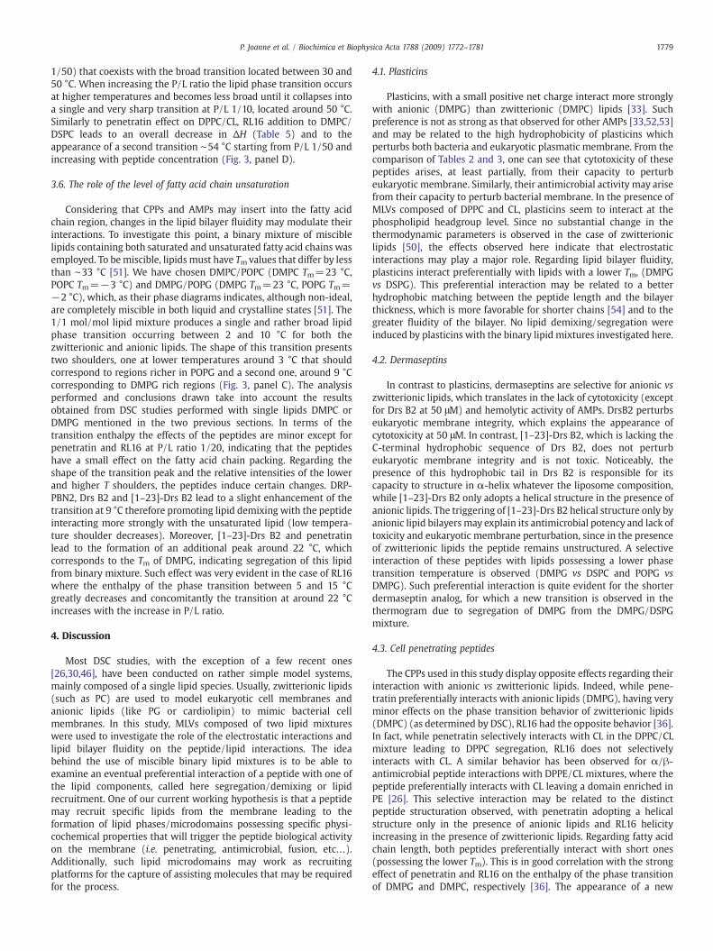
Fig. 3. High-sensitivity DSC heating scans illustrating the effect of the addition of DRP-PBN2behavior of vesicles composed of DMPG/CL (7/3 mol/mol) (panel A), DMPG/DSPG (1/1 mo1 mol/mol) (panel D) and DMPC/POPC (1/1 mol/mol) (panel E). Thermodynamic parametlipid mixtures only (DMPC/DSPC and DMPC/POPC) and not with the anionic mixtures (DMthe opposite was true for all the other peptides investigated in this study. The gel to fluid phabetter readability, the zero value in Cp was incrementally added by 0.5, 1, 0.5, 0.4 and 0.6 U
peptides to interact and or disrupt the lipid acyl chain packing providinginsight into their interaction mechanism. In the case of binary lipidmixtures, a careful selection of the lipids used in the mixture has to beconsidered because: i) not all the lipids are miscible, ii) the type ofinformation obtained regarding peptide/lipid interactions will necessa-rily depend on the appropriate choice of the lipid partners. This will befurther discussed in the following sections of the manuscript.
3.4.1. The role of electrostatic forces on the peptide/lipid interactionConsidering that the peptides investigated here are quite rich in
positively charged amino acids (Table 1) the investigation of theirinteraction with negatively charged membranes is mandatory. More-over, their antibacterial activities suggest that they may have apreferential interaction with charged lipids such as PG or cardiolipinthat are especially present in the outer leaflet of bacterial membranesand often used in studies involving lipid model systems. Indeed, mostof the peptides under study lead to a greater perturbation in the phasetransition of DMPG vs DMPC, with the exception of RL16, suggesting astronger interaction and perturbation of the fatty acid chain packing(data not shown) [33,36].
Herein, to get further insight into the role of electrostatics in thepeptide/lipid interaction and possible alteration in terms of lateral
, DRP-PD36KF, Drs B2, [1–23]-Drs B2, RL16 and penetratin on the thermotropic phasel/mol) (panel B), DMPG/POPG (1/1 mol/mol) (panel C) at a P/L 1/20, DMPC/DSPC (1/ers are given in Tables 4, 5 and 6. The interaction of RL16 was studied with zwitterionicPG/DSPG and DMPG/POPG) because the lipid interacts strongly with the former lipids,se transition temperatures (Tm) of the lipids are indicated to aid interpretation [61]. Forfor panels A, B, C, D, and E, respectively.

Table 5Thermodynamic parameters obtained by DSC for the interaction of the dermaseptin andplasticin analogs as well as penetratinwith DMPG/DSPG and for the interaction of RL16with DMPC/DSPC (1/1 mol/mol).
Peptides P/L Tm (°C) ΔH (Kcal/mol)
No peptide DMPG/DSPG No peptide 29.8/44 15.6DRP-PBN2 1/100 29.3/43.7 14.5
1/20 27.9/44.7 11.8DRP-PD36KF 1/100 29.9/44.5 15
1/20 31.2/45.3 11.5Drs B2 1/200 30/43.7 14.8
1/20 b/44.6 12.5[1–23]-Drs B2 1/200 29/43.2 12.9
1/20 b/45 12.8Penetratin 1/100 29/43.6 12.2
1/20 b/51.8 6.2No peptide DMPC/DSPC 31/44.7 15.6RL16 1/100 31/44.7 17.8
1/20 30.8/45/54.3a 10
= No changes observed.a Phase transition temperature of a second transition peak.b The phase transition disappears.
1778 P. Joanne et al. / Biochimica et Biophysica Acta 1788 (2009) 1772–1781
lipid organization (lipid segregation), we have used binary lipidmixtures composed of DPPC and cardiolipin (7/3mol/mol). While therole of electrostatics could be studied by just comparing the effect ofthe peptide on zwitterionic vs anionic lipid membranes (single lipidcomponent), binary lipid mixtures containing lipids with sufficientlydifferent phase transition temperatures are mandatory to evaluate thepossible lipid recruitment induced by the peptide. Cardiolipin frombovine heart possess a high content of unsaturated fatty acids (whichexplains the lack of phase transition above 0 °C), in addition to theelectrostatic effect investigated with the DPPC/CL there is also theimpact of fatty acid chain unsaturation. The mixture of lipids oftenresults in a reduction and broadening of the phase transition due tolipid intercalation, as a consequence of that, a relatively high lipidconcentration was used (always with fixed P/L molar ratios) allowinga reasonable DSC signal to be observed. DPPC lipid phase transitionoccurs at 41 °C and CL has no observable transition between 0 and100 °C. As it can be seen in panel A of Fig. 3, this lipidmixture producesa single (although broad) transition confirming good lipid miscibility(below and above the phase transition region). Very distinct effects onthe thermogram were induced by the presence of the peptides withreductions or increases in the enthalpy of the transition accompaniedor not by changes in Tm. In view of the broad transition observed, anaccurate determination of Tm and ΔH is not straightforward. However,significant effects, such as the appearance of additional transitionpeaks at higher temperatures deserve consideration.
Both plasticin and dermaseptin analogs lead to changes in theenthalpy and Tm of the DPPC/CL transition suggesting that theyinteract with these lipids. Lack of change in the general form of thetransition peak suggests the absence of lipid lateral redistribution inthe presence of these AMPs. Similarly, RL16 leads to no change in theform of the transition but produces a strong decrease in the enthalpyof the transition, particularly at a P/L ratio of 1/20, which indicates aperturbation of the packing of the acyl chains. The most noticeableresult was obtained with penetratin, with the appearance of a secondtransition around 41 °C, starting at P/L ratio of 1/50 and increasing inmagnitude with peptide concentration. One should note thatpenetratin does not lead to any change in the phase transitiontemperature of DMPC [36]. This strongly suggests that penetratinpreferentially binds and recruits CL, leaving a domain highly enrichedin DPPC, which gives rise to the sharp transition at 41 °C.
3.5. The role of the fatty acid chain length
To investigate the potential role the lipid bilayer thickness plays inthe lipid/peptide interactions, DSC studies were performed on binarylipid mixtures composed of lipids with distinct fatty acid chain length.We have chosen DMPC/DSPC or DMPG/DSPG (1/1 mol/mol) lipids
Table 4Thermodynamic parameters obtained by DSC for the interaction of the peptides withDPPC/CL (7/3 mol/mol).
Peptides P/L Tm (°C) ΔH (Kcal/mol)
Lipid only No peptide 28 5.2DRP-PBN2 1/100 29 8.6
1/20 29.5 8.3DRP-PD36KF 1/100 29 8.6
1/20 29.5 7.8Drs B2 1/200 28 5.2
1/20 29 6[1–23]-Drs B2 1/200 28 5.2
1/20 29.5 5.2RL16 1/100 29.5 6.8
1/20 29.5 2.4Penetratin 1/100 29 4.2
1/20 29/41.8a 2.7
= No changes observed.a Phase transition temperature of a second transition peak.
with chain lengths differing by four carbons, the maximum chainlength difference, that can be used before observing complete lateralphase separation [48,49]. This mixture is not ideal with a broadtransition located between about 30 and 50 °C composed of twopeaks, one towards 30 °C that corresponds to a DMPG rich region andanother one around 44 °C that corresponds to a DSPG rich region(Fig. 3, panel B). Anionic lipid binary mixtures (DMPG/DSPG) wereemployed for all the peptides due to their preferential electrostaticinteraction with this lipid (determined by previous studies [33,35]),except for RL16 that interacts preferentially with zwitterionic lipids[36]. Moreover, RL16 has a tendency to induce aggregation in thepresence of anionic lipids [36]. Both plasticin and dermaseptinanalogs change the overall shape of the transition with a strongdecrease in the area under the peak located on the left side of thebroad transition, which has been attributed to the DMPG rich region(see explanation above) (Fig. 3, panel B). For the dermaseptinanalogs, the transition peak located on the left side of the broadtransition was completely abolished at P/L ratio of 1/20. Studiesperformed in our laboratories have shown that both plasticins anddermaseptins lead to changes in both the Tm and the ΔH of DMPG[35,50]. The results suggest a stronger interaction of the peptideswith the shorter lipid DMPG without observable DSPG segregation.Penetratin also leads to a decrease in the enthalpy of the transitionand the appearance of a second transition at ∼52 °C (starting at P/L
Table 6Thermodynamic parameters obtained by DSC for the interaction of the dermaseptin andplasticin analogs as well as penetratin with DMPG/POPG (1/1 mol/mol) and for theinteraction of RL16 with DMPC/POPC (1/1 mol/mol).
Peptides P/L Tm (°C) ΔH (Kcal/mol)
No peptide DMPG/POPG 3.2/9.3 5.4DRP-PBN2 1/100 4.1/9.1 6.7
1/20 6.0/9.8 4.3DRP-PD36KF 1/100 4.6/10.5 6
1/20 ND 4.4Drs B2 1/100 4.5/10.9 5.2
1/20 4.6/10.6 4.5[1–23]-Drs B2 1/100 6.3/9.8 6
1/20 5/10.8/22a 4.4Penetratin 1/100 2.8/9.3 5.2
1/20 3.1/9.5/22.2a 4No peptide DMPC/POPC 6.8/12.1 5.2RL16 1/100 6.8/12.1 4.8
1/20 6.9/12.3/23.8a 2.6
= No changes observed.a Phase transition temperature of a second transition peak.
1779P. Joanne et al. / Biochimica et Biophysica Acta 1788 (2009) 1772–1781
1/50) that coexists with the broad transition located between 30 and50 °C. When increasing the P/L ratio the lipid phase transition occursat higher temperatures and becomes less broad until it collapses intoa single and very sharp transition at P/L 1/10, located around 50 °C.Similarly to penetratin effect on DPPC/CL, RL16 addition to DMPC/DSPC leads to an overall decrease in ΔH (Table 5) and to theappearance of a second transition ∼54 °C starting from P/L 1/50 andincreasing with peptide concentration (Fig. 3, panel D).
3.6. The role of the level of fatty acid chain unsaturation
Considering that CPPs and AMPs may insert into the fatty acidchain region, changes in the lipid bilayer fluidity may modulate theirinteractions. To investigate this point, a binary mixture of misciblelipids containing both saturated and unsaturated fatty acid chains wasemployed. To bemiscible, lipids must have Tmvalues that differ by lessthan ∼33 °C [51]. We have chosen DMPC/POPC (DMPC Tm=23 °C,POPC Tm=−3 °C) and DMPG/POPG (DMPG Tm=23 °C, POPG Tm=−2 °C), which, as their phase diagrams indicates, although non-ideal,are completely miscible in both liquid and crystalline states [51]. The1/1 mol/mol lipid mixture produces a single and rather broad lipidphase transition occurring between 2 and 10 °C for both thezwitterionic and anionic lipids. The shape of this transition presentstwo shoulders, one at lower temperatures around 3 °C that shouldcorrespond to regions richer in POPG and a second one, around 9 °Ccorresponding to DMPG rich regions (Fig. 3, panel C). The analysisperformed and conclusions drawn take into account the resultsobtained from DSC studies performed with single lipids DMPC orDMPG mentioned in the two previous sections. In terms of thetransition enthalpy the effects of the peptides are minor except forpenetratin and RL16 at P/L ratio 1/20, indicating that the peptideshave a small effect on the fatty acid chain packing. Regarding theshape of the transition peak and the relative intensities of the lowerand higher T shoulders, the peptides induce certain changes. DRP-PBN2, Drs B2 and [1–23]-Drs B2 lead to a slight enhancement of thetransition at 9 °C therefore promoting lipid demixing with the peptideinteracting more strongly with the unsaturated lipid (low tempera-ture shoulder decreases). Moreover, [1–23]-Drs B2 and penetratinlead to the formation of an additional peak around 22 °C, whichcorresponds to the Tm of DMPG, indicating segregation of this lipidfrom binary mixture. Such effect was very evident in the case of RL16where the enthalpy of the phase transition between 5 and 15 °Cgreatly decreases and concomitantly the transition at around 22 °Cincreases with the increase in P/L ratio.
4. Discussion
Most DSC studies, with the exception of a few recent ones[26,30,46], have been conducted on rather simple model systems,mainly composed of a single lipid species. Usually, zwitterionic lipids(such as PC) are used to model eukaryotic cell membranes andanionic lipids (like PG or cardiolipin) to mimic bacterial cellmembranes. In this study, MLVs composed of two lipid mixtureswere used to investigate the role of the electrostatic interactions andlipid bilayer fluidity on the peptide/lipid interactions. The ideabehind the use of miscible binary lipid mixtures is to be able toexamine an eventual preferential interaction of a peptide with one ofthe lipid components, called here segregation/demixing or lipidrecruitment. One of our current working hypothesis is that a peptidemay recruit specific lipids from the membrane leading to theformation of lipid phases/microdomains possessing specific physi-cochemical properties that will trigger the peptide biological activityon the membrane (i.e. penetrating, antimicrobial, fusion, etc…).Additionally, such lipid microdomains may work as recruitingplatforms for the capture of assisting molecules that may be requiredfor the process.
4.1. Plasticins
Plasticins, with a small positive net charge interact more stronglywith anionic (DMPG) than zwitterionic (DMPC) lipids [33]. Suchpreference is not as strong as that observed for other AMPs [33,52,53]and may be related to the high hydrophobicity of plasticins whichperturbs both bacteria and eukaryotic plasmatic membrane. From thecomparison of Tables 2 and 3, one can see that cytotoxicity of thesepeptides arises, at least partially, from their capacity to perturbeukaryotic membrane. Similarly, their antimicrobial activity may arisefrom their capacity to perturb bacterial membrane. In the presence ofMLVs composed of DPPC and CL, plasticins seem to interact at thephospholipid headgroup level. Since no substantial change in thethermodynamic parameters is observed in the case of zwitterioniclipids [50], the effects observed here indicate that electrostaticinteractions may play a major role. Regarding lipid bilayer fluidity,plasticins interact preferentially with lipids with a lower Tm, (DMPGvs DSPG). This preferential interaction may be related to a betterhydrophobic matching between the peptide length and the bilayerthickness, which is more favorable for shorter chains [54] and to thegreater fluidity of the bilayer. No lipid demixing/segregation wereinduced by plasticins with the binary lipid mixtures investigated here.
4.2. Dermaseptins
In contrast to plasticins, dermaseptins are selective for anionic vszwitterionic lipids, which translates in the lack of cytotoxicity (exceptfor Drs B2 at 50 μM) and hemolytic activity of AMPs. DrsB2 perturbseukaryotic membrane integrity, which explains the appearance ofcytotoxicity at 50 μM. In contrast, [1–23]-Drs B2, which is lacking theC-terminal hydrophobic sequence of Drs B2, does not perturbeukaryotic membrane integrity and is not toxic. Noticeably, thepresence of this hydrophobic tail in Drs B2 is responsible for itscapacity to structure in α-helix whatever the liposome composition,while [1–23]-Drs B2 only adopts a helical structure in the presence ofanionic lipids. The triggering of [1–23]-Drs B2 helical structure only byanionic lipid bilayers may explain its antimicrobial potency and lack oftoxicity and eukaryotic membrane perturbation, since in the presenceof zwitterionic lipids the peptide remains unstructured. A selectiveinteraction of these peptides with lipids possessing a lower phasetransition temperature is observed (DMPG vs DSPC and POPG vsDMPG). Such preferential interaction is quite evident for the shorterdermaseptin analog, for which a new transition is observed in thethermogram due to segregation of DMPG from the DMPG/DSPGmixture.
4.3. Cell penetrating peptides
The CPPs used in this study display opposite effects regarding theirinteraction with anionic vs zwitterionic lipids. Indeed, while pene-tratin preferentially interacts with anionic lipids (DMPG), having veryminor effects on the phase transition behavior of zwitterionic lipids(DMPC) (as determined by DSC), RL16 had the opposite behavior [36].In fact, while penetratin selectively interacts with CL in the DPPC/CLmixture leading to DPPC segregation, RL16 does not selectivelyinteracts with CL. A similar behavior has been observed for α/β-antimicrobial peptide interactions with DPPE/CL mixtures, where thepeptide preferentially interacts with CL leaving a domain enriched inPE [26]. This selective interaction may be related to the distinctpeptide structuration observed, with penetratin adopting a helicalstructure only in the presence of anionic lipids and RL16 helicityincreasing in the presence of zwitterionic lipids. Regarding fatty acidchain length, both peptides preferentially interact with short ones(possessing the lower Tm). This is in good correlation with the strongeffect of penetratin and RL16 on the enthalpy of the phase transitionof DMPG and DMPC, respectively [36]. The appearance of a new

1780 P. Joanne et al. / Biochimica et Biophysica Acta 1788 (2009) 1772–1781
transition at ∼54 °C starting from P/L 1/50, which is accompanied bya decrease in the magnitude of the original broad transition observedin absence of peptide, is attributed to the segregation of DSPC fromthe binary lipid mixture (DSPC Tm is ∼53 °C). Such lipid segregationis a consequence of the preferential interaction of the peptide withshorter aliphatic chains (DMPC). The observation that RL16 selec-tively interacts with shorter chains may be related to the formationof small vesicles (carpet type mechanism, proposed to occur for thispeptide [36]) occurring more specifically with shorter than longerchains. Additionally, both peptides recruit unsaturated fatty acidchains, this effect being more noticeable in the case of RL16. Apreferential interaction of the peptides with the unsaturated lipidmay be a consequence of their higher fluidity arising from theirlower phase transition temperature, correlating well with thefavorable interaction with shorter vs longer fatty acid chains. Thefact that such behavior was more substantially marked for RL16 thanpenetratin may be related with the different level of insertions intothe lipid core region. In the presence of CL, both electrostatic andfatty acid chain unsaturation may play a role, but the peptideimmobilization on the surface due to electrostatic interactions, mayexplain the lack of preferential interaction with the fatty acid chain(unsaturated vs saturated).
All the peptides investigated, except RL16, interact with the lipids,at a first level through electrostatic interactions established betweenthe positively charged amino acids of the peptides and the negativelycharged lipid headgroups. The present data clearly demonstrate thatboth CPPs and AMPs induce specific lipid segregation of binary lipidmixtures possessing lipid components with markedly different phasetransition temperatures. It is noteworthy to mention that whenselective lipid interactions were observed, the lipid being recruitedfrom the binary mixture always corresponded to the one having thelowest phase transition temperature. A certain hierarchy in thedemixing/segregationwas also observed: the unsaturated lipids werepreferentially recruited first, before lipids with shorter fatty acidchains, again this probably being a consequence of the lower Tm ofunsaturated lipids. Such preference may be related to the higherfluidity (reduced fatty acid chain packing) of these lipids, which betteradapt to lipid rearrangements taking place during the cell transloca-tion and/or perturbation of the lipid bilayer. Regarding, the prefer-ential interaction of the AMPs with anionic lipids (CL, data shownhere; DMPG vs DMPC, data not shown) and their better antimicrobialactivity for E. coli vs S. aureus, interesting observations can be drawn,taking into account the membrane lipid composition of E. coli (80% PE,15% PG and 5% CL) and S. aureus (58% PG and 42% CL, no PE). Thus, abetter antimicrobial activity would exist in bilayers where bothanionic and zwitterionic lipids exist, rather than in bilayers solelycomposed of anionic lipids. A similar analysis has been reported byEpand et al. for the interaction of several antimicrobial peptides withbinary lipid mixtures [26,30–32] and their antimicrobial activities in alarge range of bacterial strains. Based both on the antimicrobialactivities for the different bacterial strains and from lipid domainformation observed by DSC, the authors concluded that one of thecauses for the differences in species selectivity with AMPs may comefrom clustering of anionic lipids and their segregation in domains. Incontrast to dermaseptins and plasticins, RL16 which has no prefer-ential interaction with CL nor DMPG, has low antimicrobial activity inE. coli (50 μM) but higher with S. aureus (9 μM).
Finally, the formation of lateral membrane heterogeneity inbilayers may have important consequences for the uptake andantimicrobial activities of CPPs and AMPs, respectively. Therefore,the lipid domains formed may present the necessary physicochemicalproperties to promote CPPs passage across the bilayer (by a non-endocytic pathway) via either the formation of inverted micelles[36–38,55,56] or other lipid structures such as tubes [40,57] or therecruitment of the necessary auxiliary proteins (e.g. clathrins,caveolins, etc…) and other molecules needed for their uptake
(when an endocytic pathway is taken). Moreover, peptide-inducedsegregation of lipids into phases/domains can destabilize themembrane by introducing phase boundary defects between lipiddomains [58,59]. Such boundary defects, offering reduced surfacetension, may facilitate peptide penetration and/or leakage [24,25,60].
Acknowledgments
This work was supported by the Agence Nationale pour laRecherche (ANR-Prob DOM). Authors wish to thank Dr. ChristophePiesse from the “Ingénierie des protéines et synthèse peptidique”service of the “Institut de Biologie intégrative”, IFR 83, for the synthesisof dermaseptin and plasticin analogs.
References
[1] C. Palm, S. Netzereab, M. Hallbrink, Quantitatively determined uptake of cell-penetrating peptides in non-mammalian cells with an evaluation of degradationand antimicrobial effects, Peptides 27 (2006) 1710–1716.
[2] S.T. Henriques, M.N. Melo, M.A. Castanho, Cell-penetrating peptides andantimicrobial peptides: how different are they? Biochem. J. 399 (2006) 1–7.
[3] M. B.-H, J.S. Wadia, S.F. Dowdy, Protein Transport, 1st Ed.CRC Press, New York,2002.
[4] A.D. Frankel, C.O. Pabo, Cellular uptake of the tat protein from humanimmunodeficiency virus, Cell 55 (1988) 1189–1193.
[5] A. Joliot, C. Pernelle, H. Deagostini-Bazin, A. Prochiantz, Antennapedia homeoboxpeptide regulates neural morphogenesis, Proc. Natl. Acad Sci. U. S. A. 88 (1991)1864–1868.
[6] B. Lebleu, I. Robbins, L. Bastide, E. Vives, J.E. Gee, Pharmacokinetics ofoligonucleotides in cell culture, Ciba Found. Symp. 209 (1997) 47–54 discussion54–49.
[7] D. Derossi, A.H. Joliot, G. Chassaing, A. Prochiantz, The third helix of theAntennapedia homeodomain translocates through biological membranes, J. Biol.Chem. 269 (1994) 10444–10450.
[8] M. Magzoub, A. Graslund, Cell-penetrating peptides: from inception to applica-tion, Q. Rev. Biophys. 37 (2004) 147–195.
[9] M. Zorko, U. Langel, Cell-penetrating peptides: mechanism and kinetics of cargodelivery, Adv. Drug Deliv. Rev. 57 (2005) 529–545.
[10] M. Zasloff, Antimicrobial peptides of multicellular organisms, Nature 415 (2002)389–395.
[11] R.E. Hancock, Peptide antibiotics, Lancet 349 (1997) 418–422.[12] R.M. Epand, H.J. Vogel, Diversity of antimicrobial peptides and their mechanisms
of action, Biochim. Biophys. Acta 1462 (1999) 11–28.[13] Y. Shai, Mode of action of membrane active antimicrobial peptides, Biopolymers
66 (2002) 236–248.[14] K. Matsuzaki, Magainins as paradigm for the mode of action of pore forming
polypeptides, Biochim. Biophys. Acta 1376 (1998) 391–400.[15] R.M. Epand, Y. Shai, J.P. Segrest, G.M. Anantharamaiah, Mechanisms for the
modulation of membrane bilayer properties by amphipathic helical peptides,Biopolymers 37 (1995) 319–338.
[16] B. Bechinger, K. Lohner, Detergent-like actions of linear amphipathic cationicantimicrobial peptides, Biochim. Biophys. Acta 1758 (2006) 1529–1539.
[17] P. Nicolas, C. El Amri, The dermaseptin superfamily: a gene-based combinatoriallibrary of antimicrobial peptides, Biochim. Biophys. Acta (2008).
[18] C. El Amri, P. Nicolas, Plasticins: membrane-damaging peptides with ‘chameleon-like’ properties, Cell Mol. Life Sci. 65 (2008) 895–909.
[19] D. Marsh, CRC Hanbook of Lipid Bilayers, CRC Press, Boca Raton, 1990.[20] L.O. Bergelson, GawrischK. , FerrettiA. , BlumenthalR. , Special issue on domain
organization in biological membranes, Mol. Membr. Biol. 12 (1995) 1–162.[21] O.P. Karlsson, M. Rytomaa, A. Dahlqvist, P.K. Kinnunen, A. Wieslander, Correlation
between bilayer lipid dynamics and activity of the diglucosyldiacylglycerolsynthase from Acholeplasma laidlawii membranes, Biochemistry 35 (1996)10094–10102.
[22] T. Honger, K. Jorgensen, R.L. Biltonen, O.G. Mouritsen, Systematic relationshipbetween phospholipase A2 activity and dynamic lipid bilayer microheterogeneity,Biochemistry 35 (1996) 9003–9006.
[23] P. Verkade, K. Simons, Robert Feulgen Lecture 1997. Lipid microdomains andmembrane trafficking in mammalian cells, Histochem. Cell Biol. 108 (1997)211–220.
[24] F. Jean-Francois, S. Castano, B. Desbat, B. Odaert, M. Roux, M.H. Metz-Boutigue, E.J.Dufourc, Aggregation of cateslytin beta-sheets on negatively charged lipidspromotes rigid membrane domains. A new mode of action for antimicrobialpeptides? Biochemistry 47 (2008) 6394–6402.
[25] R.M. Epand, R.F. Epand, Lipid domains in bacterial membranes and the action ofantimicrobial agents, Biochim. Biophys. Acta 1788 (2009) 289–294.
[26] R.F. Epand, M.A. Schmitt, S.H. Gellman, R.M. Epand, Role of membrane lipids in themechanism of bacterial species selective toxicity by two alpha/beta-antimicrobialpeptides, Biochim. Biophys. Acta 1758 (2006) 1343–1350.
[27] R.N. McElhaney, The use of differential scanning calorimetry and differentialthermal analysis in studies of model and biological membranes, Chem. Phys.Lipids 30 (1982) 229–259.

1781P. Joanne et al. / Biochimica et Biophysica Acta 1788 (2009) 1772–1781
[28] T. Wieprecht, O. Apostolov, M. Beyermann, J. Seelig, Membrane binding and poreformation of the antibacterial peptide PGLa: thermodynamic and mechanisticaspects, Biochemistry 39 (2000) 442–452.
[29] E.J. Prenner, R.N. Lewis, L.H. Kondejewski, R.S. Hodges, R.N. McElhaney,Differential scanning calorimetric study of the effect of the antimicrobial peptidegramicidin S on the thermotropic phase behavior of phosphatidylcholine,phosphatidylethanolamine and phosphatidylglycerol lipid bilayer membranes,Biochim. Biophys. Acta 1417 (1999) 211–223.
[30] R.F. Epand, B.P. Mowery, S.E. Lee, S.S. Stahl, R.I. Lehrer, S.H. Gellman, R.M. Epand,Dual mechanism of bacterial lethality for a cationic sequence–randomcopolymer that mimics host–defense antimicrobial peptides, J. Mol. Biol. 379(2008) 38–50.
[31] J.E. Shaw, R.F. Epand, J.C. Hsu, G.C. Mo, R.M. Epand, C.M. Yip, Cationic peptide-induced remodelling of model membranes: direct visualization by in situ atomicforce microscopy, J. Struct. Biol. 162 (2008) 121–138.
[32] R.M. Epand, S. Rotem, A. Mor, B. Berno, R.F. Epand, Bacterial membranes aspredictors of antimicrobial potency, J. Am. Chem. Soc. 130 (2008) 14346–14352.
[33] C. El Amri, C. Lacombe, K. Zimmerman, A. Ladram, M. Amiche, P. Nicolas, F.Bruston, The plasticins: membrane adsorption, lipid disorders, and biologicalactivity, Biochemistry 45 (2006) 14285–14297.
[34] O. Lequin, F. Bruston, O. Convert, G. Chassaing, P. Nicolas, Helical structure ofdermaseptin B2 in a membrane-mimetic environment, Biochemistry 42 (2003)10311–10323.
[35] C. Galanth, F. Abbassi, O. Lequin, J. Ayala-Sanmartin, A. Ladram, P. Nicolas, M.Amiche, Mechanism of antibacterial action of dermaseptin B2: interplay betweenhelix–hinge–helix structure and membrane curvature strain, Biochemistry 48(2009) 313–327.
[36] I.D. Alves, N. Goasdoue, I. Correia, S. Aubry, C. Galanth, S. Sagan, S. Lavielle, G.Chassaing, Membrane interaction and perturbation mechanisms induced by twocationic cell penetrating peptides with distinct charge distribution, Biochim.Biophys. Acta (2008).
[37] D. Derossi, S. Calvet, A. Trembleau, A. Brunissen, G. Chassaing, A. Prochiantz, Cellinternalization of the third helix of the Antennapedia homeodomain is receptor-independent, J. Biol. Chem. 271 (1996) 18188–18193.
[38] D. Derossi, G. Chassaing, A. Prochiantz, Trojan peptides: the penetratin system forintracellular delivery, Trends Cell Biol. 8 (1998) 84–87.
[39] F. Bruston, C. Lacombe, K. Zimmermann, C. Piesse, P. Nicolas, C. El Amri, Structuralmalleability of plasticins: preorganized conformations in solution and relevancefor antimicrobial activity, Biopolymers 86 (2007) 42–56.
[40] A. Lamaziere, F. Burlina, C. Wolf, G. Chassaing, G. Trugnan, J. Ayala-Sanmartin,Non-metabolic membrane tubulation and permeability induced by bioactivepeptides, PLoS ONE 2 (2007) e201.
[41] S. El-Andaloussi, P. Jarver, H.J. Johansson, U. Langel, Cargo-dependent cytotoxicityand delivery efficacy of cell-penetrating peptides: a comparative study, Biochem. J.407 (2007) 285–292.
[42] H. Duclohier, Bilayer lipid composition modulates the activity of dermaseptins,polycationic antimicrobial peptides, Eur. Biophys. J. 35 (2006) 401–409.
[43] H. Tournois, B. de Kruijff, Polymorphic phospholipid phase transitions as tools tounderstand peptide–lipid interactions, Chem. Phys. Lipids 57 (1991) 327–340.
[44] K. Lohner, E.J. Prenner, Differential scanning calorimetry and X-ray diffractionstudies of the specificity of the interaction of antimicrobial peptides withmembrane-mimetic systems, Biochim. Biophys. Acta 1462 (1999) 141–156.
[45] S.E. Blondelle, K. Lohner, M. Aguilar, Lipid-induced conformation and lipid-
binding properties of cytolytic and antimicrobial peptides: determination andbiological specificity, Biochim. Biophys. Acta 1462 (1999) 89–108.
[46] L.M. Contreras, R.F. de Almeida, J. Villalain, A. Fedorov, M. Prieto, Interaction ofalpha-melanocyte stimulating hormone with binary phospholipid membranes:structural changes and relevanceof phase behavior, Biophys. J. 80 (2001)2273–2283.
[47] W. Jing, H.N. Hunter, J. Hagel, H.J. Vogel, The structure of the antimicrobial peptideAc-RRWWRF-NH2 bound to micelles and its interactions with phospholipidbilayers, J. Pept. Res. 61 (2003) 219–229.
[48] C. Leidy, W.F. Wolkers, K. Jorgensen, O.G. Mouritsen, J.H. Crowe, Lateralorganization and domain formation in a two-component lipid membrane system,Biophys. J. 80 (2001) 1819–1828.
[49] E.J. Shimshick, H.M. McConnell, Lateral phase separations in binary mixtures ofcholesterol and phospholipids, Biochem. Biophys. Res. Commun. 53 (1973) 446–451.
[50] P. Joanne, Falord, M., Lacombe, C., Castano, S., Desbat, B., Chesneau, O., Desmadril,M., Nicaise, M., Auvynet, C., Nicolas, P., Msadek, T., El Amri C., Antistaphyloccocalactivities of plasticins: relationships between resistance and membrane interac-tions., Biochem. J. Submitted (2009).
[51] W. Curatolo, B. Sears, L.J. Neuringer, A calorimetry and deuterium NMR study ofmixed model membranes of 1-palmitoyl-2-oleylphosphatidylcholine and satu-rated phosphatidylcholines, Biochim. Biophys. Acta 817 (1985) 261–270.
[52] J.P. Powers, A. Tan, A. Ramamoorthy, R.E. Hancock, Solution structure andinteraction of the antimicrobial polyphemusins with lipid membranes, Biochem-istry 44 (2005) 15504–15513.
[53] G.W. Seto, S. Marwaha, D.M. Kobewka, R.N. Lewis, F. Separovic, R.N. McElhaney,Interactions of the Australian tree frog antimicrobial peptides aurein 1.2, citropin1.1 and maculatin 1.1 with lipid model membranes: differential scanningcalorimetric and Fourier transform infrared spectroscopic studies, Biochim.Biophys. Acta (2007).
[54] A.N. Ridder, W. van de Hoef, J. Stam, A. Kuhn, B. de Kruijff, J.A. Killian, Importanceof hydrophobic matching for spontaneous insertion of a single-spanningmembrane protein, Biochemistry 41 (2002) 4946–4952.
[55] S. Afonin, A. Frey, S. Bayerl, D. Fischer, P. Wadhwani, S. Weinkauf, A.S. Ulrich, Thecell-penetrating peptide TAT(48–60) induces a non-lamellar phase in DMPCmembranes, Chemphyschem. 7 (2006) 2134–2142.
[56] A. Lamaziere, C. Wolf, O. Lambert, G. Chassaing, G. Trugnan, J. Ayala-Sanmartin,The homeodomain derived peptide Penetratin induces curvature of fluidmembrane domains, PLoS ONE 3 (2008) e1938.
[57] W. Romer, L. Berland, V. Chambon, K. Gaus, B. Windschiegl, D. Tenza, M.R. Aly, V.Fraisier, J.C. Florent, D. Perrais, C. Lamaze, G. Raposo, C. Steinem, P. Sens, P.Bassereau, L. Johannes, Shiga toxin induces tubular membrane invaginations forits uptake into cells, Nature 450 (2007) 670–675.
[58] S.G. Clerc, T.E. Thompson, Permeability of dimyristoyl phosphatidylcholine/dipalmitoyl phosphatidylcholine bilayer membranes with coexisting gel andliquid–crystalline phases, Biophys. J. 68 (1995) 2333–2341.
[59] H. Komatsu, S. Okada, Increased permeability of phase-separated liposomalmembranes with mixtures of ethanol-induced interdigitated and non-interdigi-tated structures, Biochim. Biophys. Acta 1237 (1995) 169–175.
[60] F.M. Menger, V.A. Seredyuk, M.V. Kitaeva, A.A. Yaroslavov, N.S. Melik-Nubarov,Migration of poly-L-lysine through a lipid bilayer, J. Am. Chem. Soc. 125 (2003)2846–2847.
[61] C. Huang, S. Li, Calorimetric and molecular mechanics studies of the thermotropicphase behavior of membrane phospholipids, Biochim. Biophys. Acta 1422 (1999)273–307.
Related Documents