ف عا ن م ا ي ه م ل ع ال ى م س م ا ل عا ع م ا ة ي ر ح ل ى ا ن ها ح ن م ت لا ة ي ر ح ل ا ة ي ر ي خ ل ر ا) لي ا ات, ئ ي ه و م ن ي ت) ب ي ة ي ر ح ل ا ة ي ك وز رة ح دماء) ب .ً دعا) ئ مE ن ك ئ ل ة قJ ش ع, ة وا) ي رق حي ة وا ي ف س م غ ن ل واحد، وا م ع ى علY ك م ما ن ه ا زِ ّ ك ز م. م ه ل ل ا عJ ش ت ل، و ق ع ل دح ا ق ن ال، و) زح ل ل ا ق ص ت ها نl ا فE ن ح م ل ا) بً زعا ذ ق ص ت لا ص. ي خ ز ق ف خ م ل ، وا وت ف م م لJ ش ا ق ل ر، وا صف ل ط عا ل ، واE ن س ح ي ماٍ , رئ م ل ا ك ة م ن ف

Lipid Metabolism.ppt
Nov 09, 2014
داتا الدكتور جميل من غير تعديل
كان فاضلى slide 2 واخلصها لكن البرنامج هنج وحذف كل حاجة
حاليا عندى depression دعواتكم بقى والسلام عليكم
كان فاضلى slide 2 واخلصها لكن البرنامج هنج وحذف كل حاجة
حاليا عندى depression دعواتكم بقى والسلام عليكم
Welcome message from author
This document is posted to help you gain knowledge. Please leave a comment to let me know what you think about it! Share it to your friends and learn new things together.
Transcript
ن ف معا اسمى ال علمهيا نى امع عالمالحرية
تمنحها ال الخيرية الحرية البر هيئاتينمو نبت وزكية الحرية حرة بدماء
به واحترق فيه وانغمس واحد، عمل على اهتمامك رك,ز
. . مبدعا لتكن وأعشقه
،العقل وتقدح الرجال، تصقل فإنها بالمحن ذرعا. تضق ال
. الهمم وتشعل
والفاشل صفر، والعاطل يحسن، ما امرئٍ كل قيمة. رخيص والمخفق ممقوت،
2
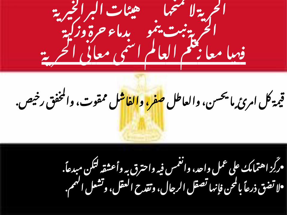
1- كربوهيدرات او سكريات
�وَن� و �ع!ِق�ل ي " $ِق�و!ٍم ل �ة% آلي ��َك َذ�ل ف�ي �َن* ِإ %ا ًن �َح�َس ًق%ا و�ر�ْز! ا �ر% ك �س �!ُه م�ًن �*ِخ�ُذ�وَن �َّت َت �اِب� �ْع!ًن و�اَأل *ِخ�يِل� الًن ات� ��م�ر َث م�ن
سورة - 67آية - 16الًنحِل
2- البروَتيًنات او اللحوٍم
ر� �ِّش$ و�ب �م! ه�د�اك م�ا ْع�ل�ى �*ُه الل وا �$ر �ب �ك �َّت ل �م! �ك ل ه�ا �ِخ*ر �س ��ُذ�ل�َك ك �م! م�ًنك *ِق!و�ى الَّت ��ُه �ال �ًن ي �ك�ن و�ل ِد�م�اُؤ�ه�ا و�ال �ح�وم�ه�ا ل �*ُه الل ��اَل �ًن ي �ن ل
��ين ًن !م�ح!َس� سورة - ال 37آية - 22الحج
الدهوَن-3
��وَن �د!ه�ًن ف�ي ��د!ه�ن َت �و! ل سورة - و�ِدFوا 9آية - 68الِقلم
�وَن� أ• مFد!ه�ًن �م �نَّت أ !ح�د�يِث� ال �ه�ُذ�ا سورة - ف�ب 81آية - 56الواًقعة
• ��ين �ل ك $آْل! ل !ٍغ" و�ِص�ب �الدFه!ن� ب ��ُت �ًنب َت ��اَء !ًن ي �س ُط�ور� م�ن �ُج ��ِخ!ر َت ة% �َج�ر �سورة - و�َش 20آية - 23المؤمًنوَن
الِقرأَن فى الحيوية للكيمياَء الرئيَسية االًقَساٍمالكريم
3
Lipid Metabolism
By Dr. Gamil Abdalla
4
Course outlines
1. Lipid digestion and absorption and their errors
2. Fate of absorbed lipids
3. Lipolysis and Lipogenesis
4. Fatty acid oxidation and synthesis
5. Ketogenesis and ketolysis
6. Cholesterol and Lipiprotein metabolis
7. Fatty liver
5
6
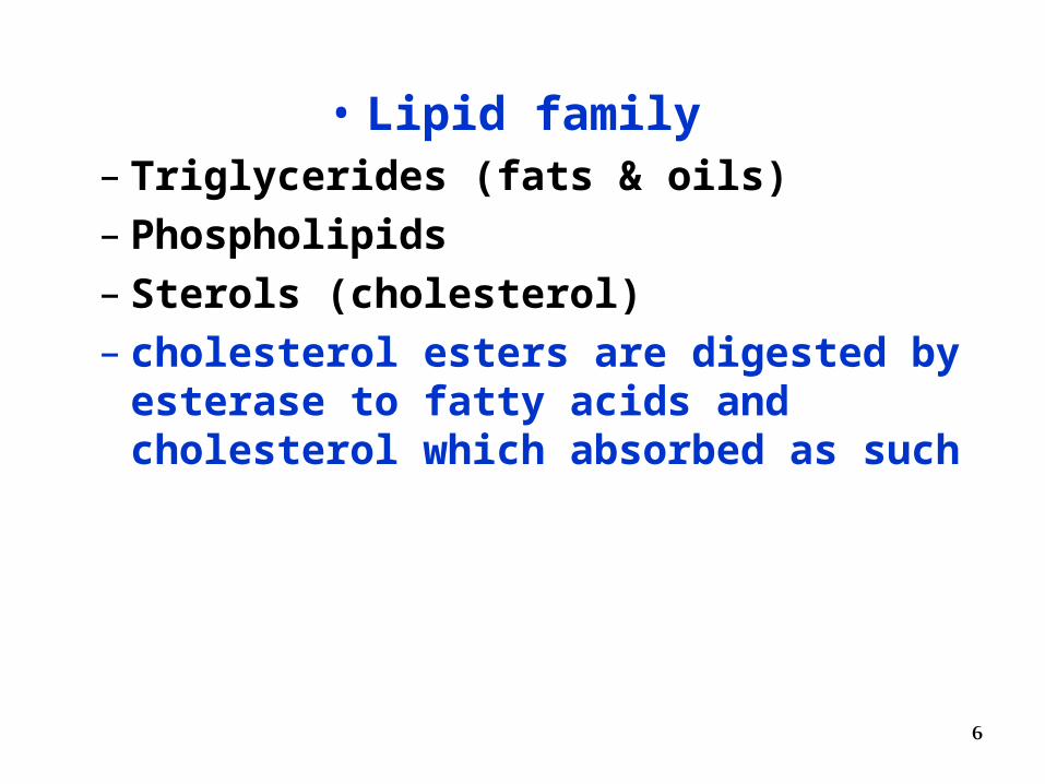
• Lipid family– Triglycerides (fats & oils)– Phospholipids– Sterols (cholesterol)– cholesterol esters are digested by esterase
to fatty acids and cholesterol which absorbed as such
7
Importance of lipids
• As storage and transport form of metabolic fuel
• To keep the body temperature
• Source for essential FA and oil soluble vitamins
• To protect important organs
8
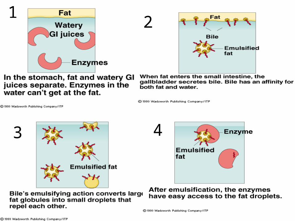
Lipid Digestion
• Challenges– Lipids are not water soluble– Triglycerides too large to be absorbed
• Digestive solution– Triglycerides mix with bile and pancreatic
secretions• Emulsification and digestion
9
Digestion of Triglycerides
• Minor digestion of triacylglycerols in
1. Mouth by lingual lipase
2. Stomach by gastric lipase (in infants only).
• Major digestion of all lipids in the lumen of
the duodenum/ jejunum by Pancreatic lipases
10
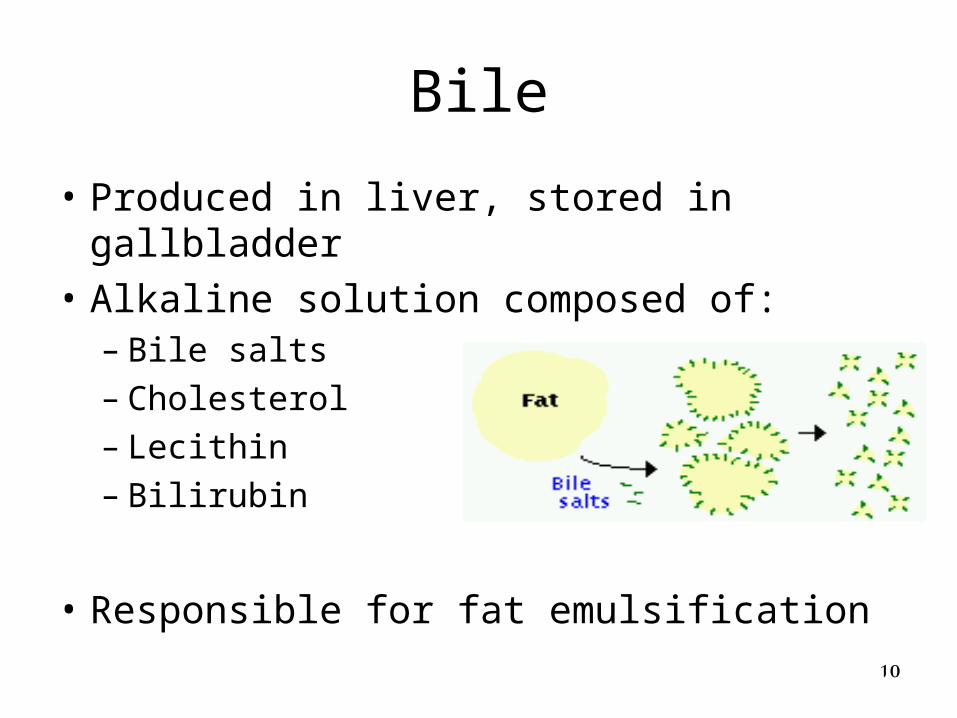
Bile
• Produced in liver, stored in gallbladder
• Alkaline solution composed of:– Bile salts– Cholesterol– Lecithin– Bilirubin
• Responsible for fat emulsification
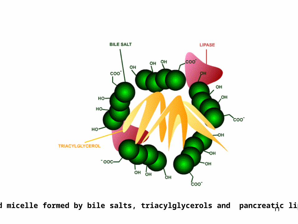
11Mixed micelle formed by bile salts, triacylglycerols and pancreatic lipase.
12
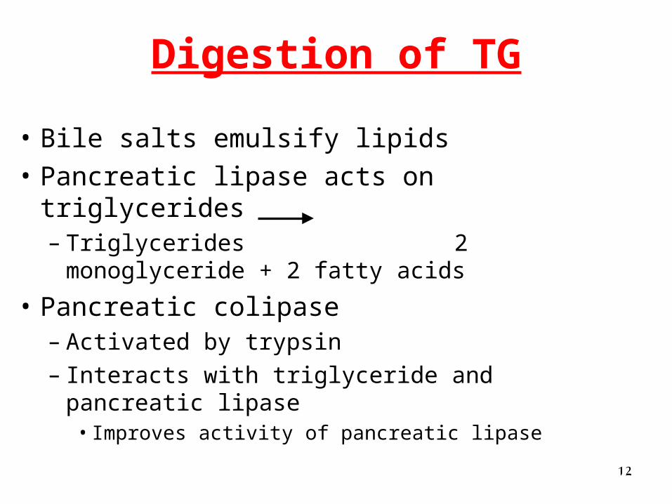
Digestion of TG
• Bile salts emulsify lipids
• Pancreatic lipase acts on triglycerides– Triglycerides 2 monoglyceride + 2
fatty acids
• Pancreatic colipase– Activated by trypsin– Interacts with triglyceride and pancreatic lipase
• Improves activity of pancreatic lipase
13
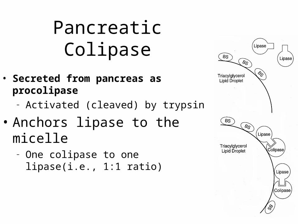
Pancreatic Colipase
• Secreted from pancreas as procolipase– Activated (cleaved) by trypsin
• Anchors lipase to the micelle– One colipase to one lipase(i.e., 1:1 ratio)
14
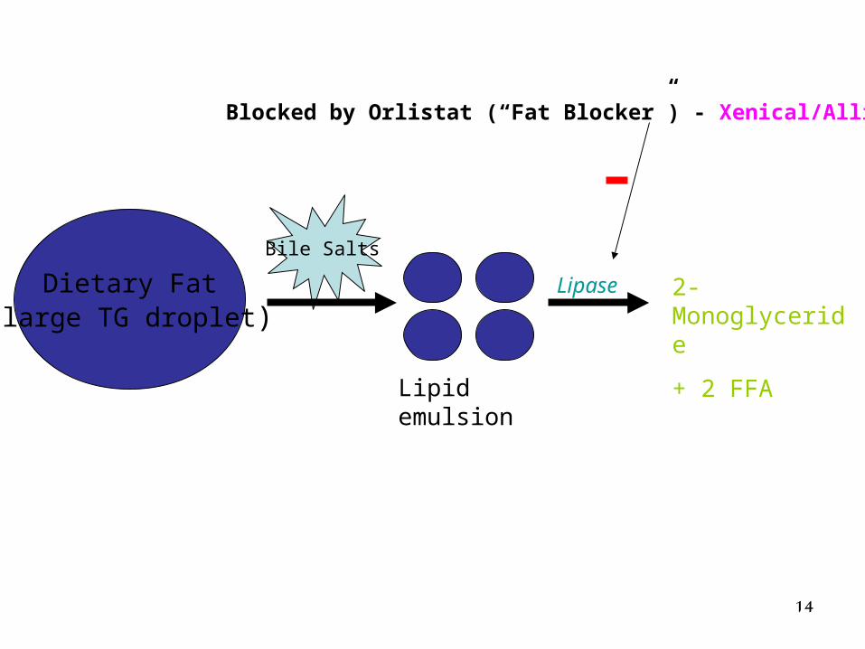
Dietary Fat(large TG droplet)
Bile Salts
Lipid emulsion
Lipase 2-Monoglyceride
+ 2 FFA
Blocked by Orlistat (“Fat Blocker”) - Xenical/Alli
15
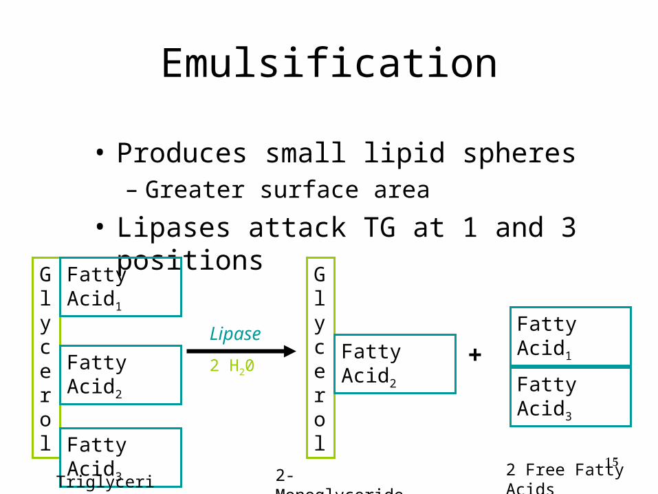
Emulsification
• Produces small lipid spheres– Greater surface area
• Lipases attack TG at 1 and 3 positions
Glycerol
Fatty Acid1
Fatty Acid2
Fatty Acid3
Lipase
Glycerol
Fatty Acid3
Fatty Acid1Fatty
Acid2
Triglyceride 2-Monoglyceride
+
2 Free Fatty Acids
2 H20
16
12
3 4
17
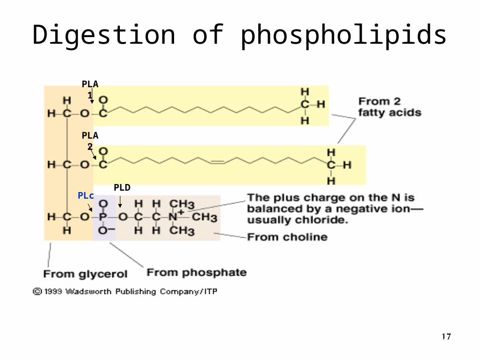
Digestion of phospholipids
PLA2
PLA1
PLcPLD
18
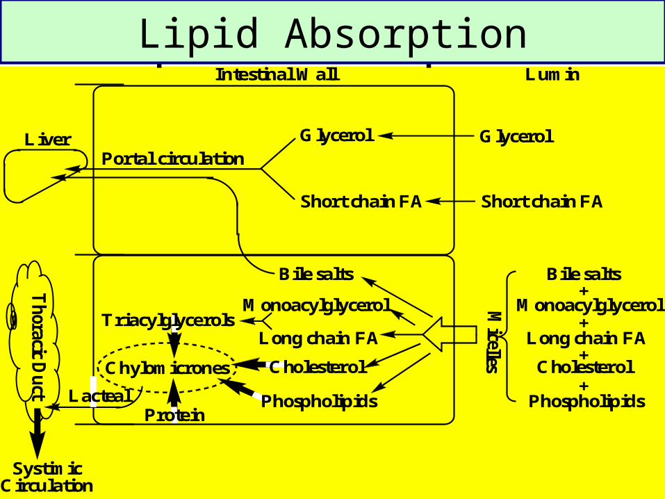
Intestinal Wall
Liver GlycerolPortal circulation
Short chain FA
Lumin
Glycerol
Short chain FA
Triacylglycerols
Chylomicrones
Protein
SystimicCirculation
Th
oracicD
uct
Bile salts
Monoacylglycerol
Long chain FA
Cholesterol
Phospholipids
+
+
+
+
Bile salts
Monoacylglycerol
Long chain FA
Cholesterol
PhospholipidsLacteal
Lipid AbsorptionLipid Absorption
19
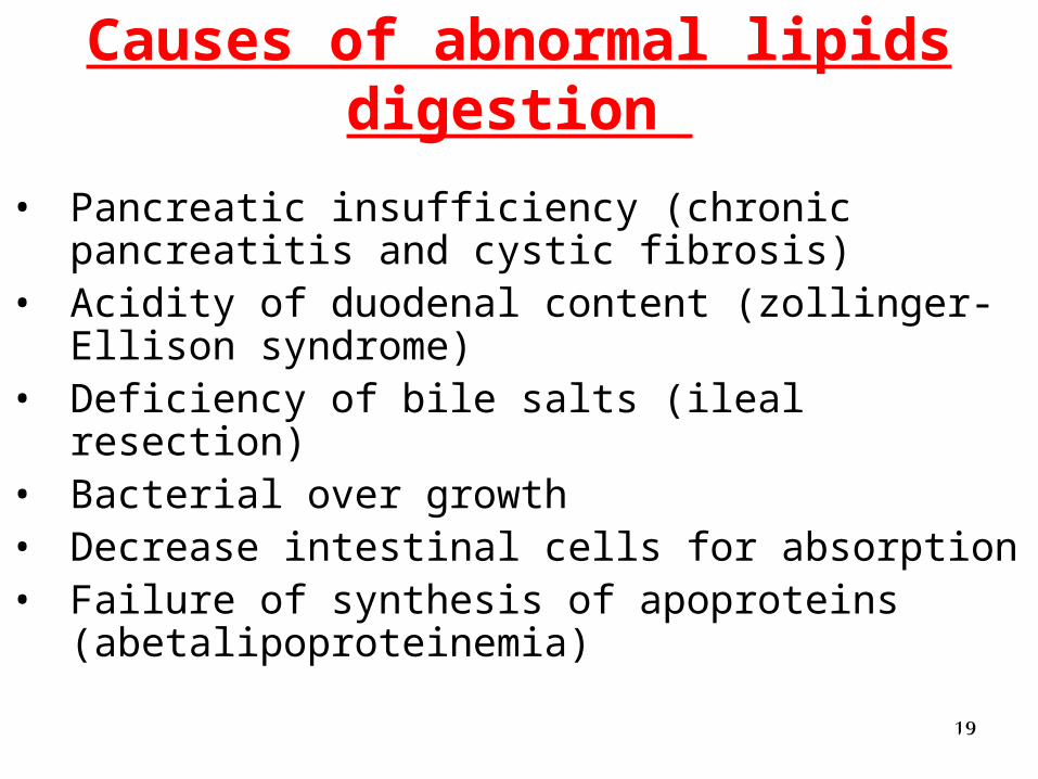
• Pancreatic insufficiency (chronic pancreatitis and cystic fibrosis)
• Acidity of duodenal content (zollinger-Ellison syndrome)
• Deficiency of bile salts (ileal resection)• Bacterial over growth • Decrease intestinal cells for absorption • Failure of synthesis of apoproteins
(abetalipoproteinemia)
Causes of abnormal lipids digestion
20
Errors of lipid digestion and absorption
1. Steatorhoea
stool fat > 5 gm per day
2. Chyluria (milky urine)
Abnormal connections between lymphatics and urinary system.
21
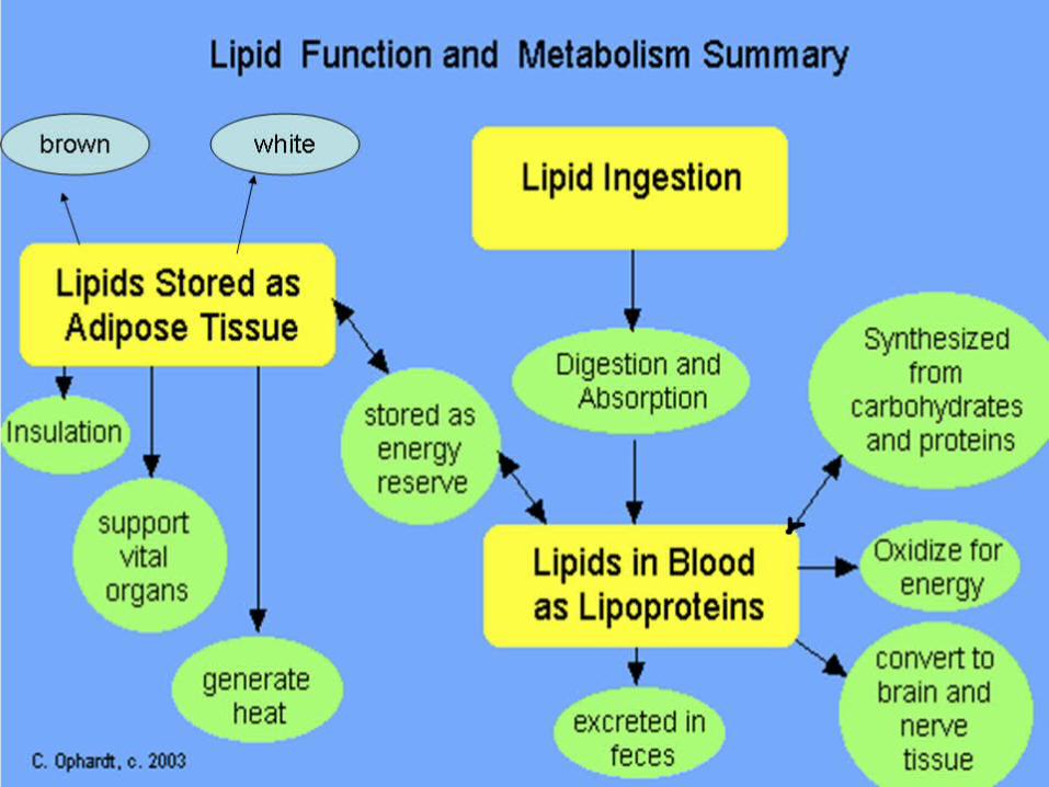
Fate of absorbed lipids
1. Storage
2. Energy production
3. Gluconeogenesis
4. Synthesis of
• Cellular structures
• Biological active compounds eg. Prostaglandins

22
Body lipids
Tissue lipidsAdipose tissue lipid
(depot fat)
WhiteUnder skin &breast Around vital organs
BrownMitochonderiaCytochromes
BVs
23
Fat Storage
• Mainly as triacylglycerols (triglycerides) in adipose cells
• Constitute 84% of stored energy
24
25
MITOCHONDRION
cell membrane
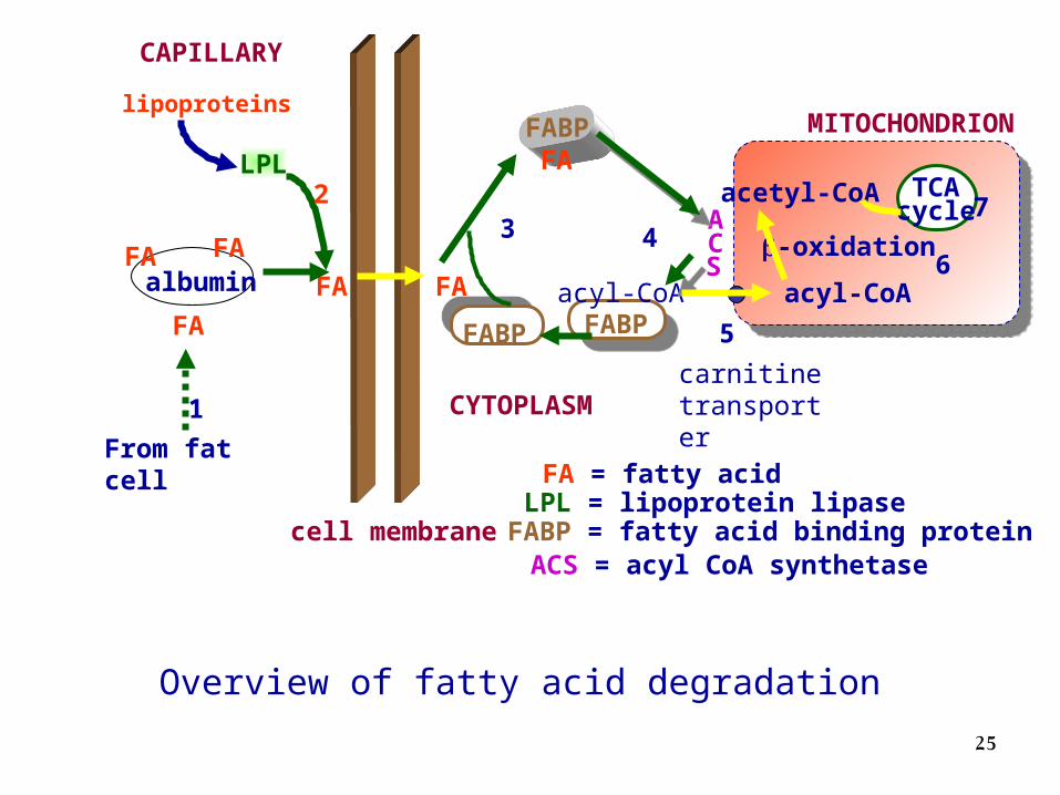
FA = fatty acidLPL = lipoprotein lipaseFABP = fatty acid binding protein
ACS
FABP
FABPFA
3
FABPacyl-CoA
4
CYTOPLASM
CAPILLARY
LPL
lipoproteins
2
FAFA
1
albuminFA FA
FA
From fat cell
carnitinetransporter
acyl-CoA
5
Overview of fatty acid degradation
ACS = acyl CoA synthetase
acetyl-CoA TCAcycle
-oxidation6
7
2626
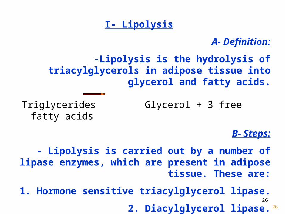
I- Lipolysis
A- Definition:
-Lipolysis is the hydrolysis of triacylglycerols in adipose tissue into glycerol and fatty acids.
Triglycerides Glycerol + 3 free fatty acids
B- Steps:
- Lipolysis is carried out by a number of lipase enzymes, which are present in adipose tissue. These are:
1. Hormone sensitive triacylglycerol lipase.
2. Diacylglycerol lipase.
3. Monoacylglycerol lipase.
2727
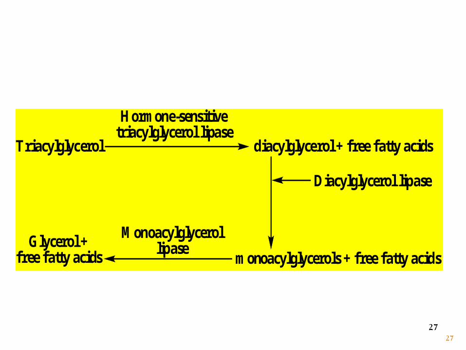
Triacylglycerol
Hormone-sensitivetriacylglycerol lipase
diacylglycerol + free fatty acids
Diacylglycerol lipase
Glycerol +free fatty acids
Monoacylglycerollipase
monoacylglycerols + free fatty acids
28
Lipolysis products
Fatty acids Glycerol
29
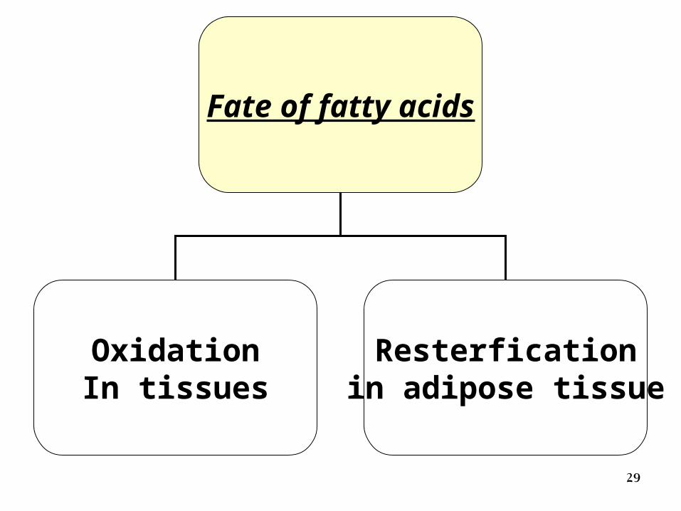
Fate of fatty acids
OxidationIn tissues
Resterficationin adipose tissue
30
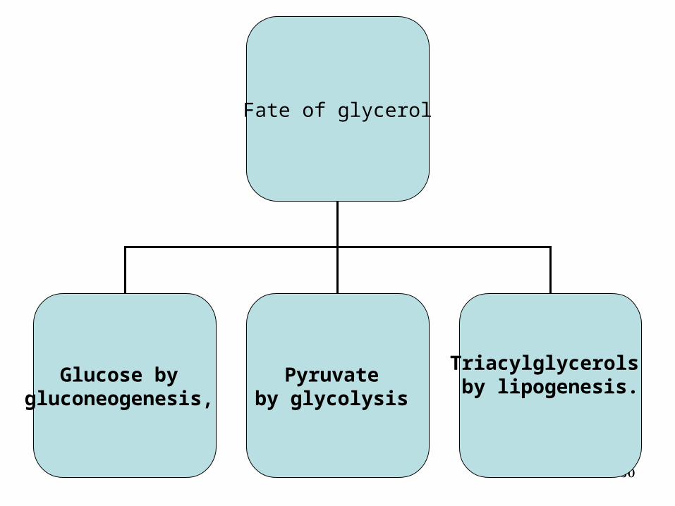
Fate of glycerol
Glucose by gluconeogenesis,
Pyruvate by glycolysis
Triacylglycerols by lipogenesis.
31
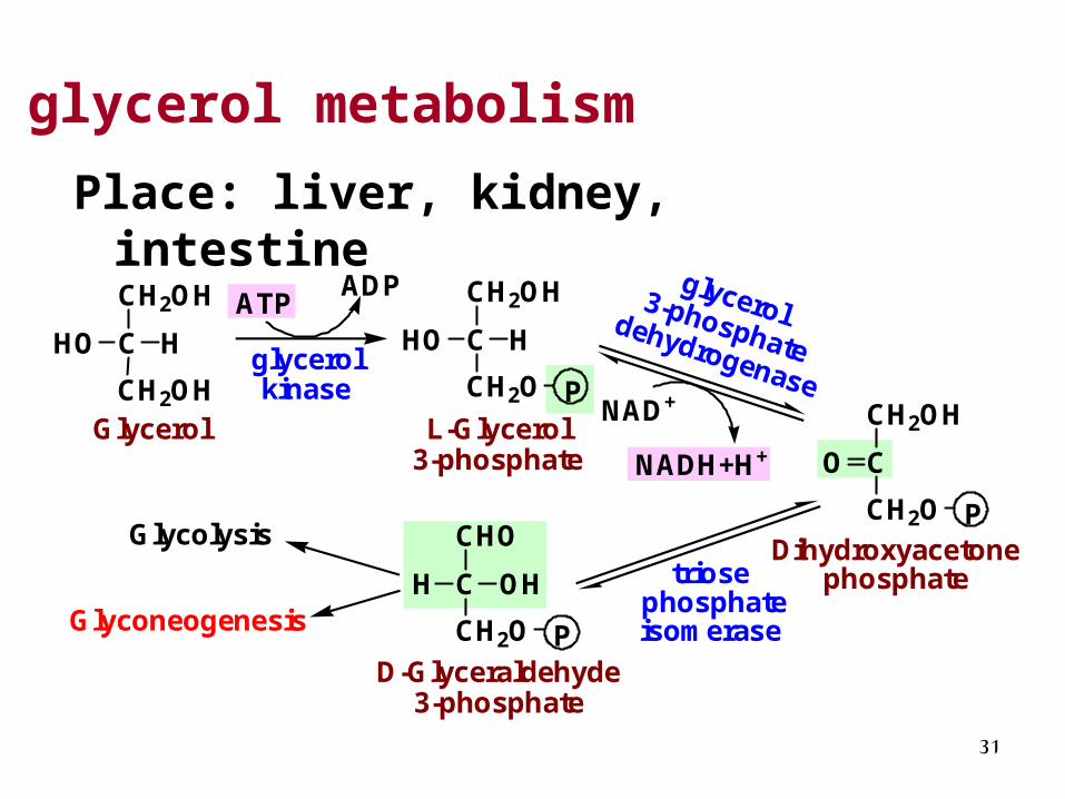
glycerol metabolism
Place: liver, kidney, intestine
CH2OH
CHO H
CH2OHglycerolkinase
CH2OH
CHO H
CH2O PGlycerol L-Glycerol
3-phosphate
ATPADP
CH2OH
CO
CH2O PDihydroxyacetone
phosphate
D-Glyceraldehyde 3-phosphate
Glycolysis
NAD+
NADH+H+
CHO
CH
CH2O P
OH triose phosphateisomerase
glycerol 3-phosphatedehydrogenase
Glyconeogenesis
32
Note
• In muscle cells and adipocytes, the activity of glycerol kinase is low, so these tissues cannot use glycerol as fuel.
3333
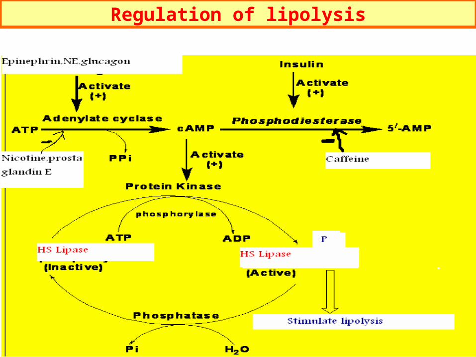
Regulation of lipolysis
34
Hormone sensitive lipase (HSL)
• TG lipase is the rate-limiting enzyme in the TG degradation in adipose tissue. It is also named HSL because it is regulated by some hormones.
35
Effect of hormones on lipolysis
• Lipolytic Hormones:
epinephrine
norepinephrine
adrenocorticotropic hormone (ACTH)
thyroid stimulating hormone (TSH)
Glucagon etc.
• Antilipolytic Hormones: insulin
36
Causes of excessive lipolysis:
• - In conditions where the need for energy is increased e.g.:
• 1- Starvation.
• 2- Diabetes mellitus.
• 3- Low carbohydrate diet.
37
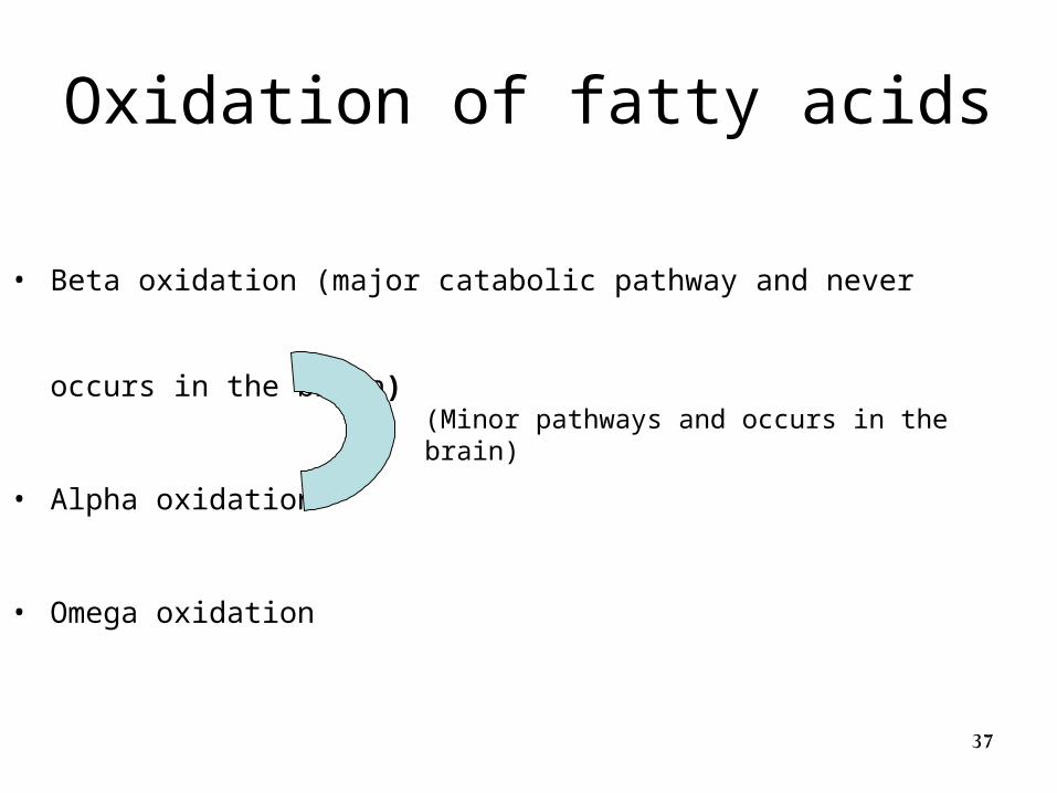
Oxidation of fatty acids
• Beta oxidation (major catabolic pathway and never occurs in the brain)
• Alpha oxidation
• Omega oxidation
(Minor pathways and occurs in the brain)
38
Beta Oxidation
• Cleavage of fatty acids to acetate in tissues
• Occurs in the mitochondria of liver, kidney and heart
• Never occur in the brain•Fatty acid catabolism can be subdivided into 3 stages.
39
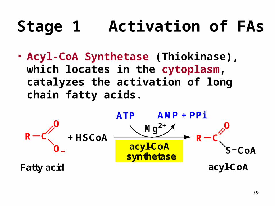
Stage 1 Activation of FAs
• Acyl-CoA Synthetase (Thiokinase), which locates in the cytoplasm, catalyzes the activation of long chain fatty acids.
+ HSCoAacyl-CoA
synthetase
Mg2+ATP AMP + PPi
R CO
O
Fatty acid
R CO
S CoA
acyl-CoA
40
Key points of FA activation
1. Irreversible
2. Consume 2 ~P
3. Site: cytosol
41
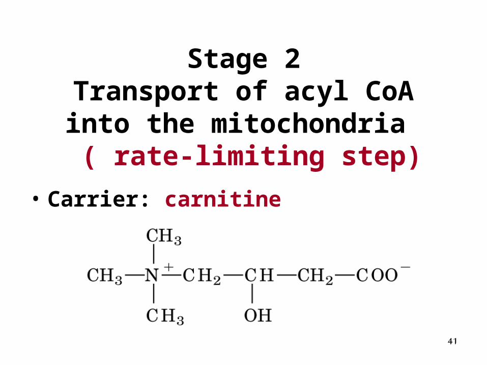
Stage 2Transport of acyl CoA into the
mitochondria ( rate-limiting step)
• Carrier: carnitine
42
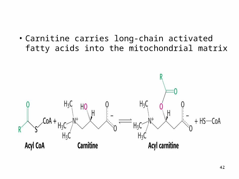
• Carnitine carries long-chain activated fatty acids into the mitochondrial matrix
43
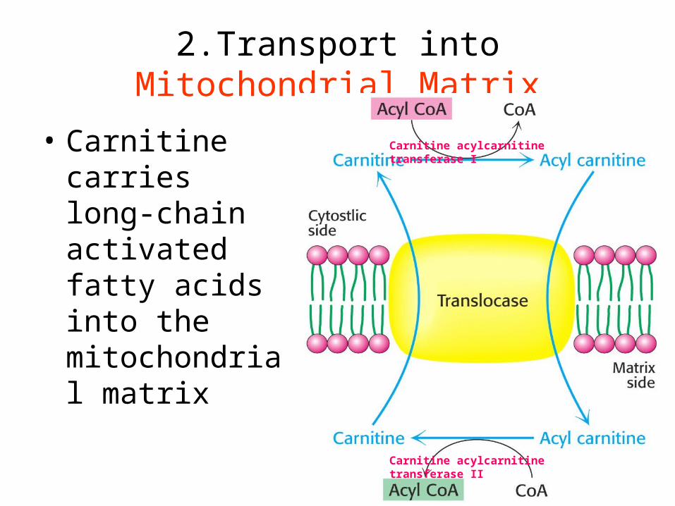
• Carnitine carries long-chain activated fatty acids into the mitochondrial matrix
2.Transport into Mitochondrial Matrix
Carnitine acylcarnitine transferase I
Carnitine acylcarnitine transferase II
44
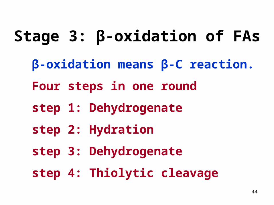
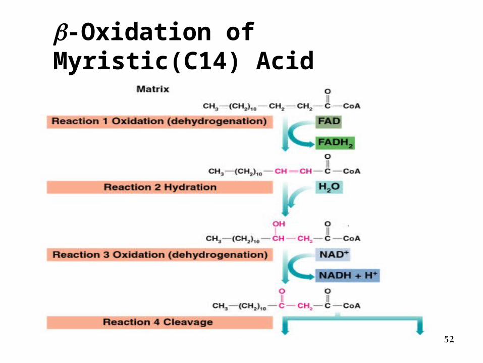
Stage 3: β-oxidation of FAs
β-oxidation means β-C reaction.
Four steps in one round
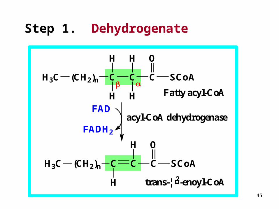
step 1: Dehydrogenate
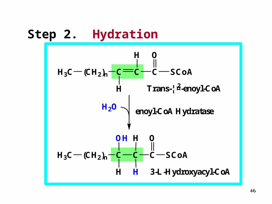
step 2: Hydration
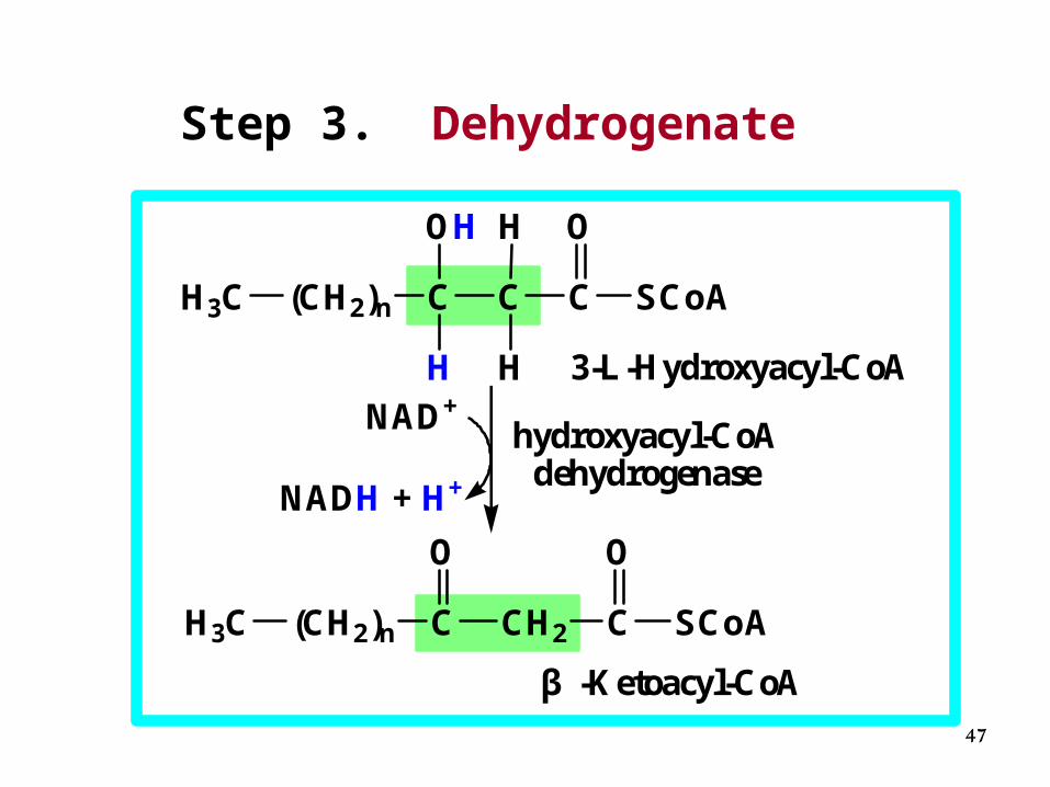
step 3: Dehydrogenate
step 4: Thiolytic cleavage
45
Step 1. Dehydrogenate
H3C (CH2)n C C C SCoA
H
H
H
H O
H3C (CH2)n C C C SCoA
H
H O
FADH2
FAD
Fatty acyl-CoA
acyl-CoA dehydrogenase
trans-¦¤2-enoyl-CoA
46
Step 2. Hydration
H3C (CH2)n C C C SCoA
H
H O
H3C (CH2)n C C C SCoA
H
O
H2O
OH
Trans-¦¤2-enoyl-CoA
H
H 3-L-Hydroxyacyl-CoA
enoyl-CoA Hydratase
47
Step 3. Dehydrogenate
H3C (CH2)n C C C SCoA
H
OOH
H3C (CH2)n C CH2 C SCoA
OO
NADH + H+
NAD+
H
H 3-L-Hydroxyacyl-CoA
hydroxyacyl-CoAdehydrogenase
β -Ketoacyl-CoA

48
Step 4. Thiolytic cleavage
H3C (CH2)n C CH2 C SCoA
OO
CH3 C SCoA
O
H3C (CH2)n C SCoA +
O
HSCoAβ -Ketoacyl-CoA
Acetyl-CoAFatty acyl-CoA(2C shorter)
β -Ketothiolase
49
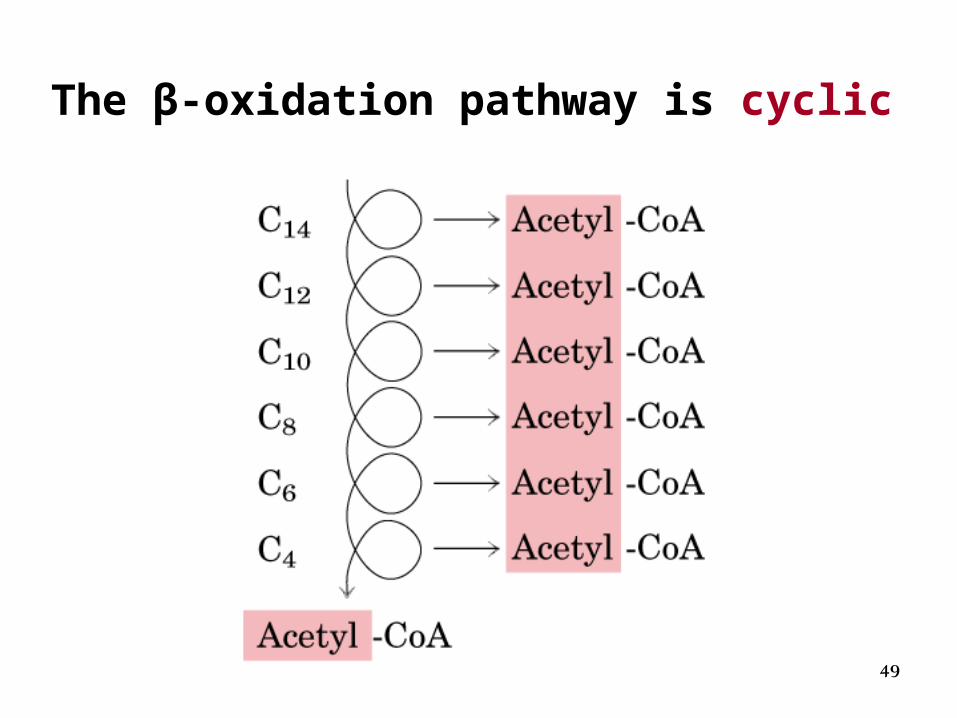
The β-oxidation pathway is cyclic
50
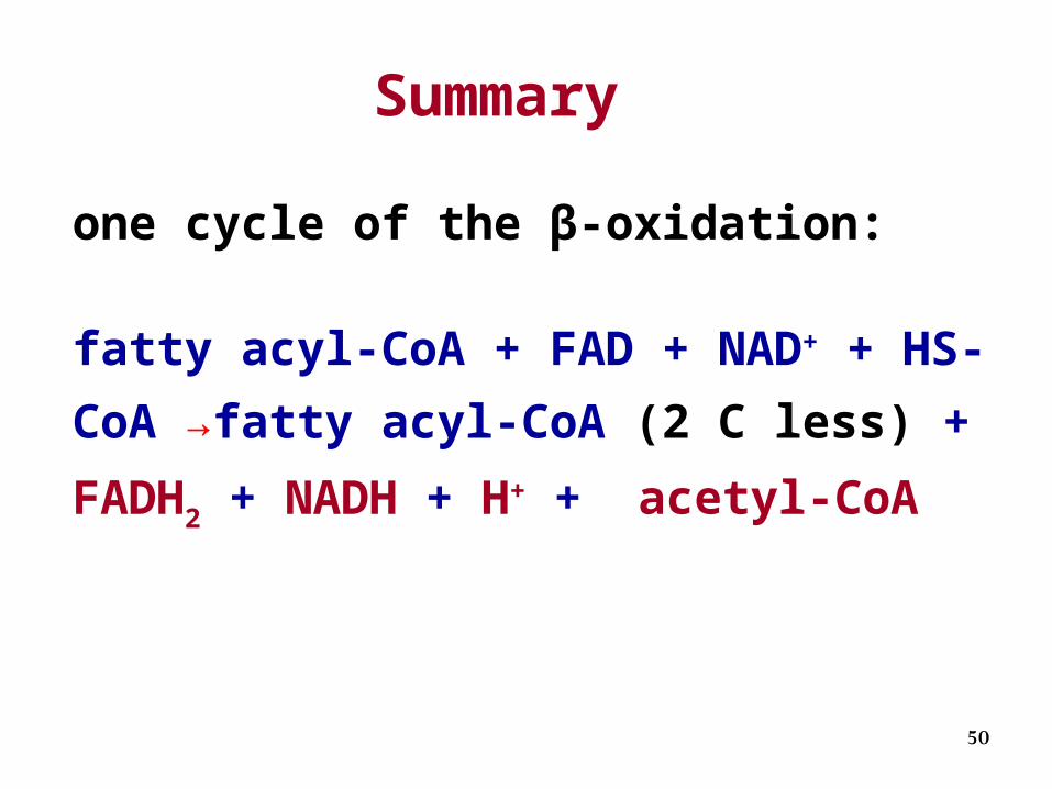
one cycle of the β-oxidation:
fatty acyl-CoA + FAD + NAD+ + HS-CoA
→fatty acyl-CoA (2 C less) + FADH2 +
NADH + H+ + acetyl-CoA
Summary
51
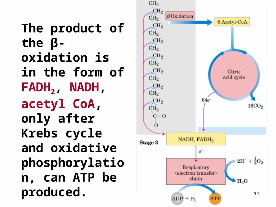
The product of the β-oxidation is in the form of FADH2, NADH, acetyl CoA, only after Krebs cycle and oxidative phosphorylation, can ATP be produced.
52
-Oxidation of Myristic(C14) Acid
53
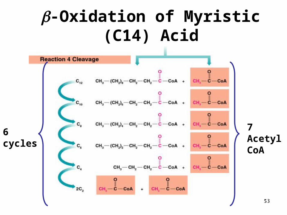
-Oxidation of Myristic (C14) Acid
7 Acetyl CoA
6 cycles
54
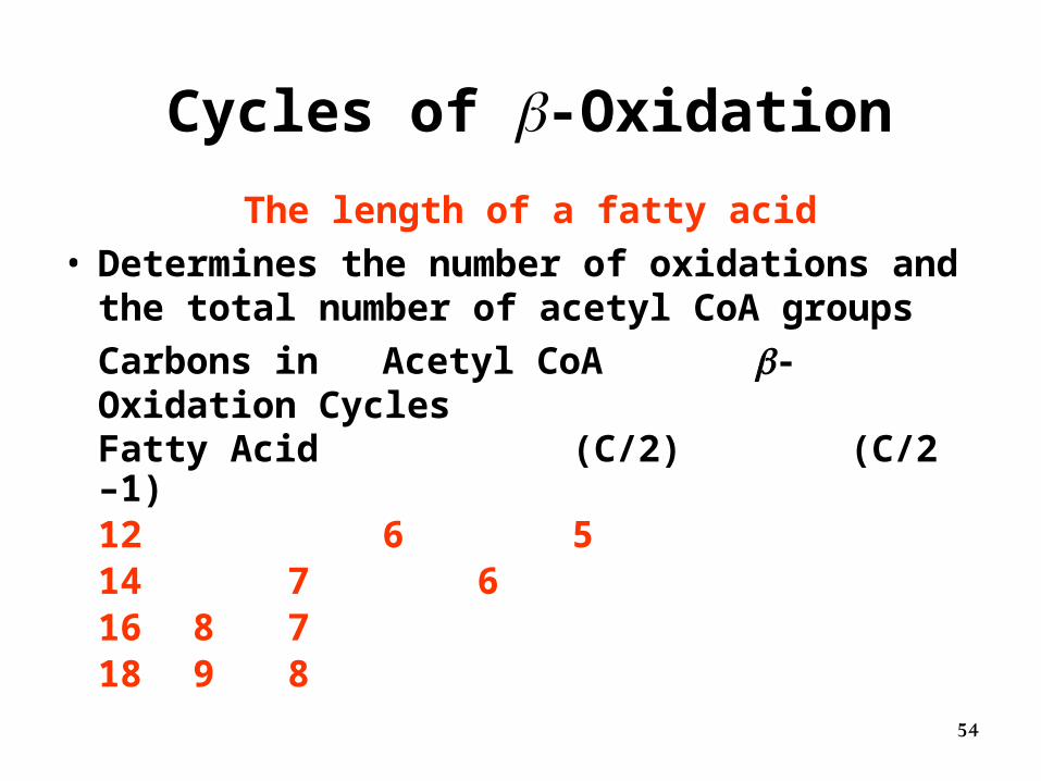
Cycles of -Oxidation
The length of a fatty acid• Determines the number of oxidations and the
total number of acetyl CoA groups
Carbons in Acetyl CoA -Oxidation CyclesFatty Acid (C/2) (C/2 –1)12 6 514 7 616 8 718 9 8
55
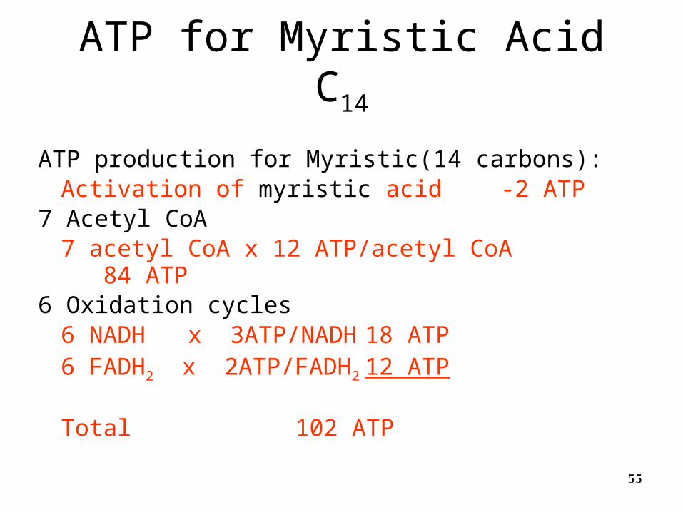
ATP for Myristic Acid C14
ATP production for Myristic(14 carbons):Activation of myristic acid -2 ATP
7 Acetyl CoA7 acetyl CoA x 12 ATP/acetyl CoA 84 ATP
6 Oxidation cycles 6 NADH x 3ATP/NADH 18 ATP6 FADH2 x 2ATP/FADH2 12 ATP
Total 102 ATP
56
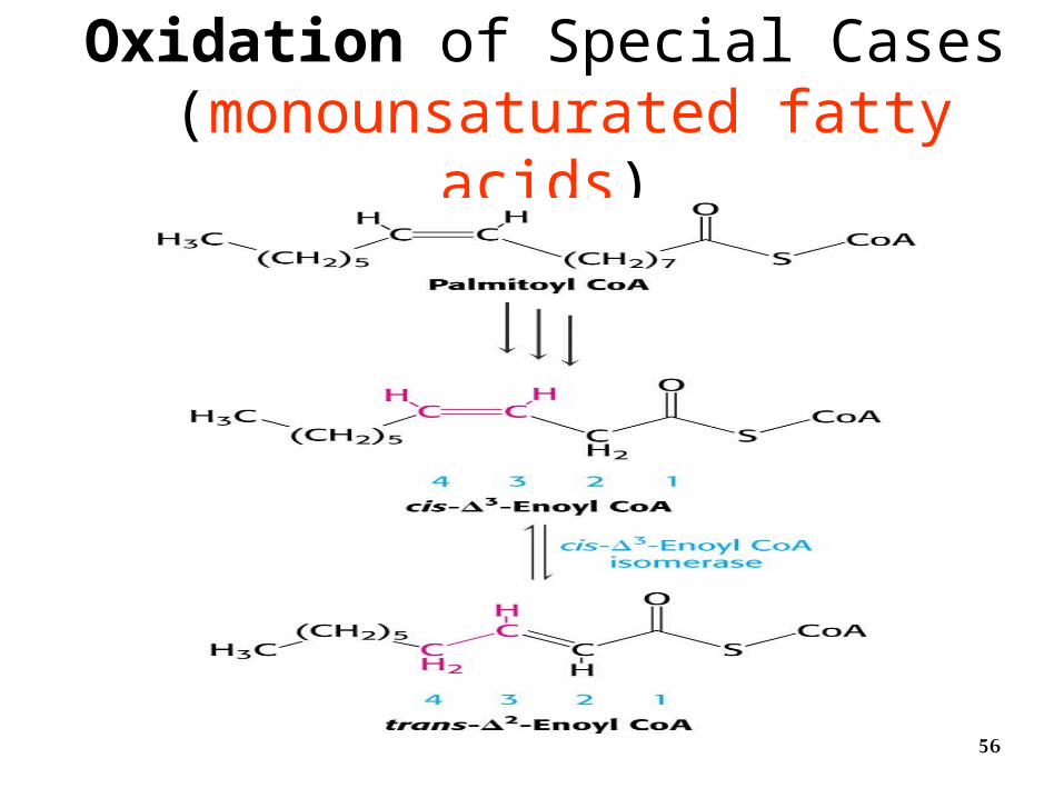
Oxidation of Special Cases (monounsaturated fatty acids)
57
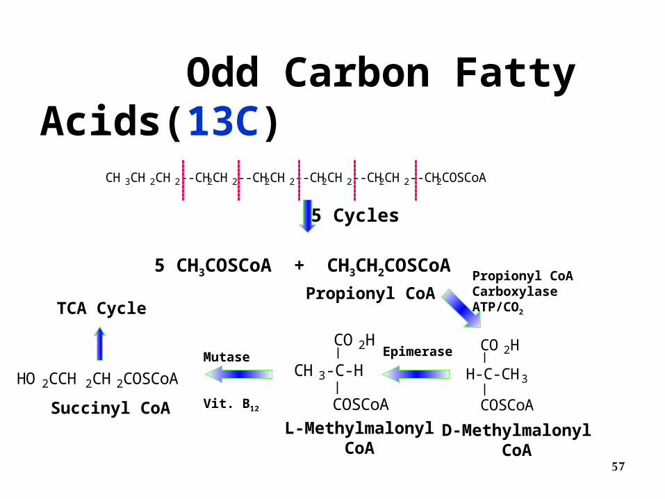
Odd Carbon Fatty Acids(13C)
CH3CH2CH2--CH2CH2--CH2CH2--CH2CH2--CH2CH2--CH2COSCoA
5 Cycles
5 CH3COSCoA + CH3CH2COSCoA
Propionyl CoA
CO2H
COSCoA
H-C-CH3
CO2H
COSCoA
CH3-C-HHO2CCH 2CH2COSCoA
D-MethylmalonylCoA
L-MethylmalonylCoA
Succinyl CoA
TCA Cycle
Propionyl CoA CarboxylaseATP/CO2
EpimeraseMutase
Vit. B12
58
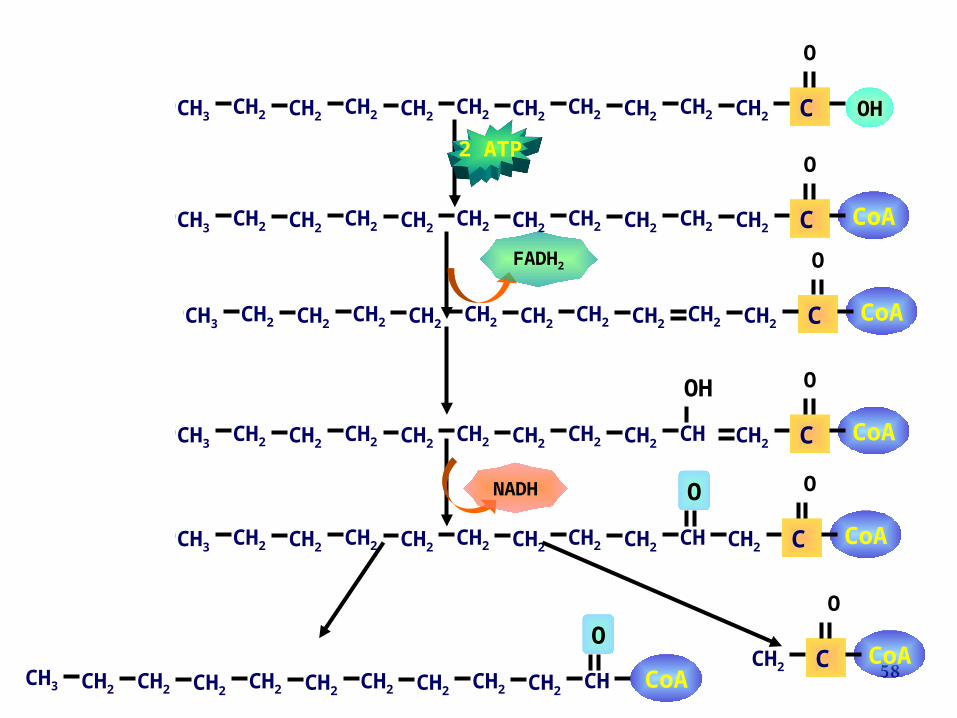
OHCH3 C
O
CH3 CH2 CH2 CH2 CH2 CH2 CH2 CH2 CH2 CH2 CH2
CoAC
O
CH3 CH2 CH2 CH2 CH2 CH2 CH2 CH2 CH2 CH2 CH2
CoAC
O
CH3 CH2 CH2 CH2 CH2 CH2 CH2 CH2 CH2 CH CH2
OH
CoAC
O
CH3 CH2 CH2 CH2 CH2 CH2 CH2 CH2 CH2 CH CH2
O
CH2CoAC
O
CH3 CH2 CH2 CH2 CH2 CH2 CH2 CH2 CH2 CH2 CH
O
CoA
CoAC
O
CH3 CH2 CH2 CH2 CH2 CH2 CH2 CH2 CH2 CH2 CH2
NADH
FADH
2
2 ATP
59
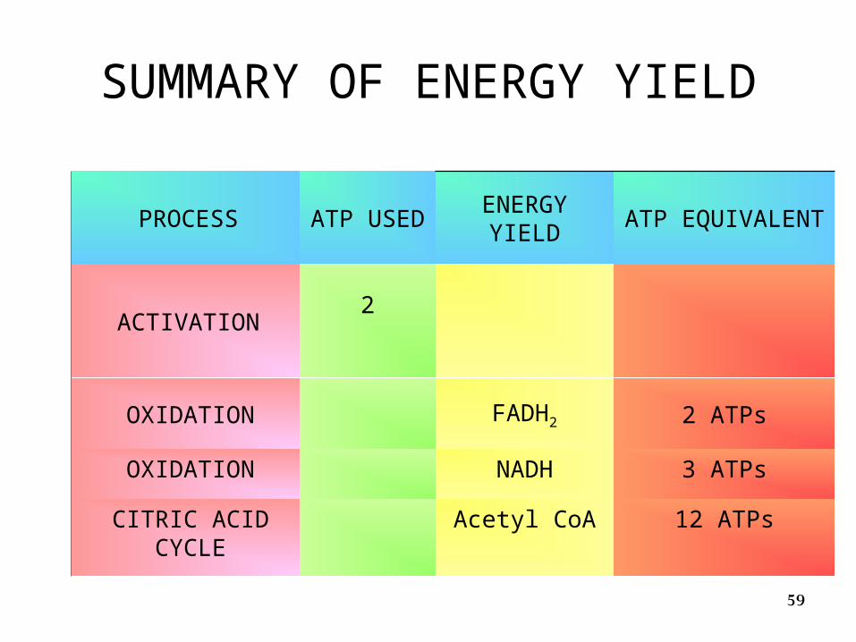
SUMMARY OF ENERGY YIELD
PROCESSATP
USEDENERGY
YIELDATP EQUIVALENT
ACTIVATION2
OXIDATION FADH2 2 ATPs
OXIDATION NADH 3 ATPs
CITRIC ACID CYCLE
Acetyl CoA 12 ATPs
60
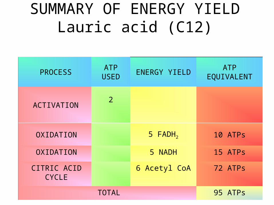
SUMMARY OF ENERGY YIELD Lauric acid (C12)
PROCESSATP
USEDENERGY YIELD
ATP EQUIVALENT
ACTIVATION2
OXIDATION 5 FADH2 10 ATPs
OXIDATION 5 NADH 15 ATPs
CITRIC ACID CYCLE
6 Acetyl CoA 72 ATPs
TOTAL 95 ATPs
61
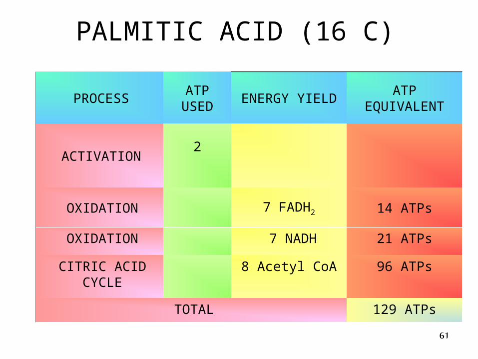
PALMITIC ACID (16 C)
PROCESSATP
USEDENERGY YIELD
ATP EQUIVALENT
ACTIVATION2
OXIDATION 7 FADH2 14 ATPs
OXIDATION 7 NADH 21 ATPs
CITRIC ACID CYCLE
8 Acetyl CoA 96 ATPs
TOTAL 129 ATPs
62
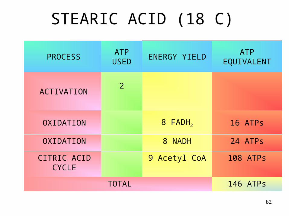
STEARIC ACID (18 C)
PROCESSATP
USEDENERGY YIELD
ATP EQUIVALENT
ACTIVATION2
OXIDATION 8 FADH2 16 ATPs
OXIDATION 8 NADH 24 ATPs
CITRIC ACID CYCLE
9 Acetyl CoA 108 ATPs
TOTAL 146 ATPs
63
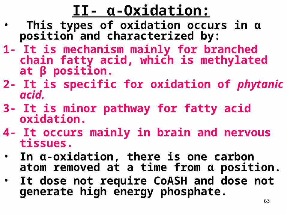
II- α-Oxidation:• This types of oxidation occurs in α position and
characterized by:1- It is mechanism mainly for branched chain fatty
acid, which is methylated at β position.2- It is specific for oxidation of phytanic acid.3- It is minor pathway for fatty acid oxidation.4- It occurs mainly in brain and nervous tissues.• In α-oxidation, there is one carbon atom removed
at a time from α position.• It dose not require CoASH and dose not generate
high energy phosphate.
64
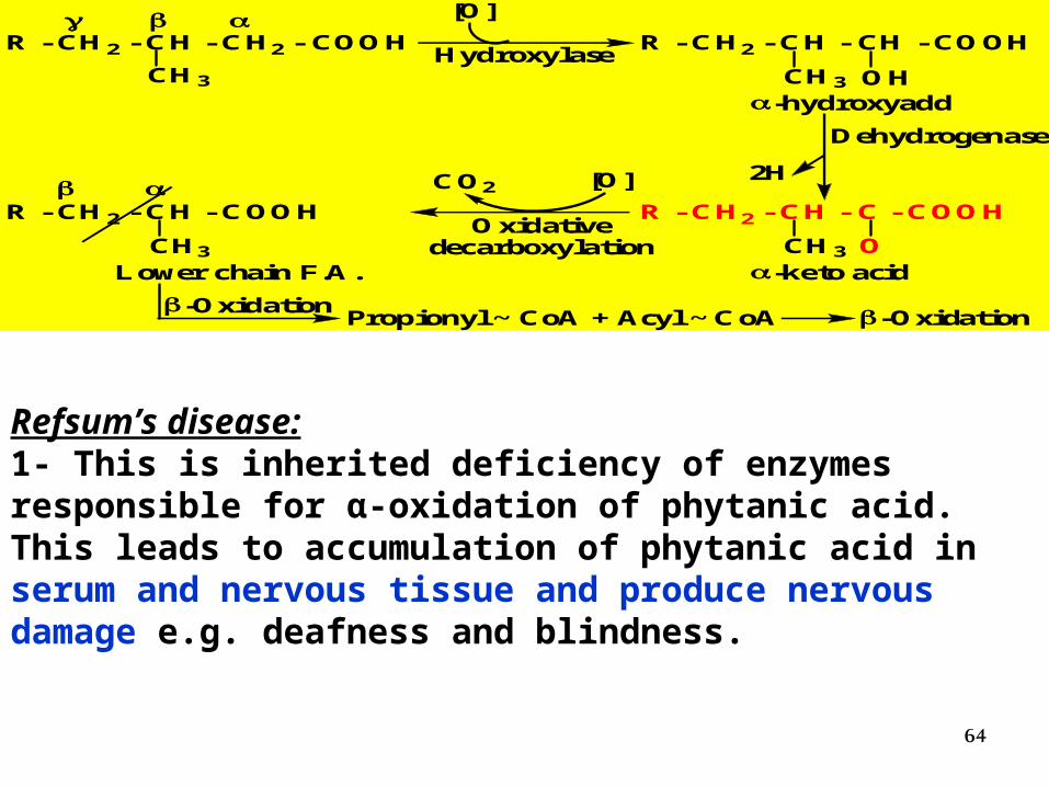
R - CH2 - CH - CH2 - COOH R - CH2 - CH - CH - COOH
CH3
[O]
HydroxylaseCH3 OH
-hydroxyadd
R - CH2 - CH - C - COOH
CH3 O-keto acid
2H
Dehydrogenase
R - CH2 - CH - COOH
CH3
Lower chain F.A.
Oxidativedecarboxylation
CO2 [O]
Propionyl ~ CoA + Acyl ~ CoA -Oxidation-Oxidation
Refsum’s disease:1- This is inherited deficiency of enzymes responsible for α-oxidation of phytanic acid. This leads to accumulation of phytanic acid in serum and nervous tissue and produce nervous damage e.g. deafness and blindness.
65
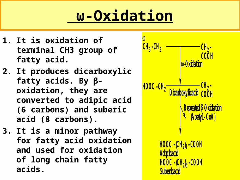
ω-Oxidation
1. It is oxidation of terminal CH3 group of fatty acid.
2. It produces dicarboxylic fatty acids. By β-oxidation, they are converted to adipic acid (6 carbons) and suberic acid (8 carbons).
3. It is a minor pathway for fatty acid oxidation and used for oxidation of long chain fatty acids.
CH3 - CH2 CH2 -COOH
HOOC - CH2 CH2 -COOH
HOOC - (CH2)4 - COOHAdipicacidHOOC - (CH2)6 - COOHSubericacid
-Oxidation
Repeated -Oxidation(Aoetyl - CoA)
Dicarboxylicacid
66
67
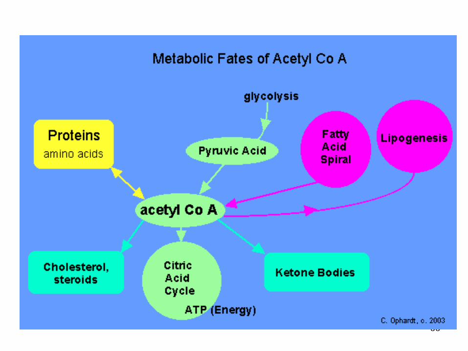
Ketone Bodies
68
Formation and Utilization
• Ketone bodies are:
1. water-soluble fuels
2. Normally exported by the liver
3. overproduced during fasting or in untreated diabetes mellitus.
69
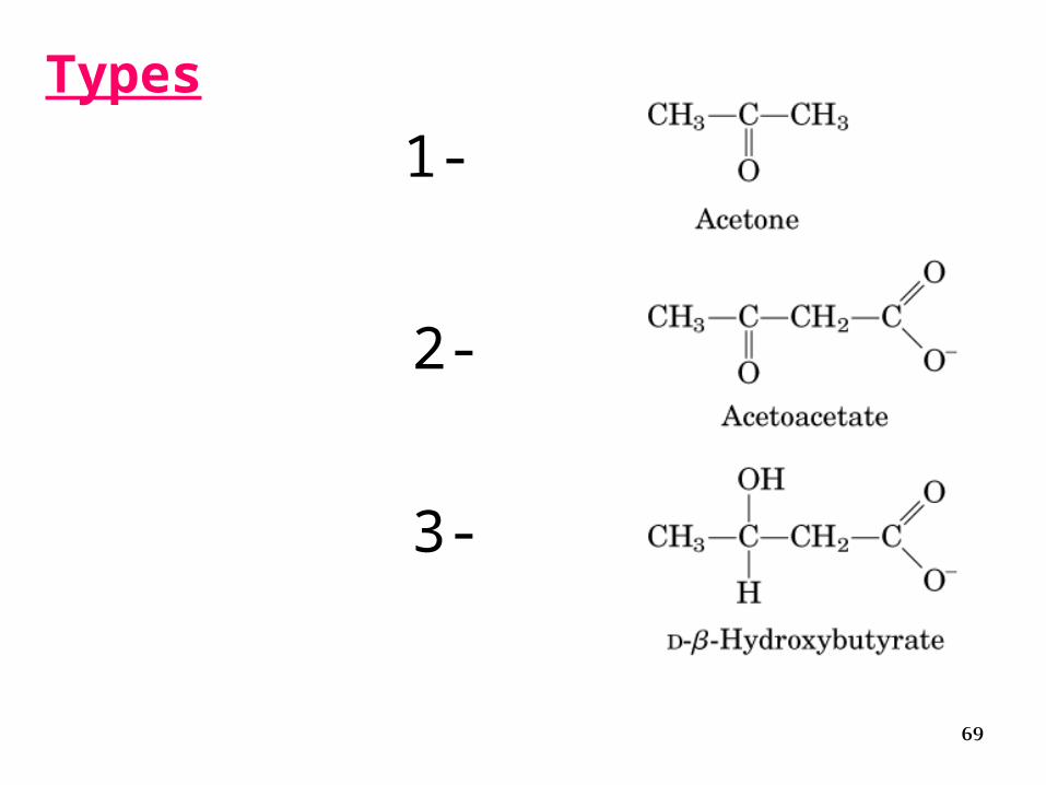
Types
1-
2-
3-
70
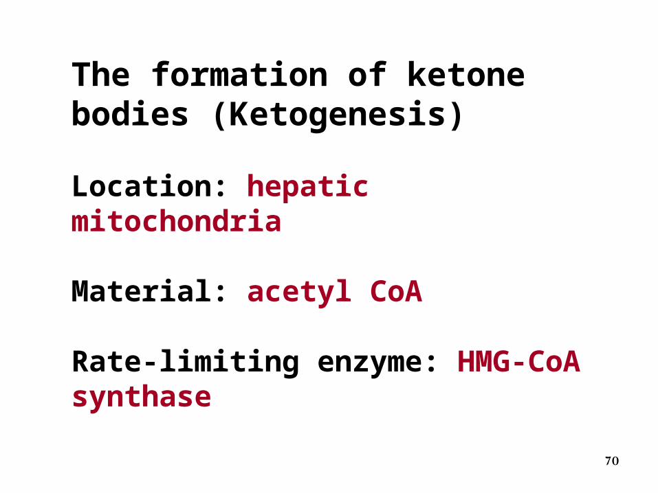
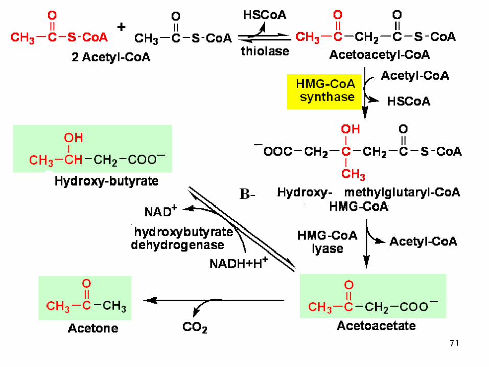
The formation of ketone bodies (Ketogenesis)
Location: hepatic mitochondria
Material: acetyl CoA
Rate-limiting enzyme: HMG-CoA synthase
71

72
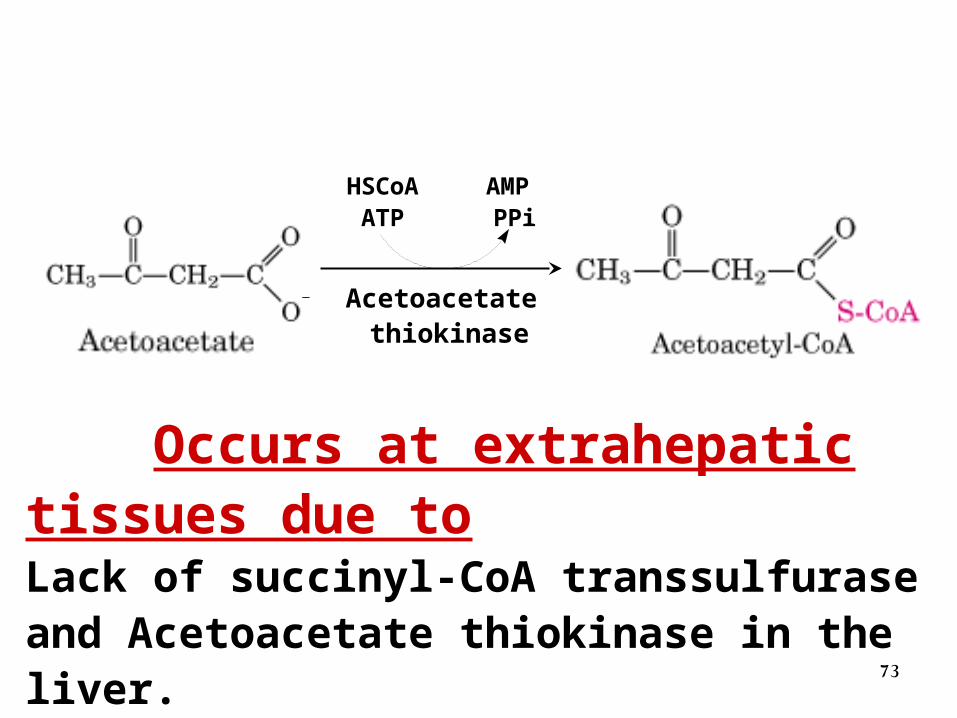
Utilization of ketone bodies (ketolysis) Occurs at extrahepatic tissues
Succinyl-CoA transsulfurase
73
HSCoAATP
AMP PPi
Acetoacetate thiokinase
-
Occurs at extrahepatic tissues due toLack of succinyl-CoA transsulfurase and Acetoacetate thiokinase in the liver.
74
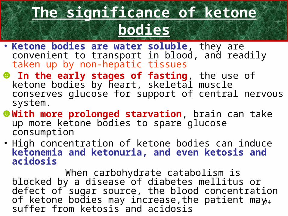
The significance of ketone bodies
• Ketone bodies are water soluble, they are convenient to transport in blood, and readily taken up by non-hepatic tissues
☻ In the early stages of fasting, the use of ketone bodies by heart, skeletal muscle conserves glucose for support of central nervous system.
☻With more prolonged starvation, brain can take up more ketone bodies to spare glucose consumption
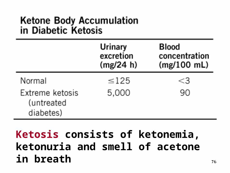
• High concentration of ketone bodies can induce ketonemia and ketonuria, and even ketosis and acidosis
When carbohydrate catabolism is blocked by a disease of diabetes mellitus or defect of sugar source, the blood concentration of ketone bodies may increase,the patient may suffer from ketosis and acidosis

75
Glucose Glucose exported as fuel for tissues such as brain
oxaloacetate
Fattyacids Acetyl-CoA
β-oxidation
gluconeogenesis
CitricAcid cycle
Ketone bodiesexported as energy source for heart, skeletal muscle, kidney, and brain
Ketone body formation
Hepatocyte
Acetoacetate, β-hydroxybutyrate,
acetone
CoA
76
Ketosis consists of ketonemia, ketonuria and smell of acetone in breath
77
Causes for ketosis
• Severe diabetes mellitus
• Starvation
• Hyperemesis (vomiting) in early pregnancy
78
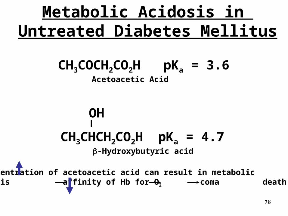
CH3COCH2CO2H pKa = 3.6 Acetoacetic Acid
CH3CHCH2CO2H pKa = 4.7 -Hydroxybutyric acid
OH
Concentration of acetoacetic acid can result in metabolic acidosis affinity of Hb for O2 coma death
Metabolic Acidosis in Untreated Diabetes Mellitus
Lipogenesis
8080
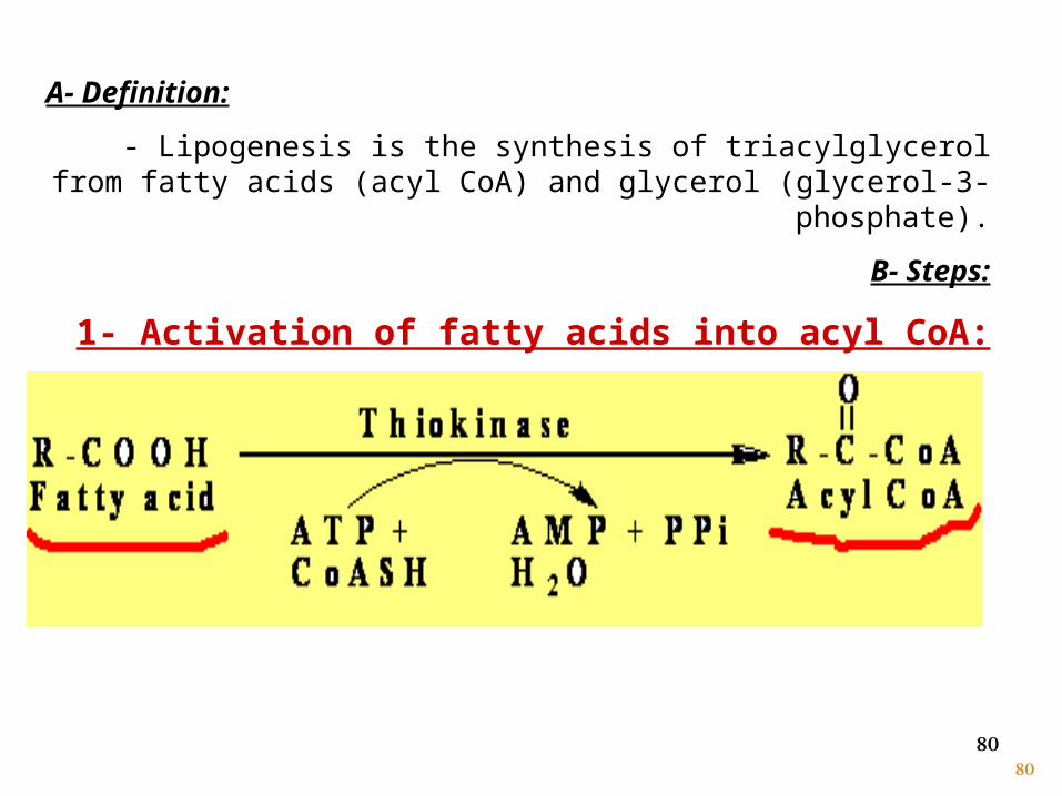
A- Definition:
- Lipogenesis is the synthesis of triacylglycerol from fatty acids (acyl CoA) and glycerol (glycerol-3-phosphate).
B- Steps:
1- Activation of fatty acids into acyl CoA:
81
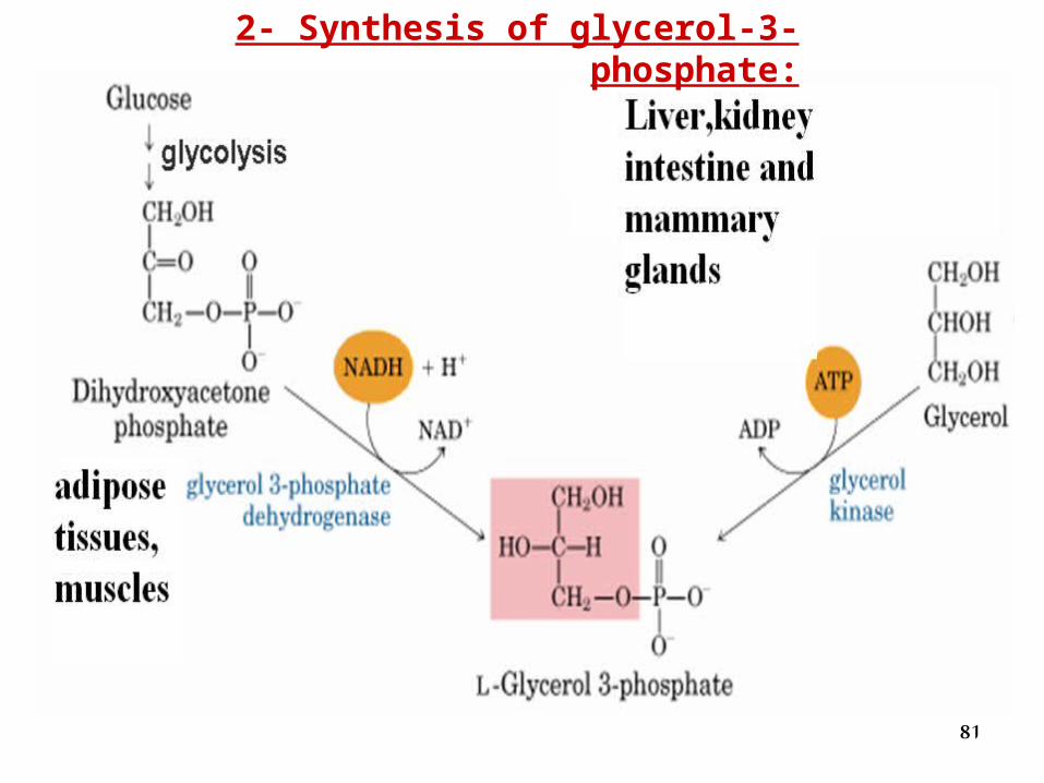
2- Synthesis of glycerol-3- phosphate:
82
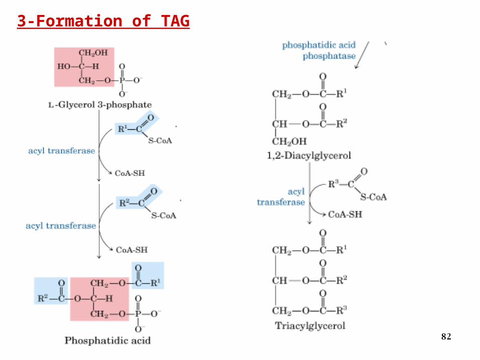
3-Formation of TAG
8383
Regulation of lipogenesis
After meal, lipogenesis is stimulated:
- Insulin is secreted which stimulates glycolysis. Glycolysis supplies
dihydroxyacetone phosphate that converted into glycerol-3-phosphate
in adipose tissue, so lipogenesis is stimulated.
During fasting lipogenesis is inhibited:
- Anti-insulin hormones are secreted. These inhibit lipogenesis and
stimulate lipolysis
84
Fatty Acid Biosynthesis
85
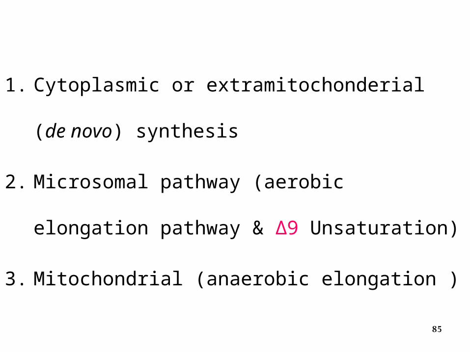
1. Cytoplasmic or extramitochonderial (de novo)
synthesis
2. Microsomal pathway (aerobic elongation
pathway & ∆9 Unsaturation)
3. Mitochondrial (anaerobic elongation )
86
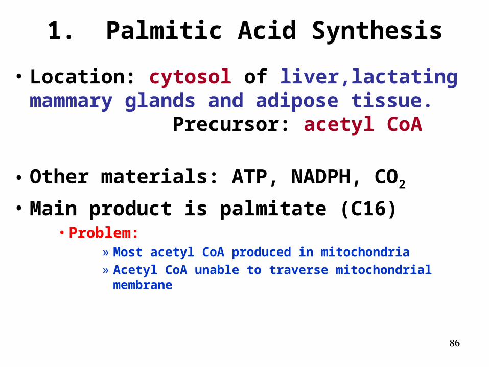
1. Palmitic Acid Synthesis
• Location: cytosol of liver,lactating mammary glands and adipose tissue. Precursor: acetyl CoA
• Other materials: ATP, NADPH, CO2
• Main product is palmitate (C16)• Problem:
» Most acetyl CoA produced in mitochondria» Acetyl CoA unable to traverse mitochondrial
membrane
87
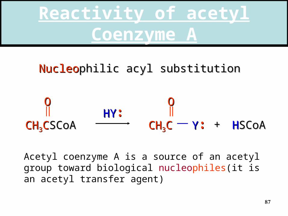
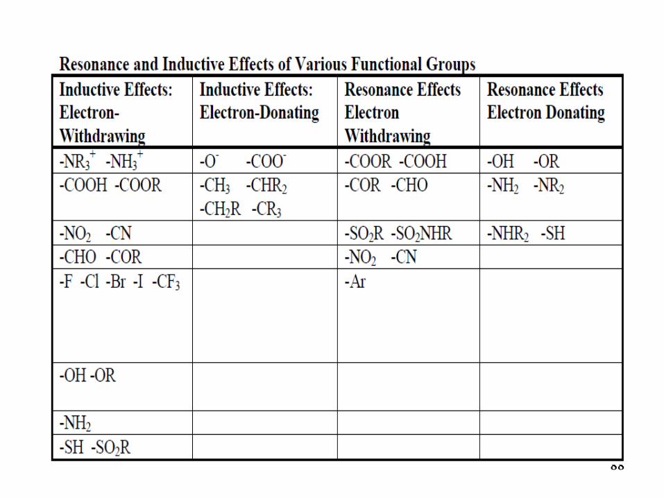
Reactivity of acetyl Coenzyme A
NucleoNucleophilic acyl substitutionphilic acyl substitution
CHCH33CCSCoASCoA
OOHYHY••••
CHCH33CC
OO
YY •••• ++ HHSCoASCoA
Acetyl coenzyme A is a source of an acetyl group toward biological nucleophiles(it is an acetyl transfer agent)
88

89
can react via enolcan react via enol
CHCH33CCSCoASCoA
OO
Acetyl coenzyme A reacts with biological electrophiles at its α carbon atom
CCSCoASCoA
OHOH
HH22CC
EE++
CHCH22CCSCoASCoA
OO
EE
Reactivity of acetyl Coenzyme A
90
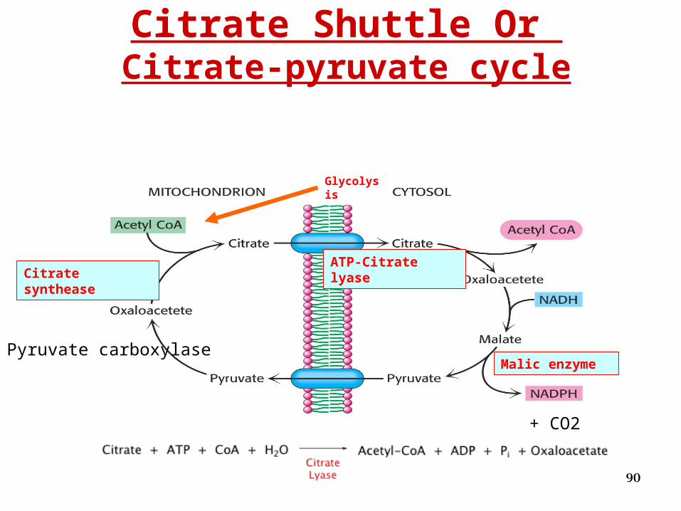
Citrate Shuttle Or Citrate-pyruvate cycle
Citrate syntheaseATP-Citrate lyase
Malic enzyme
Glycolysis
+ CO2
Pyruvate carboxylase
91
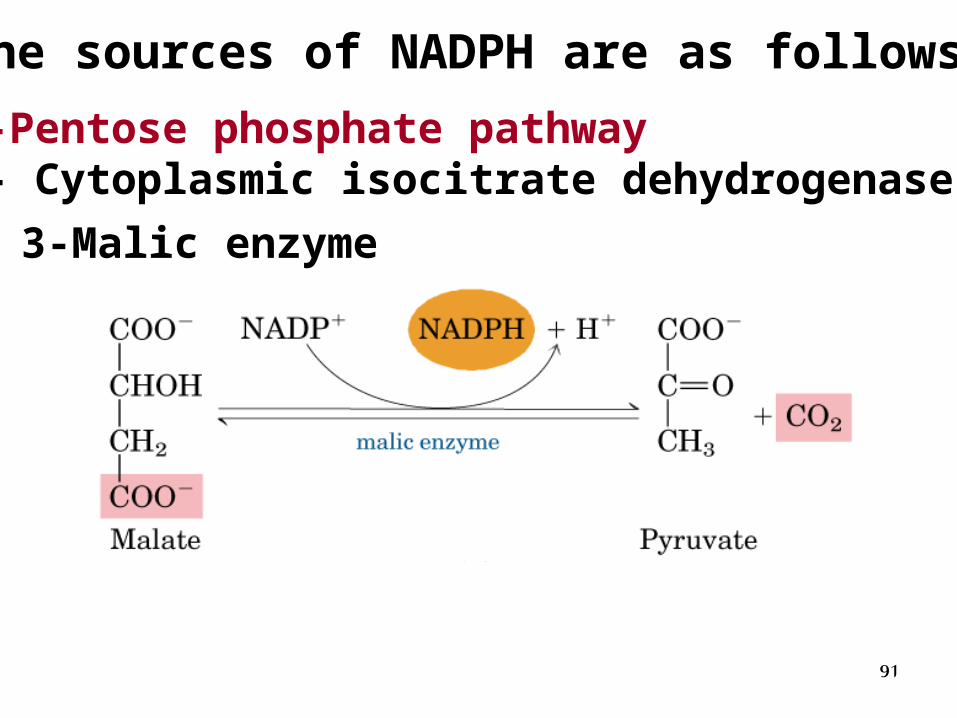
The sources of NADPH are as follows:
1-Pentose phosphate pathway3- Cytoplasmic isocitrate dehydrogenase
3-Malic enzyme
92
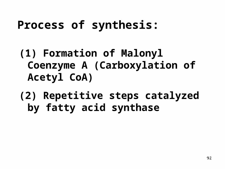
Process of synthesis:
(1) Formation of Malonyl Coenzyme A (Carboxylation of Acetyl CoA)
(2) Repetitive steps catalyzed by fatty acid synthase
93
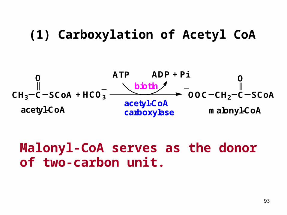
(1) Carboxylation of Acetyl CoA
Malonyl-CoA serves as the donor of two-carbon unit.
CH3 C
O
SCoA
acetyl-CoA
+ HCO3acetyl-CoAcarboxylase
ATP ADP + Pibiotin
OOC CH2 C SCoA
O
malonyl-CoA
94
Acetyl-CoA Carboxylase is the rate limiting enzyme of the fatty acid synthesis pathway.
The mammalian enzyme is regulated, by
phosphorylation
allosteric regulation by local metabolites.
95
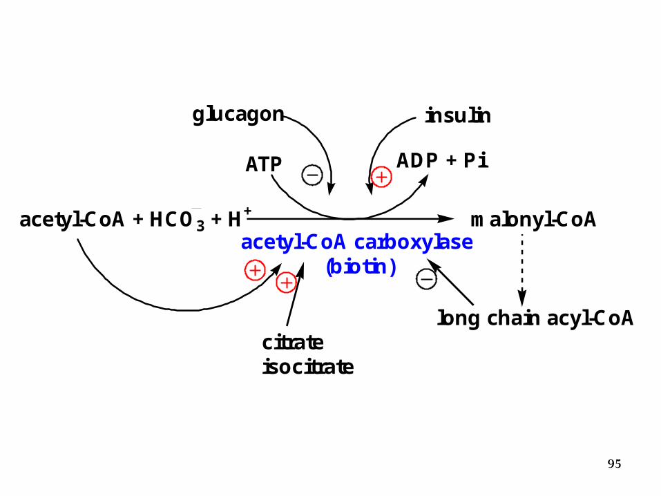
acetyl-CoA + HCO3 + H+
acetyl-CoA carboxylase (biotin)
malonyl-CoA
long chain acyl-CoA
ATP ADP + Pi
glucagon insulin
citrateisocitrate
96
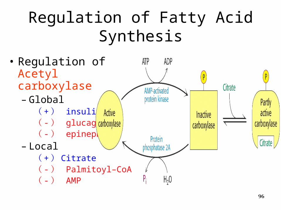
• Regulation of Acetyl carboxylase– Global
( + ) insulin( - ) glucagon( - ) epinephrine
– Local( + ) Citrate( - ) Palmitoyl–CoA( - ) AMP
Regulation of Fatty Acid Synthesis
97
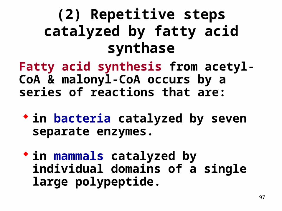
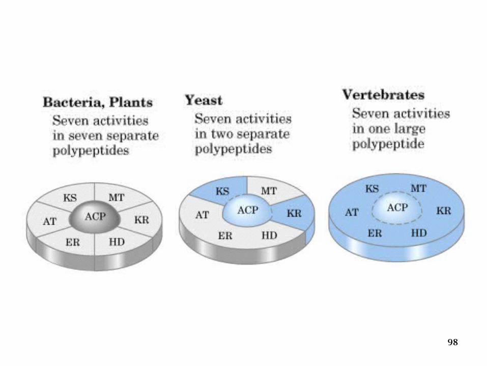
Fatty acid synthesis from acetyl-CoA & malonyl-CoA occurs by a series of reactions that are:
in bacteria catalyzed by seven separate enzymes.
in mammals catalyzed by individual domains of a single large polypeptide.
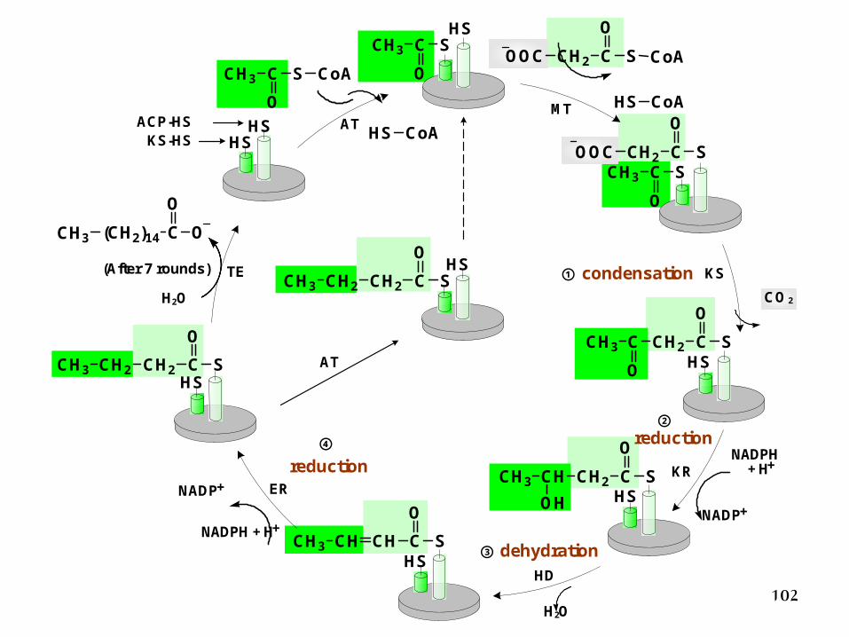
(2) Repetitive steps catalyzed by fatty acid synthase
98
99
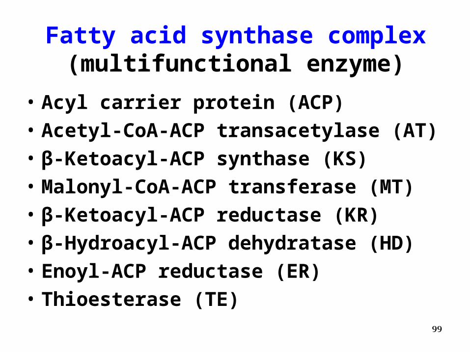
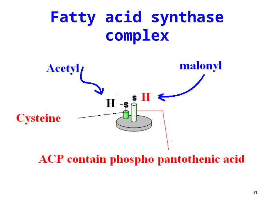
Fatty acid synthase complex(multifunctional enzyme)
• Acyl carrier protein (ACP)
• Acetyl-CoA-ACP transacetylase (AT)
• β-Ketoacyl-ACP synthase (KS)
• Malonyl-CoA-ACP transferase (MT)
• β-Ketoacyl-ACP reductase (KR)
• β-Hydroacyl-ACP dehydratase (HD)
• Enoyl-ACP reductase (ER)
• Thioesterase (TE)
100
Cys
HS
PhP
HS
AT
KS
MTHD ER KR
ACP
TE
Cys
HS
PhP
HS
AT
KS
MTHDERKR
ACP
TE
Fu
nctio
nal
divisio
n
Subunitdivision
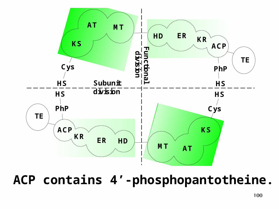
ACP contains 4’-phosphopantotheine.
101
Fatty acid synthase complex
102
ATMT
KS① condensation
②
KR
③ dehydration
④
HD
ER
AT
TE
NADPH + H+
NADP+
(CH2)14 C O
O
CH3
NADP+
+ H+NADPH
CH3 C S
O
CH3 C S
O
OOC CH2 C S
O
C CH2 C S
O
O
CH3
CH CH2 C S
O
OH
CH3
CH CH C S
O
CH3
CH2 CH2 C S
O
CH3
KS-HSACP-HS
CH2 CH2 C S
O
CH3CO 2
H2O
H2O
OOC CH2 C S CoA
O
CH3 C S
O
CoA
HS CoA
HS
reduction
(After 7 rounds)
HS CoA
HS
HS
HS
HS
HS
HSHS
reduction
103
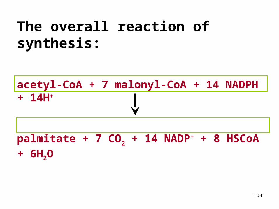
acetyl-CoA + 7 malonyl-CoA + 14 NADPH + 14H+
palmitate + 7 CO2 + 14 NADP+ + 8 HSCoA + 6H2O
The overall reaction of synthesis:
104
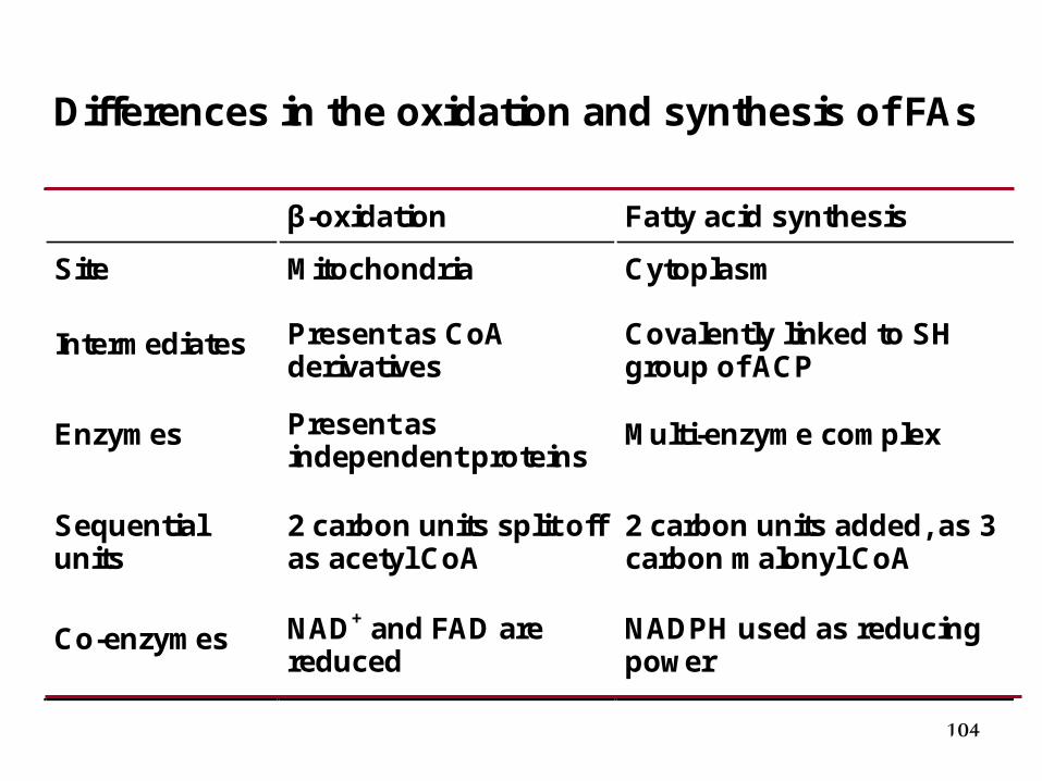
Differences in the oxidation and synthesis of FAs β-oxidation Fatty acid synthesis
Site Mitochondria Cytoplasm
Intermediates Present as CoA derivatives
Covalently linked to SH group of ACP
Enzymes Present as independent proteins
Multi-enzyme complex
Sequential units
2 carbon units split off as acetyl CoA
2 carbon units added, as 3 carbon malonyl CoA
Co-enzymes NAD+ and FAD are reduced
NADPH used as reducing power
105
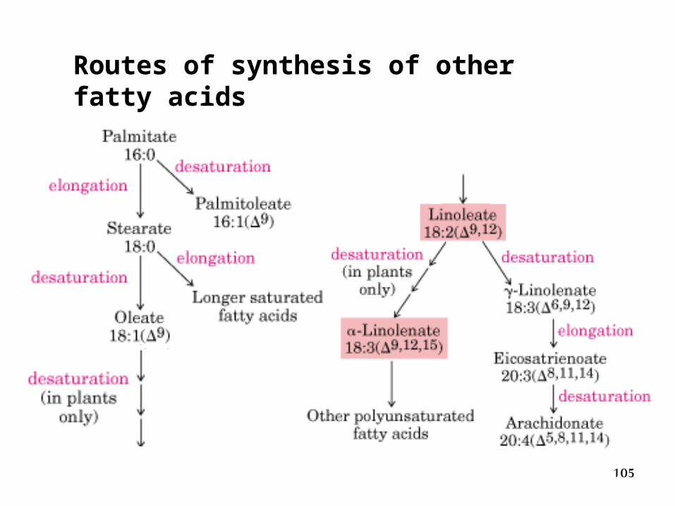
Routes of synthesis of other fatty acids
106
2. Elongation of palmitate
Elongation beyond the 16-C length of the palmitate occurs in mitochondria and endoplasmic reticulum (ER).
107
Fatty acid elongation within mitochondria uses the acetyl-CoA as donor of 2-carbon units and NADPH serves as electron donor for the final reduction step.
Fatty acids esterified to coenzyme A are substrates for the ER elongation machinery, which uses malonyl-CoA as donor of 2-carbon units.
108
3. The synthesis of unsaturated fatty acid
• Formation of a double bond in a fatty acid involves several endoplasmic reticulum membrane proteins in mammalian cells
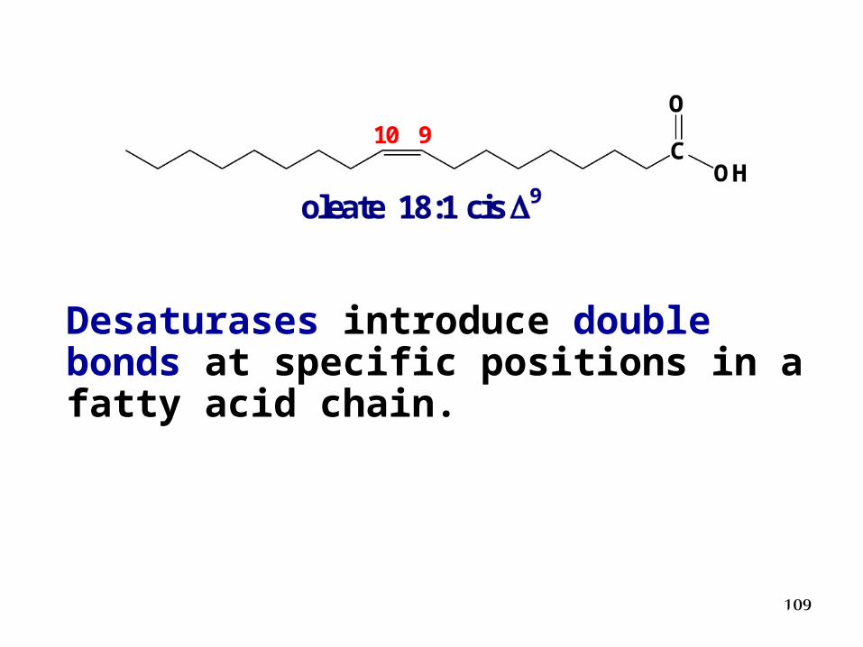
109
Desaturases introduce double bonds at specific positions in a fatty acid chain.
110
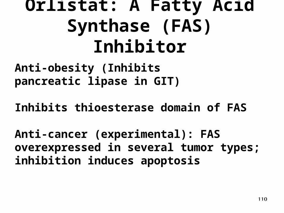
Orlistat: A Fatty Acid Synthase (FAS) Inhibitor
Anti-obesity (Inhibitspancreatic lipase in GIT)
Inhibits thioesterase domain of FAS
Anti-cancer (experimental): FAS overexpressed in several tumor types; inhibition induces apoptosis
Metabolism of phospholipids
112
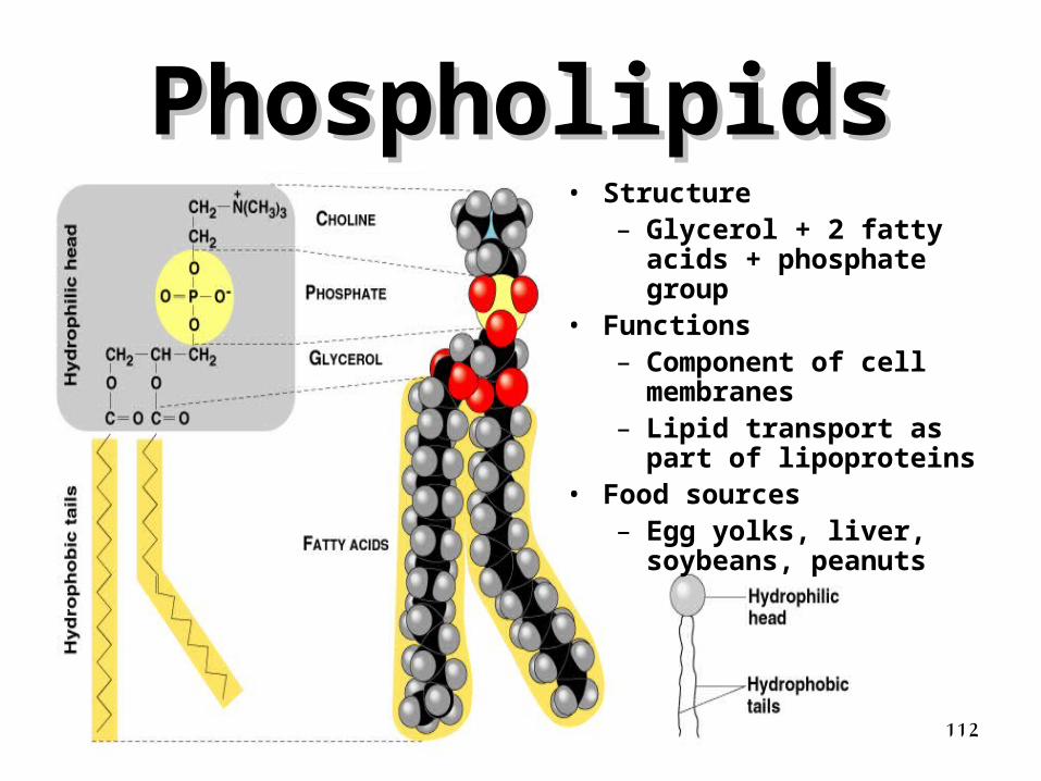
PhospholipidsPhospholipids• Structure
– Glycerol + 2 fatty acids + phosphate group
• Functions– Component of cell
membranes– Lipid transport as part of
lipoproteins• Food sources
– Egg yolks, liver, soybeans, peanuts
113
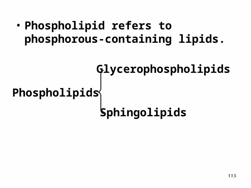
• Phospholipid refers to phosphorous-containing lipids.
Phospholipids
Glycerophospholipids
Sphingolipids
114
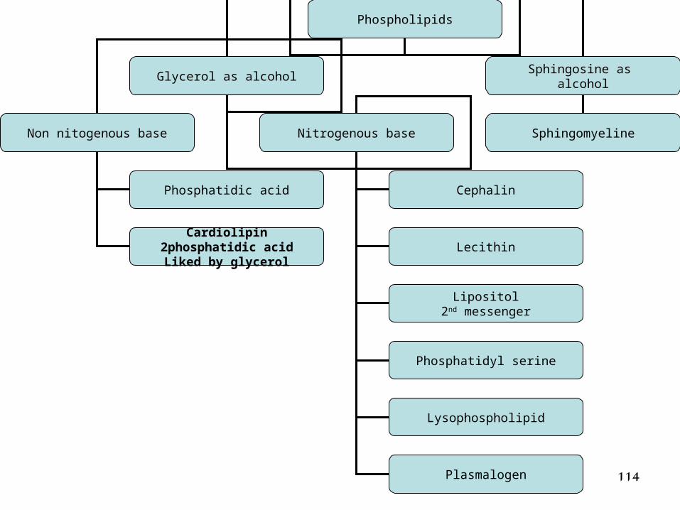
Phospholipids
Glycerol as alcoholSphingosine as
alcohol
SphingomyelineNon nitogenous base Nitrogenous base
Cephalin
Lecithin
Lipositol2nd messenger
Phosphatidic acid
Cardiolipin2phosphatidic acidLiked by glycerol
Phosphatidyl serine
Lysophospholipid
Plasmalogen
115
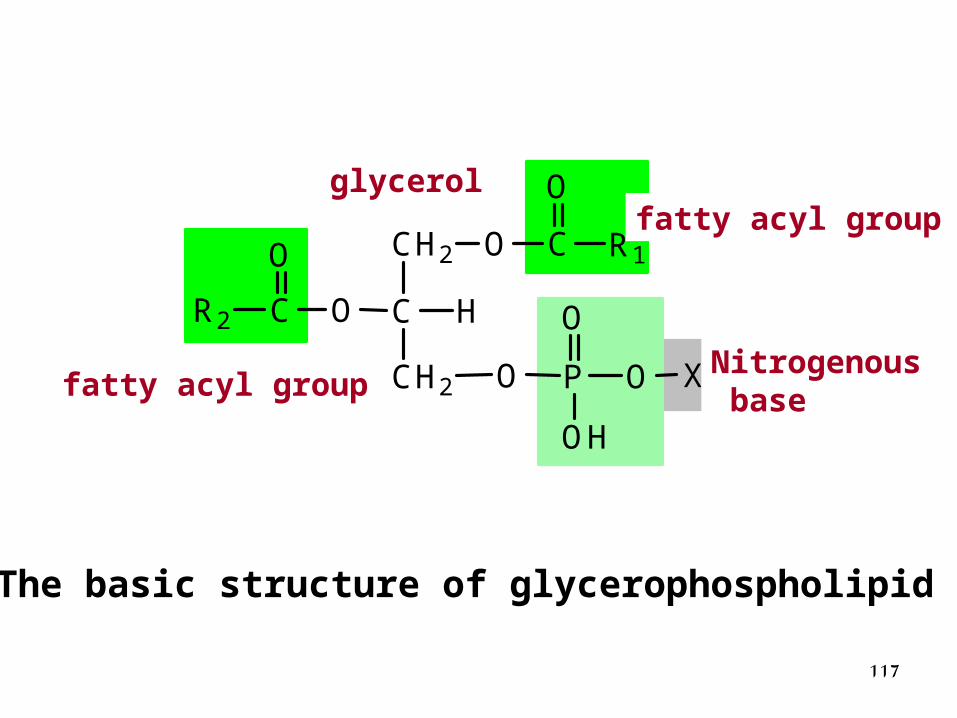
Classification and Structure of Glycerophospholipids
• Glycerophospholipids are lipids with a glycerol, fatty acids, a phosphate group and a nitrogenous base.
116
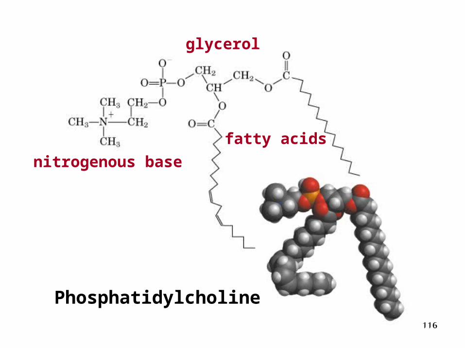
Phosphatidylcholine
fatty acids
nitrogenous base
glycerol
117
CH2 O
C H
CH2
O
O
C
C
P
R1
R2
O
O
O
O
OH
X
甘油
脂酰基
脂酰基
含氮化合物
The basic structure of glycerophospholipid
glycerolfatty acyl group
Nitrogenous basefatty acyl group
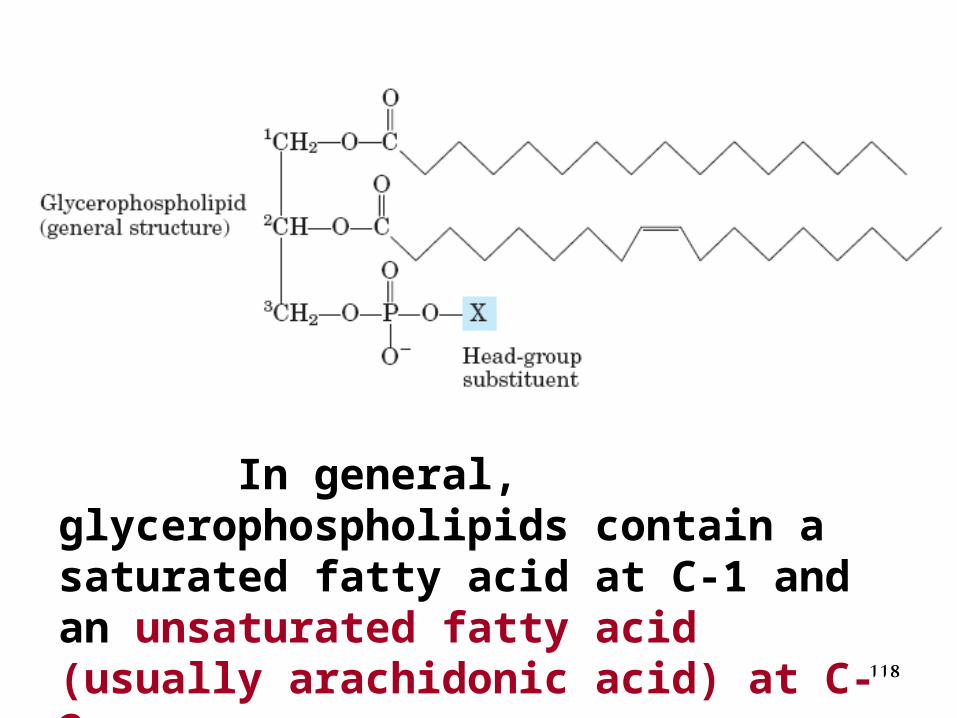
118
In general, glycerophospholipids contain a saturated fatty acid at C-1 and an unsaturated fatty acid (usually arachidonic acid) at C-2.
119
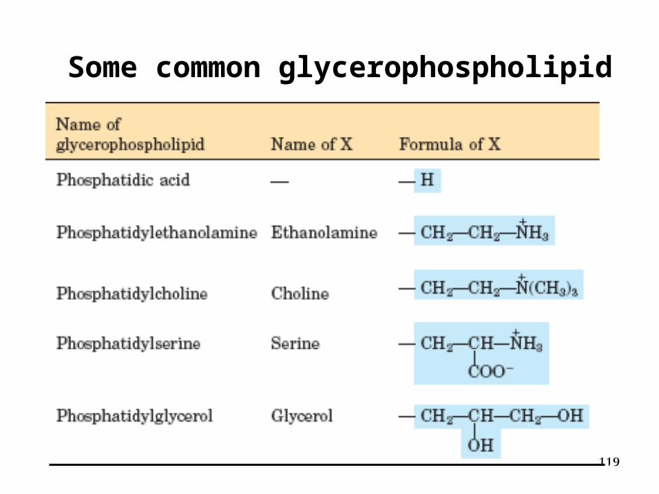
Some common glycerophospholipid
120
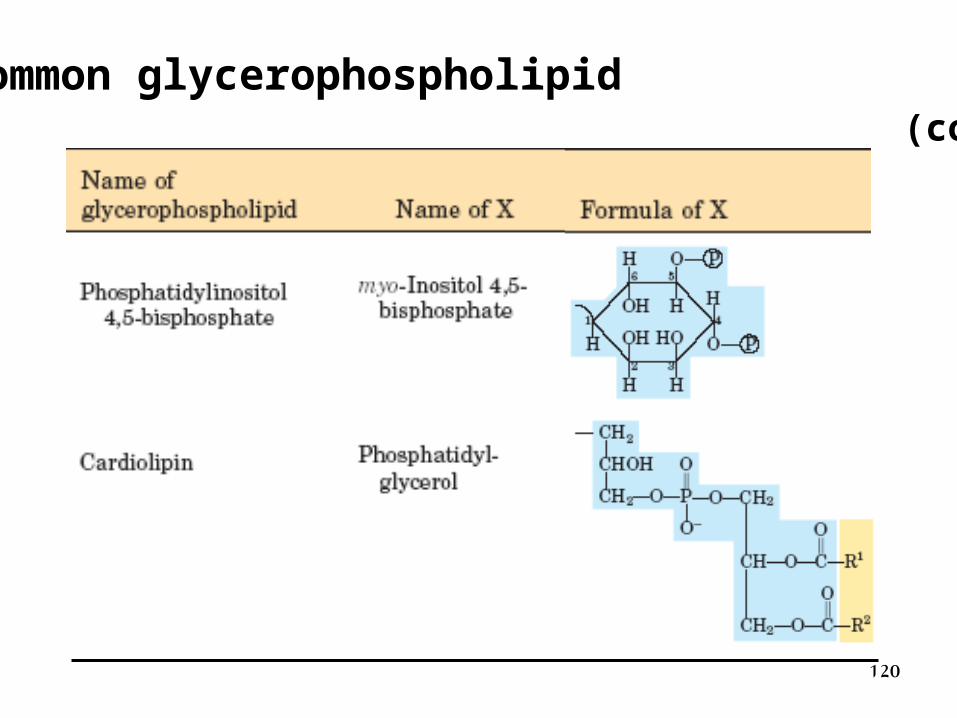
Some common glycerophospholipid (continue)
121
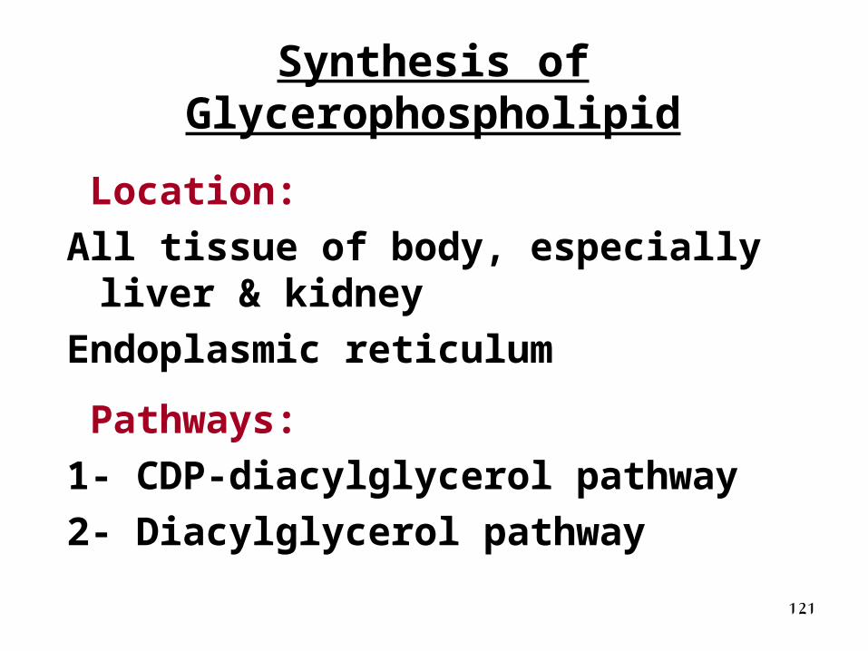
Synthesis of Glycerophospholipid
Location:
All tissue of body, especially liver & kidney
Endoplasmic reticulum
Pathways:
1- CDP-diacylglycerol pathway
2- Diacylglycerol pathway
122
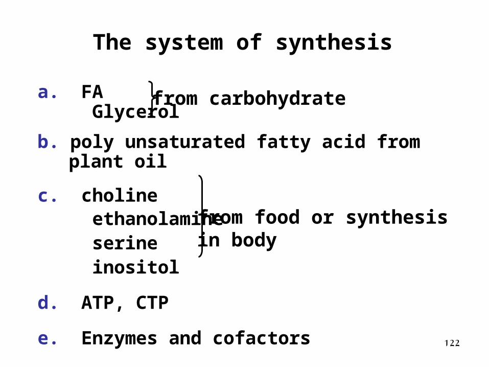
a. FA Glycerol
b. poly unsaturated fatty acid from plant oil c. choline ethanolamine serine inositol
d. ATP, CTP
e. Enzymes and cofactors
The system of synthesis
from carbohydrate
from food or synthesis in body
123
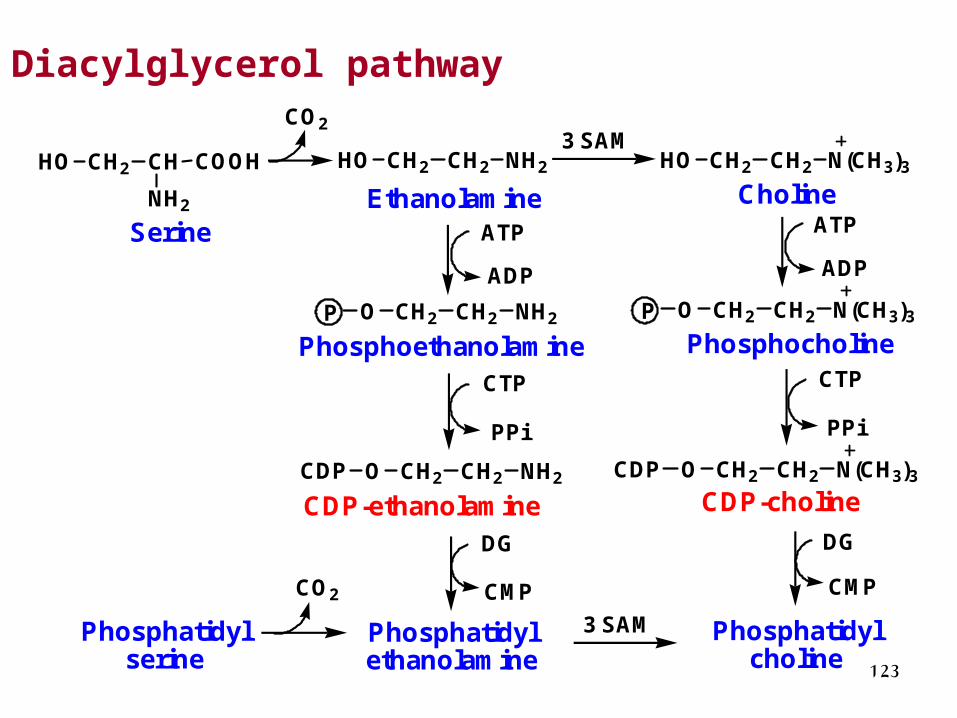
Diacylglycerol pathway
SerineEthanolamine
CO2
ATP
ADP
CTP
PPi
DG
CMPCO2
ATP
ADP
CTP
PPi
DG
CMP
3 SAMHO CH2 CH
NH2
COOH HO CH2 CH2 NH2 HO CH2 CH2 N(CH3)3
Choline
PhosphoethanolamineO CH2 CH2 NH2P O CH2 CH2 N(CH3)3
CDP
P
Phosphocholine
CDP-ethanolamineO CH2 CH2 NH2 O CH2 CH2 N(CH3)3CDP
CDP-choline
Phosphatidylethanolamine
Phosphatidylcholine
3 SAMPhosphatidylserine
124
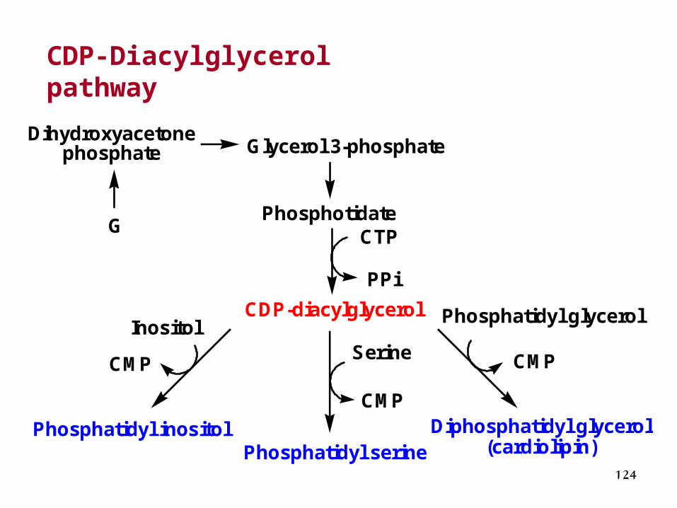
CDP-Diacylglycerol pathway
PhosphotidateCTP
PPi
CDP-diacylglycerol
CMP
CMP
CMP
Glycerol 3-phosphate
G
Phosphatidyl serinePhosphatidyl inositol
Phosphatidyl glycerol
Diphosphatidyl glycerol(cardiolipin)
SerineInositol
Dihydroxyacetonephosphate
125
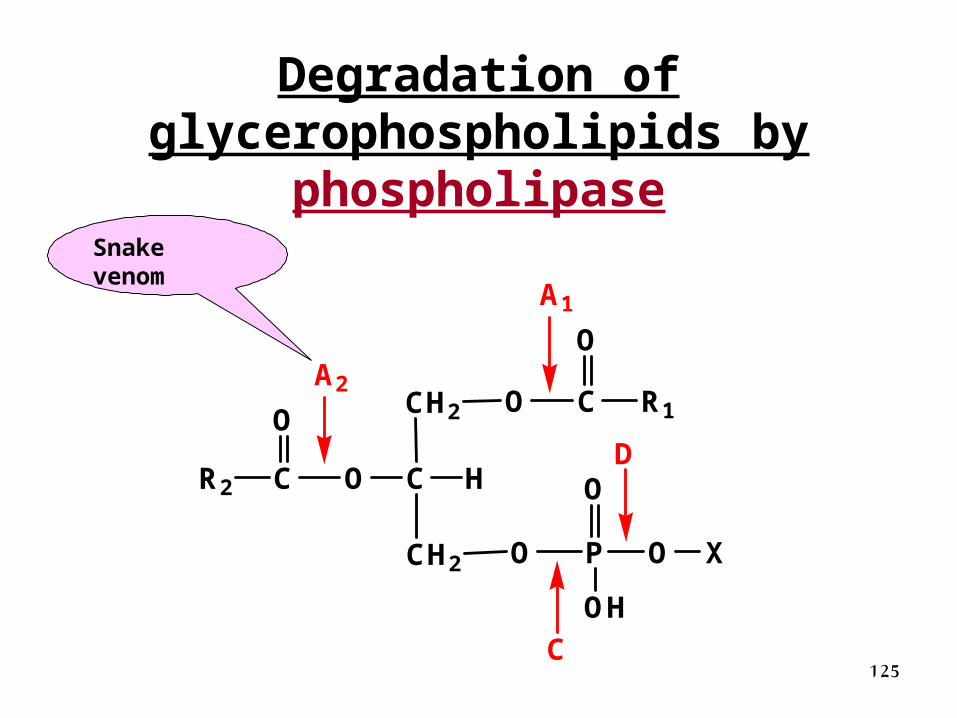
Degradation of glycerophospholipids by
phospholipase
CH2 O
C H
CH2
O
O
C
C
P
R1
R2
O
O
O
O
OH
X
A2
A1
C
D
Snake venom
126
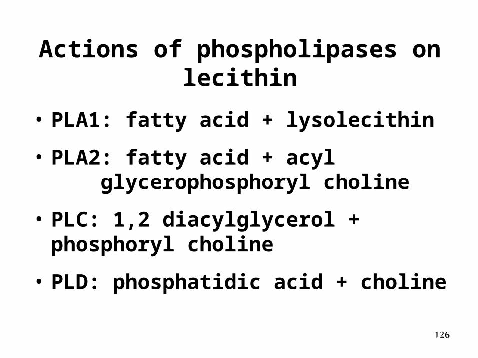
Actions of phospholipases on lecithin
• PLA1: fatty acid + lysolecithin
• PLA2: fatty acid + acyl glycerophosphoryl choline
• PLC: 1,2 diacylglycerol + phosphoryl choline
• PLD: phosphatidic acid + choline
127
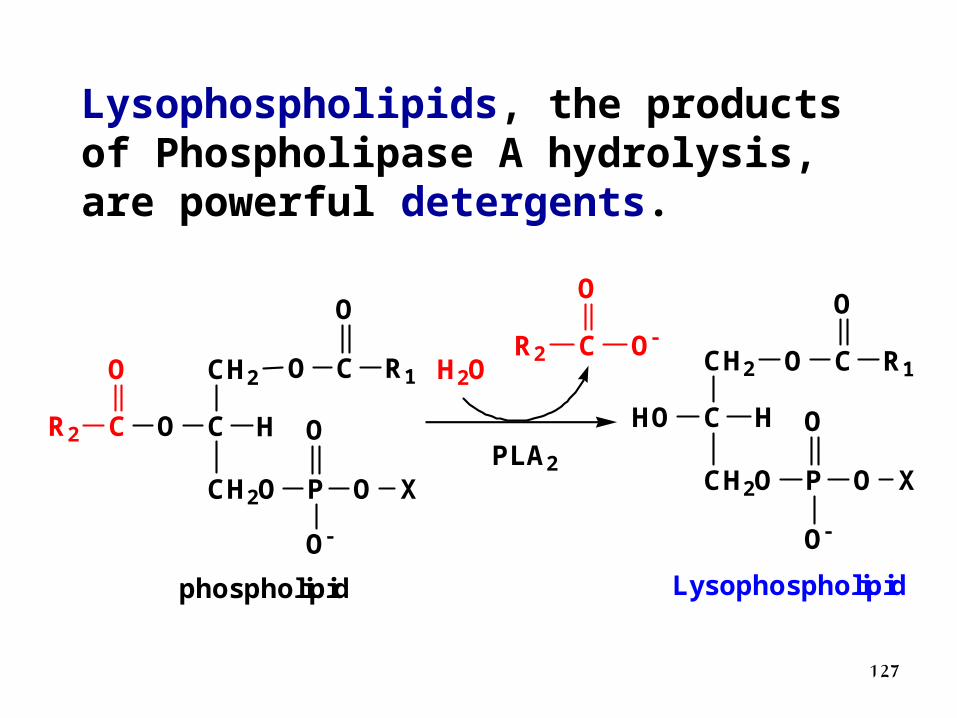
Lysophospholipids, the products of Phospholipase A hydrolysis, are powerful detergents.
CH2
C HO
CH2O
O C R1
O
P O
O
O
X
H2O
CR2
OOCR2
O
CH2
C HHO
CH2O
O C R1
O
P O
O
O
X
Lysophospholipidphospholipid
PLA2
128
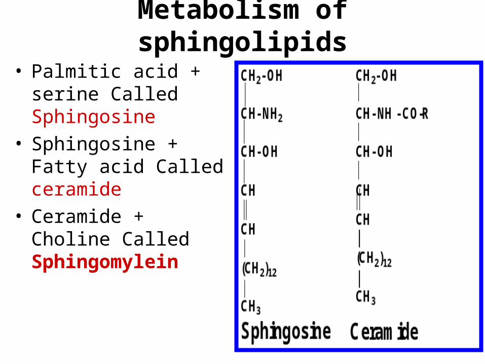
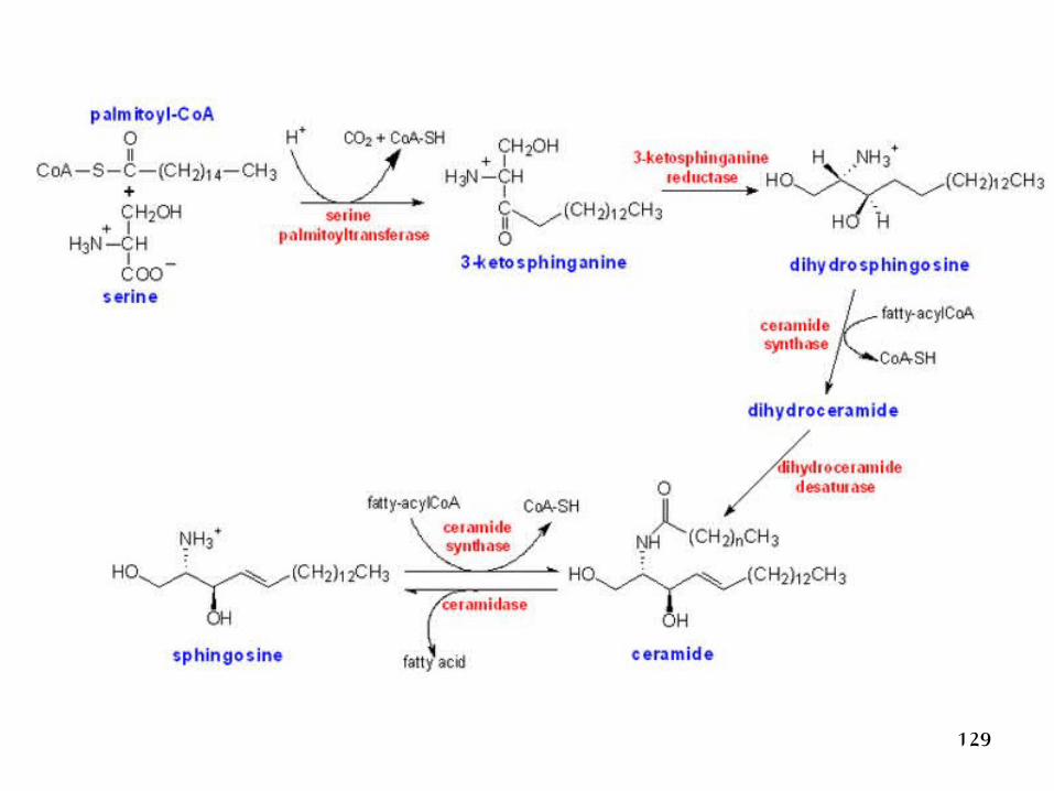
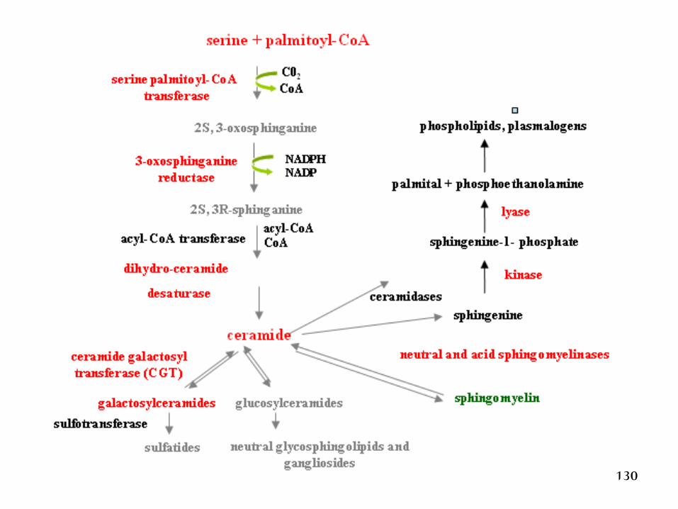
Metabolism of sphingolipids
• Palmitic acid + serine Called Sphingosine
• Sphingosine + Fatty acid Called ceramide
• Ceramide + Choline Called Sphingomylein
129
130
131
Respiratory Distress Syndrome
Most frequently seen in premature infants
Also called hyaline membrane disease
Failure to produce sufficient dipalmitoyl phosphatidylcholine,which normally is found in the extracellular fluid surroundingalveoli; decreases surface tension of fluid to prevent lung collapse
Treatment in infants born before 30 weeks includes
administration of artificial lung surfactant (e.g., Exosurf orPumactant)
132
Glycolipids
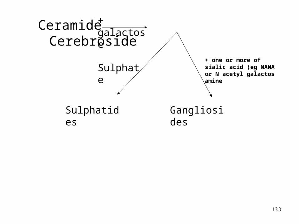
133
Ceramide Cerebroside
Sulphatides Gangliosides
Sulphate+ one or more of sialic acid (eg NANA or N acetyl galactos amine
+ galactose
134
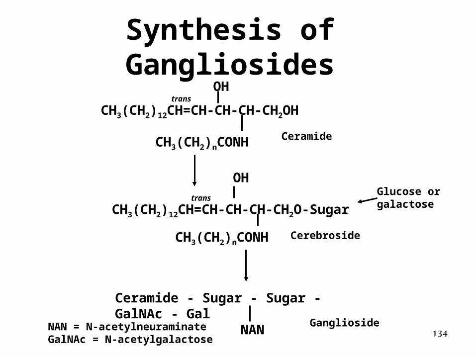
Synthesis of Gangliosides
CH3(CH2)12CH=CH-CH-CH-CH2OH
CH3(CH2)nCONH
OH
Ceramide
CH3(CH2)12CH=CH-CH-CH-CH2O-Sugar
CH3(CH2)nCONH
OH
Cerebroside
Ganglioside
trans
transGlucose orgalactose
Ceramide - Sugar - Sugar - GalNAc - Gal
NANNAN = N-acetylneuraminateGalNAc = N-acetylgalactose
135
Lipid Storage Diseases(Gangliosidoses)
136
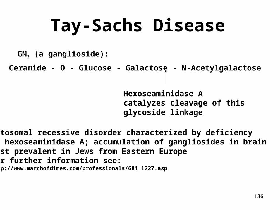
Tay-Sachs Disease
Ceramide - O - Glucose - Galactose - N-Acetylgalactose
Hexoseaminidase Acatalyzes cleavage of this glycoside linkage
GM2 (a ganglioside):
Autosomal recessive disorder characterized by deficiencyof hexoseaminidase A; accumulation of gangliosides in brainMost prevalent in Jews from Eastern EuropeFor further information see: http://www.marchofdimes.com/professionals/681_1227.asp
137
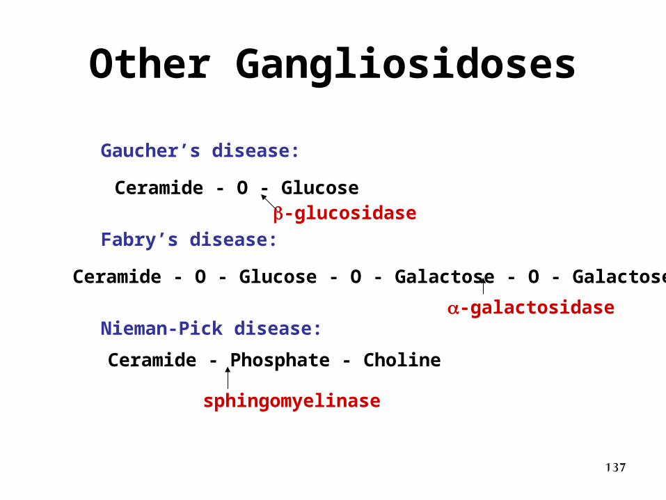
Other Gangliosidoses
Gaucher’s disease:
Fabry’s disease:
Nieman-Pick disease:
Ceramide - O - Glucose
Ceramide - O - Glucose - O - Galactose - O - Galactose
Ceramide - Phosphate - Choline
-glucosidase
-galactosidase
sphingomyelinase
138
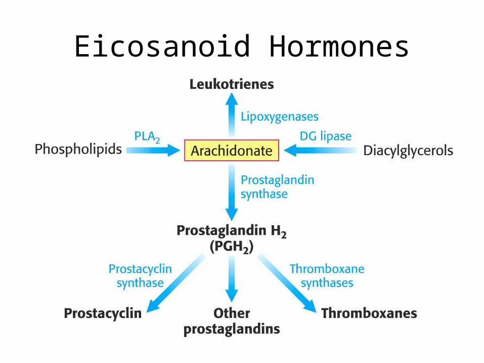
• Eicosanoid horomones are synthesized from arachadonic acid (20:4)– Prostaglandins
• 20-carbon fatty acid containing 5-carbon ring
• Prostacyclins
• Thromboxanes
– Leukotrienes• contain three conjugated double bonds
Eicosanoid Hormones
139
Eicosanoid Hormones
140
Cholesterol MetabolismCholesterol Metabolism
141
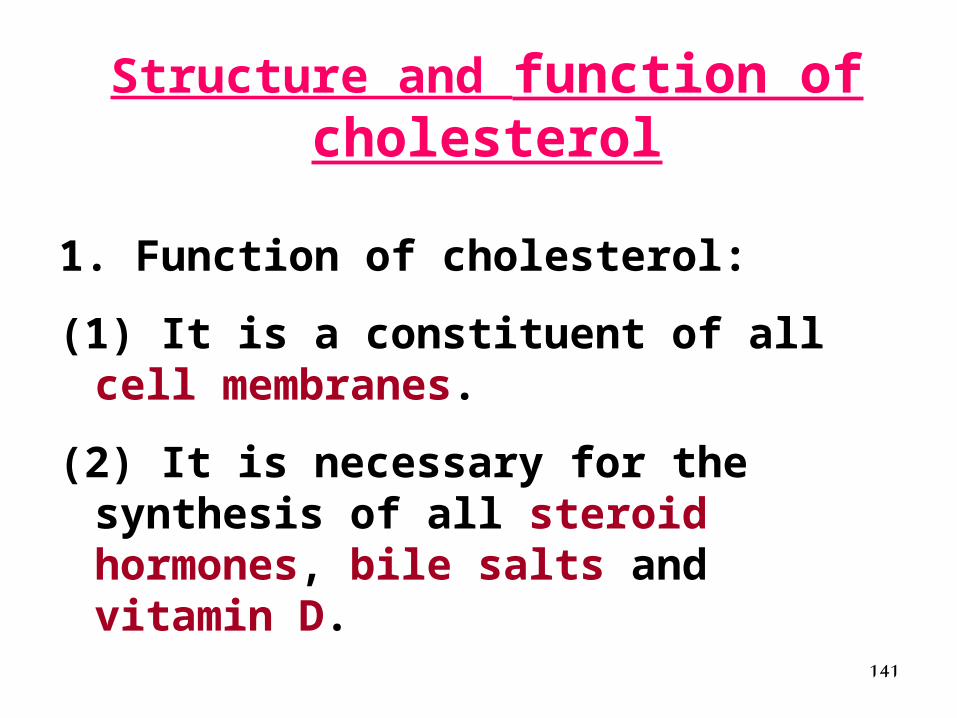
Structure and function of cholesterol
1. Function of cholesterol:
(1) It is a constituent of all cell membranes.
(2) It is necessary for the synthesis of all steroid hormones, bile salts and vitamin D.
142
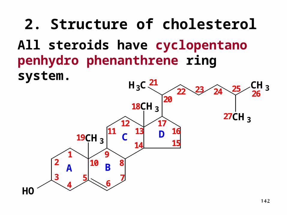
2. Structure of cholesterol
All steroids have cyclopentano penhydro phenanthrene ring system.
CH3
CH3
HO
H3C CH3
CH3
A B
C D
12
34
56
7
89
10
1112
13
14 15
1617
18
19
20
2122 23 24 25
26
27
143
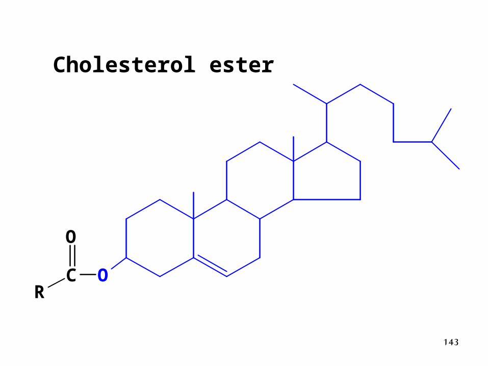
Cholesterol ester
OCR
O

144
Synthesis of cholesterolLocation:
• All tissue except brain and mature red blood cells.
• The major organ is liver (80%).
• Enzymes locate in cytosol and endoplasmic reticulum.
Materials:
Acetyl CoA, NADPH(H+), ATP
145
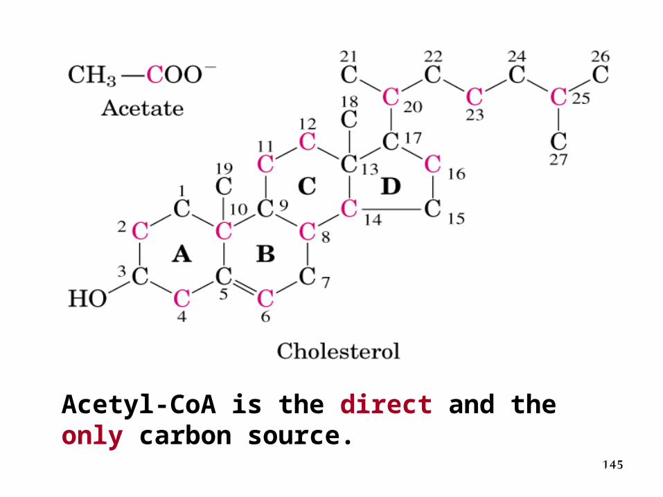
Acetyl-CoA is the direct and the only carbon source.
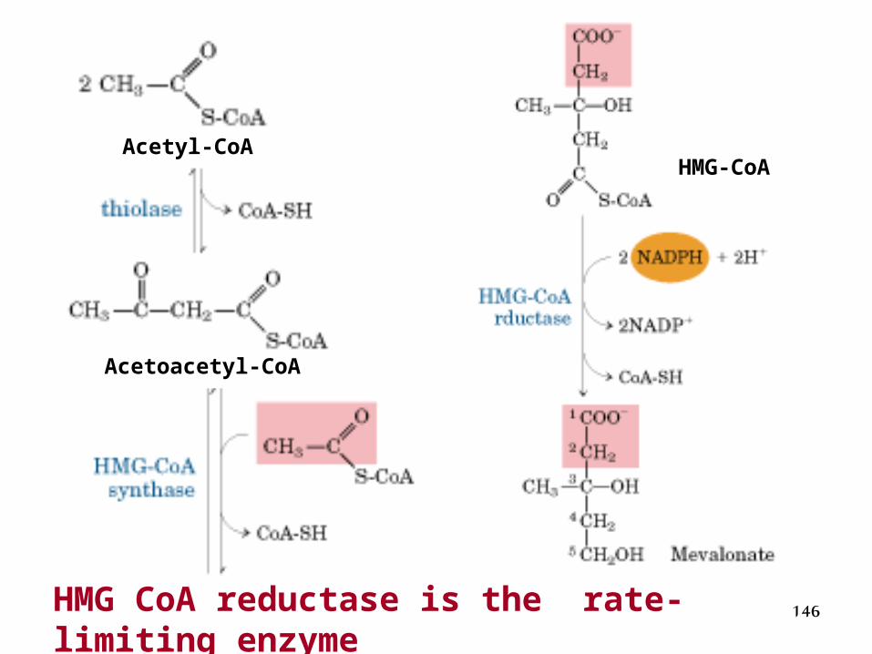
146HMG CoA reductase is the rate-limiting enzyme
Acetoacetyl-CoA
Acetyl-CoAHMG-CoA
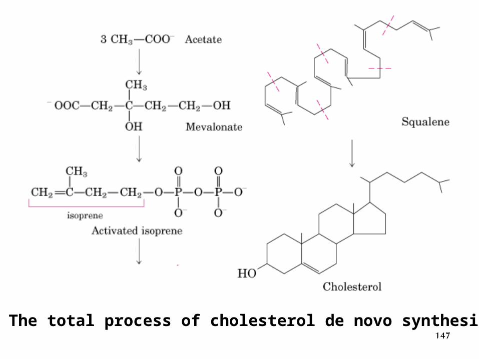
147The total process of cholesterol de novo synthesis
148
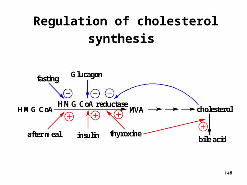
Regulation of cholesterol synthesis
MVAHMG CoA reductase
cholesterol
bile acid
fasting Glucagon
after meal insulin thyroxine
HMG CoA
149
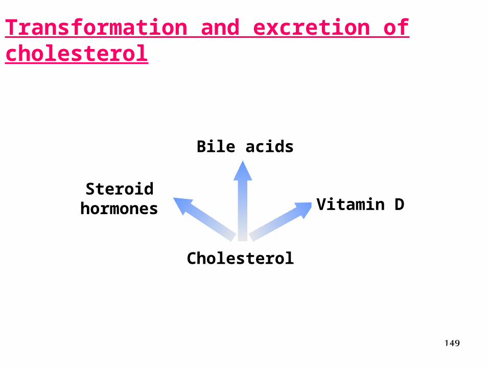
Transformation and excretion of cholesterol
Steroidhormones
Bile acids
Cholesterol
Vitamin D
150
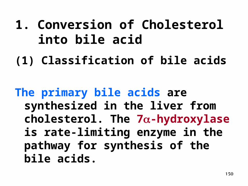
1. Conversion of Cholesterol into bile acid
(1) Classification of bile acids
The primary bile acids are synthesized in the liver from cholesterol. The 7-hydroxylase is rate-limiting enzyme in the pathway for synthesis of the bile acids.
151
The secondary bile acids are products that the primary bile acids in the intestine are subjected to some further changes by the activity of the intestinal bacteria.
152
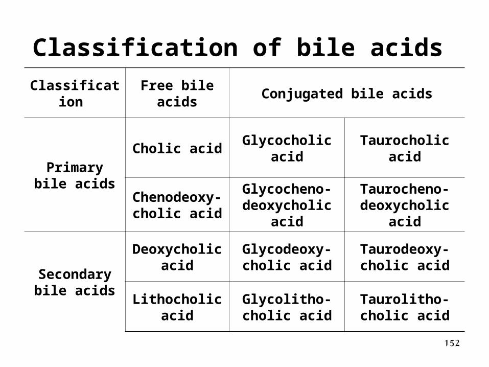
Classification of bile acids
Classification Free bile
acidsConjugated bile acids
Primary bile acids
Cholic acidGlycocholic
acidTaurocholic acid
Chenodeoxy-cholic acid
Glycocheno-deoxycholic
acid
Taurocheno-deoxycholic acid
Secondary bile acids
Deoxycholic acid
Glycodeoxy-cholic acid
Taurodeoxy-cholic acid
Lithocholic acid
Glycolitho-cholic acid
Taurolitho-cholic acid
153
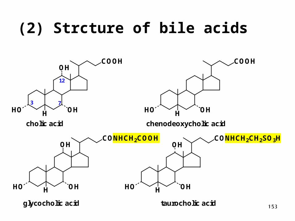
(2) Strcture of bile acids
HO OH
OH
H
COOH
HO OH
OH
H
CONHCH2COOH
HO OHH
COOH
HO OH
OH
H
CONHCH2CH2SO3H
cholic acid chenodeoxycholic acid
glycocholic acid taurocholic acid
3 7
12
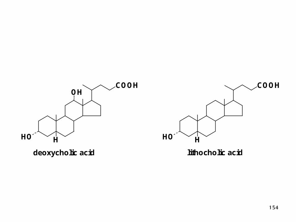
154
HO
OH
H
COOH
HO H
COOH
deoxycholic acid lithocholic acid
155
(3) Enterohepatic Cycle of bile acids
Conversion to bile salts, that are secreted into the intestine, is the only mechanism by which cholesterol is excreted.
Most bile acids are reabsorbed in the ileum , returned to the liver by the portal vein, and re-secreted into the intestine. This is the enterohepatic cycle.
156
(4) Function of bile acids
Bile acids are amphipathic, with detergent properties.
• Emulsify fat and aid digestion of fats & fat-soluble vitamins in the intestine.
• Increase solubility of cholesterol in bile.
157
2. Conversion of cholesterol into steroid hormones
• Tissues: adrenal cortex, gonads
• Steroid hormones: cortisol (glucocorti-coid), corticosterone and aldosterone (mineralocorticoid), progesterone, testosterone, and estradiol
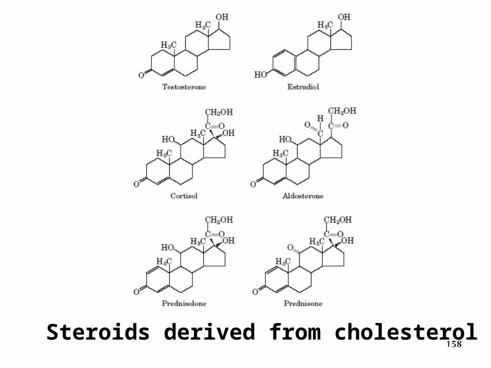
158Steroids derived from cholesterol
159
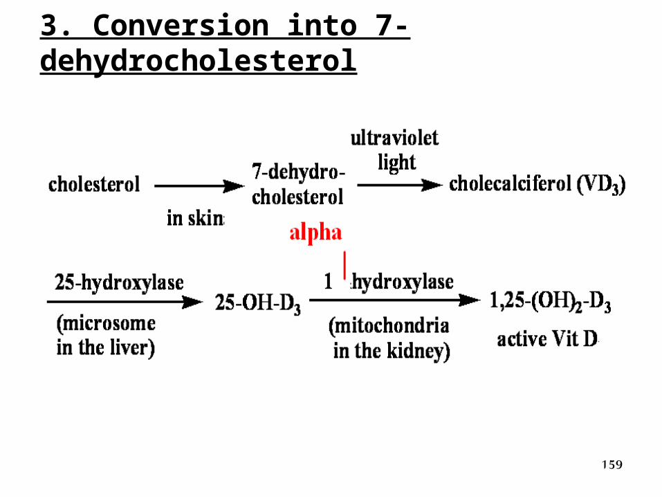
3. Conversion into 7-dehydrocholesterol
160
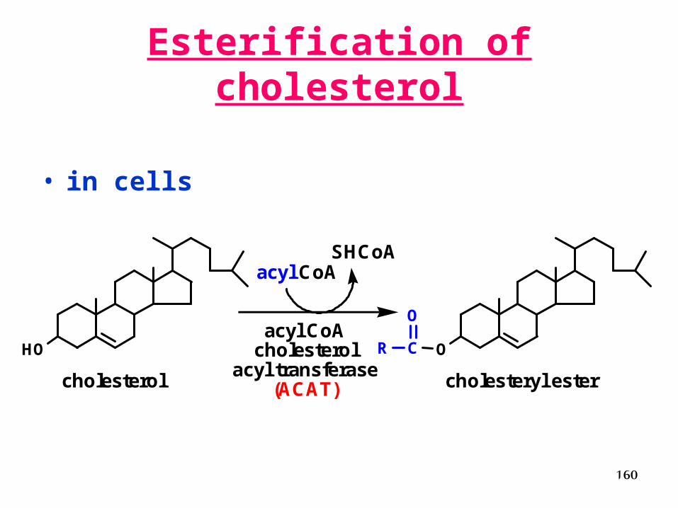
Esterification of cholesterol
• in cells
HO OCR
O
cholesterol cholesteryl ester
acyl CoA cholesterol
acyl transferase(ACAT)
acyl CoASHCoA
161
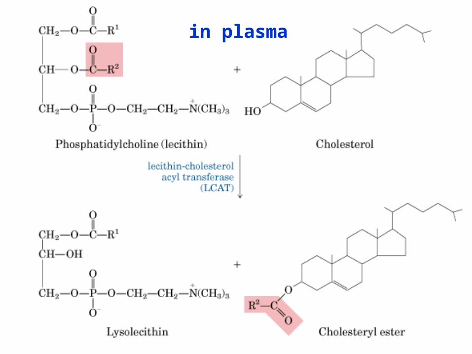
in plasma
162
Plasma Lipoproteins
163
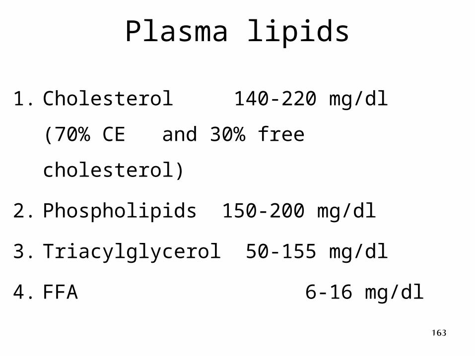
Plasma lipids
1. Cholesterol 140-220 mg/dl (70% CE
and 30% free cholesterol)
2. Phospholipids 150-200 mg/dl
3. Triacylglycerol 50-155 mg/dl
4. FFA 6-16 mg/dl
164
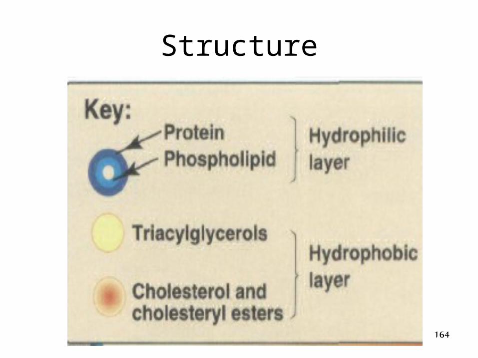
Structure
165
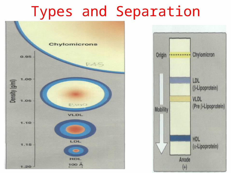
Types and Separation
166
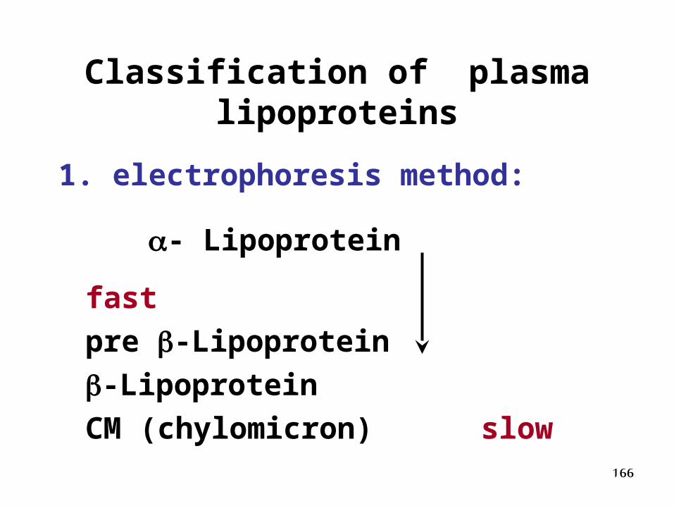
Classification of plasma lipoproteins
1. electrophoresis method:
- Lipoprotein fast
pre -Lipoprotein
-Lipoprotein
CM (chylomicron) slow
167
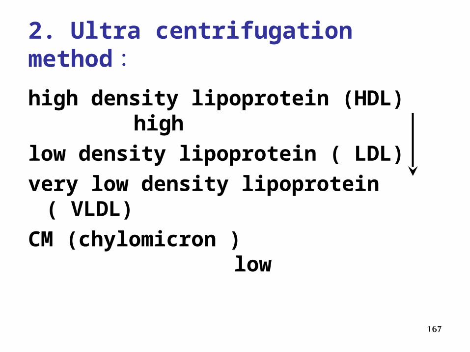
2. Ultra centrifugation method:
high density lipoprotein (HDL) high
low density lipoprotein ( LDL)
very low density lipoprotein ( VLDL)
CM (chylomicron ) low
168
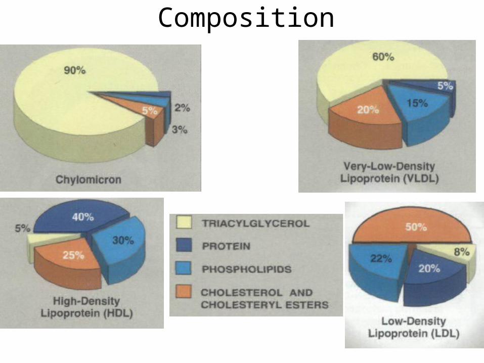
Composition
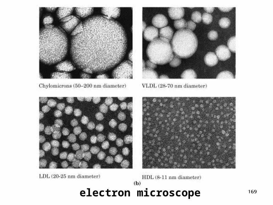
169electron microscope

170
- +
Origin CM
LDL VLDL HDL
Pre-
CM
Separation of plasma lipoproteins by electrophoresis on agarose gel
171
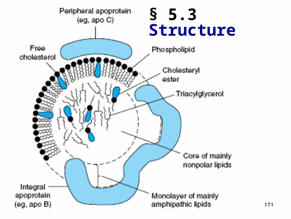
§ 5.3 Structure
172
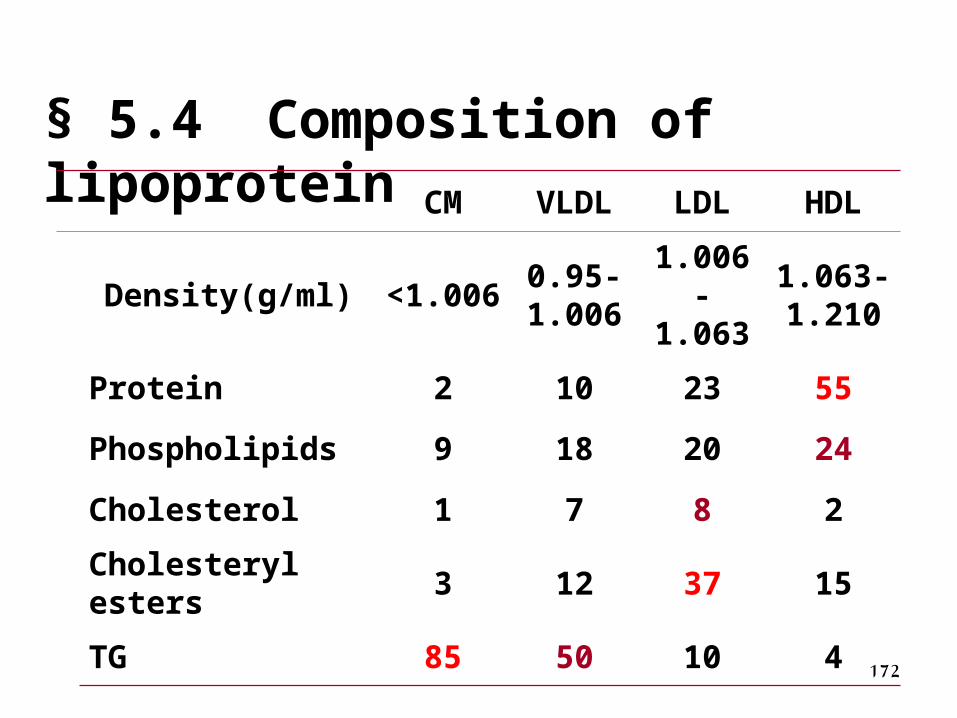
§ 5.4 Composition of lipoprotein
CM VLDL LDL HDL
Density(g/ml) <1.0060.95-1.006
1.006-1.063
1.063-1.210
Protein 2 10 23 55
Phospholipids 9 18 20 24
Cholesterol 1 7 8 2
Cholesteryl esters 3 12 37 15
TG 85 50 10 4
173
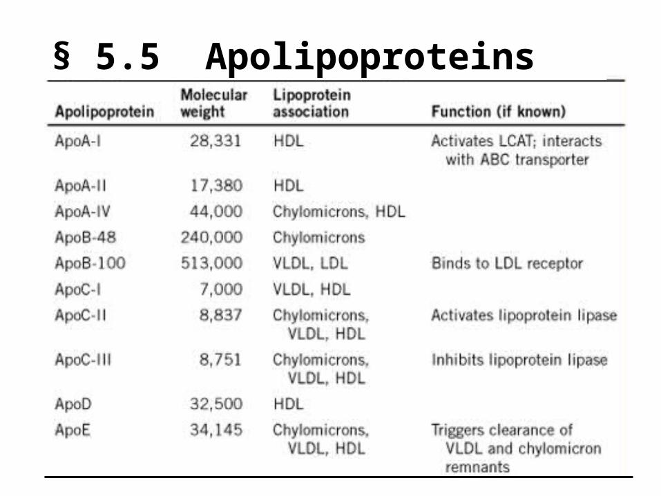
§ 5.5 Apolipoproteins
174
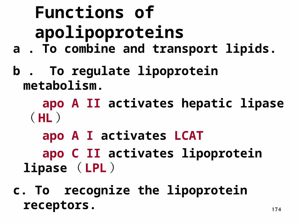
Functions of apolipoproteins
a . To combine and transport lipids.
b . To regulate lipoprotein metabolism.
apo A II activates hepatic lipase ( HL) apo A I activates LCAT
apo C II activates lipoprotein lipase ( LPL)
c. To recognize the lipoprotein receptors.
175
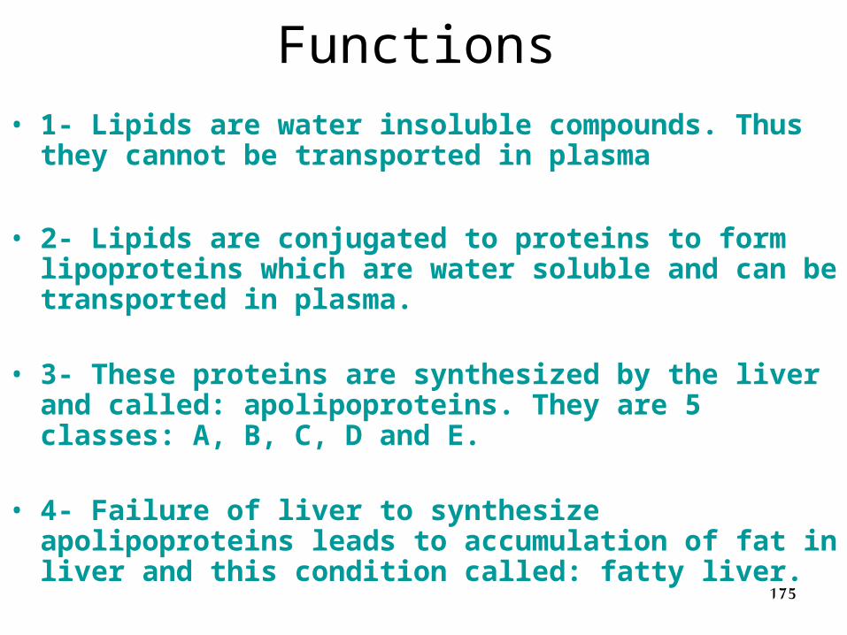
Functions• 1- Lipids are water insoluble compounds. Thus they cannot
be transported in plasma
• 2- Lipids are conjugated to proteins to form lipoproteins which are water soluble and can be transported in plasma.
• 3- These proteins are synthesized by the liver and called: apolipoproteins. They are 5 classes: A, B, C, D and E.
• 4- Failure of liver to synthesize apolipoproteins leads to accumulation of fat in liver and this condition called: fatty liver.
176
1. CM
• Chylomicrons are formed in the intestinal mucosal cells and secreted into the lacteals of lymphatic system.
177
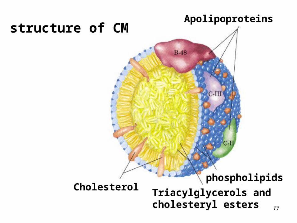
Cholesterol phospholipids
Triacylglycerols andcholesteryl esters
Apolipoproteins structure of CM
178
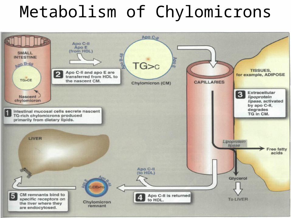
Metabolism of Chylomicrons
179
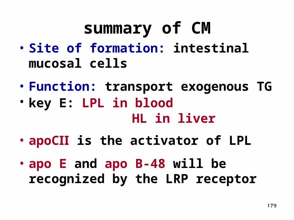
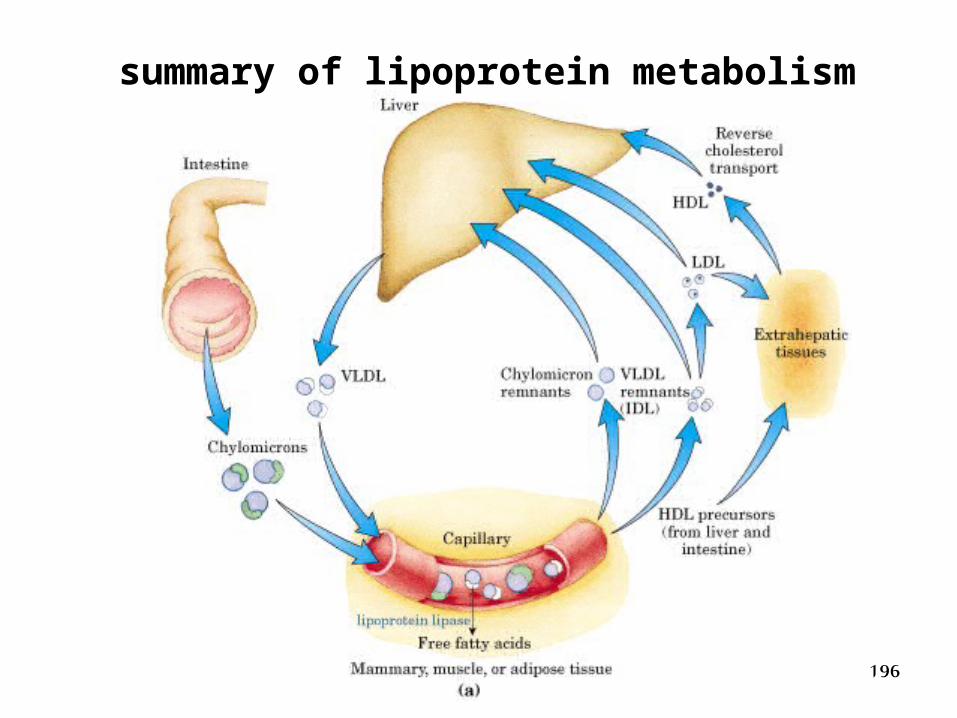
summary of CM• Site of formation: intestinal mucosal
cells
• Function: transport exogenous TG• key E: LPL in blood HL in liver
• apoCⅡ is the activator of LPL
• apo E and apo B-48 will be recognized by the LRP receptor
180
2. VLDL
• Very low density lipoproteins (VLDL) are synthesized in the liver and produce a turbidity in plasma.
181
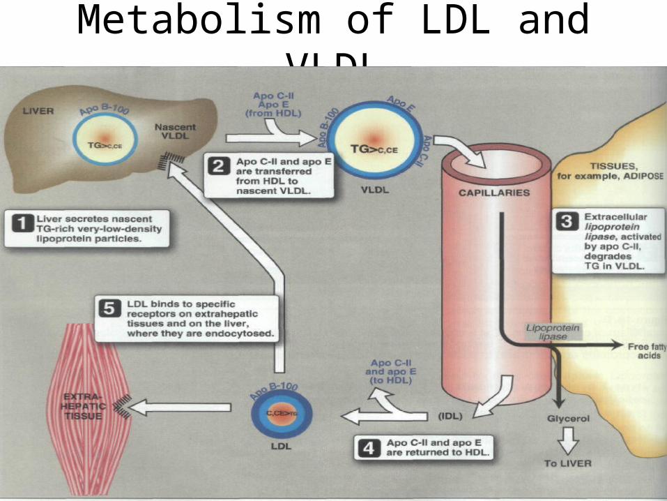
Metabolism of LDL and VLDL
182
Summary of VLDL
• Formation site: liver
• Function: VLDL carries endogenous triglycerides from liver to peripheral tissues for energy needs.
• key E: LPL in blood
HL in liver

183
3. LDL
• Most of the LDL particles are derived from VLDL, but a small part is directly released from liver. They are cholesterol rich lipoprotein molecules containing only apo B-100.
184
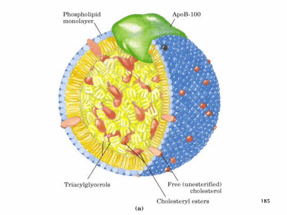
3. LDL
• Most of the LDL particles are derived from VLDL, but a small part is directly released from liver. They are cholesterol rich lipoprotein molecules containing only apo B-100.
185
186
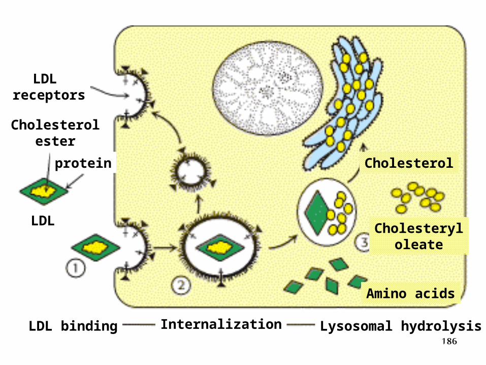
Internalization Lysosomal hydrolysisLDL binding
LDL receptors
Cholesterolester
protein
LDL
Cholesterol
Cholesteryloleate
Amino acids
187
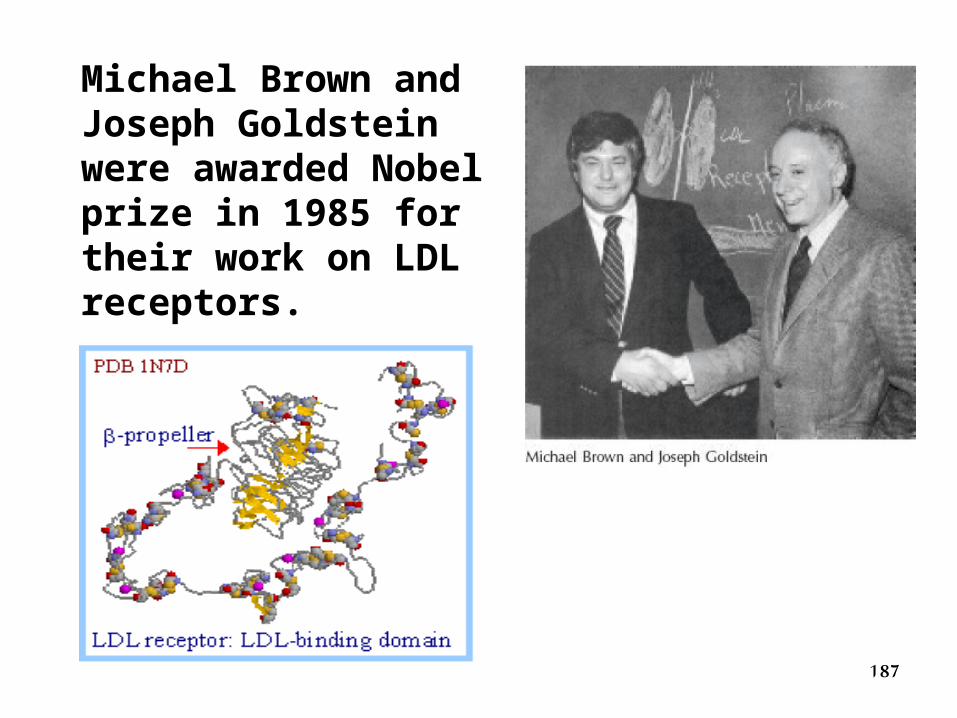
Michael Brown and Joseph Goldstein were awarded Nobel prize in 1985 for their work on LDL receptors.
188
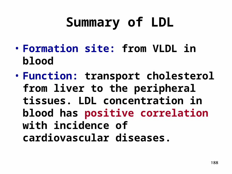
Summary of LDL
• Formation site: from VLDL in blood
• Function: transport cholesterol from liver to the peripheral tissues. LDL concentration in blood has positive correlation with incidence of cardiovascular diseases.
189
4. HDL
• LDL variety is called “ bad cholesterol” whereas HDL is known as “ good cholesterol” .
190
VLDL LDL
HDL
Cholesterol
HeartLiver
“BAD”
Deposit
Excretion
“Good”
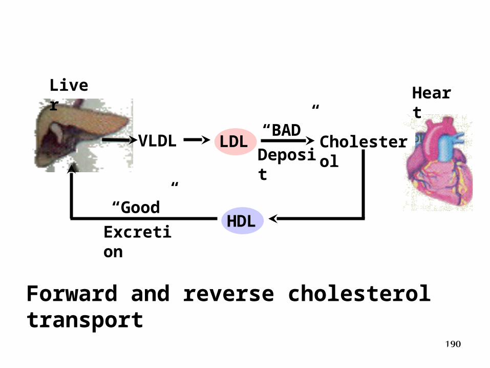
Forward and reverse cholesterol transport
191
Reverse cholesterol transport
• Cholesterol from tissues reach liver, and is later excreted. This is called reverse cholesterol transport by HDL.
192
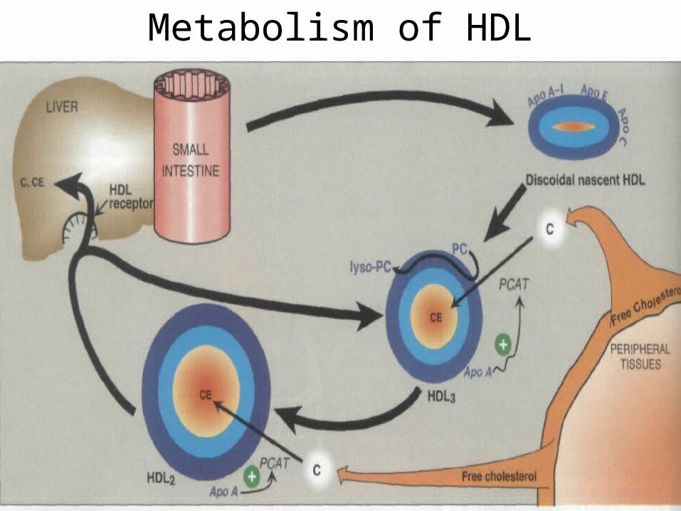
Metabolism of HDL
193
194
CETP
• Cholesterol ester transfer protein (CETP) transfer cholesterol ester in HDL to VLDL and LDL.
195
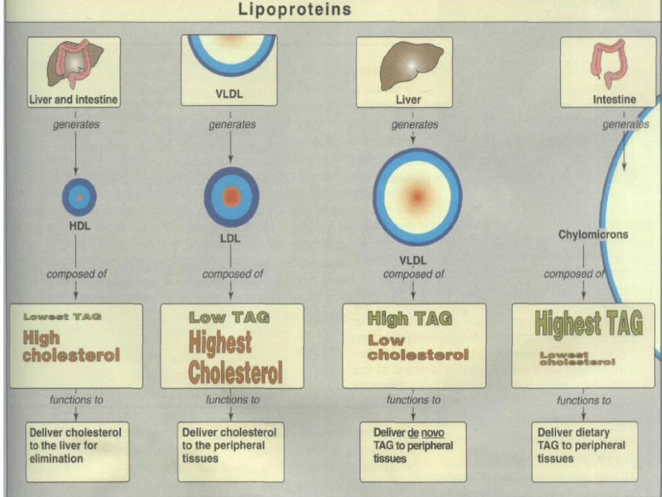
Summary of HDL
• Formation site: liver and intestine
• Function: transport cholesterol from peripheral tissues to liver
196
summary of lipoprotein metabolism
197
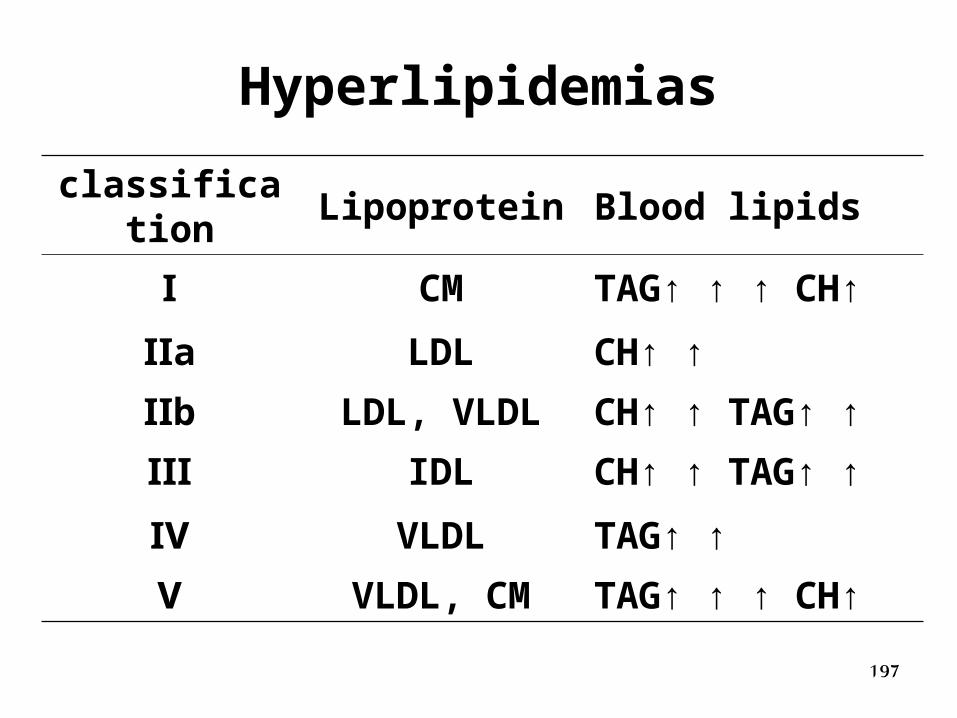
Hyperlipidemias
classification Lipoprotein Blood lipids
Ⅰ CM TAG↑ ↑ ↑ CH↑
Ⅱa LDL CH↑ ↑
Ⅱb LDL, VLDL CH↑ ↑ TAG↑ ↑
Ⅲ IDL CH↑ ↑ TAG↑ ↑
Ⅳ VLDL TAG↑ ↑
Ⅴ VLDL, CM TAG↑ ↑ ↑ CH↑
198
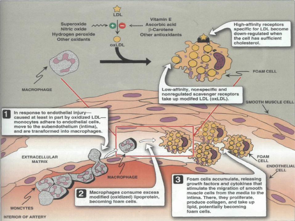
Role of Ox LDL in plaque formation and atherosclerosis
199
200
2- Secondary hyperlipoproteinemia:These abnormalities are associated with other diseases as:a) Diabetes mellitus. b)Hypothyroidism.c) Nephrotic syndrome.d) Obesity.e) Obstructive jaundice.
B- Hypolipoproteinemia:1) Abetalipoproteinemia:- Characterized by absence of LDL (β-lipoprotein). It is
associated with low concentrations of chylomicrons and VLDL.
2) Tangier disease:a- Due to deficiency of LCAT enzyme.b- Characterized by low concentration of HDL with
accumulation of cholesterol in tissues.
201
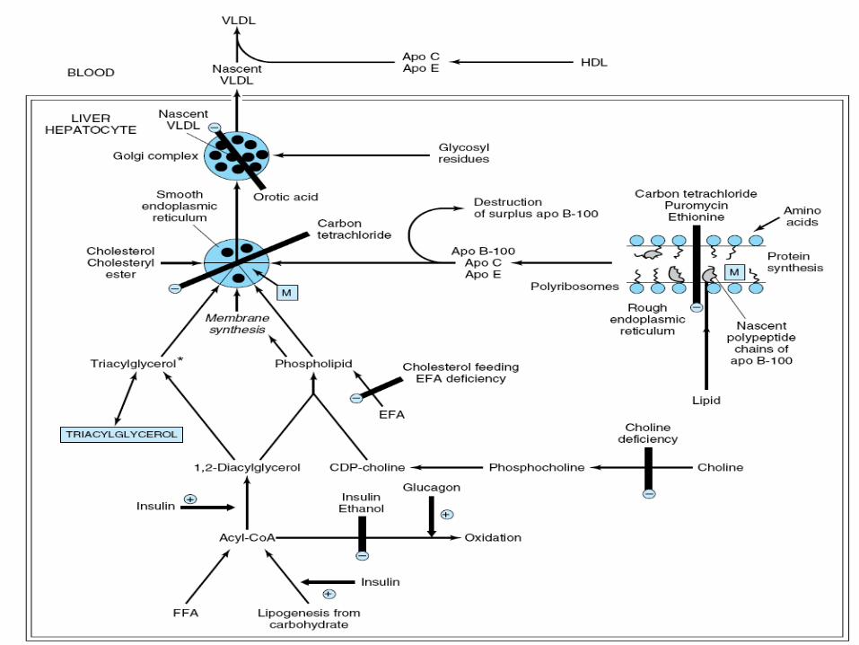
FATTY LIVER
I. Definition:-This is an accumulation of abnormal amount of fat in the liver for a long time with subsequent compression of liver cells.
II- Causes:
A- Over mobilization of fat from extrahepatic tissue to the liver.
B- During high carbohydrate diet.
C- Under mobilization of fat from the liver to the plasma.
202
A. Causes of over mobilization of fat from extrahepatic tissue to the liver:
1- During high fat diet.
2- Due to excessive lipolysis as in carbohydrate low diet, starvation and diabetes mellitus.
B. High carbohydrate diet:
- On high carbohydrate diet, liver is first saturated with glycogen, then any further amount of carbohydrate will be converted to triacylglycerols (lipogenesis).
203
C. Causes of under mobilization of fat from liver to the plasma:
- This is due deficiency any factor essential for plasma lipoproteins formation.
These factors are:1- Decreased synthesis of apoprotein (lipoprotein).2- Failure in formation of phospholipids.3- Failure in conjugation of apoprotein with triacylglycerols
or phospholipids.4- Failure in secretion of lipoprotein from liver to plasma.5- Liver poisons: As carbon tetrachloride, chloroform, lead
and arsenic. They cause fatty liver either by:a- Inhibition of formation of apoprotein.b- Inhibition of conjugation of apoprotein with lipids.c- Inhibition of secretion of lipoprotein.6- Alcoholism: Ethanol stimulates lipogenesis, inhibiting fatty
acid oxidation.
204

205
Related Documents