Protein Science (1997). 62360-872. Cambridge University Press. Printed in the USA. Copyright 0 1997 The Protein Society Limited proteolysis of ribonuclease A with thermolysin in trifluoroethanol PATRIZIA POLVERINO de LAURETO,] ELENA SCARAMELLA,' VINCENZO DE FILIPPIS,' MARTA BRUIX? MANUEL RICO? AND ANGEL0 FONTANA' 'CRIBI Biotechnology Centre, University of Padua, Via Trieste 75, 1-35121 Padua, Italy *Institute de Estructura de la Materia, CSIC, Serrano 119, E-28006 Madrid, Spain (RECEIVED October 15, 1996; ACCEPTED January 14, 1997) Abstract We have examined the proteolysis of bovine pancreatic ribonuclease A (RNase) by thermolysin when dissolved in aqueous buffer, pH 7.0, in the presence of 50% (v/v) trifluoroethanol (TFE). Under these solvent conditions, RNase acquires a conformational state characterized by an enhanced content of secondary structure (helix) and reduced tertiary structure, as given by CD measurements. It was found that the TFE-resistant thermolysin, despite its broad substrate specificity, selectively cleaves the 124-residue chain of RNase in its TFE state (20-42"C, 6-24 h) at peptide bond Asn 34-Leu 35, followed by a slower cleavage at peptide bond Thr 45-Phe 46. In the absence of TFE, native RNase is resistant to proteolysis by thermolysin. Two nicked RNase species, resulting from cleavages at one or two peptide bonds and thus constituted by two (1-34 and 35-124) (RNase Thl) or three (1-34, 35-45 and 46-124) (RNase Th2) fragments linked covalently by the four disulfide bonds of the protein, were isolated to homogeneity by chromatography and characterized. CD measurements provided evidence that RNase Thl maintains the overall conformational features of the native protein, but shows a reduced thermal stability with respect to that of the intact species (-ATm 16 "C); RNase Th2 instead is fully unfolded at room temperature. That the structure of RNase Thl is closely similar to that of the intact protein was confirmed unambiguously by two-dimensional NMR measurements. Structural differences be- tween the two protein species are located only at the level of the chain segment 30-41, i.e., at residues nearby the cleaved Asn 34-Leu 35 peptide bond. RNase Thl retained about 20% of the catalytic activity of the native enzyme, whereas RNase Th2 was inactive. The 3 1-39 segment of the polypeptide chain in native RNase forms an exposed and highly flexible loop, whereas the 41-48 region forms a &strand secondary structure containing active site residues. Thus, the conformational, stability, and functional properties of nicked RNase Thl and Th2 are in line with the concept that proteins appear to tolerate extensive structural variations only at their flexible or loose parts exposed to solvent. We discuss the conformational features of RNase in its TFE-state that likely dictate the selective proteolysis phenomenon by thermolysin. Keywords: CD; limited proteolysis; NMR; ribonuclease A; thermolysin; trifluoroethanol Short-chain alcohols, such as methanol, ethanol, propanol, chlo- roethanol, and, in particular, trifluoroethanol, have been used fre- quently to induce a-helical conformations in otherwise randomly coiled or partially structured polypeptides (Toniolo et al., 1975; Reprint requests to: Angelo Fontana, CRIBI Biotechnology Centre, Via Trieste 75, 35121 Padua, Italy; e-mail: [email protected]. Abbreviations: RNase, bovine pancreatic ribonuclease A (EC 3.1.27.5); RNase Thl, a folded, partially active derivative of RNase A with a chain fission at the peptide bond Asn 34-Leu 35 [RNase(1-34)(35-124)]; RNase Th2, a derivative of RNase A with chain fissions at peptide bonds Asn 34- Leu 35 and Thr45-Phe46 [RNase(l-34)(35-45)(46-124)]; TFE, 2.2.2- trifluoroethanol; [e], mean residue ellipticity; TFA, trifluoroacetic acid; PTC, phenylthiocarbamoyl; PTH, phenylthiohydantoin; Tris, tris(hydroxy- ethyllglycine; Dm, dithiothreitol; TOCSY, total correlation spectroscopy; methy1)aminomethane; Tricine, N-[2-hydroxy-l,l-bis(hydroxymethyl) NOESY, nuclear Overhauser effect spectroscopy; TSP, sodium 3-trimethyl- silyl (2,2,3,3-2H4) propionate; T,,,, the temperature at which a thermal transition is one-half complete; RP-HPLC, reverse-phase HPLC. Nelson & Kallenbach, 1986; Lehrman et ai., 1990; Segawa et ai., 1991; Sonnichsen et ai., 1992). It was shown that, on adding TFE to an aqueous solution of the S-peptide of bovine pancreatic ribo- nuclease A, this relatively small peptide acquires the helical con- formation that it adopts in the native protein (Tamburro et ai., 1968). Since then, TFE became the cosolvent of choice for induc- ing and stabilizing the helical secondary structure of peptides. The helix-inducing effect of TFE, however, is not occurring indepen- dently of the amino acid sequence of the polypeptide chain, but rather it seems that its main effect is that of increasing the helical population of those peptides and protein fragments that intrinsi- cally show a high propensity to form helices (Lehrman et ai., 1990; Segawa et ai., 1991). Weakly polar alcohols, including TFE, also have been shown to disrupt the native conformation of globular proteins, but the re- sulting denatured state was much different from the random-coil state observed in the presence of chemical denaturants such as urea 860

Welcome message from author
This document is posted to help you gain knowledge. Please leave a comment to let me know what you think about it! Share it to your friends and learn new things together.
Transcript
Protein Science (1997). 62360-872. Cambridge University Press. Printed in the USA. Copyright 0 1997 The Protein Society
Limited proteolysis of ribonuclease A with thermolysin in trifluoroethanol
PATRIZIA POLVERINO de LAURETO,] ELENA SCARAMELLA,' VINCENZO DE FILIPPIS,' MARTA BRUIX? MANUEL RICO? AND ANGEL0 FONTANA' 'CRIBI Biotechnology Centre, University of Padua, Via Trieste 75, 1-35121 Padua, Italy *Institute de Estructura de la Materia, CSIC, Serrano 119, E-28006 Madrid, Spain
(RECEIVED October 15, 1996; ACCEPTED January 14, 1997)
Abstract
We have examined the proteolysis of bovine pancreatic ribonuclease A (RNase) by thermolysin when dissolved in aqueous buffer, pH 7.0, in the presence of 50% (v/v) trifluoroethanol (TFE). Under these solvent conditions, RNase acquires a conformational state characterized by an enhanced content of secondary structure (helix) and reduced tertiary structure, as given by CD measurements. It was found that the TFE-resistant thermolysin, despite its broad substrate specificity, selectively cleaves the 124-residue chain of RNase in its TFE state (20-42"C, 6-24 h) at peptide bond Asn 34-Leu 35, followed by a slower cleavage at peptide bond Thr 45-Phe 46. In the absence of TFE, native RNase is resistant to proteolysis by thermolysin. Two nicked RNase species, resulting from cleavages at one or two peptide bonds and thus constituted by two (1-34 and 35-124) (RNase Thl) or three (1-34, 35-45 and 46-124) (RNase Th2) fragments linked covalently by the four disulfide bonds of the protein, were isolated to homogeneity by chromatography and characterized. CD measurements provided evidence that RNase Thl maintains the overall conformational features of the native protein, but shows a reduced thermal stability with respect to that of the intact species ( -ATm 16 "C); RNase Th2 instead is fully unfolded at room temperature. That the structure of RNase Thl is closely similar to that of the intact protein was confirmed unambiguously by two-dimensional NMR measurements. Structural differences be- tween the two protein species are located only at the level of the chain segment 30-41, i.e., at residues nearby the cleaved Asn 34-Leu 35 peptide bond. RNase Thl retained about 20% of the catalytic activity of the native enzyme, whereas RNase Th2 was inactive. The 3 1-39 segment of the polypeptide chain in native RNase forms an exposed and highly flexible loop, whereas the 41-48 region forms a &strand secondary structure containing active site residues. Thus, the conformational, stability, and functional properties of nicked RNase Thl and Th2 are in line with the concept that proteins appear to tolerate extensive structural variations only at their flexible or loose parts exposed to solvent. We discuss the conformational features of RNase in its TFE-state that likely dictate the selective proteolysis phenomenon by thermolysin.
Keywords: CD; limited proteolysis; NMR; ribonuclease A; thermolysin; trifluoroethanol
Short-chain alcohols, such as methanol, ethanol, propanol, chlo- roethanol, and, in particular, trifluoroethanol, have been used fre- quently to induce a-helical conformations in otherwise randomly coiled or partially structured polypeptides (Toniolo et al., 1975;
Reprint requests to: Angelo Fontana, CRIBI Biotechnology Centre, Via Trieste 75, 35121 Padua, Italy; e-mail: [email protected].
Abbreviations: RNase, bovine pancreatic ribonuclease A (EC 3.1.27.5); RNase Thl, a folded, partially active derivative of RNase A with a chain fission at the peptide bond Asn 34-Leu 35 [RNase(1-34)(35-124)]; RNase Th2, a derivative of RNase A with chain fissions at peptide bonds Asn 34- Leu 35 and Thr 45-Phe 46 [RNase(l-34)(35-45)(46-124)]; TFE, 2.2.2- trifluoroethanol; [e], mean residue ellipticity; TFA, trifluoroacetic acid; PTC, phenylthiocarbamoyl; PTH, phenylthiohydantoin; Tris, tris(hydroxy-
ethyllglycine; Dm, dithiothreitol; TOCSY, total correlation spectroscopy; methy1)aminomethane; Tricine, N-[2-hydroxy-l,l-bis(hydroxymethyl)
NOESY, nuclear Overhauser effect spectroscopy; TSP, sodium 3-trimethyl- silyl (2,2,3,3-2H4) propionate; T,,,, the temperature at which a thermal transition is one-half complete; RP-HPLC, reverse-phase HPLC.
Nelson & Kallenbach, 1986; Lehrman et ai., 1990; Segawa et ai., 1991; Sonnichsen et ai., 1992). It was shown that, on adding TFE to an aqueous solution of the S-peptide of bovine pancreatic ribo- nuclease A, this relatively small peptide acquires the helical con- formation that it adopts in the native protein (Tamburro et ai., 1968). Since then, TFE became the cosolvent of choice for induc- ing and stabilizing the helical secondary structure of peptides. The helix-inducing effect of TFE, however, is not occurring indepen- dently of the amino acid sequence of the polypeptide chain, but rather it seems that its main effect is that of increasing the helical population of those peptides and protein fragments that intrinsi- cally show a high propensity to form helices (Lehrman et ai., 1990; Segawa et ai., 1991).
Weakly polar alcohols, including TFE, also have been shown to disrupt the native conformation of globular proteins, but the re- sulting denatured state was much different from the random-coil state observed in the presence of chemical denaturants such as urea
860

Proteolysis of ribonuclease A in trifuoroethanol 86 1
or guanidine hydrochloride. Recent studies, based on CD and NMR measurements, have shown that the TFE-state of a protein, e.g., as obtained by dissolving the protein in 50% (v/v) aqueous TFE, is a stable, partially folded state with a high content of a-helical con- formation, but lacking the specific tertiary interactions of the na- tive protein (Buck et al., 1993, 1995; Fan et al., 1993; Alexandrescu et al., 1994; Shiraki et al., 1994). This TFE-state appears to be a noncompact, expanded conformational state characterized by an ensemble of fluctuating helices. However, the TFE-state does not result from a gross structural reorganization of the protein to an- other unrelated structure, because the large majority of amides protected from exchange in the TFE-state are also protected in the native state of the protein, thus implying a similar location of helical segments along the polypeptide chain in both native and TFE-state (Buck et al., 1993; Alexandrescu et al., 1994). One intriguing observation of these studies was that proteins in 50% (v/v) TFE acquire a higher content of helical secondary structure than the native species, implying that TFE also induces extra, non-native helices (Galat et ai., 1985; Buck et al., 1993, 1995; Alexandrescu et al., 1994; Shiraki et al., 1994). Indeed, evidence has been provided that TFE, and other alcohols, can induce the a-helical conformation in globular proteins consisting mainly of @sheets (Dufour & Haertlk, 1990; Fan et al., 1993; Liu et al., 1994; Shiraki et al., 1994). Overall, these various conformational properties of the TFE state can be related to those of the “molten globule” state of a protein, in which a polypeptide chain acquires a high content of secondary structure, and the side chains do not have fixed conformations (Kuwajima, 1989; Ptitsyn, 1992, 1995).
The precise physical mechanism of a-helix induction by TFE is not fully understood and is the subject of intense and recent in- vestigations (Thomas & Dill, 1993; Jasanoff & Fersht, 1994; Shiraki et al., 1994; Cammers-Goodwin et al., 1996). Current views are that TFE favors the formation of the intramolecular hydrogen bonds of the a-helical conformation rather than hydrogen bonds to sol- vent. Even if helix induction and stabilization by TFE is the most frequently observed phenomenon, evidence for TFE-mediated sta- bilization of P-bends and P-hairpins in relatively short peptides has been provided (Blanco et al., 1994) and thus it may be pro- posed that aqueous TFE, in general, appears to strengthen the backbone-backbone hydrogen bonds.
The aim of the present work was to probe the conformational state of RNase when dissolved in aqueous TFE by using proteo- lytic enzymes as probes of protein structure and dynamics (Mi- halyi, 1978; Neurath, 1980; Fontana et al., 1989, 1993; Price & Johnson, 1990; Signor et al., 1990; Wilson, 1991). This experi- mental approach relies on the fact that proteolysis of a polypeptide chain requires the binding of a chain segment of 6-8 amino acid residues of the polypeptide substrate at the active site cleft of the protease (Berger & Schechter, 1970). This is difficult to achieve with native, folded proteins, which are therefore usually quite re- sistant to proteolysis. considering that native (N) proteins are in equilibrium with the unfolded, denatured (D) species, it is con- ceivable to suggest that protein degradation by proteolysis occurs via the unfolded species only, so that the N H D equilibrium actually dictates and control the rate of protein degradation to small peptides, if a protease of low substrate specificity is em- ployed. Nevertheless, native globular proteins also can act as sub- strates for proteolysis, but, in this case, usually selective or preferential proteolysis at the level of very few peptide bonds is observed, leading to “nicked” protein species constituted by rela- tively large protein fragments associated in a stable and often func-
tional complex (Mihalyi, 1978). A detailed survey of the structural characteristics of the sites of limited proteolysis in globular proteins of known three-dimensional structure enabled us to observe that pep- tide bond fissions are located at flexible loops of the protein and never at chain segments embedded in rigid secondary structure elements such as helices (Fontana et al., 1989, 1993; Polverino de Laureto et al., 1994a, 1994b). Thus, limited proteolysis of globular proteins is expected to occur at sites characterized by the motility required for an effective and stereospecific binding at the active site of the protease in order to form the idealized transition state of the hydro- lytic reaction. Indeed, a clear-cut correlation exists between sites of limited proteolysis and sites of high segmental mobility (B-factor) determined crystallographically (Fontana et al., 1986). It is our be- lief that notions of exposure, accessibility, or protrusion (Novotny & Bruccoleri, 1987; Hubbard et al., 1991) are clearly not sufficient to explain the limited proteolysis phenomenon, because it is evident that, even in a small globular protein, there are many exposed sites (see also Hubbard et al., 1994).
For this study of limited proteolysis of a model protein in its TFE-state, the thermophilic enzyme thermolysin (Heinrikson, 1977; Holmes & Matthews, 1982) appeared to be a most suitable pro- teolytic probe, because of its noteworthy stability under relatively harsh solvent conditions, including aqueous organic solvents (Wayne & Fruton, 1983; Welinder, 1988; Kitaguchi & Klibanov, 1989) and broad substrate specificity (Heinrikson, 1977; Keil, 1982). Thus, it was anticipated that thermolysin would cleave the polypeptide chain of RNase in its TFE-state at sites characterized by the flex- ibility required for an efficient proteolysis and not by the speci- ficity of the protease. Moreover, RNase was considered a convenient choice as a model protein because of the extensive literature re- garding its structure, dynamics, folding, and function, and because limited proteolysis played an important role in the elucidation of its structure and mechanism of action (Richards & Wyckoff, 1971; Blackburn & Moore, 1982). The subtilisin-mediated cleavage of na- tive RNase at the Ala 20-Ser 21 peptide bond leading to RNase S, the nicked protein species RNase[( 1-20)(21-124)] (Richards & Vithayathil, 1959), can be considered the most classical and fruitful limited proteolysis experiment, which, over the years, enabled a num- ber of additional studies of the RNase molecule that contributed a great deal to present day knowledge and understanding of protein structure and function.
Here we report the isolation and characterization of two nicked species of RNase with one (RNase Thl) and two (RNase Th2) pep- tide bonds cleaved along the disulfide-crosslinked, 124-residue chain of the protein. Structural properties of RNase dissolved in aqueous TFE that likely contribute to its specific nicking by thermolysin are discussed. Conformational and functional properties of the two nicked RNase molecules are interpreted, taking advantage of the known three-dimensional structure and dynamics of native RNase in the crystal state (Wyckoff et al., 1970; Wlodawer et al., 1982, 1988) or in solution (Rico et al., 1989; Santoro et al., 1993)?
Results
Proteolysis of RNase by thermolysin in the presence of TFE
Initial experiments of proteolysis of RNase by thermolysin in the presence of aqueous TFE were conducted by varying the alcohol
3Presented at the Ninth Symposium of the Protein Society, Boston, July 8-12, 1995 [Commun. 7OO-S].

862
concentration. the protease/RNase ratio. as well as the time and temperature of incubation. Samples were withdrawn periodically from the reaction mixture and proteolysis stopped by acidification. The pattern of protein fragmentation was monitored by SDS- PAGE under reducing conditions and utilizing the Tricine buffer system (Schagger & von Jagow, 1987) (Fig. I ). In the absence of TFE, RNase is resistant to proteolysis by thermolysin (3% by mass) when incubated in Tris buffer. pH 7.0, at 20-42°C for several hours, because only the band of the intact protein (-14 kDa) was observed in the stained gel (Fig. I ) . At higher temperatures (50-65°C) and as the time of incubation increased (up to 24 h), the band corresponding to the intact protein disap- peared gradually from the gel, whereas other bands of peptide/ protein material were not seen in the gel. I t is likely that the native-denatured conformational transition of RNase dictates the rate of protein degradation and that the unfolded protein is digested to small peptides not stained by the Coomassie dye of the SDS- PAGE system.
The results of the SDS-PAGE analysis (Fig. IA) of the proteo- lytic mixture obtained by reacting RNase with thermolysin (4 h, 42°C) in the presence of increasing concentrations of TFE (up to 50% by volume) show that RNase is cleaved if the alcohol con- centration is >20%. The interesting observation is that few and rather large fragments are produced. In 30-509 TFE, three main protein fragments of approximately 10, 9, and 4 kDa are seen in the stained gel. besides traces of the intact protein (-I4 kDa) (Fig. IA). The SDS-PAGE analysis of the time-course proteolysis of RNase in 50% TFE at 20°C is shown in Figure IB. It is seen that the initial cleavage of the protein (after 4 h ) leads to two main fragments of - IO and -4 kDa, implying that the protein is cleaved at a single peptide bond, considering that the sum of the molecular masses of the two fragments corresponds to that of the intact protein (-14 kDa). The gel of Figure IB clearly shows that the -10 kDa fragment is converted subsequently and slowly to a fragment of a lower mass, so that, after 30 h reaction, essentially a -9-kDa and -4-kDa fragment appear to be the end products of the reaction (note that small peptides are not stained in the gel). The pattern of proteolysis of RNase in 30% TFE was similar to that obtained in 50% TFE (see Fig. IA). These last solvent conditions were used routinely for the preparative isolation of nicked RNase (see below), because this solvent mixture was used in previous studies of the TFE-induced structure of some proteins (Buck et al.,
A
kDa o IO 20 30 40 W%TFE
16.8- 14.4- 10.8- 8.4- 6.1 - 2.5 -
P: Polverino de Lmreto et al.
1993; Alexandrescu et al.. 1994; Fontana et al.. 1995; Polverino de Laureto et al., 1995a. 199%).
In order to identify the thermolytic peptide bond fission(s) along the polypeptide chain of RNase, the peptide/protein bands of the SDS-PAGE gel were electroblotted (Matsudaira. 1987) onto a Pro- Blott membrane and then subjected to Edman degradation using the automatic sequencer (see Materials and methods). These pre- liminary N-terminal sequence data of the electroblotted fragments allowed us to suggest that the initial single nick of the chain occurs at peptide bond Asn 34-Leu 35, followed by an additional cleav- age at Thr 45-Phe 46 (see below for more precise analytical data of the nicked RNase species isolated to homogeneity).
Isolation and characteri:ntion qf nicked RNase
The results of SDS-PAGE analysis (above) were complemented by those obtained by ion-exchange and RP-HPLC. In particular, col- umn chromatography was employed for the purification of nicked RNase species in sufficient quantity for subsequent analysis. It was clear from the SDS-PAGE analysis (Fig. IB) that a proteolytic mixture leading mostly to the hydrolysis of a single peptide bond (see above) was that resulting from a reaction conducted in 50% TFE at 20°C for 4 h (Fig. 1 B). In practice. we have found that proteolysis at 42 "C for 30 min leads to a quite similar proteolytic mixture. A sample of this mixture was applied to a Mono-S col- umn, which was eluted with a salt gradient (Fig. 2A). The peptide/ protein material of the major peak of the chromatogram (named T h l , Fig. 2A) gave two bands in the SDS-PAGE gel (reducing conditions) of - 10 and -4 kDa, respectively.
In order to produce a nicked RNase with more than one peptide bond hydrolyzed (see above), proteolysis was conducted in 508 TFE for 6 h at 42°C. This sample was applied to a Vydac C4 column eluted with a gradient of acetonitrile. The chromatogram of Figure 2B shows that a major peak of protein material is eluted from the column at -37% acetonitrile. This protein sample (named RNase Th2) was shown by SDS-PAGE (reducing conditions) to be constituted by two fragments of molecular masses of -9 kDa and -4 kDa.
The disulfide-crosslinked protein material RNase T h l and RNase Th2, isolated by ion-exchange and RP-HPLC, respectively, was reduced with DTT and subsequently S-carboxamidomethylated (see Materials and methods). From these reaction mixtures, the protein
B 0 4 6 9 24 30 hours
Fig. 1. SDS-PAGE of proteolytic mixture of RNase with thermolysin. A: Reaction proceeded for 4 h at 42 "C. in 50 mM Tris-HCI, 5 mM CaCL, pH 7.0. at an enzyme to substrate ratio of 3 6 (by weight) in the presence of TFE from 0 to 5 0 8 (by volume). Molecular weight markers are shown on the left in kDa. R: Time-course of the proteolysis of RNase with thermolysin in Tris buffer containing 50% TFE at 20°C.

Proteolysis of ribonuclease A in tr@uoroethanol 863
I I I I
A
/ /
/ /
/
I ' /
/ - /
-hi / /
/
/ /
/
B rh;
, . /-
/ I
I I
I I
I I "-" I
A) I
5 10 15 20 25 RETENTION TIME, min
50
40
2 2o ; ,30 E
0
0 K
10 h
0
RETENTION TIME, min
Fig. 2. Chromatographic analysis of the proteolytic mixture of RNase with thermolysin. A: Ion-exchange HPLC of RNase reacted with thermolysin in 50% TFE. Reaction proceeded for 30 min at 42°C using an enzyme/substrate ratio of 3% (by weight) in 50 mM Tris-HC1 buffer, pH 7.0, containing 5 mM CaCI2 and 50% TFE. The separation was performed on a Mono-S HR 5/5 column (Pharmacia-LKB), equilibrated with 50 mM KHlPOd buffer, pH 7.0, and eluted with a gradient (B) of the same buffer containing 0.8 M KCI. B: RP-HPLC of RNase reacted with thermolysin in 50% TFE. Proteolysis was conducted under the same conditions given in A and allowed to proceed for 6 h at 42 "C. The separation was performed on a Vydac C4 column eluted at a flow rate of 0.6 mL/min with a gradient of acetonitrile containing 0.05% TFA (by volume).
fragments constituting RNase Thl and RNase Th2 were isolated by RP-HPLC (see Fig. S.2, Electronic Appendix) and subjected to further analysis (amino acid composition after acid hydrolysis, N-terminal sequencing, and determination of the molecular mass by mass spectrometry). The analytical data of the fragments, as well as those of the parent disulfide-crosslinked protein species (see Table S.1, Electronic Appendix), when compared with the known amino acid sequence of RNase (Smyth et al., 1963; see Fig. S.l, Electronic Appendix), allowed us to establish unambig- uously the chemical identity of the nicked protein species. RNase Thl is a nicked protein with the single Asn 34-Leu 35 peptide bond hydrolyzed, whereas RNase Th2 is with both Asn 34-Leu 35 and Thr 45-Phe 46 hydrolyzed. RNase Thl is therefore constituted by two fragments, 1-34 and 35-124, and RNase Th2 by three fragments, 1-34, 35-45, and 46-124. It should be noted that the fragments in both RNase Thl and Th2 are linked covalently by disulfide bonds, because the eight cysteine residues of the RNase molecule form four disulfide bridges between residues 26 and 84, 40 and 95, 58 and 110, and 65 and 72 (Smyth et al., 1963).
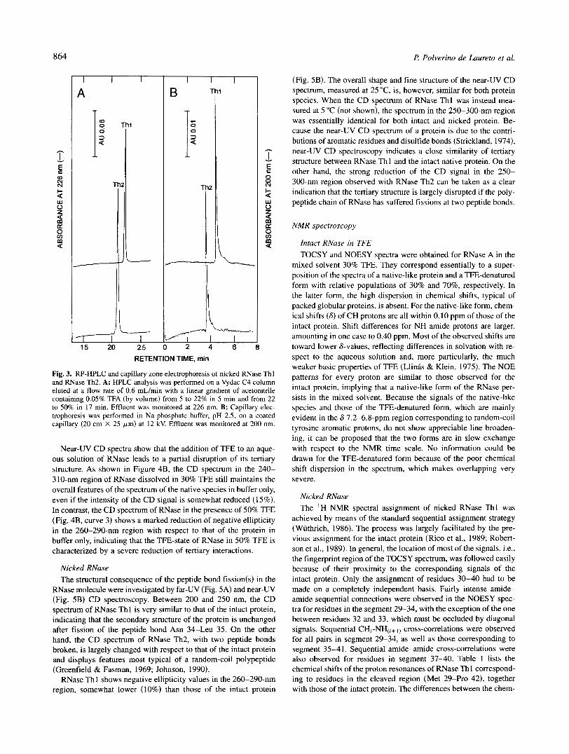
Because the aim of the present work was also to characterize the structural and functional properties of the nicked protein species, samples of purified RNase Thl and RNase Th2 were further an- alyzed by RP-HPLC and by capillary electrophoresis. The results of these analyses demonstrated that indeed the isolated protein samples were highly homogeneous (Fig. 3). Moreover, gel filtra- tion experiments also allowed us to establish the purity of nicked proteins (not shown). Of note, RNase Thl eluted from a Superose-12
column at the same position as that of the intact protein, whereas RNase Th2 eluted somewhat earlier, signifying a higher hydro- dynamic volume o f RNase Th2 with respect to that of intact and RNase Th 1 and therefore a more expanded protein species (Corbett & Roche, 1984).
CD measurements
Intact RNase in TFE The effect of increasing concentrations of TFE on the confor-
mation of intact RNase was investigated by far-UV (Fig. 4A) and near-UV (Fig. 4B) CD spectroscopy. If dissolved in 30% aqueous TFE at pH 7.0, RNase acquires a slightly enhanced content of a-helix with respect to that of the protein in buffer alone. In fact, the intensity of the negative ellipticity in the 200-250-nm region of RNase in aqueous TFE is greater than that of the protein in buffer only, and shows characteristics typical of helical polypep- tides with minima of ellipticity near 208 and 222 nm (Greenfield & Fasman, 1969; Johnson, 1990). The enhanced content of helicity is much more evident if the protein is dissolved in 50% TFE (Fig. 4A, curve 3). A quantitative analysis of the far-UV CD spec- tra in terms of percentages of secondary structure (Chen et al., 1972) allowed us to estimate that RNase acquires a conformational state characterized by 33% and 45% helicity in 30% and 50% TFE, respectively. These figures should be compared with the 30% he- lical content of native RNase, derived from the crystal structure of the protein.
Th
Thl
B Thl
ThZ
RETENTION TIME, min
Fig. 3. RP-HPLC and capillary zone electrophoresis of nicked RNase Thl and RNase Th2. A: HPLC analysis was performed on a Vydac C4 column eluted at a flow rate of 0.6 mL/min with a linear gradient of acetonitrile containing 0.05% TFA (by volume) from 5 to 22% in 5 min and from 22 to 50% in 17 min. Effluent was monitored at 226 nm. B: Capillary elec- trophoresis was performed in Na-phosphate buffer, pH 2.5, on a coated capillary (20 cm X 25 pm) at 12 kV. Effluent was monitored at 200 nm.
Near-UV CD spectra show that the addition of TFE to an aque- ous solution of RNase leads to a partial disruption of its tertiary structure. As shown in Figure 4B, the CD spectrum in the 240- 310-nm region of RNase dissolved in 30% TFE still maintains the overall features of the spectrum of the native species in buffer only, even if the intensity of the CD signal is somewhat reduced (15%). In contrast, the CD spectrum of RNase in the presence of 50% TFE (Fig. 4B, curve 3) shows a marked reduction of negative ellipticity in the 260-290-nm region with respect to that of the protein in buffer only, indicating that the TFE-state of RNase in 50% TFE is characterized by a severe reduction of tertiary interactions.
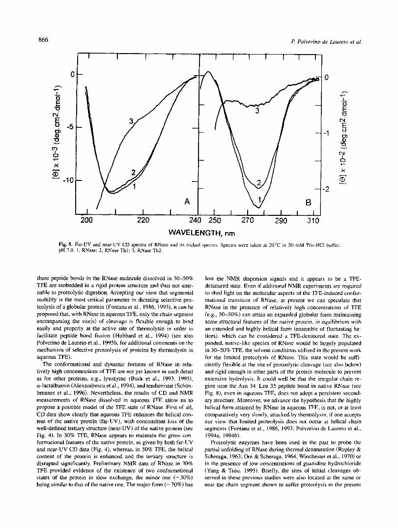
Nicked RNase The structural consequence of the peptide bond fission(s) in the
RNase molecule were investigated by far-UV (Fig. 5A) and near-UV (Fig. 5B) CD spectroscopy. Between 200 and 250 nm, the CD spectrum of RNase Thl is very similar to that of the intact protein, indicating that the secondary structure of the protein is unchanged after fission of the peptide bond Asn 34-Leu 35. On the other hand, the CD spectrum of RNase Th2, with two peptide bonds broken, is largely changed with respect to that of the intact protein and displays features most typical of a random-coil polypeptide (Greenfield & Fasman, 1969; Johnson, 1990).
RNase Thl shows negative ellipticity values in the 260-290-nm region, somewhat lower (10%) than those of the intact protein
I? Polverino de Laureto et al.
(Fig. 5B). The overall shape and fine structure of the near-UV CD spectrum, measured at 25 "C, is, however, similar for both protein species. When the CD spectrum of RNase Thl was instead mea- sured at 5 "C (not shown), the spectrum in the 250-300-nm region was essentially identical for both intact and nicked protein. Be- cause the near-UV CD spectrum of a protein is due to the contri- butions of aromatic residues and disulfide bonds (Strickland, 1974), near-UV CD spectroscopy indicates a close similarity of tertiary structure between RNase Thl and the intact native protein. On the other hand, the strong reduction of the CD signal in the 250- 300-nm region observed with RNase Th2 can be taken as a clear indication that the tertiary structure is largely disrupted if the poly- peptide chain of RNase has suffered fissions at two peptide bonds.
NMR spectroscopy
Intact RNase in TFE TOCSY and NOESY spectra were obtained for RNase A in the
mixed-solvent 30% TFE. They correspond essentially to a super- position of the spectra of a native-like protein and a TFE-denatured form with relative populations of 30% and 70%, respectively. In the latter form, the high dispersion in chemical shifts, typical of packed globular proteins, is absent. For the native-like form, chem- ical shifts (6) of CH protons are all within 0.10 ppm of those of the intact protein. Shift differences for NH amide protons are larger, amounting in one case to 0.40 ppm. Most of the observed shifts are toward lower 6-values, reflecting differences in solvation with re- spect to the aqueous solution and, more particularly, the much weaker basic properties of TFE (Llinis & Klein, 1975). The NOE patterns for every proton are similar to those observed for the intact protein, implying that a native-like form of the RNase per- sists in the mixed solvent. Because the signals of the native-like species and those of the TFE-denatured form, which are mainly evident in the 6 7.2-6.8-ppm region corresponding to random-coil tyrosine aromatic protons, do not show appreciable line broaden- ing, it can be proposed that the two forms are in slow exchange with respect to the NMR time scale. No information could be drawn for the TFE-denatured form because of the poor chemical shift dispersion in the spectrum, which makes overlapping very severe.
Nicked RNase The 'H NMR spectral assignment of nicked RNase Thl was
achieved by means of the standard sequential assignment strategy (Wiithrich, 1986). The process was largely facilitated by the pre- vious assignment for the intact protein (Rico et al., 1989; Robert- son et al., 1989). In general, the location of most of the signals, i.e., the fingerprint region of the TOCSY spectrum, was followed easily because of their proximity to the corresponding signals of the intact protein. Only the assignment of residues 30-40 had to be made on a completely independent basis. Fairly intense amide- amide sequential connections were observed in the NOESY spec- tra for residues in the segment 29-34, with the exception of the one between residues 32 and 33, which must be occluded by diagonal signals. Sequential CH,-NH(,+ I ) cross-correlations were observed for all pairs in segment 29-34, as well as those corresponding to segment 35-41. Sequential amide-amide cross-correlations were also observed for residues in segment 37-40. Table 1 lists the chemical shifts of the proton resonances of RNase Th 1 correspond- ing to residues in the cleaved region (Met 29-Pro 42). together with those of the intact protein. The differences between the chem-

Proteolysis of ribonuclease A in trlyuoroethanol 865
I 1 1 I I I 1 I 1 1 I I
A B
200 220 240 250 270 290 3, I I I 1 I I I I I I I
WAVELENGTH, nm
Fig. 4. Effect of TFE on the (A) far-UV and (B) near-UV CD spectra of RNase. Protein was dissolved in 50 mM Tris-HCI, pH 7.0, in the absence of TFE (curve 1) and in the presence of 30% TFE (curve 2) and 50% TFE (curve 3). Protein concentration was 0.1 and 0.5 mg/mL for CD measurements conducted in the far-UV and near-UV region, respectively.
ical shifts (6) of CH and NH resonances between RNase Thl and the intact protein were plotted versus the sequence number. These chemical shifts (6) depend on both the nature of the amino acid and on the electromagnetic environment of the peptide bond (Wuthrich, 1986; Wishart et al., 1991).
As shown in Figure 6, the largest differences in chemical shifts are largely confined at residues located nearby the cleaved peptide bond Asn 34-Leu 35. Differences as large as 1.7 ppm (Lys 41-NH) and some others larger than 0.5 ppm have been detected for pro- tons in residues in that region. These differences in chemical shift have their origin in (1) the newly generated -COO- and NH3+ groups at residues 34 and 35; (2) the conformational change un- dergone by the residues located near the hydrolyzed peptide bond, most probably toward more flexible conformations; (3) the effect of electric and magnetic anisotropy of peptide groups on the cor- responding protons, which will be nonvanishing in the intact pro- tein; and (4) ring current effects arising from the nearby aromatic rings of Tyr 92 and Tyr 97 and even those of His 12, Tyr 25, and Phe 46. Moreover, differences in chemical shifts can be observed in regions far in the sequence but proximate in the space, like those in the chain segment Lys 7-Asp 14 and Arg 85-Cys 95. Long- range tertiary effects of anisotropic groups on protons in the intact protein are probably the main origin of the observed differences in chemical shifts. Chemical shifts in all other regions of the protein are nearly identical, perhaps with the exception of some reso- nances shifted by small differences in pH (Phe 120-NH). Also, the NOE patterns observed for all these protons correspond very closely with those shown by the analogous protons in the intact protein. Altogether, these NMR results confirm unambiguously that RNase Thl has a cleaved 34-35 peptide bond and also that the structure of this nicked protein is closely similar to that of the intact form (Santoro et al., 1993).
Thermal denaturation of RNase
Fission of the Asn 34-Leu 35 peptide bond destabilizes RNase markedly. The thermal unfolding transitions of both nicked RNase Thl and intact protein were measured at pH 7.0 by monitoring the temperature dependence of the CD signal at 220 nm. The midpoint of the thermal transition of RNase Thl is reduced by 16°C with respect to that of the intact protein (Fig. 7).
Enzymatic activity
The breaking of the peptide bond Asn 34-Leu 35 in RNase leads to a reduction of the enzymatic activity. With cytidine 2',3'- phosphate as substrate, the relative activity at 25 "C of RNase Thl is decreased to -20% with respect to that of the intact enzyme. On the other hand, the RNase Th2 species, with peptide bonds Asn 34- Leu 35 and Thr 45-Phe 46 hydrolyzed, was inactive.
Discussion
Limited proteolysis
The results presented in this paper show that thermolysin cleaves RNase in aqueous TFE at a very restricted region of its 124-residue polypeptide chain. The peptide bond Asn 34-Leu 35 is the site of selective initial cleavage, followed by an additional, but slower, cut at Thr 45-Phe 46. This selective hydrolysis is quite striking, if one considers that thermolysin shows a broad substrate specificity with only some preferences for hydrophobic and bulky amino acid residues (Ile, Leu, Phe) (Heinrikson, 1977; Keil, 1982). Therefore, there are plenty of peptide bonds in the RNase polypeptide chain (see Fig. S.1, Electronic Appendix) that can be cleaved by ther- molysin and, because this is not occurring, we must propose that
866 I? Polverino de Laureto et al.
I I I I I I 1 I I I 1
-
200 220 240 250 270 290 310
I
WAVELENGTH, nm Fig. 5. Far-UV and near-UV CD spectra of RNase and its nicked species. Spectra were taken at 20°C in 50 mM Tris-HC1 buffer. pH 7.0. 1 , RNase; 2, RNase T h l ; 3, RNase Th2.
these peptide bonds in the RNase molecule dissolved in 30-50% TFE are embedded in a rigid protein structure and thus not ame- nable to proteolytic digestion. Accepting our view that segmental mobility is the most critical parameter in dictating selective pro- teolysis of a globular protein (Fontana et al., 1986, 1993), it can be proposed that, with RNase in aqueous TFE, only the chain segment encompassing the site(s) of cleavage is flexible enough to bind easily and properly at the active site of thermolysin in order to facilitate peptide bond fission (Hubbard et al., 1994) (see also Polverino de Laureto et al., 1995b. for additional comments on the mechanism of selective proteolysis of proteins by thermolysin in aqueous TFE).
The conformational and dynamic features of RNase in rela- tively high concentrations of TFE are not yet known in such detail as for other proteins, e.g., lysozyme (Buck et al., 1993, 1995). a-lactalbumin (Alexandrescu et al., 1994), and tendamistat (Schon- brunner et al., 1996). Nevertheless, the results of CD and NMR measurements of RNase dissolved in aqueous TFE allow us to propose a possible model of the TFE state of RNase. First of all, CD data show clearly that aqueous TFE enhances the helical con- tent of the native protein (far-UV), with concomitant loss of the well-defined tertiary structure (near-UV) of the native protein (see Fig. 4). In 30% TFE, RNase appears to maintain the gross con- formational features of the native protein, as given by both far-UV and near-UV CD data (Fig. 4), whereas, in 50% TFE, the helical content of the protein is enhanced and the tertiary structure is disrupted significantly. Preliminary NMR data of RNase in 30% TFE provided evidence of the existence of two conformational states of the protein in slow exchange, the minor one (-30%) being similar to that of the native one. The major form (-70%) has
lost the NMR dispersion signals and it appears to be a TFE- denatured state. Even if additional NMR experiments are required to shed light on the molecular aspects of the TFE-induced confor- mational transition of RNase, at present we can speculate that RNase in the presence of relatively high concentrations of TFE (e.g., 30-50%) can attain an expanded globular form maintaining some structural features of the native protein, in equilibrium with an extended and highly helical form (ensemble of fluctuating he- lices), which can be considered a TFE-denatured state. The ex- panded, native-like species of RNase would be largely populated in 30-50% TFE, the solvent conditions utilized in the present work for the limited proteolysis of RNase. This state would be suffi- ciently flexible at the site of proteolytic cleavage (see also below) and rigid enough in other parts of the protein molecule to prevent extensive hydrolysis. It could well be that the irregular chain re- gion near the Asn 34-Leu 35 peptide bond in native RNase (see Fig. 8), even in aqueous TFE, does not adopt a persistent second- ary structure. Moreover, we advance the hypothesis that the highly helical form attained by RNase in aqueous TFE, is not, or at least comparatively very slowly, attacked by thermolysin, if one accepts our view that limited proteolysis does not occur at helical chain segments (Fontana et al., 1986, 1993; Polverino de Laureto et al., 1994a, 1994b).
Proteolytic enzymes have been used in the past to probe the partial unfolding of RNase during thermal denaturation (Rupley & Scheraga, 1963; Ooi & Scheraga, 1964; Winchester et al., 1970) or in the presence of low concentrations of guanidine hydrochloride (Yang & Tsou, 1995). Briefly, the sites of initial cleavages ob- served in these previous studies were also located at the same or near the chain segment shown to suffer proteolysis in the present

Proteolysis of ribonuclease A in trifluoroethanol 867
Table 1. Chemical shifts (6, ppm from TSP) of proton resonances of RNase Thl and RNase A (italics) corresponding to residues in the cleaved regiona
Residue NH H, Hp-Hp H,-H,-
Met 29 8.30 8.50
Met 30 8.49 8.68
Lys 31 7.02 6.57
Ser 32 7.68 8.60
Arg 33 7.73 7.80
Asn 34 8.20 7.95
Leu 35 8.12
Thr 36 8.63 7.64
Lys 37 8.41 7.01
Asp 38 8.60 8.81
Arg 39 7.74 7.72
Cys 40 7.43 9.00
Lys 41 9.01 7.33
Pro 42 -
4.08 4.14
4.50 4.26
4.30 4.36
4.5 1 4.19
4.27 4.44
4.30 4.82
4.14 4.63
4.40 5.40
4.26 4.20
4.66 4.39
4.48 3.64
4.65 4.78
4.55 4.48
4.54 4.53
0.97, 0.73 0.94, 0.85
1.92, 1.70 1.73, 1.40
1.95, 1.95 1.38, 1.89
4.05, 3.90 3.98, 3.91
1.60, 1.29 1.83, 2.27
2.48 3.17, 2.95
1.76 2.00, 1.60
4. I7 4.84
1.75 1.80
2.99, 2.84 2.77, 2.61
1.70 1.78, 1.52
2.95, 2.78 2.79, 2.94
1.80 2.00, 1.63
2.50, 2.13 2.48, 2.13
1.40, 1.05 1.37, 1.30
2.35, 0.91 2.44, 0.58
1.41 1.43
1.45 1.85, 1.78
1.55 1.55
1.24 1.20
1.40 1.46, 1.39
1.76, 1.67 1.39, 1.10
1.52 1.64, 1.51
2.23, 2.13 2.25, 2.15
1.60 I . 71
2.99, 2.91 3.42, 3.12
7.37, 6.77
1 . IO , 0.96 0.89, 0.77
1.73
3.37, 3.12 3.05
1.70, 1.60 1.83, 1.47
4.16, 4.00 4.09. 3.92
1.32 1.23 (Me)
1.60 1.50 (Me)
3.02 3.01
7.06 7.30
2.95 3.04
7.36 7.20
2.98, 2.84 3.34, 3.19
7.50
7.5 1
aMeasurements were performed at 35 "C, pH 4.0.
study. For example, at 60°C, the initial cleavages of RNase by trypsin (Ooi & Scheraga, 1964) occur at Lys 31-Ser 32, Arg 33- Asn 34, and Lys 37-Asp 38, whereas chymotrypsin (Rupley & Scheraga, 1963) splits rapidly Tyr 25-Lys 26, followed by Leu 35- Thr 36 and Phe 46-Val 47. More recently, Arnold et al. (1996) reinvestigated the use of proteolytic probes for the thermal unfold- ing of RNase utilizing several proteases. Interestingly, these au- thors also used thermolysin in their study and concluded that initial cleavages on heating occur at the same peptide bonds here found to be cleaved in RNase dissolved in aqueous TFE. These previous results allow us to propose that both TFE and heat induce a relaxed state of the RNase molecule, characterized by a highly flexible 30-46 chain segment favoring selective proteolysis. After the ini- tial cut at Asn 34-Leu 35, the resulting RNase Thl molecule is strongly destabilized (see below) and suffers an additional cut at the Thr 45-Phe 46 peptide bond.
Structure-stability-function of nicked RNase
Although RNase Th2 was inactive toward cytidine 2',3'-phosphate, RNase Th 1 retained enzymatic activity, even if lower (-20%) than
that of the intact protein. Both CD and NMR measurements were used by us to correlate the enzymological properties of the nicked proteins with their conformational features. Far-UV and near-UV CD data indicate that RNase Th2 is a largely unfolded molecule, thus explaining the lack of activity of this nicked species. RNase Thl instead possesses the same secondary structure and a slightly perturbed tertiary structure with respect to those of the intact pro- tein, as given by CD measurements (see Fig. 5). Of interest, the CD data of RNase Thl closely parallel those of RNase S, the enzymatically active nicked protein obtained by limited proteoly- sis of RNase with subtilisin (cleavage at the Ala 20-Ser 21 peptide bond) (Strickland, 1974).
A more detailed structural analysis of the RNase Thl molecule was conducted by 2D-NMR measurements. The differences in the chemical shifts (6) of the CH and NH resonances between RNase Thl and the native protein were measured as a function of the amino acid sequence. The figures for 6 depend on both the nature of the amino acid and on the electromagnetic environment of the peptide bond (Wiithrich, 1986; Wishart et al., 1991). The measured differences in 6 between the nicked and intact protein are largely confined at the chain segment where proteolytic cleavage oc-

868 t? Polverino de Luureto et al.
CaH
’“1 I 0.54
-1.0 0 20 40 60 80 100 120
Sequence number
NH 1.69
Sequence number
Fig. 6. Plots of the differences in the chemical shifts (6) of the CH (top) and NH (bottom) resonances between RNase Thl and the native protein versus amino acid sequence.
curred. Therefore, 2D-NMR spectra indicate that, apart from the expected local changes around the site of peptide bond fission. the backbone conformation and the tertiary interactions of RNase Th I are much similar to those of the intact protein.
Current understanding of protein structure-stability relation- ships, as well as the structural data available for intact RNase, allow us to explain why the fission of the Asn 34-Leu 35 peptide bond does not lead to major alteration of the structure of the protein (see above). First of all, the results of numerous experi- ments of site-directed mutagenesis conducted in recent years on a number of (model) proteins have indicated clearly that mutations can be introduced at surface sites. and in particular at chain loops, without major effects on protein structure and stability, whereas mutations of the rigid core of the protein, or those disrupting secondary structure elements, are much more detrimental (Alber, 1989; Fontana, 199 1 ; Matthews, 1996). The site of fission in RNase Thl (Asn 34-Leu 35, see Fig. 8) is located at a flexible loop displaying the largest dispersion values of the backbone angles of the RNase molecule, as given by NMR spectroscopy (Santoro et al., 1993). Even the refined X-ray structure of RNase revealed that “Asn 34 is located in a surface region with uncertain electron
1.0
0
a \ a
0.8 - Y - Y
0.6
0.4 20 30 40 50 60 70 80
TEMPERATURE, O C
Fig. 7. Temperature dependence of the mean residue ellipticity, [I?]. at 220 nm of nicked RNase Thl and intact RNase. Melting profile of the proteins was measured in SO mM Tris-HCI buffer, pH 7.0 (see Materials and methods). [e],, is the ellipticity measured at 15°C.
itoring the refolding process of RNase by using trypsin as a pro- teolytic probe of protein structure. It was found that this segment is not protected significantly from tryptic attack during the folding of the protein (Lang & Schmid, 1986). implying that this site maintains some chain flexibility even if a large portion of the RNase molecule is already refolded to a native-like form. Finally, hydrogen exchange measurements by NMR of RNase under mildly denaturing conditions revealed that chain segment 30-36 ex- changes protons at a much faster rate than other sites of the protein, and that the rate was too fast to be measured (Kiefhaber & Bald- win, 1995). All these structural data therefore indicate that the site
density and its conformation is still unknown,” thus indicating a static/dynamic disorder of this site in the intact protein (Wlodawer a-helices (al-(yz) and P-sheets ( P I - P 6 ) are depicted in this diagram Fig. 8. Ribbon diagram of the three-dimensional structure of RNase. The
et al.7 1982). Indirect evidence Of the of chain segment (adaoted from Kolbanovskava et al.. 1992). Arrows ooint toward the sites 3 1-39, forming a loop in the native RNase, was obtained by mon- of proteolysis.
Proteolysis of ribonuclease A in trijluoroethanol 869
of initial thermolytic cleavage is exposed and flexible in the intact protein and thus cleavage of the Asn 34-Leu 35 peptide bond does not lead to disruption of a rigid structural moiety. On the other hand, in RNase Th2, the additional cleavage of the Thr 45-Phe 46 peptide bond is very disadvantageous thermodynamically because, after cleavage, the P-sheet encompassing this peptide bond (see Fig. 8) is likely destroyed and thus all hydrogen bonds coopera- tively stabilizing this element of secondary structure are broken.
RNase Thl was found less stable to heat than the intact protein (-ATm 16"C), whereas RNase Th2 was fully unfolded at room temperature. The peptide bond hydrolysis is expected to destabi- lize a protein for purely entropic reasons, analogous to the reduc- tive cleavage of a disulfide bond in a protein (Johnson et al., 1978; Pace et al., 1988). In fact, the destabilization due to fission of a covalent bond is related to the entropy gain in the denatured state, even if enthalpy and heat capacity effects may be included (see Krokoszynska & Otlewski, 1996, for a recent discussion). Of in- terest, the same -AT, of 16 "C was determined previously for the difference in heat stability between nicked RNase S and intact RNase (Takahashi et al., 1969).
Concluding remarks
The results of this study show that proteolysis of RNase in aqueous TFE occurs very selectively, enabling the isolation of nicked pro- tein species of structural and functional interest. Because RNase is one of the most studied protein molecules and currently is a pro- totype model protein utilized by many investigators to study structure-stability-folding aspects of proteins, likely the procedure described here can be utilized to produce interesting nicked pro- teins within the RNase superfamily (Beintema, 1987; Eftink & Biltonen, 1987). This study and our previous ones (Fontana et al., 1995; Polverino de Laureto et al., 1995a, 1995b) show that prote- olysis of proteins in their TFE-state by the TFE-resistant thermol- ysin is very selective and that it can be used as a procedure to produce nicked proteins or, in general, rather large protein frag- ments for protein chemistry and/or biophysical studies. Finally, the results reported here provide some insights into the partly folded TFE-state of proteins utilizing a proteolytic probe, and comple- ment those obtained by other investigators using spectroscopic techniques (Buck et al., 1993, 1995; Alexandrescu et al., 1994; Shiraki et al., 1994; Schonbrunner et al., 1996). This study repre- sents a continuation of our efforts to demonstrate that proteolytic enzymes can be used as reliable probes of protein structure and dynamics (Vita et al., 1985; Fontana et al., 1989, 1993; Signor et al., 1990; Polverino de Laureto et al., 1994a, 1994b, 1995a, 1995b; Vindigni et al., 1994).
Materials and methods
Materials
Bovine pancreatic RNase (type XII-A) and thermolysin from Ba- cillus thermoproteolyticus were purchased from Sigma (St. Louis, Missouri) and used without further purification. The reagents used for N-terminal sequence analysis were from Applied Biosystems (Foster City, California) and those for amino acid analysis were from Waters-Millipore (Milford, Massachusetts). All other chem- icals were of analytical reagent grade and were obtained from Sigma or Fluka (Buchs, Switzerland).
Proteolysis of RNase with thermolysin
Limited proteolysis of RNase with thermolysin was performed in 50 mM Tris-HC1 buffer, pH 7.0, containing 5 mM CaC12, in the presence of increasing concentrations of TFE, using an enzyme to substrate ratio of 3: 100 (by weight) for 45 min or 6 h at 20-22,42, or 50°C. The products of proteolytic cleavage were purified by ion-exchange chromatography on a Mono-S HR 5/5 column (Pharmacia-LKB, Uppsala, Sweden), eluted with a linear gradient of 50 mM KH2P04 buffer, pH 7.0, containing from 0 to 0.8 M NaCl(O-60% in 19 min), or by RP-HPLC on a Vydac C4 column (The Separations Group, Hesperia, California) using a linear gra- dient of acetonitrile containing 0.05% TFA (by volume) from 5 to 22% in 5 min and from 22 to 50% in 17 min. The elution condi- tions were at a flow rate of 0.6 mL/min and the effluent was monitored at 226 nm.
Reduction and S-alkylation of nicked forms of RNase
Reduction of disulfide bonds of nicked forms of RNase was per- formed with DTT in 0.5 M Tris-HC1 buffer, pH 8.0, containing 6 M guanidine hydrochloride and 2 mM EDTA. The reaction mixture was kept at 37 "C for 3 h under nitrogen atmosphere and then 50 equivalents of iodoacetamide were added to the reaction mixture. After 1 h in the dark, excess DTT and reaction byproducts were removed by RP-HPLC on a Vydac C4 column using the same acetonitrile/water gradient employed for the purification of RNase Thl and Th2.
Electrophoresis
SDS-PAGE was performed using the Tricine buffer system de- scribed by Schagger and von Jagow (1987). Capillary zone elec- trophoresis was performed on a Bio-Rad HPE-100 system with coated capillaries (20 cm X 25 pm). Samples were dissolved in 0.1 M Na2HP04 buffer, pH 2.5. The analysis was run at 12 kV and the effluent was monitored at 200 nm.
Size-exclusion chromatography
Size-exclusion chromatography was conducted at room tempera- ture on a Superose-I2 HR 10/30 (Pharmacia-LKB) column, eluted at a flow rate of 230 pL/min with 20 mM sodium phosphate buffer, pH 7.2, containing 0.15 M NaCI. Effluent from the column was monitored at 280 nm. Calibration of the Superose column was obtained with a mixture of bovine serum albumin (66.0 m a ) , ovalbumin (43.0 kDa), carbonic anydrase (29.0 kDa), myoglobin (15.9 kDa), RNase (13.7 kDa), and aprotinin (6.5 kDa).
Amino acid analysis and sequencing
For amino acid determination, the proteidfragment samples were hydrolyzed in gas phase with 6 N HCI containing 0.1 % phenol for 1 h at 150 "C utilizing the Pico-Tag workstation (Waters, Milford, California). Free amino acids were reacted with phenylisothiocya- nate and the resulting PTC-derivatives analyzed by RP-HPLC (Hei- nrikson & Meredith, 1984) on a Pico-Tag C18 column (3.9 X 150 mm; Waters) utilizing a Perkin-Elmer liquid chromatograph (model 410-Bio) connected to an automatic injection system (Perkin- Elmer, model ISS-101). Automatic Edman degradation was per- formed with an Applied Biosystems protein sequencer (model 477A) equipped with an on-line analyzer (model 120A) of PTH-derivatives of amino acids.

870 I? Polverino de Laureto et al.
Mass determination
Mass spectra were recorded using a time-of-flight matrix-assisted laser desorption mass spectrometer (Reflex, Bruker). A mixture of 5 p L of a 10 p M analyte solution and 5 p L of 0.01 mM sinapinic acid in 20% acetonitrile was applied to the metallic probe tip and dried in vacuo. A pulsed laser beam (337 nm, 3 ns) was focused on the sample and the ions were accelerated with a linear potential of 30 kV. The mass spectrometer was calibrated with horse heart myoglobin utilized as an internal standard. Raw data were ana- lyzed and stored using X-Mass software provided by Bruker.
CD
CD measurements were conducted on a Jasco (Tokyo, Japan) J-7 10 spectropolarimeter utilizing 1 -mm or 5-mm pathlength quartz cell in the far-UV and near-UV region, respectively. The instrument was calibrated with ammonium d-IO-camphorsulfonic acid. The mean residue ellipticity [e] (deg X cm2 X dmol-') was calculated from the formula [e] = (t90bJ10) X (MRW/lc), where Oobs is the ellipticity in deg, MRW is the mean residue molecular weight, c is the protein concentration in g/mL, and I is the pathlength of the cuvette in cm. The concentration of RNase and its nicked forms was determined spectrophotometrically by using a molar absor- bance of 9.8 X cm" M" at 278 nm (Lang & Schmid, 1986) and MRW was taken as 110. Thermal unfolding was monitored by recording the decrease of the CD signal at 220 nm as a function of sample temperature. Denaturation experiments for native and nicked RNase were conducted at a protein concentration of 0.05 mg/mL in 50 mM Na2HP04 buffer, pH 7.0, containing 0.1 M NaCI, using a rectangular I-cm pathlength quartz cuvette heated by a water cir- culating bath (Neslab, Newington, New Hampshire) at a heating rate of 50 "C/h. CD and temperature data were recorded and elab- orated by a computer program provided by Jasco. Analysis of far-UV CD spectra to estimate the percent of protein secondary structure was performed with a computer program provided by Jasco and based on the method of Chen et al. (1972).
NMR spectroscopy
NMR samples in aqueous solution were prepared by dissolving the protein at 2 mM concentration in H20/D20 9: 1. For samples in TFE, the appropriate volume of deuterated TFE (Cambridge Iso- tope Chem.) was added to the aqueous NMR sample. TSP was used as an internal reference. Two-dimensional 'H-NMR experi- ments were performed on a Bruker AMX-600 spectrometer. The two-dimensional spectra were acquired in the phase-sensitive mode using the time-proportional phase incrementation technique (Marion & Wuthrich, 1983) with presaturation of the water signal. TOCSY (Bax & Davis, 1985) spectra were performed using the standard MLEV 17 spinlock sequence and 75-111s mixing time. NOESY (Ku- mar et al., 1980; Macura & Ernst, 1980) spectra were recorded using standard phase-cycling sequences with a mixing time of 100 ms. The size of the data matrix was 2,048 X 5 12 words inf2 andf,, respectively, and was zero-filled up to 4,096 X 1,024 prior to Fourier transform.
Enzymatic assays
Assays of the enzymatic activity of RNase and its nicked forms Thl and Th2 were determined at 25 "C following the increase in absorbance at 287 nm of a 3-mL solution of 0.15 M cytidine
2',3'-phosphate in 0.1 M Tris acetate buffer, pH 7.0, after addition of 20 pg of enzyme (Crook et al., 1960).
Supplementary material in Electronic Appendix
Figures showing the amino acid sequence of RNase and the RP- HPLC chromatograms of reduced and S-carboxamidomethylated samples of RNase Thl and Th2, as well as a table with the amino acid composition, N-terminal sequence, and molecular mass of the polypeptide chains constituting RNase Th 1 and Th2, can be found in the Electronic Appendix.
Acknowledgments
We thank Mr. M. Zambonin for excellent technical assistance and Mrs. A. Mocavero for typing the manuscript. This work was supported in part by the Italian National Council of Research (CNR).
References
Alber T. 1989. Stabilization energies of protein conformation. In: Fasman GD. ed. Prediction of protein structure and the principles of protein cr~nforma- tion. New York: Plenum Press. pp 161Ll92.
Alexandrescu AI; Ng YL, Dobson CM. 1994. Characterization of a TFE- induced partially folded state of a-lactalbumin. J Mol Biol 235587-599.
Arnold U, Rucknagel KP, Schierhorn A, Ulbrich-Hofman R. 1996. Thermal unfolding and proteolytic susceptibility of ribonuclease. Eur J Biochem 237862-869.
Bax A, Davis DG. 1985. Practical aspects of two-dimensional transverse NOE spectroscopy. J Magn Reson 65:355-360.
Beintema JJ. 1987. Structure, properties and molecular evolution of pancreatic type ribonucleases. Life Chem Rep 4:333-389.
Berger A, Shechter 1. 1970. Mapping the active site of papain with the aid of the peptide substrate and inhibitors. Philos R Soc Lund Biol 257249-264.
Blackburn P, Moore S. 1982. Pancreatic ribonuclease. The enzymes 15317-433. Blanco FJ. Jimenez MA, Pineda A, Rico M, Santoro J, Nieto JL. 1994. NMR
solution structure of the isolated N-terminal fragment of protein-(; B, do- main: Evidence of trifluoroethanol-induced native-like &hairpin formation. Biochemistn 33:6004-6014.
Buck M. Radford SE, Dobson CM. 1993. A partially folded state of hen egg- white lysozyme in trifluoroethanol: Structural characterization and implica- tions for protein folding. Biochemistp 32:669-678.
Buck M. Schwalbe H, Dobson CM. 1995. Characterization of conformational preferences in a partly folded protein by heteronuclear NMR specmoscopy: Assignment and secondary structure analysis of hen egg-white lysozyme in trifluoroethanol. Biochemistp 3: 13219-13232.
Cammers-Goodwin A, Allen JJ, Oslick SL. McLure KF, Lee JH. Kemp DS. 1996. Mechanism of stabilization of helical conformations of polypeptides by low water containing trifluoroethanol. J A m Chem Soc 118:3082-3090.
Chen YH, Yang JT, Martinez HM. 1972. Determination of the secondary struc- tures of proteins by circular dichroism and optical rotatory dispersion. Bio-
Corbett RJT, Roche RS. 1984. Use of high-speed size-exclusion chromatogra- chernisrv //:4120-4131.
phy for the study of protein folding and stability. Biochemistp 23:1888- 1894.
Crook EM, Mathias AP, Rabin BR. 1960. Spectrophotometric assay of bovine pancreatic ribonuclease by the use of cytidine 2'.3'-phosphate. Biochem J 74:234-238.
Dufour E, Haertle T. 1990. Alcohol-induced changes of P-lactoglobulin-retinol
Eftlnk MR, Biltonen RL. 19x7. Pancreatic ribonuclease A: The most studied binding stoichiometry. Protein Eng 4: 185-190.
endonuclease. In: Neuberger A. Blocklerhurst K. eds. Hydro/yric enzymes. Amsterdam: Elsevier. pp 333-375.
Fan PF, Bracken C, Baum J. 1993. Structural characterization of monellin in the alcohol-denatured state by NMR: Evidence for P-sheet to a-helix conver-
Fonlana A. 1991. Analysis and modulation of protein stability. Curr Opin Bio- sion. Biochemist? 32: 1573-1582.
techno1 2551-560. Fontana A, Fassina G, Vita C, Dalzoppo D, Zamai M. Zambonin M. 1986.
Correlation between segmental mobility and sites of limited proteolysis in thermolysin. Biochemistv 25: 1847-185 I .
Fontana A, Polverino de Laureto P, De Filippis V. 1993. Molecular aspects of proteolysis of globular proteins. In: Van den Tweel W, Harder A, Buitelaar
Proteolysis of ribonuclease A in tr$i'uoroethanol 87 1
M, eds. Protein stabilit). and stabilization. Amsterdam: Elsevier Science. pp 101-1 10.
Fontana A, Vita C, Dalzoppo D, Zambonin M. 1989. Limited proteolysis as a tool to detect structure and dynamic features of globular proteins: Studies on thermolysin. In: Wittman-Liebold B, ed. Methods in protein sequence anal- ysis. Berlin: Springer Verlag. pp 315-324.
Fontana A, Zambonin M, De Filippis V, Bosco M, Polverino de Laureto P. 1995. Limited proteolysis of cytochrome c in trifluoroethanol. FEBS Lett 362:266- 270.
Galat A. 1985. Early stages in the trifluoroethanol-induced unfolding of egg- white lysozyme and its complex with (Glc-NAc),. Biochim Biophvs Acto 827221-227.
Greenfield N. Fasman GD. 1969. Computed circular dichroism spectra for the evaluation of protein conformation. Biochemistv 84108-41 16.
Heinrikson RL. 1977. Applications of thermolysin in protein structural analysis. Methods Enzymol 47175-189.
Heinrikson RL, Meredith SC. 1984. Amino acid analysis by reverse-phase high- performance liquid chromatography: Pre-column derivatization with phe-
Holmes MA, Matthews BW. 1982. Structure of thermolysin refined at 1.6 A nylisothiocyanate. Anal Biochem /36:65-74.
Hubbard SJ, Campbell SF, Thornton JM. 1991. Molecular recognition: Confor- resolution. J Mol Biol 160623-639.
mational analysis of limited proteolytic sites and serine proteinase protein inhibitors. J Mol Biol 220507-530.
Hubbard SJ. Eisenmenger F, Thomton JM. 1994. Modeling studies of the change in conformation required for cleavage of limited proteolysis sites. Protein Sci 3:757-768.
Jasanoff A, Fersht AR. 1994. Quantitative determination of helical propensities from trifluoroethanol titration curves. Biochemistp 33:2129-2135.
Johnson RE, Adams P, Rupley JA. 1978. Thermodynamics of protein crosslinks. Biochemisrp 1 7 1479-1484.
Johnson WC. 1990. Protein secondary structure and circular dichroism: A prac- tical guide. Proteins Srruct Funct Genet 7205-214.
Keil B. 1982. Enzymic cleavage of proteins. In: E17inga M, ed. Methods in
Kiethaber T, Baldwin RL. 1995. Kinetics of hydrogen bond breakage in the protein sequence analysis. Clifton, New Jersey: Humana Press. pp 291-304.
process of unfolding of ribonuclease A measured by pulsed hydrogen ex- change. Proc Natl Acad Sci USA 92:2657-2661.
Kitaguchi H. Klibanov AM. 1989. Enzymatic peptide synthesis via segment condensation in the presence of water mimics. J Am Chem Soc I 1 /:9272- 9273.
Kolbanovskaya EY, Sathyanarayana BK, Wlodawer A, Karpeisky MY. 1992. Intramolecular interactions in pancreatic ribonucleases. Protein Sci I : 1050- 1060.
Krokoszynska 1. Otlewski J. 1996. Thermodynamic stability effects of single peptide bond hydrolysis in protein inhibitors of serine proteinases. J Mol Biol ?56:793-802.
Kumar A, Ernst RR, Wuthrich K. 1980. A two-dimensional NOE experiment for the elucidation of complete proton-proton cross-relaxation networks in bio- logical macromolecules. Biochem Biophys Res Commun 95: 1-6.
Kuwajima K. 1989. The molten globule state as a clue for understanding the folding and cooperativity of globular-protein structure. Proteins Strucr Funrr Gener 6:87-103.
Lang K, Schmid FX. 1986. Use o f a trypsin-pulse method to study the refolding pathway of ribonuclease. Eur J Biochem /59:275-281.
Lehrman SR, Tuls JL, Lund M. 1990. Peptide a-helicity in aqueous TFE: Corre- lations with predicted a-helicity and the secondary structure of the corre- sponding regions of bovine growth hormone. Biochemistry 295590-5596.
Liu ZP, Rizo J, Gierash LM. 1994. Equilibrium folding studies of cellular retinoic acid binding protein, a predominantly P-sheet protein. Biochemistp 33:134-142.
Llinas M, Klein MP. 1975. Charge relay at the peptide bond: A proton magnetic resonance study of solvation effects on the amide electron density distribu- tion. J A m Chem Soc 974731-4737.
Macura S, Ernst RR. 1980. Elucidation of cross relaxation in liquids by two- dimensional NMR spectroscopy. Mol Phys 41:95-117.
Marion D. Wiithrich K. 1993. Application of phase-sensitive two-dimensional correlated spectroscopy (COSY) for measurements of proton-proton spin- spin coupling constants. Biochem Biophys Res Commun I13:967-974.
Matsudaira P. 1987. Sequence from picomoles quantities of protein electroblot- ted onto polyvinylidene difluoride membranes. J Biol Chem 26210035- 10038.
Matthews BW. 1996. Structural and genetic analysis of the folding and function of T4 lysozyme. FASEB J 1035-41.
Mihalyi E. 1978. Application ofpruteolytic enzymes to protein structure studies. West Palm Beach, Florida: CRC Press.
Nelson JW. Kallenbach NR. 1986. Stabilization of the ribonuclease S-peptide a-helix by trifluoroethanol. Proteins Srruct Funct Genet /:211-217.
Neurath H. 1980. Limited proteolysis, protein folding and physiological regu- lation. In: Jaenicke R, ed. Proteinfolding AmsterdadNew York: Elsevierl Nonh Holland Biomedical Press. pp 501-525.
Novotny I , Bruccoleri RE. 1987. Correlation among sites of limited proteol- ysis. enzyme accessibility and segmental mobility. FEBS Left 211:185- 189.
Ooi T, Scheraga HA. 1964. Structural studies of ribonuclease. XII. Enzymic hydrolysis of active tryptic modifications of ribonuclease. Biochemistry 3:641-648.
Pace CN, Grimsley GR, Thomson JA, Bamett BJ. 1988. Conformational sta- bility and activity of ribonuclease T, with zero. one and two intact disulfide bonds. J Biol Chem 263: I 1820-1 1825.
Polverino de Laureto P, De Filippis V, Bertolero F, Orsini G, Fontana A. 1994a. Probing the structure of a human interleukin-6 mutant by limited proteol- ysis. In: Crabb JW, ed. Techniques in protein chemistry, vol 5. New York: Academic Press. pp 381-388.
Polverino de Laureto P, De Filippis V, Di Bello M, Zambonin M, Fontana A. 1995a. Probing the molten globule state of a-lactalbumin by limited pro- teolysis. Biochemist. 34: 12596-12604.
Polverino de Laureto P, De Filippis V, Scaramella E, Zambonin M, Fontana A. 1995b. Limited proteolysis of lysozyme in trifluoroethanol: Isolation and characterization of a partially active enzyme derivative. Eur J Biochem 230:779-787.
Polverino de Laureto P, Toma S, Tonon G, Fontana A. 1994b. Probing the structure of human growth hormone by limited proteolysis. Inr J Peptide Protein Res 45:2O0-208.
Price NC. Johnson CM. 1990. Proteinases as probes of conformation of soluble proteins. In: Beynon RJ, Bond JS, eds. Proteolytic enzymes. A practical approach. Oxford: IRL Press. pp 163-180.
Ptitsyn OB. 1992. The molten globule state. In: Creighton TE. ed. Protein folding. New York: Freeman. pp 243-300.
Ptitsyn OB. 1995. Molten globule and protein folding. AdvProrein Chem 4783- 229.
Richards FM, Vithayathil PI. 1959. The preparation of subtilisin-modified ri- bonuclease and the separation of the peptide and protein components. J Biol Chem 2341459-1464.
Richards FM, Wyckoff HW. 1971. Bovine pancreatic ribonuclease A. The enzymes 4:647-806.
Rico M, Bruix M, Santoro J , Gonzalez C, Neira JL, Nieto JL, Herranz J. 1989. Sequential 'H NMR assignment and solution structure of bovine pancreatic ribonuclease A. Eur J Biochem /83:623-638.
Robertson AD, Purisima EO, Eastman MA, Scheraga HA. 1989. Proton NMR assignments and regular backbone structure of bovine pancreatic ribonucle- ase A in aqueous solution. Biochemistry 285930-5938.
Rupley JA. Scheraga HA. 1963. Structural studies of ribonuclease. VII. Chy- motryptic hydrolysis of ribonuclease A at elevated temperatures. Biochem- ist. 2:421-431.
Santoro J, Gonzales C, Bruix M, Neira JL, Nieto JL, Herranz J, Rico M. 1993. High-resolution three-dimensional structure of ribonuclease A in solution by nuclear magnetic resonance spectroscopy. J Mol Biol229722-734.
Schagger H, von Jagow G. 1987. Tricine sodium dodecyl sulfate polyacrylamide gel electrophoresis for the separation of proteins in the range from I kDa to I O 0 kDa. Anal Biochem /66:368-379.
Schonbrunner N, Wey J. Engels J, Holger G, Kiefhaber T. 1996. Native-like structure in a trifluoroethanol-induced partially folded state of the all+- sheet protein tendamistat. J Mol Biol 260:432-435.
Segawa SI, Fukuno T, Fujiwara K, Noda Y. 1991. Local structures in unfolded lysoryme and correlation with secondary structures in the native conforma- tion: Helix-forming or -breaking propensity of peptide segments. Biopoly- mers 3/:497-509.
Signor G, Vita C, Fontana A, Frigerio F, Bolognesi M, Toma S. Gianna R, De Gregoriis E, Grandi G. 1990. Structural features of neutral protease from Bacillus subtilis deduced from model-building and limited proteolysis ex- periments. Eur J Biochern 189:221-227.
Shiraki K, Nishikawa K, Goto Y. 1994. Trifluoroethanol-induced stabilization of the a-helical structure of P-lactoglobulin: implication for non-hierarchical protein folding. J Mol Biol 245:180-194.
Smyth DG, Stein WH, Moore S. 1963. The sequence of amino acid residues in bovine pancreatic ribonuclease: Revisions and confirmations. J Biol Chem 238127-234.
Sonnichsen F D , van Eyk JE, Hodges RS, Sykes BD. 1992. Effect of TFE on protein secondary structure: An NMR and CD study using a synthetic actin peptide. Biochemistry 3/:8790-8798.
Strickland EH. 1974. Aromatic contributions lo circular dichroism spectra of proteins. CRC Crir Rev Biochem 3:113-175.
Takahashi T. Irie M, Ukita T. 1969. A comparative study on enzymatic activity of bovine pancreatic ribonuclease A, ribonuclease S and ribonuclease S'. J Biochern (Tokyo) 6555-62.

872 P. Polverino de Luureto et al.
Tamburro AM, Scatturin A, Rocchi R, Marchiori F, Bonn G, Scoffone E. 1968. Conformational transitions of bovine pancreatic ribonuclease S-peptide. FEBS Lert 1:298-300.
Thomas PD, Dill KA. 1993. Local and nonlocal interactions in globular proteins and mechanisms of alcohol denaturation. Prorein Sci 2:2050-2065.
Toniolo C, Fontana A, Scoffone E. 1975. Conformational studies of equilibrium structures in fragments of horse heart cytochrome c. Eur JBiochem 50367- 374.
Vindigni A, De Filippis V, Zanotti G, Visco C, Orsini G, Fontana A. 1994. Probing the structure of hirudin from Hirudinaria manillensis by limited proteolysis: Isolation, characterization and thrombin-inhibitory properties of N-terminal fragments. Eur J Biochem 226323-333.
Vita C, Dalzoppo D, Fontana A. 1985. Limited proteolysis of thermolysin by subtilisin: Isolation and characterization of a partially active enzyme deriv- ative. Biochemistry 24: 1798-1806.
Wayne SI, Fruton JS. 1983. Thermolysin-catalyzed peptide bond synthesis. Proc Nut1 Acad Sci USA 80:3241-3244.
Welinder KG. 1988. Generation of peptides suitable for sequence analysis by proteolytic cleavage in reversed-phase high-performance liquid chromatog- raphy solvents. Anal Biochem I74:54-64.
Wilson JE. 1991. The use of monoclonal antibodies and limited proteolysis in
elucidation of structure-function relationships in proteins. Methods Bio- chem Anal 351:207-250.
Winchester BG, Mathias AP, Rabin BR. 1970. Study of the thermal denaturation of ribonuclease A by differential thermal analysis and susceptibility to pro- teolysis. Biochem J 11 7299-307.
Wishart DS, Sykes BD, Richards FM. 1991. Relationship between nuclear magnetic resonance chemical shift and protein secondary structure. J Mol Biol 222:311-333.
Wlodawer A, Bott R, Sjolin L. 1982. The refined crystal structure of rihonu- clease A at 2.0 8, resolution. J Bioi Chem 2 5 7 1325-1 332.
Wlodawer A, Svensson LA, Sjolin L, Gilliland GL. 1988. Structure of phosphate- free ribonuclease A refined at 1.26 A resolution. Biochemistry 272705- 2717.
Wiithrich K. 1986. NMR of proteins and nucleic acids. New York: Wiley-
Wyckoff HW, Tsemoglou D, Hanson AW, Knox JR, Lee B, Richards FM. 1970. Interscience.
The three-dimensional structure of ribonuclease A. J B i d Chem 245:305- 328.
Yang HJ, Tsou CL. 1995. Inactivation during denaturation of ribonuclease A in guanidinium hydrochloride is accompanied by unfolding of the active site. Biochem J 305:379-384.
Related Documents


