RESEARCH ARTICLE Open Access Limited importance of the dominant-negative effect of TP53 missense mutations Ewelina Stoczynska-Fidelus 1 , Malgorzata Szybka 1 , Sylwester Piaskowski 1 , Michal Bienkowski 1 , Krystyna Hulas-Bigoszewska 1 , Mateusz Banaszczyk 1 , Izabela Zawlik 1 , Dorota Jesionek-Kupnicka 2 , Radzislaw Kordek 2 , Pawel P Liberski 1 and Piotr Rieske 1* Abstract Background: Heterozygosity of TP53 missense mutations is related to the phenomenon of the dominant-negative effect (DNE). To estimate the importance of the DNE of TP53 mutations, we analysed the percentage of cancer cases showing a single heterozygous mutation of TP53 and searched for a cell line with a single heterozygous mutation of this gene. This approach was based on the knowledge that genes with evident DNE, such as EGFR and IDH1, represent nearly 100% of single heterozygous mutations in tumour specimens and cell lines. Methods: Genetic analyses (LOH and sequencing) performed for early and late passages of several cell lines originally described as showing single heterozygous TP53 mutations (H-318, G-16, PF-382, MOLT-13, ST-486 and LS-123). Statistical analysis of IARC TP53 and SANGER databases. Genetic analyses of N-RAS, FBXW7, PTEN and STR markers to test cross-contamination and cell line identity. Cell cloning, fluorescence-activated cell sorting and SSCP performed for the PF-382 cell line. Results: A database study revealed TP53 single heterozygous mutations in 35% of in vivo (surgical and biopsy) samples and only 10% of cultured cells (in vitro), although those numbers appeared to be overestimated. We deem that published in vivo TP53 mutation analyses are not as rigorous as studies in vitro, and we did not find any cell line showing a stable, single heterozygous mutation. G16, PF-382 and MOLT-13 cells harboured single heterozygous mutations temporarily. ST-486, H-318 and LS-123 cell lines were misclassified. Specific mutations, such as R175H, R273H, R273L or R273P, which are reported in the literature to exert a DNE, showed the lowest percentage of single heterozygous mutations in vitro (about 5%). Conclusion: We suggest that the currently reported percentage of TP53 single heterozygous mutations in tumour samples and cancer cell lines is overestimated. Thus, the magnitude of the DNE of TP53 mutations is questionable. This scepticism is supported by database investigations showing that retention of the wild-type allele occurs with the same frequency as either nonsense or missense TP53 mutations. Keywords: TP53, heterozygous mutation, dominant-negative effect, cancer cell lines Background Single heterozygous mutations of TP53 are associated with gain-of-function (GOF) phenomena and, especially, the dominant-negative effect (DNE) [1,2]. Nonetheless, GOF can be attributed to hemizygous or homozygous, as well as heterozygous, mutations [3]. DNE may be linked to many processes, including the effectiveness of chemotherapy and the failure of TP53 gene therapy [4,5]. Dominant-negative TP53 mutations are frequently described as very strong, completely abolishing the func- tion of the wild-type protein [6]. Nevertheless, certain observations undermine the importance of the dominant- negative effect of missense heterozygous mutations. According to Dearth et al., approximately 60% of tumours with a missense mutation in TP53 have lost the wild-type allele, making a case against the absolute importance of the dominant-negative effect [7]. Moreover, Heyne et al. * Correspondence: [email protected] 1 Department of Molecular Pathology and Neuropathology, Chair of Oncology, Medical University of Lodz, Czechoslowacka 8/10, 92-216 Lodz, Poland Full list of author information is available at the end of the article Stoczynska-Fidelus et al. BMC Cancer 2011, 11:243 http://www.biomedcentral.com/1471-2407/11/243 © 2011 Stoczynska-Fidelus et al; licensee BioMed Central Ltd. This is an Open Access article distributed under the terms of the Creative Commons Attribution License (http://creativecommons.org/licenses/by/2.0), which permits unrestricted use, distribution, and reproduction in any medium, provided the original work is properly cited.

Welcome message from author
This document is posted to help you gain knowledge. Please leave a comment to let me know what you think about it! Share it to your friends and learn new things together.
Transcript
RESEARCH ARTICLE Open Access
Limited importance of the dominant-negativeeffect of TP53 missense mutationsEwelina Stoczynska-Fidelus1, Malgorzata Szybka1, Sylwester Piaskowski1, Michal Bienkowski1,Krystyna Hulas-Bigoszewska1, Mateusz Banaszczyk1, Izabela Zawlik1, Dorota Jesionek-Kupnicka2,Radzislaw Kordek2, Pawel P Liberski1 and Piotr Rieske1*
Abstract
Background: Heterozygosity of TP53 missense mutations is related to the phenomenon of the dominant-negativeeffect (DNE). To estimate the importance of the DNE of TP53 mutations, we analysed the percentage of cancercases showing a single heterozygous mutation of TP53 and searched for a cell line with a single heterozygousmutation of this gene. This approach was based on the knowledge that genes with evident DNE, such as EGFRand IDH1, represent nearly 100% of single heterozygous mutations in tumour specimens and cell lines.
Methods: Genetic analyses (LOH and sequencing) performed for early and late passages of several cell linesoriginally described as showing single heterozygous TP53 mutations (H-318, G-16, PF-382, MOLT-13, ST-486 andLS-123). Statistical analysis of IARC TP53 and SANGER databases. Genetic analyses of N-RAS, FBXW7, PTEN and STRmarkers to test cross-contamination and cell line identity. Cell cloning, fluorescence-activated cell sorting and SSCPperformed for the PF-382 cell line.
Results: A database study revealed TP53 single heterozygous mutations in 35% of in vivo (surgical and biopsy)samples and only 10% of cultured cells (in vitro), although those numbers appeared to be overestimated. We deemthat published in vivo TP53 mutation analyses are not as rigorous as studies in vitro, and we did not find any cellline showing a stable, single heterozygous mutation. G16, PF-382 and MOLT-13 cells harboured singleheterozygous mutations temporarily. ST-486, H-318 and LS-123 cell lines were misclassified. Specific mutations, suchas R175H, R273H, R273L or R273P, which are reported in the literature to exert a DNE, showed the lowestpercentage of single heterozygous mutations in vitro (about 5%).
Conclusion: We suggest that the currently reported percentage of TP53 single heterozygous mutations in tumoursamples and cancer cell lines is overestimated. Thus, the magnitude of the DNE of TP53 mutations is questionable.This scepticism is supported by database investigations showing that retention of the wild-type allele occurs withthe same frequency as either nonsense or missense TP53 mutations.
Keywords: TP53, heterozygous mutation, dominant-negative effect, cancer cell lines
BackgroundSingle heterozygous mutations of TP53 are associatedwith gain-of-function (GOF) phenomena and, especially,the dominant-negative effect (DNE) [1,2]. Nonetheless,GOF can be attributed to hemizygous or homozygous,as well as heterozygous, mutations [3]. DNE may be
linked to many processes, including the effectiveness ofchemotherapy and the failure of TP53 gene therapy[4,5]. Dominant-negative TP53 mutations are frequentlydescribed as very strong, completely abolishing the func-tion of the wild-type protein [6]. Nevertheless, certainobservations undermine the importance of the dominant-negative effect of missense heterozygous mutations.According to Dearth et al., approximately 60% of tumourswith a missense mutation in TP53 have lost the wild-typeallele, making a case against the absolute importance ofthe dominant-negative effect [7]. Moreover, Heyne et al.
* Correspondence: [email protected] of Molecular Pathology and Neuropathology, Chair ofOncology, Medical University of Lodz, Czechoslowacka 8/10, 92-216 Lodz,PolandFull list of author information is available at the end of the article
Stoczynska-Fidelus et al. BMC Cancer 2011, 11:243http://www.biomedcentral.com/1471-2407/11/243
© 2011 Stoczynska-Fidelus et al; licensee BioMed Central Ltd. This is an Open Access article distributed under the terms of the CreativeCommons Attribution License (http://creativecommons.org/licenses/by/2.0), which permits unrestricted use, distribution, andreproduction in any medium, provided the original work is properly cited.
demonstrated the resistance of mitochondrial TP53 todominant inhibition [8]. Li-Fraumeni patients’ fibroblasts,exhibiting single heterozygous mutations, did not demon-strate clear alterations in vital functions [9]. The majorityof work focusing on the dominant-negative effect andgain-of-function were performed under artificial modelconditions using plasmids to express wild-type andmutated TP53. [10,11]. Other opportunities were offeredby a rodent animal model [12]; however, the mouse modeldoes not represent an exact human counterpart. To thisend, we proposed an alternative approach to evaluate thesignificance of TP53 mutations DNE. Cell lines directlyderived from human cancers with heterozygous TP53mutations may be more relevant, and thus, we searchedfor cell lines with a single heterozygous TP53 mutation.Because haplo-insufficiency is not attributed to TP53, thepercentage of cases demonstrating single heterozygousmutations can be used to estimate the importance of TP53mutations DNE. Obviously, genes such as IDH1 andEGFR, for which a DNE is clearly observed, exhibit almostexclusively single heterozygous mutations [13,14]. More-over, studies report differences and gradations for specificmutations [7,15]. This prompted us to perform a morecomplex analysis of databases and cell lines to estimatethe percentage of single heterozygous TP53 mutationsand, thus, the importance of TP53 mutations DNE. Ourapproach is in accordance with the suggestion of Olivier’sgroup to consider the percentage of cases showing loss ofheterozygosity (LOH) in the estimation of DNE impor-tance [15]. As they have hypothesized, thorough assess-ment of the role of DNE in cancer development requiresknowledge of the status of 17p LOH in tumours [15].Furthermore, apart from LOH status, we have also ana-lysed the occurrence of double mutations of TP53. More-over, we have broadened the spectrum of results obtainedfrom databases by analysing several cell lines in vitro.
MethodsCultured SamplesThe study included human cancer cell lines, culturedcells and corresponding tumour samples. Commerciallyavailable human cancer cell lines PF-382 and MOLT-13were obtained from German Collection of Microorgan-isms and Cell Cultures (DSMZ), Braunschweig,Germany. Cell lines ST-486 and LS-123 were obtainedfrom American Type Culture Collection (ATCC) Mana-ssas, USA. H-318 mouse cell line was kindly providedby Professor Guillermina Lozano, Department of CancerGenetics, The University of Texas M. D. Anderson Can-cer Center, Houston. Cells were cultured in RPMI 1640or MEM supplemented with 10% FBS (PAA, Linz,Austria) and penicillin/streptomycin/glutamin (GIBCOBRL, Paisley, UK), in 5% CO2. G-16 cultured cells wereestablished from a surgically resected tumour from a
patient diagnosed with glioblastoma. Archival paraffinsections of the tumour, from which G-16 cells werederived, were obtained from the Department of Pathol-ogy, Medical University of Lodz. G-16 cells were cul-tured in MEM supplemented with 10% FBS (PAA, Linz,Austria) and penicillin/streptomycin/glutamin (GIBCOBRL, Paisley, UK), in 5% CO2.
Database AnalysisOur analysis of TP53 mutation status in human cancercell lines and tumour samples was performed using twodatabases: the Sanger Institute Catalog Of SomaticMutations In Cancer (COSMIC) that gathers informa-tion on genetic alterations (taken from the literatureand in-house sequencing in human tumour samples andtumour cell lines), and the IARC TP53 Mutation Data-base that compiles all TP53 gene variations identified inhuman populations and tumour samples [15-18]. Analy-sis was confirmed using the TP53 Mutation Handbook -the last release of the UMD_p53 database, that collectsinformation on the TP53 status in cell lines publishedbetween 1989 and 2008 [19]. The data were organizedinto 2 × 2 tables according to given variables and Χ2
(chi squared) test was performed.
DNA and RNA ExtractionTotal cellular DNA and RNA were isolated from celllines, cultured samples, frozen tissues (stored at -80°C)and frozen leukocytes of peripheral blood obtained fromhealthy volunteers and the patients using AllPrep DNA/RNA Mini Kit (Qiagen, Germany) according to themanufacturer’s protocol. RNA samples were treatedwith DNase. RNA and DNA concentrations were mea-sured spectrophotometrically. 100 ng of total RNA wasreversetranscribed into single-stranded cDNA in a finalvolume of 40 μl containing 50 mM DTT, 1.5 μg oligo(dT), 0.5 mM dNTP, 40 units RNase inhibitor and200 units M-MLV reverse transcriptase (Promega).
DNA Extraction from Archival Formalin-fixed Paraffin-embedded BlocksSpecimens from a patient diagnosed with glioblastoma(classified according to the World Health Organizationcriteria for the classification of brain tumours) wereobtained from the Department of Pathology, MedicalUniversity of Lodz. Tissue samples were fixed with 4%neutralized formalin and embedded in paraffin. Beforetissue processing, histopathological examination of thosespecimens with H&E staining was necessary to confirmthe target areas of tissue for DNA extraction. The sameareas in the paraffin blocks were matched to accuratelylocate the target tissue for scraping. Two differentmarked target areas from paraffin block were carefullyscraped with the surgical blade, to a maximum depth of
Stoczynska-Fidelus et al. BMC Cancer 2011, 11:243http://www.biomedcentral.com/1471-2407/11/243
Page 2 of 11
1 mm, to avoid contamination with underlying normaltissue. The scraped tissues were collected into 2 mlEppendorf tubes, de-paraffinized with xylene, washedwith ethanol and dried. 200 μl lysis buffer containingapproximately 1 μg/μl KCev PRoteinase (Roche Diag-nostics, Mannheim, Germany) was added to each tubeand digested at 55°C overnight. DNA was then purifiedand extracted with phenol, phenol/chloroform mixtureand chloroform, precipitated with 98% ethanol anddried in a speed vacuum. The obtained DNA was dis-solved in 20 μl TE buffer.
Loss of Heterozygosity Analysis (STR Analysis)Loss of heterozygosity analysis (LOH) was performedusing paired tumour specimens and corresponding per-ipheral blood samples. The following LOH markerswere used: D17S1828, D17S976. Twenty additional mar-kers were used for cross contamination analysis. Theforward primers were 5’-end-fluorescence-labeled. PCRwas performed in thermocycling conditions individuallyestablished for each pair of primers. Approximately 0.5μl of each PCR product was denatured and gel electro-phoresis in LiCor automatic sequencer system (Lincoln,NE) was applied for the separation and analysis of PCR-generated alleles. LOH was assigned when the intensityof the tumour alleles differed by at least 50% from thatobserved in the corresponding control DNA. STR analy-sis allowed to exclude possible cross contamination ofanalysed cell lines excluding H-318 (murine cell line).
Single Strand Conformation Polymorphism (SSCP)Exon 8 of the TP53 gene was amplified by PCR on DNAtemplate in standard conditions 32x (92°C, 55°C, 72°C)using the 5’-end-fluorescence-labeled primers and muta-tions were detected by SSCP analysis. For SSCP analysis,PCR product was mixed with gel loading buffer (95%formamide, 20mM EDTA, 0.05% bromophenol blue and0.05% xylene cyanole). The mixture was heat-denaturedat 100°C for 5 min and rapidly chilled on ice. Denaturedproducts were loaded on to the polyacrylamide gel sup-plemented with 10% glycerol and gel electrophoresis inLiCor automatic sequencer system (Lincoln, NE) wasapplied for the analysis.
TP53 DNA and cDNA SequencingExons from 2 to 11, introns from 4 to 8 and 3’UTRregion of the TP53 gene were amplified by PCR onDNA template in standard conditions 32x (92°C, 55°C,72°C) and sequenced using the dideoxy terminationmethod and SequiTherm Excel DNA Sequencing Kit(Epicentre Technologies) following the protocol of man-ufacturer. Exons 5-8 of the TP53 gene were amplified byPCR on cDNA template as described before andsequenced using the dideoxy termination method and
SequiTherm Excel DNA Sequencing Kit (EpicentreTechnologies) [20]. To verify the results of sequencingthe semiquantitative densitometric analysis was per-formed. The intensity of wild-type and mutated bandswas estimated by comparing them to the neighbouringbands in the same sequencing lane used as a reference.Primer sequences provided as supporting information.
Single Cell-cloningIn order to yield a homogeneous cell population, severalcell lines were established by single-cell cloning from theprimary culture of PF-382 cells provided by German Col-lection of Microorganisms and Cell Cultures, Braunsch-weig, Germany. Cloning was performed by limitingdilution into 96-well plates. Clonality was assessed imme-diately by microscopic observation to identify wells con-taining only a single cell. Among the clones, two rapidlygrowing lines, cell line A and B, were selected for furtheranalysis based on DNA and cDNA sequencing, and com-pared with primary PF-382 cells and each other.
Cell SortingPF-382 cell line was sorted under non-sterile conditionsto retrieve subpopulations for DNA extraction and formorphological analysis. Cells from culture were centri-fuged for 5 min at 1 500 rpm, washed three times in PBSat 37°C for 5 minutes. After this, cells were fixed with 4%paraformaldehyde at 4°C for 15 minutes and again centri-fuged for 5 min at 1 500 rpm and then washed threetimes in PBS at 4°C for 5 minutes. For sorting, fixed cellswere resuspended in 3 ml PBS. Light scatter parameterswere used to establish sorting gates to distinguish thesubpopulations of viable, apoptotic and dead cells. Theanalysis and separation of 1 000 000 cells was performedusing FACSAria II cell sorter (BD Biosciences).
ResultsDatabase AnalysisAnalysis of the Sanger and IARC databases with respectto TP53 status in surgical and biopsy samples and incultured cells yielded the results presented in Figure 1.Most importantly, the percentage of surgical and biopsyspecimens described as single heterozygous was veryhigh (35%) compared to cultured cancer cell lines sup-posedly carrying such a mutation (10%) (Figure 1A, 1B).Cell lines show a lower proportion of wild-type TP53retention because of more frequent 17p LOH and sec-ond heterozygous TP53 mutations (Figure 1C). The per-centage of samples with at least one mutation outsideexons 5-8 is higher in cancer cell lines, with at least twomutations, than in tumour samples with two mutations(42% in cell lines and 31% in tumour samples - not sta-tistically significant) (Figure 1D). The frequency ofretention of the wild-type allele in cancer cell lines with a
Stoczynska-Fidelus et al. BMC Cancer 2011, 11:243http://www.biomedcentral.com/1471-2407/11/243
Page 3 of 11
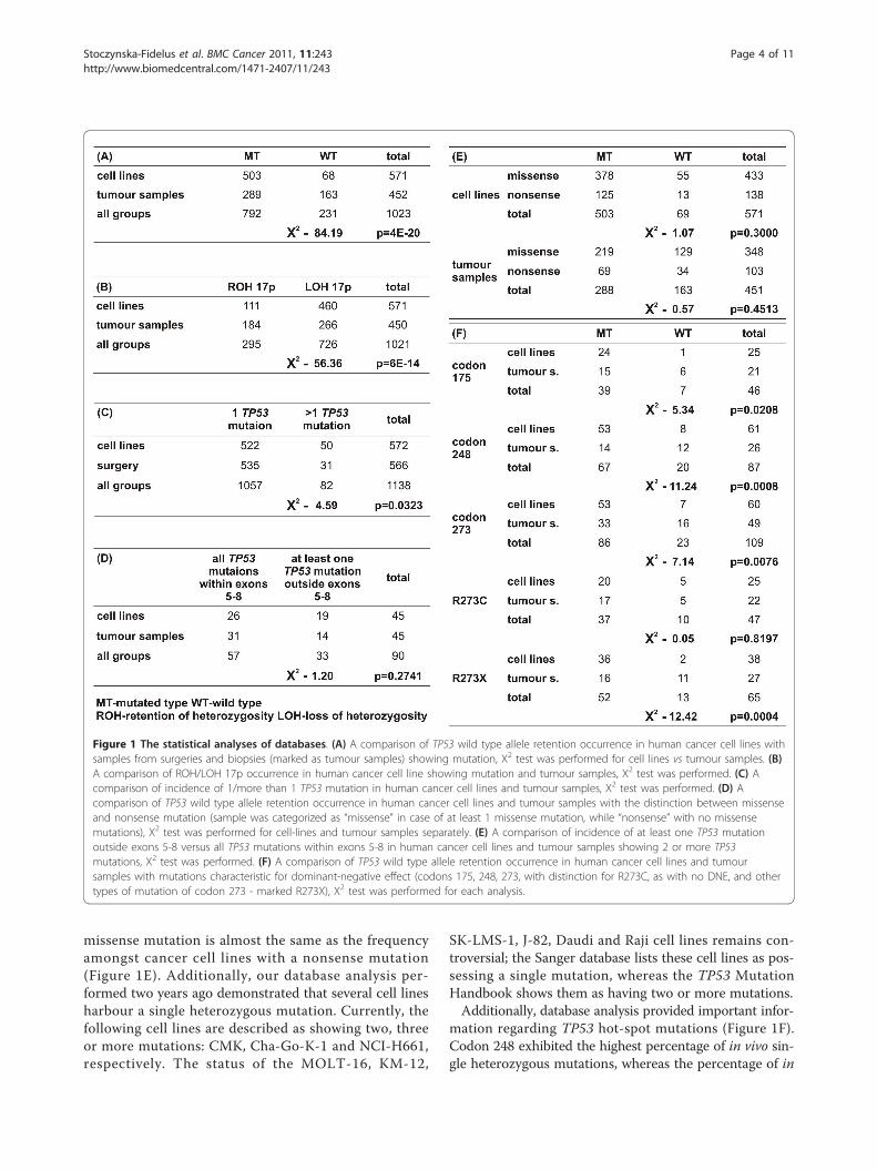
missense mutation is almost the same as the frequencyamongst cancer cell lines with a nonsense mutation(Figure 1E). Additionally, our database analysis per-formed two years ago demonstrated that several cell linesharbour a single heterozygous mutation. Currently, thefollowing cell lines are described as showing two, threeor more mutations: CMK, Cha-Go-K-1 and NCI-H661,respectively. The status of the MOLT-16, KM-12,
SK-LMS-1, J-82, Daudi and Raji cell lines remains con-troversial; the Sanger database lists these cell lines as pos-sessing a single mutation, whereas the TP53 MutationHandbook shows them as having two or more mutations.Additionally, database analysis provided important infor-
mation regarding TP53 hot-spot mutations (Figure 1F).Codon 248 exhibited the highest percentage of in vivo sin-gle heterozygous mutations, whereas the percentage of in
Figure 1 The statistical analyses of databases. (A) A comparison of TP53 wild type allele retention occurrence in human cancer cell lines withsamples from surgeries and biopsies (marked as tumour samples) showing mutation, Χ2 test was performed for cell lines vs tumour samples. (B)A comparison of ROH/LOH 17p occurrence in human cancer cell line showing mutation and tumour samples, Χ2 test was performed. (C) Acomparison of incidence of 1/more than 1 TP53 mutation in human cancer cell lines and tumour samples, Χ2 test was performed. (D) Acomparison of TP53 wild type allele retention occurrence in human cancer cell lines and tumour samples with the distinction between missenseand nonsense mutation (sample was categorized as “missense” in case of at least 1 missense mutation, while “nonsense” with no missensemutations), Χ2 test was performed for cell-lines and tumour samples separately. (E) A comparison of incidence of at least one TP53 mutationoutside exons 5-8 versus all TP53 mutations within exons 5-8 in human cancer cell lines and tumour samples showing 2 or more TP53mutations, Χ2 test was performed. (F) A comparison of TP53 wild type allele retention occurrence in human cancer cell lines and tumoursamples with mutations characteristic for dominant-negative effect (codons 175, 248, 273, with distinction for R273C, as with no DNE, and othertypes of mutation of codon 273 - marked R273X), Χ2 test was performed for each analysis.
Stoczynska-Fidelus et al. BMC Cancer 2011, 11:243http://www.biomedcentral.com/1471-2407/11/243
Page 4 of 11
vitro mutations in this codon was as low as any mutationof TP53. Codon 175 demonstrated a very low percentageof single heterozygous mutations in vitro and an averagepercentage in vivo. Analysis of codon 273 provided addi-tional information, specifically, that mutation R273C was asingle heterozygous mutation in approximately 20% of celllines, both in vivo and in vitro. For other mutations of thiscodon, the incidence was 42% and 5% in vivo and in vitro,respectively.
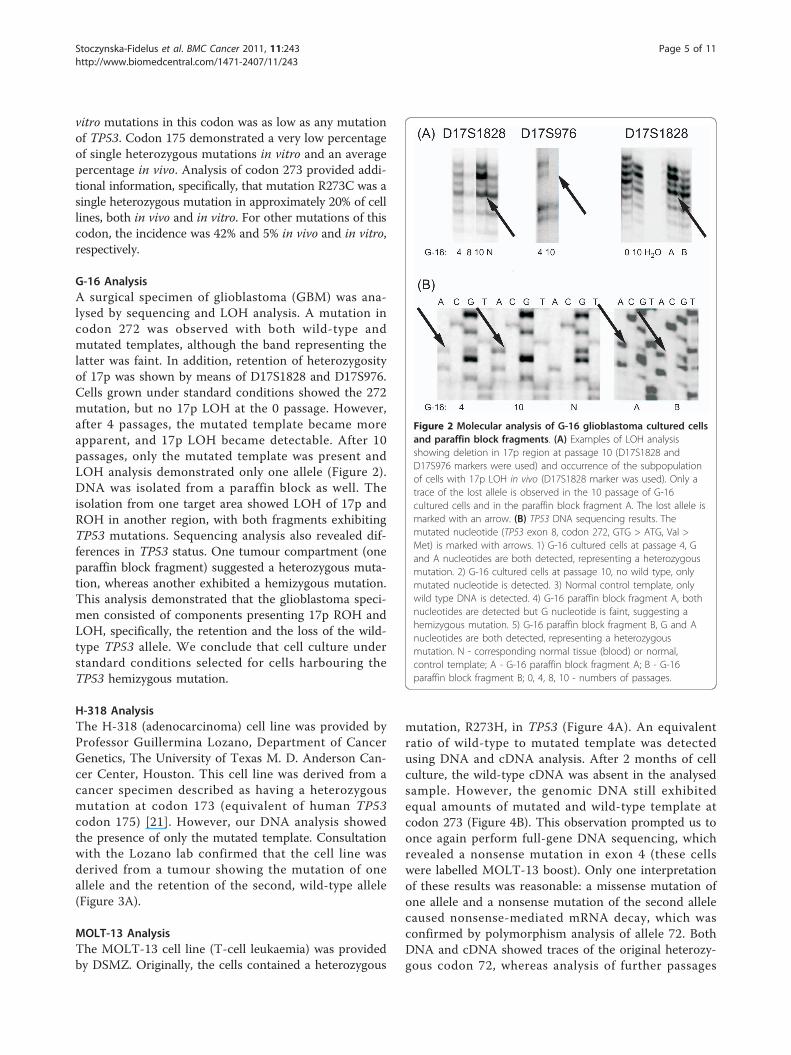
G-16 AnalysisA surgical specimen of glioblastoma (GBM) was ana-lysed by sequencing and LOH analysis. A mutation incodon 272 was observed with both wild-type andmutated templates, although the band representing thelatter was faint. In addition, retention of heterozygosityof 17p was shown by means of D17S1828 and D17S976.Cells grown under standard conditions showed the 272mutation, but no 17p LOH at the 0 passage. However,after 4 passages, the mutated template became moreapparent, and 17p LOH became detectable. After 10passages, only the mutated template was present andLOH analysis demonstrated only one allele (Figure 2).DNA was isolated from a paraffin block as well. Theisolation from one target area showed LOH of 17p andROH in another region, with both fragments exhibitingTP53 mutations. Sequencing analysis also revealed dif-ferences in TP53 status. One tumour compartment (oneparaffin block fragment) suggested a heterozygous muta-tion, whereas another exhibited a hemizygous mutation.This analysis demonstrated that the glioblastoma speci-men consisted of components presenting 17p ROH andLOH, specifically, the retention and the loss of the wild-type TP53 allele. We conclude that cell culture understandard conditions selected for cells harbouring theTP53 hemizygous mutation.
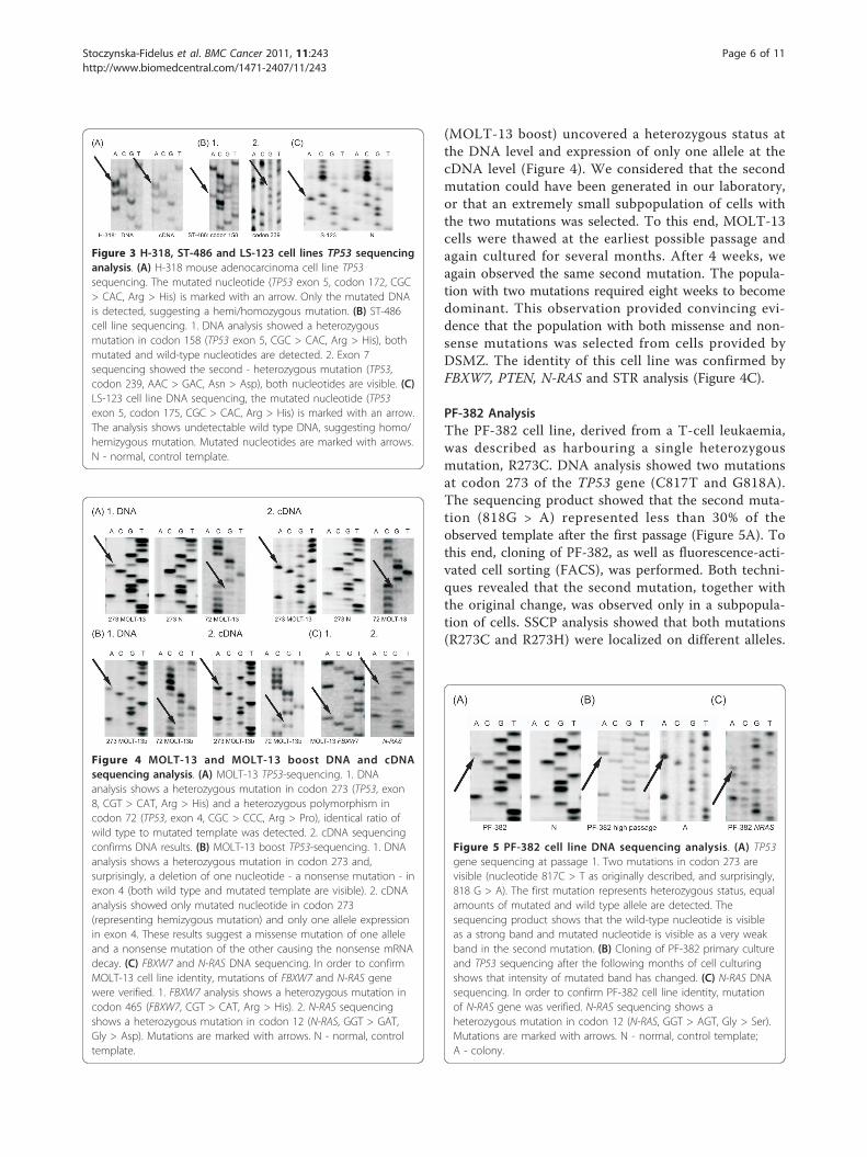
H-318 AnalysisThe H-318 (adenocarcinoma) cell line was provided byProfessor Guillermina Lozano, Department of CancerGenetics, The University of Texas M. D. Anderson Can-cer Center, Houston. This cell line was derived from acancer specimen described as having a heterozygousmutation at codon 173 (equivalent of human TP53codon 175) [21]. However, our DNA analysis showedthe presence of only the mutated template. Consultationwith the Lozano lab confirmed that the cell line wasderived from a tumour showing the mutation of oneallele and the retention of the second, wild-type allele(Figure 3A).
MOLT-13 AnalysisThe MOLT-13 cell line (T-cell leukaemia) was providedby DSMZ. Originally, the cells contained a heterozygous
mutation, R273H, in TP53 (Figure 4A). An equivalentratio of wild-type to mutated template was detectedusing DNA and cDNA analysis. After 2 months of cellculture, the wild-type cDNA was absent in the analysedsample. However, the genomic DNA still exhibitedequal amounts of mutated and wild-type template atcodon 273 (Figure 4B). This observation prompted us toonce again perform full-gene DNA sequencing, whichrevealed a nonsense mutation in exon 4 (these cellswere labelled MOLT-13 boost). Only one interpretationof these results was reasonable: a missense mutation ofone allele and a nonsense mutation of the second allelecaused nonsense-mediated mRNA decay, which wasconfirmed by polymorphism analysis of allele 72. BothDNA and cDNA showed traces of the original heterozy-gous codon 72, whereas analysis of further passages
Figure 2 Molecular analysis of G-16 glioblastoma cultured cellsand paraffin block fragments. (A) Examples of LOH analysisshowing deletion in 17p region at passage 10 (D17S1828 andD17S976 markers were used) and occurrence of the subpopulationof cells with 17p LOH in vivo (D17S1828 marker was used). Only atrace of the lost allele is observed in the 10 passage of G-16cultured cells and in the paraffin block fragment A. The lost allele ismarked with an arrow. (B) TP53 DNA sequencing results. Themutated nucleotide (TP53 exon 8, codon 272, GTG > ATG, Val >Met) is marked with arrows. 1) G-16 cultured cells at passage 4, Gand A nucleotides are both detected, representing a heterozygousmutation. 2) G-16 cultured cells at passage 10, no wild type, onlymutated nucleotide is detected. 3) Normal control template, onlywild type DNA is detected. 4) G-16 paraffin block fragment A, bothnucleotides are detected but G nucleotide is faint, suggesting ahemizygous mutation. 5) G-16 paraffin block fragment B, G and Anucleotides are both detected, representing a heterozygousmutation. N - corresponding normal tissue (blood) or normal,control template; A - G-16 paraffin block fragment A; B - G-16paraffin block fragment B; 0, 4, 8, 10 - numbers of passages.
Stoczynska-Fidelus et al. BMC Cancer 2011, 11:243http://www.biomedcentral.com/1471-2407/11/243
Page 5 of 11
(MOLT-13 boost) uncovered a heterozygous status atthe DNA level and expression of only one allele at thecDNA level (Figure 4). We considered that the secondmutation could have been generated in our laboratory,or that an extremely small subpopulation of cells withthe two mutations was selected. To this end, MOLT-13cells were thawed at the earliest possible passage andagain cultured for several months. After 4 weeks, weagain observed the same second mutation. The popula-tion with two mutations required eight weeks to becomedominant. This observation provided convincing evi-dence that the population with both missense and non-sense mutations was selected from cells provided byDSMZ. The identity of this cell line was confirmed byFBXW7, PTEN, N-RAS and STR analysis (Figure 4C).
PF-382 AnalysisThe PF-382 cell line, derived from a T-cell leukaemia,was described as harbouring a single heterozygousmutation, R273C. DNA analysis showed two mutationsat codon 273 of the TP53 gene (C817T and G818A).The sequencing product showed that the second muta-tion (818G > A) represented less than 30% of theobserved template after the first passage (Figure 5A). Tothis end, cloning of PF-382, as well as fluorescence-acti-vated cell sorting (FACS), was performed. Both techni-ques revealed that the second mutation, together withthe original change, was observed only in a subpopula-tion of cells. SSCP analysis showed that both mutations(R273C and R273H) were localized on different alleles.
Figure 5 PF-382 cell line DNA sequencing analysis. (A) TP53gene sequencing at passage 1. Two mutations in codon 273 arevisible (nucleotide 817C > T as originally described, and surprisingly,818 G > A). The first mutation represents heterozygous status, equalamounts of mutated and wild type allele are detected. Thesequencing product shows that the wild-type nucleotide is visibleas a strong band and mutated nucleotide is visible as a very weakband in the second mutation. (B) Cloning of PF-382 primary cultureand TP53 sequencing after the following months of cell culturingshows that intensity of mutated band has changed. (C) N-RAS DNAsequencing. In order to confirm PF-382 cell line identity, mutationof N-RAS gene was verified. N-RAS sequencing shows aheterozygous mutation in codon 12 (N-RAS, GGT > AGT, Gly > Ser).Mutations are marked with arrows. N - normal, control template;A - colony.
Figure 4 MOLT-13 and MOLT-13 boost DNA and cDNAsequencing analysis. (A) MOLT-13 TP53-sequencing. 1. DNAanalysis shows a heterozygous mutation in codon 273 (TP53, exon8, CGT > CAT, Arg > His) and a heterozygous polymorphism incodon 72 (TP53, exon 4, CGC > CCC, Arg > Pro), identical ratio ofwild type to mutated template was detected. 2. cDNA sequencingconfirms DNA results. (B) MOLT-13 boost TP53-sequencing. 1. DNAanalysis shows a heterozygous mutation in codon 273 and,surprisingly, a deletion of one nucleotide - a nonsense mutation - inexon 4 (both wild type and mutated template are visible). 2. cDNAanalysis showed only mutated nucleotide in codon 273(representing hemizygous mutation) and only one allele expressionin exon 4. These results suggest a missense mutation of one alleleand a nonsense mutation of the other causing the nonsense mRNAdecay. (C) FBXW7 and N-RAS DNA sequencing. In order to confirmMOLT-13 cell line identity, mutations of FBXW7 and N-RAS genewere verified. 1. FBXW7 analysis shows a heterozygous mutation incodon 465 (FBXW7, CGT > CAT, Arg > His). 2. N-RAS sequencingshows a heterozygous mutation in codon 12 (N-RAS, GGT > GAT,Gly > Asp). Mutations are marked with arrows. N - normal, controltemplate.
Figure 3 H-318, ST-486 and LS-123 cell lines TP53 sequencinganalysis. (A) H-318 mouse adenocarcinoma cell line TP53sequencing. The mutated nucleotide (TP53 exon 5, codon 172, CGC> CAC, Arg > His) is marked with an arrow. Only the mutated DNAis detected, suggesting a hemi/homozygous mutation. (B) ST-486cell line sequencing. 1. DNA analysis showed a heterozygousmutation in codon 158 (TP53 exon 5, CGC > CAC, Arg > His), bothmutated and wild-type nucleotides are detected. 2. Exon 7sequencing showed the second - heterozygous mutation (TP53,codon 239, AAC > GAC, Asn > Asp), both nucleotides are visible. (C)LS-123 cell line DNA sequencing, the mutated nucleotide (TP53exon 5, codon 175, CGC > CAC, Arg > His) is marked with an arrow.The analysis shows undetectable wild type DNA, suggesting homo/hemizygous mutation. Mutated nucleotides are marked with arrows.N - normal, control template.
Stoczynska-Fidelus et al. BMC Cancer 2011, 11:243http://www.biomedcentral.com/1471-2407/11/243
Page 6 of 11
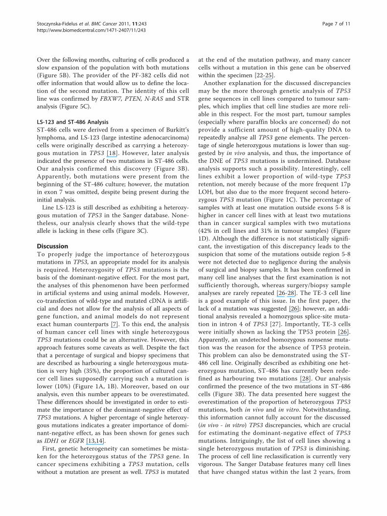
Over the following months, culturing of cells produced aslow expansion of the population with both mutations(Figure 5B). The provider of the PF-382 cells did notoffer information that would allow us to define the loca-tion of the second mutation. The identity of this cellline was confirmed by FBXW7, PTEN, N-RAS and STRanalysis (Figure 5C).
LS-123 and ST-486 AnalysisST-486 cells were derived from a specimen of Burkitt’slymphoma, and LS-123 (large intestine adenocarcinoma)cells were originally described as carrying a heterozy-gous mutation in TP53 [18]. However, later analysisindicated the presence of two mutations in ST-486 cells.Our analysis confirmed this discovery (Figure 3B).Apparently, both mutations were present from thebeginning of the ST-486 culture; however, the mutationin exon 7 was omitted, despite being present during theinitial analysis.Line LS-123 is still described as exhibiting a heterozy-
gous mutation of TP53 in the Sanger database. None-theless, our analysis clearly shows that the wild-typeallele is lacking in these cells (Figure 3C).
DiscussionTo properly judge the importance of heterozygousmutations in TP53, an appropriate model for its analysisis required. Heterozygosity of TP53 mutations is thebasis of the dominant-negative effect. For the most part,the analyses of this phenomenon have been performedin artificial systems and using animal models. However,co-transfection of wild-type and mutated cDNA is artifi-cial and does not allow for the analysis of all aspects ofgene function, and animal models do not representexact human counterparts [7]. To this end, the analysisof human cancer cell lines with single heterozygousTP53 mutations could be an alternative. However, thisapproach features some caveats as well. Despite the factthat a percentage of surgical and biopsy specimens thatare described as harbouring a single heterozygous muta-tion is very high (35%), the proportion of cultured can-cer cell lines supposedly carrying such a mutation islower (10%) (Figure 1A, 1B). Moreover, based on ouranalysis, even this number appears to be overestimated.These differences should be investigated in order to esti-mate the importance of the dominant-negative effect ofTP53 mutations. A higher percentage of single heterozy-gous mutations indicates a greater importance of domi-nant-negative effect, as has been shown for genes suchas IDH1 or EGFR [13,14].First, genetic heterogeneity can sometimes be mista-
ken for the heterozygous status of the TP53 gene. Incancer specimens exhibiting a TP53 mutation, cellswithout a mutation are present as well. TP53 is mutated
at the end of the mutation pathway, and many cancercells without a mutation in this gene can be observedwithin the specimen [22-25].Another explanation for the discussed discrepancies
may be the more thorough genetic analysis of TP53gene sequences in cell lines compared to tumour sam-ples, which implies that cell line studies are more reli-able in this respect. For the most part, tumour samples(especially where paraffin blocks are concerned) do notprovide a sufficient amount of high-quality DNA torepeatedly analyse all TP53 gene elements. The percen-tage of single heterozygous mutations is lower than sug-gested by in vivo analysis, and thus, the importance ofthe DNE of TP53 mutations is undermined. Databaseanalysis supports such a possibility. Interestingly, celllines exhibit a lower proportion of wild-type TP53retention, not merely because of the more frequent 17pLOH, but also due to the more frequent second hetero-zygous TP53 mutation (Figure 1C). The percentage ofsamples with at least one mutation outside exons 5-8 ishigher in cancer cell lines with at least two mutationsthan in cancer surgical samples with two mutations(42% in cell lines and 31% in tumour samples) (Figure1D). Although the difference is not statistically signifi-cant, the investigation of this discrepancy leads to thesuspicion that some of the mutations outside region 5-8were not detected due to negligence during the analysisof surgical and biopsy samples. It has been confirmed inmany cell line analyses that the first examination is notsufficiently thorough, whereas surgery/biopsy sampleanalyses are rarely repeated [26-28]. The TE-3 cell lineis a good example of this issue. In the first paper, thelack of a mutation was suggested [26]; however, an addi-tional analysis revealed a homozygous splice-site muta-tion in intron 4 of TP53 [27]. Importantly, TE-3 cellswere initially shown as lacking the TP53 protein [26].Apparently, an undetected homozygous nonsense muta-tion was the reason for the absence of TP53 protein.This problem can also be demonstrated using the ST-486 cell line. Originally described as exhibiting one het-erozygous mutation, ST-486 has currently been rede-fined as harbouring two mutations [28]. Our analysisconfirmed the presence of the two mutations in ST-486cells (Figure 3B). The data presented here suggest theoverestimation of the proportion of heterozygous TP53mutations, both in vivo and in vitro. Notwithstanding,this information cannot fully account for the discussed(in vivo - in vitro) TP53 discrepancies, which are crucialfor estimating the dominant-negative effect of TP53mutations. Intriguingly, the list of cell lines showing asingle heterozygous mutation of TP53 is diminishing.The process of cell line reclassification is currently veryvigorous. The Sanger Database features many cell linesthat have changed status within the last 2 years, from
Stoczynska-Fidelus et al. BMC Cancer 2011, 11:243http://www.biomedcentral.com/1471-2407/11/243
Page 7 of 11
having a single heterozygous mutation to exhibitingmore than one mutation (vide cell lines: CMK, Cha-Go-K-1 and NCI-H661). It is highly unlikely that eachpair of double mutations affected the same allele. More-over, some cell lines are reported differently betweendatabases: MOLT-16, KM-12, SK-LMS-1, J-82, Daudiand Raji. Notably, the LS-123 cell line, which is still cat-alogued as heterozygous, was provided to us with ahemizygous TP53 mutation (Figure 3C). The reasons forthese on-going changes in the databases are not clearbecause we are unable to determine whether the latediscovery of the second mutation is the result of its gen-eration in vitro, selection of an originally small subpopu-lation of cells with this mutation in vivo, experimentalerrors or technical inaccuracies. The difficulty in discri-minating between the first two potential explanationswill be the subject of the following paragraphs.The in vitro selection of cells exhibiting a hemizygous
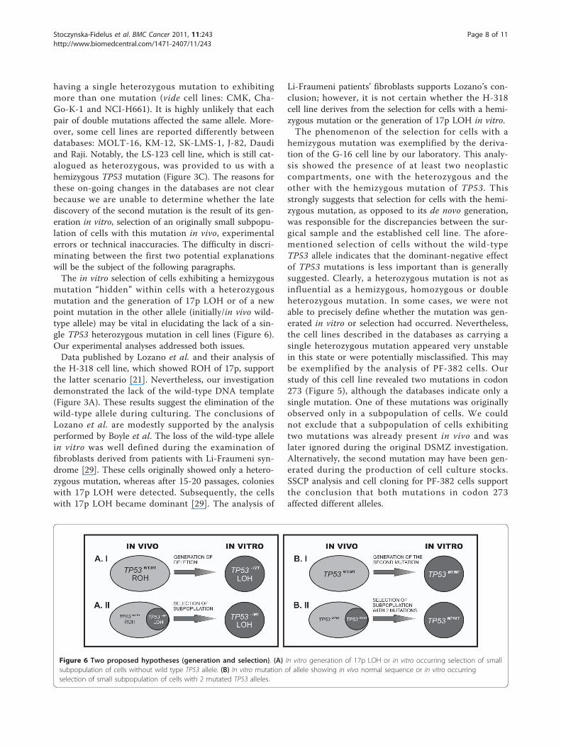
mutation “hidden” within cells with a heterozygousmutation and the generation of 17p LOH or of a newpoint mutation in the other allele (initially/in vivo wild-type allele) may be vital in elucidating the lack of a sin-gle TP53 heterozygous mutation in cell lines (Figure 6).Our experimental analyses addressed both issues.Data published by Lozano et al. and their analysis of
the H-318 cell line, which showed ROH of 17p, supportthe latter scenario [21]. Nevertheless, our investigationdemonstrated the lack of the wild-type DNA template(Figure 3A). These results suggest the elimination of thewild-type allele during culturing. The conclusions ofLozano et al. are modestly supported by the analysisperformed by Boyle et al. The loss of the wild-type allelein vitro was well defined during the examination offibroblasts derived from patients with Li-Fraumeni syn-drome [29]. These cells originally showed only a hetero-zygous mutation, whereas after 15-20 passages, colonieswith 17p LOH were detected. Subsequently, the cellswith 17p LOH became dominant [29]. The analysis of
Li-Fraumeni patients’ fibroblasts supports Lozano’s con-clusion; however, it is not certain whether the H-318cell line derives from the selection for cells with a hemi-zygous mutation or the generation of 17p LOH in vitro.The phenomenon of the selection for cells with a
hemizygous mutation was exemplified by the deriva-tion of the G-16 cell line by our laboratory. This analy-sis showed the presence of at least two neoplasticcompartments, one with the heterozygous and theother with the hemizygous mutation of TP53. Thisstrongly suggests that selection for cells with the hemi-zygous mutation, as opposed to its de novo generation,was responsible for the discrepancies between the sur-gical sample and the established cell line. The afore-mentioned selection of cells without the wild-typeTP53 allele indicates that the dominant-negative effectof TP53 mutations is less important than is generallysuggested. Clearly, a heterozygous mutation is not asinfluential as a hemizygous, homozygous or doubleheterozygous mutation. In some cases, we were notable to precisely define whether the mutation was gen-erated in vitro or selection had occurred. Nevertheless,the cell lines described in the databases as carrying asingle heterozygous mutation appeared very unstablein this state or were potentially misclassified. This maybe exemplified by the analysis of PF-382 cells. Ourstudy of this cell line revealed two mutations in codon273 (Figure 5), although the databases indicate only asingle mutation. One of these mutations was originallyobserved only in a subpopulation of cells. We couldnot exclude that a subpopulation of cells exhibitingtwo mutations was already present in vivo and waslater ignored during the original DSMZ investigation.Alternatively, the second mutation may have been gen-erated during the production of cell culture stocks.SSCP analysis and cell cloning for PF-382 cells supportthe conclusion that both mutations in codon 273affected different alleles.
Figure 6 Two proposed hypotheses (generation and selection). (A) In vitro generation of 17p LOH or in vitro occurring selection of smallsubpopulation of cells without wild type TP53 allele. (B) In vitro mutation of allele showing in vivo normal sequence or in vitro occurringselection of small subpopulation of cells with 2 mutated TP53 alleles.
Stoczynska-Fidelus et al. BMC Cancer 2011, 11:243http://www.biomedcentral.com/1471-2407/11/243
Page 8 of 11
The analysis of another cell line demonstrated similarresults. The MOLT-13 cell line was provided to ourlaboratory by DSMZ, and only one heterozygous muta-tion in codon 273 of the TP53 gene was detectable inthe early passages. After 2 months of culturing understandard conditions, the second mutation was detectedduring a routine screening (Figure 4B), which promptedus to restart the culture of the original MOLT-13 cells.Culturing these cells for only four weeks produced cellswith both mutations, which established that the pre-sence of cells with the two mutations was the result ofselection in our laboratory, rather than de novo genera-tion. The data provided by DSMZ did not allow us toexclude that the second mutation was not present in theoriginal patient sample and was generated during theproduction of vendor stocks. In any case, if generationor selection occurs in vitro, the importance of the domi-nant-negative effect is undermined by the instability ofsingle heterozygous mutations, as well as the enhancedin vitro survival of cells exhibiting a lack of wild-typeTP53.The data presented above show that although surgical
specimens are analysed less precisely than cell lines, sin-gle heterozygous mutations are still observed more fre-quently in vivo than in vitro. The G16, MOLT13-boostand PF-382 cell lines consist of cells that were selectedfrom subpopulations arising in vivo (in the case of theMOLT13-boost minor subpopulation), whereas H-318cells probably acquired the 17p LOH in vitro (adapta-tion to in vitro conditions and further stages of tumori-genesis). Thus, it may be presumed that it is possible todetect many biological differences between cellsobserved in vivo and in vitro in terms of TP53 status.For example, artificial selection forces acting in vitromay change the TP53 status. Alternatively, rapid selec-tion and generation of subpopulations of the moreadvanced neoplastic cells may be observed under suchconditions. The latter interpretation would favour invitro conditions as selecting the most effective impair-ment of TP53, i.e., the selection of cells at moreadvanced stages of carcinogenesis.Nonetheless, an effective DNE would not be easily
replaced by elimination or impairment of the secondallele under artificial or in vivo selection pressure. More-over, it is very unlikely that a mechanism that is suffi-ciently effective in vivo would become utterlysuppressed in vitro. In general, in vitro selection andgeneration of cells presenting hemi/homozygous muta-tions undermines the importance of dominant-negativeinactivation of TP53. Still, we cannot exclude that invitro cell culture does not optimally recapitulate allaspects of tumorigenesis involving TP53.We are aware that DNE has been presented many
times for some of the TP53 mutations. Our analysis
suggests that DNE TP53 mutations, which are albeitlimited in general, differ with respect to specific muta-tions. First, the mutation R175H seems to exhibit aweaker DNE than mutations in codon 248. Furthermore,our results are very intriguing for DNE mutations incodon 273 (R273L, R273P, R273H) in comparison toR273C, a mutation exhibiting a lack of DNE, as demon-strated by Dearth et al. In part, it could be expectedthat the percentage of single heterozygous mutationscorrelates with the magnitude of DNE and the impor-tance of specific dominant-negative mutations definedby Dearth et al. Namely, the percentage of mutations incodon 273 defined by Dearth et al. as exhibiting a DNEwas higher in vivo than this number for the R273Cmutation (42% vs. 23%). On the other hand, the percen-tage of single heterozygous R273C mutations was 4times higher in vitro than other mutations in this codon(20% vs. 5%), undermining the importance of TP53mutations DNE in vitro. Nevertheless, segregating TP53mutations into hot spots changes the overall results verylittle. None of the hot spots exhibit a percentage of sin-gle heterozygous mutations higher than 50% (Figure 1F),bearing in mind that these values are overestimatedaccording to our analyses. Still, the data do not imply acomplete lack of DNE. It may be inferred that DNE is amechanism operating solely in vivo, which, however,remains in contradiction with the DNE model. More-over, other tumour suppressor genes, such as APC andRb, also show similar differences between the frequencyof single heterozygous mutations in vivo and in vitro[17,18]. Obviously, APC and Rb exhibit nonsense muta-tions, and DNE is not attributed to these genes, whereasepigenetic silencing is [30-33]. However, considering thelatter observation, another analysis of the database isvery appealing. The retention of the wild-type allele incancer cell lines with a missense TP53 mutation isalmost the same as the frequency amongst cancer celllines with a nonsense mutation (Figure 1E). This indi-cates that a missense mutation does not predispose cellsto retain the wild-type TP53 allele any more than does anonsense mutation, whereas a dominant-negative effectis expected to be the attribute of missense mutationsonly. In our opinion, this suggests that the dominant-negative effect can be confused with gain of functionand even mistaken for an unknown mechanism of wild-type TP53 inhibition if a single heterozygous mutationoccurs. Our group has already demonstrated the predo-minance of mutated mRNA over wild-type mRNA inglioblastoma specimens showing putative single hetero-zygous mutation of TP53 [20].
ConclusionConsidering all the data presented here, the DNE ofTP53 mutations seems to be less important than the
Stoczynska-Fidelus et al. BMC Cancer 2011, 11:243http://www.biomedcentral.com/1471-2407/11/243
Page 9 of 11
majority of reports that describe this phenomenon sug-gest. Nevertheless, this effect should not be ignored, andwe do not exclude that it plays an important role at theearliest stages of tumorigenesis.In conclusion, we summarize the following facts dis-
cussed in our article undermining the influence of thedominant-negative effect of TP53 missense mutations:
- the percentage of TP53 single heterozygous muta-tions described in databases is low (35% in vivo and10% in vitro),- the percentage of single heterozygous mutations incancer specimens (in vivo) is overestimated indatabases,- the percentage of single heterozygous mutations incancer cell lines (in vitro) is overestimated indatabases,- cancer cells exhibiting a lack of wild-type TP53 aregenerated and/or selected for in vitro,- the percentage of single heterozygous mutations ofTP53 is higher in cancer specimens than in cancercell lines,- mutations considered to show DNE, R175H andR273X (excluding R273C), represent about 5% ofsingle heterozygous mutations in vitro, according tothe IARC database,- the frequency of retention of the wild-type allele incancer cell lines and tumour samples with a mis-sense mutation is almost the same as the frequencyamongst cancer cell lines and tumour samples witha nonsense mutation, according to databases.
AcknowledgementsWe would like to thank Professor Lozano for providing H-318 cell line.This study was supported by the Ministry of Science and Higher Educationprojects no. N N 401 020635 and partially sponsored by a PolpharmaScience Foundation.
Author details1Department of Molecular Pathology and Neuropathology, Chair ofOncology, Medical University of Lodz, Czechoslowacka 8/10, 92-216 Lodz,Poland. 2Department of Pathology, Chair of Oncology, Medical University ofLodz, Paderewskiego 4, 93-509 Lodz, Poland.
Authors’ contributionsESF conceived study design, performed database and genetic analyses,wrote the manuscript, contributed to data interpretation and cell culturing.MS performed genetic analyses, sequenced TP53 and contributed to datainterpretation. SP participated in the conception and design of the study,genetic analyses and data interpretation. MiB participated in the studydesign, genetic analyses, provided help in manuscript preparation and datainterpretation, performed the database and statistical analysis. KHB wasresponsible for cell culturing. MaB carried out genetic analyses and databaseanalysis. IZ participated in the acquisition of funding, contributed to geneticanalyses and data interpretation. DJK and RK provided histopathology resultsand participated in data interpretation. PPL participated in the manuscriptpreparation, revised the manuscript critically. PR supervised the project,conceived study design and provided help in manuscript preparation,participated in the acquisition of funding, contributed to data interpretation
and cell culture. All authors have given final approval of the version to bepublished.
Competing interestsThe authors declare that they have no competing interests.
Received: 24 November 2010 Accepted: 13 June 2011Published: 13 June 2011
References1. Roemer K: Mutant p53: gain-of-function oncoproteins and wild-type p53
inactivators. Biol Chem 1999, 380:879-887.2. Sigal A, Rotter V: Oncogenic mutations of the p53 tumor suppressor:
the demons of the guardian of the genome. Cancer Res 2000,60:6788-6793.
3. Waldman YY, Tuller T, Sharan R, Ruppin E: TP53 cancerous mutationsexhibit selection for translation efficiency. Cancer Res 2009, 69:8807-13.
4. Vinyals A, Peinado MA, Gonzalez-Garrigues M, Monzó M, Bonfil RD, Fabra A:Failure of wild-type p53 gene therapy in human cancer cells expressinga mutant p53 protein. Gene Ther 1999, 6:22-33.
5. Cuddihy AR, Jalali F, Coackley C, Bristow RG: WTp53 induction does notoverride MTp53 chemoresistance and radioresistance due to gain-of-function in lung cancer cells. Mol Cancer Ther 2008, 7:980-92.
6. Willis A, Jung EJ, Wakefield T, Chen X: Mutant p53 exerts a dominantnegative effect by preventing wild-type p53 from binding to thepromoter of its target genes. Oncogene 2004, 23:2330-8.
7. Dearth LR, Qian H, Wang T, Baroni TE, Zeng J, Chen SW, Yi SY,Brachmann RK: Inactive full-length p53 mutants lacking dominant wild-type p53 inhibition highlight loss of heterozygosity as an importantaspect of p53 status in human cancers. Carcinogenesis 2007, 28:289-98.
8. Heyne K, Schmitt K, Mueller D, Armbruester V, Mestres P, Roemer K:Resistance of mitochondrial p53 to dominant inhibition. Mol Cancer 2008,12:7-54.
9. Ford JM, Hanawalt PC: Li-Fraumeni syndrome fibroblasts homozygous forp53 mutations are deficient in global DNA repair but exhibit normaltranscription-coupled repair and enhanced UV resistance. Proc Natl AcadSci USA 1995, 92:8876-80.
10. Nicholls CD, McLure KG, Shields MA, Lee PW: Biogenesis of p53 involvescotranslational dimerization of monomers and posttranslationaldimerization of dimers. Implications on the dominant negative effect. JBiol Chem 2002, 277:12937-45.
11. Chan WM, Siu WY, Lau A, Poon RY: How many mutant p53 molecules areneeded to inactivate a tetramer? Mol Cell Biol 2004, 24:3536-3551.
12. Olive KP, Tuveson DA, Ruhe ZC, Yin B, Willis NA, Bronson RT, Crowley D,Jacks T: Mutant p53 gain of function in two mouse models of Li-Fraumeni syndrome. Cell 2004, 119:847-860.
13. Zhao S, Guan KL: IDH1 mutant structures reveal a mechanism ofdominant inhibition. Cell Res 2010.
14. Lynch TJ, Bell DW, Sordella R, Gurubhagavatula S, Okimoto RA,Brannigan BW, Harris PL, Haserlat SM, Supko JG, Haluska FG, Louis DN,Christiani DC, Settleman J, Haber DA: Activating mutations in theepidermal growth factor receptor underlying responsiveness of non-small-cell lung cancer to gefitinib. N Engl J Med 2004, 350:2129-39.
15. Petitjean A, Mathe E, Kato S, Ishioka C, Tavtigian SV, Hainaut P, Olivier M:Impact of mutant p53 functional properties on TP53 mutation patternsand tumour phenotype: lessons from recent developments in the IARCTP53 database. Hum Mutat 2007, 28:622-9.
16. Ikediobi ON, Davies H, Bignell G, Edkins S, Stevens C, O’Meara S, Santarius T,Avis T, Barthorpe S, Brackenbury L, et al: Mutation analysis of 24 knowncancer genes in the NCI-60 cell line set. Mol Cancer Ther 2006, 5:2606-12.
17. Forbes S, Clements J, Dawson E, Bamford S, Webb T, Dogan A, Flanagan A,Teague J, Wooster R, Futreal PA, Stratton MR: The Catalogue of SomaticMutations in Cancer COSMIC 2005. Br J Cancer 2006, 94:318-22.
18. Forbes Simon A, Tang G, Bindal N, Sally Bamford, Elisabeth Dawson,Charlotte Cole, Kok ChY, Jia M, Ewing R, Menzies A, Teague JW,Stratton MR, Futreal PA: COSMIC (the Catalogue of Somatic Mutations inCancer): a resource to investigate acquired mutations in human cancer.Nucleic Acids Res 2010, 38:D652-7.
19. [http://p53.free.fr].20. Szybka M, Zawlik I, Kulczycka D, Golanska E, Jesien E, Kupnicka D, Stawski R,
Piaskowski S, Bieniek E, Zakrzewska M, Kordek R, Liberski PP, Rieske P:
Stoczynska-Fidelus et al. BMC Cancer 2011, 11:243http://www.biomedcentral.com/1471-2407/11/243
Page 10 of 11
Elimination of wild-type P53 mRNA in glioblastomas showingheterozygous mutations of P53. Br J Cancer 2008, 98:1431-3.
21. Lang GA, Iwakuma T, Suh YA, Liu G, Rao VA, Parant JM, Valentin-Vega YA,Terzian T, Caldwell LC, Strong LC, El-Naggar AK, Lozano G: Gain of functionof a p53 hot spot mutation in a mouse model of Li-Fraumeni syndrome.Cell 2004, 119:861-72.
22. Giaretti W, Rapallo A, Sciutto A, Macciocu B, Geido E, Hermsen MA,Postma C, Baak JP, Williams RA, Meijer GA: Intratumor heterogeneity of k-ras and p53 mutations among human colorectal adenomas containingearly cancer. Anal Cell Pathol 2000, 21:49-57.
23. Wu XR: Urothelial tumorigenesis: a tale of divergent pathways. Nat RevCancer 2005, 5:713-25.
24. Tamura G: Genetic and epigenetic alterations of tumor suppressor andtumor-related genes in gastric cancer. Histol Histopathol 2002, 17:323-9.
25. Goranova TE, Ohue M, Kato K: Putative precursor cancer cells in humancolorectal cancer tissue. Int J Clin Exp Pathol 2009, 2:154-62.
26. Barnas C, Martel-Planche G, Furukawa Y, Hollstein M, Montesano R,Hainaut P: Inactivation of the p53 protein in cell lines derived fromhuman esophageal cancers. Int J Cancer 1997, 71:79-87.
27. Boonstra JJ, et al: Mistaken Identity of Widely Used EsophagealAdenocarcinoma Cell Line TE-7. Cancer Res 2007, 67:7996-8001.
28. Bhatia K, Goldschmidts W, Gutierrez M, Gaidano G, Dalla-Favera R,Magrath I: Hemi- or homozygosity: a requirement for some but notother p53 mutant proteins to accumulate and exert a pathogeneticeffect. FASEB J 1993, 7:951-6.
29. Boyle JM, Spreadborough AR, Greaves MJ, Birch JM, Varley JM, Scott D:Delayed chromosome changes in gamma-irradiated normal and Li-Fraumeni fibroblasts. Radiat Res 2002, 157:158-65.
30. Laurent-Puig P, Béroud C, Soussi T: APC gene: database of germline andsomatic mutations in human tumors and cell lines. Nucleic Acids Res1998, 26:269-70.
31. Harbour JW: Overview of RB gene mutations in patients withretinoblastoma. Implications for clinical genetic screening.Ophthalmology 1998, 105:1442-7.
32. Chen J, Röcken C, Lofton-Day C, Schulz HU, Müller O, Kutzner N,Malfertheiner P, Ebert MP: Molecular analysis of APC promotermethylation and protein expression in colorectal cancer metastasis.Carcinogenesis 2005, 26:37-43.
33. Stirzaker C, Millar DS, Paul CL, Warnecke PM, Harrison J, Vincent PC,Frommer M, Clark SJ: Extensive DNA methylation spanning the Rbpromoter in retinoblastoma tumors. Cancer Res 1997, 57:2229-37.
Pre-publication historyThe pre-publication history for this paper can be accessed here:http://www.biomedcentral.com/1471-2407/11/243/prepub
doi:10.1186/1471-2407-11-243Cite this article as: Stoczynska-Fidelus et al.: Limited importance of thedominant-negative effect of TP53 missense mutations. BMC Cancer 201111:243.
Submit your next manuscript to BioMed Centraland take full advantage of:
• Convenient online submission
• Thorough peer review
• No space constraints or color figure charges
• Immediate publication on acceptance
• Inclusion in PubMed, CAS, Scopus and Google Scholar
• Research which is freely available for redistribution
Submit your manuscript at www.biomedcentral.com/submit
Stoczynska-Fidelus et al. BMC Cancer 2011, 11:243http://www.biomedcentral.com/1471-2407/11/243
Page 11 of 11
Related Documents