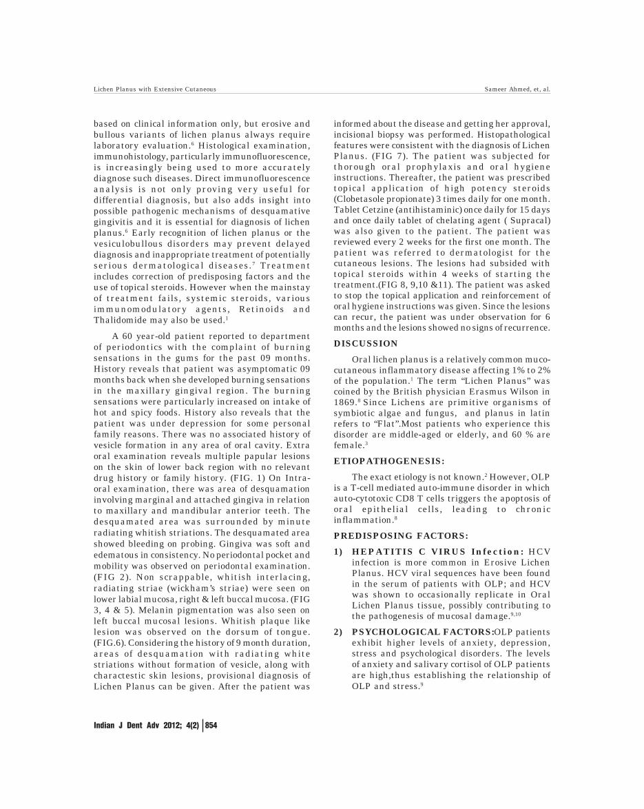
Indian J Dent Adv 2012; 4(2) 853 Lichen Planus with Extensive Cutaneous and oral Manifestations - A Case Report And Review of Literature Sameer Ahmed 1 , Shamimul Hasan 2 , Abhishek Singhvi 3 ABSTRACT: Oral Lichen Planus is a common chronic inflammatory disorder affecting stratified squamous epithelium of skin, oral mucosa and genitalia. It is a T-cell mediated auto- immune disorder in which auto-cytotoxic CD8 T-cells triggers the apoptosis of oral epithelial cells and leads to chronic inflammation. Skin lesions are usually self limiting and cause pruritis, whereas oral lesions are chronic, rarely undergo remission, are potentially premalignant and may even cause death. Usually clinical presentation is sufficient for diagnosis, especially if characterstic skin and oral features are present. However, Biopsy and immunofluorescent studies are done for a confirmatory diagnosis. The mainstay of treatment remains the use of topical steroids but newer therapies are also been in current use. This paper presents a case of Lichen Planus with extensive oral and skin lesions. The patients were treated with topical corticosteroids for oral lesions and was referred to dermatologist for the skin lesions. Key words: Lichen Planus, Desquamative gingivitis, Auto immune disorders, Topical steroids CASE REPORT doi: ........................... 1 Assistant professor Department of Periodontics, Vyas Dental College and Hospital, Jodhpur 2 Assistant professor Department of Oral Medicine and Radiology, Jamia millia Islamia, New Delhi 3 Assistant professor Department of Oral pathology, Vyas Dental college and Hospital, Jodhpur Article Info: Received: April 14, 2012; Review Completed: May, 13, 2012; Accepted: June 12, 2012 Published Online: August, 2012 (www. nacd. in) © NAD, 2012 - All rights reserved Email for correspondence: [email protected] Quick Response Code INTRODUCTION Lichen planus is a common muco-cutaneous disorder, more frequent within the oral cavity, where it appears as either white reticular, or erosive lesions. 1 In the majority of patients with oral lichen planus (OLP) there is no associated cutaneous lichen planus or lichen planus at other mucosal sites. This may be called “isolated” OLP. 2 Although the exact etiology is unknown, it is generally considered to be an immunological hypersensitivity reaction, characterized by an intensive T-cell infiltrate localized in the epithelium-connective tissue interface. 3 However, there are various factors that may predispose to its etio- pathogenesis. Most patients with lichen planus are middle-aged or older adults. Women predominate in most series of cases, usually by a 3:2 ratio over men. 1 The oral lesions are reticular, papular, bullous, plaque -like, atrophic, erosive and ulcerated. A rarely encountered form of lichen planus is the bullous variant. 4 Lesions are usually seen on the buccal mucosa and less common on the tongue, inner aspect of the lips and gingiva. 5 Gingival manifestations of lichen planus are relatively rare. Lichen planus is often diagnosed INDIAN JOURNAL OF DENTAL ADVANCEMENTS Journal homepage: www. nacd. in

Welcome message from author
This document is posted to help you gain knowledge. Please leave a comment to let me know what you think about it! Share it to your friends and learn new things together.
Transcript

Indian J Dent Adv 2012; 4(2) 853
Lichen Planus with Extensive Cutaneousand oral Manifestations - A Case Report
And Review of LiteratureSameer Ahmed1, Shamimul Hasan2, Abhishek Singhvi3
ABSTRACT:
Oral Lichen Planus is a common chronic inflammatory disorder
affecting stratified squamous epithelium of skin, oral mucosa
and genitalia. It is a T-cell mediated auto- immune disorder in
which auto-cytotoxic CD8 T-cells triggers the apoptosis of oral
epithelial cells and leads to chronic inflammation. Skin lesions
are usually self limiting and cause pruritis, whereas oral lesions
are chronic, rarely undergo remission, are potentially
premalignant and may even cause death. Usually clinical
presentation is sufficient for diagnosis, especially if characterstic
skin and oral features are present. However, Biopsy and
immunofluorescent studies are done for a confirmatory
diagnosis. The mainstay of treatment remains the use of topical
steroids but newer therapies are also been in current use. This
paper presents a case of Lichen Planus with extensive oral and
skin lesions. The patients were treated with topical
corticosteroids for oral lesions and was referred to dermatologist
for the skin lesions.
Key words: Lichen Planus, Desquamative gingivitis, Auto
immune disorders, Topical steroids
C A S E R E P O R T
doi: ...........................
1Assistant professorDepartment of Periodontics, Vyas Dental College andHospital, Jodhpur
2Assistant professorDepartment of Oral Medicine and Radiology,Jamia millia Islamia, New Delhi
3Assistant professorDepartment of Oral pathology,Vyas Dental college and Hospital, Jodhpur
Article Info:
Received: April 14, 2012;Review Completed: May, 13, 2012;Accepted: June 12, 2012Published Online: August, 2012 (www. nacd. in)© NAD, 2012 - All rights reserved
Email for correspondence:[email protected]
Quick Response Code
INTRODUCTIONLichen planus is a common muco-cutaneous disorder, more frequent within the oral cavity, where it
appears as either white reticular, or erosive lesions.1 In the majority of patients with oral lichen planus(OLP) there is no associated cutaneous lichen planus or lichen planus at other mucosal sites. This may becalled “isolated” OLP.2 Although the exact etiology is unknown, it is generally considered to be animmunological hypersensitivity reaction, characterized by an intensive T-cell infiltrate localized in theepithelium-connective tissue interface.3 However, there are various factors that may predispose to its etio-pathogenesis. Most patients with lichen planus are middle-aged or older adults. Women predominate inmost series of cases, usually by a 3:2 ratio over men.1 The oral lesions are reticular, papular, bullous, plaque-like, atrophic, erosive and ulcerated. A rarely encountered form of lichen planus is the bullous variant.4
Lesions are usually seen on the buccal mucosa and less common on the tongue, inner aspect of the lips andgingiva.5 Gingival manifestations of lichen planus are relatively rare. Lichen planus is often diagnosed
INDIAN JOURNAL OF DENTAL ADVANCEMENTS
Jour nal homepage: www. nacd. in
Indian J Dent Adv 2012; 4(2) 854
based on clinical information only, but erosive andbullous variants of lichen planus always requirelaboratory evaluation.6 Histological examination,immunohistology, particularly immunofluorescence,is increasingly being used to more accuratelydiagnose such diseases. Direct immunofluorescenceanalysis is not only proving very useful fordifferential diagnosis, but also adds insight intopossible pathogenic mechanisms of desquamativegingivitis and it is essential for diagnosis of lichenplanus.6 Early recognition of lichen planus or thevesiculobullous disorders may prevent delayeddiagnosis and inappropriate treatment of potentiallyserious dermatological diseases.7 Treatmentincludes correction of predisposing factors and theuse of topical steroids. However when the mainstayof treatment fails, systemic steroids, variousimmunomodulatory agents, Retinoids andThalidomide may also be used.1
A 60 year-old patient reported to departmentof periodontics with the complaint of burningsensations in the gums for the past 09 months.History reveals that patient was asymptomatic 09months back when she developed burning sensationsin the maxillary gingival region. The burningsensations were particularly increased on intake ofhot and spicy foods. History also reveals that thepatient was under depression for some personalfamily reasons. There was no associated history ofvesicle formation in any area of oral cavity. Extraoral examination reveals multiple papular lesionson the skin of lower back region with no relevantdrug history or family history. (FIG. 1) On Intra-oral examination, there was area of desquamationinvolving marginal and attached gingiva in relationto maxillary and mandibular anterior teeth. Thedesquamated area was surrounded by minuteradiating whitish striations. The desquamated areashowed bleeding on probing. Gingiva was soft andedematous in consistency. No periodontal pocket andmobility was observed on periodontal examination.(FIG 2). Non scrappable, whitish interlacing,radiating striae (wickham’s striae) were seen onlower labial mucosa, right & left buccal mucosa. (FIG3, 4 & 5). Melanin pigmentation was also seen onleft buccal mucosal lesions. Whitish plaque likelesion was observed on the dorsum of tongue.(FIG.6). Considering the history of 9 month duration,areas of desquamation with radiating whitestriations without formation of vesicle, along withcharactestic skin lesions, provisional diagnosis ofLichen Planus can be given. After the patient was
informed about the disease and getting her approval,incisional biopsy was performed. Histopathologicalfeatures were consistent with the diagnosis of LichenPlanus. (FIG 7). The patient was subjected forthorough oral prophylaxis and oral hygieneinstructions. Thereafter, the patient was prescribedtopical application of high potency steroids(Clobetasole propionate) 3 times daily for one month.Tablet Cetzine (antihistaminic) once daily for 15 daysand once daily tablet of chelating agent ( Supracal)was also given to the patient. The patient wasreviewed every 2 weeks for the first one month. Thepatient was referred to dermatologist for thecutaneous lesions. The lesions had subsided withtopical steroids within 4 weeks of starting thetreatment.(FIG 8, 9,10 &11). The patient was askedto stop the topical application and reinforcement oforal hygiene instructions was given. Since the lesionscan recur, the patient was under observation for 6months and the lesions showed no signs of recurrence.
DISCUSSION
Oral lichen planus is a relatively common muco-cutaneous inflammatory disease affecting 1% to 2%of the population.1 The term “Lichen Planus” wascoined by the British physician Erasmus Wilson in1869.8 Since Lichens are primitive organisms ofsymbiotic algae and fungus, and planus in latinrefers to “Flat”.Most patients who experience thisdisorder are middle-aged or elderly, and 60 % arefemale.3
ETIOPATHOGENESIS:
The exact etiology is not known.2 However, OLPis a T-cell mediated auto-immune disorder in whichauto-cytotoxic CD8 T cells triggers the apoptosis oforal epithelial cells, leading to chronicinflammation.8
PREDISPOSING FACTORS:
1) HEPATITIS C VIRUS Infection: HCVinfection is more common in Erosive LichenPlanus. HCV viral sequences have been foundin the serum of patients with OLP; and HCVwas shown to occasionally replicate in OralLichen Planus tissue, possibly contributing tothe pathogenesis of mucosal damage.9,10
2) PSYCHOLOGICAL FACTORS:OLP patientsexhibit higher levels of anxiety, depression,stress and psychological disorders. The levelsof anxiety and salivary cortisol of OLP patientsare high,thus establishing the relationship ofOLP and stress.9
Lichen Planus with Extensive Cutaneous Sameer Ahmed, et, al.

Indian J Dent Adv 2012; 4(2) 855
3) ORAL LICHENOID REACTIONS:a) Dental restorative materials:
Amalgams, composite resins,cobalt,goldand even flavouring agents may lead to orallichenoid reactions.9
b) Drugs:NSAIDS, ACE inhibitors(captopril), beta blockers,Penicillamine maybe implicated as a cause. However,lichenoid reactions are usually unilateraland resolves on discontinuation of theoffending factors.11
4) MECHANICAL TRAUMA:Dental procedures,friction from sharp cusps,rough dentalrestorations,poorly fitting prosthesis anddeleterious oral habits are exacerbatingfactors.KOEBNERS PHENOMENON,wherethe lesions develop in response totrauma,explains why erosive lesions arecommon in areas subjected to trauma.(buccalmucosa and lateral border of tongue.9,12
5 PLAQUE AND CALCULUS: May result inworsening gingival lesions LP and are associatedwith a higher incidence of erosive lesions.
6 DIABETES AND HYPERTENSION: There isno literature supporting the association of LPwith Diabetes and Hypertension. HoweverGRINSPAN’S SYNDROME is the associationof LP, Diabetes and Vascular Hypertension.13
A) EXTRA-ORAL MANIFESTATIONS:
1) CUTANEOUS LESIONS: consists of purple,pruritic, polygonal papules, often overlined byradiating lines (WICKHAM’S STRIAE).13 Skinlesions develop several months after theappearance of oral lesions and are usually selflimiting. Genital mucosa is the most commonextraoral site of involvement.
VULVOVAGINAL-GINGIVAL SYNDROME- is the association of LP of vulva,vagina andgingiva in female patients. Patients usuallycomplains of burning,pain,vaginal dischargedyspareunia.9
PENO-GINGIVAL SYNDROME - is the malecounterpart of vulvovaginal-gingival syndromeof LP.9
2) SCALP AND HAIR FOLLICLES: Lichenplanopilaris / Graham’s little syndromerepresents LP involvement of scalp and hairfollicles, resulting in scarring alopecia.9
3) NAILS: Thinning and ridging of the nail plateand splitting of the distal free edge of thenail.Healing with scar producesPTYERGIUM.9
4) ESOPHAGEAL LESIONS: Dysphagia is thecommonest feature. Chronic pain and stricturesmay also be seen.
B) ORAL MANIFESTATIONS:The clinical evaluation of the oral lesions is
based on the six clinical forms described byAndreason: reticular, papular, plaque, atrophic,erosive, and bullous.14 Mucosal lesions, which aremultiple, generally have a symmetric distribution,particularly on the mucosa of the cheeks, adjacentto molars, and on the tongue mucosa, less frequenton the labial mucosa (lichenous cheilitis) and on thegums (desquamative gingivitis).14 The most commonform is reticular type with characterstic slenderwhite radiating interlacing striations.15The lesionsfrequently occurs bilaterally and are mostlyasymptomatic.15 Erosive LP most often appears asa mixture of intensely erythematous mucosa withlarge areas of irregularly shaped ulceration with awhitish-yellowish pseudomembrane.4 Erosive andatrophic LP results in burning sensations.Erythematous lesions that affect the gingival causedesquamative gingivitis, the most common type ofgingival LP. The plaque like forms of LP mayresemble leukoplakia, particularly proliferativeverrucous leukoplakia and appears as slightly raisedor flat area on oral mucous membrane.16 The mostcommon site of plaque like LP is tongue. BullousLP is extremely rare form in oral cavity..The bullaerupture almost immediately, leaving an ulcerationon a bed of inflamed mucosa. Bullous LP mostcommonly affects the posterior buccal mucosa.16
MALIGNANT POTENTIAL:The most important complication of OLP is thedevelopment of Oral squamous cell carcinoma.17 Thefirst case of carcinoma arising in LP of oral mucosawas described by HALLAPEAU in 1910.18 The riskof malignant transformation varies from 0.4% toover 5% a period of 5 – 20 years. Accumulation ofinducible nitric oxide synthetase with 8-nitroguanine and 8-oxo-7,8-dihydro-2’deoxyganosine in oral epithelium in OLP may causeoxidative and nitrative damage to DNA and couldbe the basis of malignancy. The risk of malignanttransformation is independent of the clinical typeof OLP or the treatment used.19
Lichen Planus with Extensive Cutaneous Sameer Ahmed, et, al.

Indian J Dent Adv 2012; 4(2) 856
DIAGNOSIS:
1) CLINICAL DIAGNOSIS: is sufficient toestablish a diagnosis of OLP, if charactersticoral and skin lesions are present.
2) HISTOPATHOLOGY:9
Essential features:-superficial band like infiltrate of T lymphocytes-Basal cell liquefaction degeneration-Normal epithelial maturation pattern
Additional features:-SAW TOOTH rete pegs-Civatte /colloid bodies-Separation of epithelium from lamina propria-Max joseph spaces
3) IMMUNOFLUORESCENCE OF PERI-LESIONAL MUCOSA: 9
-Fibrin and shaggy fibrinogen in a linear patternat basement membrane zone-Cytoids in the absence of deposition offibrinogen
TREATMENT:Generally, no medication is necessary for the
benign form of this disease (reticular lichen planus).In the case of severe pain and a burning sensation,high-potency topical corticosteroids remain the mostre-liably effective treatment modality. Oral hygieneand corrective dentistry play a major role in themanagement of OLP and consultation with a dentistor oral medicine specialist is helpful.9
DRUG TREATMENT:Drug treatment with topical steroids is preferreddue to fewer side effects. Systemic steroids may beused if the lesions are extensive,or there arerecalcitrant disease.
TOPICAL CORTICOSTEROIDS:Most effective topical steroids are the medium potentsteroids (triamcinolone), high potent steroids(fluocinolone acetonide) and superpotenthalogenated steroids (clobetasol propionate). Elixirforms are also used such as dexamethasone,triamcinolone and clobetasol.These are used fordiffuse oral involvement,elderly patients or forpatients having difficulty in applying themedications. The greatest difficulty in using topicalsteroids is is the lack of mucosal adherence for asufficient period of time. Therefore,topical steroidsmay be used with adhesive pastes(orabase).9 A
regular follow up should be done for prolonged useof topical steroids for the following adverse effects:
a) secondary candidal infectionb) Tachyphylaxis (diminished biological
effectiveness)c) Adrenal suppressiond) atrophy of the oral mucosa
INTRALESIONAL STEROIDS:
Used for intractable erosive OLP lesions.Triamcinolone acetonide(10-20 mg / ml) is used andrepeated every 2-4 weeks.Frequent steroid injectionsare are painful and may result in an unwantedsystemic dose.9
SYSTEMIC STEROIDS:
Should be reserved for recalcitrant cases of Erosiveor Erythematous OLP or for widespread OLP withskin,genital,scalp or esophageal involvement.Dailydoses of 40-80 mg is usually sufficient to achieve aresponse.9
OTHER TOPICAL AGENTS:
a) TOPICAL CYCLOSPORINE(100 mg / ml asa mouth rinse)-may be beneficial inrecalcitrant OLP cases.Systemic absorption isgenerally low, but it is expensive and lesseffective than topical steroids in inducingclinical improvement in OLP.9
b) TOPICAL TACROLIMUS: is a steroid freetopical immunosuppressive agent. It was usedprimarily for atopic dermatitis. The exact modeof action is not known they inhibit T cellactivation and proliferation. Burning is the mostcommon side effect with tacrolimus.Other sideeffects include-carcinogenicity, mutagenesisand infertility.20
c) TOPICAL RETINOIDS: Tretinoin and iso-treinointrophic are used for erosive-atrophicforms.Side effects include teratogenicity andliver dysfunction.
OTHER THERAPIES:
-PUVA THERAPY- Photochemotherapy with 8-methoxypsoralen and long-wave ultraviolet light(PUVA) has been used successfully in the treatmentof skin lesions and cu-taneous lichen planus.21,22 Itwas first used in the treatment of recalcitrant OLP.23
Eighty-seven percent of patients treated withultraviolet-A, without a sys-temic or topicalphotosensitizer, improved signifi-cantly.24 Some
Lichen Planus with Extensive Cutaneous Sameer Ahmed, et, al.

Indian J Dent Adv 2012; 4(2) 857
studies have indicated that PUVA therapy mightalso have therapeutic effects.23 To avoid PUVA sideeffects, photosensitization with topical 0.01%trioxsalen can be used for the treat-ment,25 althoughoral mucosa seems more resistant to phototoxicdamage in comparison to skin.26 PUVA with 8-methoxypsoralen has various side effects, such asnausea, dizziness, eye symptoms, paraesthe-sia, andheadache.27 Photochemotherapy may be use-ful forsevere forms of erosive OLP that do not re-spond toconventional treatment.28 Moreover, one matter ofconcern is that PUVA therapy has been shown tohave oncogenic potential.29 -CRYOSURGERY- hasalso been used, particularly in erosive drug resistantOLP, but the lesions may develop in healing woundsand result in scars.
-LASER THERAPY-CO2 lasers are used to treatmulticentric lesions or lesions in difficult areas.
ADDITIONAL DRUG THERAPY:
Griesofulvin, Thalidomide, Levamisole and Dapsonemay also be used.
SURGERY:Resection my be done for isolated plaques or non-healing erosions. Free soft tissue grafts have beenused for localized areas of erosive OLP. However,periodontal surgery have been reported to provokeOLP.
CONCLUSION: OLP are a relatively common oralmucosal disease process encountered in clinicalpractice.Given the apparent risk of oral SCC, regularfollow up of these patients is mandatory.
REFERENCES1. Vincent SD, Fotos LPG, Baker KA, Williams TP. Oral lichen
planus: the clinical, historical, and therapeutic features of100 cases. Oral Surg Oral Med Oral Pathol 1990; 70: 165–171.
2. Al Hashimi I,Shifter M, Lockhart PB, Wrap D, Brennan M,Migliorate et al :OLP and Oral lichenoid lesions OOO S25;e;2007;103: 1-12.
3. Sapp JP, Eversole LR, Wysocki GP: Contemporary Oral andMaxillofacial Pathology, Second Edition, Mosby, St.Louis,2004.
4. Kuffer R; Lombardi T; Erosion and ulcerations occurring inoral lichen planus ; Dermatology 2003; 207(3): 340.
5. Dissemond J. Oral lichen planus: an overview. J DermatologTreat 2004; 15(3):136-140.
6. Raghu AR, Nirmala NR, Sreekumaran N: Directimmunofluorescence in oral lichen planus and oral lichenoidreactions. Quintessence Int. 2002;33(3):234-239.
7. Mignogna MD, Lo Russo L, Fedele S: Gingival involvementof oral lichen planus in a series of 700 patients. J ClinPeriodontol.2005; 32(10):1029-1033.
8. Axell T, Rundquist L. Oral lichen planus-a demographicstudy. Community Dent Oral Epidemiol. Feb 1987;15(1):52.
9. D Eisen; M Carrozzo ; J-V Bagan Sebastian ; K Thongprasan:Oral lichen planus: Clinical features and management:Oraldiseases (2005); 11: 338-349.
10. Lodi G; Porter SR; Hepatitis c virus and OLP: A short review:oral diseases:1997; 3: 77-89.
11. Dinkova AT; D Gospidava –Interdisciplinary approach incomplex treatment of Oral lichen planus-Review and casereport-Journal of IMAB.
12. Katta R ;Am Fam Physicians 2000; 63: 3319-3324.13. A Text book of oral pathology- Shaffers; Hine; Nevy; 5 th edition.14. Andreason JO; Oral lichen planus- A clinical evaluation of
115 cases ; OOO;1968; 25: 31-42.15. Lozada-Nur F, Miranda C: Oral lichen planus: epidemiology.
Clinical characteristics, and associated diseases. SeminCutan Med Surg. 1997;16(4):273-277.
16. Scott S; De Rossi; Katherine N Ciarrocca : Dental clinics ofnorth America 2005; 49: 77-89
17. Meij van der EH, Reibel J, Slootweg PJ, Wal JE van der.Interobserver and intraobserver variability in the histologicassessment of oral lichen planus. J Oral Pathol Med 1999;28: 274–277
18. Hallopeau H. Sur un cas de lichen de Wilson gingival avecneoplasie voisine dans la region maxillaire. Bull Soc Fr DermSyph 1910; 17: 33
19. Chiayarit P; Jontell M; Hiraku Y et al; Nitrative andOxidative damage of DNA in OLP; Cancer Sciences; 2005;96; 553-559
20. Nazzaro G; Cestari R; ;Topical Tacrolimus ointment inulcerative lichen planus; An alternative therapeuticapproach; European journal Dermatology july 2002; 12:4;321
21. PUVA treatment in lichen planus: comparison of oral andexternal methoxsalen regimens. Photodermatol 1987; 4:265-268
22. Jansen CT, Lehtinen R, Happonen RP, Lehtinen A,Soder-lund K. Mouth PUVA:new treatment for recalcitrant orallichen lanus. Photodermatol 1987; 4: 165-166
23. Chen HR. A newly developed method for treatment of orallichen planus with ultraviolet irradiation. Taiwan Yi XueHui Za Zhi 1989; 88:248-252.
24. Lehtinen R, Happonen RP, Kuusilehto A, Jansén C. A clini-cal trial of PUVA treatment in oral lichen planus. Proc FinnDent Soc 1989; 85:29-33.
25. Kuusilehto A, Lehtinen R, Happonen RP, Heikinheimo K,Lehtimäki K, Jansén CT. An open clinical trial of a newmouth-PUVA variant in the treatment of oral lichenoid le-sions. Oral Surg Oral Med Oral Pathol Oral Radiol Endod1997; 84:502-505.
26. Kuusilehto A, Lehtinen R, Jansen CT. Comparison of theminimal phototoxic dose in topical 4, 59,8-trimethylpsoralenPUVA treatment of Caucasian skin and of oral mucousmembrane. Acta Derm Venereol 1990; 70:508-509.
27. Lundquist G, Forsgren H, Gajecki M, Emtestam L. Photo-chemotherapy of oral lichen planus. A controlled study. OralSurg Oral Med Oral Pathol Oral Radiol Endod 1995; 79:554-558.
28. Seoane J, Vazquez J, Romero MA, Aguado A, Pomareda M.[Photochemotherapy in the treatment of oral erosive lichenplanus. Letter]. Acta Otorrinolaringol Esp 1997; 48:251-253.
29. Lindelöf B, Sigurgeirsson B, Tegner E, Larkö O, JohannessonA, Berne B, et al. PUVA and cancer: a large- scaleepidemiological study. Lancet 1991;338:91-93
Lichen Planus with Extensive Cutaneous Sameer Ahmed, et, al.

Indian J Dent Adv 2012; 4(2) 858
Figure 10&11: Resolution of lesions on lower labial mucosa and tongue.Figure 9: Marked reduction in erythema of marginaland attached gingival.
Figure 8: Resolution of skin lesions.Figure 7: Histopathology showing basal celldegeneration & chronic inflammatory cells.
Figure 6: Plaque like lesions on the dorsal aspect of tongue.Figure 4&5: Whitish interlacing striae affecting the right & left buccal mucosa.
Figure 3: Desquamative gingivitis affecting themandibular labial gingival with radiatingwhitish straiations of lower labial mucosa.
Figure 2: Erythematous marginal and attachedgingiva of maxillary anterior teeth.
Figure 1: Papular lesionson the lower back skin.
Lichen Planus with Extensive Cutaneous Sameer Ahmed, et, al.
Related Documents