Libro consigliato: Immunobiology, Janeway 6th edizione Date appelli: 30 Maggio ore 9.30 14 giugno 4 luglio 13 settembre 16 novembre 14 dicembre Esame orale

Libro consigliato: Immunobiology, Janeway 6th edizione Date appelli: 30 Maggio ore 9.30 14 giugno 4 luglio 13 settembre 16 novembre 14 dicembre Esame orale.
Dec 18, 2015
Welcome message from author
This document is posted to help you gain knowledge. Please leave a comment to let me know what you think about it! Share it to your friends and learn new things together.
Transcript

Libro consigliato: Immunobiology, Janeway 6th edizione
Date appelli:
30 Maggio ore 9.3014 giugno 4 luglio 13 settembre 16 novembre 14 dicembre
Esame orale

Immune System Function
• Defends the body against the outside world– Microorganisms
• Defends the body against the inside world– Abnormal cells
• Functions by modulation– Highly complex up and down regulatory mechanisms

Types of Immunity
Two major types
Innate immunity or natural immunity
Acquired immunity or specific immunity

Self Recognition Concept
• Immune system must recognize what is part of the body and what is not part of the body– Different classes of histocompatibility complex
proteins• Important in immune stimulation

Innate immunity
first front line of defensenot specificno immunologic memory (does not getstronger with more exposures)

Innate or Natural Immunity
• Mechanisms include– Mucous barriers– Natural killer cells– Polymorphonuclear and mononuclear
phagocytic cells

Macrophage
Neutrophil
Phagocytosis of Bacteria by Macrophages and Neutrophils- First Line of Defense
Listeria

Acquired Immunity
• It is specific and generally increases with exposure to foreign substances
• Two kinds of acquired immune response– Humoral immunity
• Immunoglobulins– Protein called antibody and substances that react with antibodies
are called antigens
– Cell-mediated immunity• Specialized cells that destroy foreign target cells
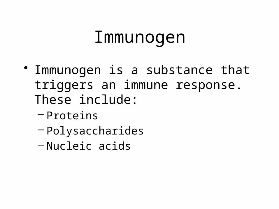
Immunogen
• Immunogen is a substance that triggers an immune response. These include:– Proteins– Polysaccharides– Nucleic acids

Overview of Immune Mechanisms

Recognition of Self
• Genetic variations in specific proteins– Class I and Class II major histocompatibility
complex (MHC)– Immune system develops with a concept of self

Cell Types
• Polymorphonuclear phagocytes• Mononuclear phagocytes• Lymphocytes• Antigen presenting cells
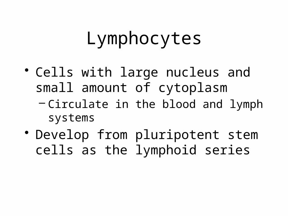
Lymphocytes
• Cells with large nucleus and small amount of cytoplasm– Circulate in the blood and lymph systems
• Develop from pluripotent stem cells as the lymphoid series

Blood Cell
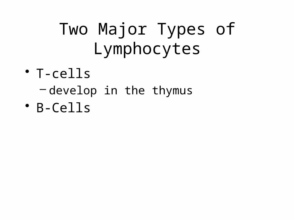
Two Major Types of Lymphocytes
• T-cells – develop in the thymus
• B-Cells

T-Lymphocytes
• T-lymphocytes – Initiating an immune response– Modulating an immune response
• T-lymphocytes have the following cluster differentiation (CD) or surface proteins– CD3+ – CD4+
• T-helper cells– CD8+
• T-suppressor cells• T-cytotoxic cells

B-Lymphocytes
• Develop and mature in the bone marrow• Produce class specific Immunoglobulins
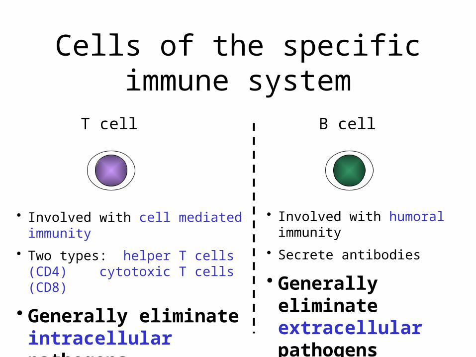
Cells of the specific immune system
T cell B cell
• Involved with cell mediated immunity
• Two types: helper T cells (CD4)cytotoxic T cells
(CD8)
• Generally eliminate intracellular pathogens
• Involved with humoral immunity
• Secrete antibodies
• Generally eliminate extracellular pathogens

Immunity mediated by T cells• T cells recognise and destroy cells infected with foreign
antigen– e.g. viral infection, intracellular bacteria (mycobacterium
tuberculosis), intracellular parasites)
• T cells can either:– kill infected cells themselves
• (CD8+ T cells also called cytotoxic T cells) or
– recruit help to eliminate the infected cell by means of soluble mediators called cytokines• (CD4+ T cells also called helper T cells)

How do T cells recognise specific antigenic epitopes?

CD4 and CD8 are co-receptors that serve to aid TCR signalling
CD4 or CD8
co-receptor
TCR
CD3 signalling
complex

How does the TCR get to see specific epitopes derived from
and intracellular foreign antigen?
i.e. if the infecting agent such as a virus is within its target cell how does the T cell get access?

Antigen Presentation
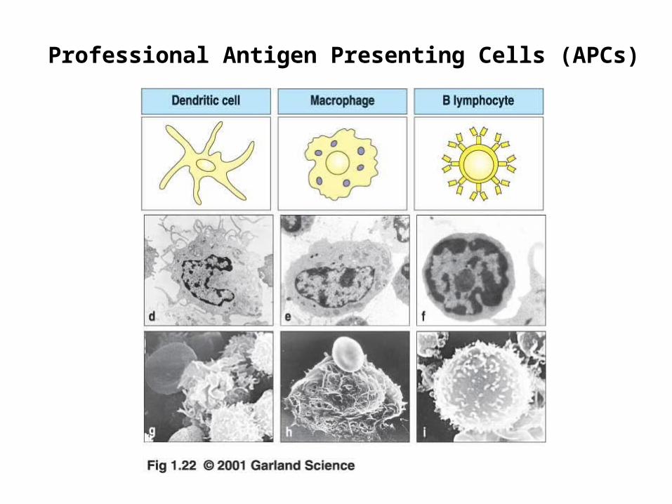
Professional Antigen Presenting Cells (APCs)

Major Histocompatibility Complex(MHC)
• Two types– MHC class I
• Expressed on the surface of all nucleated cells in your body
– MHC class II• Expressed on the surface of professional antigen-
presenting cells e.g. macrophages and dendritic cells

• CD4+ T cells interact with MHC class II on antigen-presenting cells
• CD8+ T cells interact with MHC class I which is expressed on all nucleated cells within your body

Processing of intracellular foreign antigen
• Presentation of antigen on MHC class II molecules, example:– Macrophages/Dendritic cells phagocytose bacteria
which are digested into small antigen fragments– These fragments are processed in the cytoplasm and
bound to MHC class II molecules– Antigen/MHC II complexes are subsequently expressed
on the surface of the antigen-presenting cell

• Presentation of antigen on MHC class I molecules, example:– Influenza virus infects a cell and becomes
incorporated into the host DNA where it can replicate itself
– Each cell in your body constantly screens itself by processing ‘self’ antigens and binding them to MHC class I molecules which are subsequently expressed on the cell surface
– If viral protein is present it to will be processed and expressed on the cell surface in the context of MHC class I

Interaction between CD4+ T cells and antigen/MHC II complexes
Activation and secretion of cytokines
CD4
Stabilises the MHC antigen complex
TCR
MHC II/antigen
Processing of microbial fragments onto MHC class II by
macrophage
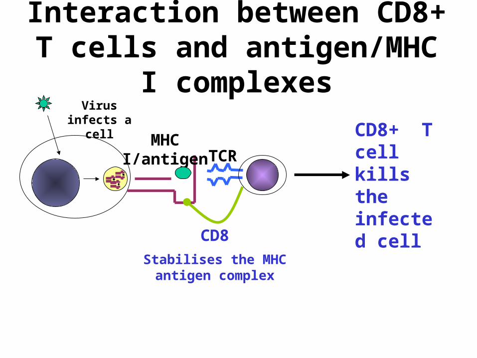
Interaction between CD8+ T cells and antigen/MHC I complexes
CD8+ T cell kills the infected cell
CD8
Stabilises the MHC antigen complex
TCRMHC I/antigen
Virus infects a cell


T Cell Development (I)- Thymus

T Cell Development (II)- Periphery
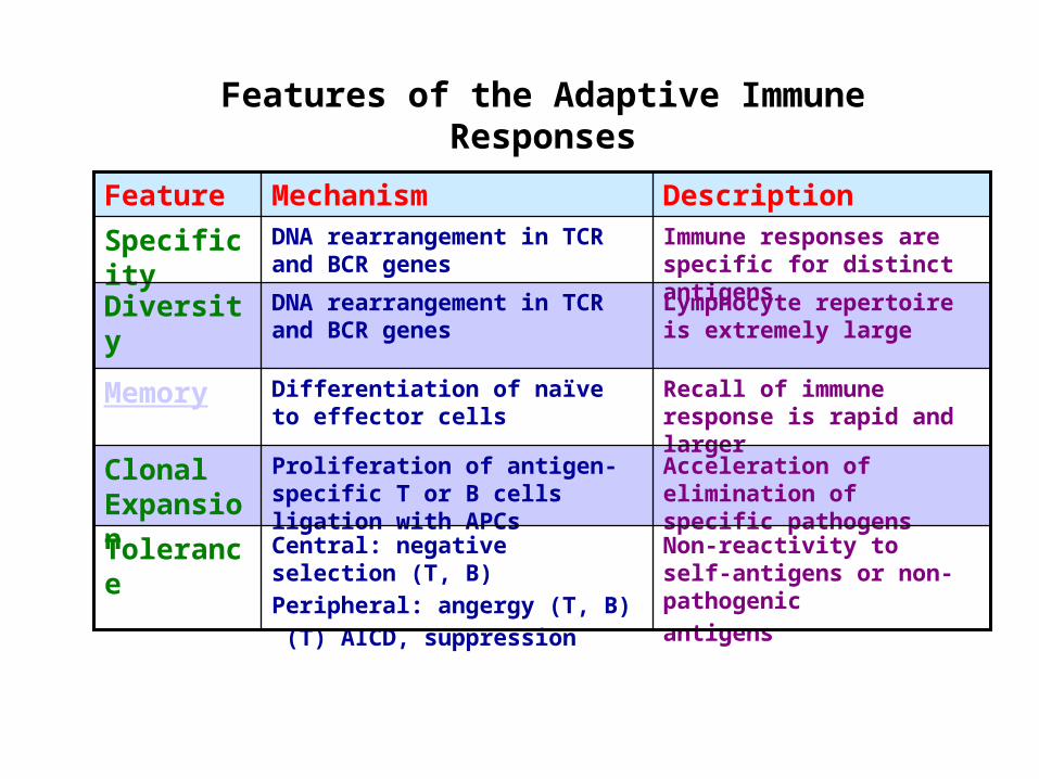
Features of the Adaptive Immune Responses
DescriptionMechanismFeature
Non-reactivity to self-antigens or non-pathogenic
antigens
Central: negative selection (T, B)
Peripheral: angergy (T, B)
(T) AICD, suppression
Tolerance
Acceleration of elimination of specific pathogens
Proliferation of antigen-specific T or B cells ligation with APCs
Clonal Expansion
Recall of immune response is rapid and larger
Differentiation of naïve to effector cells
Memory
Lymphocyte repertoire is extremely large
DNA rearrangement in TCR and BCR genes
Diversity
Immune responses are specific for distinct antigens
DNA rearrangement in TCR and BCR genes
Specificity

EffectorAg elimination
MemoryMemory maintenance
RecognitionAg presentation
ActivationDifferentiation
NaïveT/B
Activated T/B
Humoral& Cellular Immunity
MemoryT/B
Different Phases of Adaptive Immune Response
Modified from Cell. Mol. Immunol. 5th ed. Abbas et al.
Days after antigen exposure
0 7 14 >30
Nu
mb
er o
f an
tig
en-
spec
ific
T
/B c
ells
Antigen challenge
Clonal Expansion
ApoptosisHomeostasis
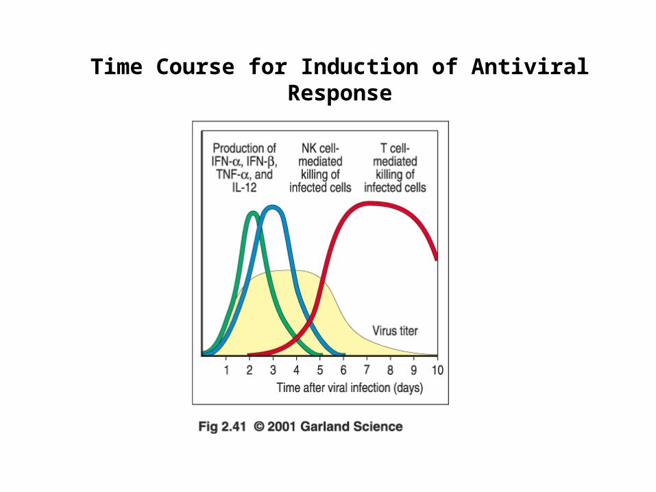
Time Course for Induction of Antiviral Response

Where Pathogens Enter the Body
Skin : DC, macrophages
Gut : GALT (Gut-Associated Lymphoid Tissue)
Lung : BALT (Bronchia-Associated Lymphoid Tissue )
Blood : Spleen/lymph nodes
Genital duct: MALT (Mucosal-Associated Lymphoid Tissue)

Where T/B Cells Meet Foreign Antigens-Spleen and Lymph Nodes

Induction and Effector Phases of Cell-mediated Immunity
Cell. Mol. Immunol. 5th ed. Abbas et al.

Requirements for a Professional APC
1. Able to ingest and present antigens
2. Express both MHC I and MHC II
3. Express Co-stimulatory molecules

Molecules Involved in the Interactions of T Cells and APCs during T Cell Activation
T cell APC
TCR-CD3 complex MHC-peptides
CD4/CD8 (coreceptor) MHC I/II
Adhesion molecules Ligands
Costimulatory molecule Ligands

Adhesion Molecules involved in the Interactions of TCells with APCs

Costimulatory Molecules of T Cells and APCs
T cell APC
CD28, CTLA-4 B7.1 B7.2
ICOS LICOS
4-1BB ( on CD8) 4-1BBL
PD-1 PD-L
Blue: Constitutive expressionRed: Inducible expression

Activation of Naïve T Cells Requires Two IndependentSignals- TCR + Costimulatory Signals

Mechanism of Peripheral Tolerance- TCR Ligation Without Costimulatory Molecules

Role of Costimulation and Th Cells in the Differentiation of CD8 T Cells
CD8
IL-2
1. CTL differentiation without Th cells
CD8
4-1BBL4-1BB
IL-2
3. Th cells enhance APCs to stimulate CTL differentiation
Modified from Cell. Mol. Immunol. 5th ed. Abbas et al.
CD8IL-2
2. Th cells produce cyto- kines to stimulate CTL differentiation

Distribution of Different APCs in Lymph Node

Features of Mature DC
Increased expression of MHC I and II
Expression of CCR7 for homing to 2o Lymphoid organs
Expression of costimulatory molecules B7
Increased expression of DC-SIGN
Secret cytokines IL-12 and TNFa Secret DC-CK to attract naïve T cells

Dendritic Cells are Immature in Peripheral Tissues and Become Mature after Taking up Antigens

DC Take up Antigen in the Skin and Migrate to Lymphoid Organs Where They Present It to T Cells

Involvement of TLR in Linking Innate Immunity to Adaptive Immunity
Nature Immunology 2001 2:675

Microbial Substances Induce Co-stimulatory Activity in Macrophages

B Cells Use Ig Receptor to Capture Specific Antigens

Summary of Different Properties of APCs
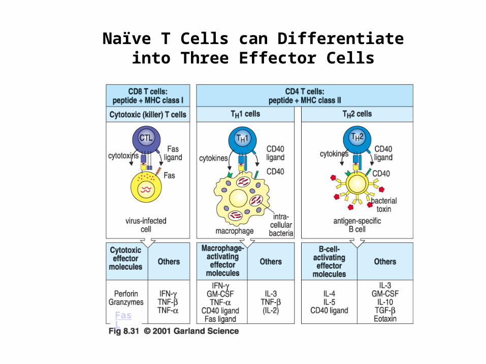
Naïve T Cells can Differentiate into Three Effector Cells
FasL

Fas-FasL-mediated Apoptosis Pathway (Extrinsic Pathway)
Back

Release of Effector Molecules is Localized on Contact Sites of T and Target Cells

Cytotoxic Effector Proteins Released by T Cells
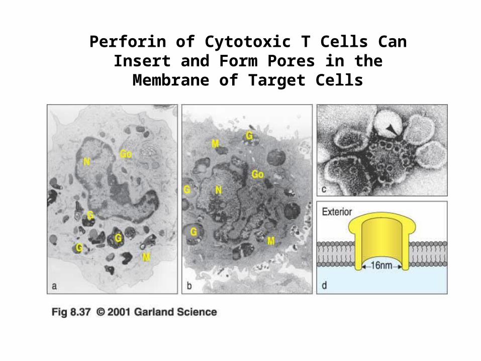
Perforin of Cytotoxic T Cells Can Insert and Form Pores in the Membrane of Target Cells

Differentiation of Immature CD4 Helper T Cells
In terms of cytokine production

Genes Dev. 2000 14:1693
Factors Involved in the Differentiation of Helper T Cells
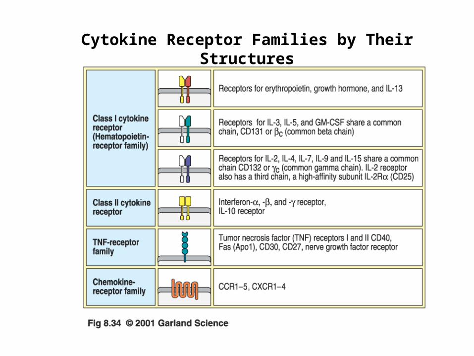
Cytokine Receptor Families by Their Structures

Functions of Th1 Cytokines

Effector Functions of Th1 Cells
Cell. Mol. Immunol. 5th ed. Abbas et al.

Th1 Cells Activate Macrophages to Become Highly Microbicidal

Functions of Th2 Cytokines

Effector Functions of Th2 Cells
Cell. Mol. Immunol. 5th ed. Abbas et al.
Related Documents