Leptospira interrogans Endostatin-Like Outer Membrane Proteins Bind Host Fibronectin, Laminin and Regulators of Complement Brian Stevenson 1 *, Henry A. Choy 2,3 , Marija Pinne 2,3 , Matthew L. Rotondi 4¤a , M. Clarke Miller 4¤b , Edward DeMoll 4 , Peter Kraiczy 5 , Anne E. Cooley 1 , Trevor P. Creamer 6 , Marc A. Suchard 7 , Catherine A. Brissette 1 , Ashutosh Verma 1,8 , David A. Haake 2,3 1 Department of Microbiology, Immunology, and Molecular Genetics, University of Kentucky College of Medicine, Lexington, Kentucky, United States of America, 2 Division of Infectious Diseases, Veterans Affairs Greater Los Angeles Health Care System, Los Angeles, California, United States of America, 3 Department of Medicine, University of California Los Angeles School of Medicine, Los Angeles, California, United States of America, 4 Department of Chemistry, University of Kentucky, Lexington, Kentucky, United States of America, 5 Institute of Medical Microbiology and Infection Control, University Hospital of Frankfurt, Frankfurt am Main, Germany, 6 Department of Molecular and Cellular Biochemistry, University of Kentucky College of Medicine, Lexington, Kentucky, United States of America, 7 Department of Biomathematics, University of California Los Angeles School of Medicine, Los Angeles, California, United States of America, 8 Department of Veterinary Sciences, University of Kentucky, Lexington, Kentucky, United States of America The pathogenic spirochete Leptospira interrogans disseminates throughout its hosts via the bloodstream, then invades and colonizes a variety of host tissues. Infectious leptospires are resistant to killing by their hosts’ alternative pathway of complement- mediated killing, and interact with various host extracellular matrix (ECM) components. The LenA outer surface protein (formerly called LfhA and Lsa24) was previously shown to bind the host ECM component laminin and the complement regulators factor H and factor H-related protein-1. We now demonstrate that infectious L. interrogans contain five additional paralogs of lenA, which we designated lenB, lenC, lenD, lenE and lenF. All six genes encode domains predicted to bear structural and functional similarities with mammalian endostatins. Sequence analyses of genes from seven infectious L. interrogans serovars indicated development of sequence diversity through recombination and intragenic duplication. LenB was found to bind human factor H, and all of the newly-described Len proteins bound laminin. In addition, LenB, LenC, LenD, LenE and LenF all exhibited affinities for fibronectin, a distinct host extracellular matrix protein. These characteristics suggest that Len proteins together facilitate invasion and colonization of host tissues, and protect against host immune responses during mammalian infection. Citation: Stevenson B, Choy HA, Pinne M, Rotondi ML, Miller MC, et al (2007) Leptospira interrogans Endostatin-Like Outer Membrane Proteins Bind Host Fibronectin, Laminin and Regulators of Complement. PLoS ONE 2(11): e1188. doi:10.1371/journal.pone.0001188 INTRODUCTION Leptospirosis is a zoonotic disease of humans caused by the spirochete Leptospira interrogans and several other members of that genus [1]. The prevalence of leptospirosis in many parts of the world is due to chronic kidney infection of a wide variety of domestic, peridomestic and wild reservoir host mammals, in- cluding rodents, pigs, cattle, horses and dogs. Colonization of the renal tubules of carrier animals results in shedding of virulent leptospires in the urine. Leptospires persist in fresh water until infection of a new host occurs via the conjunctiva, breaks in the skin or by invasion of mucous membranes in the respiratory or digestive tract. A hallmark of leptospiral infection is early and widespread hematogenous dissemination manifested by fever, myalgia, conjunctivitis, meningitis, uveitis and/or jaundice. Between 5 and 10% of patients progress to the more dangerous, icteric phase of leptospirosis, which may lead to death due to acute renal failure, pulmonary hemorrhage, intracerebral hemorrhage, and multiorgan system failure [1]. Infectious Leptospira spp. are endemic in many tropical and temperate areas of the world, presenting health threats to inhabitants of both rural and urban areas, as well as military personnel, aid workers, and tourists. Presumably as mechanisms that facilitate tissue invasion and colonization, pathogenic leptospires interact with a variety of host extracellular matrix (ECM) components, and some of the bacterial adhesins have been identified [reference [2–6] and this work]. L. interrogans is highly resistant to the alternative pathway of host complement activation [7–12], a feature that is associated with binding of factor H to the bacterial outer membrane, degradation of C3b and C3 convertase, and inhibition of membrane-attack complex formation [11,12]. The capacities to bind host ECM and factor H are associated with virulence, as those traits are held by infectious Leptospira species but are lacking from non-infectious species of Leptospira [2,11,12]. A previous study which screened an L. interrogans expression library for proteins capable of binding host factor H identified an approximately 25 kDa outer membrane protein, designated LfhA ( leptospiral factor H-binding protein A) [12]. LfhA was also found Academic Editor: Adam Ratner, Columbia University, United States of America Received August 27, 2007; Accepted October 24, 2007; Published November 14, 2007 This is an open-access article distributed under the terms of the Creative Commons Public Domain declaration which stipulates that, once placed in the public domain, this work may be freely reproduced, distributed, transmitted, modified, built upon, or otherwise used by anyone for any lawful purpose. Funding: This work was funded in part by VA Medical Research funds and National 10 Institute of Allergy and Infectious Diseases grant AI-34431 to D. Haake. M. Suchard is an Alfred P. Sloan Research Fellow. A. Verma was supported by a Paul Mellon Fellowship in Equine Studies. Competing Interests: The authors have declared that no competing interests exist. * To whom correspondence should be addressed. E-mail: brian.stevenson@uky. edu ¤a Current address: Weill Medical College of Cornell University, New York, New York, United States of America ¤b Current address: Brown Cancer Center, University of Louisville, Louisville, Kentucky, United States of America PLoS ONE | www.plosone.org 1 November 2007 | Issue 11 | e1188

Welcome message from author
This document is posted to help you gain knowledge. Please leave a comment to let me know what you think about it! Share it to your friends and learn new things together.
Transcript
Leptospira interrogans Endostatin-Like Outer MembraneProteins Bind Host Fibronectin, Laminin and Regulatorsof ComplementBrian Stevenson1*, Henry A. Choy2,3, Marija Pinne2,3, Matthew L. Rotondi4¤a, M. Clarke Miller4¤b, Edward DeMoll4, Peter Kraiczy5, Anne E.Cooley1, Trevor P. Creamer6, Marc A. Suchard7, Catherine A. Brissette1, Ashutosh Verma1,8, David A. Haake2,3
1 Department of Microbiology, Immunology, and Molecular Genetics, University of Kentucky College of Medicine, Lexington, Kentucky, United Statesof America, 2 Division of Infectious Diseases, Veterans Affairs Greater Los Angeles Health Care System, Los Angeles, California, United States ofAmerica, 3 Department of Medicine, University of California Los Angeles School of Medicine, Los Angeles, California, United States of America,4 Department of Chemistry, University of Kentucky, Lexington, Kentucky, United States of America, 5 Institute of Medical Microbiology and InfectionControl, University Hospital of Frankfurt, Frankfurt am Main, Germany, 6 Department of Molecular and Cellular Biochemistry, University of KentuckyCollege of Medicine, Lexington, Kentucky, United States of America, 7 Department of Biomathematics, University of California Los Angeles School ofMedicine, Los Angeles, California, United States of America, 8 Department of Veterinary Sciences, University of Kentucky, Lexington, Kentucky, UnitedStates of America
The pathogenic spirochete Leptospira interrogans disseminates throughout its hosts via the bloodstream, then invades andcolonizes a variety of host tissues. Infectious leptospires are resistant to killing by their hosts’ alternative pathway of complement-mediated killing, and interact with various host extracellular matrix (ECM) components. The LenA outer surface protein (formerlycalled LfhA and Lsa24) was previously shown to bind the host ECM component laminin and the complement regulators factor Hand factor H-related protein-1. We now demonstrate that infectious L. interrogans contain five additional paralogs of lenA, whichwe designated lenB, lenC, lenD, lenE and lenF. All six genes encode domains predicted to bear structural and functional similaritieswith mammalian endostatins. Sequence analyses of genes from seven infectious L. interrogans serovars indicated development ofsequence diversity through recombination and intragenic duplication. LenB was found to bind human factor H, and all of thenewly-described Len proteins bound laminin. In addition, LenB, LenC, LenD, LenE and LenF all exhibited affinities for fibronectin,a distinct host extracellular matrix protein. These characteristics suggest that Len proteins together facilitate invasion andcolonization of host tissues, and protect against host immune responses during mammalian infection.
Citation: Stevenson B, Choy HA, Pinne M, Rotondi ML, Miller MC, et al (2007) Leptospira interrogans Endostatin-Like Outer Membrane Proteins BindHost Fibronectin, Laminin and Regulators of Complement. PLoS ONE 2(11): e1188. doi:10.1371/journal.pone.0001188
INTRODUCTIONLeptospirosis is a zoonotic disease of humans caused by the
spirochete Leptospira interrogans and several other members of that
genus [1]. The prevalence of leptospirosis in many parts of the
world is due to chronic kidney infection of a wide variety of
domestic, peridomestic and wild reservoir host mammals, in-
cluding rodents, pigs, cattle, horses and dogs. Colonization of the
renal tubules of carrier animals results in shedding of virulent
leptospires in the urine. Leptospires persist in fresh water until
infection of a new host occurs via the conjunctiva, breaks in the
skin or by invasion of mucous membranes in the respiratory or
digestive tract. A hallmark of leptospiral infection is early and
widespread hematogenous dissemination manifested by fever,
myalgia, conjunctivitis, meningitis, uveitis and/or jaundice.
Between 5 and 10% of patients progress to the more dangerous,
icteric phase of leptospirosis, which may lead to death due to acute
renal failure, pulmonary hemorrhage, intracerebral hemorrhage,
and multiorgan system failure [1]. Infectious Leptospira spp. are
endemic in many tropical and temperate areas of the world,
presenting health threats to inhabitants of both rural and urban
areas, as well as military personnel, aid workers, and tourists.
Presumably as mechanisms that facilitate tissue invasion and
colonization, pathogenic leptospires interact with a variety of host
extracellular matrix (ECM) components, and some of the bacterial
adhesins have been identified [reference [2–6] and this work]. L.
interrogans is highly resistant to the alternative pathway of host
complement activation [7–12], a feature that is associated with
binding of factor H to the bacterial outer membrane, degradation
of C3b and C3 convertase, and inhibition of membrane-attack
complex formation [11,12]. The capacities to bind host ECM and
factor H are associated with virulence, as those traits are held by
infectious Leptospira species but are lacking from non-infectious
species of Leptospira [2,11,12].
A previous study which screened an L. interrogans expression
library for proteins capable of binding host factor H identified an
approximately 25 kDa outer membrane protein, designated LfhA
(leptospiral factor H-binding protein A) [12]. LfhA was also found
Academic Editor: Adam Ratner, Columbia University, United States of America
Received August 27, 2007; Accepted October 24, 2007; Published November 14,2007
This is an open-access article distributed under the terms of the CreativeCommons Public Domain declaration which stipulates that, once placed in thepublic domain, this work may be freely reproduced, distributed, transmitted,modified, built upon, or otherwise used by anyone for any lawful purpose.
Funding: This work was funded in part by VA Medical Research funds andNational 10 Institute of Allergy and Infectious Diseases grant AI-34431 to D.Haake. M. Suchard is an Alfred P. Sloan Research Fellow. A. Verma was supportedby a Paul Mellon Fellowship in Equine Studies.
Competing Interests: The authors have declared that no competing interestsexist.
* To whom correspondence should be addressed. E-mail: [email protected]
¤a Current address: Weill Medical College of Cornell University, New York, NewYork, United States of America¤b Current address: Brown Cancer Center, University of Louisville, Louisville,Kentucky, United States of America
PLoS ONE | www.plosone.org 1 November 2007 | Issue 11 | e1188
to bind human factor H-related protein 1 (FHR-1), a distinct
protein whose carboxy-terminus is very similar to that of factor H
[12,13]. LfhA did not bind human factor H-like protein 1 (FHL-1),
a splice variant of the same gene as factor H which consists of the
first seven short consensus repeat units of factor H [12,13].
A subsequent study by Barbosa and colleagues identified an L.
interrogans outer membrane laminin-binding protein, which they
designated Lsa24 (leptospiral surface adhesin 24kDa) [2]. Intrigu-
ingly, Lsa24 and LfhA are the same protein, indicating that this
single protein is able to bind host factor H, FHR-1 and laminin.
As described in the present report, L. interrogans carries 5
additional genes homologous to lfhA/lsa24. Modeling of the
predicted proteins encoded by these six paralogous genes indicated
substantial similarities to mammalian endostatins. In order to unify
the nomenclatures of these leptospiral genes, we renamed lfhA/
lsa24 as ‘‘lenA’’ (leptospiral endostatin-like protein A) and named
the newly-described paralogs lenB, lenC, lenD, lenE and lenF. At the
present time, there are no tools available to specifically mutate L.
interrogans, so it is impossible to study protein function in situ by
deletion and complementation mutagenesis. That being so, in
order to better understand these genes and their roles in leptospiral
pathogenesis, we characterized relationships of len genes among
several distinct L. interrogans strains, performed biophysical
characterization of the proteins, and examined interactions
between Len and host ECM and complement-regulatory proteins.
RESULTS
Paralogous L. interrogans len genesIn a previous study [12], members of our laboratories demon-
strated that the lenA (lfhA) genes of serovar Pomona strain JEN4
and serovar Lai strain 56601 encode a factor H/FHR-1 binding
protein. Sequence analyses of additional L. interrogans strains from
five other serovars indicated that all contain lenA loci. The lenA
gene sequences of serovars Lai, Copenhageni, Grippotyphosa and
Hardjo are all highly conserved and are predicted to encode
membrane-associated lipoproteins (Fig. 1) [14,15]. The lenA loci of
serovars Pomona, Bratislava and Canicola are nearly identical to
those of the aforementioned serovars, but include mutations that
would preclude their synthesis of intact lipoproteins.
The sequenced genomes of L. interrogans serovar Copenhageni
strain Fiocruz L1-130 and serovar Lai strain 56601 both contain
an additional locus with size and sequence similar to lenA, which
we named lenB (Table 1, Fig. 1). All other L. interrogans strains we
examined also contain a lenB locus, bordered by sequences similar
to those of serovars Copenhageni and Lai. The lenB loci of serovars
Bratislava and Hardjo each encode a protein with a lipoprotein
leader polypeptide and a cleavage/lipidation sequence (Fig. 1)
[14,15]. However, all other analyzed serovars lack the initial 19
codons of lenB, as well as a substantial portion of upstream DNA,
and appear to be defective genes. Thus, only serovars Bratislava
and Hardjo are predicted to produce membrane-associated LenB
lipoproteins. The 59 non-coding regions of the Bratislava and
Hardjo lenB loci contain extensive homologies to the 59 non-coding
regions of lenA loci (Fig. 1D).
Four additional paralogs of lenA and lenB were identified in the
genome sequences of Copenhageni Fiocruz L1-130 and Lai
56601, which we named lenC, lenD, lenE, and lenF (Table 1, Fig. 1).
The latter four genes were sequenced in their entirety from five
additional L. interrogans isolates. Each of the four genes in all seven
strains was intact and predicted to encode a protein of
approximately 50kDa. The nearly two-fold increase in size
compared to LenA and LenB was accounted for by the presence
of two motifs that each resemble LenA and LenB, which we refer
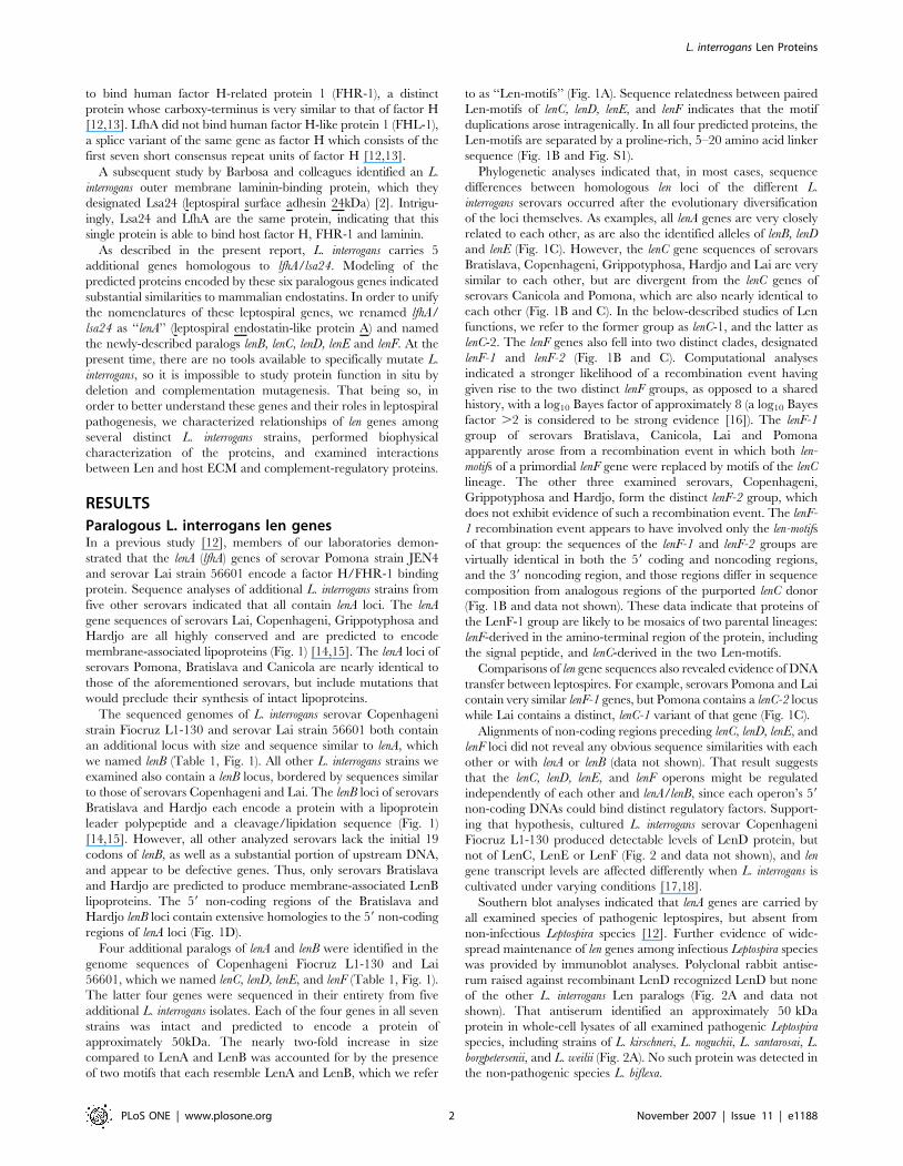
to as ‘‘Len-motifs’’ (Fig. 1A). Sequence relatedness between paired
Len-motifs of lenC, lenD, lenE, and lenF indicates that the motif
duplications arose intragenically. In all four predicted proteins, the
Len-motifs are separated by a proline-rich, 5–20 amino acid linker
sequence (Fig. 1B and Fig. S1).
Phylogenetic analyses indicated that, in most cases, sequence
differences between homologous len loci of the different L.
interrogans serovars occurred after the evolutionary diversification
of the loci themselves. As examples, all lenA genes are very closely
related to each other, as are also the identified alleles of lenB, lenD
and lenE (Fig. 1C). However, the lenC gene sequences of serovars
Bratislava, Copenhageni, Grippotyphosa, Hardjo and Lai are very
similar to each other, but are divergent from the lenC genes of
serovars Canicola and Pomona, which are also nearly identical to
each other (Fig. 1B and C). In the below-described studies of Len
functions, we refer to the former group as lenC-1, and the latter as
lenC-2. The lenF genes also fell into two distinct clades, designated
lenF-1 and lenF-2 (Fig. 1B and C). Computational analyses
indicated a stronger likelihood of a recombination event having
given rise to the two distinct lenF groups, as opposed to a shared
history, with a log10 Bayes factor of approximately 8 (a log10 Bayes
factor .2 is considered to be strong evidence [16]). The lenF-1
group of serovars Bratislava, Canicola, Lai and Pomona
apparently arose from a recombination event in which both len-
motifs of a primordial lenF gene were replaced by motifs of the lenC
lineage. The other three examined serovars, Copenhageni,
Grippotyphosa and Hardjo, form the distinct lenF-2 group, which
does not exhibit evidence of such a recombination event. The lenF-
1 recombination event appears to have involved only the len-motifs
of that group: the sequences of the lenF-1 and lenF-2 groups are
virtually identical in both the 59 coding and noncoding regions,
and the 39 noncoding region, and those regions differ in sequence
composition from analogous regions of the purported lenC donor
(Fig. 1B and data not shown). These data indicate that proteins of
the LenF-1 group are likely to be mosaics of two parental lineages:
lenF-derived in the amino-terminal region of the protein, including
the signal peptide, and lenC-derived in the two Len-motifs.
Comparisons of len gene sequences also revealed evidence of DNA
transfer between leptospires. For example, serovars Pomona and Lai
contain very similar lenF-1 genes, but Pomona contains a lenC-2 locus
while Lai contains a distinct, lenC-1 variant of that gene (Fig. 1C).
Alignments of non-coding regions preceding lenC, lenD, lenE, and
lenF loci did not reveal any obvious sequence similarities with each
other or with lenA or lenB (data not shown). That result suggests
that the lenC, lenD, lenE, and lenF operons might be regulated
independently of each other and lenA/lenB, since each operon’s 59
non-coding DNAs could bind distinct regulatory factors. Support-
ing that hypothesis, cultured L. interrogans serovar Copenhageni
Fiocruz L1-130 produced detectable levels of LenD protein, but
not of LenC, LenE or LenF (Fig. 2 and data not shown), and len
gene transcript levels are affected differently when L. interrogans is
cultivated under varying conditions [17,18].
Southern blot analyses indicated that lenA genes are carried by
all examined species of pathogenic leptospires, but absent from
non-infectious Leptospira species [12]. Further evidence of wide-
spread maintenance of len genes among infectious Leptospira species
was provided by immunoblot analyses. Polyclonal rabbit antise-
rum raised against recombinant LenD recognized LenD but none
of the other L. interrogans Len paralogs (Fig. 2A and data not
shown). That antiserum identified an approximately 50 kDa
protein in whole-cell lysates of all examined pathogenic Leptospira
species, including strains of L. kirschneri, L. noguchii, L. santarosai, L.
borgpetersenii, and L. weilii (Fig. 2A). No such protein was detected in
the non-pathogenic species L. biflexa.
L. interrogans Len Proteins
PLoS ONE | www.plosone.org 2 November 2007 | Issue 11 | e1188
Figure 1. Relationships between Len proteins and genes. (A) Schematic of Len proteins, with individual Len-motifs indicated by grey rectangles.LenC, LenD, LenE and LenF each consist of 2 Len-motifs, bridged by proline-rich linkers. (B) Alignment of predicted amino acid sequences ofrepresentative proteins: serovar Lai LenA (LenA, Lai), serovar Bratislava LenB (LenB, Brat), serovar Bratislava LenC-1 (LenC, Brat), serovar Canicola LenC-2 (LenC, Can), serovar Pomona LenD (LenD, Pom), serovar Grippotyphosa LenE (LenE, Gripp), serovar Pomona LenF-1 (LenF,. Pom), and serovar HardjoLenF-1 (LenF, Har). Sequences of the proteins possessing two Len-motifs (LenC, LenD, LenE and LenF) were divided in the middle, after the well-conserved internal CVEQ sequence, to permit alignment of each Len-motif, and the amino- and carboxy-terminal Len-motifs are indicated by ‘‘-N’’and ‘‘-C’’, respectively. An alignment of these same proteins, undivided, is presented in Figure S1. Identical amino acids found in the majority ofproteins are boxed and shaded. Cysteine residues that may serve as amino-terminal lipidation sites are circled. (C) Unrooted phylogenetic tree ofpredicted amino acid sequences of each identified Len protein. Bootstrap values of each major node are indicated. (D) Alignment of sequenceslocated 59 of lenA and lenB genes. Identical nucleotides are boxed and shaded.doi:10.1371/journal.pone.0001188.g001
L. interrogans Len Proteins
PLoS ONE | www.plosone.org 3 November 2007 | Issue 11 | e1188
Cellular localization and biophysical
characterization of Len proteinsLenA is an outer membrane protein [2,12]. All Len proteins bear
at least some hallmarks of being lipoproteins, with LenA, LenB,
LenC, LenD and LenF having high probabilities of being so
according to analyses using the spirochete-specific lipoprotein
algorithm SpLip [14,15]. Using the above-described anti-LenD
antiserum, cellular localization of that protein was assessed by
Triton X-114 detergent solubilization and phase partitioning of
live leptospires. The reliability of this technique has been validated
by comparisons with results obtained by sucrose density gradient
isolation of outer membrane vesicles [19]. This method yields
three fractions: a detergent fraction consisting of outer membrane
components, an aqueous fraction consisting of periplasmic
contents, and a pellet consisting of inner membrane-associated
components, cytoplasmic contents, and undisrupted cells [20].
LenD was found in the detergent fraction (Fig. 2B). Control
analysis of the endoflagellar sheath protein FlaA1 (which is
attached to the inner membrane) confirmed that the detergent
phase was not contaminated with inner membrane components
(Fig. 2B). Presence of LenD in the pellet fraction is typical of
leptospiral outer membrane proteins [21–23], and is indicative of
incomplete Triton X-114 extraction. These results indicate that
LenD is also an outer membrane protein.
The LenC, LenD, LenE and LenF proteins appear to be fused
dimers of LenA/LenB-like proteins. This suggested to us that
LenA and LenB might function as dimers, with each dimer being
the equivalent of a single LenC, LenD, LenE or LenF protein. To
explore that possibility, we examined whether or not recombinant
LenA forms dimers. However, HPLC through a size-exclusion
column yielded a single peak, with a calculated molecular mass of
24.2 kDa (data not shown), comparable to the calculated
molecular mass of 24.4 kDa for the recombinant LenA monomer.
Circular dichroism (CD) analysis of recombinant LenA indicated
that it is composed of 36% b-sheet, 23% turns, and 41%
unstructured/other, with no evidence of any a-helices (Fig. 3A).
These data are in line with previous CD analyses indicating that
LenA contains b-sheets [2]. Recombinant LenA was found to be
a relatively stable protein, with a melting point of 53uC (62uC)
(Fig. 3B). Due to the limited solubility of the other recombinant Len
paralogs, those proteins could not be analyzed by these biophysical
techniques. PHYRE modeling of the predicated amino acid
sequences of all Len proteins indicated moderate to strong
probabilities (ranging between 25 and 60% estimated precision) for
each Len-motif folding into a structure similar to those of
mammalian endostatins, which are derived from the carboxy-
termini of collagens XVIII and XV [24–26]. Among other functions,
endostatins bind various ECM components, including laminin,
[25,26]. We did not detect sequence or predicted structural
similarities between Len proteins and any other known adhesins.
Functional characterization of Len paralogsThrough use of both affinity blot analyses and surface plasmon
resonance, members of our laboratories previously demonstrated
that LenA binds host complement factor H [12]. Ligand affinity
blot analyses of LenB indicated that it, too, can bind human factor
H (Fig. 4). However, none of the other Len proteins exhibited
binding of factor H, as assessed by ligand affinity blot.
A previous study indicated that recombinant LenA (Lsa24)
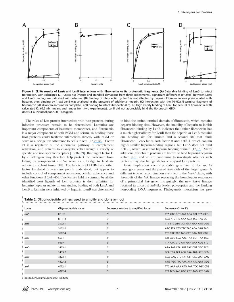
bound laminin [2]. We therefore examined whether or not the five
newly-identified L. interrogans proteins shared that property. Each
recombinant protein was solubilized in SDS buffer, subjected to
SDS-PAGE and transferred to nitrocellulose membranes, then
examined for ability to bind soluble laminin. All the recombinant
Len proteins bound laminin, although LenC-1, LenC-2, LenE,
LenF-1 and LenF-2 consistently yielded the strongest affinity blot
signals, with LenD, LenB and then LenA exhibiting progressively
weaker relative binding of laminin (Fig. 5). No laminin binding by
control protein BSA was detected, even with extended film
exposure times, demonstrating that binding of laminin by the Len
proteins was specific.
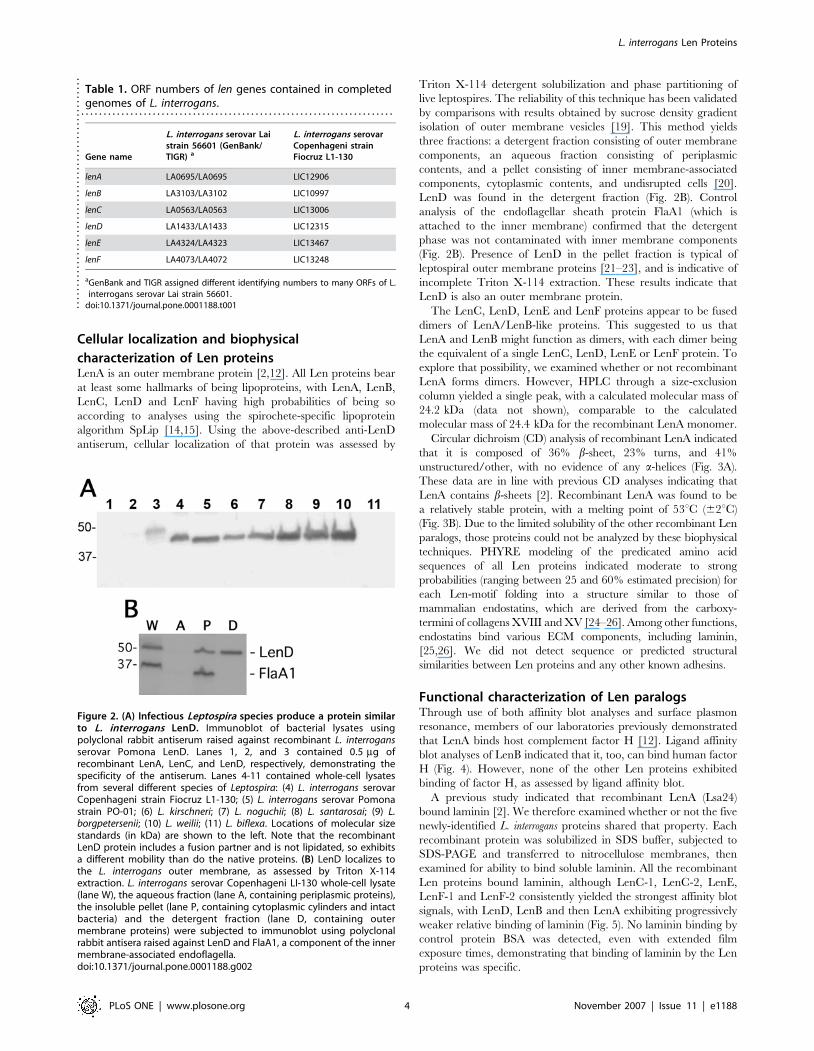
Table 1. ORF numbers of len genes contained in completedgenomes of L. interrogans.
. . . . . . . . . . . . . . . . . . . . . . . . . . . . . . . . . . . . . . . . . . . . . . . . . . . . . . . . . . . . . . . . . . . . . .
Gene name
L. interrogans serovar Laistrain 56601 (GenBank/TIGR) a
L. interrogans serovarCopenhageni strainFiocruz L1-130
lenA LA0695/LA0695 LIC12906
lenB LA3103/LA3102 LIC10997
lenC LA0563/LA0563 LIC13006
lenD LA1433/LA1433 LIC12315
lenE LA4324/LA4323 LIC13467
lenF LA4073/LA4072 LIC13248
aGenBank and TIGR assigned different identifying numbers to many ORFs of L.interrogans serovar Lai strain 56601.
doi:10.1371/journal.pone.0001188.t001....
....
....
....
....
....
....
....
....
....
....
....
....
Figure 2. (A) Infectious Leptospira species produce a protein similarto L. interrogans LenD. Immunoblot of bacterial lysates usingpolyclonal rabbit antiserum raised against recombinant L. interrogansserovar Pomona LenD. Lanes 1, 2, and 3 contained 0.5 mg ofrecombinant LenA, LenC, and LenD, respectively, demonstrating thespecificity of the antiserum. Lanes 4-11 contained whole-cell lysatesfrom several different species of Leptospira: (4) L. interrogans serovarCopenhageni strain Fiocruz L1-130; (5) L. interrogans serovar Pomonastrain PO-01; (6) L. kirschneri; (7) L. noguchii; (8) L. santarosai; (9) L.borgpetersenii; (10) L. weilii; (11) L. biflexa. Locations of molecular sizestandards (in kDa) are shown to the left. Note that the recombinantLenD protein includes a fusion partner and is not lipidated, so exhibitsa different mobility than do the native proteins. (B) LenD localizes tothe L. interrogans outer membrane, as assessed by Triton X-114extraction. L. interrogans serovar Copenhageni LI-130 whole-cell lysate(lane W), the aqueous fraction (lane A, containing periplasmic proteins),the insoluble pellet (lane P, containing cytoplasmic cylinders and intactbacteria) and the detergent fraction (lane D, containing outermembrane proteins) were subjected to immunoblot using polyclonalrabbit antisera raised against LenD and FlaA1, a component of the innermembrane-associated endoflagella.doi:10.1371/journal.pone.0001188.g002
L. interrogans Len Proteins
PLoS ONE | www.plosone.org 4 November 2007 | Issue 11 | e1188
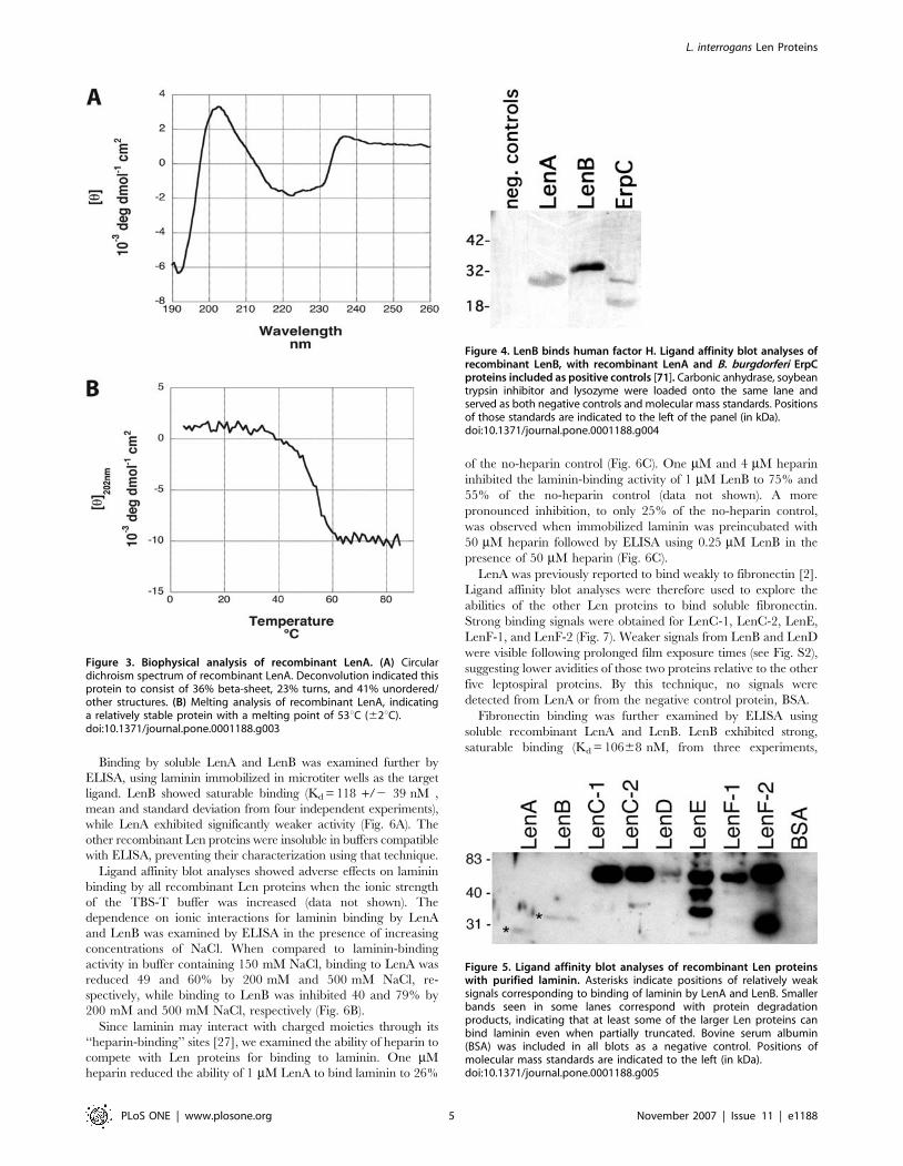
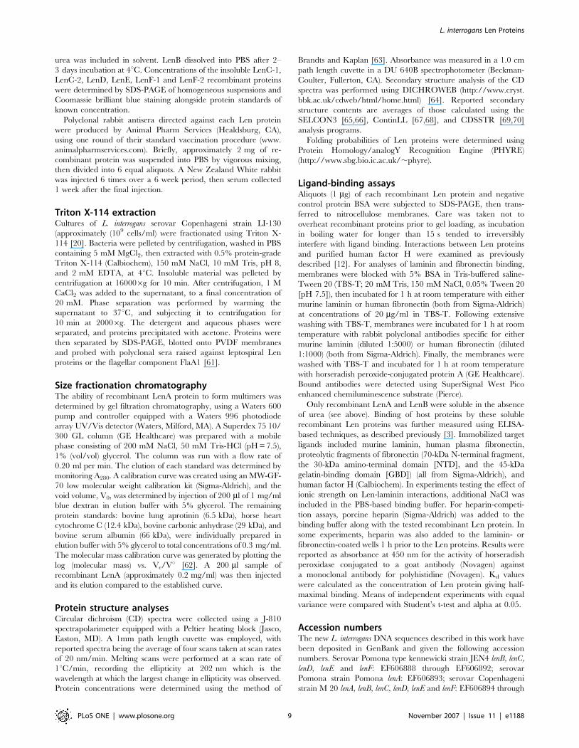
Binding by soluble LenA and LenB was examined further by
ELISA, using laminin immobilized in microtiter wells as the target
ligand. LenB showed saturable binding (Kd = 118 +/2 39 nM ,
mean and standard deviation from four independent experiments),
while LenA exhibited significantly weaker activity (Fig. 6A). The
other recombinant Len proteins were insoluble in buffers compatible
with ELISA, preventing their characterization using that technique.
Ligand affinity blot analyses showed adverse effects on laminin
binding by all recombinant Len proteins when the ionic strength
of the TBS-T buffer was increased (data not shown). The
dependence on ionic interactions for laminin binding by LenA
and LenB was examined by ELISA in the presence of increasing
concentrations of NaCl. When compared to laminin-binding
activity in buffer containing 150 mM NaCl, binding to LenA was
reduced 49 and 60% by 200 mM and 500 mM NaCl, re-
spectively, while binding to LenB was inhibited 40 and 79% by
200 mM and 500 mM NaCl, respectively (Fig. 6B).
Since laminin may interact with charged moieties through its
‘‘heparin-binding’’ sites [27], we examined the ability of heparin to
compete with Len proteins for binding to laminin. One mM
heparin reduced the ability of 1 mM LenA to bind laminin to 26%
of the no-heparin control (Fig. 6C). One mM and 4 mM heparin
inhibited the laminin-binding activity of 1 mM LenB to 75% and
55% of the no-heparin control (data not shown). A more
pronounced inhibition, to only 25% of the no-heparin control,
was observed when immobilized laminin was preincubated with
50 mM heparin followed by ELISA using 0.25 mM LenB in the
presence of 50 mM heparin (Fig. 6C).
LenA was previously reported to bind weakly to fibronectin [2].
Ligand affinity blot analyses were therefore used to explore the
abilities of the other Len proteins to bind soluble fibronectin.
Strong binding signals were obtained for LenC-1, LenC-2, LenE,
LenF-1, and LenF-2 (Fig. 7). Weaker signals from LenB and LenD
were visible following prolonged film exposure times (see Fig. S2),
suggesting lower avidities of those two proteins relative to the other
five leptospiral proteins. By this technique, no signals were
detected from LenA or from the negative control protein, BSA.
Fibronectin binding was further examined by ELISA using
soluble recombinant LenA and LenB. LenB exhibited strong,
saturable binding (Kd = 10668 nM, from three experiments,
Figure 4. LenB binds human factor H. Ligand affinity blot analyses ofrecombinant LenB, with recombinant LenA and B. burgdorferi ErpCproteins included as positive controls [71]. Carbonic anhydrase, soybeantrypsin inhibitor and lysozyme were loaded onto the same lane andserved as both negative controls and molecular mass standards. Positionsof those standards are indicated to the left of the panel (in kDa).doi:10.1371/journal.pone.0001188.g004
Figure 5. Ligand affinity blot analyses of recombinant Len proteinswith purified laminin. Asterisks indicate positions of relatively weaksignals corresponding to binding of laminin by LenA and LenB. Smallerbands seen in some lanes correspond with protein degradationproducts, indicating that at least some of the larger Len proteins canbind laminin even when partially truncated. Bovine serum albumin(BSA) was included in all blots as a negative control. Positions ofmolecular mass standards are indicated to the left (in kDa).doi:10.1371/journal.pone.0001188.g005
Figure 3. Biophysical analysis of recombinant LenA. (A) Circulardichroism spectrum of recombinant LenA. Deconvolution indicated thisprotein to consist of 36% beta-sheet, 23% turns, and 41% unordered/other structures. (B) Melting analysis of recombinant LenA, indicatinga relatively stable protein with a melting point of 53uC (62uC).doi:10.1371/journal.pone.0001188.g003
L. interrogans Len Proteins
PLoS ONE | www.plosone.org 5 November 2007 | Issue 11 | e1188
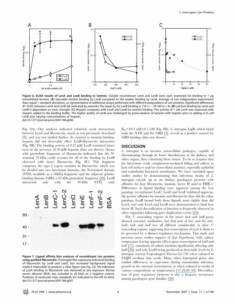
Fig. 8A). Our analyses indicated relatively weak interactions
between LenA and fibronectin, much as was previously described
[2], and was not studied further. In contrast to laminin binding,
heparin did not detectably affect LenB-fibronectin interaction
(Fig. 8B). The binding activity of 0.25 mM LenB remained intact
even in the presence of 50 mM heparin (data not shown). Assays
with proteolytic fragments of fibronectin indicated that the N-
terminal 70 kDa could account for all of the binding by LenB
observed with intact fibronectin (Fig. 8C). This fragment
comprises the type I repeat modules in fibronectin, which can
be divided into two functional domains, the N-terminal domain
(NTD; available as a 30kDa fragment) and the adjacent gelatin-
binding domain (GBD; a 45 kDa proteolytic fragment) [28]. LenB
interacted only with the 30 kDa NTD, with
Kd = 69.5 nM60.7 nM (Fig. 8D). L. interrogans LigB, which binds
both the NTD and the GBD [3], served as a positive control for
GBD binding (data not shown).
DISCUSSIONL. interrogans is an invasive extracellular pathogen, capable of
disseminating through its hosts’ bloodstream to the kidneys and
other organs, then colonizing those tissues. To do so requires that
the bacterium evade complement-mediated killing and adhere to
host cell surfaces and/or extracellular matrices, especially epithelial
and endothelial basement membranes. We have extended upon
earlier studies by demonstrating that infectious strains of L.
interrogans encode up to six distinct paralogous proteins with
affinities for host fibronectin, laminin, factor H and/or FHR-1.
Differences in ligand binding were apparent among the Len
paralogs: recombinant LenC, LenE and LenF exhibited apprecia-
bly greater affinities for laminin and fibronectin than did the other
paralogs, LenB bound both those ligands more tightly than did
LenA, and only LenA and LenB were demonstrated to bind host
factor H. Such diversification of function is frequently observed in
other organisms following gene duplication events [29].
The 59 noncoding regions of the intact lenA and lenB genes
showed extensive similarities, but that pair of loci and the lenC,
lenD, lenE and lenF loci all differed considerably in their 59
noncoding regions, suggesting that transcription of each is likely to
be governed by a distinct regulatory mechanism. This study and
previous array studies support of that hypothesis, with culture
temperature having opposite effects upon transcription of lenD and
lenE [17], osmolarity of culture medium significantly affecting only
lenD [30], and only LenD being produced at detectable levels by L.
interrogans serovar Copenhageni Fiocruz L1-130 when cultured in
EMJH medium (this work). Many other leptospiral genes also
exhibit differences in expression during mammalian infection,
growth in the external environment, or when cultured in media of
various compositions or temperatures [17,18,30–33]. Diversifica-
tion of gene regulatory elements is also a frequent occurrence
among paralogous gene families [34].
Figure 6. ELISA results of LenA and LenB binding to laminin. Soluble recombinant LenA and LenB were each examined for binding to 1 mgimmobilized laminin. (A) Saturable laminin binding by LenB compared to the weaker binding by LenA. Average of two independent experiments(bars equal 1 standard deviation), as representative of additional assays performed with different preparations of Len proteins. Significant differences(P,0.05) between LenA and LenB are indicated by asterisks The mean Kd for LenB binding is 118 +/2 39 nM (n = 4). (B) Laminin binding by LenA andLenB is dependent on ionic strength. (C) Heparin competes with LenA and LenB for laminin binding. The activity of 1 mM LenA was measured withheparin added to the binding buffer. The higher avidity of LenB was challenged by preincubation of laminin with heparin prior to adding 0.25 mMLenB plus varying concentrations of heparin.doi:10.1371/journal.pone.0001188.g006
Figure 7. Ligand affinity blot analyses of recombinant Len proteinsusing purified fibronectin. Prolonged film exposures indicated bindingof fibronectin by LenB and LenD, but increased background signalmade it impossible to produce a clear figure (see Fig. S2). No indicationof LenA binding to fibronectin was observed at any exposure. Bovineserum albumin (BSA) was included in all blots as a negative control.Positions of molecular mass standards are indicated to the left (in kDa).doi:10.1371/journal.pone.0001188.g007
L. interrogans Len Proteins
PLoS ONE | www.plosone.org 6 November 2007 | Issue 11 | e1188
The roles of Len protein interactions with host proteins during
infection processes remain to be determined. Laminins are
important components of basement membranes, and fibronectin
is a major component of both ECM and serum, so binding those
host proteins could facilitate interactions directly with ECM or
serve as a bridge for adherence to cell surfaces [27,28,35]. Factor
H is a regulator of the alternative pathway of complement
activation, and adheres to eukaryotic cells through a variety of
specific and non-specific receptors [13,36–39]. Binding of factor H
by L. interrogans may therefore help protect the bacterium from
killing by complement and/or serve as a bridge to facilitate
adherence to host tissues [40]. The functions of FHR-1 and other
factor H-related proteins are poorly understood, but appear to
include control of complement activation, cellular adherence and
other functions [13,41–45]. One feature held in common by all the
identified host ligands of Len proteins is their affinities for
heparin/heparan sulfate. In our studies, binding of both LenA and
LenB to laminin were inhibited by heparin. LenB was determined
to bind the amino-terminal domain of fibronectin, which contains
heparin-binding sites. However, the inability of heparin to inhibit
fibronectin-binding by LenB indicates that either fibronectin has
a much higher affinity for LenB than for heparin or LenB contains
one binding site for laminin and a second site that binds
fibronectin. LenA binds both factor H and FHR-1, which contain
highly similar heparin-binding regions, but LenA does not bind
FHL-1, which lacks that heparin binding domain [12,13]. Many
additional vertebrate proteins are known to bind heparin/heparan
sulfate [46], and we are continuing to investigate whether such
proteins may also be ligands for leptospiral Len proteins.
Gene duplication events probably gave rise to the six len
paralogous genes and the paired len-motifs of the larger genes. A
different type of recombination event led to the lenF-1 clade, with
len-motifs of the lenC lineage replacing the homologous sequences
of a primordial lenF gene. Intriguingly, the new lenF-1 lineage
retained its ancestral lenF-like leader polypeptide and the flanking
non-coding DNA sequences. Phylogenetic mosaicism has pre-
Table 2. Oligonucleotide primers used to amplify and clone len loci.. . . . . . . . . . . . . . . . . . . . . . . . . . . . . . . . . . . . . . . . . . . . . . . . . . . . . . . . . . . . . . . . . . . . . . . . . . . . . . . . . . . . . . . . . . . . . . . . . . . . . . . . . . . . . . . . . . . . . . . . . . . . . . . . . . . . . . . . . . . . . . . . . .
Locus Oligonucleotide name Sequence relative to amplified locus Sequence (59 to 39)
lenA LFH-2 59 TTA GTC GGT AAT AGA GTT TTA GCG
LFH-11 39 ACA ATC TTC CAA AGA TCC TAA CG
lenB 3102-1 59 TTT TTG ATG GCT GCA GAA ATG GGG
3102-2 39 AAC TTA CTG TTC TAC ACA GAG TAG
3102-4 39 TTC TAC TAT TAG CCT GAA AGC CTG
lenC 563-1 59 ATT ACG CCA AAC TAA CGT TAA TCG
563-4 39 TTA CTC GTC ATT GAA AAA AGG TTG
lenD 1433-1 59 AAA TAT CTA AGT TAC CGT CGC TCG
1433-2 39 TCA TCA TCT ACG CAA AGA ATT GCG
lenE 4323-1 59 ACA GAA GTC TAT CTT CAG AAT GAG
4323-2 39 ATG AGA TTC AAA ATA ATC GAT CGG
lenF 4072-1 59 TTG AAA AAA ATG AAA TCC AGC CTG
4072-4 39 TTT TCG AAC GGG CCT AAG ATT GAG
doi:10.1371/journal.pone.0001188.t002....
....
....
....
....
....
....
....
....
....
....
....
....
....
....
....
.
Figure 8. ELISA results of LenA and LenB interactions with fibronectin or its proteolytic fragments. (A) Saturable binding of LenB to intactfibronectin, with calculated Kd 10668 nM (means and standard deviations from three experiments). Significant differences (P,0.05) between LenAand LenB binding are indicated with asterisks. (B) Binding of fibronectin by LenB is not affected by heparin. Fibronectin was preincubated withheparin, then binding by 1 mM LenB was analyzed in the presence of additional heparin. (C) Interaction with the 70-kDa N-terminal fragment offibronectin (70 kDa) can account for complete LenB binding to intact fibronectin (Fn). (D) High avidity binding of LenB to the NTD of fibronectin, withcalculated Kd 69.5 nM (means and ranges from two experiments). LenB did not appreciably bind the fibronectin GBD.doi:10.1371/journal.pone.0001188.g008
L. interrogans Len Proteins
PLoS ONE | www.plosone.org 7 November 2007 | Issue 11 | e1188

viously been observed for another leptospiral gene, which encodes
the OmpL1 porin [47].
In conclusion, infectious L. interrogans encode six paralogous Len
proteins that can interact with host laminin, fibronectin, and/or
complement regulatory proteins. All six members of the Len
family share structural and functional characteristics with
mammalian endostatins, fragments of collagens XVIII and XV
which bind laminin and other cell surface and tissue proteins. The
apparently widespread distribution of len genes among virulent
leptospires, their presence in multiple copies in L. interrogans
genomes, and their absence from non-pathogenic Leptospira
species, suggest that Len proteins perform important roles during
pathogenesis and have provided a selective advantage during
mammalian infection. Generation of len sequence diversity
occurred early during leptospiral evolution through genetic drift
and recombination between len genes, prior to the development of
distinct antigenic serovars. Diversity within the paralogous Len
family appears to have resulted in functional differences, which
may facilitate colonization of multiple niches and hosts. While site-
specific genetic manipulation of L. interrogans is currently
impossible, further in vitro studies on functions of the Len proteins
and analyses of their expression during infection will continue to
increase our understanding of the mechanisms of host tissue
interactions and complement evasion employed by this pathogen.
MATERIALS AND METHODS
Bacterial strains and culture conditionsInfectious L. interrogans serovar Copenhageni strain Fiocruz L1-130
[48] was obtained from Albert Ko (Goncalo Moniz Research
Center, Oswaldo Cruz Foundation, Brazilian Ministry of Health,
Salvador, Bahia, Brazil). Infectious L. interrogans serovar Lai strain
56601 [49] was obtained from Mathieu Picardeau (Pasteur
Institute, Paris, France). Infectious L. interrogans serovar Pomona
type kennewicki strain JEN4 was isolated from an infected horse in
Kentucky, USA [32]. L. interrogans reference organisms Pomona
strain Pomona, Copenhageni strain M 20, Canicola strain Hond
Utrech IV, Grippotyphosa strain Andaman, Hardjo strain
Hardjoprajitno, and Bratislava strain Jez Bratislava were obtained
from Michael Donahue (Livestock Disease Diagnostic Center,
University of Kentucky, Lexington, KY) and were of undeter-
mined infectivity. Infectious L. interrogans serovar Pomona strain
PO-01, L. kirschneri serovar Grippotyphosa strain RM52, L. noguchii
serovar Proechymis strain LT796, L. santarosai serovar Bakeri
strain LT79, L. borgpetersenii serovar Hardjo strain HB-15B7/93U,
and the non-infectious saprophyte L. biflexa serovar Patoc strain
Patoc I were obtained from the National Animal Disease Center,
Agricultural Research Service, United States Department of
Agriculture (Ames, IA). Infectious L. weilii strain Ecochallenge
was isolated from the blood of an infected human who participated
in Eco-Challenge 2000 held in the Malaysian Borneo [50]. All
leptospires were grown at 30uC in Ellinghausen-McCullough-
Johnson-Harris (EMJH) broth containing 1% rabbit serum [51].
Analyses of L. interrogans len gene and predicted
Len protein sequenceslenA and paralogous genes of the previously sequenced L. interrogans
serovars Lai and Copenhageni [48,49] were identified by BLAST-
P analyses of GenBank (http://www.ncbi.nlm.nih.gov/blast) and
the Institute for Genomic Research (TIGR) Comprehensive
Microbial Resource database (http://tigrblast.tigr.org/cmr-blast).
Genomic DNA from L. interrogans strains was isolated from 5 ml
cultures, as previously described [52]. DNA segments that
included each len locus were PCR amplified using rTaq DNA
polymerase (Takara, Otsu, Japan) and 25 cycles of 94uC for 1 min,
55uC for 1 min and 72uC for 2 min. Oligonucleotide primers
utilized (Table 2) were complementary to conserved sequences
located 59 and 39 of the len genes of serovar Pomona strain JEN4,
serovar Canicola strain Fiocruz L1-130, and serovar Lai strain
56601 [12,48,49]. Amplicons were cloned into pCR2.1 (Invitro-
gen, Carlsbad, CA) and both strands sequenced completely (Davis
Sequencing, Davis, CA).
DNA and protein alignments were performed using Clustal X
[53]. Phylogenies were reconstructed both by the neighbor joining
method under default settings of amino acid substitution using
PAUP* version 4.0b10 software [54] and by Bayesian inference
under the Hasegawa, Kishino, Yano nucleotide substitution model
[55] and a relaxed molecular clock [56] using BEAST 1.4
software. In the former case, bootstrapping provided measures of
clade credibility [57]. To estimate the Bayes factor in favor of
a recombination event [58] giving rise to the lenF-1 group over the
alternative hypothesis in which lenF-1 and lenF-2 sharing
a common history, an unconstrained tree topology model and
a model in which lenF-1 and lenF-2 sequences are constrained to be
monophyletic were fit. From each model, the marginal likelihood
was estimated using the harmonic mean estimator [59] and the
Bayes factor found by taking the ratio. If two hypotheses are
equally likely a priori, then the Bayes factor is the relative posterior
probabilities of the competing hypotheses; a log10 Bayes factor .2
is generally taken as strong evidence [16].
The spirochete-specific lipoprotein algorithm SpLip [15] was
used to determine the probabilities that each len gene encodes
a lipoprotein. This algorithm is a hybrid weight matrix approach
using 28 experimentally verified spirochetal lipoproteins in the
training set. All lipoproteins contain a hydrophobic amino-
terminal leader polypeptide, followed by variable 3–4 amino acid
‘‘lipobox’’, then a cysteine residue [14,60]. During processing of
the pre-protein to the mature lipoprotein, the leader polypeptide is
removed and lipid moieties attached to the cysteine. The 21
position relative to the cysteine is the most constrained position in
the lipobox, and is typically populated by a small, uncharged
amino acid. The four most frequently-occurring residues at the 21
position in leptospiral lipoproteins are serine, asparagine, glycine
and alanine (listed in descending order of frequency).
Recombinant Len proteins and antibodiesA polyhistidine-tagged recombinant LenA protein, based upon the
lenA gene of serovar Lai, was previously described [12]. Additional
polyhistidine-tagged recombinant proteins were produced using
pET200 (Invitrogen). Recombinant LenB was produced from the
gene of serovar Bratislava, one of the two serovars identified as
having a complete lenB ORF. Recombinant proteins LenC-1 and
LenC-2 were produced from the genes of serovars Bratislava and
Canicola, respectively, representatives of the two lenC allele
groups. As the lenD and lenE genes each form a tight phylogenetic
cluster (Fig. 1C), serovars Pomona and Grippotyphosa were
chosen at random for production of recombinant proteins LenD
and LenE, respectively. The lenF genes of serovars Pomona and
Hardjo served as templates for recombinant proteins LenF-1 and
LenF-2, respectively, representatives of the two allele groups of
that gene. Recombinant proteins formed insoluble inclusion
bodies when produced in Escherichia coli, and so were purified in
the presence of 8M urea using MagneHis nickel conjugated
magnetic beads (Promega, Madison, WI). As a final step in
purification, recombinant proteins were dialyzed against PBS
using 10 kDa Slide-a-Lyzer cassettes (Pierce, Rockford, IL). Each
of the new recombinant proteins precipitated out of solution
during dialysis, and all except LenB remained insoluble unless 8M
L. interrogans Len Proteins
PLoS ONE | www.plosone.org 8 November 2007 | Issue 11 | e1188
urea was included in solvent. LenB dissolved into PBS after 2–
3 days incubation at 4uC. Concentrations of the insoluble LenC-1,
LenC-2, LenD, LenE, LenF-1 and LenF-2 recombinant proteins
were determined by SDS-PAGE of homogeneous suspensions and
Coomassie brilliant blue staining alongside protein standards of
known concentration.
Polyclonal rabbit antisera directed against each Len protein
were produced by Animal Pharm Services (Healdsburg, CA),
using one round of their standard vaccination procedure (www.
animalpharmservices.com). Briefly, approximately 2 mg of re-
combinant protein was suspended into PBS by vigorous mixing,
then divided into 6 equal aliquots. A New Zealand White rabbit
was injected 6 times over a 6 week period, then serum collected
1 week after the final injection.
Triton X-114 extractionCultures of L. interrogans serovar Copenhageni strain LI-130
(approximately (109 cells/ml) were fractionated using Triton X-
114 [20]. Bacteria were pelleted by centrifugation, washed in PBS
containing 5 mM MgCl2, then extracted with 0.5% protein-grade
Triton X-114 (Calbiochem), 150 mM NaCl, 10 mM Tris, pH 8,
and 2 mM EDTA, at 4uC. Insoluble material was pelleted by
centrifugation at 160006g for 10 min. After centrifugation, 1 M
CaCl2 was added to the supernatant, to a final concentration of
20 mM. Phase separation was performed by warming the
supernatant to 37uC, and subjecting it to centrifugation for
10 min at 20006g. The detergent and aqueous phases were
separated, and proteins precipitated with acetone. Proteins were
then separated by SDS-PAGE, blotted onto PVDF membranes
and probed with polyclonal sera raised against leptospiral Len
proteins or the flagellar component FlaA1 [61].
Size fractionation chromatographyThe ability of recombinant LenA protein to form multimers was
determined by gel filtration chromatography, using a Waters 600
pump and controller equipped with a Waters 996 photodiode
array UV/Vis detector (Waters, Milford, MA). A Superdex 75 10/
300 GL column (GE Healthcare) was prepared with a mobile
phase consisting of 200 mM NaCl, 50 mM Tris-HCl (pH = 7.5),
1% (vol/vol) glycerol. The column was run with a flow rate of
0.20 ml per min. The elution of each standard was determined by
monitoring A280. A calibration curve was created using an MW-GF-
70 low molecular weight calibration kit (Sigma-Aldrich), and the
void volume, V0, was determined by injection of 200 ml of 1 mg/ml
blue dextran in elution buffer with 5% glycerol. The remaining
protein standards: bovine lung aprotinin (6.5 kDa), horse heart
cytochrome C (12.4 kDa), bovine carbonic anhydrase (29 kDa), and
bovine serum albumin (66 kDa), were individually prepared in
elution buffer with 5% glycerol to total concentrations of 0.3 mg/ml.
The molecular mass calibration curve was generated by plotting the
log (molecular mass) vs. Ve/Vu [62]. A 200 ml sample of
recombinant LenA (approximately 0.2 mg/ml) was then injected
and its elution compared to the established curve.
Protein structure analysesCircular dichroism (CD) spectra were collected using a J-810
spectrapolarimeter equipped with a Peltier heating block (Jasco,
Easton, MD). A 1mm path length cuvette was employed, with
reported spectra being the average of four scans taken at scan rates
of 20 nm/min. Melting scans were performed at a scan rate of
1uC/min, recording the ellipticity at 202 nm which is the
wavelength at which the largest change in ellipticity was observed.
Protein concentrations were determined using the method of
Brandts and Kaplan [63]. Absorbance was measured in a 1.0 cm
path length cuvette in a DU 640B spectrophotometer (Beckman-
Coulter, Fullerton, CA). Secondary structure analysis of the CD
spectra was performed using DICHROWEB (http://www.cryst.
bbk.ac.uk/cdweb/html/home.html) [64]. Reported secondary
structure contents are averages of those calculated using the
SELCON3 [65,66], ContinLL [67,68], and CDSSTR [69,70]
analysis programs.
Folding probabilities of Len proteins were determined using
Protein Homology/analogY Recognition Engine (PHYRE)
(http://www.sbg.bio.ic.ac.uk/,phyre).
Ligand-binding assaysAliquots (1 mg) of each recombinant Len protein and negative
control protein BSA were subjected to SDS-PAGE, then trans-
ferred to nitrocellulose membranes. Care was taken not to
overheat recombinant proteins prior to gel loading, as incubation
in boiling water for longer than 15 s tended to irreversibly
interfere with ligand binding. Interactions between Len proteins
and purified human factor H were examined as previously
described [12]. For analyses of laminin and fibronectin binding,
membranes were blocked with 5% BSA in Tris-buffered saline-
Tween 20 (TBS-T; 20 mM Tris, 150 mM NaCl, 0.05% Tween 20
[pH 7.5]), then incubated for 1 h at room temperature with either
murine laminin or human fibronectin (both from Sigma-Aldrich)
at concentrations of 20 mg/ml in TBS-T. Following extensive
washing with TBS-T, membranes were incubated for 1 h at room
temperature with rabbit polyclonal antibodies specific for either
murine laminin (diluted 1:5000) or human fibronectin (diluted
1:1000) (both from Sigma-Aldrich). Finally, the membranes were
washed with TBS-T and incubated for 1 h at room temperature
with horseradish peroxide-conjugated protein A (GE Healthcare).
Bound antibodies were detected using SuperSignal West Pico
enhanced chemiluminescence substrate (Pierce).
Only recombinant LenA and LenB were soluble in the absence
of urea (see above). Binding of host proteins by these soluble
recombinant Len proteins was further measured using ELISA-
based techniques, as described previously [3]. Immobilized target
ligands included murine laminin, human plasma fibronectin,
proteolytic fragments of fibronectin (70-kDa N-terminal fragment,
the 30-kDa amino-terminal domain [NTD], and the 45-kDa
gelatin-binding domain [GBD]) (all from Sigma-Aldrich), and
human factor H (Calbiochem). In experiments testing the effect of
ionic strength on Len-laminin interactions, additional NaCl was
included in the PBS-based binding buffer. For heparin-competi-
tion assays, porcine heparin (Sigma-Aldrich) was added to the
binding buffer along with the tested recombinant Len protein. In
some experiments, heparin was also added to the laminin- or
fibronectin-coated wells 1 h prior to the Len proteins. Results were
reported as absorbance at 450 nm for the activity of horseradish
peroxidase conjugated to a goat antibody (Novagen) against
a monoclonal antibody for polyhistidine (Novagen). Kd values
were calculated as the concentration of Len protein giving half-
maximal binding. Means of independent experiments with equal
variance were compared with Student’s t-test and alpha at 0.05.
Accession numbersThe new L. interrogans DNA sequences described in this work have
been deposited in GenBank and given the following accession
numbers. Serovar Pomona type kennewicki strain JEN4 lenB, lenC,
lenD, lenE and lenF: EF606888 through EF606892; serovar
Pomona strain Pomona lenA: EF606893; serovar Copenhageni
strain M 20 lenA, lenB, lenC, lenD, lenE and lenF: EF606894 through
L. interrogans Len Proteins
PLoS ONE | www.plosone.org 9 November 2007 | Issue 11 | e1188
EF606899; serovar Bratislava strain Jez Bratislava lenA, lenB, lenC,
lenE and lenF: EF632554 through EF632558; serovar Canicola
strain Hond Utrech IV lenA, lenB, lenC, lenD, lenE and lenF:
EF611235 through EF611240; serovar Grippotyphosa strain
Andaman lenA, lenB, lenC, lenD, lenE and lenF: EF999884 through
EF999889; and serovar Hardjo strain Hardjoprajitno lenA, lenB,
lenC, lenD, lenE and lenF: EF999890 through EF999895.
SUPPORTING INFORMATION
Figure S1 Alignment of predicted amino acid sequences of
representative proteins: serovar Lai LenA (LenA, Lai), serovar
Bratislava LenB (LenB, Brat), serovar Bratislava LenC-1 (LenC,
Brat), serovar Canicola LenC-2 (LenC, Can), serovar Pomona
LenD (LenD, Pom), serovar Grippotyphosa LenE (LenE, Gripp),
serovar Pomona LenF-1 (LenF,. Pom), and serovar Hardjo LenF-1
(LenF, Har). An alignment of these same proteins, with the
proteins having two Len-motifs divided after the well-conserved
internal CVEQ sequence, is presented in Figure 1. Identical amino
acids found in the majority of proteins are boxed and shaded.
Cysteine residues that may serve as amino-terminal lipidation sites
are circled.
Found at: doi:10.1371/journal.pone.0001188.s001 (133.57 MB
TIF)
Figure S2 Extended film exposure of ligand affinity blot analyses
of recombinant Len proteins with purified fibronectin. Signals
corresponding with fibronectin bound to LenB and LenD are
indicated by asterisks above each band. No indication of LenA
binding to fibronectin was observed at any exposure. Bovine
serum albumin (BSA) was included as a negative control. Positions
of molecular mass standards are indicated to the left (in kDa).
Found at: doi:10.1371/journal.pone.0001188.s002 (0.30 MB TIF)
ACKNOWLEDGMENTSWe thank Michael Donahue, Albert Ko and Mathieu Picardeau for
providing bacterial strains; Kelly Babb, Sara Bair, Logan Burns, Tomasz
Bykowski, Sarah Kearns, James Matsunaga, Natalie Mickelson, Sean
Riley, John Timoney, and Michael Woodman for assistance and helpful
comments on this work.
Author Contributions
Conceived and designed the experiments: MS DH BS HC AV. Performed
the experiments: TC BS HC MP MR MM PK AC CB AV. Analyzed the
data: TC MS DH BS HC ED AV. Contributed reagents/materials/
analysis tools: TC MS DH ED PK. Wrote the paper: DH BS.
REFERENCES1. Bharti AR, Nally JE, Ricaldi JN, Matthias MA, Diaz MM, et al. (2003)
Leptospirosis: a zoonotic disease of global importance. Lancet Inf Dis 3:
757–771.
2. Barbosa AS, Abreu PAE, Neves FO, Atzingen MV, Watanabe MM, et al. (2006)
A newly identified leptospiral adhesin mediates attachment to laminin. Infect
Immun 74: 6356–6364.
3. Choy HA, Kelley MM, Chen TL, Møller AK, Matsunaga J, et al. (2007)
Physiological osmotic induction of Leptospira interrogans adhesion: LigA and LigB
bind extracellular matrix proteins and fibrinogen. Infect Immun 75: 2441–2450.
4. Cinco M, Cini B, Perticarari S, Presani G (2002) Leptospira interrogans binds to the
CR3 receptor on mammalian cells. Microb Pathog 33: 299–305.
5. Ito T, Yanagawa R (1987) Leptospiral attachment to four structural components
of extracellular matrix. Jpn J Vet Sci 49: 875–882.
6. Merien F, Truccolo J, Baranton G, Perolat P (2000) Identification of a 36-kDa
fibronectin-binding protein expressed by a virulent variant of Leptospira interrogans
serovar icterohaemorrhagiae. FEMS Microbiol Lett 185: 17–22.
7. Cinco M, Banfi E (1983) Activation of complement by leptospires and its
bacteriocidal activity. Zbl Bakt Hyg A 254: 261–265.
8. Johnson RC, Muschel LH (1965) Antileptospiral activity of normal serum.
J Bacteriol 89: 1625–1626.
9. Johnson RC, Muschel LH (1966) Antileptospiral activity of serum I: normal and
immune serum. J Bacteriol 91: 1403–1409.
10. Johnson RC, Harris VG (1967) Antileptospiral activity of serum II: leptospiral
virulence factor. J Bacteriol 93: 513–519.
11. Meri T, Murgia R, Stefanel P, Meri S, Cinco M (2005) Regulation of
complement activation at the C3-level by serum resistant leptospires. Microb
Pathog 39: 139–147.
12. Verma A, Hellwage J, Artiushin S, Zipfel PF, Kraiczy P, et al. (2006) LfhA,
a novel factor H-binding protein of Leptospira interrogans. Infect Immun 74:
2659–2666.
13. Zipfel PF, Skerka C, Hellwage J, Jokiranta ST, Meri S, et al. (2002) Factor H
family proteins: on complement, microbes and human diseases. Biochem Soc
Trans 30: 971–978.
14. Cullen PA, Haake DA, Adler B (2004) Outer membrane proteins of pathogenic
spirochetes. FEMS Microbiol Rev 28: 291–318.
15. Setubal JC, Reis M, Matsunaga J, Haake DA (2006) Lipoprotein computational
prediction in spirochaetal genomes. Microbiology 152: 113–121.
16. Kass RE, Raftery AE (1995) Bayes factors. J Am Stat Assn 90: 773–795.
17. Lo M, Bulach DM, Powell DR, Haake DA, Matsunaga J, et al. (2006) Effects of
temperature on gene expression patterns in Leptospira interrogans serovar Lai as
assessed by whole-genome microarrays. Infect Immun 74: 5848–5859.
18. Matsunaga J, Lo M, Bulach DM, Zuerner RL, Adler B, et al. (2007) Response of
Leptospira interrogans to physiologic osmolarity: relevance in signaling the
environment-to-host transition. Infect Immun 75: doi:10.1128/IAI.01619-06.
19. Haake DA, Matsunaga J (2002) Characterization of the leptospiral outer
membrane and description of three novel leptospiral membrane proteins. Infect
Immun 70: 4936–4945.
20. Haake DA, Walker EM, Blanco DR, Bolin CA, Miller JN, et al. (1991) Changes
in the surface of Leptospira interrogans serovar grippotyphosa during in vitro
cultivation. Infect Immun 59: 1131–1140.
21. Haake DA, Champion CI, Martinich C, Shang ES, Blanco DR, et al. (1993)
Molecular cloning and sequence analysis of the gene encoding OmpL1,
a transmembrane outer membrane protein of pathogenic Leptospira spp.
J Bacteriol 175: 4225–4234.
22. Shang ES, Summers TA, Haake DA (1996) Molecular cloning and sequence
analysis of the gene encoding LipL41, a surface-exposed lipoprotein of
pathogenic Leptospira species. Infect Immun 64: 2322–2330.
23. Matsunaga J, Werneid K, Zuerner RL, Frank A, Haake DA (2006) LipL46 is
a novel surface-exposed lipoprotein expressed during leptospiral dissemination in
the mammalian host. Microbiology 152: 3777–3786.
24. Hohenester E, Sasaki T, Olsen BJ, Timpl R (1998) Crystal structure of the
angiogenesis inhibitor endostatin at 1.5 A resolution. EMBO J 17: 1656–1664.
25. Iozzo RV (2005) Basement membrane proteoglycans: from cellar to ceiling. Nat
Rev Molec Cell Biol 6: 646–656.
26. Marneros AG, Olsen BR (2005) Physiological role of collagen XVIII and
endostatin. FASEB J 19: 716–728.
27. Colognato H, Yurchenco PD (2000) Form and function: the laminin family of
heterotrimers. Dev Dynamics 218: 213–234.
28. Pankov R, Yamada KM (2002) Fibronectin at a glance. J Cell Science 115:
3861–3863.
29. Gogarten JP, Olendzenski L (1999) Orthologs, paralogs and genome
comparisons. Curr Opin Genet Dev 9: 630–636.
30. Matsunaga J, Nedeiros MA, Sanchez Y, Werneid KF, Ko AI (2007) Osmotic
regulation of expression of two extracellular matrix-binding proteins and
a haemolysin of Leptospira interrogans: differential effects on LigA and Sph2
extracellular release. Microbiology 153: 3390–3398.
31. Haake DA, Martinich C, Summers TA, Shang ES, Pruetz JD, et al. (1998)
Characterization of a leptospiral outer membrane lipoprotein LipL36: down-
regulation associated with late-log-phase growth and mammalian infection.
Infect Immun 66: 1579–1587.
32. Nally JE, Timoney JF, Stevenson B (2001) Temperature-regulated protein
synthesis by Leptospira interrogans. Infect Immun 69: 400–404.
33. Nally JE, Chow E, Fishbein MC, Blanco DR, Lovett MA (2005) Changes in
lipopolysaccharide O antigen distinguish acute versus chronic Leptospira interrogans
infections. Infect Immun 73: 3251–3260.
34. Force A, Lynch M, Pickett FB, Amores A, Yan Y, et al. (1999) Preservation of
duplicate genes by complementary, degenerative mutations. Genetics 151:
1531–1545.
35. van Putten JPM, Duensing TD, Cole RL (1998) Entry of Opa+ gonococci into
HEp-2 cells requires concerted action of glycosaminoglycans, fibronectin and
integrin receptors. Mol Microbiol 29: 369–379.
36. DiScipio RG, Daffern PJ, Schraufstatter IU, Srirsmarao P (1998) Human
polymorphonuclear leukocytes adhere to complement factor H through an
interaction that involves <MH2 (CD11b/CD18). J Immunol 160: 4057–4066.
L. interrogans Len Proteins
PLoS ONE | www.plosone.org 10 November 2007 | Issue 11 | e1188
37. Jokiranta TS, Cheng ZZ, Seeberger H, Jozsi M, Heinen S, et al. (2005) Binding
of complement factor H to endothelial cells is mediated by the carboxy-terminal
glycosaminoglycan binding site. Am J Pathol 167: 1173–1181.
38. Malhotra R, Ward M, Sim RB, Bird MI (1999) Identification of human
complement factor H as a ligand for L-selectin. Biochem J 341: 61–69.
39. Vaziri-Sani F, Hellwage J, Zipfel PF, Sjoholm AG, Iancu R, et al. (2005) Factor
H binds to washed human platelets. J Thromb Haemostasis 3: 154–162.
40. Hammerschmidt S, Agarwal V, Kunert A, Haelbich H, Skerka C, et al. (2007)
The host immune regulator factor H interacts via two contact sites with the PspC
protein of Streptococcus pneumoniae and mediates adhesion to host epithelial cells.
J Immunol 178: 5848–5858.
41. Hellwage J, Jokiranta TS, Koistinen V, Vaarala O, Meri S, et al. (1999)
Functional properties of complement factor H-related proteins FHR-3 and
FHR-4: binding to the C3d region of C3b and differential regulation by heparin.
FEBS Lett 462: 345–352.
42. Hellwage J, Eberle F, Babuke T, Seeberger H, Richter H, et al. (2006) Two
factor H-related proteins from the mouse: expression, analysis and functional
characterization. Immunogenetics 58: 883–893.
43. McRae JL, Duthy TG, Griggs KM, Ormsby RJ, Cowan PJ, et al. (2005) Human
factor H-related protein 5 has cofactor activity, inhibits C3 convertase activity,
binds heparin and C-reactive protein, and associates with lipoprotein. J Immunol
174: 6250–6256.
44. Park CT, Wright SD (1996) Plasma lipopolysaccharide-binding protein is found
associated with a particle containing apolipoprotein A-I, phospholipid, and
factor H-related proteins. J Biol Chem 271: 18054–18060.
45. Zipfel PF, Edey M, Heinen S, Jozzi M, Richter H, et al. (2007) Deletion of
complement factor H-related genes CFHR1 and CFHR3 is associated with
atypical hemolytic uremic syndrome. PLoS Gentics 3: e41.
46. Saito A, Munakata H (2007) Analysis of plasma proteins that bind to
glycosaminoglycans. Biochim Biophys Acta 1770: 241–246.
47. Haake DA, Suchard MA, Kelley MM, Dundoo M, Alt DP, et al. (2004)
Molecular evolution and mosaicism of leptospiral outer membrane proteins
involves horizontal DNA transfer. J Bacteriol 186: 2818–2828.
48. Nascimento AL, Ko AI, Martins EA, Monteiro-Vitorello CB, Ho PL, et al.
(2004) Comparative genomics of two Leptospira interrogans serovars reveals novel
insights into physiology and pathogenesis. J Bacteriol 186: 2164–2172.
49. Ren S-X, Fu G, Jiang X-G, Zeng R, Miao Y-G, et al. (2003) Unique
physiological and pathogenic features of Leptospira interrogans revealed by whole-
genome sequencing. Nature 422: 888–893.
50. Haake DA, Dundoo M, Cader R, Kubak BM, Hartskeerl RA, et al. (2002)
Leptospirosis, water sports, and chemoprophylaxis. Clin Infect Dis 34: e40–e43.
51. Johnson RC, Harris VG (1967) Differentiation of pathogenic and saprophytic
leptospires: I. Growth at low temperatures. J Bacteriol 94: 27–31.
52. Artushin S, Timoney JF, Nally J, Verma A (2004) Host-inducible immunogenic
sphyngomyelinase-like protein, Lk73.5, of Leptospira interrogans. Infect Immun 72:
742–749.
53. Thompson JD, Gibson TJ, Plewniak F, Jeanmougin F, Higgins DG (1997) The
Clustal X window interface: flexible strategies for multiple sequence alignmentaided by quality analyses tools. Nucleic Acids Res 24: 4876–4882.
54. Swofford DL (2000) PAUP*, phylogenetic analysis using parsimony (*and other
methods), version 4. Sunderland, MA: Sinauer Associates.55. Hasegawa M, Kishino H, Yano T (1985) Dating the human-ape splitting by
a molecular clock of mitochondrial DNA. J Mol Evol 22: 160–174.56. Drummond AJ, Ho SYW, Phillips MJ, Rambaut A (2006) Relaxed
phylogenetics and dating with confidence. PLoS Biology 4: e88.
57. Efron B, Halloran E, Holmes S (1996) Bootstrap confidence levels forphylogenetic trees. Proc Natl Acad Sci USA 93: 13429–13434.
58. Suchard MA, Weiss RE, Dorman KS, Sinsheimer JS (2002) Oh brother, whereart thou? A Bayes factor test for recombination with uncertain heritage.
Systematic Biol 51: 715–728.59. Suchard MA (2005) Stochastic models for horizontal gene transfer: taking
a random walk in tree-space. Genetics 170: 419–431.
60. Haake DA (2000) Spirochetal lipoproteins and pathogenesis. Microbiology 146:1491–1504.
61. Cullen PA, Xu X, Matsunaga J, Sanchez Y, Ko AI, et al. (2005) Surfaceome ofLeptospira spp. Infect Immun 73: 4853–4863.
62. Andrews P (1964) Estimation of the molecular weights of proteins by Sephadex
gel-filtration. Biochem J 91: 222–233.63. Brandts JF, Kaplan LJ (1973) Derivative spectroscopy applied to tyrosyl
chromophores. Studies on ribonuclease, lima bean inhibitors, insulin, andpancreatic trypsin inhibitor. Biochemistry 12: 2011–2024.
64. Lobley A, Whitmore L, Wallace BA (2002) DICHROWEB: an interactivewebsite for the analysis of protein secondary structure from circular dichroism
spectra. Bioinformatics 18: 211–212.
65. Sreerama N, Venyaminov SY, Woody RW (1999) Estimation of the number ofalpha-helical and beta-strand segments in proteins using circular dichroism
spectroscopy. Protein Sci 8: 370–380.66. Sreerama N, Woody RW (1993) A self-consistent method for the analysis of
protein secondary structure from circular dichroism. Anal Biochem 209: 32–44.
67. Provencher SW, Glockner J (1981) Estimation of globular protein secondarystructure from circular dichroism. Biochemistry 20: 33–37.
68. van Stokkum IH, Spoelder HJ, Bloemendal M, van Grondelle R, Groen FC(1990) Estimation of protein secondary structure and error analysis from circular
dichroism spectra. Anal Biochem 191: 110–118.69. Manavalan P, Johnson WC (1987) Variable selection method improves the
prediction of protein secondary structure from circular dichroism spectra. Anal
Biochem 167: 76–85.70. Sreerama N, Woody RW (2000) Estimation of protein secondary structure from
circular dichroism spectra: comparison of CONTIN, SELCON, and CDSSTRmethods with an expanded reference set. Anal Biochem 287: 252–260.
71. Stevenson B, El-Hage N, Hines MA, Miller JC, Babb K (2002) Differential
binding of host complement inhibitor factor H by Borrelia burgdorferi Erp surfaceproteins: a possible mechanism underlying the expansive host range of Lyme
disease spirochetes. Infect Immun 70: 491–497.
L. interrogans Len Proteins
PLoS ONE | www.plosone.org 11 November 2007 | Issue 11 | e1188
Related Documents


